Embed Size (px)
Citation preview
Draft
Effect of vitamin D on isoprenaline induced myocardial
infarction in rats; possible role of Peroxisome Proliferator Activated Receptor- ɣ (PPAR-ɣ)
Journal: Canadian Journal of Physiology and Pharmacology
Manuscript ID cjpp-2016-0150.R2
Manuscript Type: Article
Date Submitted by the Author: 20-Jul-2016
Complete List of Authors: El-Gohary, Ola; Benha University, physiology allam, mona; B
Keyword: vitamin D, isoprenaline, myocardial infarction, Peroxisome Proliferator Activated Receptor- ɣ, oxidative stress.
https://mc06.manuscriptcentral.com/cjpp-pubs
Canadian Journal of Physiology and Pharmacology
Draft
1
Title page
Effect of vitamin D on isoprenaline induced myocardial infarction in rats; possible
role of Peroxisome Proliferator Activated Receptor- ɣ (PPAR-ɣ)
Ola Ahmed El-Gohary and Mona Maher Allam
Physiology department, Faculty of Medicine, Benha University, Egypt.
Running title: Effect of vitamin D on isoprenaline induced myocardial infarction in rats and
role of PPAR-ɣ.
Key words: vitamin D, isoprenaline, myocardial infarction, Peroxisome Proliferator
Activated Receptor- ɣ, oxidative stress.
Name of Authors:
1. Ola Ahmed El-Gohary ([email protected],
[email protected] , [email protected]) – Benha Faculty of
Medicine – Benha, Egypt
2. Mona Maher Allam ([email protected]) – Benha Faculty of
Medicine – Benha, Egypt
The Corresponding author: Dr. Ola Ahmed El-Gohary
E.Mail: [email protected] ,
Mobile: 00201284499665.
Address: Physiology department, Benha Faculty of Medicine, Benha, Qualubia, Egypt.
(www.fmed.bu.edu.eg)
Page 1 of 26
https://mc06.manuscriptcentral.com/cjpp-pubs
Canadian Journal of Physiology and Pharmacology
Draft
2
Effect of vitamin D on isoprenaline induced myocardial infarction in rats; possible
role of Peroxisome Proliferator Activated Receptor- ɣ (PPAR-ɣ)
Ola A. EL-Gohary and Mona M. Allam
Department of Physiology
Faculty of Medicine, Benha University, Benha, Egypt.
Abstract:
Infarct-like lesion induced by isoprenaline is a well-known model to study myocardial
infarction (MI). Vitamin D has been shown to have anti-inflammatory and antioxidant
effects. Recent studies highlighted cross talk between vitamin D and peroxisome
proliferator-activated receptor gamma (PPAR- ɣ). The present study was designed to
investigate the effect of pretreatment with vitamin D on the isoprenaline-induced infarct-
like lesion in rats and the role of PPAR- ɣ as a novel mechanism in vitamin D-mediated
cardio protective effect. Markers chosen to assess cardiac damage included serum level
of creatine kinase (CPK), lactate dehydrogenase (LDH), tumor necrosis factor-alpha
(TNF-α), and interleukin-6 (IL-6). Cardiac contents of malondialdehyde (MDA),
superoxide dismutase (SOD) and glutathione peroxidase (GSH) have been also assessed.
Furthermore, ECG monitoring and measurement of injury extension were carried out.
Isoprenaline increased the level of cardiac enzymes as well as inflammatory and
oxidative stress biomarkers. In addition, it produced ST-segment elevation. Pretreatment
with vitamin D significantly improved previous parameters. The prior treatment with
PPAR- ɣ antagonist; bisphenol A diglycidyl ether; (BADGE) significantly attenuated the
protective effect of vitamin D. In conclusion, vitamin D can be demonstrated as a
Page 2 of 26
https://mc06.manuscriptcentral.com/cjpp-pubs
Canadian Journal of Physiology and Pharmacology
Draft
3
promising cardio-protective agent in MI and PPAR- ɣ significantly contributes toward
vitamin D-mediated protection.
Key words: vitamin D, isoprenaline, myocardial infarction, Peroxisome Proliferator
Activated Receptor- ɣ, oxidative stress.
Page 3 of 26
https://mc06.manuscriptcentral.com/cjpp-pubs
Canadian Journal of Physiology and Pharmacology
Draft
4
1. Introduction:
Despite great efforts during the last decades, cardiovascular diseases (CVDs) remain
the major cause of death worldwide, increasing their prevalence every year (Basak et al.
2015). Myocardial infarction (MI) is a cardiovascular disease that occurs when the blood
supply to a part of the heart is interrupted, causing death to the heart tissue. It is a
complex phenomenon affecting the mechanical, electrical, structural, and biochemical
properties of the heart (Bharti et al. 2010; Upaganlawar et al. 2010).
Isoprenaline (ISO), a β adrenergic agonist, has been reported to induce infarct-like lesions
in rats and other animal species (Zhang et al. 2008). It has been shown to exhibit many
metabolic and morphological aberrations in the heart tissue of experimental animals
similar to those seen in humans with myocardial infarction (Panda and Naik 2009).
Injected isoprenaline undergoes auto-oxidation resulting in the generation of free radicals
that stimulate lipid peroxidation which causes destruction and damage to the myocardial
cell membrane (Loh et al. 2007; Upaganlawar et al. 2010). Inflammation is a key process
involved in mediating myocardial tissue damage through the release of proteolytic
enzymes (Jordan et al. 1999).
Vitamin D plays a classical hormonal role in skeletal structures by regulating calcium and
phosphorus metabolism. Recently, much research has focused on the cardio protective
effects of vitamin D (Brandenburg et al. 2012; Mancuso et al. 2008) and on its anti-
inflammatory and anti-oxidant actions (Shab-Bidar et al. 2014; Zittermann and Koerfer
2008), which may play a role during acute infarction. It has been shown that insufficient
vitamin D levels are linked to nonskeletal major chronic diseases, especially
cardiovascular diseases (Wang et al. 2013). Vitamin D deficiency has been found in a
Page 4 of 26
https://mc06.manuscriptcentral.com/cjpp-pubs
Canadian Journal of Physiology and Pharmacology
Draft
5
high proportion of patients with myocardial infarction (Goleniewska et al. 2014; Hlaing
et al. 2014) and has been associated with coronary heart diseases and myocardial
infarctions (Lee et al. 2011).
Vitamin D receptors have been found in all major cardiovascular cell types including
cardiomyocytes, arterial wall cells, and immune cells. It has been established that they are
closely related to the Peroxisome Proliferator Activated Receptor-ɣ (PPAR-ɣ) (Al Mheid
et al. 2013). Both vitamin D receptors and PPAR-ɣ are ligand-activated nuclear receptors.
Recently, a few in vitro studies suggested cross talk between these two nuclear receptors
with involvement of PPAR-ɣ in vitamin D mediated biological responses (Dai et al.
2008).
In spite of the accumulating data about the cardiovascular effect of vitamin D, the role of
vitamin D supplementation in the management of cardiovascular diseases remains to be
established. Hence, the present study was designed to investigate the effect of vitamin D
on the outcome of ISO- induced myocardial infarct-like lesions in rats, and also to outline
the role of (PPAR-ɣ) as a novel mechanism for this effect if found. Parameters chosen to
assess the myocardial damage and the protective effect of vitamin D included; serum
cardiac enzymes, inflammatory markers and oxidative stress parameters in addition to
ECG monitoring and measurement of injury extension.
2. Material and Methods
2.1. Animals:
This study was conducted on 50 adult Wistar albino male rats, 6-8 weeks old and
weighing between 200 and 250 g. Animals were housed in the animal laboratory at the
Page 5 of 26
https://mc06.manuscriptcentral.com/cjpp-pubs
Canadian Journal of Physiology and Pharmacology
Draft
6
Medical Research Center of Benha Faculty of Medicine. They were housed at room
temperature (25°C) and 12h/12h light/dark cycle. All Rats were fed a standard diet and
water. The study was carried out according to the guidelines of the Ethics Committee,
Faculty of Medicine, Benha University.
2.2. Experimental design:
The rats were randomly divided into five groups (n:10). Group I (Control group):
Received no medication and was given free access to food and water. Group II (Vit. D
group): Received Vitamin D ( 0.5 µg/kg; i.p.) once daily for 7 days (Akanksha et al.
2013). Group III (ISO group): Infarct-like lesion was induced in the rats by
subcutaneous injection of 100mg/ kg isoprenaline hydrochloride dissolved in saline once
daily for two successive days (Kumaran and Prince 2010). Group IV (ISO+Vit. D
group): Received Vitamin D ( 0.5 µg/kg; i.p.) once daily for 7 days. For induction of
infarct-like lesion, rats received isoprenaline (100mg/kg; s.c.) after 1 h of vitamin D
administration on the last 2 days of the treatment period. Group V (ISO+Vit. D+ BADGE
group): This group was treated with PPAR-γ antagonist; Bisphenol A diglycidyl ether
(BADGE); It was dissolved in minimal volume of ethanol, diluted with saline and
injected intraperitoneally at a dose of (30 mg/kg) given 30 min before vitamin D injection
( 0.5 µg/kg; i.p.) once daily for 7 days (Akanksha et al. 2013). For induction of infarct-
like lesion, rats received isoprenaline (100mg/kg; s.c.) after 1 h of vitamin D
administration on the last 2 days of the treatment period. Isoprenaline hydrochloride,
vitamin D and Bisphenol A diglycidyl ether (BADGE) were purchased from Sigma–
Aldrich (USA).
Page 6 of 26
https://mc06.manuscriptcentral.com/cjpp-pubs
Canadian Journal of Physiology and Pharmacology
Draft
7
24 hours after the last treatment, overnight- fasted rats were anaesthetized with urethane
(1.5 g/kg; i.p.) for ECG monitoring (Abood and Elshal 2015; Zaafan et al. 2013).
Thereafter, blood samples were collected via cardiac puncture for serum separation and
estimation of cardiac marker enzymes such as serum level of creatine kinase (CPK) and
lactate dehydrogenase (LDH) as well as inflammatory markers such as tumor necrosis
factor- α (TNF-α) and interleukin-6 (IL-6). The rats were then sacrificed by decapitation
and their hearts were rapidly isolated. The injury extension was measured in the excised
hearts. Portions of the heart tissues were used to determine the oxidative stress as
malondialdehyde (MDA) and antioxidant enzymes such as superoxide dismutase (SOD)
and glutathione peroxidase (GSH).
2.3. ECG monitoring:
The anesthetized rats were placed in the supine position on a board and ECG was
recorded continuously with standard artifact free lead II (right forelimb to left hind limb).
Needle electrodes were inserted subcutaneously into the paw pads of each rat, and
connected to Biocare ECG 101 (Shenzhen Biocare Electronics Co., Ltd., China). The
ECG was measured to determine the duration and amplitude of the P wave, the QRS
complex, and the ST segment alterations.
2.4. Measurement of extension of cardiac injury:
The excised beating heart was submerged in cold (8ºC) 30 mmol KCl to achieve diastolic
arrest. The right ventricle and both atria were excised to isolate the left ventricle (the
septum and free wall). The left ventricle was then sectioned by a sharp surgical scissor
into transverse slices, each of about 1.5mm thick. The slices were stained in a 1.5%
triphenyltetrazolium chloride (TTC) (MP biomedical, France) in phosphate buffer, PH
Page 7 of 26
https://mc06.manuscriptcentral.com/cjpp-pubs
Canadian Journal of Physiology and Pharmacology
Draft
8
7.4, for 10-15 minutes at 37ºC. The TTC stain formed red color precipitates in the
presence of an intact dehydrogenase enzyme system. Areas of necrosis lost
dehydrogenase activity and therefore failed to stain (Sharma and Singh 2000; Vivaldi et
al. 1985). The slices were washed with saline and then clear glass plates were placed over
both sides of each slice. Epicardial and endocardial outlines as well as the TTC stained
and non- stained areas were traced on clear plastic sheets. The plastic sheets were then
fixed on an E.C.G paper and the small squares occupying the stained and non-stained
areas were counted giving each in mm2. The sum of the stained and the non-stained areas
give the surface area of the whole heart slices. The surface area of the whole left ventricle
was calculated by adding the surface areas of all cardiac sections measured on E.C.G
paper. The surface area of the injured tissue of the whole heart was obtained by adding
the surface area of the injured tissue in all cardiac slices and the injury extension was
calculated as percentage of the sum of the injured areas to the sum of surface areas of all
the slices (Evans et al. 1985).
2.5. Biochemical analysis:
The serum was separated by centrifugation (5000 rpm for 5 min) and used for
biochemical analysis. Cardiac marker enzymes such as CPK and LDH were detected
using Stanbio CK-MB diagnostic kit (USA). In addition inflammatory markers such as
TNF-α and IL-6 were determined by the ELISA technique using standard kits (Ray
Biotech, Inc., USA). The sensitivities of the methods as stated in the instructions of
enzyme immunoassay kits are 12 pg/ml for IL-6 and 11.2 pg/ml for TNFα. The intra-
assay coefficient of variation for the measurements was <5%.
Page 8 of 26
https://mc06.manuscriptcentral.com/cjpp-pubs
Canadian Journal of Physiology and Pharmacology
Draft
9
Portions of the heart tissue were homogenized in a saline solution (0.9%) and centrifuged
at 3000 rpm for 15 min; the supernatant was kept at – 20 C and used to determine the
oxidative stress as level of MDA (Uchiyama and Mihara 1978) and antioxidant enzymes
such as SOD (Das et al. 2000) and GSH (Moron et al. 1979).
2.6. Statistical analysis:
All analyses were performed using the program "Statistical Package for Social Sciences
(SPSS) version 16" (SPSS Inc, Chicago, IL, USA). The data are presented as the mean ±
standard deviation (SD). Comparisons among groups, in all studied parameters, were
analyzed by using one-way analysis of variance (ANOVA) test and Post-Hoc multiple
comparisons (LSD). Probability of chance (P value) < 0.05 was considered statistically
significant.
3. Results
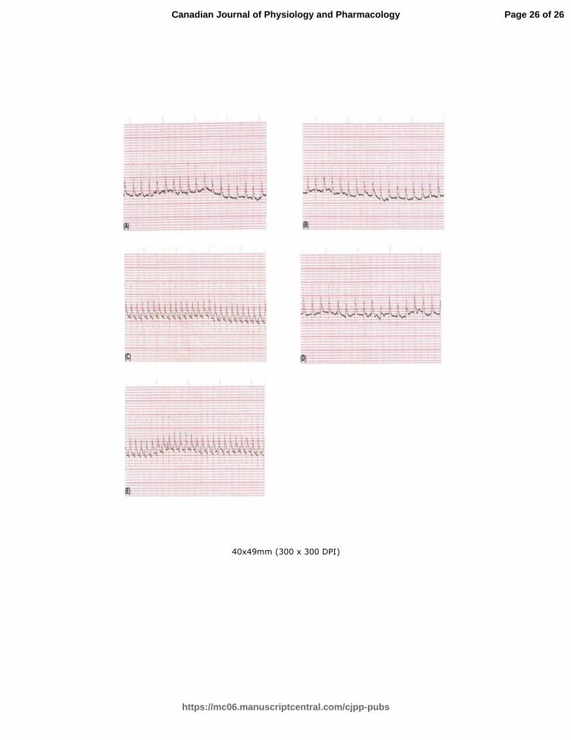
3.1. Effect of vitamin D on ECG parameters and extension of cardiac injury in
isoprenaline-induced infarct-like lesion in rats and the role of PPAR-ɣ (Fig. 1, Table
2):
Isoprenaline injection induced infarct-like lesion represented by ST segment elevation,
and a decrease in R wave amplitude as compared to the normal group. Pretreatment with
vitamin D resulted in a reduction in the ST segment elevation with an increase in R wave
amplitude as compared to the isoprenaline group. The BADGE pretreatment abolished
the protective effect of vitamin D in rats subjected to infarct-like lesion (Fig. 1).
As regards the extension of cardiac injury, vitamin D treatment significantly decreased
the injury extension (P<0.05) as compared to the isoprenaline group. Moreover, BADGE
Page 9 of 26
https://mc06.manuscriptcentral.com/cjpp-pubs
Canadian Journal of Physiology and Pharmacology
Draft
10
pretreatment in rats receiving vitamin D and undergone infarct-like lesion significantly
increased the injury extension (P<0.05) that was still reduced (P<0.05) as compared to
isoprenaline group. (Table 2).
3.2. Effect of vitamin D on the biochemical changes associated with isoprenaline-
induced infarct-like lesion in rats and the role of PPAR-ɣ (Table1, 2):
The levels of cardiac enzymes (CPK and LDH) in the plasma showed a significant rise
(P˂0.05) in the ISO group, ISO + Vit.D group and ISO + Vit. D + BADGE group
compared to the control group. Pretreatment with vitamin D significantly reduced the
cardiac enzymes to near normal values. On the other hand, BADGE pretreatment in the
rats that received vitamin D and subjected to infarct-like lesion significantly increased
(P<0.05) the plasma levels of cardiac enzymes that still showed significant reduction
(P<0.05) as compared to the isoprenaline group. (Table 1).
As regards pro-inflammatory cytokines, TNF-α and IL-6 increased significantly (P˂0.05)
in the ISO group, ISO + Vit.D group and ISO + Vit. D + BADGE group as compared
with the control group. However, these cytokines showed significant reduction in the
ISO-injected rats pretreated with vitamin D (P˂0.05) compared to the ISO group.
Furthermore, the pretreatment with BADGE in the rats that received vitamin D and
subjected to infarct-like lesion produced significant elevation (P˂0.05) of pro-
inflammatory cytokines that still revealed significant reduction (P<0.05) as compared to
isoprenaline group. (Table 1).
Similarly, cardiac MDA significantly increased (P˂0.05) with a parallel significant
decrease in SOD and GSH content (P˂0.05) in the ISO group, ISO + Vit.D group and
Page 10 of 26
https://mc06.manuscriptcentral.com/cjpp-pubs
Canadian Journal of Physiology and Pharmacology
Draft
11
ISO + Vit. D + BADGE group as compared to the normal rats. Pretreatment with vitamin
D resulted in a significant decrease in MDA (P˂0.05) and a significant increase in SOD
and GSH content (P˂0.05) as compared to the isoprenaline group. Similar to other
parameters, the BADGE pretreatment abolished the protective effect of vitamin D in rats
subjected to infarct-like lesion. There was a significant decrease (P<0.05) in cardiac
MDA while there was a significant increase (P<0.05) in SOD content and a non-
significant increase (P>0.05) in GSH content comparing the ISO + Vit. D + BADGE
group to the ISO group. (Table 2).
4. Discussion
MI continues to be a major health problem worldwide and contributes significantly to
mortality statistics (Gao et al. 2001). The term MI is thought to reflect death of cardiac
myocytes due to prolonged ischemia (Patel et al. 2011). In the current study, infarct-like
lesion was induced in rats by intraperitoneal injection of ISO. It has been reported that
isoprenaline administration in high doses to animals produces ‘infarct like’ lesions in the
heart (Vennila et al. 2010). The mechanisms proposed to explain isoprenaline-induced
cardiac damage include increases in heart rate, myocardial contractility, hypoxia due to
myocardial hyperactivity, depletion in energy reserve, calcium overload, excessive
production of highly cytotoxic free radicals, and mitochondrial injury or dysfunction
(Mohanty et al. 2004; Rathore et al. 2000).
In the present study, isoproterenol injection was seen to result in significant alterations in
ECG patterns such as ST segment elevation coupled with marked decrease in R wave
amplitude that reflect isoprenaline-induced infarct-like lesion. ECG pattern alterations by
isoprenaline were previously demonstrated by other investigators (Prince and Sathya
Page 11 of 26
https://mc06.manuscriptcentral.com/cjpp-pubs
Canadian Journal of Physiology and Pharmacology
Draft
12
2010; Tawfik et al. 2010). In addition to the measurement of injury extension, ECG
abnormalities were the main criteria used for the diagnosis of infarct-like lesion. These
alterations could be due to the consecutive loss of cell membrane potential in the injured
myocardium as a result of oxidative stress (Peacock et al. 2007). It was reported that ST
elevation correlates well with the leak of creatine kinase from the myocardium (Mohan et
al. 2010).
Cardiac enzymes, as a marker for acute myocardial infarction were all significantly
elevated in our model. Since theses enzymes are released from necrotic cells to the
extracellular fluid upon the incidence of infarction, their high levels give another
evidence for necrosis and infarction (Wang et al. 2009). Our findings are in agreement
with those of Kurian et al. (2005) and Rajadurai et al. (2007), who reported that these
cardio specific marker enzymes were released from the heart into the blood during
myocardial damage.
Inflammation is a key process involved in mediating myocardial tissue damage after an
ischemic event. In the current study, isoprenaline produced an increase in the serum level
of pro-inflammatory cytokine TNF-α and IL-6, an effect that is in accordance with the
work of other investigators (Cusack et al. 2002; Tawfik et al. 2010). The injured region
undergoes local necrosis and myocyte apoptosis resulting in complement activation, free
radicals generation, and accumulation of cellular debris. Phagocytosis of the resultant
cellular debris by macrophages and neutrophils triggers the inflammatory cytokines as
TNF-α (Frangogiannis et al. 2002).
The increase in the cardiac MDA content (an indicator of lipid peroxidation) and the
Page 12 of 26
https://mc06.manuscriptcentral.com/cjpp-pubs
Canadian Journal of Physiology and Pharmacology
Draft
13
decrease in the cardiac content of SOD and GSH (cardiac antioxidants) in isoprenaline
group could be expected. Isoprenaline causes oxidative stress, which results from a
serious imbalance between the generation of reactive oxygen species (ROS) and their
clearance by the body’s endogenous antioxidative defenses. Our findings are in
agreement with those of Shikalgar and Naikwade (2010) and Upaganlawar et al. (2010).
Administration of vitamin D for 7 days before induction of infarct-like lesion resulted in
amelioration of cardiac injury. Specifically, there was improvement in ECG parameters,
cardiac injury extension and cardiac enzymes of ISO-injected rats pretreated with vitamin
D. These findings are in line with a recent study of Abood and Elshal (2015), who
reported a cardio-protective effect against MI through vitamin D receptor (VDR)
stimulation based on improvement of ECG criteria in addition to return of cardiac
enzyme parameters to near normal.
To explain the protective effect of vitamin D on isoprenaline-induced infarct-like lesion
we measured plasma TNF-α and IL-6, as markers for inflammation, together with cardiac
MDA, SOD and GSH content, as markers for the oxidative state. The down regulation of
inflammatory cytokines demonstrated by vitamin D administration in ISO-injected rats
denotes that vitamin D exerts its protective effect, at least in part, by an anti-
inflammatory action. The reduction of inflammatory cytokines as evidenced in our study
has previously been reported by Arnson et al. (2013). Contrarily, Witham et al (2013)
showed no reduction of inflammatory cytokines after 8 weeks of vitamin D
supplementation. This contradiction is probably due to genetic polymorphism that may
modulate the response to vitamin D supplementation (Gagnon et al. 2014). In addition,
pretreatment with vitamin D in ISO-injected rats was found to be associated with a
Page 13 of 26
https://mc06.manuscriptcentral.com/cjpp-pubs
Canadian Journal of Physiology and Pharmacology
Draft
14
significant reduction in lipid peroxidation products resulting in amelioration of the
oxidative stress. This conforms to the recent data published by Cavalcante and colleagues
(2015) regarding the antioxidant effect of vitamin D.
We revealed that vitamin D treatment reduced ISO-induced infarct-like lesion through
PPAR- ɣ activation. The PPAR- ɣ agonists are well documented to protect against IRI in
various tissues through down-regulation of molecular pathways like nuclear factor kappa
beta, thromboxane synthase, monocyte chemoattractant protein-1, inducible nitric oxide
synthase, and fibronectin (Chatterjee et al. 2004; Panchapakesan et al. 2005). Moreover,
the PPAR- ɣ agonists are proposed to inhibit Jun N-terminal kinase phosphorylation that
belongs to mitogen-activated protein kinase and production of endothelin-1 that are
produced in response to stress stimuli including cytokines and are responsible for cellular
apoptosis and damage (Delerive et al. 1999). There are previous studies that reported the
protective action of vitamin D on the heart (Abood and Elshal 2015; Creighton et al.
2012; Schleithoff et al. 2006). To the best of our knowledge, our study reports for the
first time that vitamin D essentially involves PPAR- ɣ activation for its cardio protective
effect, as pharmacologic inhibition of PPAR- ɣ using BADGE abolished vitamin D-
mediated cardio protection, supporting the involvement of PPAR- ɣ in protective action
of vitamin D.
Conclusion:
In conclusion, the present study revealed that vitamin D pretreatment could improve the
outcome of isoprenaline-induced infarct-like lesion in rats. This protective effect could be
attributed to the strong anti-inflammatory and antioxidant effects of vitamin D. The study
also demonstrated for the first time that PPAR- ɣ significantly contributes toward vitamin
Page 14 of 26
https://mc06.manuscriptcentral.com/cjpp-pubs
Canadian Journal of Physiology and Pharmacology
Draft
15
D mediated cardio protection.
Declaration of interest statement:
The authors declare that they have no conflict of interest.
References:
Abood, A.M., and Elshal, M.F. 2015. VDR stimulation improves outcome of isoprenaline-
induced myocardial infarction in rats via down-regulation of cardiac inos gene expression.
Biomed. Res. 26 (4): 755–764.
Kapil, A., Pal Singh, J., Kaur, T., Singh, B., and Pal Singh, A. 2013. Involvement of peroxisome
proliferator–activated receptor gamma in vitamin D–mediated protection against acute kidney
injury in rats. J. Surg. Res. 185 (2): 774–783. doi: http://dx.doi.org/10.1016/j.jss.2013.07.017.
Al Mheid, I., Patel, R.S., Tangpricha, V., and Quyyumi, A.A. 2013. “Vitamin D and
cardiovascular disease: is the evidence solid?” Eur. Heart J. 34(48): 3691–3698. doi:
http://dx.doi.org/10.1093/eurheartj/eht166 3691-3698.
Arnson, Y., Itzhaky, D., Mosseri, M. Barak, V., Tzur, B., Agmon-Levin, N. et al. 2013. Vitamin
D inflammatory cytokines and coronary events: a comprehensive review. Clin. Rev. Allergy
Immunol. 45(2): 236–247. doi: 10.1007/s12016-013-8356-0.
Basak, T., Varshney, S., Akhtar, S., and Sengupta, S. 2015. Understanding different facets of
cardiovascular diseases based on model systems to human studies: a proteomic and metabolomic
perspective. J. Proteomics, 8(127): 50–60. doi: 10.1016/j.jprot.2015.04.027. PMID: 25956427
Bharti, S., Arora, S., and Arya, S.D. 2010. Evaluation of morphological changes in experimental
models of myocardial infarction: In electron and light microscopically evidence. In Microscopy:
science, technology, applications and education. Edited by A. Mendez-Vilas and J. Dıaz.
Page 15 of 26
https://mc06.manuscriptcentral.com/cjpp-pubs
Canadian Journal of Physiology and Pharmacology
Draft
16
Formatex, Spain. pp.361–371.
Brandenburg, V.M., Vervloet, M.G., and Marx, N. 2012. The role of vitamin D in cardiovascular
disease: From present evidence to future perspectives. Atherosclerosis, 225: 253–263. doi:
10.1016/j.atherosclerosis.2012.08.005. PMID: 22921424.
Cavalcante, I.G., Silva, A.S., Costa, M.J., Persuhn, D.C., Issa, C.I., de Luna Freire, T.L. et al.
2015. Effect of vitamin D3 supplementation and influence of BsmI polymorphism of the VDR
gene of the inflammatory profile and oxidative stress in eldery women with vitamin D
insufficiency: Vitamin D3 megadose reduces inflammatory markers. Exp. Gerontol. 66: 10–16.
doi: 10.1016/j.exger.2015.03.011. PMID: 25827670.
Chatterjee, P.K., Patel, N.S., Cuzzocrea, S., Brown, P.A., Stewart, K.N., Mota-Filipe, H. et al.
2004. The cyclopentenone prostaglandin 15- deoxydelta (12,14)-prostaglandin J2 ameliorates
ischemic acute renal failure. Cardiovasc. Res. 61(3): 630–43. PMID: 14962493.
Creighton, D., Ignaszewski, A., and Francis, G. 2012. Vitamin D: New D-fence against
cardiovascular disease? B.C.Med. J. 54(3): 136–140.
Cusack, M.R., Marber, M.S., Lambiase, P.D., Bucknall, C.A., and Redwood, S.R. 2002. Systemic
inflammation in unstable angina is the result of myocardial necrosis. J. Am. Coll. Cardiol. 39(12):
1917–1923. PMID: 12084588.
Dai, X., Sayama, K., Shirakata, Y., Tokumaru, S., Yang, L., Tohyama, M. et al. 2008. PPAR
gamma is an important transcription factor in 1 alpha,25- dihydroxyvitamin D3-induced
involucrin expression. J. Dermatol. Sci, 50: 53–60. doi:
http://dx.doi.org/10.1016/j.jdermsci.2007.10.011.
Page 16 of 26
https://mc06.manuscriptcentral.com/cjpp-pubs
Canadian Journal of Physiology and Pharmacology
Draft
17
Das, S., Vasisht, S., Snehlata, R., Das, N., and Srivastava, L.M. 2000. Correlation between total
antioxidant status and lipid peroxidation in hypercholesterolemia. Curr. Sci. 78(4): 486–487.
Delerive, P., Martin-Nizard, F., Chinetti, G., Trottein, F., Fruchart, J.C., Najib, J. et al. 1999.
Peroxisome proliferator-activated receptor activators inhibit thrombin-induced endothelin-1
production in human vascular endothelial cells by inhibiting the activator protein-1 signaling
pathway. Circ. Res. 85(5): 394–402. PMID: 10473669
Evans, R.C., Mejios, J.E., Kulevish, J., Fischer, V.W. and Muller, H.S. 1985. Evaluation of a rat
model for assessing intervention to salvage ischemic myocardium. Cardiovasc. Res. 19 (3): 132–
138. doi: http://dx.doi.org/10.1093/cvr/19.3.132.
Frangogiannis, N.G., Wayne-Smith, C., and Entman, M.L. 2002. The inflammation response in
myocardial infarction. Cardiovasc. Res. 53(1): 31–47. doi: http://dx.doi.org/10.1016/S0008-
6363(01)00434-5.
Gagnon, C., Daly, R.M., Carpentier, A., Lu, Z.X., Shore- Lorenti, C., Sikaris, K. et al. 2014.
Effects of combined calcium and vitamin D supplementation on insulin secretion, insulin
sensitivity and β-cell function in multiethnic vitamin D-deficient adults at risk for type 2 diabetes:
a pilot randomized, placebo-controlled trial. PLOS One, 9 (10):
e109607.doi: 10.1371/journal.pone.0109607. PMCID: PMC4192133.
Gao, F., Gong, B., Christopher, T.A., Lopez, B.L., Karasawa, A., and Ma, X.L. 2001.
Antiapoptotic effect of benidipine, a long-lasting vasodilating calcium antagonist, in
ischaemic/reperfused myocardial cells. Br. J. Pharmacol. 132: 869–878.
doi:10.1038/sj.bjp.0703881.
Goleniewska, B., Kacprzak, M., and Zielińska, M. 2014. Vitamin D level and extent of coronary
Page 17 of 26
https://mc06.manuscriptcentral.com/cjpp-pubs
Canadian Journal of Physiology and Pharmacology
Draft
18
stenotic lesions in patients with first acute myocardial infarction. Cardiol. J. 21(1): 18–23. doi:
10.5603/CJ.a2013.0048. PMID: 23677723.
Hlaing, S.M., Garcia, L.A., Contreras, J.R., Norris, K.C., Ferrini, M.G., and Artaza1, J.N. 2014.
1,25-vitamin D3 promotes cardiac differentiation through modulation of the WNT signaling
pathway. J. Mol. Endocrinol. 53 (3): 303–317. doi: 10.1530/JME-14-0168. PMCID:
PMC4198487.
Jordan, J.E., Zhao, Z.Q., and Vinten-Johansen, J. 1999. The role of neutrophils in myocardial
ischemia-reperfusion injury. Cardiovasc. Res. 43: 860–878. doi: http://dx.doi.org/10.1016/S0008-
6363(99)00187-X 860-878.
Kumaran, K.S., and Prince, P.S. 2010. Caffeic acid protects rat heart mitochondria against
isoproterenol-induced oxidative damage. Cell Stress Chaperon, 15 (6): 791–806. doi:
10.1007/s12192-010-0187-9. PMID: 20376586.
Kurian, G.A., Philip, S., and Varghese, T. 2005. Effect of aqueous extract of the Desmodium
gangeticum DC root in the severity of myocardial infarction. Ethnopharmacol. 97:457–461.
PMID: 15740881.
Lee, J.H., Gadi, R., Spertus, J.A., Tang, F., and O'Keefe, J.H. 2011. Prevalence of vitamin D
deficiency in patients with acute myocardial infarction. Am. J. Cardiol. 107 (11): 1636–1638. doi:
10.1016/j.amjcard.2011.01.048. PMID: 21439530.
Loh, H.K., Sahoo, K.C., Kishore, K., Ray, R., Nag, T.C., Kumari, S. et al. 2007. Effects of
thalidomide on isoprenaline-induced acute myocardial injury: a haemodynamic, histopathological
and ultrastructural study. Basic Clin. Pharmacol. Toxicol. 100(4): 233–239. PMID: 17371527.
Mancuso, P., Rahman, A., Hershey, S.D., Dandu, L., Nibbelink, K.A., Simpson, R.U. 2008. 1,25-
Page 18 of 26
https://mc06.manuscriptcentral.com/cjpp-pubs
Canadian Journal of Physiology and Pharmacology
Draft
19
Dihydroxyvitamin-D3 treatment reduces cardiac hypertrophy and left ventricular diameter in
spontaneously hypertensive heart failure-prone (cp/) rats independent of changes in serum leptin.
J. Cardiovasc. Pharmacol. 51(6): 559–564. doi: 10.1097/FJC.0b013e3181761906. PMID:
18496147.
Mohan, M., Patankar, P., Ghadi, P., and Kasture, S. 2010. Cardioprotective potential of Punica
granatum extract in isoproterenol-induced myocardial infarction in Wistar rats. J. Pharmacol.
Pharmacother. 1:32–37. doi: 10.4103/0976-500X.64533. PMCID: PMC3142755.
Mohanty, I., Arya, D.S., Dinda, A., Talwar, K.K., Joshi, S., and Gupta, S.K. 2004. Mechanisms
of cardioprotective effects of Withania somnifera in experimentally induced myocardial
infarction. Basic Clin. Pharmacol. Toxicol. 94: 184–190. doi: 10.1111/j.1742-
7843.2004.pto940405.x.
Moron, M.S., Defierre, J.W., and Mannervik, B. 1979. Levels of glutathione, glutathione
reductase and glutathione S-transferase activities in rat lung and liver. Biochim. Biophys. Acta,
582: 67–68. doi:10.1016/0304-4165(79)90289-7.
Panchapakesan, U., Sumual, S., Pollock, C.A. and Chen, X. 2005. PPAR gamma agonists exert
antifibrotic effects in renal tubular cells exposed to high glucose. Am. J. Physiol. 289: F1153-
1158. PMID: 15886275.
Panda, V.S., and Naik, S.R. 2009. Evaluation of cardioprotective activity of Ginkgo biloba and
Ocimum sanctum in rodents. Altern. Med. Rev. 14: 161-171.
Patel, S.S., Verma, N.K., Rathore, B., Nayak, G., Singhai, A.K., and Singh, P. 2011. Cardio-
protective effect of Bombax ceiba flowers against acute adriamycin-induced myocardial
infarction in rats. Rev. Bras. Farmacogn. 21: 704–709. doi: http://dx.doi.org/10.1590/S0102-
Page 19 of 26
https://mc06.manuscriptcentral.com/cjpp-pubs
Canadian Journal of Physiology and Pharmacology
Draft
20
695X2011005000090.
Peacock, W.E., Hollander, J.E., Smalling, R.W., and Bresler, M.J. 2007. Reperfusion strategies in
the emergency treatment of ST-segment elevation myocardial infarction. Am. J. Emerg. Med. 25:
353–366. doi:10.1016/j.ajem.2006.07.013.
Prince, P.S., and Sathya, B. 2010. Pretreatment with quercetin ameliorates lipids, lipoproteins and
marker enzymes of lipid metabolism in isoproterenol treated cardiotoxic male Wistar rats. Eur. J.
Pharmacol. 635(1–3): 142–148. doi:10.1016/j.ejphar.2010.02.019.
Rajadurai, M., and Prince, S.M.P. 2007. Preventive effect of naringin on cardiac markers,
electrocardiographic patterns and lysosomal hydrolases in normal and isoproterenol-induced
myocardial infarction in Wistar rats. Toxicology, 230 (2–3): 178–188. PMID: 17188415.
Rathore, N., Kale, M., John, S., and Bhatnagar, D. 2000. Lipid peroxidation and antioxidant
enzymes in isoproterenol induced oxidative stress in rat erythrocytes. Indian J. Physiol.
Pharmacol. 44: 161–166.
Schleithoff, S.S., Zittermann, A., Tenderich, G., Berthold, H.K., Stehle, P., and Koerfer, R. 2006.
Vitamin D supplementation improves cytokine profiles in patients with congestive heart failure: a
double-blind, randomized, placebo-controlled trial. Am. J. Clin. Nutr. 83: 754–9. PMID:
16600924.
Shab-Bidar, S., Neyestani, T.R., and Djazayery, A. 2014. The interactive effect of improvement
of vitamin D status and VDR FokI variants on oxidative stress in type 2 diabetic subjects: a
randomized controlled trial. Eur. J. Clin. Nutr. 69: 216-222. doi:10.1038/ejcn.2014.240.
Sharma, A., and Singh. M. 2000. Effect of isoproterenol amiloride, a Na+-H+ exchange inhibitor,
Page 20 of 26
https://mc06.manuscriptcentral.com/cjpp-pubs
Canadian Journal of Physiology and Pharmacology
Draft
21
on cardioprotective effect of ischemic and angiotensin preconditioning. Mol. Cell Biochem. 214:
31 – 8. doi:10.1023/A:1007167519596.
Shikalgar, T.S., and Naikwade, N.S. 2010. Verapamil ameliorates cardio protective potential of
vitamin E in myocardial oxidative damage induced by isoproterenol: a biochemical study. J
Pharma Sci. Technol. 2: 298–302.
Tawfik, M.K., Ghattas, M.H., Abo-Elmatty, D.M., and Abdel-Aziz, N.A. 2010. Atorvastatin
restores the balance between pro-inflammatory and anti-inflammatory mediators in rats with
acute myocardial infarction. Eur. Rev. Med. Pharmacol. Sci. 14(6): 499–506. PMID: 20712256.
Uchiyama, M., and Mihara, M. 1978. 1978. Determination of malonaldehyde precursor in tissues
by thiobarbituric acid test. Anal. Biochem. 86: 271–278. doi:10.1016/0003-2697(78)90342-1.
Upaganlawar, A., Gandhi, H., and Balaraman, R. 2010. Effect of vitamin E alone and in
combination with lycopene on biochemical and histopathological alterations in isoproterenol-
induced myocardial infarction in rats. J. Pharmacol. Pharmacother. 1: 24–31. doi: 10.4103/0976-
500X.64532 PMCID: PMC3142754.
Vennila, L., and Pugalendi, K.V. 2010. Protective effect of sesamol against myocardial infarction
caused by isoproterenol in Wistar rats. Redox Rep. 15: 36–42.
doi:10.1179/174329210X12650506623168.
Vivaldi, M.T., Kloner, R.A. and Frederick, J.S. 1985. Triphenyltertezolium staining of
irreversible ischemic injury following coronary artery occlusion in rats. Am. J. Pathol. 121: 522 –
30. PMCID: PMC1887916.
Page 21 of 26
https://mc06.manuscriptcentral.com/cjpp-pubs
Canadian Journal of Physiology and Pharmacology
Draft
22
Wang, L., Ma, J., Manson, J.E., Buring, J.E., Gaziano, J.M., and Sesso, H.D. 2013. A prospective
study of plasma vitamin D metabolites, vitamin D receptor gene polymorphisms, and risk of
hypertension in men. Eur. J. Nutr. 52(7) :1771–1779.doi:10.1007/s00394-012-0480-8.
Wang, S.B., Tian, S., Yang, F., Yang, H.G., Yang, X.Y., and Du, G.H. 2009. Cardioprotective
effect of salvianolic acid A on isoproterenol-induced myocardial infarction in rats. Eur. J.
Pharmacol. 615: 125–132. doi: 10.1016/j.ejphar.2009.04.061. PMID: 19445921.
Witham, M.D., Adams, F., Kabir, G., Kennedy, G., Belch, J.J., and Khan, F. 2013. Effect of short
term vitamin D supplementation on markers of vascular health in South Asian women living in
UK: A randomised controlled trial. Atherosclerosis, 230: 293–299. doi:
10.1016/j.atherosclerosis.2013.08.005. PMID: 24075759.
Zaafan, M.A., Zaki, H.F. , El-Brairy, A.I., and Kenawy, S.A. 2013. Protective effects of
atorvastatin and quercetin on isoprenaline-induced myocardial infarction in rats. Bulletin of
Faculty of Pharmacy, Cairo University, 51(1): 35–41. doi:10.1016/j.bfopcu.2013.03.001.
Zhang, J., Knapton, A., Lipshultz, S.E., Weaver, J.L., and Herman, E.H. 2008. Isoproterenol-
induced cardiotoxicity in Sprague Dawley rats: correlation of reversible and irreversible
myocardial injury with release of cardiac troponin T and roles of iNOS in myocardial injury.
Toxicol. Pathol. 36: 277–278. doi: 10.1177/0192623307313010. PMID: 18349426.
Zittermann, A., and Koerfer, R. 2008. Protective and toxic effects of vitamin D on vascular
calcification: clinical implications. Mol. Aspects Med. 29: 423–432. doi:
10.1016/j.mam.2008.04.002. PMID: 18538838.
Page 22 of 26
https://mc06.manuscriptcentral.com/cjpp-pubs
Canadian Journal of Physiology and Pharmacology
Draft
23
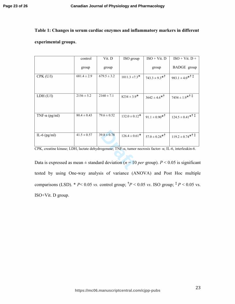
Table 1: Changes in serum cardiac enzymes and inflammatory markers in different
experimental groups.
control
group
Vit. D
group
ISO group ISO + Vit. D
group
ISO + Vit. D +
BADGE group
CPK (U/l) 681.4 ± 2.9 679.5 ± 3.2 1011.3 ±7.3*
743.3 ± 9.3*†
983.1 ± 4.0*† ‡
LDH (U/l) 2156 ± 5.2 2160 ± 7.1 8234 ± 3.9*
3642 ± 4.6*†
7454 ± 1.9*† ‡
TNF-α (pg/ml) 80.4 ± 0.43
79.6 ± 0.52
132.0 ± 0.12*
91.1 ± 0.90*†
124.5 ± 0.41*† ‡
IL-6 (pg/ml) 41.5 ± 0.57 39.8 ± 0.78 126.4 ± 0.61* 57.0 ± 0.24*† 119.2 ± 0.74*† ‡
CPK, creatine kinase; LDH, lactate dehydrogenase; TNF-α, tumor necrosis factor- α; IL-6, interleukin-6.
Data is expressed as mean ± standard deviation (n = 10 per group). P < 0.05 is significant
tested by using One-way analysis of variance (ANOVA) and Post Hoc multiple
comparisons (LSD). * P< 0.05 vs. control group; †P < 0.05 vs. ISO group; ‡ P < 0.05 vs.
ISO+Vit. D group.
Page 23 of 26
https://mc06.manuscriptcentral.com/cjpp-pubs
Canadian Journal of Physiology and Pharmacology
Draft
24
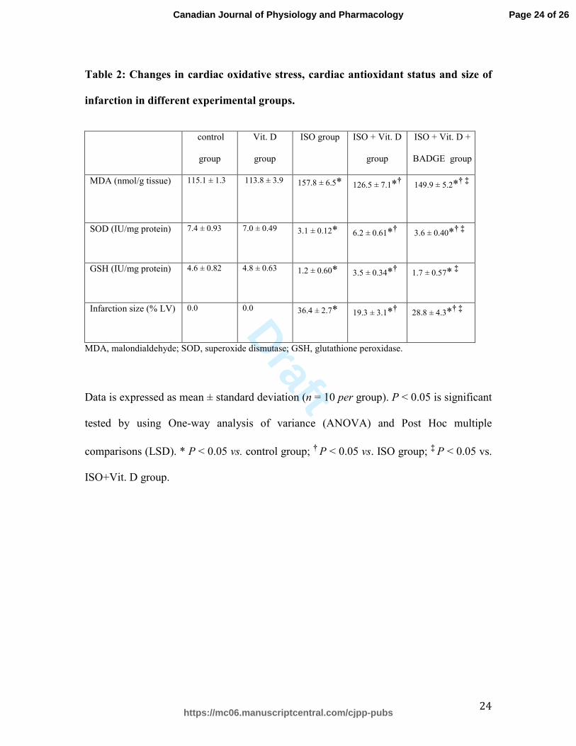
Table 2: Changes in cardiac oxidative stress, cardiac antioxidant status and size of
infarction in different experimental groups.
control
group
Vit. D
group
ISO group ISO + Vit. D
group
ISO + Vit. D +
BADGE group
MDA (nmol/g tissue) 115.1 ± 1.3 113.8 ± 3.9 157.8 ± 6.5*
126.5 ± 7.1*†
149.9 ± 5.2*† ‡
SOD (IU/mg protein) 7.4 ± 0.93 7.0 ± 0.49 3.1 ± 0.12* 6.2 ± 0.61*† 3.6 ± 0.40*† ‡
GSH (IU/mg protein) 4.6 ± 0.82 4.8 ± 0.63 1.2 ± 0.60* 3.5 ± 0.34*† 1.7 ± 0.57*
‡
Infarction size (% LV) 0.0 0.0 36.4 ± 2.7* 19.3 ± 3.1*† 28.8 ± 4.3*† ‡
MDA, malondialdehyde; SOD, superoxide dismutase; GSH, glutathione peroxidase.
Data is expressed as mean ± standard deviation (n = 10 per group). P < 0.05 is significant
tested by using One-way analysis of variance (ANOVA) and Post Hoc multiple
comparisons (LSD). * P < 0.05 vs. control group; † P < 0.05 vs. ISO group; ‡ P < 0.05 vs.
ISO+Vit. D group.
Page 24 of 26
https://mc06.manuscriptcentral.com/cjpp-pubs
Canadian Journal of Physiology and Pharmacology
Draft
25
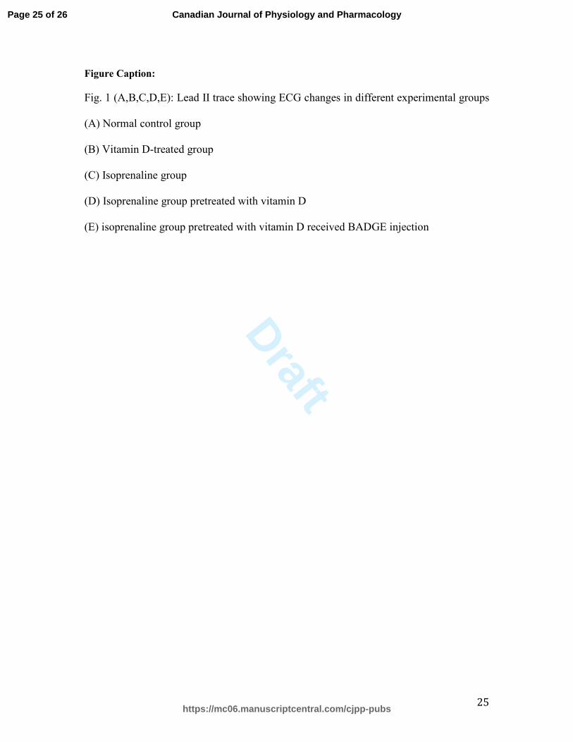
Figure Caption:
Fig. 1 (A,B,C,D,E): Lead II trace showing ECG changes in different experimental groups
(A) Normal control group
(B) Vitamin D-treated group
(C) Isoprenaline group
(D) Isoprenaline group pretreated with vitamin D
(E) isoprenaline group pretreated with vitamin D received BADGE injection
Page 25 of 26
https://mc06.manuscriptcentral.com/cjpp-pubs
Canadian Journal of Physiology and Pharmacology
Draft
40x49mm (300 x 300 DPI)
Page 26 of 26
https://mc06.manuscriptcentral.com/cjpp-pubs
Canadian Journal of Physiology and Pharmacology