Embed Size (px)
Citation preview
The University of ToledoThe University of Toledo Digital Repository
Theses and Dissertations
2012
Activation of glutamate transporter 1 attenuatesrelapse to alcohol-seeking behavior in ratsAbeer QrunflehThe University of Toledo
Follow this and additional works at: http://utdr.utoledo.edu/theses-dissertations
This Thesis is brought to you for free and open access by The University of Toledo Digital Repository. It has been accepted for inclusion in Theses andDissertations by an authorized administrator of The University of Toledo Digital Repository. For more information, please see the repository's Aboutpage.
Recommended CitationQrunfleh, Abeer, "Activation of glutamate transporter 1 attenuates relapse to alcohol-seeking behavior in rats" (2012). Theses andDissertations. 408.http://utdr.utoledo.edu/theses-dissertations/408
i
A Thesis
Entitled
Activation of Glutamate Transporter 1 Attenuates Relapse
to Alcohol-Seeking Behavior in Rats
By Abeer Qrunfleh
Submitted to the Graduate Faculty as a partial fulfillment of the requirement for the
Master of Science in Pharmacology and Toxicology
_____________________________________
Dr. Youssef Sari, Committee Chair
_____________________________________
Dr. Ming-Cheh Liu, Committee Member
_____________________________________
Dr. Zahoor Shah, Committee Member
_____________________________________ Dr. Patricia Komuniecki, Dean College of Graduate Studies
The University of Toledo May 2012

ii
Copyright 2012, Abeer Qrunfleh
This document is copyrighted material. Under copyright law, no parts of this document may be reproduced without the expressed permission of the author.
iii
An Abstract of
Activation of Glutamate Transporter 1 Attenuates Relapse
to Alcohol-Seeking Behavior in Rats
By Abeer Qrunfleh
Submitted to the Graduate Faculty as a partial fulfillment of the requirement for the Master Degree in Pharmacology and Toxicology
The University of Toledo
May 2012
Relapse to alcohol use after prolonged withdrawal periods is a major
problem in the treatment of alcohol addiction in humans. Recent preclinical
research has focused on identifying long-term neuroadaptive changes and
identifying the behavioral, environmental, and neuronal mechanisms underlying
drug relapse. By doing so, potential new avenues for relapse prevention may be
developed. Current research suggests that changes in glutamatergic
neurotransmission may significantly contribute to alcohol relapse and alcohol
addiction. Alcohol dependence has been linked specifically to the increased
extracellular glutamate levels in key regions of neurocircuits mainly the
mesocorticolimbic circuit. Based on the fact that glutamate transporter1 (GLT1) is
responsible for the removal of the majority of extracellular glutamate, we
hypothesized that the activation of GLT1 by the use of ceftriaxone, a -lactam
antibiotic, known to elevate GLT1 expression, would attenuate alcohol
consumption in alcohol-preferring (P) rats and ultimately prevent relapse to
alcohol-seeking behavior. Statistical analyses showed that P rats treated
intraperitoneally with ceftriaxone, exhibited a significant reduction in alcohol
consumption followed by a period of alcohol deprivation as compared to saline-
treated P rats. Preliminary data with Western blot suggests that activation of
GLT1 may play a key role in preventing relapse to alcohol-seeking behavior, and
iv
ultimately implicate its potential role as a therapeutic-target for treatment of
alcohol dependence.
Acknowledgments
Without the help and support of many people this thesis would have never been
completed. Sincere thanks and gratitude goes to Dr. Youssef Sari for giving me the
advice, support and the opportunity to participate in this project. I would like to extend
my gratitude to the committee members, Dr. Ming-Cheh Liu and Dr. Zahoor Shah.
Many thanks go to my lab colleagues Sai Nandini Sreemantula, Manal Gergis, Adnan
Alazizi, and Jessica Ciesler for their help and support.
Last but not least, my greatest thanks and gratitude goes to my husband Haitham for his
endless support, love and care through this journey. My father who is my idol, my
inspiration and my mentor. My mother, who believed in me, and was there for me every
step of the way. Your encouragement and support made me who I am today.
v
Contents
Abstract iii
Acknowledgments iv
Contents v
List of Figures vi
Introduction 1
1.1 Alcoholism 1
1.2 Neurotransmitter systems involved in alcohol dependence 3
1.3 Neurocircuitry of reward 8
1.4 Animal models of alcoholism 10
1.5 Animal models of alcohol relapse 12
1.6 Glutamate and glutamate transporters 16
1.7 Activation of GLT1 by ceftriaxone, a -lactam antibiotic 19
1.8 Objectives and goals 21
Material and methods 22
2.1 Animals 22
2.2 Behavioral drinking paradigm 22
2.3 Brain tissue harvesting 24
2.4 Protein tissue extraction protocol 25
2.5 Protein quantification 25
vi
2.6 Western blot procedures 26
Results 28
3.1 Effects of ceftriaxone treatment on alcohol drinking behavior 28
3.2 Effects of ceftriaxone treatment on water intake 30
3.3 Effects of ceftriaxone treatment on body weight 32
3.4 Effects of ceftriaxone treatment on sucrose drinking behavior 33
3.5 Effects of ceftriaxone treatment on GLT1 expression 34
Discussion 37
References 43
vii
List of Figures
Figure 1-1. Circuitry in the development and expression of drug addiction....……..…….10
Figure 1-2. Ceftriaxone chemical structure………………………….…..…………...............19
Figure 3-1. Effects of ceftriaxone treatment on alcohol drinking behavior………………….30
Figure 3-2. Effects of ceftriaxone treatment on water intake………………………………...31
Figure 3-3. Effects of ceftriaxone treatment on body weight………………………………. 32
Figure 3-4. Effects of ceftriaxone treatment on sucrose drinking behavior………………... 34
Figure 3-5. Effects of ceftriaxone treatment on GLT1 expression in the core…………….. 35
Figure 3-6 Effects of ceftriaxone treatment on GLT1 expression in the prefrontal cortex…36
1
Introduction
1.1 Alcoholism
Alcoholism is a chronic relapsing disorder that is progressive and has serious
detrimental health outcomes. The development of alcoholism is characterized by frequent
episodes of intoxication, preoccupation with alcohol, use of alcohol despite adverse
consequences, compulsion to seek and consume alcohol, loss of control in over its
consumption, and emergence of a negative emotional state in the absence of the drug
(American Psychiatric Association 1994). According to the National Institute on Alcohol
Abuse and Alcoholism (NIAAA), more than 17 million people in the United States either
abuse or are dependent on alcohol (NIAAA 2007), with a cost to U.S. society of over
$180 billion annually (NIAAA 2004). Nearly 79,000 annual deaths are caused directly or
indirectly by excessive alcohol consumption (Center for Disease Control and Prevention,
2008).
The Therapeutic treatments of alcoholism are not effective in alleviating this
disorder, partly because alcohol is generally viewed as an unspecific pharmacological
agent that has only a few known primary targets, and mainly because the neurobiology
behind alcoholism is very complex. Recent work has begun to define the neurocircuits
responsible for excessive ethanol drinking (Koob et al., 1998).
A neural circuit can be viewed as a group of neurons that are interconnected and
relay information related to a specific function. Within the neural circuit, information is
passed between neurons thru various electrochemical signaling processes. Activated
2
neurons release neurotransmitters that bind to specific receptors on other neurons.
Depending on the neurotransmitter involved, this binding leads to the electrical excitation
or inhibition of subsequent neurons in the circuit (Gilpin and Koob, 2008).
Despite the fact that drugs of abuse possess various neuropharmacological
profiles, activation of the mesocorticolimbic system, particularly the ventral tegmental
area, nucleus accumbens, amygdala and prefrontal cortex via dopaminergic, GABAergic
and glutamatergic pathways, constitutes a common neurocircuitry by which diverse
drugs of abuse mediate their acute reinforcing effects, whereas, long-term
neuroadaptations in this circuitry most seemingly underlie the transition to drug
dependence and cycles of relapse (Feltenstein and See, 2008).
Within the brain’s central reward neurocircuits and stress circuits, alcohol
interacts with several neurotransmitter systems. The interactions between alcohol and the
neurotransmitter systems produce alcohol’s acute reinforcing effects. However, in
chronic exposure to alcohol, these interactions ultimately result in changes in neuronal
function that underlie the development of tolerance, withdrawal, and dependence (Gilpin
and Koob, 2008).
3
1.2 Neurotransmitter Systems Involved in Alcohol
Dependence
Alcohol reinforcement appears to be mediated by several neurotransmitter
systems including GABAergic system, opioid system, dopaminergic system, serotonergic
system, and glutamatergic system (Koob et al., 1998).
-Aminobutyric Acid System (GABA)
-Aminobutyric acid (GABA) is the major inhibitory neurotransmitter in the
brain. It plays a role in regulating the neuronal excitability in the nervous system. There
are two classes of GABA receptors: GABAA and GABAB.
GABAA receptors are ligand-gated ion channels, whereas, GABAB receptors are G
protein-coupled receptors.
Alcohol can increase GABA activity in the brain either by acting on the GABA-releasing
(presynaptic) neuron, leading to an increased GABA release. Or by acting on the signal-
receiving (postsynaptic) neuron, resulting in facilitating the activity of the GABAA
receptor ( Gilpin and Koob, 2008).
Ethanol allosterically appears to modulate the GABA receptor complex and to
basically open the chloride channel and hyperpolarize cells or at least potentiates the
hyperpolarization produced by GABA (Dietrich et al., 1989)
4
Previous studies have demonstrated that alcohol drinking is suppressed by
GABAA receptor antagonists, which interferes with the actions of the GABAA receptor in
the brain regions of the extended amygdala such as the nucleus accumbens, ventral
pallidum, bed nucleus of the stria terminalis and amygdala (Gilpin and Koob, 2008). Of
these regions, the central nucleus of the amygdala is especially sensitive to the
suppression of alcohol drinking by GABAergic compounds (Hyytia and Koob, 1995).
Acute alcohol exposure (short-term alcohol consumption) seems to increase the GABAA
receptor function, whereas, prolonged drinking due to counteradaptive processes has the
opposite effects. The effects of chronic alcohol use are linked to over-excitation due to
compensatory mechanisms associated with repeated administration. Chronic alcohol use
has been shown to decrease the cell surface expression of GABAA receptors and decrease
their sensitivity (Mharte et al., 1993).
Opioid System
Endogenous opioids are small naturally occurring peptides that are produced in
the body and have been associated in the actions of opiate drugs and alcohol for a long
time. There are three classes of endogenous opioids: endorphins, enkephalins, and
dynorphins (Gilpin and Koob, 2008). These endogenous opioids produce their effects by
interacting with three subtypes of opioid receptors: , , and .
The positive alcohol reinforcement, certain properties of alcohol leading to dependence,
is mediated at least in part by the release of endogenous opioids in the brain (Ulm et al.,
1995). This hypothesis is indeed supported by various studies demonstrating that opioid
antagonists acting on opioid receptors suppress alcohol drinking in several animal
5
models. Moreover, mice lacking -opioid receptor show reduction in alcohol intake
(Roberts et al., 2000). The agent naltrexone, a pharmacological substance that binds to
endorphin receptors, preventing endorphin receptor binding, and reducing the craving for
abuse substances, is classified as a nonspecific opioid receptor antagonist, which is
currently used as a treatment for alcoholism and narcotic addiction in humans, and is
particularly effective in reducing heavy drinking to alcohol (Gilpin and Koob, 2008).
Alcohol induces an increase in the extracellular endorphin level in the nucleus
accumbens (Olive et al., 2001). The opioid system influences alcohol drinking behavior
by direct and indirect interactions with the mesolimbic dopamine system (Gilpin and
Koob, 2008). Opioid receptor antagonists interfere with alcohol’s rewarding effects by
acting in various brain regions including the ventral tegmental area, nucleus accumbens,
and central nucleus of the amygdala (Koob, 2003).
Dopaminergic System
Dopamine is a monoamine neurotransmitter involved in a circuit called the
mesolimbic system, which projects from the ventral tegmental area to the nucleus
accumbens (Gilpin and Koob, 2008). This circuit influences incentive changes in the
environment, and it affects incentive motivation (Biggio et al., 2007). Studies suggest that
dopamine also has a role in the incentive motivation associated with acute alcohol
intoxication. Moreover, studies demonstrated that alcohol consumption can be blocked by
administration of dopamine antagonist directly into the nucleus accumbens (Hodge et al.,
1997). Additionally, alcohol ingestion and anticipation to the availability of alcohol
6
produce dopamine release in the nucleus accumbens as determined by microdialysis
study (Weiss et al., 1993).
Serotonergic System
Serotonin (5-hydroxy-tryptamine or 5-HT) is a monoamine neurotransmitter that
is considered a target of interest for potential therapy of alcohol dependence because of a
well-established link between serotonin depletion, impulsivity, and alcohol-drinking
behavior in rats, mice, and humans (Gilpin and Koob, 2008).Therapeutic compounds that
target the serotonin system either by inhibiting neuronal reuptake of serotonin, therefore
prolonging its actions, or by blocking specific serotonin receptor subtypes have been
shown to reduce the voluntary alcohol consumption in rats (Jhonson, 2008). Serotonin
release in the nucleus accumbens of rats is suppressed during alcohol withdrawal, and
this reduction in serotonin release is partially reversed by self-administration of alcohol
(Weiss et al., 1996). Studies have shown that SSRIs effectively maintain the attenuation
of alcohol intake achieved during treatment of alcohol dependence for at least 6 months
after pharmacological intervention in some alcoholics (Sari et al., 2011). For example,
Escitalopram, an effective SSRI, reduces alcohol consumption in alcoholics.
Glutamatergic System
Glutamate is the chief excitatory neurotransmitter in the brain; it exerts its effects
by interacting with several receptor subtypes, including the N-methyl-D-aspartate
(NMDA) receptor (Gilpin and Koob, 2008). The glutamatergic system has long been
implicated in the acute as well as chronic reinforcing actions of alcohol. In contrast to
7
alcohol’s acute effects on GABA, alcohol inhibits glutamate activity in the brain.
Furthermore, acute alcohol exposure reduces the extracellular glutamate levels in the
striatum, which contains the nucleus accumbens, among other structures (Gilpin and
Koob, 2008). Alternatively, chronic alcohol exposure leads to an increase in glutamate
output, and impairs ability in glutamate transport, leading ultimately to excessive
glutamatergic neurotransmission within the mesolimbic circuit. Importantly, alcohol
affects glutamate transmission most likely by altering the functions of both NMDA
receptors (Lovinger et al., 1989) and also metabotropic glutamate subtype 5 receptors
(mGluR5) (Blednov and Harris, 2008).The involvement of NMDA receptors in
alcoholism might be associated with neuroplasticity, a process characterized by neural
reorganization that likely contributes to hyperexcitability and craving during alcohol
withdrawal (Pulvirenti and Diana, 2001).
Currently, compounds targeting the glutamatergic system are being used in the
treatment of alcohol dependence. The agent acamprosate blocks excessive alcohol
consumption by reducing excessive glutamate activity. This process seems to depend on
the involvement of genes such as Per2, which is implicated in maintaining the normal
daily rhythm (the circadian clock) of an organism (Spanagel et al., 2005). The drug
acamprosate has been approved for the treatment of alcoholism in humans, primarily due
to its ability to suppress alcohol drinking among diverse species (Gilpin and Koob, 2008).
Acamprosate tend to dampen excessive glutamate activity, and modulate glutamate
transmission by acting on NMDA and/or metabotropic glutamate receptor (Littleton,
2007).
8
1.3 Neurocircuitry of Reward
The understanding of the neurobiological factors involved in alcohol
reinforcement and dependence would lead to the development of effective
pharmacotherapies for the prevention of alcohol relapse after periods of abstinence
(Spanagel and Zieglgansberger, 1997). Understanding relapse lies in the same
neurochemical elements that are compromised by repeated alcohol use. Studies have
focused on examining motivational reward circuits through stimulation sites of specific
brain regions. Most of these diverse stimulation sites later were identified to be linked
through a common neural pathway; a complex bundle of axons termed the medial
forebrain bundle (Phillips, 1984). Dopamine was identified later as a critical
neurotransmitter in producing the rewarding effects by intracranial self-stimulation
(ICSS) of the medial forebrain bundle (Liebman and Butcher, 1973; Phillips and Fibiger,
1978). It has been shown that low doses of alcohol produce a dose-dependent increase in
the firing rate of dopaminergic neurons in the ventral tegmental area (Gessa et al., 1985).
The brain regions that mediate the reinforcing effects of alcohol include: the
extended amygdala and the mesolimbic dopaminergic pathway, including the ventral
tegmental area, the nucleus accumbens and the prefrontal cortex, these are the main sites
in the brain that mediate alcohol reinforcement (Vengeliene et al, 2008). An integral part
of the brain, the nucleus accumbens, which is located in the ventral striatum, has been
well studied for its role in reward, as well as addiction. Currently, it is believed that the
nucleus accumbens serves to integrate information contained in the mesocorticolimbic
circuit and projects that information out to the motor system to produce appropriate
9
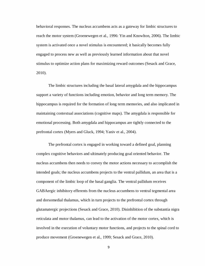
behavioral responses. The nucleus accumbens acts as a gateway for limbic structures to
reach the motor system (Groenewegen et al., 1996: Yin and Knowlton, 2006). The limbic
system is activated once a novel stimulus is encountered; it basically becomes fully
engaged to process new as well as previously learned information about that novel
stimulus to optimize action plans for maximizing reward outcomes (Sesack and Grace,
2010).
The limbic structures including the basal lateral amygdala and the hippocampus
support a variety of functions including emotion, behavior and long term memory. The
hippocampus is required for the formation of long term memories, and also implicated in
maintaining contextual associations (cognitive maps). The amygdala is responsible for
emotional processing. Both amygdala and hippocampus are tightly connected to the
prefrontal cortex (Myers and Gluck, 1994; Yaniv et al., 2004).
The prefrontal cortex is engaged in working toward a defined goal, planning
complex cognitive behaviors and ultimately producing goal oriented behavior. The
nucleus accumbens then needs to convey the motor actions necessary to accomplish the
intended goals; the nucleus accumbens projects to the ventral pallidum, an area that is a
component of the limbic loop of the basal ganglia. The ventral pallidum receives
GABAergic inhibitory efferents from the nucleus accumbens to ventral tegmental area
and dorsomedial thalamus, which in turn projects to the prefrontal cortex through
glutamatergic projections (Sesack and Grace, 2010). Disinhibition of the substantia nigra
reticulata and motor thalamus, can lead to the activation of the motor cortex, which is
involved in the execution of voluntary motor functions, and projects to the spinal cord to
produce movement (Groenewegen et al., 1999; Sesack and Grace, 2010).
10
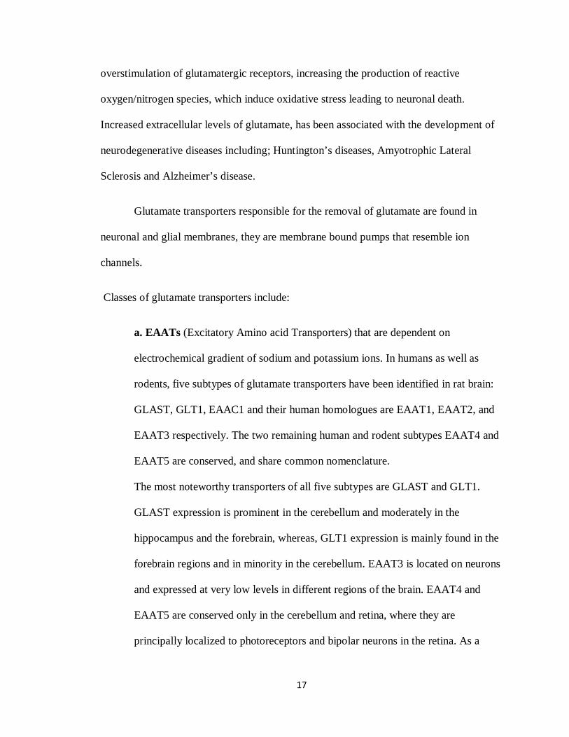
1.4 Animal Models of Alcoholism
The large variability in alcohol preference among animal models has set forth the
selectively of breeding rats for differential alcohol preference, low or high alcohol
consumption levels (Spanagel, 2000). Examples of the animal models of alcohol
preference include: Alko Alcohol (AA) and Alko Nonalcohol (ANA) rats, which
comprises two strains of albino rats that were selectively bred based on their selection or
rejection of a 10% alcohol solution and water (Eriksson, 1968). The alcohol-preferring
(P) rats, originally bred at Indiana University Institute, voluntarily consume 5 -8 grams of
Figure 1-1 Circuitry in the development and expression of drug addiction
11
alcohol per kilogram of body weight per day (g/kg/day), attaining blood alcohol
concentrations of 50-200 mg/100 ml, whereas the non-alcohol-preferring rats (NP)
consume less than 0.5 g/kg/day alcohol (McBride and Li, 1998). The Sardinian alcohol-
preferring (sP) rats also have been selectively bred for high alcohol preference and
consumption for more than 20 years (Colombo, 1997). These models have been used as a
tool for characterizing the behavioral, neurochemical, and molecular correlates of
differential voluntary alcohol consumption and preference (Spanagel, 2000).
Animal models attempt to parallel various aspects of human conditions, but most
animal models are limited by the fact that animals do not express the wide range of
behaviors that humans produce. The following criteria has been proposed for an animal
model of alcoholism (Lester and Freed, 1973) : (1) the animal should orally self
administer ethanol; (2) the amount of ethanol consumed should result in
pharmacologically relevant blood ethanol levels; (3) ethanol should be consumed for its
post-ingestive pharmacological effect, and not strictly for its caloric value or taste; (4)
ethanol should be positively reinforcing, or in other words the animals must be willing to
work to receive ethanol; (5) chronic ethanol consumption should lead to the expression of
metabolic and functional tolerance; (6) chronic consumption of ethanol should lead to
dependence, as indicated by withdrawal symptoms after access to ethanol is terminated.
More recently, a 7th criterion has been added that states animal model of alcoholism
should also display characteristics associated with relapse (McBride, Li, 1998) because
alcoholics generally go through episodes of abstinence and relapse.
12
The (P) rat has been well characterized both behaviorally and neurobiologically
(Li et al., 1979; McBride and Li, 1998) and satisfies criteria proposed as essential for an
animal model of alcoholism (Lester and Freed, 1973)
First, P rats readily self-administer greater than 5/g/kg of ethanol per day (Rodd-Henricks
et al., 2000). Second, P rats can achieve blood ethanol concentrations (BECs) of 200
mg% or greater during 24-hour free-choice ethanol self administration, although typically
these rats maintain BECs in the range of 50-70 mg% (Waller et al., 1982). Third, the
alcohol preference of P rats appears to be stable even in the presence of dietary changes.
Studies demonstrated that despite varying the carbohydrate content of the solid food
(22%, 59%, or 78%), the P rats maintained high ethanol intake and preference over water
(Rodd et al., 2004). Fourth, P rats will self administer ethanol under operant conditions.
Using a one-lever design procedure, free-fed P rats will exceed 1000 bar presses for
ethanol in a 24 hour period (Penn et al., 1978). Ethanol naïve P rats do not require fluid
deprivation, food restriction, or sucrose substitution procedures to acquire ethanol self-
administration under operant conditions. Furthermore, P rats will self administer ethanol
intragastrically, which precludes the influence of taste (Waller et al., 1984). Fifth, P rats
develop metabolic and neuronal tolerance under 24-hour free choice alcohol drinking
conditions (Lumeng and Li, 1986). After chronic free-choice ethanol drinking, P rats
display tolerance to the motor impairing (Gatto et al., 1987) and aversive effects of
ethanol. Sixth, P rats develop dependence after chronic free-choice ethanol drinking
(Waller et al., 1982).
13
1.5 Animal Models of Alcohol Relapse
There are three proposed animal models of alcohol relapse:
a. The Reinstatement Model.
b. The ADE (Alcohol Deprivation Effect) Model.
c. The Point Of-No-Return Model.
a. The Reinstatement Model: In the alcohol addiction field of study,
Chiamulera and colleagues (1995) reported the first alcohol reinstatement study in
rats. Rats were trained over several months to press a lever in order to self
administer alcohol. Once stable lever pressing was obtained, the rats received
water instead of alcohol in order to extinguish the lever pressing behavior. After
animals extinguished their behavior, reinstatement to alcohol seeking behavior
was initiated by administration of a small quantity of alcohol (Spanagel, 2000).
The limitations of the reinstatement model include:
Researchers have not conclusively proved that the rats undergoing
the reinstatement procedure exhibit uncontrolled drinking
behavior.
Investigators following the alcohol extinction period present
various stimuli to reinstate responding to alcohol, whereas
alcoholics that undergo treatment of alcohol addiction tend to
avoid exposure to alcohol during abstinence. The animal
14
reinstatement procedure may not accurately reflect the situation of
abstinent alcoholics experiencing craving and relapse.
b. The Alcohol Deprivation Effect (ADE) Model: ADE is considered to
be a long-term model of alcohol self-administration with repeated alcohol
deprivation phases that can mimic abstinent alcoholics experiencing craving and
relapse. Male wistar rats were exposed to three alcohol solutions (5%, 10%,
20%), for two months of continued access, and then, the rats were deprived from
alcohol for several days before being offered again different alcohol solutions
(Spanagel and Holter, 1999). The rats that had renewed availability of alcohol
solutions following the deprivation phase demonstrated a pronounced temporary
rise in alcohol intake and preference. Such pattern of relapse-like drinking is
observed across several species including rats, mice, monkeys and even human
social drinkers (Spanagel, 2000). The term ADE was then defined as the“
temporary increase in the ratio of alcohol/total fluid intake and voluntary intake of
ethanol solutions over baseline drinking conditions when ethanol is reinstated
following a period of alcohol deprivation” (Spanagel, 2000).
Characteristics of ADE include:
ADE involves changes in the animal’s alcohol intake patterns;
animals consume large amounts of concentrated alcohol solution at
inappropriate times during their daily cycle.
15
ADE can persist over long periods of abstinence indicating that
there is a specific memory for alcohol. This behavior is similar to
human alcoholics who can relapse even after years of abstinence.
Animals that exhibit ADE have strong motivation for alcohol; they
are motivated to perform a task to receive alcohol.
When adulterating the taste of alcoholic solution with Quinine (a
bitter tasting substance), animals that exhibited ADE would still
consume large amounts of quinine containing alcohol solution.
This demonstrates loss of control over drinking which a major
criterion for defining addiction in animals (Spanagel, 2000).
Additional studies demonstrated that rats undergoing ADE
exhibited tolerance, physical and psychological signs of
withdrawal, and stress induced drinking (Spanagel and Holter,
2000).
The Diagnostic criteria for alcoholism listed in the 4th edition of
DSMIV (Diagnostic and Statistical Manual of Mental Disorders)
of the American Psychiatric Associate (1994) are covered by the
alcohol deprivation model.
c. The Point Of-No-Return Model: Is an animal model on the
development of loss of control in which there is an assumed irreversible point
indicative of alcohol addiction. Rats were exposed to free access to water and
three alcohol solutions (5%, 10%, and 20%); their drinking behavior was
monitored to reflect an acquisition phase, in which the rats experimented different
16
alcohol doses for one to two weeks. The rats eventually developed an individual
drinking pattern that was stable for months which reflects controlled behavior.
However, after 6 months, rats gradually changed their alcohol drinking behavior;
they demonstrated increasing alcohol consumption over the next few months
(Wolffgramm et al., 1999). This transition from controlled to uncontrolled
drinking, suggests that a point of No return does exist.
1.6 Glutamate and Glutamate Transporters
Glutamate is the major excitatory neurotransmitter in the brain, and the primary
excitatory amino acid neurotransmitter in the central nervous system, however, it is also a
potent neurotoxin that may lead to neuronal death. Glutamatergic neurons are
prominently represented in the cerebral cortex and limbic regions of the brain. The
concentration of glutamate in the synaptic cleft, and the duration of its action are tightly
regulated in order to maintain homeostasis of glutamate. Maintaining a normal
physiological level of extracellular glutamate is the key to prevent neurotoxicity that
occurs under a variety of pathological conditions (Kim et al., 2010).
Under normal conditions, glutamate released from the synaptic neurons, results
in activation of ionotropic glutamate receptors present on the postsynaptic neurons. This
activation of glutamate receptors results in the influx of Na+ and Ca+, leading to
membrane depolarization and generation of action potentials. When glutamate is
released, it participates in the signaling process through different types of glutamatergic
receptors, and then must be taken up from the synaptic cleft (Kanai and Hediger, 2003).
The accumulation of excess extracellular glutamate will subsequently lead to
17
overstimulation of glutamatergic receptors, increasing the production of reactive
oxygen/nitrogen species, which induce oxidative stress leading to neuronal death.
Increased extracellular levels of glutamate, has been associated with the development of
neurodegenerative diseases including; Huntington’s diseases, Amyotrophic Lateral
Sclerosis and Alzheimer’s disease.
Glutamate transporters responsible for the removal of glutamate are found in
neuronal and glial membranes, they are membrane bound pumps that resemble ion
channels.
Classes of glutamate transporters include:
a. EAATs (Excitatory Amino acid Transporters) that are dependent on
electrochemical gradient of sodium and potassium ions. In humans as well as
rodents, five subtypes of glutamate transporters have been identified in rat brain:
GLAST, GLT1, EAAC1 and their human homologues are EAAT1, EAAT2, and
EAAT3 respectively. The two remaining human and rodent subtypes EAAT4 and
EAAT5 are conserved, and share common nomenclature.
The most noteworthy transporters of all five subtypes are GLAST and GLT1.
GLAST expression is prominent in the cerebellum and moderately in the
hippocampus and the forebrain, whereas, GLT1 expression is mainly found in the
forebrain regions and in minority in the cerebellum. EAAT3 is located on neurons
and expressed at very low levels in different regions of the brain. EAAT4 and
EAAT5 are conserved only in the cerebellum and retina, where they are
principally localized to photoreceptors and bipolar neurons in the retina. As a
18
result of an action potential glutamate is released, and then rapidly removed from
the extracellular space by the EAATs (Beart and O’Shea, 2007). Glutamate is
taken up into Glia cells and then converted to glutamine (a molecule which does
not cause excitotoxicity) and then stored in vesicles for further synaptic release.
The rodent GLT1 and its human homologue EAAT2, which is primarily
expressed in astrocytes, are considered selective glutamate transporters that keep
extracellular glutamate levels below the excitotoxic levels by clearing glutamate
from neuronal synapses in the CNS (Rothstein et al., 1996). EAAT2 is
responsible for 90% of total glutamate uptake. (Mitani and Tanaka, 2003;
Rothstein et al., 1995).
b. VGLUTs (Vesicular Glutamate Transporters) which are sodium independent
transporters. Four types of VGLUTs are known, VGLUT 1 to 3 and the
glutamate/aspartate transporter. These transporters are involved in vesicular
glutamate uptake. VGLUTs are dependent on the proton gradients with the
vesicles being more acidic than the cytosol.
The role of vesicular glutamate transporter 1 and 2 (VGLUT1 and VGLUT2) has
been was studied in P rat model (Zhou et al., 2006). This study demonstrated that
the number of VGLUT1 immunostained terminals was unchanged in the extended
amygdala in either a continuously exposed alcohol group, or in a repeated
deprivation alcohol group. On contrast, the number of VGLUT2 immunostained
terminals increased in the shell of the nucleus accumbens of the repeated
deprivation alcohol group compared to the control water group. It is noteworthy
19
to include that VGLUT2-bearing glutamate fibers, are associated with the motor
circuit, while the VGLUT1-bearing glutamate fibers, and are associated with the
cognitive circuit. The findings of this study, demonstrate that repeated alcohol
deprivation may result in an increase in glutamate terminals in the nucleus
accumbens shell bearing the VGLUT2, which represents the greater population of
glutamate terminals. The repeated deprivation of alcohol can ultimately change
the ratio of glutamate to dopamine innervations in the nucleus accumbens shell,
which plays a critical role in reward related processes.
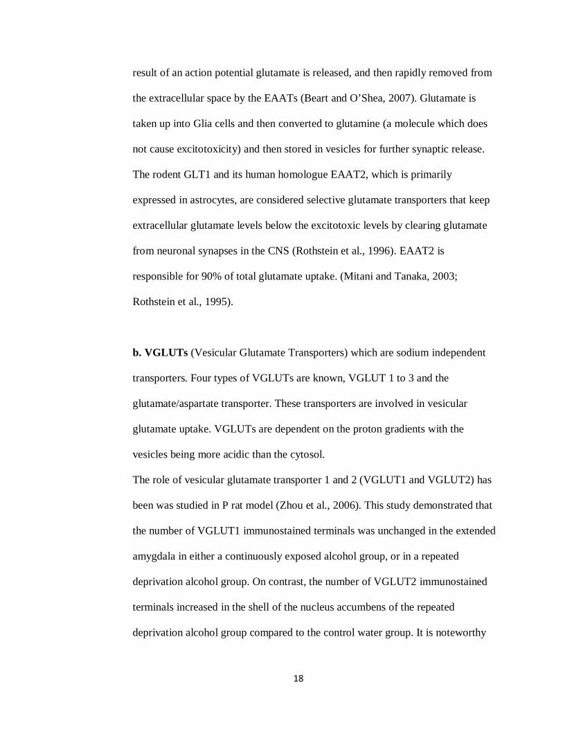
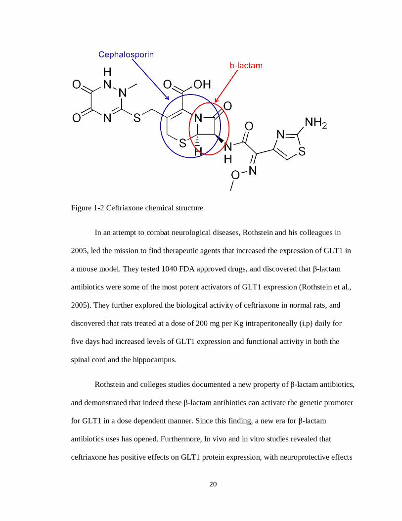
1.7 Activation of GLT1 by Ceftriaxone, a -lactam antibiotic
Ceftriaxone is a -lactam broad spectrum antibiotic, belonging to the third
generation of cephalosporins. It works by interfering with the formation of the bacteria's
cell wall so that the wall ruptures, resulting in the death of the bacteria. Ceftriaxone is
mainly used by intravenous or intramuscular administration, in the treatment of many
bacterial infections, such as respiratory tract infections, urinary tract infections and
meningitis.
20
Figure 1-2 Ceftriaxone chemical structure
In an attempt to combat neurological diseases, Rothstein and his colleagues in
2005, led the mission to find therapeutic agents that increased the expression of GLT1 in
a mouse model. They tested 1040 FDA approved drugs, and discovered that -lactam
antibiotics were some of the most potent activators of GLT1 expression (Rothstein et al.,
2005). They further explored the biological activity of ceftriaxone in normal rats, and
discovered that rats treated at a dose of 200 mg per Kg intraperitoneally (i.p) daily for
five days had increased levels of GLT1 expression and functional activity in both the
spinal cord and the hippocampus.
Rothstein and colleges studies documented a new property of -lactam antibiotics,
and demonstrated that indeed these -lactam antibiotics can activate the genetic promoter
for GLT1 in a dose dependent manner. Since this finding, a new era for -lactam
antibiotics uses has opened. Furthermore, In vivo and in vitro studies revealed that
ceftriaxone has positive effects on GLT1 protein expression, with neuroprotective effects
21
based on its ability to inhibit neuronal cell death, by preventing glutamate excitotoxicity.
Although the molecular mechanism of the action of GLT1 is unclear, analysis of the 2.5-
kb human EAAT2 promoter revealed that NF- B is a very important regulator of
EAAT2 expression in astrocytes, the -lactam antibiotics are transcriptional activators of
EAAT2 resulting in increased EAAT2 protein levels (Kim et al., 2010). Ceftriaxone
increases EAAT2 transcription in primary human fetal astrocytes through the NF- B
signaling pathway. Studies revealed that the 272 position of NF- B binding site is critical
in ceftriaxone mediated EAAT2 protein induction. Indeed, there is a promising potential
utility of EAAT2 promoter for developing screening assays to identify novel regulators of
glutamate transport in the brain.
1.8 Objectives and Goals
The main goal of this thesis is to investigate the role of GLT1 in relapse to alcohol
seeking behavior in a high drinking alcohol-preferring (P) rat model. Based on the facts,
P rats are an established model of alcoholism, and will readily consume intoxicating
levels of alcohol (McBride and Li, 1998: Rodd-Henricks, et al., 2002; Rodd, et al., 2005).
In addition, P rats find alcohol positively reinforcing and consume alcohol for its
pharmacological effects in relevant blood alcohol concentrations (0.05-0.2) (Li e al.,
1987). Further studies have revealed that chronic alcohol consumption in P rats leads to
tolerance and dependence, whereas deprivation leads to relapse (McBride and Li, 1998).
22
In this research, we based our hypothesis on the fact that glutamate transporter
GLT1 is responsible for the removal of most extracellular glutamate, we explored the
hypothesis, that up-regulation or activation of GLT1 by the use of the -lactam antibiotic
ceftriaxone, known to elevate GLT1 expression, would attenuate ethanol consumption
after a period of alcohol deprivation and ultimately prevent relapse. If an increase in
glutamate transmission plays a critical role in alcohol relapse, then up-regulation of
GLT1 should attenuate this response, and correlate with an increase of GLT1 in the
nucleus accumbens and or prefrontal cortex.
Materials and methods
2.1 Animals
All the animals that were used in this study were male alcohol-preferring (P) rats.
At the beginning of the experiment, the animals weighed in a range of 317 gm to 395 gm.
Animals were obtained from the Indiana University Medical Center (Indianapolis, IN) at
approximately 4 weeks of age and housed in groups of two in standard plastic tubs, in a
temperature (21°C) and humidity (~50%) controlled vivarium maintained on a 12 hour
light/dark cycle, at the main campus of the University of Toledo. The total number of
animals used in the study was 44. After habituation to the vivarium, two weeks prior to
23
90 days old, animals were moved to the Department of Laboratory Animal Resources
(DLAR) and were singly housed. All Experimental procedures began when animals were
approximately 90 days old. Animal procedures were approved (animal protocol #106966)
by the Institutional Animal Care and Use Committee, and in accordance with the
guidelines of the National Institutes of Health.
2.2 Behavioral Drinking Paradigms
Male P rats were given free choice access to food, tap water, 15%, and 30%
ethanol for five weeks at approximately three months old. 190 proof ethanol was
purchased from PHARMCO-AAPER (Shelbyville, KY), diluted with distilled water to
make the appropriate desired concentrations of the alcohol that was offered to rats, and
then alcohol was placed in glass bottles. The daily consumption of alcohol was measured
by bottle weight. Alcohol bottles were replaced three times a week (Mondays,
Wednesdays, and Fridays) at noon. At that time animals were weighed by placing them in
a plastic beaker on a Sartorius scale (max = 820g). Alcohol measurements were made by
subtracting the weight of the bottle from the previous weight of the bottle 2 or 3 days
prior. Alcohol consumption for each animal was measured as grams of alcohol consumed
per kilogram of body weight per day for 5 weeks. At the end of five weeks, animals that
did not meet the requirement (must have drank more than or equal to 4 grams of alcohol
per kilogram body weight on baseline) for chronic alcohol drinking behavior were
removed from the study. Baseline calculations were the average measurements taken
across the last two weeks of the five week alcohol drinking paradigm. The requirement
24
for chronic alcohol drinking behavior was obtained from a previous model (Li et al.,
1987). On week six, animals were separated into three different groups and deprived
from alcohol for 2 weeks. During the last five days of the 2 weeks deprivation period,
animals were injected intraperitoneally (i.p.) with either saline (n = 8), ceftriaxone 50
mg/kg (n = 8), or ceftriaxone 100 mg/kg (n = 8). ceftriaxone was purchased from the
pharmacy of the University Of Toledo’s hospital, as a powder and dissolved in a saline
solution immediately before being administered to the animals. After 10 days of alcohol
deprivation, ceftriaxone was administered (i.p.) once a day at noon for five consecutive
days. Animal body weights and water bottles were measured daily right before injections
throughout the deprivation period. After the five days of treatment, animals were re-
exposed to the alcohol drinking paradigm for an additional nine days to determine the
effects of ceftriaxone treatment in relapse to alcohol seeking behavior. During this time,
weights of animals and their water/alcohol bottles were recorded daily as previously
described. On day 10 animals were euthanized with isoflurane, rapidly decapitated with a
guillotine, and their brains were dissected out and immediately frozen on dry ice and
stored at (-70ºC) for immunoblotting. An additional naive control group of animals (n =
6) was treated with saline for five days to undergo a similar level of stress as the other
tested animals, then after ten days were euthanized (at the same age of the other groups),
decapitated, and their brains were dissected out and immediately frozen on dry ice and
stored at (-70ºC) for immunoblotting. A similar experimental design was applied to
sucrose consumption in order to test ceftriaxone’s specificity on alcohol consumption
protocol. At approximately three months old, animals (n = 14) were placed on a free-
choice 10% sucrose drinking paradigm (sucrose, SIGMA; diluted with distilled water) for
25
three weeks before undergoing the two week deprivation period. On week four, animals
were deprived of sucrose for two weeks, during the last five days of the two week sucrose
deprivation period, animals were treated with either saline (n = 4), ceftriaxone 50 mg/kg
(n = 5), or ceftriaxone 100 mg/kg (n= 5) once a day for five days . After the five days of
treatment, animals were re-exposed to the sucrose drinking paradigm for an additional
nine days to determine the long-lasting effects of ceftriaxone treatment. During this time,
weights of animals and their water/sucrose bottles were recorded daily as previously
described. On Day 10, animals were euthanized with isoflurane.
2.3 Brain Tissue Harvesting
In this study, we assessed GLT1 expression levels in the nucleus accumbens and
prefrontal cortex, in a set of animals exposed to free choice-ethanol (15% and 30% v/v)
and water for 5 weeks and then deprived for 2 weeks, afterwards treated with ceftriaxone
50, 100 mg/kg, or saline for 5 days, then re-exposed to alcohol for nine days. On Day 10,
animals were euthanized by isoflurane, rapidly decapitated with a guillotine, and their
brains were dissected out and immediately frozen on dry ice and stored at (-70º C). The
prefrontal cortex and nucleus accumbens were punctured streotaxically using cryostat
apparatus and frozen brain regions were stored for immunoblotting assays, to examine
GLT1 protein levels.
2.4 Protein Tissue Extraction Protocol
Procedures were conducted on nucleus accumbens and prefrontal cortex samples
from animals sacrificed on day 10 of re-exposure. Samples were homogenized using
26
filtered lysis buffer (2.5mL 1M Tris HCL, 2.5mL 3 M NaCl, 0.1mL 0.5M EDTA, 2.5mL
10% NP-40, 5mL 10% Triton, 0.5mL 10%SDS, 3 mL of dissolved protease inhibitor
tablet in water, and 33.9 mL Millipore water). 300 µL lysis buffer was added to each
sample in a 1.5 µL eppendorf tubes and the tissue was grounded with a pestle until no
solid mass remained. The samples were then placed on ice for 30 minutes to allow
homogenization to complete. They were then centrifuged at 13,200 RPM for 15 minutes
at 4º C. The supernatant was aliquoted and immediately frozen on dry ice and the pellet
was discarded.
2.5 Protein Quantification
A Lowry protein quantification assay was conducted on one aliquot of each
sample. All samples were assayed in quadruplicates in a 96-well plate. Bovine serum
albumin (BSA) (1.48mg/mL, New England Biolabs) was used to make a standard curve.
The curve was prepared using serial BSA dilutions. The wells containing the proteins
samples contained 1µL of sample diluted in 4 µL of lysis buffer. Afterwards, 3mL
reagent A (Biorad Laboratories) was mixed with 60 µL reagent S (BioRad Laboratories)
and 25µL of this mixture was added to each well. 200µL of reagent B (BioRad
Laboratories) was then added to each well and the reaction was allowed to sit at room
temperature for 15 minutes before being read on a multiskan FC spectrophotometer
(Thermo Scientific) at a wavelength of 750 nm. The quadruplicate optical density values
were averaged and the blank optical density was subtracted from each measurement. A
standard curve was made by plotting the BSA optical density versus the diluted BSA
27
protein concentrations. The protein concentrations were then calculated by comparing
their optical densities to the standard BSA curve.
2.6 Western Blot Procedures
Water (n = 6), saline (n = 8), ceftriaxone 50mg/kg (n = 8), and ceftriaxone
100mg/kg (n = 8) samples were used for the western blot analysis of GLT1 total protein
concentration in the nucleus accumbens and prefrontal cortex. Using the protein
quantification data, each sample was diluted to 8µg/20µL with the same lysis buffer.
Laemmli dye was added to each sample (5µL dye per 20µL sample) and mixed well,
samples were vortexed then heated for 4 minutes at 98º C in a digital dry bath (Labnet
International Inc.), vortexed again, and then centrifuged for 3 minutes at 4º C at 13,200
RPM (Centrifuge 5414 R, Eppendorf ).The 10-20% Tris=glycine gels (Invitrogen) were
placed into an electrophoresis apparatus and submerged in 1X laemmli buffer (10X
laemmli buffer = 30.2 Tris Base, 144g Glycine, 10g SDS, qsp to 1 L). A 20 µL of each
sample was placed into a well of the gel and proteins were separated by electrophoresis
(1 hour at 200 volts). After completion, the gels containing the proteins were removed
from the electrophoresis apparatus and transferred on an immobilon-P membrane
(Millipore, Fisher Scientific, Inc.) using a transfer apparatus (Idea Scientific Company,
MN). Protein transfer was carried out by filling the transfer chamber with transfer buffer
(3.2 L distilled water, 28.8g Glycine, 5.9g Tris Base, 800 mL methanol) which was then
hooked up to electrophoresis electrodes for 2.5 hours at a 24 volts. Membranes were
placed in Ponceau dye for one minute, to see the bands, then washed with water 4 times
quickly, membranes were then blocked with blocking buffer for 30 minutes (10mL/blot;
28
3% milk made from 3 g dry milk and 100 mL 1X TBST). With the membranes still
soaking in the blocking buffer, primary GLT1 antibody (AB1783 GP X Glutamate
Transporter, Millipore) was added (2µL/blot, 1:5000) and was allowed to incubate
overnight shaking at 300 RPM in the fridge or cold room. The next day, each membrane
was washed with 1X TBST 5 times for 5 minutes each and then incubated again with
blocking buffer for 30 minutes. Each membrane was then incubated in secondary anti-
guinea pig (anti-guinea pig IgG HRP-linked antibody, Cell Signaling Technology, Inc.)
antibody (2µL/blot, 1:5000 for 1.5 hour before being washed again with IX TBST 5
times for 5 minutes each. The membranes were dried on Whatman paper and incubated
with a Super Signal West Pico developer kit (Pierce; 1mL reagent A and 1 mL reagent B
per blot) for one minute to detect the chemiluminescent signal of HRP. The membranes
were dried again on Whatman paper and developed on Kodak BioMax MR Film (Thermo
Fisher Scientific) with an SRX-101A machine. Protein loading was then normalized
using -tubulin immunoblotting as a loading control. After exposure, membranes were
washed with 1X TBST 5 times for 5 minutes each before being placed in blocking buffer
for 30 minutes. The membranes were incubated in -tubulin primary antibody (2µL/blot,
1:5000) overnight. The next day, membranes were washed with 1X TBST 5 times for 5
minutes each and then incubated again with blocking buffer for 30 minutes. Each
membrane was then incubated in secondary anti-mouse (Anti-mouse IgG HRP –linked
antibody (2µL/blot, 1:5000) for 1.5 hour before being washed again with IX TBST 5
times for 5 minutes each. The membranes were dried on Whatman paper and incubated
with a Super Signal West Pico developer kit (Pierce; 1mL reagent A and 1 mL reagent B
per blot) for one minute to detect the chemiluminescent signal of HRP. The membranes
29
were dried again on Whatman paper and developed on Kodak BioMax MR Film (Thermo
Fisher Scientific) with an SRX-101A machine. Digitalized images of the immunoreactive
proteins were quantified using an MCID system and the data are reported as percentage
ratios
Results
3.1 Effects of Ceftriaxone Treatment on Alcohol Drinking Behavior
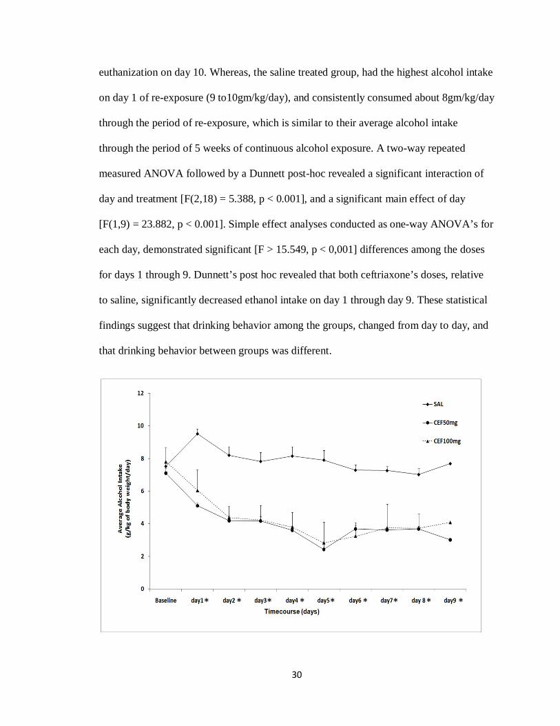
During the 9 days of re-exposure to alcohol after 5 days of treatment with either
saline, ceftriaxone 50 or 100 mg/kg, alcohol consumption was measured daily as
g/kg/day for 9 days, starting on the first day of alcohol re-exposure. The data presented in
Figure 3-1 show the average alcohol consumption for 9 days for saline (n = 8),
ceftriaxone 50 mg/kg (n = 8), ceftriaxone 100mg/kg (n = 8) with the baseline being the
average of rat’s alcohol consumption for the last two weeks of continuous alcohol
exposure. Note that rats were initially exposed to free choice of water, ethanol 15% and
30% for 5 weeks. Alcohol intake was significantly reduced (p < 0.001) for the groups
treated with ceftriaxone since day 1 through day 9 of re-exposure. Whereas, the saline
treated group regressed back to their normal drinking behavior, as soon as they were re-
exposed to alcohol (from day 1). Alcohol intake for the two groups treated with
ceftriaxone was reduced at both doses, which displayed a long-lasting effect, reached the
lowest alcohol intake of 2 to 3 g/kg/day on day 5 of re-exposure, progressively increased
and then reached a plateau, yet overall remained around under 4g/kg/day until their
30
euthanization on day 10. Whereas, the saline treated group, had the highest alcohol intake
on day 1 of re-exposure (9 to10gm/kg/day), and consistently consumed about 8gm/kg/day
through the period of re-exposure, which is similar to their average alcohol intake
through the period of 5 weeks of continuous alcohol exposure. A two-way repeated
measured ANOVA followed by a Dunnett post-hoc revealed a significant interaction of
day and treatment [F(2,18) = 5.388, p < 0.001], and a significant main effect of day
[F(1,9) = 23.882, p < 0.001]. Simple effect analyses conducted as one-way ANOVA’s for
each day, demonstrated significant [F > 15.549, p < 0,001] differences among the doses
for days 1 through 9. Dunnett’s post hoc revealed that both ceftriaxone’s doses, relative
to saline, significantly decreased ethanol intake on day 1 through day 9. These statistical
findings suggest that drinking behavior among the groups, changed from day to day, and
that drinking behavior between groups was different.
* * * * * * * * *
31
Figure 3-1:
Daily alcohol intake of male P rats for 9 days of alcohol re-exposure, following 5 days of treatment during relapse period with saline (n = 8) or ceftriaxone 50 mg/kg (n = 8)and ceftriaxone 100 mg/kg (n = 8). The graph represents average daily alcohol consumption (±SEM) during the 9 days of alcohol re-exposure. A two-way, repeated-measures ANOVA revealed a significant reduction in average daily alcohol consumption during the duration of re-exposure for both ceftriaxone-treated groups as compared to the saline treated group. *, depicts a significant (p < 0.001) one-way ANOVA across doses for the respective days.
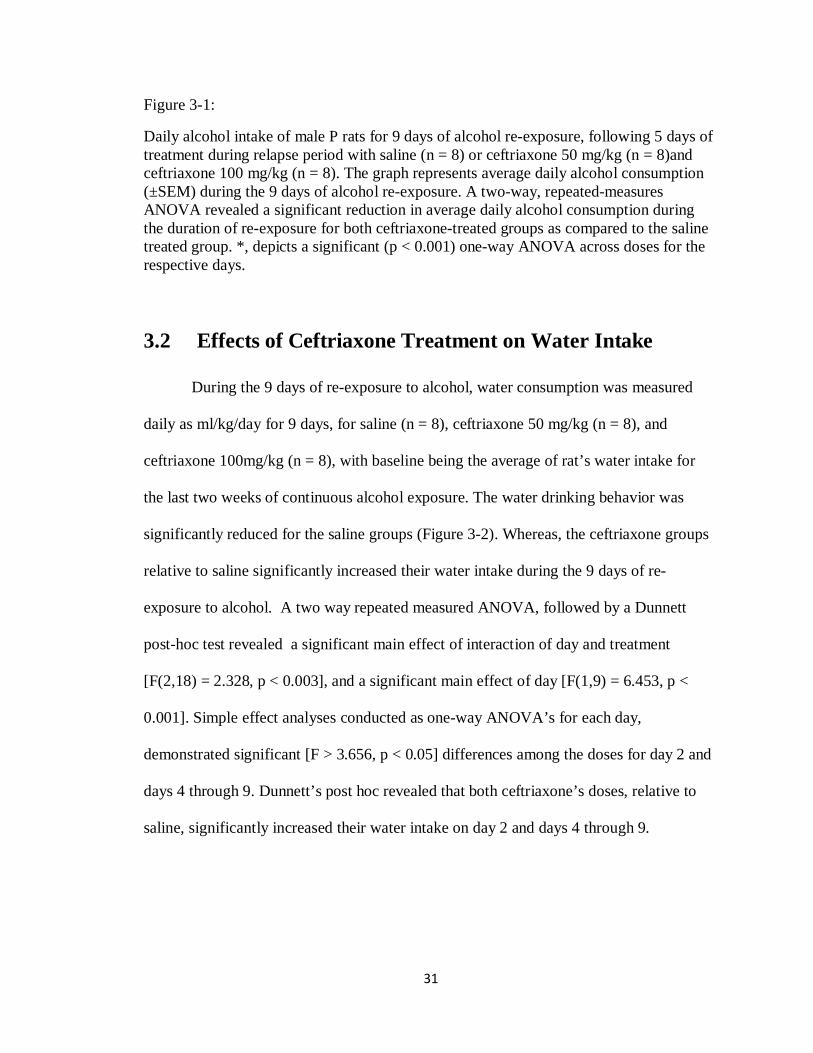
3.2 Effects of Ceftriaxone Treatment on Water Intake
During the 9 days of re-exposure to alcohol, water consumption was measured
daily as ml/kg/day for 9 days, for saline (n = 8), ceftriaxone 50 mg/kg (n = 8), and
ceftriaxone 100mg/kg (n = 8), with baseline being the average of rat’s water intake for
the last two weeks of continuous alcohol exposure. The water drinking behavior was
significantly reduced for the saline groups (Figure 3-2). Whereas, the ceftriaxone groups
relative to saline significantly increased their water intake during the 9 days of re-
exposure to alcohol. A two way repeated measured ANOVA, followed by a Dunnett
post-hoc test revealed a significant main effect of interaction of day and treatment
[F(2,18) = 2.328, p < 0.003], and a significant main effect of day [F(1,9) = 6.453, p <
0.001]. Simple effect analyses conducted as one-way ANOVA’s for each day,
demonstrated significant [F > 3.656, p < 0.05] differences among the doses for day 2 and
days 4 through 9. Dunnett’s post hoc revealed that both ceftriaxone’s doses, relative to
saline, significantly increased their water intake on day 2 and days 4 through 9.
32
Figure 3-2:
Daily water intake of male P rats for 9 days, following 5 days of treatment with saline (n = 8) or ceftriaxone 50 mg/kg (n = 8) , and ceftriaxone 100 mg/kg (n = 8). The graph represents average daily water consumption (±SEM) during the 9 days of alcohol re-exposure. A two-way, repeated-measures ANOVA revealed a significant increase in average daily water consumption during the duration of re-exposure for both ceftriaxone-treated groups as compared to the saline treated group. *, depicts a significant (p < 0.05) one-way ANOVA across doses for the respective day.
3.3 Effects of Ceftriaxone Treatment on Body Weight
During the 9 days of re-exposure to alcohol, body weight was measured daily as
gm (±SEM) for 9 days, for saline (n = 8), ceftriaxone 50mg/kg (n = 8), and ceftriaxone
100 mg/kg (n = 8), with baseline being the average of rat’s weight for the last two weeks
of continuous alcohol exposure (Figure 3-3). A two way repeated measured ANOVA,
followed by a Dunnett post-hoc test revealed a significant main effect of interaction of
day and treatment [F(2,18) = 3.369, p < 0.001], and a significant main effect of day
[F(1,9) = 68.251, p < 0.001]. However, a simple effect analyses conducted as one-way
* * * * * * *
33
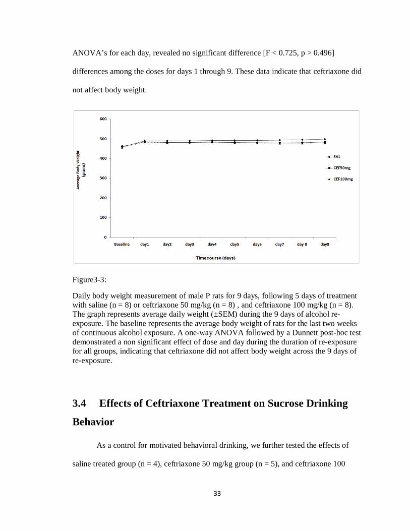
ANOVA’s for each day, revealed no significant difference [F < 0.725, p > 0.496]
differences among the doses for days 1 through 9. These data indicate that ceftriaxone did
not affect body weight.
Figure3-3:
Daily body weight measurement of male P rats for 9 days, following 5 days of treatment with saline (n = 8) or ceftriaxone 50 mg/kg (n = 8) , and ceftriaxone 100 mg/kg (n = 8). The graph represents average daily weight (±SEM) during the 9 days of alcohol re-exposure. The baseline represents the average body weight of rats for the last two weeks of continuous alcohol exposure. A one-way ANOVA followed by a Dunnett post-hoc test demonstrated a non significant effect of dose and day during the duration of re-exposure for all groups, indicating that ceftriaxone did not affect body weight across the 9 days of re-exposure.
3.4 Effects of Ceftriaxone Treatment on Sucrose Drinking
Behavior
As a control for motivated behavioral drinking, we further tested the effects of
saline treated group (n = 4), ceftriaxone 50 mg/kg group (n = 5), and ceftriaxone 100
34
mg/kg group (n = 5) on sucrose (10%) consumption. Sucrose intake was examined over a
period of 9 days. During the 9 days of re-exposure to sucrose (10%) after 5 days of
treatment with either saline, ceftriaxone 50 mg/kg or ceftriaxone 100 mg/kg, sucrose
consumption was measured daily as ml/kg/day for 9 days, starting on the first day of
sucrose re-exposure. The data presented in Figure 3-4 shows the average sucrose
consumption for 9 days for saline (n = 4), ceftriaxone 50 mg/kg (n = 5), ceftriaxone 100
mg/kg (n = 5) with the baseline being the average of rat’s alcohol intake for the last week
of continuous sucrose exposure, starting on the first day of sucrose re-exposure. A two
way repeated measured ANOVA, followed by a Dunnett post-hoc test revealed a non
significant main effect of interaction of day and treatment [F(2,18) = 0.173, p = 1.00],
and a significant main effect of day [F(1,9) = 4.152, p < 0.001]. Simple effect analyses
conducted as one-way ANOVA’s for each day, demonstrated a non significant [F <
0.514, p > 0.612] differences among the doses for days 1 through 9. These data indicate
that ceftriaxone did not affect sucrose intake.
35
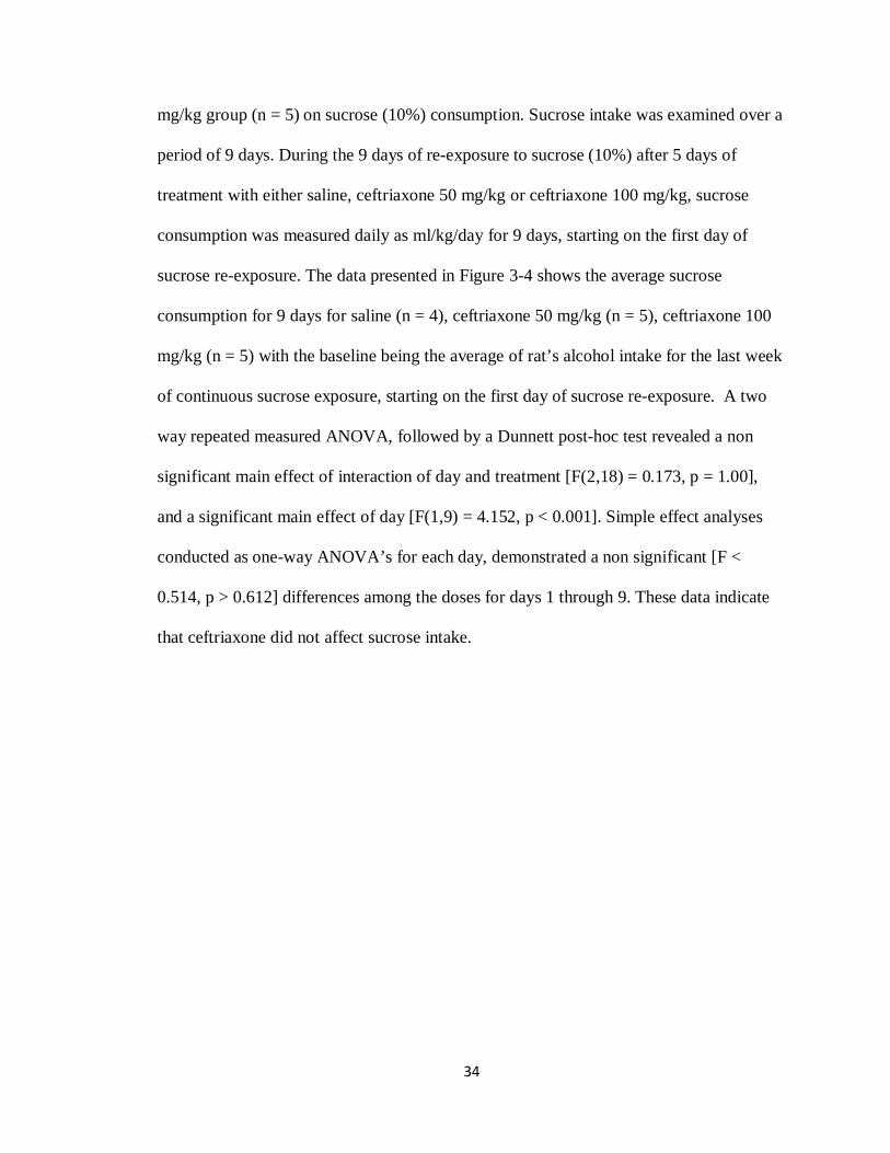
Figure 3-4:
Daily sucrose (10%)l intake of male P rats for 9 days, following 5 days of treatment with saline (n = 4) or ceftriaxone 50 mg/kg (n = 5) , and ceftriaxone 100 mg/kg (n = 5). The graph represents average daily sucrose (10%) consumption (±SEM) during the 9 days of sucrose (10%) re-exposure. While the day main effect was significant (p < 0.001), neither the interaction by day and treatment (p = 1.00) nor the day main effect (p > 0.612) were significant. Thus, ceftriaxone did not affect sucrose intake.
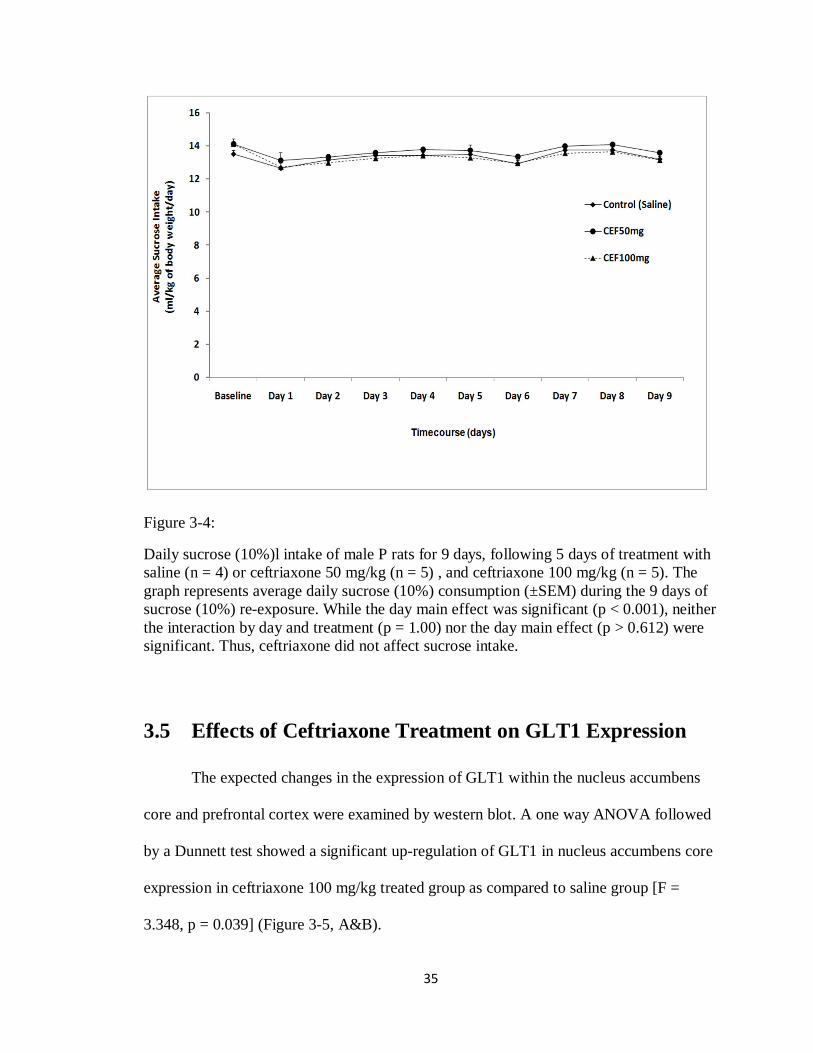
3.5 Effects of Ceftriaxone Treatment on GLT1 Expression
The expected changes in the expression of GLT1 within the nucleus accumbens
core and prefrontal cortex were examined by western blot. A one way ANOVA followed
by a Dunnett test showed a significant up-regulation of GLT1 in nucleus accumbens core
expression in ceftriaxone 100 mg/kg treated group as compared to saline group [F =
3.348, p = 0.039] (Figure 3-5, A&B).
36
Fig. 3-5 (A&B): Effects of naive (n = 4), saline (n = 4), ceftriaxone 50mg/kg (n = 4), ceftriaxone 100mg/kg (n = 4) on GLT1 expression in the nucleus accumbens core.( A) Each panel presents immunoblots for -tubulin, which was used as a control loading protein, and GLT1. B) Quantitative analysis revealed a significant increase in the ratio of
B
Saline CEF-50 CEF-1000
100
200
300
GLT
1/b-
tubu
lin (%
ratio
to N
aive
con
trol)
*
37
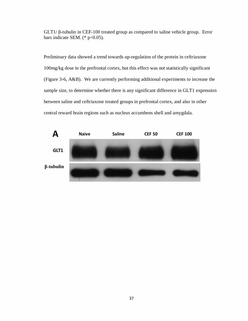
GLT1/ -tubulin in CEF-100 treated group as compared to saline vehicle group. Error bars indicate SEM. (* p<0.05).
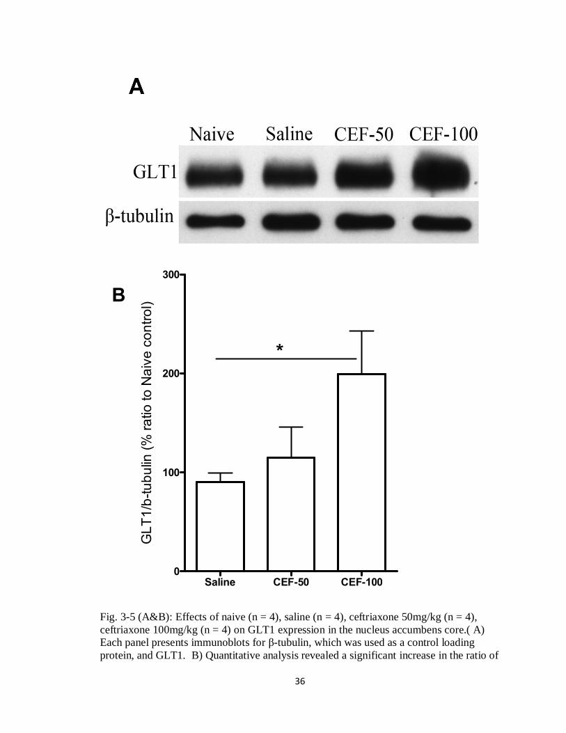
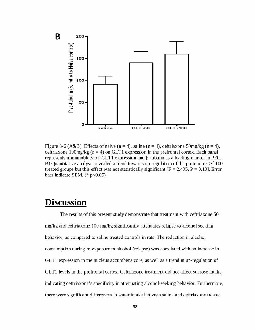
Preliminary data showed a trend towards up-regulation of the protein in ceftriaxone
100mg/kg dose in the prefrontal cortex, but this effect was not statistically significant
(Figure 3-6, A&B). We are currently performing additional experiments to increase the
sample size, to determine whether there is any significant difference in GLT1 expression
between saline and ceftriaxone treated groups in prefrontal cortex, and also in other
central reward brain regions such as nucleus accumbens shell and amygdala.
Naive Saline CEF 50 CEF 100
-tubulin
A
38
Figure 3-6 (A&B): Effects of naive (n = 4), saline (n = 4), ceftriaxone 50mg/kg (n = 4), ceftriaxone 100mg/kg (n = 4) on GLT1 expression in the prefrontal cortex. Each panel represents immunoblots for GLT1 expression and -tubulin as a loading marker in PFC. B) Quantitative analysis revealed a trend towards up-regulation of the protein in Cef-100 treated groups but this effect was not statistically significant [F = 2.405, P = 0.10]. Error bars indicate SEM. (* p<0.05)
Discussion
The results of this present study demonstrate that treatment with ceftriaxone 50
mg/kg and ceftriaxone 100 mg/kg significantly attenuates relapse to alcohol seeking
behavior, as compared to saline treated controls in rats. The reduction in alcohol
consumption during re-exposure to alcohol (relapse) was correlated with an increase in
GLT1 expression in the nucleus accumbens core, as well as a trend in up-regulation of
GLT1 levels in the prefrontal cortex. Ceftriaxone treatment did not affect sucrose intake,
indicating ceftriaxone’s specificity in attenuating alcohol-seeking behavior. Furthermore,
there were significant differences in water intake between saline and ceftriaxone treated
B
39
groups, however, there were no significant differences in the body weight between all the
tested groups indicating that ceftriaxone does not affect body weight of animals. The
increase in water intake in ceftriaxone treated groups could be explained by the fact that
the decrease in their alcohol intake during relapse was compensated, in part, by the
increase in water intake.
In this study, we investigated GLT1 protein levels in the prefrontal cortex and
nucleus accumbens core. It has been established, that glutamate transmission in key brain
regions of the reward circuit, plays a critical role in alcohol dependence and drug-seeking
behavior. The glutamatergic system in the prefrontal cortex has been suggested to be
involved in cocaine reinforcement (Sari et al., 2009). Animal studies of drugs of abuse,
supports the importance of glutamate projections from prefrontal cortex to the nucleus
accumbens and the ventral tegmental area (Goldstein and Volkow, 2002). It has been
shown that elevations in extracellular glutamate during alcohol withdrawal from chronic
alcohol exposure, are associated with increased number and function of N-methyl-D-
aspartate (NMDA) glutamate receptors (Rossetti et al., 1999).The effect of NMDA
receptor stimulation on the extracellular levels of glutamate in Sprague-Dawley rats
withdrawn from chronic alcohol exposure by the administration of NMDA directly in the
striatum of dependent rats, showed a significant increased glutamate output compared to
sucrose controls as measured by microdialysis (Rossetti et al., 1999). The results suggest
that glutamate-induced NMDA receptor-mediated elevations of extracellular glutamate
during alcohol withdrawal, may contribute to the neuropathology associated with
alcoholism, implicating the role of increased glutamate release in alcohol dependence
(Rossetti et al., 1999). Further evidence indicates that chronic alcohol exposure leads to
40
an increase in glutamate output as well as an impaired ability in glutamate transport. The
effect of chronic ethanol consumption on glutamate transporter binding and function was
studied in the cerebral cortex of alcohol-preferring c AA rats (Schreiber and Freund,
2000). In this study, the alcohol-preferring c AA rats were chronically exposed to 10%
v/v alcohol for 20 months, rats showed a down-regulation of neuronal and/or glial
glutamate transporter activity, with limited re-uptake of synaptic glutamate (Schreiber
and Freund, 2000).
The excessive amount of glutamate seen within the prefrontal cortex during
alcohol withdrawal, causes hyperactivation of neurons that project to the nucleus
accumbens, and promotes alcohol-seeking behavior. The medial prefrontal cortex, a
terminal region of the mesocorticolimbic dopamine system, is composed of pyramidal
glutamatergic neurons that are the output of this unit, these glutamatergic neurons are
modulated by numerous neurotransmitter systems, including GABAergic interneurons
that inhibit the firing of the glutamatergic neurons, preventing excessive glutamate
release (Steketee, 2003). With chronic alcohol consumption, a decreased GABAA
receptor density and sensitivity is observed; in contrast, an increased NMDA receptor
density and sensitivity is encountered, resulting in the loss of inhibitory influence of the
GABAergic interneurons over the cortical neurons. Alcohol-exposure alters
glutamatergic activity in the mesocorticolimbic circuit, and the glutamatergic projections
from the prefrontal cortex to the nucleus accumbens and the ventral tegmental area, are
important in mediating craving for alcohol.
41
The specific role of GLT1 in addiction has been studied in various drug abuse
models. For example, activation of GLT1 by MS-153 attenuated cocaine, morphine and
methamphetamine conditioned place preference in mice (Nakagawa et al., 2005). In
accordance, (Sari et al., 2009) reported that ceftriaxone attenuates cue-induced cocaine
relapse in a dose-dependent manner. GLT1 is one of the major glutamate transporters,
expressed predominantly in astroglial cells, and is responsible for the uptake of more than
90% of extracellular glutamate (Danbolt, 2001; Robinson, 1998). The glutamate
transporters tightly regulate glutamate concentration. If an increase in glutamate
transmission plays a major role in relapse to alcohol seeking behavior, then up-regulation
of GLT1 should attenuate such response. We have tested this hypothesis using
ceftriaxone, -lactam antibiotic known to up-regulate GLT1, in our established male P rat
model. Although, our present preliminary study in prefrontal cortex and studies from ours
and others demonstrated that ceftriaxone administration for 5 days can lead to up-
regulation of GLT1 levels (Sari et al., 2009, Sari et al., 2011, Rothstein et al., 2005)
ceftriaxone has been shown to increase GLT1 activity independent of an increase in
GLT1 expression. A study on wistar rats (Thone-Reinke et al., 2008) used a single dose
of ceftriaxone treatment 200 mg/kg intraperitoneally (i.p), this treatment increased
glutamate transporter activity without increasing GLT1 expression in a stroke model. It is
therefore possible that ceftriaxone could reduce alcohol consumption by other
mechanisms, or perhaps the GLT1 up-regulation could be secondary to an unknown
primary mechanism.
Interestingly, chronic alcohol exposure is linked with a decrease in brain
glutathione (GSH) and an increase in oxidized glutathione in vivo (Gotz et al., 2001).
42
Furthermore, alcohol withdrawal is associated with increases in oxygen-derived free
radicals which by oxidation of thiol groups located on the glutamate transporters, may
inhibit glutamate uptake (Volterra et al., 1994). Additionally, it has been shown that
ceftriaxone increased both GSH and cysteine/glutamate exchanger (xCT) levels, leading
to the reversal of the glutamate transporter deficits caused by the increased levels of free
radical oxidation (Lewerenz et al., 2009). The xCT expression was increased by the use
of ceftriaxone, in the nucleus accumbens of a cocaine relapse-behavior in a rat model
(Knacksteddt et al., 2010). This could explain the decrease in alcohol drinking after
treatment with ceftriaxone. It is possible that ceftriaxone-induced increase in xCT, and
subsequent increase in the levels of GSH, corrected the deficits of the glutamate
transporter, independent of GLT1 up-regulation.
Indeed this present study reports a correlation between increased GLT1 protein
expression in the nucleus accumbens core, and the reduced alcohol drinking in relapse to
alcohol seeking behavior, however, the next steps of our project could include:
I. Increasing the n value for the prefrontal cortex.
II. Identifying brain regions in which ceftriaxone has increased GLT1 protein
expression, and other regions in which it had no effect. Additional key
brain regions involved in the reward neurocircuitry such as the nucleus
accumbens shell, amygdala and hippocampus need to be examined as
well.
III. Additionally, brain regions of the reward circuitry, could be further
separated into their sub-components to seek the possibility of any
43
differential effects. For example, the prefrontal cortex can be further
subdivided into prelimbic cortex, infralimbic cortex and cingulated cortex.
IV. Future studies are needed to examine the mechanisms by which
ceftriaxone reduces alcohol drinking. For example, dihydrokainic acid
(DHK), a selective GLT1 antagonist, could be co-administered with
ceftriaxone to ensure that the activation of GLT1 by ceftriaxone is the
direct mechanism of action in the reduction of alcohol intake, rather than
other unknown mechanisms of the drug.
V. Moreover, the role of other transporters, such as VGLUTs and GLAST in
relapse to alcohol seeking behavior should be examined.
In conclusion, our findings demonstrate that activation of GLT1 by ceftriaxone
attenuates relapse to alcohol-seeking behavior in male P rats. Studies revealed that the
effect of ceftriaxone appears to be selective to GLT1 since other glutamate transporters
are unaffected by this drug. Our results suggest that activation of GLT1 counteracts
elevated extracellular glutamate in central reward brain regions that are involved in drug
abuse, including alcohol. Thus, we suggest that GLT1 may be a potential target for
decreasing the extracellular glutamate level, and consequently attenuating relapse to
alcohol-drinking behavior.
44
References Beart, P. M. and O'Shea, R. D. “Transporters for L-glutamate: An update on their
molecular pharmacology and pathological involvement”. British Journal of
Pharmacology, 150 (2007): 5–17.
Biggio G, Concas A, Follesa P, et al. “Stress, ethanol, and neuroactive steroids”.
Pharmacology and Therapeutics.116 (2007): 140–171.
Blednov YA, Harris AR.“Metabotropic glutamate receptor 5 (mGluR5) regulation of
ethanol sedation, dependence and consumption: Relationship to acamprosate
actions.” International Journal of Neuropsychopharmacology. 11 (2008): 775-793.
Danbolt NC. “ Glutamate uptake”. ProgNeurobiol 65 (2001): 1-105.
Deitrich RA, Dunwiddie TV, Harris RA, Erwin VG. “Mechanism of action of ethanol:
initial central nervous system actions”. Pharmacological Reviews. 41 (1989): 489-
537.
Eriksson K. “Ethyl alcohol consumption: valid measurement in albino rats.” Science
(New York, N. Y.). 161 (1968): 76-7.
Feltenstein, M. W. and See, R. E. “The neurocircuitry of addiction: an overview”.
British Journal of Pharmacology. 154 (2008): 261–274.
Gatto GJ, Murphy JM, Waller MB, McBride WJ, Lumeng L, Li TK. “Chronic
ethanol tolerance through free-choice drinking in the P line of alcohol-preferring
rats”. Pharmacology,Biochemistry,and Behavior. 28 (1987): 111-115.
45
Gessa GL, Muntoni F, Collu M, Vargiu L, Mereu G. “Low doses of ethanol activate
dopaminergic neurons in the ventral tegmental area.”Brain Research. 348 (1985):
201-203.
Gilpin NW, Koob GF.“Neurobiology of Alcohol Dependence: Focus on Motivational
Mechanisms”. Alcohol Research & Health. 31 (2008): 185-195.
Goldstein RZ, Volkow ND . “Drug addiction and its underlying neurobiological basis:
neuroimaging evidence for the involvement of the frontal cortex.” TheAmerican
journal of Psychiatry.159 (2002): 1642-1652.
Gotz, M. E., Janetzky, B., Pohli, S., Gottschalk, A., Gsell, W., Tatschner, T., et al.
“Chronic alcohol consumption and cerebral indices of oxidative stress: is there a
link? “Alcoholism, Clinical &Expermental Research, 25 (2001): 717-725.
Groenewegen, H. J., Galis-de Graaf, Y., & Smeets, W. J. ”Integration and segregation
of limbic cortico-striatal loops at the thalamic level: an experimental tracing study
in rats.” Journal of Chemical Neuroanatomy, 16 (1999): 167-185.
Groenewegen, H. J., Wright, C. I., & Beijer, A. V. “The nucleus accumbens: gateway
for limbic structures to each the motor system?”. Progress in Brain Research, 107
(1996): 485-511.
Hodge CW, Samson HH, Chappelle AM. “Alcohol self-administration: Further
examination of the role of dopamine receptors in the nucleus accumbens”.
Alcoholism: Clinical and Experimental Research.21 (1997): 1083–1091.
Hyytia P, Koob GF. “GABAA receptor antagonism in the extended amygdala decreases
ethanol self-administration in rats”. European Journal of Pharmacology. 283
(1995): 151–159.
46
Johnson BA.“Update on neuropharmacological treatments for alcoholism: Scientific
basis and clinical findings.” Biochemical Pharmacology.75 (2008): 34–56.
Kanai Y, Hediger MA. “The glutamate and neutral amino acid transporter family:
physiological and pharmacological implications” European Journal of
Pharmacology. 479 (2003): 237-247.
Kim, K., Lee, S.-G., Kegelman, T. P., Su, Z.-Z., Das, S. K., Dash, R., Dasgupta, S.,
Barral, P. M., Hedvat, M., Diaz, P., Reed, J. C., Stebbins, J. L., Pellecchia,
M., Sarkar, D. and Fisher, P. B. “Role of Excitatory Amino Acid Transporter-2
(EAAT2) and glutamate in neurodegeneration: Opportunities for developing
novel therapeutics.” Journal of Cellular Physiology, 226 (2011): 2484–2493.
Knackstedt, L. A., Melendez, R. I., &Kalivas, P. W.“Ceftriaxone restores glutamate
homeostasis and prevents relapse to cocaine seeking”. Biological Psychiatry, 67
(2010): 81-84.
Koob GF. “Alcoholism: Allostasis and beyond.” Alcoholism: Clinical and Experimental
Research.27 (2003): 232–243.
Koob, G. F., Roberts, A. J., Schulteis, G., Parsons, L. H., Heyser, C. J., Hyytiä, P.,
Merlo-Pich, E. and Weiss, F. “Neurocircuitry Targets in Ethanol Reward and
Dependence”. Alcoholism: Clinical and Experimental Research. 22 (1998): 3–9.
Lester D, Freed EX. “Criteria for an animal model of alcoholism.”Pharmacoloy,
Biochemistry, and Behavior. 1 (1973): 103-107
47
Lewerenz, J., Albrecht, P., Tien, M. L., Henke, N., Karumbayaram , S., Kornblum,
H. I., et al. “Induction of Nrf2 and Xct are involved in the action of the
neuroprotective antibiotic ceftriaxone in vitro.” Journal of Neurochemistry. 111
(2009): 332-343.
Liebman, J.M., & Butcher, L. L.“Effects on self-stimulation behavior of drugs
influencing dopaminergic neurotransmission mechanisms.”
NaunynSchmiedebergsArchieves of Pharmacology, 277 (1973): 305-318.
Li TK, Lumeng L, McBride WJ, Waller MB. “Progress toward a voluntary oral
consumption model of alcoholism.” Drug and Alcohol Dependence. 4 (1979): 45-
60.
Li TK, Lumeng L, McBride WJ, Murphy JM. “Rodent lines selected for factors
affecting alcohol consumption.”Alcohol and Alcoholism. 1 (1987): 91-96.
Littleton JM.“Acamprosate in alcohol dependence: Implications of a unique mechanism
of action”. Journal of Addiction Medicine. 1 (2007): 115–125.
Lovinger DM, White G, Weight FF. “Ethanol inhibits NMDA-activated ion current in
hippocampal neurons.”Science.243 (1989): 1721–1724.
Lumeng L, Li TK.“The development of metabolic tolerance in the alcohol-preferring P
rats: comparison of forced and free-choice drinking of ethanol.” Pharmacology,
Biochemistry,and Behavior. 25 (1986): 1013-1020.
McBride, W. J., & Li T.K.“Animal models of alcoholism: neurobiology of high
alcohol-drinking behavior in rodents .” Critical Reviews in Neurobiology, 12
(1998): 339-369.
48
Mhatre MC, Pena G, Sieghart W, Ticku MK. “Antibodies specific for GABAA
receptor alpha subunits reveal that chronic alcohol treatment down-regulates
alpha-subunit expression in rat brain regions”. Journal of neurochemistry. 61
(1993): 1620-1625.
Mitani, A., & Tanaka, K.“Functional changes of glial glutamate transporter GLT-1
during ischemia: an in vivo study in the hippocampal CA1 of normal mice and
mutant mice lacking GLT-1”. Journal of Neuroscience, 23 (2003): 7176-7182.
Myers, C. E., & Gluck, M. A. “Context, conditioning, and hippocampal rerepresentation
in animal learning.” Behavioral Neuroscience, 108 (1994): 835-847.
Nakagawa T, Fujio M, Ozawa T, Minami M, Satoh M.“Effect of MS-153, a glutamate
transporter activator, on the conditioned rewarding effects of morphine,
methamphetamine and cocaine in mice.” Behavioural Brain Research.156 (2005): 233-
239.
Nicholas W. Gilpin, Ph.D. and George F. Koob, Ph.D.“Neurobiology of Alcohol
Dependence: Focus on Motivational Mechanisms” Alcohol Res Health. 31 (2008): 185-195.
Olive MF, Koenig HN, Nannini MA, Hodge CW.“Stimulation of endorphin
neurotransmission in the nucleus accumbens by ethanol, cocaine, and
amphetamine”. Journal of Neuroscience.21 (2001): RC184.
Penn PE, McBride WJ, Lumeng L, Gaff TM, Li TK. “Neurochemical and operant
behavioral studies of a strain of alcohol-preferring rats.”Pharmacology,
Biochemistry,and Behavior. 8 (1978): 475-481.
49
Phillips, A. G. “Brain reward circuitry: a case for separate systems.” Brain Research
Bulletin, 12 (1984): 195-201.
Phillips, A. G., & Fibiger, H. C. “The role of dopamine in maintaining intracranial self-
stimulation in the ventral tegmentum, nucleus accumbens, and medial prefrontal
cortex”. Canadian Journal of Psychology, 32 (1978): 58-66.
Pulvirenti L, Diana M.“Drug dependence as a disorder of neural plasticity: Focus on
dopamine and glutamate.” Reviews in the Neurosciences. 12 (2001): 141–158.
Roberts AJ, Mcdonald JS, Heyser CJ, et al. -Opioid receptor knockout mice do not
self-administer alcohol”. Journal of Pharmacology and Experimental
Therapeutics. 293 (2000): 1002–1008.
Robinson MB.”The family of sodium-dependent glutamate transporters: a focus on the
GLT1/EAAT2 subtype”. Neurochem Int. 33 (1998): 479-91.
Rodd-Hendricks, Z. A., Bell, R. L., Kuc, K. A., Murphy, J. M., McBride, W. J.,
Lumeng, L., et al.“Effects of ethanol exposure on subsequent acquisition and
extinction of ethanol self-administration and expression of alcohol-seeking
behavior in adult alcohol-preferring (P) rats: II. Adult exposure”. Alcohol
Clinical & Experimental Research, 26 (2002): 1642-1652.
Rodd-Henricks ZA, McKinzie DL, Shaikh SR, Murphy JM, McBride WJ, Lumeng
L, Li TK.
“Alcohol deprivation effect is prolonged in the alcohol preferring (P) rat after
repeated deprivations.”Alcoholism, Clinical and Experimental Research. 24
(2000): 8-16.
50
Rodd, Z. A., Bell, R. L., McQueen, V. K., Davids, M. R., Hsu, C. C., Murphy, J. M.,
et al. “Prolonged increase in the sensitivity of the posterior ventral tegmental area
to the reinforcing effects of ethanol following repeated exposure to cycles of
ethanol access and deprivation.”The Journal of Pharmacology & Experimental
Therapeutics, 315 (2005): 648-657.
Rodd ZA, Bell RL, Sable HJ, Murphy JM, McBride WJ. “Recent advances in animal
models of alcohol craving and relapse.” Pharmacology, Biochemistry,and
Behavior. 79 (2004): 439-450.
Rossetti, Z. L., Carboni, S., &Fadda, F.“Glutamate-induced increase of extracellular
glutamate through N-methyl-D-aspartate receptors in ethanol withdrawal.”
Neuroscience, 93 (1999): 1135-1140.
Rothstein JD, Dykes-Hoberg M, Pardo CA, Bristol LA, Jin L, Kuncl RW, Kanai Y,
Hediger MA, Wang Y, Schielke JP, Welty DF . “Knockout of glutamate
transporters reveals a major role for astroglial transport in excitotoxicity and
clearance of glutamate.” Neuron. 16 (1996): 675-686.
Rothstein, J. D., Patel, S., Regan, M. R., Haenggeli, C., Huang, Y. H., Bergles, D. E.,
et al. “Beta-lactam antibiotics offer neuroprotection by increasing glutamate
transporter expression.” Nature, 433 (2005): 73-77.
Rothstein, J. D., Van Kmmen, M., Levey, A. I., Martin, L. J., &Kuncl, R. W.
“Selective loss of glial glutamate transporter GLT-1 in amyotrophic lateral
sclerosis”. Annals of Neurology, 38 (1995): 73-84.
Sari Y, Johnson VR, Weedman JM.“Role of the serotonergic system in alcohol
dependence: from animal models to clinics.”Progress in Molecular Biology
Translational Science. 98 (2011): 401-443.
51
Sari Y, Smith KD, Ali PK,Rebec GV. “Upregulation of GLT1 attenuates cue-induced
reinstatement of cocaine-seeking behavior in rats.” The Journal of
Neuroscience.29 (2009): 9239-9243.
Schreiber, R., & Freund, W.D. “Glutamate transport is downregulated in the cerebral
cortex of alcohol-preferring rats.” Medical Science Monitor: international medical
journal of experimental & clinical research, 6 (2000), 649-652.
Seasack, S.R., & Grace, A. A. “Cortico-Basal Ganglia reward network: microcircuitry.”
Neuropsychopharmacology, 35 (2010): 27-47.
Spanagel R. “Recent animal models of alcoholism.” Alcohol Research & Health: the
journal of National Institute on Alcohol Abuse & Alcoholism. 24 (2000): 124-
131.
Spanagel R, Hölter SM. “Long-term alcohol self-administration with repeated alcohol
deprivation phases: an animal model of alcoholism?”Alcohol and Alcoholism. 34
(1999): 231-243.
Spanagel R, Hölter SM. “Pharmacological validation of a new animal model of
alcoholism.” Journal of Neural Transmission.107 (2000): 669-680.
Spanagel R, Pendyala G, Abarca C, et al. “The clock gene Per2 influences the
glutamatergic system and modulates alcohol consumption.” Nature Medicine.11
(2005): 35–42.
Spanagel R, Zieglgänsberger W.“Anti-craving compounds for ethanol: new
pharmacological tools to study addictive processes.”Trends in Pharmacological
Sciences. 18 (1997): 54-90.
52
Steketee, J. D.“Neurotransmitter systems of the medial prefrontal cortex: potential role
in sensitization to psychostimulants.” Brain Research Brain Research Reviews. 41
(2003): 203-228.
Thone-Reineke, C., Neumann, C., Namsolleck, P ., Schmerbach, K., Krikv, M.,
Schefe, J. H., et al.“The beta-lactam antibiotic, ceftriaxone, dramatically
improves survival, increases glutamate uptake and induces neurotrophins in
stroke”. Journal of Hypertension, 26 (2008): 2426-2435.
Ulm RR, Volpicelli JR, Volpicelli LA.“Opiates and alcohol self-administration in
animals” The Journal of Clinical Psychiatry. 56 Suppl 7 (1995): 5-14.
Volterra, A., Trotti, D., Tromba, C., Floridi, S., &Racagani, G. “Glutamate uptake
inhibition by oxygen free radicals in rat cortical astrocytes.” The Journal of
Neuroscience, 14 (1994): 2924-2932.
V Vengeliene, A Bilbao, A Molander, R Spanagel. “Neuropharmacology of alcohol
addiction” British Journal of Pharmacoloy. 154 (2008): 299-315
Waller MB, McBride WJ, Gatto GJ, Lumeng L, Li TK. “Intragastric self-infusion of
ethanol by ethanol-preferring and -nonpreferring lines of rats.”Science. 225
(1984): 78-80.
Waller MB, McBride WJ, Lumeng L, Li TK. “Effects of intravenous ethanol and of 4-
methylpyrazole on alcohol drinking in alcohol-preferring rats.”Pharmacology,
Biochemistry, and Behavior.17 (1982): 763-768.
Weiss F, Lorang MT, Bloom FE, Koob GF. “Oral alcohol self-administration
stimulates dopamine release in the rat nucleus accumbens: Genetic and
motivational determinants.” Journal of Pharmacology and Experimental
Therapeutics.267 (1993): 250–258.
53
Weiss F, Parsons LH, Schulteis G, et al.“Ethanol self-administration restores
withdrawal-associated deficiencies in accumbal dopamine and 5-
hydroxytryptamine release in dependent rats.” Journal of Neuroscience.16 (1996):
3474–3485.
Wolffgramm J, Galli G, Thimm F, Heyne A. “Animal models of addiction: models for
therapeutic strategies?”.Journal of Neural Transmission. 107 (2000): 649-668.
Yaniv, D., Desmedt, A., Jaffard, R., & Richter-Levin, G. “The amygdala and appraisal
processes: stimulus and response complexity as an organizing factor.” Brain
Research Brain Research Review, 44 (2004): 179-186.
Yin, H. H., & Knowlton, B. J.“The role of the basal ganglia in habit formation.”
Nature Reviews Neuroscience, 7 (2006): 464-476.
Zhou FC, Sahr RN, Sari Y, Behbahani K.“Glutamate and dopamine synaptic
terminals in extended amygdala after 14-week chronic alcohol drinking in inbred
alcohol-preferring rats.” Alcohol. 39 (2006): 39-49.