Embed Size (px)
Citation preview

Laboratory Title: Cells
Your Name: Felisha Borg
Lab Goals:
Lab Objectives: Students will. . .
identify important organelles within a plant cell identify important organelles within an animal cell distinguish the difference between animal and plant cells discuss the functions of organelles in plant/animal cells create their own cell modles be able to create keys and relate importance of them
Benchmark(s) Addressed:
Science
Grade 3Grade 4Grade 5Grade 6P=Physical science; L=Life science; E=Earth and Space science; S=Scientific inquiry; D=Design (engineering)
MathGrade 3
Grade 4
Grade 5
Grade 6
Materials and Costs:
List the equipment and non-consumable material and estimated cost of each
Item CostC ............................................................................................................................$S ............................................................................................................................$S ............................................................................................................................$V ............................................................................................................................$O ............................................................................................................................$S ............................................................................................................................$C ............................................................................................................................$6 ............................................................................................................................$

Estimated total, one-time, start-up cost:..................................................................$
List the consumable supplies and estimated cost for presenting to a class of 30 students
Item CostP ............................................................................................................................$G ............................................................................................................................$D ............................................................................................................................$R ............................................................................................................................$L ............................................................................................................................$C ............................................................................................................................$
Estimated total, one-time, start-up cost:..................................................................$
Time:
Preparation time:
Set up time:
Instruction time:
Clean-up time:
Assessment (include all assessment materials):
Extensions/changes for younger grades:
Changes:Extensions:

Background information:
http://en.wikipedia.org/wiki/Cell_(biology )
The cell is the functional basic unit of life. It was discovered by Robert Hooke and is the functional unit of all known living organisms. It is the smallest unit of life that is classified as a living thing, and is often called the building block of life.[1] Some organisms, such as most bacteria, are unicellular (consist of a single cell). Other organisms, such as humans, are multicellular. (Humans have an estimated 100 trillion or 1014 cells; a typical cell size is 10 µm; a typical cell mass is 1 nanogram.) The largest known cell is an unfertilised ostrich egg cell.[clarification needed How large?][2]
In 1835, before the final cell theory was developed, Jan Evangelista Purkyně observed small "granules" while looking at the plant tissue through a microscope. The cell theory, first developed in 1839 by Matthias Jakob Schleiden and Theodor Schwann, states that all organisms are composed of one or more cells, that all cells come from preexisting cells, that vital functions of an organism occur within cells, and that all cells contain the hereditary information necessary for regulating cell functions and for transmitting information to the next generation of cells.[3]
The word cell comes from the Latin cellula, meaning, a small room. The descriptive term for the smallest living biological structure was coined by Robert Hooke in a book he published in 1665 when he compared the cork cells he saw through his microscope to the small rooms monks lived in.[4]
Anatomy of cells
There are two types of cells: eukaryotic and prokaryotic. Prokaryotic cells are usually independent, while eukaryotic cells are often found in multicellular organisms.
Prokaryotic cellsMain article: Prokaryote

Diagram of a typical prokaryotic cell
The prokaryote cell is simpler, and therefore smaller, than a eukaryote cell, lacking a nucleus and most of the other organelles of eukaryotes. There are two kinds of prokaryotes: bacteria and archaea; these share a similar overall structure.
A prokaryotic cell has three architectural regions:
On the outside, flagella and pili project from the cell's surface. These are structures (not present in all prokaryotes) made of proteins that facilitate movement and communication between cells;
Enclosing the cell is the cell envelope – generally consisting of a cell wall covering a plasma membrane though some bacteria also have a further covering layer called a capsule. The envelope gives rigidity to the cell and separates the interior of the cell from its environment, serving as a protective filter. Though most prokaryotes have a cell wall, there are exceptions such as Mycoplasma (bacteria) and Thermoplasma (archaea). The cell wall consists of peptidoglycan in bacteria, and acts as an additional barrier against exterior forces. It also prevents the cell from expanding and finally bursting (cytolysis) from osmotic pressure against a hypotonic environment. Some eukaryote cells (plant cells and fungi cells) also have a cell wall;
Inside the cell is the cytoplasmic region that contains the cell genome (DNA) and ribosomes and various sorts of inclusions. A prokaryotic chromosome is usually a circular molecule (an exception is that of the bacterium Borrelia burgdorferi, which causes Lyme disease). Though not forming a nucleus, the DNA is condensed in a nucleoid. Prokaryotes can carry extrachromosomal DNA elements called plasmids, which are usually circular. Plasmids enable additional functions, such as antibiotic resistance.

Eukaryotic cellsMain article: Eukaryote
Diagram of a typical animal (eukaryotic) cell, showing subcellular components.Organelles:(1) nucleolus(2) nucleus(3) ribosome(4) vesicle(5) rough endoplasmic reticulum (ER)(6) Golgi apparatus(7) Cytoskeleton(8) smooth endoplasmic reticulum(9) mitochondria(10) vacuole(11) cytoplasm(12) lysosome(13) centrioles within centrosome
Eukaryotic cells are about 15 times wider than a typical prokaryote and can be as much as 1000 times greater in volume. The major difference between prokaryotes and eukaryotes is that eukaryotic cells contain membrane-bound compartments in which specific metabolic activities take place. Most important among these is the presence of a cell nucleus, a membrane-delineated compartment that houses the eukaryotic cell's DNA. It is this nucleus that gives the eukaryote its name, which means "true nucleus." Other differences include:
The plasma membrane resembles that of prokaryotes in function, with minor differences in the setup. Cell walls may or may not be present.
The eukaryotic DNA is organized in one or more linear molecules, called chromosomes, which are associated with histone proteins. All chromosomal DNA is stored in the cell nucleus, separated from the cytoplasm by a membrane. Some eukaryotic organelles such as mitochondria also contain some DNA.

Many eukaryotic cells are ciliated with primary cilia. Primary cilia play important roles in chemosensation, mechanosensation, and thermosensation. Cilia may thus be "viewed as sensory cellular antennae that coordinate a large number of cellular signaling pathways, sometimes coupling the signaling to ciliary motility or alternatively to cell division and differentiation."[5]
Eukaryotes can move using motile cilia or flagella. The flagella are more complex than those of prokaryotes.
Table 1: Comparison of features of prokaryotic and eukaryotic cells
Prokaryotes Eukaryotes
Typical organisms bacteria, archaea protists, fungi, plants, animals
Typical size ~ 1–10 µm ~ 10–100 µm (sperm cells, apart from the tail, are smaller)
Type of nucleus nucleoid region; no real nucleus real nucleus with double membrane
DNA circular (usually) linear molecules (chromosomes) with histone proteins
RNA-/protein-synthesis coupled in cytoplasm RNA-synthesis inside the nucleus
protein synthesis in cytoplasm
Ribosomes 50S+30S 60S+40S
Cytoplasmatic structure very few structures highly structured by endomembranes and a
cytoskeleton
Cell movement flagella made of flagellin
flagella and cilia containing microtubules; lamellipodia and filopodia containing actin
Mitochondria none one to several thousand (though some lack mitochondria)
Chloroplasts none in algae and plants
Organization usually single cells single cells, colonies, higher multicellular organisms with specialized cells
Cell division Binary fission (simple division)
Mitosis (fission or budding)Meiosis
Table 2: Comparison of structures between animal and plant cells
Typical animal cell Typical plant cell
Organelles Nucleus Nucleus

o Nucleolus (within nucleus) Rough endoplasmic reticulum (ER) Smooth ER Ribosomes Cytoskeleton Golgi apparatus Cytoplasm Mitochondria Vesicles Lysosomes Centrosome
o Centrioles
o Nucleolus (within nucleus) Rough ER Smooth ER Ribosomes Cytoskeleton Golgi apparatus (dictiosomes) Cytoplasm Mitochondria Vacuole (s)
Cell wall
Subcellular components
The cells of eukaryotes (left) and prokaryotes (right)
All cells, whether prokaryotic or eukaryotic, have a membrane that envelops the cell, separates its interior from its environment, regulates what moves in and out (selectively permeable), and maintains the electric potential of the cell. Inside the membrane, a salty cytoplasm takes up most of the cell volume. All cells possess DNA, the hereditary material of genes, and RNA, containing the information necessary to build various proteins such as enzymes, the cell's primary machinery. There are also other kinds of biomolecules in cells. This article will list these primary components of the cell, then briefly describe their function.
Cell membrane: A cell's defining boundaryMain article: Cell membrane
The cytoplasm of a cell is surrounded by a cell membrane or plasma membrane. The plasma membrane in plants and prokaryotes is usually covered by a cell wall. This membrane serves to separate and protect a cell from its surrounding environment and is made mostly from a double layer of lipids (hydrophobic fat-like molecules) and hydrophilic phosphorus molecules. Hence, the layer is called a phospholipid bilayer. It may also be called a fluid mosaic membrane. Embedded within this membrane is a variety of protein molecules that act as channels and pumps that move different molecules into and out of the cell. The membrane is said to be 'semi-permeable', in that it can either let a substance (molecule or ion) pass through freely, pass

through to a limited extent or not pass through at all. Cell surface membranes also contain receptor proteins that allow cells to detect external signaling molecules such as hormones.
Cytoskeleton: A cell's scaffoldMain article: Cytoskeleton
Bovine Pulmonary Artery Endothelial cell: nuclei stained blue, mitochondria stained red, and F-actin, an important component in microfilaments, stained green. Cell imaged on a fluorescent microscope.
The cytoskeleton acts to organize and maintain the cell's shape; anchors organelles in place; helps during endocytosis, the uptake of external materials by a cell, and cytokinesis, the separation of daughter cells after cell division; and moves parts of the cell in processes of growth and mobility. The eukaryotic cytoskeleton is composed of microfilaments, intermediate filaments and microtubules. There is a great number of proteins associated with them, each controlling a cell's structure by directing, bundling, and aligning filaments. The prokaryotic cytoskeleton is less well-studied but is involved in the maintenance of cell shape, polarity and cytokinesis.[6]
Genetic material
Two different kinds of genetic material exist: deoxyribonucleic acid (DNA) and ribonucleic acid (RNA). Most organisms use DNA for their long-term information storage, but some viruses (e.g., retroviruses) have RNA as their genetic material. The biological information contained in an organism is encoded in its DNA or RNA sequence. RNA is also used for information transport (e.g., mRNA) and enzymatic functions (e.g., ribosomal RNA) in organisms that use DNA for the genetic code itself. Transfer RNA (tRNA) molecules are used to add specific amino acids during the process of protein translation.
Prokaryotic genetic material is organized in a simple circular DNA molecule (the bacterial chromosome) in the nucleoid region of the cytoplasm. Eukaryotic genetic material is divided into different, linear molecules called chromosomes inside a discrete nucleus, usually with additional genetic material in some organelles like mitochondria and chloroplasts (see endosymbiotic theory).
A human cell has genetic material contained in the cell nucleus (the nuclear genome) and in the mitochondria (the mitochondrial genome). In humans the nuclear genome is divided into 23 pairs

of linear DNA molecules called chromosomes. The mitochondrial genome is a circular DNA molecule distinct from the nuclear DNA. Although the mitochondrial DNA is very small compared to nuclear chromosomes, it codes for 13 proteins involved in mitochondrial energy production as well as specific tRNAs.
Foreign genetic material (most commonly DNA) can also be artificially introduced into the cell by a process called transfection. This can be transient, if the DNA is not inserted into the cell's genome, or stable, if it is. Certain viruses also insert their genetic material into the genome.
OrganellesMain article: Organelle
The human body contains many different organs, such as the heart, lung, and kidney, with each organ performing a different function. Cells also have a set of "little organs," called organelles, that are adapted and/or specialized for carrying out one or more vital functions.
There are several types of organelles within an animal cell. Some (such as the nucleus and golgi apparatus) are typically solitary, while others (such as mitochondria, peroxisomes and lysosomes) can be numerous (hundreds to thousands). The cytosol is the gelatinous fluid that fills the cell and surrounds the organelles.
Mitochondria and Chloroplasts – the power generators Mitochondria are self-replicating organelles that occur in various numbers, shapes, and sizes in the cytoplasm of all eukaryotic cells. Mitochondria play a critical role in generating energy in the eukaryotic cell. Mitochondria generate the cell's energy by the process of oxidative phosphorylation, utilizing oxygen to release energy stored in cellular nutrients (typically pertaining to glucose) to generate ATP. Mitochondria multiply by splitting in two. Respiration occurs in the cell mitochondria.Organelles that are modified chloroplasts are broadly called plastids, and are involved in energy storage through the process of photosynthesis, which utilizes solar energy to generate carbohydrates and oxygen from carbon dioxide and water.[citation needed]
Mitochondria and chloroplasts each contain their own genome, which is separate and distinct from the nuclear genome of a cell. Both of these organelles contain this DNA in circular plasmids, much like prokaryotic cells, strongly supporting the evolutionary theory of endosymbiosis; since these organelles contain their own genomes and have other similarities to prokaryotes, they are thought to have developed through a symbiotic relationship after being engulfed by a primitive cell.[citation needed]
Ribosomes The ribosome is a large complex of RNA and protein molecules. They each consist of two subunits, and act as an assembly line where RNA from the nucleus is used to synthesise proteins from amino acids. Ribosomes can be found either floating freely or bound to a membrane (the rough endoplasmatic reticulum in eukaryotes, or the cell membrane in prokaryotes).[7]

Cell nucleus – a cell's information center The cell nucleus is the most conspicuous organelle found in a eukaryotic cell. It houses the cell's chromosomes, and is the place where almost all DNA replication and RNA synthesis (transcription) occur. The nucleus is spherical in shape and separated from the cytoplasm by a double membrane called the nuclear envelope. The nuclear envelope isolates and protects a cell's DNA from various molecules that could accidentally damage its structure or interfere with its processing. During processing, DNA is transcribed, or copied into a special RNA, called mRNA. This mRNA is then transported out of the nucleus, where it is translated into a specific protein molecule. The nucleolus is a specialized region within the nucleus where ribosome subunits are assembled. In prokaryotes, DNA processing takes place in the cytoplasm.
Diagram of a cell nucleus
Endoplasmic reticulum – eukaryotes only The endoplasmic reticulum (ER) is the transport network for molecules targeted for certain modifications and specific destinations, as compared to molecules that will float freely in the cytoplasm. The ER has two forms: the rough ER, which has ribosomes on its surface and secretes proteins into the cytoplasm, and the smooth ER, which lacks them. Smooth ER plays a role in calcium sequestration and release.
Golgi apparatus – eukaryotes only The primary function of the Golgi apparatus is to process and package the macromolecules such as proteins and lipids that are synthesized by the cell. It is particularly important in the processing of proteins for secretion. The Golgi apparatus forms a part of the endomembrane system of eukaryotic cells. Vesicles that enter the Golgi apparatus are processed in a cis to trans direction, meaning they coalesce on the cis side of the apparatus and after processing pinch off on the opposite (trans) side to form a new vesicle in the animal cell.[citation needed]
Diagram of an endomembrane system
Lysosomes and Peroxisomes – eukaryotes only Lysosomes contain digestive enzymes (acid hydrolases). They digest excess or worn-out

organelles, food particles, and engulfed viruses or bacteria. Peroxisomes have enzymes that rid the cell of toxic peroxides. The cell could not house these destructive enzymes if they were not contained in a membrane-bound system. These organelles are often called a "suicide bag" because of their ability to detonate and destroy the cell.[citation needed]
Centrosome – the cytoskeleton organiser The centrosome produces the microtubules of a cell – a key component of the cytoskeleton. It directs the transport through the ER and the Golgi apparatus. Centrosomes are composed of two centrioles, which separate during cell division and help in the formation of the mitotic spindle. A single centrosome is present in the animal cells. They are also found in some fungi and algae cells.[citation needed]
Vacuoles Vacuoles store food and waste. Some vacuoles store extra water. They are often described as liquid filled space and are surrounded by a membrane. Some cells, most notably Amoeba, have contractile vacuoles, which are able to pump water out of the cell if there is too much water. The vacuoles of eukaryotic cells are usually larger in those of plants than animals.
Structures outside the cell wall
Capsule
A gelatinous capsule is present in some bacteria outside the cell wall. The capsule may be polysaccharide as in pneumococci, meningococci or polypeptide as Bacillus anthracis or hyaluronic acid as in streptococci.[citation needed] Capsules are not marked by ordinary stain and can be detected by special stain. The capsule is antigenic. The capsule has antiphagocytic function so it determines the virulence of many bacteria. It also plays a role in attachment of the organism to mucous membranes.[citation needed]
Flagella
Flagella are the organelles of cellular mobility. They arise from cytoplasm and extrude through the cell wall. They are long and thick thread-like appendages, protein in nature. Are most commonly found in bacteria cells but are found in animal cells as well.
Fimbriae (pili)
They are short and thin hair like filaments, formed of protein called pilin (antigenic). Fimbriae are responsible for attachment of bacteria to specific receptors of human cell (adherence). There are special types of pili called (sex pili) involved in the process of conjunction.[citation needed]
Cell functions
Cell growth and metabolismMain articles: Cell growth and Metabolism

Between successive cell divisions, cells grow through the functioning of cellular metabolism. Cell metabolism is the process by which individual cells process nutrient molecules. Metabolism has two distinct divisions: catabolism, in which the cell breaks down complex molecules to produce energy and reducing power, and anabolism, in which the cell uses energy and reducing power to construct complex molecules and perform other biological functions. Complex sugars consumed by the organism can be broken down into a less chemically-complex sugar molecule called glucose. Once inside the cell, glucose is broken down to make adenosine triphosphate (ATP), a form of energy, via two different pathways.
The first pathway, glycolysis, requires no oxygen and is referred to as anaerobic metabolism. Each reaction is designed to produce some hydrogen ions that can then be used to make energy packets (ATP). In prokaryotes, glycolysis is the only method used for converting energy.
The second pathway, called the Krebs cycle, or citric acid cycle, occurs inside the mitochondria and is capable of generating enough ATP to run all the cell functions.
An overview of protein synthesis.Within the nucleus of the cell (light blue), genes (DNA, dark blue) are transcribed into RNA. This RNA is then subject to post-transcriptional modification and control, resulting in a mature mRNA (red) that is then transported out of the nucleus and into the cytoplasm (peach), where it undergoes translation into a protein. mRNA is translated by ribosomes (purple) that match the three-base codons of the mRNA to the three-base anti-codons of the appropriate tRNA. Newly-synthesized proteins (black) are often further modified, such as by binding to an effector molecule (orange), to become fully active.

Creation of new cellsMain article: Cell division
Cell division involves a single cell (called a mother cell) dividing into two daughter cells. This leads to growth in multicellular organisms (the growth of tissue) and to procreation (vegetative reproduction) in unicellular organisms.
Prokaryotic cells divide by binary fission. Eukaryotic cells usually undergo a process of nuclear division, called mitosis, followed by division of the cell, called cytokinesis. A diploid cell may also undergo meiosis to produce haploid cells, usually four. Haploid cells serve as gametes in multicellular organisms, fusing to form new diploid cells.
DNA replication, or the process of duplicating a cell's genome, is required every time a cell divides. Replication, like all cellular activities, requires specialized proteins for carrying out the job.
Protein synthesisMain article: Protein biosynthesis
Cells are capable of synthesizing new proteins, which are essential for the modulation and maintenance of cellular activities. This process involves the formation of new protein molecules from amino acid building blocks based on information encoded in DNA/RNA. Protein synthesis generally consists of two major steps: transcription and translation.
Transcription is the process where genetic information in DNA is used to produce a complementary RNA strand. This RNA strand is then processed to give messenger RNA (mRNA), which is free to migrate through the cell. mRNA molecules bind to protein-RNA complexes called ribosomes located in the cytosol, where they are translated into polypeptide sequences. The ribosome mediates the formation of a polypeptide sequence based on the mRNA sequence. The mRNA sequence directly relates to the polypeptide sequence by binding to transfer RNA (tRNA) adapter molecules in binding pockets within the ribosome. The new polypeptide then folds into a functional three-dimensional protein molecule.
Cell movement or motility
Cells can move during many processes: such as wound healing, the immune response and cancer metastasis. For wound healing to occur, white blood cells and cells that ingest bacteria move to the wound site to kill the microorganisms that cause infection.At the same time fibroblasts (connective tissue cells) move there to remodel damaged structures. In the case of tumor development, cells from a primary tumor move away and spread to other parts of the body. Cell motility involves many receptors, crosslinking, bundling, binding, adhesion, motor and other proteins.[8] The process is divided into three steps – protrusion of the leading edge of the cell, adhesion of the leading edge and de-adhesion at the cell body and rear, and cytoskeletal contraction to pull the cell forward. Each of these steps is driven by physical forces generated by unique segments of the cytoskeleton.[9][10]

EvolutionMain article: Evolutionary history of life
The origin of cells has to do with the origin of life, which began the history of life on Earth.
Origin of the first cellFurther information: Abiogenesis
There are a number of theories about the origin of small molecules that could lead to life in an early Earth. One is that they came from meteorites (see Murchison meteorite). Another is that they were created at deep-sea vents. A third is that they were synthesized by lightning in a reducing atmosphere (see Miller–Urey experiment); althougyze chemical reactions (see RNA world hypothesis). But some other entity with the potential to self-replicate could have preceded RNA, like clay or peptide nucleic acid.[11]
Cells emerged at least 4.0–4.3 billion years ago. The current belief is that these cells were heterotrophs. An important characteristic of cells is the cell membrane, composed of a bilayer of lipids. The early cell membranes were probably more simple and permeable than modern ones, with only a single fatty acid chain per lipid. Lipids are known to spontaneously form bilayered vesicles in water, and could have preceded RNA. But the first cell membranes could also have been produced by catalytic RNA, or even have required structural proteins before they could form.
Origin of eukaryotic cells
The eukaryotic cell seems to have evolved from a symbiotic community of prokaryotic cells. It is almost certain that DNA-bearing organelles like the mitochondria and the chloroplasts are what remains of ancient symbiotic oxygen-breathing proteobacteria and cyanobacteria, respectively, where the rest of the cell seems to be derived from an ancestral archaean prokaryote cell – a theory termed the endosymbiotic theory.
There is still considerable debate about whether organelles like the hydrogenosome predated the origin of mitochondria, or viceversa: see the hydrogen hypothesis for the origin of eukaryotic cells.
Sex, as the stereotyped choreography of meiosis and syngamy that persists in nearly all extant eukaryotes, may have played a role in the transition from prokaryotes to eukaryotes. An 'origin of sex as vaccination' theory suggests that the eukaryote genome accreted from prokaryan parasite genomes in numerous rounds of lateral gene transfer. Sex-as-syngamy (fusion sex) arose when infected hosts began swapping nuclearized genomes containing co-evolved, vertically transmitted symbionts that conveyed protection against horizontal infection by more virulent symbionts.
http://www.enchantedlearning.com/subjects/animals/cell/
The following is a glossary of animal cell terms:

cell membrane - the thin layer of protein and fat that surrounds the cell. The cell membrane is semipermeable, allowing some substances to pass into the cell and blocking others.centrosome - (also called the "microtubule organizing center") a small body located near the nucleus - it has a dense center and radiating tubules. The centrosomes is where microtubules are made. During cell division (mitosis), the centrosome divides and the two parts move to opposite sides of the dividing cell. The centriole is the dense center of the centrosome.cytoplasm - the jellylike material outside the cell nucleus in which the organelles are located.Golgi body - (also called the Golgi apparatus or golgi complex) a flattened, layered, sac-like organelle that looks like a stack of pancakes and is located near the nucleus. It produces the membranes that surround the lysosomes. The Golgi body packages proteins and carbohydrates into membrane-bound vesicles for "export" from the cell.lysosome - (also called cell vesicles) round organelles surrounded by a membrane and containing digestive enzymes. This is where the digestion of cell nutrients takes place.mitochondrion - spherical to rod-shaped organelles with a double membrane. The inner membrane is infolded many times, forming a series of projections (called cristae). The mitochondrion converts the energy stored in glucose into ATP (adenosine triphosphate) for the cell.nuclear membrane - the membrane that surrounds the nucleus.nucleolus - an organelle within the nucleus - it is where ribosomal RNA is produced. Some cells have more than one nucleolus.nucleus - spherical body containing many organelles, including the nucleolus. The nucleus controls many of the functions of the cell (by controlling protein synthesis) and contains DNA (in chromosomes). The nucleus is surrounded by the nuclear membrane.ribosome - small organelles composed of RNA-rich cytoplasmic granules that are sites of protein synthesis.rough endoplasmic reticulum - (rough ER) a vast system of interconnected, membranous, infolded and convoluted sacks that are located in the cell's cytoplasm (the ER is continuous with the outer nuclear membrane). Rough ER is covered with ribosomes that give it a rough appearance. Rough ER transports materials through the cell and produces proteins in sacks called cisternae (which are sent to the Golgi body, or inserted into the cell membrane).smooth endoplasmic reticulum - (smooth ER) a vast system of interconnected, membranous, infolded and convoluted tubes that are located in the cell's cytoplasm (the ER is continuous with the outer nuclear membrane). The space within the ER is called the ER lumen. Smooth ER transports materials through the cell. It contains enzymes and produces and digests lipids (fats) and membrane proteins; smooth ER buds off from rough ER, moving the newly-made proteins and lipids to the Golgi body, lysosomes, and membranes.vacuole - fluid-filled, membrane-surrounded cavities inside a cell. The vacuole fills with food being digested and waste material that is on its way out of the cell.
Plant cells:
amyloplast - an organelle in some plant cells that stores starch. Amyloplasts are found in starchy plants like tubers and fruits.ATP - ATP is short for adenosine triphosphate; it is a high-energy molecule used for energy storage by organisms. In plant cells, ATP is produced in the cristae of mitochondria and chloroplasts.

cell membrane - the thin layer of protein and fat that surrounds the cell, but is inside the cell wall. The cell membrane is semipermeable, allowing some substances to pass into the cell and blocking others.cell wall - a thick, rigid membrane that surrounds a plant cell. This layer of cellulose fiber gives the cell most of its support and structure. The cell wall also bonds with other cell walls to form the structure of the plant.centrosome - (also called the "microtubule organizing center") a small body located near the nucleus - it has a dense center and radiating tubules. The centrosomes is where microtubules are made. During cell division (mitosis), the centrosome divides and the two parts move to opposite sides of the dividing cell. Unlike the centrosomes in animal cells, plant cell centrosomes do not have centrioles.chlorophyll - chlorophyll is a molecule that can use light energy from sunlight to turn water and carbon dioxide gas into sugar and oxygen (this process is called photosynthesis). Chlorophyll is magnesium based and is usually green.chloroplast - an elongated or disc-shaped organelle containing chlorophyll. Photosynthesis (in which energy from sunlight is converted into chemical energy - food) takes place in the chloroplasts.christae - (singular crista) the multiply-folded inner membrane of a cell's mitochondrion that are finger-like projections. The walls of the cristae are the site of the cell's energy production (it is where ATP is generated).cytoplasm - the jellylike material outside the cell nucleus in which the organelles are located.Golgi body - (also called the golgi apparatus or golgi complex) a flattened, layered, sac-like organelle that looks like a stack of pancakes and is located near the nucleus. The golgi body packages proteins and carbohydrates into membrane-bound vesicles for "export" from the cell.granum - (plural grana) A stack of thylakoid disks within the chloroplast is called a granum.mitochondrion - spherical to rod-shaped organelles with a double membrane. The inner membrane is infolded many times, forming a series of projections (called cristae). The mitochondrion converts the energy stored in glucose into ATP (adenosine triphosphate) for the cell.nuclear membrane - the membrane that surrounds the nucleus.nucleolus - an organelle within the nucleus - it is where ribosomal RNA is produced.nucleus - spherical body containing many organelles, including the nucleolus. The nucleus controls many of the functions of the cell (by controlling protein synthesis) and contains DNA (in chromosomes). The nucleus is surrounded by the nuclear membranephotosynthesis - a process in which plants convert sunlight, water, and carbon dioxide into food energy (sugars and starches), oxygen and water. Chlorophyll or closely-related pigments (substances that color the plant) are essential to the photosynthetic process.ribosome - small organelles composed of RNA-rich cytoplasmic granules that are sites of protein synthesis.rough endoplasmic reticulum - (rough ER) a vast system of interconnected, membranous, infolded and convoluted sacks that are located in the cell's cytoplasm (the ER is continuous with the outer nuclear membrane). Rough ER is covered with ribosomes that give it a rough appearance. Rough ER transport materials through the cell and produces proteins in sacks called cisternae (which are sent to the Golgi body, or inserted into the cell membrane).smooth endoplasmic reticulum - (smooth ER) a vast system of interconnected, membranous, infolded and convoluted tubes that are located in the cell's cytoplasm (the ER is continuous with

the outer nuclear membrane). The space within the ER is called the ER lumen. Smooth ER transport materials through the cell. It contains enzymes and produces and digests lipids (fats) and membrane proteins; smooth ER buds off from rough ER, moving the newly-made proteins and lipids to the Golgi body and membranesstroma - part of the chloroplasts in plant cells, located within the inner membrane of chloroplasts, between the grana.thylakoid disk - thylakoid disks are disk-shaped membrane structures in chloroplasts that contain chlorophyll. Chloroplasts are made up of stacks of thylakoid disks; a stack of thylakoid disks is called a granum. Photosynthesis (the production of ATP molecules from sunlight) takes place on thylakoid disks.vacuole - a large, membrane-bound space within a plant cell that is filled with fluid. Most plant cells have a single vacuole that takes up much of the cell. It helps maintain the shape of the cell.
*** All the activities below are created so that they can be printed out for the classes. So there is a lot of space within the activities and explanations to make printing and copying easier for us teachers.
Activity #1
Activity one can be used for many different things. it can be used as a pretest to see what the students need to know. It can be used as a post test or it can be used like it was during today’s lesson as a note assistant.


Activity #2This wasn’t covered within the lesson plan. But I thought that it would be a helpful worksheet. It’s has good visuals for the division this can also be used and a post or pre test as well as a note assistant

Activity #4Create your own plant and animal cell model
This is the activity we did during the lesson plant personally I like it more as a homework assignment then an in-class activity. However I really wanted to do this activity. This activity can be used as an in class assignment or can be used as a homework project. Both options pose pros and cons. Pros for the at home and Cons in the class activity makes you have to buy all the materials, and create time in class. Cons for the at home and Pros for the in class ensures that every student will create a hands on model, and you will be there to answer any questions that they student may come across.
Homework choice: If you chose to do it as a homework assignment the only materials you will need is the paper to print the assignment on. The assignment is for each student to create their own plant and animal cells using whatever materials they wish. Each cell must include the main parts of the cell, have a key describing what things are, and label each cell. You may also want to include a size requirement depending on the location where you want to display them.
In class assignment: Materials: *****I’m a big fan of eatable things so this will be candy/food
centered but it is possible to use any other kind of materials you can find.

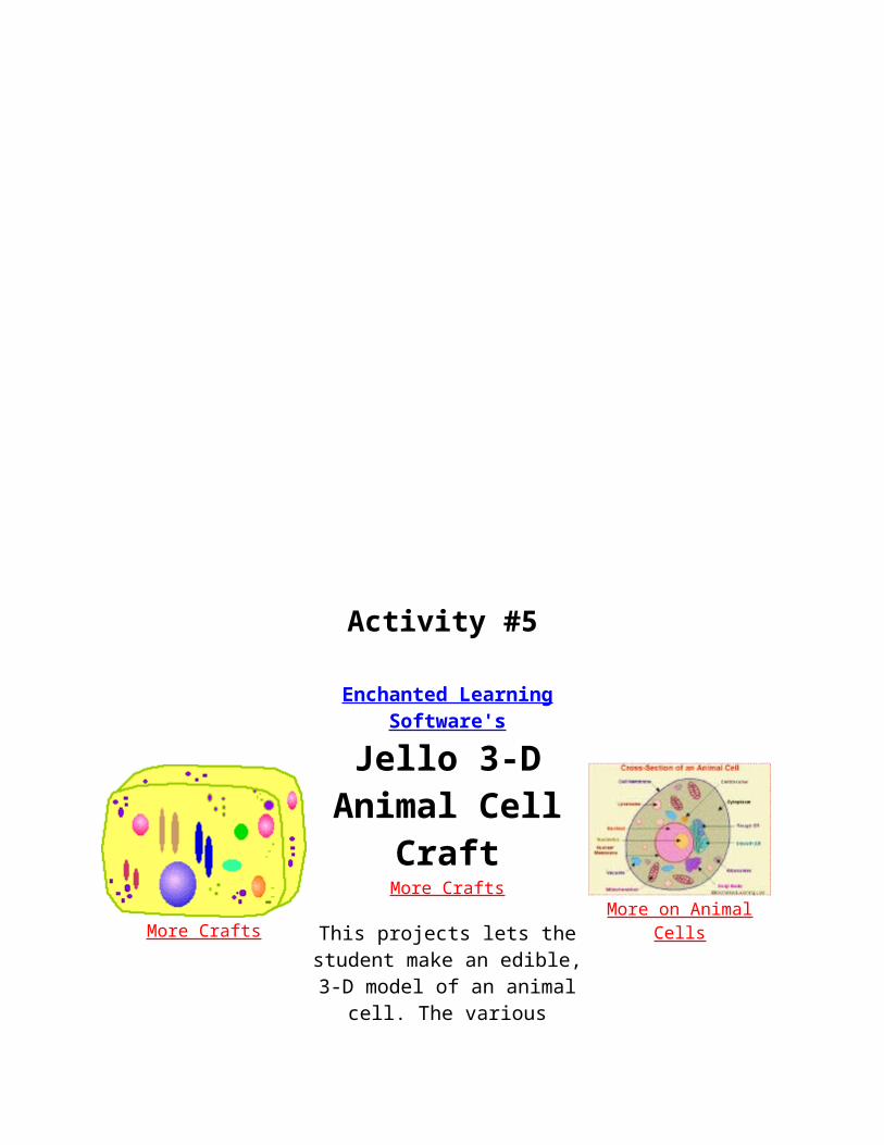
Activity #5
More Crafts
Enchanted Learning Software's
Jello 3-D Animal Cell Craft
More Crafts
This projects lets the student make an edible, 3-D model of an animal cell. The various organelles of the cell are represented by fruits and candies. When you've finished making your cell and writing
about it, you can eat it!
More on Animal Cells
Supplies:
Gelatin, either a light-colored Jello (like lemon) or unflavored gelatin with sugar or juice added
Water Spoon (to stir the gelatin) Microwave or stove (used to heat the water) A small but sturdy plastic bag to make the
gelatin in (we used 1-gallon ziplock bags) Various fruits and candies used to represent
the parts of the cell: raisins, gummy worms (plain and sour), gumdrops, gum ball, jelly beans, grapes, mandarin orange sections, sprinkles, M&M's, jaw breakers, a small stone fruit (like a plum), dried fruit, and/or hard candy. Note: marshmallows will float on top of the gelatin, so they don't work well in this craft.
Refrigerator (used to set the gelatin)
Make the light-colored Jello or gelatin, but make it with a bit less water than the instructions call for (this will make the gelatin a little stiffer and will
make the cell components stay in place better). The gelatin will represent the cytoplasm of the cell.
First, heat the water to boiling (use about three-quarters of what is called for in the instructions). Dissolve the gelatin in the hot water and carefully stir it. Carefully add the same amount of cold water.
Place an open plastic bag (we used 1-gallon ziplock bags) inside a sturdy container (like a large bowl or pan) - this makes pouring the Jello easier. Slowly pour the cooled gelatin into the bag - make sure that there is room in the bag for all the cell components that will be added later. Seal the bag and put it in the refrigerator.
When the gelatin is almost set (this takes about an hour, but depends on the temperature of your refrigerator), open the bag and start adding the components of the cell. (Also, have the student label the cell components using this printout, including the name of each cell component and what they used to represent it using the animal cell glossary.)
Cell
components (we've included what we used for our model, but you can choose whatever edible parts you like): cell membrane - the thin layer of protein and fat that surrounds the cell. It is represented by the plastic bag.centrosome - a small body located near the nucleus - it has a dense center and radiating tubules. This is where microtubules are made. During cell division (mitosis), the centrosome divides and the two parts move to opposite sides of the dividing cell. It is represented by a gum ball.cytoplasm - the jellylike material outside the cell nucleus in which the organelles are located. It is represented by the gelatin.Golgi body - (also called the Golgi apparatus or Golgi complex) a flattened, layered, sac-like organelle that looks like a stack of pancakes and is located near the nucleus. It produces the membranes that surround the lysosomes. The Golgi body packages proteins and carbohydrates into membrane-bound vesicles for "export" from the cell. It is represented by folded ribbons of hard candy.lysosome - (also called cell vesicles) round organelles surrounded by a membrane and containing digestive enzymes. This is where the digestion of cell nutrients takes place. They are represented by M&M's.mitochondrion - spherical to rod-shaped organelles with a double membrane. The inner membrane is infolded many times, forming a series of projections (called cristae). The mitochondrion converts the energy stored in glucose into ATP

(adenosine triphosphate) for the cell. They are represented by raisins.nuclear membrane - the membrane that surrounds the nucleus. It is represented by the plum's skin.nucleolus - an organelle within the nucleus - it is where ribosomal RNA is produced. Some cells have more than one nucleolus. It is represented by the plum pit.nucleus - spherical body containing many organelles, including the nucleolus. The nucleus controls many of the functions of the cell (by controlling protein synthesis) and contains DNA (in chromosomes). It is represented by the plum.ribosome - small organelles composed of RNA-rich cytoplasmic granules that are sites of protein synthesis. They are represented by candy sprinkles.rough endoplasmic reticulum - (rough ER) a vast system of interconnected, membranous, infolded and convoluted sacks that are located in the cell's cytoplasm (the ER is continuous with the outer nuclear membrane). Rough ER is covered with ribosomes that give it a rough appearance. Rough ER transports materials through the cell and produces proteins in sacks called cisternae (which are sent to the Golgi body, or inserted into the cell membrane). It is represented by sour gummy worms.smooth endoplasmic reticulum - (smooth ER) a vast system of interconnected, membranous, infolded and convoluted tubes that are located in the cell's cytoplasm (the ER is continuous with the outer nuclear membrane). The space within the ER is called the ER lumen. Smooth ER transports materials through the cell. It contains enzymes and produces and digests lipids (fats) and membrane proteins; smooth ER buds off from rough ER, moving the newly-made proteins and lipids to the Golgi body, lysosomes, and membranes. It is represented by gummy worms.vacuole - fluid-filled, membrane-surrounded cavities inside a cell. The vacuole fills with food being digested and waste material that is on its way out of the cell. They are represented by jaw breakers.
Re-seal the plastic bag and refrigerate the gelatin until it is fully set.
When the gelatin is set, you can examine your 3-D gelatin cell and then eat it.
http://www.enchantedlearning.com/subjects/animals/cell/jello/

Activity #6It's Alive, Alive, Allllllllllliiiiiiivvvvveeee!
Background: You will be in groups of three, each with your own job. The jobs to choose from are Contractor, Architect, Surveyor. Your job, as a group, is to build the most realistic life-like plant cell the world has ever seen.
Problem: What does a 3-dimensional cell look like? What are the various parts of plant cells?
Hypothesis: ______________________________________________________.
Materials: Play-doe, food coloring or tempera paints (red, purple, green, blue, white), 1 pair of gloves, yarn or undercooked spaghetti, pepper, plastic-bubble packing, aluminum foil, plastic wrap, pencil shavings, scissors, 1 large knife, glue.
Procedure:
1. Before we start be aware that on the final day you must present your cell to the class.
2. After you have decided upon your jobs, the Contractor and Architect will collaborate to design the plant cell. The design should be drawn up on a piece of paper that explains what materials will be used for each organelle. It should be colored the same color it will appear when it is built. Take your time and make a good drawing. This should be completed early on day two. Throughout this entire process the Surveyor should be writing down the order in which each organelle was designed and the order in which it will be built. Along with this the Surveyor must make a copy of the design that the group can use when building it. The Surveyor's job is to basically take notes all the way through, so if the final product doesn't come out as planned the Surveyor can look back at their notes and answer why.
3. After you have finished your design, hand it in and your teacher will approve it. If it is approved, you can start to build your cell.
4. Building should be the role of the contractor. Architect's watch the builders to make sure they are doing it exactly as planned. Surveyors should take notes on how it is built and also can assist the Architects to make sure it is being built as planned.
http://www.teach-nology.com/worksheets/science/bio/lab6/

Activity #7Picking your teeth with DNA!
Problem(s): Where is DNA found in living organisms? What is DNA made of? What are nucleotides? What are the four nitrogenous bases found in DNA? Which bases pair together in a DNA molecule?
Hypothesis: ________________________________________________________.
Materials: 30 Toothpicks, 4 different colored markers, construction paper, Glue.
Procedure:
1. You will be given 30 toothpicks. Using the four markers, completely color each toothpick any of colors you want. Make sure to color the toothpick one solid color and try use all the colors.
2. Take the piece of construction paper and cut it in half.
3. Draw the phosphate and sugar backbone on each piece of paper, as your teacher has written on the board. There will need to be 15 phosphate and 15 sugar units on each paper. Take your time!
4. Now on one of pieces of paper attach a tooth pick, with glue, to each sugar group.
5. Did you guess what the color on toothpick was for yet? Each color represents a different nitrogenous base. Look at the board, find out which bases you have made.
6. To complete your DNA molecule, color your the remaining toothpicks the color of the complementary base of your current strand and attach them to the sugar molecules of your other stand of DNA, then attach both strands together by the bases (toothpicks).
Conclusion Questions:
1. Where is DNA found in a cell?
2. What are chromosomes?
3. What is the function of DNA?
http://www.teach-nology.com/worksheets/science/bio/lab7/

Activity #8Name _______________________
How Does A Plant Cell Relate To Your School?
Directions:
In the space provided below describe the function of each cell organelle and then state what person in your school serves a similar function in your school.
Plant Organelle Function within the Plant cell Who at your school has a similar job?
Cell Wall
Plasma (Cell) Membrane
Nucleus
Cytoplasm
Chloroplast(s)
Mitochondrion
Vacuole(s)
Chromosomes (DNA)
Ribosome(s)
http://www.teach-nology.com/worksheets/science/bio/work1/
Activity #9
Name _______________ Date _____________
Cells Group Creative Writing
Directions: As a group, you have 25 minutes to write a brief story using the words below.
MITOCHONDRIA
CYTOPLASM
CHLOROPLAST
CHLOROPHYLL
NUCLEOLUS
NUCLEUS
TISSUES
ORGANS
VACUOLE
CHROMOSOME
______________________________________________________________________________
http://www.teach-nology.com/worksheets/science/cell/group/
Activity #10

Name ________________________ Date ____________________
Comparing Plant And Animal Cells VENN Diagram
Directions: Fill in the VENN Diagram to compare PLANT CELLS to ANIMAL CELLS. Use the words in the word box.
cell membrane cell wall chloroplast cytoplasmmitochondria nucleus ribosome vacuole
PLANT CELL ANIMAL CELL
http://www.teach-nology.com/worksheets/science/cell/2/
Activity #11

Name _______________ Date _____________
Bank On It! WorksheetCells
Work Bank
tissues photosynthesis, plant the important of transport. perform cells, They tissues for
Simple ____________ are also referred to as ground tissues. ____________ include the tissues known as parenchyma, collenchyma, and sclerenchyma. Parenchyma tissue is composed ____________ parenchyma ____________ which are found throughout ____________ plant. They are particularly abundant in the stems and roots. The leaf cells that carry out photosynthesis are also parenchyma cells. Unlike many other plant cells, parenchyma cells are alive at maturity and retain the ability to divide. They ____________ many functions. Some are specialized ____________ ____________ others for storage, and still others for secretion and ____________ An ____________ class of parenchyma cells makes growth tissues called meristem and cambium. These ____________ give rise to all other tissues in the ____________ body.
http://www.teach-nology.com/worksheets/science/cell/bank/
Name _______________ Date _____________

Cells Vocabulary QuizDirections: Match the vocabulary words on the left with the definitions on the right.
1. tissue the central, essential, or highly concentrated part around which other parts are grouped.
2. vacuolea musical instrument consisting of a keyboard attached to a device that forces air through a number of pipes to produce a wide range of sounds; pipe organ.
3. chromosomea membranous enclosure within a cell that contains substances isolated from the protoplasm, such as dissolved acids.
4. chlorophyll (chlorophyl) the ground protoplasm of cells that is outside the nucleus.
5. cell membraneany of the very tiny rodlike or stringlike structures that occur in nearly all cells of plants and animals, and that process food for energy.
6. chloroplast a small spherical body in the nucleus of a cell, consisting of protein and RNA.
7. cell wall the mass of like cells in an animal or plant body, esp. as they form a specific organ:
8. nucleolusone of the tiny, threadlike, DNA-containing bodies found in the cell nuclei of all plants and animals, responsible for transmitting hereditary characteristics.
9. organ the green pigment in the leaves and stems of plants that is necessary for the production of plant food by photosynthesis.
10. cytoplasm the rigid outermost layer of a plant cell, which is made of cellulose.

11. nucleus a small oval green bit of protoplasm that contains chlorophyll and is the location of photosynthesis.
12. mitochondrion the semipermeable membrane that encloses the contents of a cell; plasma membrane.
Activity # 12Community Cell

Subjects: Arts & Humanities: Visual Arts; Science: Life Sciences Grades: 6-8
Brief Description
This activity is designed to introduce students to the parts and functions of the cell. This kinesthetic lesson will engage all types of learners.
Objectives
Students learn about the parts and functions of an animal cell.
Keywords
biology, cells, cell structure, organelles
Materials Needed
For the class: dark felt cut into a circle measuring 16 to 18 inches, spray adhesive Per student group:
pieces of felt in a variety of colors cut into 1-to 2-inch squares scissors tag board glitter markers student-researched materials on cells neon-colored sticky notes plastic or paper bag
Lesson Plan
Before the lesson: Students use library, media, or Web sources to research information about cells. Some suggested Web sites:
The Cell Cell Structure and Function Cytology Mitosis
Procedures:1. Discuss the meanings of the words organelle and cell. Students can share information gathered
from research sources and/or from the suggested Web sites above. 2. Organixe students into cooperative groups of three or four. 3. Place the large dark felt circle on a chalkboard or wall. Say "This circle represents the smallest
unit of life. It has many functions, and together we are going to build a model of the human cell." 4. Distribute task cards to each group. The task cards are neon-colored sticky notes with the name
of a specific organelle written on each one. 5. Distribute a small bag of supplies to each group: glitter, scissors, square felt, markers, tag board.

6. Say "Working with your group, create a model of your assigned organelle. Look through your research materials to find a picture of the organelle. Using only the materials found in your bag, your group must construct a model of your organelle. On your tag board, clearly write the function and the name of your assigned organelle."
7. Let one group at a time come to the board or wall. Using the spray adhesive, have each group place its organelle within the large circular cell. One member from each group recites the name of the organelle and its function(s). Students sitting at their desks use separate paper to draw and label each organelle as it is presented to the class.
Assessment
Observe cooperative group behavior. Grade students for class participation, presentation of organelles, and copies of cell model.
Activity # 13

#2580. Eukaryotic Animal Cell, Candy Cell ModelScience, level: Middle Posted Mon May 13 18:16:50 PDT 2002 by Kristen Coggins ([email protected]). University of Nevada-Las Vegas, Las Vegas, NevadaMaterials Required: candy Activity Time: 50 mins Concepts Taught: The cell
Daily Lesson PlanKristen CogginsLesson Topic: Eukaryotic Animal Cell, Candy Cell Model
Objectives:
After the lesson the students will be able to:1. Identify the parts of the cell2. Describe the functions of selected cell organelles
Materials Needed:
1. Zip-lock bag (provided)2. Index cards- cell organelle card showing the name of the cell and the function (provided)3. Various types of candy (provided)4. Cell Organelle Key (provided)5. Colored pencils
Lesson and Activity:
1. Present concepts of the eukaryotic cell in short whole-class discussions (to be done in prior class period)2. Hand out Candy Cell Instruction Sheet and go over activity3. Hand out materials.4. Students will complete activity and candy cell model
Closure:
1. Do you know the parts of the cell?2. Clean Up3. Keep your cell model.
Evaluation:
Students should keep their candy cell model for later class periods and to use as a study aid to use to study for the assessment on cell organelles and functions.
Procedure: Eukaryotic Animal Cell, Candy Cell Model
DO NOT EAT THE CANDY!
KeyCell Organelle Candy
Nucleus JawbreakerMitochondria Jelly Bean Cytoskeleton Tooth PicksLysosomes SkiddlesGolgi Apparatus Swedish Fish Ribosomes Mini Gummi BearsSmooth ER Smooth Red LacesRough ER Sour LicoriceZip-Lock Bag Cell membrane (holds is all together)
Procedure
1. Cut 5 index cards in ½.2. Write the name of the cell organelle and the candy used on one side of the index card.a. Write the function on the other side.b. Use 1 color of colored pencil for both. Do not use this color again. c. Repeat this process for all of the organelles.3. You will have 9 cards total, each written in a different color. 4. Put the cell organelles and the cards in the zip-lock bag. 5. Rewrite the Key, on 1 whole index card, so that each organelle is written in the same color that you chose for the index card. Place the Key in the bag6. Keep the candy cell model to study for the test.
Closure
1. The teacher will check to see if your project has been completed.2. Chose one member of the group to keep the cell as it will be used later in class and as a study aid.