Embed Size (px)
Citation preview
Color index: IMPORTANT -‐‑ NOTES -‐‑ EXTRA -‐‑ Books
ACUTE & CHRONIC
LEUKEMIA
Objectives:
● Definition ● Historic Perspective ● Etiology and Risk Factors ● Incidence ● Classification ● Comparison of Acute and Chronic Leukemia
Team Members: Minyal Bawazir, Mohammad Almutlaq, Ashwag almajed , Badria alsabbagh
Team Leader: Haneen alsubki
Revised By: Yara aldigi and Basel almeflh
Resources: 435 team + Davidson + kumar
● Editing file ● Feedback
Ø Not all information in female’s lecture is included. The lecture is based on the male’s slides .
Leukemia
➔ Definition: ● Leukemia is a malignant disease of hematopoietic tissue characterized by the accumulation abnormal
white cells (neoplastic or leukemic) in the bone marrow leading to bone marrow failure, a raised circulating white cell count (leukocytosis) and infiltrate organs (e.g liver, spleen, lymph nodes, brain).
● A group of malignant disorders affecting the blood and blood-forming tissues of : Bone marrow, Lymph system, Spleen.
● Occurs in all age groups (males & females, 1-2:1) ● Results in an accumulation of dysfunctional cells because of a loss of regulation in cell division and
cell death , so the cells will be long-lived . ● Fatal if untreated! In acute types of leukemia the patient comes to the ER while in chronic it can be
found incidentally while doing routine check up or at the clinic ● Often thought of as a childhood disease, BUT the number of adults affected with leukemia is 10 times
that of children especially the chronic type .
➔ Historic Perspective : ● 1945 ● The initial description of leukemia as a clinical entity was made by Bennett in Scotland and in
Germany.
➔ Etiology & pathophysiology: The etiology is unknown, most from a combination of factors: oncogenes mutation & tumor suppressor gene alteration,host factors, and environmental factors. -this is bow it mostly works : mutation of special genes that are regulating the division of stem cells > will lead to accumulation of premature stem cells > leukemia ● Host factors:
1- congenital chromosomal abnormalities : increase in patients with congenital disorders that have tendency for chromosomal abnormalities e.g. Bloom’s syndrome, Fanconi syndrome, Down’s & Klinefelter syndromes (18-20 times ↑ incidence of AL in child with Down’s syndrome). 2- Immunodeficiency: ↑ incidence of lymphoid leukemia & lymphoma (with hereditary immunodeficiency e.g. ataxia-telangiectasia &sex-linked agamaglobulinemia) usually related to T & B lymphocyte gene rearrangement. 3- Chronic bone marrow dysfunction (CBMD): syndrome have an ↑ risk of acute leukemic transformation. Examples include the myelodysplastic syndromes, myeloproliferative disorders, aplastic anemia and PNH .
● Environmental factors: 1- Ionizing radiation: Leukemia is associated with exposure to ionizing radiation e.g. nuclear weapons in Hiroshima & Nagasaki. Both acute & chronic forms of leukemia including AML, ALL, CML were associated. 2- Chemical drugs: A variety of chemicals and drugs have been associated with the development of leukemic transformation e.g. Benzene, Chloramphenicol, Phenylbutazone and Cytotoxic alkylating 3- chemotherapeutic agents (AML occur after tx with alkylating agents e.g. melphalan). The presence of RAS mutations in patients with AML has been associated with specific occupational exposure to chemicals. As if a patient has a breast cancer and she has been receiving chemotherapy for a long time which lead to bone marrow suppression, you expect her to present one day with AML and usually a case like this has a poor prognosis. 4- Viruses: The human T-cell leukemia-lymphoma virus-I (HTLV-I) has been implicated as a causative agent of adult T-Cell leukemia-lymphoma (Now it has a vaccine, it’s mandatory in the some countries in high risk) . Another related virus HTLV-II has been isolated from patients with atypical hairy cell leukemia (CLL). The Epstein Barr virus (EBV) has been linked to Burkitt’s lymphoma.
-In majority of the patients you will not find a cause for their leukemia .. Although if you found a cause it’s called secondary leukemia and it has worse prognosis most often
➔ Incidence: ● In the USA 8-10 new cases per 100,000 individuals annually. ● Approximately 28,600 new cases were reported about 50% acute and 50% chronic ● Leukemia strike more in adult than children (10:1) and has slightly increase incidence in males than
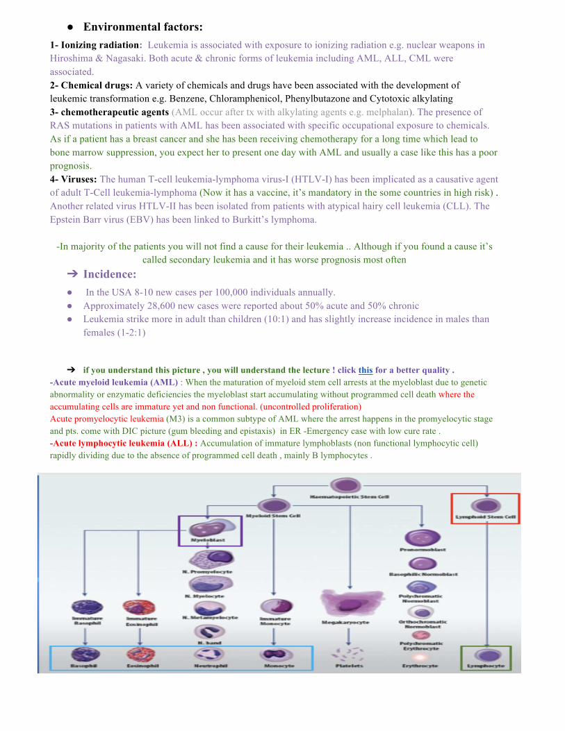
females (1-2:1) ➔ if you understand this picture , you will understand the lecture ! click this for a better quality .
-Acute myeloid leukemia (AML) : When the maturation of myeloid stem cell arrests at the myeloblast due to genetic abnormality or enzymatic deficiencies the myeloblast start accumulating without programmed cell death where the accumulating cells are immature yet and non functional. (uncontrolled proliferation) Acute promyelocytic leukemia (M3) is a common subtype of AML where the arrest happens in the promyelocytic stage and pts. come with DIC picture (gum bleeding and epistaxis) in ER -Emergency case with low cure rate . -Acute lymphocytic leukemia (ALL) : Accumulation of immature lymphoblasts (non functional lymphocytic cell) rapidly dividing due to the absence of programmed cell death , mainly B lymphocytes .

-Chronic myeloid leukemia (CML) : Abnormality (mutation) in the gene controlling the programmed cell death of the mature WBC’s (basophils, monocytes, neutrophils...etc) hence they live longer than their life span (rapidly dividing and space occupying cells , االمشكلة هھھھنا اانهھا بتسبب ززحمة في االل bone marrow ) . So It’s the accumulation of mature functioning cells. -Chronic lymphocytic leukemia (CLL) : Accumulation of mature functioning lymphocytes. (Most of the patients with CLL are only observed without treatment)
-Acute types are more dangerous and present in the ER Chronic is not fatal - treated as outpatient with chemotherapy (Pills not IV)
➔ Classification of leukemia: ● Two major types (4 subtypes) of leukemias ● Acute leukemias
- Acute lymphoblastic leukemia (ALL) - Acute myelogenous leukemia (AML) (also "myeloid" or "nonlymphocytic")
● Chronic leukemias - Chronic lymphocytic leukemia (CLL) - Chronic myeloid leukemia (CML)
● (Within these main categories, there are typically several subcategories) ➔ Comparison of acute & chronic leukemia
Acute Chronic
Age All ages Adults (old age usually)
Clinical onset Sudden Insidious (onset is more gradual)
Leukemic cells maturity
Clonal proliferation of immature hematopoietic cells (the formation of blood or blood cells).
Mature forms of WBC
Anemia Mild to severe Mild
Thrombocytopenia Mild to severe Mild
WBCs Variable Increased
Organomegaly Mild Prominent
➔ Myelogenous Leukemia: ● Myeloid tissue is a biologic tissue with the ability to perform hematopoiesis. It is mainly found as the
red bone marrow in bones, and is often synonymous with this. However, myeloid can also be present in the liver and spleen. If serious case is having hepatosplenomegaly consider AML.
● A myelocyte is a young cell of the granulocytic series, occurring normally in bone marrow, but not in circulating blood (except when caused by certain diseases).
● Granulocytes are a category of WBC characterized by the presence of granules in their cytoplasm. They are also called polymorphonuclear leukocytes (PMN or PML) because of the varying shapes of the nucleus, which is usually lobed into three segments.
● The myeloblast is a unipotent stem cell, which will differentiate into one of the actors of the granular series. myeloblast in peripheral blood : <5% is normal / >20% AML , unless the pt has M3 .
★ Acute Leukemia Acute leukemia -osmosis-
1. Acute Myelogenous Leukemia (AML)
Definition ● Leukemia characterized by proliferation of myeloid tissue (as of the bone marrow and spleen) and an abnormal increase in the number of granulocytes, myelocytes, and myeloblasts in the circulating blood.
Epidemiology ● Predominant form of leukemia in neonatal period. but only a small proportion (15- 20%) of childhood & adolescent cases.
● 25% of all leukemias. ● Accounts for 35% of all new cases of acute leukemia. ● 85% of the acute leukemias in adults,Slightly more common in males. ● Abrupt, dramatic onset (serious infections because the affected cells are WBC ,
abnormal bleeding because of thrombocytopenia ). ● Uncontrolled proliferation of myeloblasts (hyperplasia of BM and spleen).
Pathogenesis ● Characterized by clonal proliferation of myeloid precursors with reduced capacity to differentiate into more mature elements.
● Accumulation of leukemic forms in bone marrow, peripheral blood, and other tissues, with a marked reduction in red cells, platelets, and neutrophils.
Clinical presentation
● Symptoms related to pancytopenia: weakness, easy fatigue, SOB,infections, gingival bleeding, ecchymosis, epistaxis, menorrhagia.
● Infrequent bone pain (sternum, long bones)
Physical exam
● Fever: almost always due to infection, small minority have fever related solely to underlying leukemia.
● Skin: pallor, petechiae, ecchymoses, infiltrative lesions (leukemia cutis or granulocytic sarcoma)
● Eye: retinal hemorrhages and/or exudates, pale conjuctivae. candidiasis, herpetic lesions.
● Organomegaly: palpable adenopathy rare, HSM uncommon. ● Joints: polyarthritis, arthralgias, bone pain & tenderness.
2. Acute Lymphoblastic Leukemia (ALL)
Definition ● Immature lymphocytes proliferate in the bone marrow
Epidemiology Children : Most common type of leukemia in children (> 80%). Incidence peaks ages 2 to 5 yr, 30 cases per million per year in the US. Adults : - 15% of acute leukemia in adults.
Clinical manifestations
● Signs and symptoms may appear abruptly: Fever, bleeding ● Insidious with progressive: Weakness, fatigue ● Central nervous system manifestations. Why ALL present with CNS manifestations ? ● Not as we knew about AML, ALL does not cause leukostasis and occlud blood vessels
but causes CNS problems. The reason behind it that lymphoblasts are small in size unlike the myeloblast which is large (obstructing the vessels causing leukostasis) ,while the lymphoblasts are small where they can’t obstruct the vessels but cross the blood brain barrier causing CNS issues. Thus, in treatment we have to give the patient CNS directed therapy (intrathecal therapy) because the systemic therapy isn’t reaching the CNS due to BBB preventing the systemic chemotherapy from reaching CNS (ALL patients has dormant lymphoblasts in the CNS, so if not given CNS prophylaxis 50 % relapse). In ALL treatment will take 3 years because it’s hard to eradicate small cells while in AML it will just take 3-4 months .
● A major difference between therapy for ALL and AML is the need for central nervous system directed therapy , you should give CNS prophylaxis !!
● Bone marrow failure: Anemia, thrombocytopenia, neutropenia ● Lymphadenopathy, hepatomegaly, splenomegaly ● Bone pain, arthralgias (especially in children) ● Infection, fever ● Extramedullary spread: CNS involvement at diagnosis (5% children, 15% adults),
Skin , Testes (10-15% boys). ● Mediastinal mass (lymphoblastic lymphoma) or tissue mass (50% of T cell-ALL) ● tumor lysis syndrome : hyperkalemia , hyperphosphatemia .
★ Chronic leukemia chronic leukemia-osmosis-
1. Chronic Myelogenous Leukemia (CML) they present in the clinic
Definition -Excessive development of mature neoplastic granulocytes in the bone marrow. - Move into the peripheral blood in massive numbers. - Ultimately infiltrate the liver and spleen. the problem here is accumulation in the bone marrow of mature cells ررااحح يیجيیك االمريیض االعيیاددةة
. and asymptomatic mostly وو بطنهھ منفوخة
Epidemiology -CML accounts for 15% - 20% of all adults leukemia. -Occurs slightly more frequently in men than women ( 1.4 - 2.2:1) -CML was the first cancer to be shown to be caused by an underlying genetic abnormality.
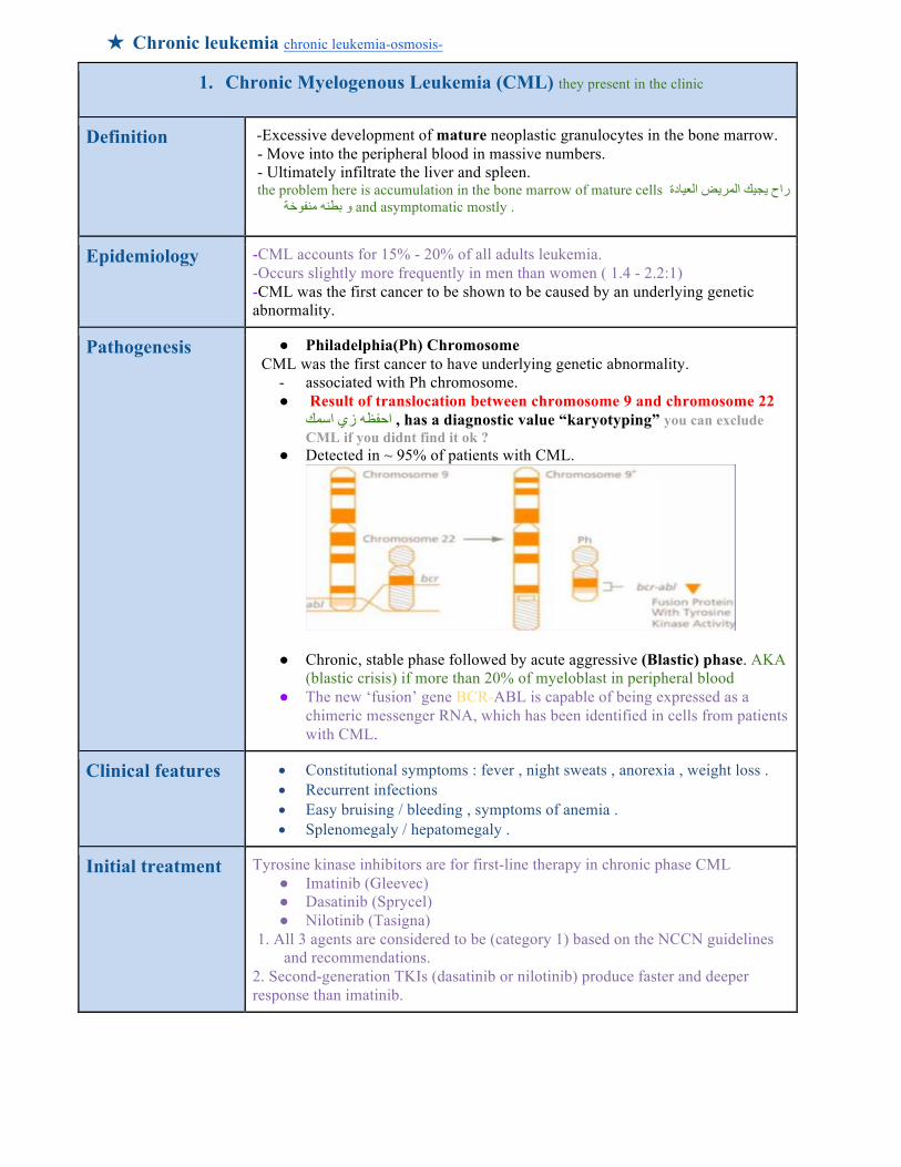
Pathogenesis ● Philadelphia(Ph) Chromosome CML was the first cancer to have underlying genetic abnormality.
- associated with Ph chromosome. ● Result of translocation between chromosome 9 and chromosome 22
has a diagnostic value “karyotyping” you can exclude , ااحفظهھ ززيي ااسمكCML if you didnt find it ok ?
● Detected in ~ 95% of patients with CML.
● Chronic, stable phase followed by acute aggressive (Blastic) phase. AKA (blastic crisis) if more than 20% of myeloblast in peripheral blood
● The new ‘fusion’ gene BCR-ABL is capable of being expressed as a chimeric messenger RNA, which has been identified in cells from patients with CML.
Clinical features • Constitutional symptoms : fever , night sweats , anorexia , weight loss . • Recurrent infections • Easy bruising / bleeding , symptoms of anemia . • Splenomegaly / hepatomegaly .
Initial treatment Tyrosine kinase inhibitors are for first-line therapy in chronic phase CML ● Imatinib (Gleevec) ● Dasatinib (Sprycel) ● Nilotinib (Tasigna)
1. All 3 agents are considered to be (category 1) based on the NCCN guidelines and recommendations.
2. Second-generation TKIs (dasatinib or nilotinib) produce faster and deeper response than imatinib.
2. Chronic Lymphocytic Leukemia (CLL) unlike CML , it doesn’t have a diagnostic test
Definition ● CLL is a neoplastic disease characterized by proliferation and accumulation (blood, marrow and lymphoid organs) of morphologically mature but immunologically dysfunctional lymphocytes.
● Production and accumulation of functionally inactive but long-lived, mature-appearing lymphocytes
● B cell involvement ● Lymph node enlargement is noticeable throughout the body , ↑ incidence of
infection -only in one situation people with CLL will come with cytopenia > if there was severe infiltration it will squeeze the cells in the bone marrow .
● The only leukemia that is not treated on early stages . Complications from early-stage CLL is rare –May develop as the disease advances –Pain, paralysis from enlarged lymph nodes causing pressure , in this situation we have to treat
Epidemiology
● Most common leukemia of Western world. ● Less frequent in Asia and Latin America. ● Male to female ratio is 2:1. ● Median age at diagnosis is 65-70 years. ● Uncommon (10%) in patients under 50 years ● In US population incidence is similar in different races.
Initial symptoms
● Approximately 40% are asymptomatic at diagnosis – discovered by a CBC ● In symptomatic cases the most common complaint is fatigue ● B symptoms – fever, sweats, weight loss ● Less often the initial complaint are enlarged nodes or the development of an
infection (bacterial).
Treatment ● Watch and wait treat if there was a compressive lesions
3. Hairy cell leukemia: The doctor completely skipped it
Definition Indolent
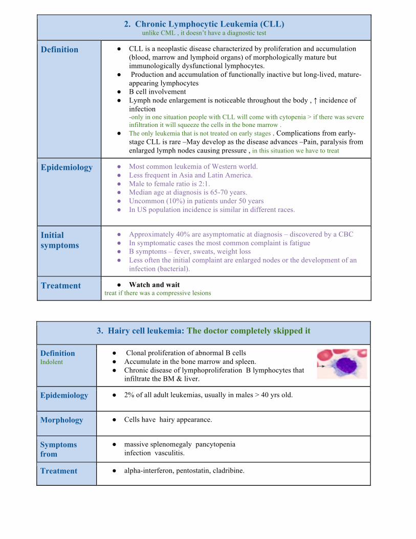
● Clonal proliferation of abnormal B cells ● Accumulate in the bone marrow and spleen. ● Chronic disease of lymphoproliferation B lymphocytes that
infiltrate the BM & liver.
Epidemiology ● 2% of all adult leukemias, usually in males > 40 yrs old.
Morphology ● Cells have hairy appearance.
Symptoms from
● massive splenomegaly pancytopenia infection vasculitis.
Treatment ● alpha-interferon, pentostatin, cladribine.
★ Unclassified leukemia: ● Subtype can’t be identified. In many cases you are not sure whether it’s AML or ALL because both
myeloblasts and lymphoblasts are high then it’s unclassified. ● Malignant leukemic cells may have lymphoid, myeloid, or mixed characteristics. ● Frequently theses patients don’t respond well to treatment (poor prognosis).
★ Differential Diagnosis of leukemia: blasts in peripheral blood will exclude anything else
1- Aplastic anemia. 2- Myelodysplastic syndrome. 3- Multiple myeloma. 4- Lymphoma. 5- Sever megaloblastic anemia. 6- Leukemoid reaction. E.g. sepsis, has a very high WBC.
★ Clinical manifestations:
Clinical manifestations related to problems caused by:
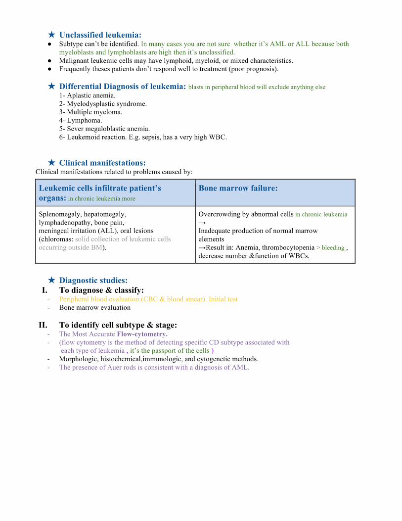
Leukemic cells infiltrate patient’s organs: in chronic leukemia more
Bone marrow failure:
Splenomegaly, hepatomegaly, lymphadenopathy, bone pain, meningeal irritation (ALL), oral lesions (chloromas: solid collection of leukemic cells occurring outside BM).
Overcrowding by abnormal cells in chronic leukemia → Inadequate production of normal marrow elements →Result in: Anemia, thrombocytopenia > bleeding , decrease number &function of WBCs.
★ Diagnostic studies: I. To diagnose & classify:
- Peripheral blood evaluation (CBC & blood smear). Initial test - Bone marrow evaluation
II. To identify cell subtype & stage:
- The Most Accurate Flow-cytometry. - (flow cytometry is the method of detecting specific CD subtype associated with
each type of leukemia , it’s the passport of the cells ) - Morphologic, histochemical,immunologic, and cytogenetic methods. - The presence of Auer rods is consistent with a diagnosis of AML.
★ Treatment: Collaborative Care
- Goal is to attain remission (when there is no longer evidence of cancer cells in the body) - What is Remission? The main aim of treatment for ALL is to give a remission. This means that the
abnormal, immature white cells or blasts can no longer be detected in the blood or bone marrow, and normal bone marrow has developed again. For many people with ALL the remission lasts indefinitely and the person is said to be cured.
-Treatment in Acute leukemia ال يیخلو من أأمريین : In all pts ALL + AML we give INDUCTION therapy to kill all the leukemic cells (28 days ) then : 1)In low risk features as in the case of AML we give CONSOLIDATION therapy for 3 months to ensure that all the leukemic cells vanished completely ! 2)High risk features we give INTENSIFICATION therapy / BMT .
➔ Chemotherapy treatment : A. Induction therapy ● Attempt to induce or bring remission. ● Seeks to destroy leukemic cells in the tissues, peripheral blood & bone marrow. ● Patient may become critically ill (provide psychological support as well). ● death can happen from infection or bleeding .
B. Intensification therapy in ALL ● High-dose therapy. ● May be given after induction therapy. ● Same drugs at higher doses and/or other drugs.
C. Consolidation therapy AML stops here ● Started after remission is achieved. ● The purpose is to eliminate remaining leukemic cells that may not be evident. ● to make sure you eradicated all the leukemic cells .
D. Maintenance therapy ● Lower dose of the same drug.
➔ Chemotherapy Regimens
Combination chemotherapy
● Mainstay treatment ● 3 purposes:
● ↓ drug resistance. ● ↓ drug toxicity to the patient by using multiple drugs with varying toxicities. ● Interrupt cell growth at multiple points in the cell cycle.
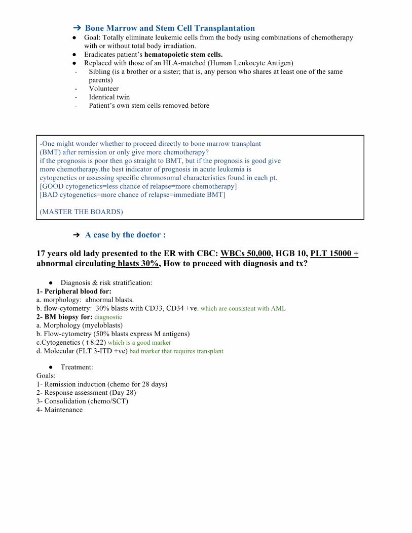
➔ Bone Marrow and Stem Cell Transplantation ● Goal: Totally eliminate leukemic cells from the body using combinations of chemotherapy
with or without total body irradiation. ● Eradicates patient’s hematopoietic stem cells. ● Replaced with those of an HLA-matched (Human Leukocyte Antigen) - Sibling (is a brother or a sister; that is, any person who shares at least one of the same
parents) - Volunteer - Identical twin - Patient’s own stem cells removed before
-One might wonder whether to proceed directly to bone marrow transplant (BMT) after remission or only give more chemotherapy? if the prognosis is poor then go straight to BMT, but if the prognosis is good give more chemotherapy.the best indicator of prognosis in acute leukemia is cytogenetics or assessing specific chromosomal characteristics found in each pt. [GOOD cytogenetics=less chance of relapse=more chemotherapy] [BAD cytogenetics=more chance of relapse=immediate BMT] (MASTER THE BOARDS)
➔ A case by the doctor : 17 years old lady presented to the ER with CBC: WBCs 50,000, HGB 10, PLT 15000 + abnormal circulating blasts 30%, How to proceed with diagnosis and tx?
● Diagnosis & risk stratification: 1- Peripheral blood for: a. morphology: abnormal blasts. b. flow-cytometry: 30% blasts with CD33, CD34 +ve. which are consistent with AML 2- BM biopsy for: diagnostic a. Morphology (myeloblasts) b. Flow-cytometry (50% blasts express M antigens) c.Cytogenetics ( t 8:22) which is a good marker d. Molecular (FLT 3-ITD +ve) bad marker that requires transplant
● Treatment: Goals: 1- Remission induction (chemo for 28 days) 2- Response assessment (Day 28) 3- Consolidation (chemo/SCT) 4- Maintenance
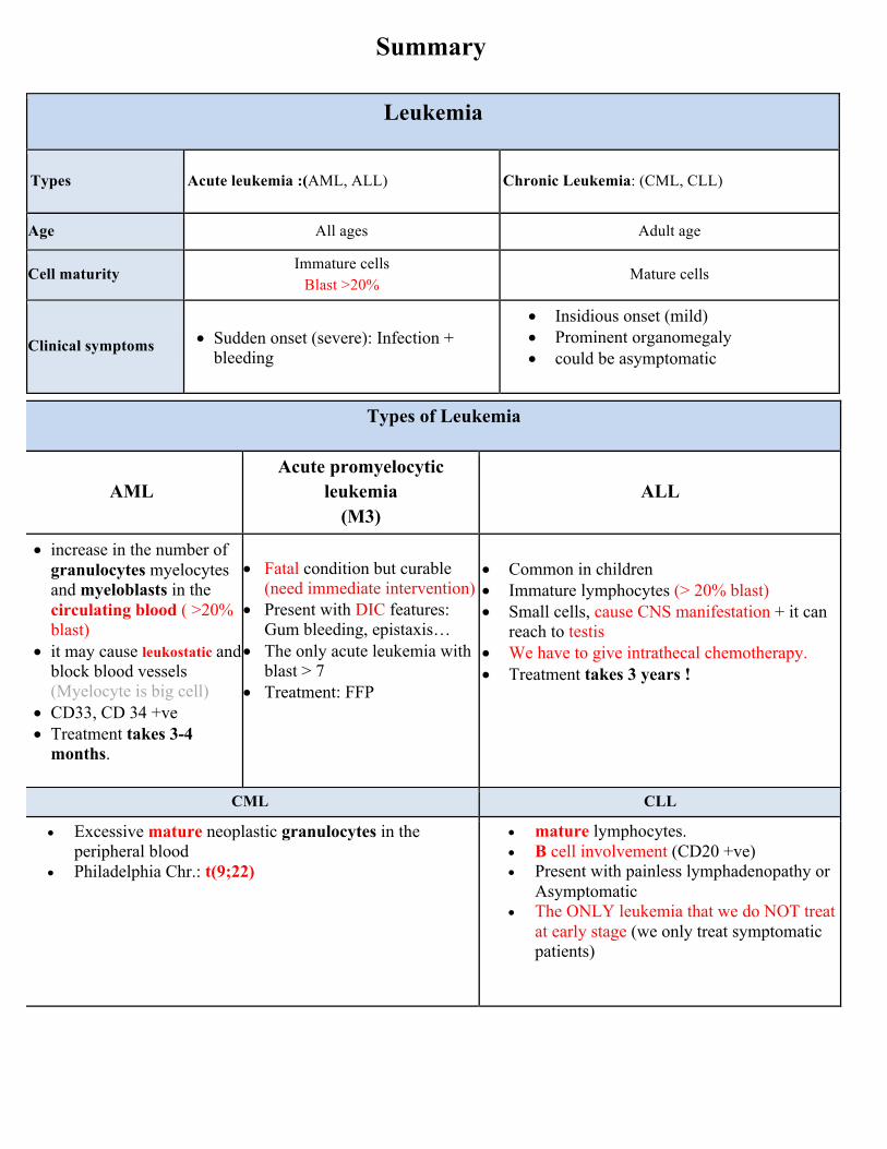
Summary
Leukemia
Types Acute leukemia :(AML, ALL) Chronic Leukemia: (CML, CLL)
Age All ages Adult age
Cell maturity Immature cells
Blast >20% Mature cells
Clinical symptoms • Sudden onset (severe): Infection + bleeding
• Insidious onset (mild) • Prominent organomegaly • could be asymptomatic
Types of Leukemia
AML Acute promyelocytic
leukemia (M3)
ALL
• increase in the number of granulocytes myelocytes and myeloblasts in the circulating blood ( >20% blast)
• it may cause leukostatic and block blood vessels (Myelocyte is big cell)
• CD33, CD 34 +ve • Treatment takes 3-4
months.
• Fatal condition but curable (need immediate intervention)
• Present with DIC features: Gum bleeding, epistaxis…
• The only acute leukemia with blast > 7
• Treatment: FFP
• Common in children • Immature lymphocytes (> 20% blast) • Small cells, cause CNS manifestation + it can
reach to testis • We have to give intrathecal chemotherapy. • Treatment takes 3 years !
CML CLL
• Excessive mature neoplastic granulocytes in the peripheral blood
• Philadelphia Chr.: t(9;22)
• mature lymphocytes. • B cell involvement (CD20 +ve) • Present with painless lymphadenopathy or
Asymptomatic • The ONLY leukemia that we do NOT treat
at early stage (we only treat symptomatic patients)

Investigation Treatment
• CBC & blood smear • Bone marrow evaluation (diagnostic test
!!!!!) • Flow cytometry: for morphology: myeloid
or lymphoid?
• Induction therapy (for all patients): to attain remission. • Intensification therapy: for acute lymphocytic
leukemia (ALL) • Consolidation therapy: started after remission is
achieved • Maintenance therapy • Bone marrow transplant: only for some types
questions 1. A 35 years lady presented to the emergency room with fever and epistaxis. Her laboratory findings were remarkable for a WBC count of 45,000 /ul, hemoglobin 11g/dl and Platelets 6,000 /ul with 30 % circulating blasts. Which ONE of the following is the first step management?
A. Chemotherapy for acute leukemia B. Immediate antibiotics and transfusion C. Immediate bone marrow biopsy D. Send peripheral blood for molecular testing
2. A 71 year old man who presents with elevated white blood cell counts and a high percentage of blasts in his peripheral blood is most likely to have :
A. B-cell acute lymphoblastic leukemia B. T-cell acute lymphoblastic leukemia. C. acute myelogenous leukemia. D. acute promyelocytic leukemia.
3. In a 11-year-old boy presenting with a new diagnosis of ALL, which of the following is not a classic sign or symptom of ALL?
A. Bone pain B. Constipation C. Fatigue D. Repeated infections
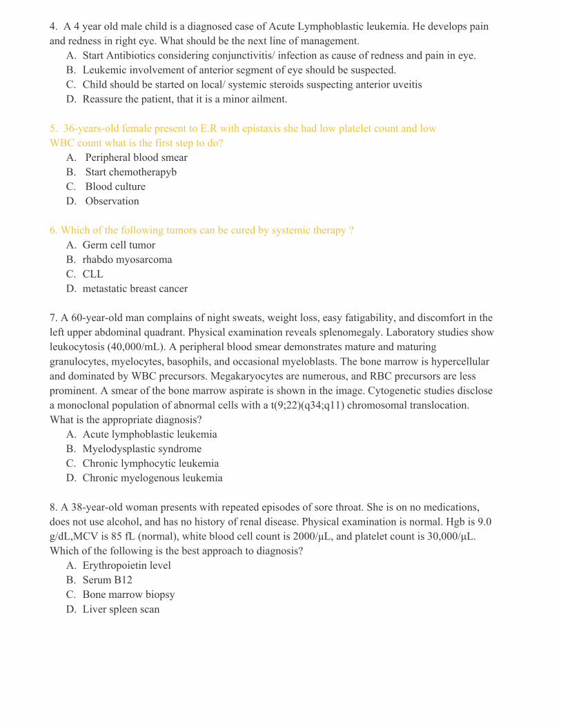
4. A 4 year old male child is a diagnosed case of Acute Lymphoblastic leukemia. He develops pain and redness in right eye. What should be the next line of management.
A. Start Antibiotics considering conjunctivitis/ infection as cause of redness and pain in eye. B. Leukemic involvement of anterior segment of eye should be suspected. C. Child should be started on local/ systemic steroids suspecting anterior uveitis D. Reassure the patient, that it is a minor ailment.
5. 36-years-old female present to E.R with epistaxis she had low platelet count and low WBC count what is the first step to do?
A. Peripheral blood smear B. Start chemotherapyb C. Blood culture D. Observation
6. Which of the following tumors can be cured by systemic therapy ?
A. Germ cell tumor B. rhabdo myosarcoma C. CLL D. metastatic breast cancer
7. A 60-year-old man complains of night sweats, weight loss, easy fatigability, and discomfort in the left upper abdominal quadrant. Physical examination reveals splenomegaly. Laboratory studies show leukocytosis (40,000/mL). A peripheral blood smear demonstrates mature and maturing granulocytes, myelocytes, basophils, and occasional myeloblasts. The bone marrow is hypercellular and dominated by WBC precursors. Megakaryocytes are numerous, and RBC precursors are less prominent. A smear of the bone marrow aspirate is shown in the image. Cytogenetic studies disclose a monoclonal population of abnormal cells with a t(9;22)(q34;q11) chromosomal translocation. What is the appropriate diagnosis?
A. Acute lymphoblastic leukemia B. Myelodysplastic syndrome C. Chronic lymphocytic leukemia D. Chronic myelogenous leukemia
8. A 38-year-old woman presents with repeated episodes of sore throat. She is on no medications, does not use alcohol, and has no history of renal disease. Physical examination is normal. Hgb is 9.0 g/dL,MCV is 85 fL (normal), white blood cell count is 2000/µL, and platelet count is 30,000/µL. Which of the following is the best approach to diagnosis?
A. Erythropoietin level B. Serum B12 C. Bone marrow biopsy D. Liver spleen scan

9. A 5-year-old girl presents with her parents who have become concerned about the small petechiae and ecchymoses on her skin. An abdominal examination reveals hepatosplenomegaly. You suspect an acute leukaemia. The most appropriate initial investigation for diagnosis is :
A. Chromosomal analysis of bone marrow cells B. Cytochemical analysis of bone marrow cells C. Direct microscopy of bone marrow cells D. Peripheral blood smear
Answers 1.A - 2.C - 3.B - 4.B - 5.A - 6.C - 7.D - 8.C - 9.D


![203341Orig1s000 - Food and Drug Administrationtreatment of adults with chronic myeloid leukemia [CP-CML, AP-CML, and BP-CML] with resistance or intolerance to prior therapy including](https://img.pdfslide.net/doc/110x75/60bcb776f8906f48904ac5cb/203341orig1s000-food-and-drug-administration-treatment-of-adults-with-chronic.jpg)