Embed Size (px)
Citation preview

Cardiovascular Boot Camp April 2009
www.cardionursing.com 1
2009 1
Acute Coronary Syndrome (ACS): Disease Management
Presented By:
Cynthia Webner BSN, RN,CCRN-CMC
www.cardionursing.com
2009 2
Acute Coronary Syndrome (ACS)
• ST Elevation
– STEMI
• No ST Elevation
– NSTEMI
– Unstable Angina (UA)
ACS refers to any rupture of plaque or thrombotic event
that leads to symptomatic ischemia or infarction.
The role of Troponin.

Cardiovascular Boot Camp April 2009
www.cardionursing.com 2
2009 3
Pathophysiology
• Deposit of lipids, calcium, fibrin, and other cellular substances within the lining of the arteries.
• Initiates a progressive inflammatory response in an effort to heal the endothelium.
• End result of inflammatory process: the production of a fibrous atherosclerotic plaque.
• Plaque can progress to cause coronary stenosis
• Plaque can also rupture prior to causing significant stenosis
2009 4
Plaque
• Stable plaque of stable angina– Thick fibrous caps separate the
lipid core from the endothelium– Less complicated than vulnerable
plaques– Tend to have smooth outlines
• Vunerable plaque of ACS – Thin caps– Edge of the fibrous cap is a
particularly vulnerable area and is commonly the location of ruptured plaque
• Limitations of stress testing and cardiac catheterization
• Intravascular ultrasound

Cardiovascular Boot Camp April 2009
www.cardionursing.com 3
2009 5
Hospitalizations in the U.S. Due to
ACS
Acute Coronary
Syndromes*
1.57 Million Hospital Admissions - ACS
UA/NSTEMI† STEMI
1.24 millionAdmissions per year
0.33 millionAdmissions per year
*Primary and secondary diagnoses. †About 0.57 million NSTEMI and 0.67 million UA.Heart Disease and Stroke Statistics – 2007 Update. Circulation 2007; 115:69–171.
2009 6
NSTEMINSTEMINSTEMINSTEMI
PresentationPresentationPresentationPresentation
Working DxWorking DxWorking DxWorking Dx
ECGECGECGECG
CardiacCardiacCardiacCardiac
BiomarkerBiomarkerBiomarkerBiomarker
Final DxFinal DxFinal DxFinal DxNQMINQMINQMINQMI Qw MIQw MIQw MIQw MI
UAUAUAUA
UnstableUnstableUnstableUnstableAnginaAnginaAnginaAngina
Ischemic DiscomfortIschemic DiscomfortIschemic DiscomfortIschemic Discomfort
Acute Coronary SyndromeAcute Coronary SyndromeAcute Coronary SyndromeAcute Coronary Syndrome
Myocardial InfarctionMyocardial InfarctionMyocardial InfarctionMyocardial Infarction
ST ElevationST ElevationST ElevationST ElevationNo ST ElevationNo ST ElevationNo ST ElevationNo ST Elevation
NonNonNonNon----ST ACSST ACSST ACSST ACS
Libby P. Circulation 2001;104:365, Hamm CW, Bertrand M, Braunwald E, Lancet 2001; 358:1533-1538; Davies MJ. Heart 2000; 83:361-366.Anderson JL, et al. J Am Coll Cardiol. 2007;50:e1-e157, Figure 1. Reprinted with permission.
Cardiovascular Boot Camp April 2009
www.cardionursing.com 4
2009 7
Acute Myocardial Infarction
• Development of myocardial necrosis caused by a critical imbalance between the oxygen supply and demand of the myocardium
• 10 seconds of oxygen deprivation: Ischemia
• 1 minutes of Ischemia: Myocardial dysfunction affected
• 20 minutes of oxygen deprivation: irreversible cell damage– STEMI
– NSTEMI
2009 8
STEMI
• < 25% of ACS patients
• Complete occlusion of a vessel by a
thrombus
• Fibrin stable clot (red clot)
• Classified more specifically by the portion
of the left ventricle suffering injury.
• Mortality is greatest within the first 24 to 48
hours of symptom onset

Cardiovascular Boot Camp April 2009
www.cardionursing.com 5
2009 9
NSTEMI
• Higher mortality and morbidity than STEMI
• Nationally under treated according to evidence based practice guidelines
• Crusade Registry
• Pathophysiology often involves a platelet plug or white clot
• Less stable clot
• Opportunity for spontaneous reperfusion
• Treatment focus = antiplatelet therapy
• Differentiated from unstable angina by troponin levels
2009 10
Causes of UA/NSTEMI*
• Thrombus or thromboembolism, usually arising on disrupted or eroded plaque– Occlusive thrombus, usually with collateral vessels†
– Subtotally occlusive thrombus on pre-existing plaque
– Distal microvascular thromboembolism from plaque-associated thrombus
– Thromboembolism from plaque erosion
• Non–plaque-associated coronary thromboembolism• Dynamic obstruction (coronary spasm‡ or vascoconstriction) of
epicardial and/or microvascular vessels• Progressive mechanical obstruction to coronary flow• Coronary arterial inflammation• Secondary UA• Coronary artery dissection§
*These causes are not mutually exclusive; some patients have 2 or more causes. †DeWood MA, et al. N Engl J Med 1986;315:417–23. ‡May occur on top of an atherosclerotic plaque, producing missed-etiology angina or UA/NSTEMI. §Rare. Modified with permission from Braunwald E. Circulation 1998;98:2219–22. Anderson JL, et al. J Am Coll Cardiol. 2007;50:e1-e157, Table 3.

Cardiovascular Boot Camp April 2009
www.cardionursing.com 6
2009 11
ACS Symptoms
• Classic Symptoms
– Stable angina
– Unstable angina
– MI
• Symptom Variations
– Women
– Elderly
– Diabetics
2009 12
Differential Diagnosis of Chest Pain
• Assessment of Pain
• Linking Patient History and Risk
factors
• Cardiac Biomarkers
• ECG Findings

Cardiovascular Boot Camp April 2009
www.cardionursing.com 7
2009 13
Assessment of Angina
• N = Normal
• O = Onset
• P = Precipitation / provoking / palliative factors
• Q = Quality or quantity
• R = Radiation and region
• S = Severity
• T = Time
2009 14
Characteristics of Angina
• Sensation of pressure, tightness, heaviness, burning, or squeezing. – Rarely described as a sharp or stabbing pain.
– Should not worsen with changes in position or respiration.
• Location behind the sternum and in the upper back, shoulder, arm, jaw, or epigastric area. – Not usually located in the middle to lower abdomen and does
usually not radiate to the lower extremities.
• Associated symptoms (or stand alone symptoms) of dyspnea, nausea, palpitations, or diaphoresis.
• Duration typically defined in minutes.
– Not typically defined in seconds or hours.
CAUTION WHEN ASKING THE PATIENT ABOUT “PAIN”!

Cardiovascular Boot Camp April 2009
www.cardionursing.com 8
2009 15
Angina
Stable
• Occurs with physical exertion
or emotional stress
• Relieved by rest or sublingual
nitroglycerin
• Predictable pattern
• Predictable = triggered by the
same amount of physical or
emotional stress and should
be easily relieved by rest or
sublingual nitroglycerin.
Unstable
• Occurs with minimal exertion
• OR increased dose of nitroglycerin is required to achieve relief.
• Prolonged rest angina is also considered unstable angina.
• Angina that increases in severity or is very severe on first presentation
• Caused by unstable or ruptured plaque that causes abrupt closure of a coronary artery which may spontaneously reperfuse.
2009 16
Angina in Women
• Delay presenting with symptoms
• Attribute symptoms to other non-cardiac causes
• Presentation
– epigastric discomfort
– less specific complaints: dyspnea or fatigue
– symptoms of discomfort from nose to navel should be evaluated for presence of CAD
• Less documented stenotic disease of major epicardial coronary arteries
• More likely to have unstable angina than MI
• Older women have higher incidence of complications

Cardiovascular Boot Camp April 2009
www.cardionursing.com 9
2009 17
Angina in the Elderly
• Generalized symptoms– weakness, dyspnea, and confusion.
• Symptoms often attributed to the aging process
• Cardiac and non cardiac co-morbidities complicate the diagnosis of ACS and increase the risk
• Don’t complain about chest pain– 37% of patients > 65
– 42% of patients > 75 years
– 75% of those > 85 years
2009 18
Angina in Diabetics
• Autonomic dysfunction can affect symptoms experienced with angina
• Less likely to experience pain.
• 25% of all patients presenting with ACS are diabetic
• Have severe multi-vessel disease
• Have higher rates of complications
• Have a greater proportion of ulcerated plaques resulting in intracoronary thrombi

Cardiovascular Boot Camp April 2009
www.cardionursing.com 10
2009 19
Acute MI Symptoms
• Symptoms occur spontaneously and are not relieved by rest or nitroglycerin
• Chest pressure or discomfort may be accompanied by nausea, vomiting, or diaphoresis
• Patient may have hemodynamic instability or cardiac arrest from ventricular fibrillation
• Acute MI patients have positive biomarkers and are classified as STEMI or NSTEMI based on ECG presentation
2009 20
Response to Symptoms
Patients with symptoms of ACS (chest discomfort with
or without radiation to the arm[s], back, neck, jaw, or
epigastria; shortness of breath; weakness;
diaphoresis; nausea; lightheadedness) should be
instructed to call 9-1-1 and should be transported to
the hospital by ambulance rather than by friends or
relatives
• Source: ACC/AHA NSTEMI Guidelines 2007

Cardiovascular Boot Camp April 2009
www.cardionursing.com 11
2009 21
STAT ECG Indications
• Chest pain or severe epigastric pain, non traumatic in origin, with components typical of myocardial ischemia or MI:– Central/substernal compression or crushing chest pain
– Pressure, tightness, heaviness, cramping, burning, aching sensation
– Unexplained indigestion, belching, epigastric pain
– Radiating pain in neck, jaw, shoulders, back, or 1 or both arms
• Associated dyspnea• Associated nausea/vomiting• Associated diaphoresis• Source: ACC / AHA NSTEMI Guidelines 2007
If non diagnostic:
�Repeat q 15 to 30 minutes
�Or use ST segment
monitoring)
�Perform V7-V9
2009 22
Cardiac Risk Factors
• Non-Modifiable Risk Factors– Previous history
– Family history• 1st degree relative
(parents, siblings)
• Men < 55; Women < 65
– Age
– Gender
– Socioeconomic Factors and Ethnicity
9 easily measured and potentially modifiable risk factors account for over 90% of the risk of an initial acute MI
• Smoking
• Hypertension
• Dyslipidemia• Diabetes
• Obesity
• Metabolic Syndrome
• Inactivity
• Alcohol
Cardiovascular Boot Camp April 2009
www.cardionursing.com 12
2009 23
Other Pertinent History
• CAD
• Cerebral vascular disease
• Peripheral vascular disease
2009 24
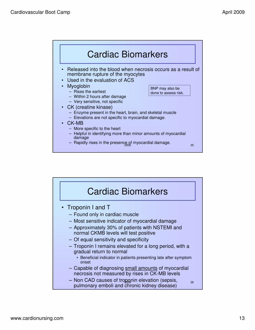
Evaluation of Oxygen Supply and Demand
• Increase myocardial oxygen demand:– Hyperthermia
– Hypertension
– Tachycardia
– Conditions producing over stimulation of the sympathetic nervous system (cocaine use, hyperthyroidism)
• Decrease myocardial oxygen delivery:– Anemia
– Pulmonary disease.
• Increase myocardial oxygen demand and decrease myocardial oxygen supply:– Aortic stenosis
– Hypertrophic cardiomyopathy
Cardiovascular Boot Camp April 2009
www.cardionursing.com 13
2009 25
Cardiac Biomarkers
• Released into the blood when necrosis occurs as a result of membrane rupture of the myocytes
• Used in the evaluation of ACS• Myoglobin
– Rises the earliest
– Within 2 hours after damage
– Very sensitive, not specific
• CK (creatine kinase)– Enzyme present in the heart, brain, and skeletal muscle
– Elevations are not specific to myocardial damage.
• CK-MB– More specific to the heart
– Helpful in identifying more than minor amounts of myocardial damage
– Rapidly rises in the presence of myocardial damage.
BNP may also be
done to assess risk.
2009 26
Cardiac Biomarkers
• Troponin I and T– Found only in cardiac muscle
– Most sensitive indicator of myocardial damage
– Approximately 30% of patients with NSTEMI and normal CKMB levels will test positive
– Of equal sensitivity and specificity
– Troponin I remains elevated for a long period, with a gradual return to normal
• Beneficial indicator in patients presenting late after symptom onset
– Capable of diagnosing small amounts of myocardial necrosis not measured by rises in CK-MB levels
– Non CAD causes of troponin elevation (sepsis, pulmonary emboli and chronic kidney disease)

Cardiovascular Boot Camp April 2009
www.cardionursing.com 14
2009 27
10 or more days 18 to 24 hours 4 to 6 hours Highly specific and sensitive
Troponin I or T
2 to 3 days 18 to 24 hours 4 to 6 hours Highly specific CK-MB
< 24 hours 4 to 10 hours Within 2 hours Sensitive but not
specific
Myoglobin
Duration Peak Rise Specificity / Sensitivity
Cardiac Biomarker
Cardiac Biomarker Summary
2009 28
Timing of Release of Various Biomarkers
After Acute Myocardial Infarction
Shapiro BP, Jaffe AS. Cardiac biomarkers. In: Murphy JG, Lloyd MA, editors. Mayo Clinic Cardiology: Concise Textbook. 3rd ed. Rochester, MN: Mayo Clinic Scientific Press and New York: Informa Healthcare USA, 2007:773–80. Anderson JL, et al. J Am Coll Cardiol 2007;50:e1–e157, Figure 5.

Cardiovascular Boot Camp April 2009
www.cardionursing.com 15
2009 29
Diagnostic Testing
• Non Invasive
– ECG Evaluation
– Stress Testing
– CT / CTA
• Invasive: Cardiac Catheterization
– IVUS
2009 30
Stress Testing
• Exercise Stress Test with or without myocardial imaging– Nuclear Scanning
– Echocardiogram– Future
• Patient conditions requiring myocardial imaging with stress testing due to lack of reliable ECG interpretation include: – Left bundle branch block
– > 1 mm ST-segment depression at rest
– Paced ventricular rhythm
– Wolf-Parkinson-White syndrome
Cardiovascular Boot Camp April 2009
www.cardionursing.com 16
2009 31
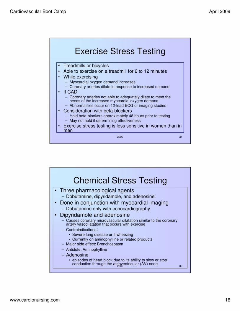
Exercise Stress Testing
• Treadmills or bicycles
• Able to exercise on a treadmill for 6 to 12 minutes
• While exercising – Myocardial oxygen demand increases
– Coronary arteries dilate in response to increased demand
• If CAD– Coronary arteries not able to adequately dilate to meet the
needs of the increased myocardial oxygen demand
– Abnormalities occur on 12-lead ECG or imaging studies
• Consideration with beta-blockers– Hold beta-blockers approximately 48 hours prior to testing
– May not hold if determining effectiveness
• Exercise stress testing is less sensitive in women than in men
2009 32
Chemical Stress Testing• Three pharmacological agents
– Dobutamine, dipyridamole, and adenosine.
• Done in conjunction with myocardial imaging– Dobutamine only with echocardiography
• Dipyridamole and adenosine – Causes coronary microvascular dilatation similar to the coronary
artery vasodilatation that occurs with exercise
– Contraindications:• Severe lung disease or if wheezing
• Currently on aminophylline or related products
– Major side effect: Bronchospasm
– Antidote: Aminophylline
– Adenosine• episodes of heart block due to its ability to slow or stop
conduction through the atrioventricular (AV) node

Cardiovascular Boot Camp April 2009
www.cardionursing.com 17
2009 33
Chemical Stress Testing
• Dobutamine
– High-dose dobutamine increases contractility
and heart rate
– Increasing myocardial oxygen demand
– More closely mimics exercise stress testing
– Side effect: Tachyarrhythmias
– Antidote: Beta blocker
2009 34
Contraindications to Stress Testing
• Acute MI <_ 2 days old
• Acute myocarditis or pericarditis
• Acute pulmonary embolism
• Acute aortic dissection
• Symptomatic heart failure
• Severe aortic stenosis
• Symptomatic arrhythmias
• High-risk unstable angina

Cardiovascular Boot Camp April 2009
www.cardionursing.com 18
2009 35
Stress Testing in Patients Presenting with Chest Pain
• Indicated when ECG and biomarkers are
not diagnostic
• Should be done before discharge or within 72 hours as outpatient
– Precautionary pharmacotherapy for low risk patients being done on outpatient basis
2009 36
CT Angiography“FAST CT”
• 64 slice and beyond
• Detailed 3D Image
• Fast
• Coronary artery calcium scoring– Shows calcified plaque
– Predictor of non-calcified plaque
• Coronary artery anatomy
• Myocardial function
• Need to lower heart rate
• Radiation exposure
• Good negative predictor

Cardiovascular Boot Camp April 2009
www.cardionursing.com 19
2009 37
Cardiac Catheterization
• Indications
– Patients with disabling angina despite medical
treatment
– Patients with high-risk criteria for coronary heart disease (CHD) on noninvasive testing
– Patients who have survived sudden cardiac death
– Patients with angina and clinical signs of CHD
– Patients with low ejection fraction and ischemia on noninvasive testing
– Patients with inadequate information obtained from
noninvasive testing
2009 38
STEMI Management • Reperfusion is number one treatment strategy
• Primary Coronary Intervention (PCI) preferred
treatment strategy if within 90 minutes
– Goal: 90 minutes from 1st medical contact
• Fibrinolytics within 30 minutes of hospital presentation (or 30 minutes from EMS to
fibrinolytics)
�Facilitated PCI with full dose fibrinolytics is not
recommended.
�Rescue PCI may be done after failed fibrinolytics.
Cardiovascular Boot Camp April 2009
www.cardionursing.com 20
2009 39
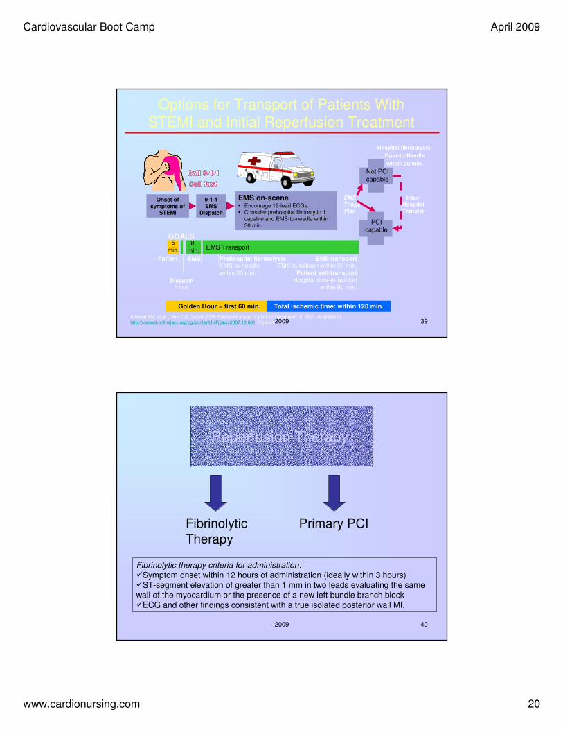
Options for Transport of Patients With
STEMI and Initial Reperfusion Treatment
EMS Transport
Onset of
symptoms of STEMI
9-1-1
EMSDispatch
EMS on-scene• Encourage 12-lead ECGs.
• Consider prehospital fibrinolytic if
capable and EMS-to-needle within
30 min.
GOALS
PCIcapable
Not PCI
capable
Hospital fibrinolysis:
Door-to-Needle
within 30 min.
EMS
Triage
Plan
Inter-Hospital
Transfer
Golden Hour = first 60 min. Total ischemic time: within 120 min.
Patient EMS Prehospital fibrinolysis
EMS-to-needle
within 30 min.
EMS transport
EMS-to-balloon within 90 min.
Patient self-transport
Hospital door-to-balloon
within 90 min.Dispatch
1 min.
5
min.8
min.
Antman EM, et al. J Am Coll Cardiol 2008. Published ahead of print on December 10, 2007. Available athttp://content.onlinejacc.org/cgi/content/full/j.jacc.2007.10.001. Figure 1.
2009 40
Reperfusion Therapy
Primary PCIFibrinolytic Therapy
Fibrinolytic therapy criteria for administration:
�Symptom onset within 12 hours of administration (ideally within 3 hours)
�ST-segment elevation of greater than 1 mm in two leads evaluating the same
wall of the myocardium or the presence of a new left bundle branch block
�ECG and other findings consistent with a true isolated posterior wall MI.

Cardiovascular Boot Camp April 2009
www.cardionursing.com 21
2009 41
Fibrinolytics Contraindications to the
administration of fibrinolytics: • Prior intracranial hemorrhage
• Known structural cerebral vascular lesion
• Malignant intracranial neoplasm
• Significant closed head injury within last 3 months
• Ischemic stroke within last 3 months (unless within last 3 hours)
• Suspected aortic dissection
• Active bleeding or bleeding diathesis (excluding menses)
• Symptoms greater than 24 hours old
• ST-segment depression (unless indicative of a true posterior wall MI)
Successful reperfusion with Fibrinolytics: • Relief of presenting symptoms
• Reduction of at least 50% of initial ST-segment segment elevation on repeat ECG
• Hemodynamic and electrical stability
• Reperfusion arrhythmias such as accelerated idioventricular rhythm
• Early peaking of the CKMB
2009 42
STEMI Anticoagulation
Fibrinolytics
• Minimum of 48 hours – up to 8 days
• Agents other than Unfractionated Heparin (UFH) if > 48 hours to reduce risk of Heparin Induced Thromocytopenia(HIT)
• Options for anticoagulation– UFH
– Enoxaparin (low molecular weight heparin (LMWH))
– Fondaparinux (indirect factor Xa inhibitor)
PCI
• UFH
• Enoxaparin
• Bivalrudin (direct thrombin inhibitor)
• Fondaparinux cannot be used as sole anticoagulant – Higher risk for catheter
thrombosis
– Need agent with anti IIaactivity

Cardiovascular Boot Camp April 2009
www.cardionursing.com 22
2009 43
Reasons for Delayed or Missed Reperfusion Therapy
• Missed performance of ECG due to atypical symptoms
• Unrecognized unequivocal ECG
• Delay in diagnosis of subtle ECG
• Failure to perform serial ECGs
• Delay in administration of therapy
• Abortion of treatment – Resolution of pain alone is not indication for aborting
therapy
2009 44

Cardiovascular Boot Camp April 2009
www.cardionursing.com 23
2009 45
2009 46
Medical Management of STEMI
• ASA • Clopidogrel (with or without reperfusion)
• Oxygen
• NTG • MS (Class I)
• D/C NSAIDS • Beta-blockers (within 24 hours)
• ACE Inhibitors (within 24 hours with impaired EF, HTN, diabetes or chronic kidney disease)
• Anticoagulants (related to reperfusion strategy)
• Intravenous insulin may be indicated in first 24 to 48 hours after STEMI to tightly control blood sugars.
Reperfusion is primary
management strategy.

Cardiovascular Boot Camp April 2009
www.cardionursing.com 24
2009 47
Treatment of NSTEMI / UA: New Guidelines
• ASA
• Oxygen (1st 6 hours)
• NTG – IV in first 48 hours for persistent ischemia, HTN, HF – Should not interfere with mortality reducing beta
blockers or ace inhibitors
• MS (if NTG unsuccessful and other anti ischemic drugs on board )
• DC – NSAIDS
• Beta Blockers (within 24 hours)
– Start PO when hemodynamically stable
– May use IV if hypertensive
• ACE Inhibitors (within 24 hours)
(in select patients – pulmonary congestion or LVEF < 40%) – may also be used in other patients
Early invasive strategy versus
early conservative strategy
2009 48
Treatment of NSTEMI / UA: New Guidelines
• Antiplatelet Therapy
– Clopidogrel
– Glycoprotein (GP)
IIb/IIIa inhibitors
– Upstream administration
for invasive strategies
– Also used in
conservative strategies
• Anticoagulation Options:
– Unfractionated heparin
– LMWH (enoxaparin)
– Direct thrombin inhibitor (bivalrudin)
– Indirect factor Xa
inhibitor
(fondaparinux)

Cardiovascular Boot Camp April 2009
www.cardionursing.com 25
2009 49
Early Invasive Option in UA / NSTEMI
• Not waiting for failed medical treatment
• Not waiting for + noninvasive test
• Angiography with intent of revascularization • Better outcomes with GP IIb/IIIa inhibitors
• Excluded: very frail elderly, severe hepatic, pulmonary or renal failure, active or inoperable cancer
• Initial conservative (selective invasive) is an alternative option
• Initial conservative strategy in low risk women
Overall reduction in mortality and increased quality of life.
2009 50
Early Invasive Indications
•Refractory angina or hemodynamic or
electrical instability– Without serious co-morbidities or contraindications to
such procedures
– May be reasonable in patients with chronic renal
insufficiency
•Initially stabilized with high risk for
clinical events – Initial conservative is also an option

Cardiovascular Boot Camp April 2009
www.cardionursing.com 26
2009 51
Algorithm for Patients with UA/NSTEMI Managed by an Initial
Invasive Strategy
Proceed to Diagnostic Angiography
ASA (Class I, LOE: A)
Clopidogrel if ASA intolerant (Class I,
LOE: A)
Diagnosis of UA/NSTEMI is Likely or
Definite
Invasive Strategy
Init ACT (Class I, LOE: A)
Acceptable options: enoxaparin or UFH (Class I, LOE: A)
bivalirudin or fondaparinux (Class I, LOE: B)
Select Management StrategyProceed with an
Initial Conservative
Strategy
Anderson JL, et al. J Am Coll Cardiol. 2007;50:e1-e157, Figure 7. ACT = anticoagulation therapy; LOE = level of evidence.
A
B
B1
B2
Prior to Angiography
Init at least one (Class I, LOE: A) or
both (Class IIa, LOE: B) of the following:
Clopidogrel
IV GP IIb/IIIa inhibitor
Factors favoring admin of both clopidogrel
and GP IIb/IIIa inhibitor include:
Delay to Angiography
High Risk Features
Early recurrent ischemic discomfort
2009 52
Init clopidogrel (Class I, LOE: A)
Consider adding IV eptifibatide or tirofiban
(Class IIb, LOE: B)
Conservative Strategy
Init ACT (Class I, LOE: A):
Acceptable options: enoxaparin or UFH (Class I,
LOE: A) or fondaparinux (Class I, LOE: B), but
enoxaparin or fondaparinux are preferable (Class IIa,
LOE: B)
Select Management Strategy
ASA (Class I, LOE: A)Clopidogrel if ASA intolerant (Class I, LOE: A)
Diagnosis of UA/NSTEMI is Likely
or Definite
Algorithm for Patients with UA/NSTEMI Managed by an Initial Conservative Strategy
Proceed with
Invasive
Strategy
(Continued)Anderson JL, et al. J Am Coll Cardiol. 2007;50:e1-e157, Figure 8. ACT = anticoagulation therapy; LOE = level of evidence.
C2
C1
A

Cardiovascular Boot Camp April 2009
www.cardionursing.com 27
2009 53
Any subsequent events necessitating
angiography?
EF greater
than 40%
Evaluate LVEF
Low Risk
Cont ASA (Class I, LOE A) Cont clopidogrel (Class I, LOE A) and ideally up to 1 yr (Class I, LOE B)
DC IV GP IIb/IIIa if started previously (Class I, LOE A)
* DC ACT (Class I, LOE A)
(Class I, LOE: B)
Proceed to Dx
Angiography
Yes
EF 40% or
less Stress Test
(Class I, LOE: A)
No
Not Low Risk
(Class IIa,
LOE: B)
Algorithm for Patients with UA/NSTEMI Managed by an Initial Conservative Strategy
(Continued)
Anderson JL, et al. J Am Coll Cardiol. 2007;50:e1-e157, Figure 8. ACT = anticoagulation therapy; LOE = level of evidence.
(Class I, LOE: A)
(Class IIa, LOE: B)
O
L
MN
K
E-1 E-2
D
(Class I,
LOE: B)
(Class I, LOE: A)
2009 54
• Cont ASA (Class I, LOE: A)
• DC clopidogrel 5 to 7 d prior to elective CABG (Class I, LOE: B)
• DC IV GP IIb/IIIa 4 h prior to CABG (Class I, LOE: B)
• Cont UFH (Class I, LOE: B); DC enoxaparin 12 to 24 h prior to
CABG; DC fondaparinux 24 h prior to CABG; DC bivalirudin 3 h prior to CABG. Dose with UFH per institutional practice (Class I, LOE: B)
• Cont ASA (Class I, LOE A) • LD of clopidogrel if not
given pre angio (Class I, LOE: A)
&
• IV GP IIb/IIIa if not started pre angio (Class I, LOE: A)
• DC ACT after PCI for
uncomplicated cases(Class I, LOE: B)
• Cont ASA (Class I, LOE: A)• LD of clopidogrel if not
given pre angio (Class I, LOE A)*• DC IV GP IIb/IIIa after
at least 12 h if started pre angio(Class I, LOE: B)
• Cont IV UFH for at least 48 h (Class
I, LOE: A) or enoxaparin or fondaparinux for dur of hosp (LOE: A); either DC bivalirudin or cont at a dose of 0.25 mg/kg/hr for up to 72 h at physician‘s discretion (Class I,
LOE: B)
Antiplatelet and ACT at physician’s discretion(Class I,
LOE: C)
No significant obstructive
CAD on angiography
CAD on angiography
Medical therapyPCICABG
Select Post Angiography Management Strategy
Dx Angiography
Management after Diagnostic Angiography in
Patients with UA/NSTEMI
Anderson JL, et al. J Am Coll Cardiol. 2007;50:e1-e157, Figure 9. ACT = anticoagulation therapy; LOE = level of evidence.
GH
I
J
F
Cardiovascular Boot Camp April 2009
www.cardionursing.com 28
2009 55
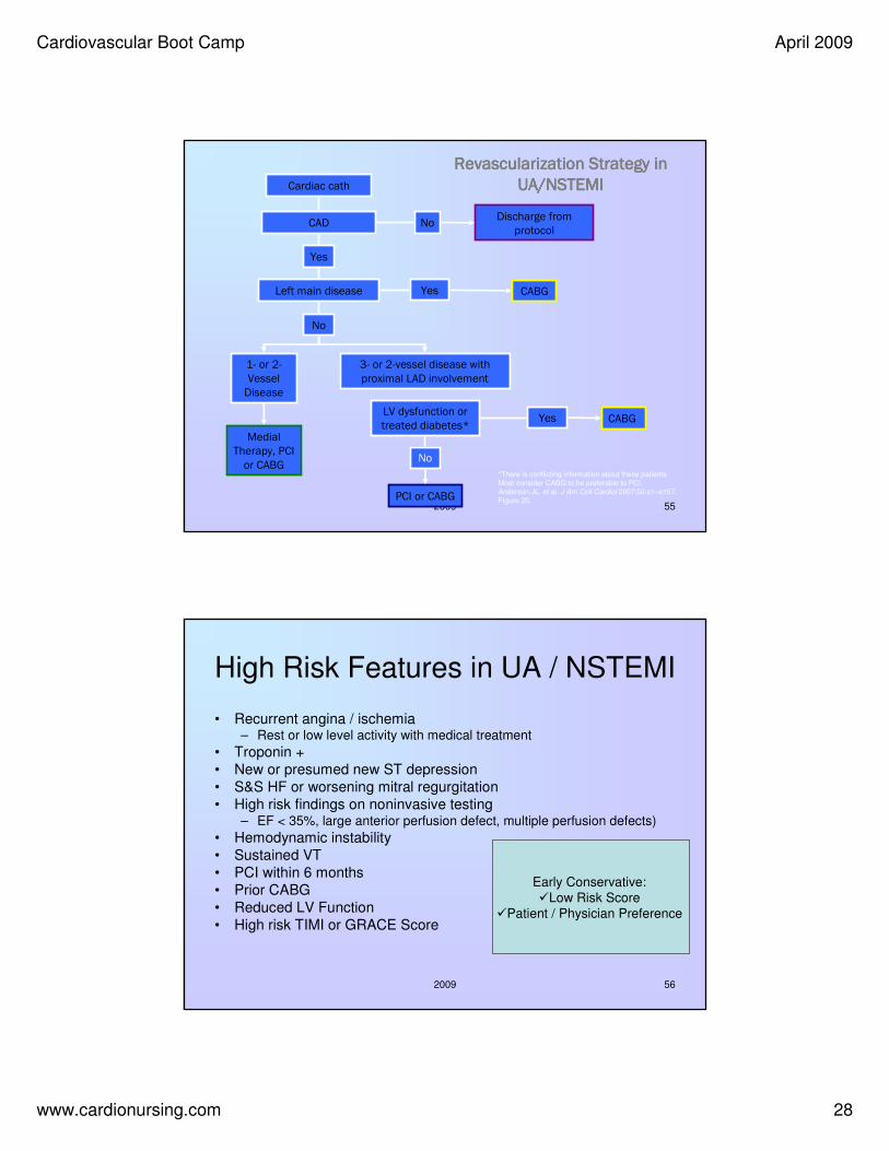
Cardiac cath
CAD NoDischarge from
protocol
Yes
Left main disease Yes CABG
No
1- or 2-
Vessel Disease
3- or 2-vessel disease with
proximal LAD involvement
LV dysfunction or treated diabetes*
No
PCI or CABG
Medial Therapy, PCI
or CABG
Yes CABG
*There is conflicting information about these patients. Most consider CABG to be preferable to PCI. Anderson JL, et al. J Am Coll Cardiol 2007;50:e1–e157, Figure 20.
Revascularization Strategy in Revascularization Strategy in Revascularization Strategy in Revascularization Strategy in
UA/NSTEMIUA/NSTEMIUA/NSTEMIUA/NSTEMI
2009 56
High Risk Features in UA / NSTEMI
• Recurrent angina / ischemia – Rest or low level activity with medical treatment
• Troponin +
• New or presumed new ST depression
• S&S HF or worsening mitral regurgitation
• High risk findings on noninvasive testing – EF < 35%, large anterior perfusion defect, multiple perfusion defects)
• Hemodynamic instability
• Sustained VT
• PCI within 6 months
• Prior CABG
• Reduced LV Function
• High risk TIMI or GRACE Score
Early Conservative:
�Low Risk Score
�Patient / Physician Preference

Cardiovascular Boot Camp April 2009
www.cardionursing.com 29
2009 57
Risk Assessment in UA / NSTEMI
• TIMI Risk Score
– Age > 65
– 3 or > risk factors for CAD
– Prior 50% or > stenosis
– ST deviation on ECG
– 2 or > anginal events in
previous 24 hours
– Use of ASA in prior 7 days
– Elevated cardiac biomarkers
• GRACE
– Older age
– Killip class
– Systolic BP
– Cardiac arrest during
presentation
– Serum creatinine
– Positive initial cardiac
markers
– HR
2009 58
•↑ Angina frequency, severity or duration
•Angina provoked at lower threshold
•New onset angina with
onset 2 wks to 2 mos prior to presentation
•Prolonged (> 20 min) rest angina, now resolved, w/ moderate/high
likelihood of CAD
•Rest angina (> 20 min) or relieved with rest or sublingual NTG
•Nocturnal angina
•New-onset or progressive CCS class III/IV angina in past 2 wks w/o prolonged (> 20 min) rest pain
but with intermediate/high likelihood of CAD
Prolonged ongoing (>
20 min) rest pain
Character Character Character Character of painof painof painof pain
Prior MI, peripheral or cerebrovascular disease, or CABG; prior ASA use
Accelerating tempo of ischemic sx in preceding 48 h
HistoryHistoryHistoryHistory
Low RiskNo high- or intermediate-
risk features but may
have any features below:
Intermediate RiskNo high-risk features, but must have
1 of the following:
High Risk≥ 1 of the
features
below must
be present:
Feature
Short-Term Risk of Death/Nonfatal MI in Patients With UA/NSTEMI

Cardiovascular Boot Camp April 2009
www.cardionursing.com 30
2009 59
Low riskIntermediate riskHigh riskFeature
NormalSlightly ↑ cardiac TnT, TnI, or CK-MB (e.g., TnT > 0.01, but < 0.1 ng/mL)
↑ Cardiac TnT, TnI, or CK-MB (e.g., TnT/TnI > 0.1 ng/mL)
Cardiac Cardiac Cardiac Cardiac markersmarkersmarkersmarkers
Normal or unchanged ECG
•T-wave changes
•Pathological Q-waves/resting ST-
depression < 1 mm in multiple lead groups (anterior, inferior, lateral)
•Angina @ rest with transient ST-segment changes > 0.5 mm
•BBB, new/presumed new
•Sustained VT
ECGECGECGECG
Age > 70 y•Pulmonary edema, most likely due to ischemia
•New/worsening MR murmur
•S3 or new/worsening rales
•Hypotension, bradycardia,
tachycardia
•Age > 75 y
Clinical Clinical Clinical Clinical findingsfindingsfindingsfindings
Estimation of the short-term risk of death and nonfatal cardiac ischemic events in UA/NSTEMI is a complex multivariable problem that cannot be fully specified in a table such as this; this table is mean to offer general guidance & illustration rather than rigid algorithms. Braunwald E, et al. AHCPR Publication No. 94-0602:1–154. Anderson JL, et al. J Am Coll Cardiol 2007;50:e1–e157, Table 7.
Short-Term Risk of Death/Nonfatal MI in Patients With UA/NSTEMI,
Continued
2009 60
Noninvasive Test Results That Predict High Risk for Adverse Outcomes
Wall-motion score > 1
Rest EF ≤ 35%
Stress Stress Stress Stress
EchocardiographyEchocardiographyEchocardiographyEchocardiography
Cardiac enlargementFall in EF ≥ 10%
Abnormal myocardial distribution with ↑ lung intake
Rest EF ≤ 35%
Abnormal myocardial tracer distribution in > 1 coronary artery region
Exercise EF ≤ 50 %
Stress Radionuclide Stress Radionuclide Stress Radionuclide Stress Radionuclide
Myocardial Perfusion Myocardial Perfusion Myocardial Perfusion Myocardial Perfusion
ImagingImagingImagingImaging
Stress Stress Stress Stress
Radionuclide Radionuclide Radionuclide Radionuclide
VentriculographyVentriculographyVentriculographyVentriculography
Adapted from O’Rourke RA, et al. J Am Coll Cardiol 1986;8:1471–83 and Cheitlin MD, et al. Circulation 1997;95:1686–744.EF = ejection fraction.

Cardiovascular Boot Camp April 2009
www.cardionursing.com 31
2009 61
Long-Term Antithrombotic Therapy at Hospital Discharge after UA/NSTEMI
Medical Therapy Medical Therapy Medical Therapy Medical Therapy
without Stentwithout Stentwithout Stentwithout Stent
Bare Metal Bare Metal Bare Metal Bare Metal
Stent GroupStent GroupStent GroupStent Group
Drug Eluting Drug Eluting Drug Eluting Drug Eluting
Stent GroupStent GroupStent GroupStent Group
ASA 162 to 325 mg/d for at least 1 ASA 162 to 325 mg/d for at least 1 ASA 162 to 325 mg/d for at least 1 ASA 162 to 325 mg/d for at least 1
month, then 75 to 162 mg/d month, then 75 to 162 mg/d month, then 75 to 162 mg/d month, then 75 to 162 mg/d
indefinitelyindefinitelyindefinitelyindefinitely (Class I, LOE: A) (Class I, LOE: A) (Class I, LOE: A) (Class I, LOE: A)
&&&&
Clopidogrel 75 mg/d for at least 1 Clopidogrel 75 mg/d for at least 1 Clopidogrel 75 mg/d for at least 1 Clopidogrel 75 mg/d for at least 1
month and up to 1 year month and up to 1 year month and up to 1 year month and up to 1 year
(Class I, LOE:B)(Class I, LOE:B)(Class I, LOE:B)(Class I, LOE:B)
Add: WarfarinAdd: WarfarinAdd: WarfarinAdd: Warfarin (INR 2.0 to 2.5)(INR 2.0 to 2.5)(INR 2.0 to 2.5)(INR 2.0 to 2.5)
(Class IIb, LOE: B)(Class IIb, LOE: B)(Class IIb, LOE: B)(Class IIb, LOE: B)
Continue with dual antiplatelet Continue with dual antiplatelet Continue with dual antiplatelet Continue with dual antiplatelet
therapy as abovetherapy as abovetherapy as abovetherapy as above
YesYesYesYes NoNoNoNo
Indication for Indication for Indication for Indication for
Anticoagulation?Anticoagulation?Anticoagulation?Anticoagulation?
ASA 75 to 162 mg/d indefinitelyASA 75 to 162 mg/d indefinitelyASA 75 to 162 mg/d indefinitelyASA 75 to 162 mg/d indefinitely
(Class I, LOE: A) (Class I, LOE: A) (Class I, LOE: A) (Class I, LOE: A)
& & & &
ClopidogrelClopidogrelClopidogrelClopidogrel 75 mg/d at least 1 75 mg/d at least 1 75 mg/d at least 1 75 mg/d at least 1
monthmonthmonthmonth (Class I, LOE: A) (Class I, LOE: A) (Class I, LOE: A) (Class I, LOE: A) and up and up and up and up
to 1 yearto 1 yearto 1 yearto 1 year (Class I, LOE: B)(Class I, LOE: B)(Class I, LOE: B)(Class I, LOE: B)
ASA 162 to 325 mg/d for at ASA 162 to 325 mg/d for at ASA 162 to 325 mg/d for at ASA 162 to 325 mg/d for at
least 3 to 6 months, then 75 least 3 to 6 months, then 75 least 3 to 6 months, then 75 least 3 to 6 months, then 75
to 162 mg/d indefinitelyto 162 mg/d indefinitelyto 162 mg/d indefinitelyto 162 mg/d indefinitely
(Class I, LOE: A)(Class I, LOE: A)(Class I, LOE: A)(Class I, LOE: A)
&&&&
Clopidogrel 75 mg/d for at Clopidogrel 75 mg/d for at Clopidogrel 75 mg/d for at Clopidogrel 75 mg/d for at
least 1 yearleast 1 yearleast 1 yearleast 1 year (Class I, LOE: B)(Class I, LOE: B)(Class I, LOE: B)(Class I, LOE: B)
Anderson JL, et al. J Am Coll Cardiol 2007;50:e1–e157, Figure 11. INR = international normalized ratio; LOE = level of evidence.
UA/NSTEMI UA/NSTEMI UA/NSTEMI UA/NSTEMI
Patient Groups at Patient Groups at Patient Groups at Patient Groups at
DischargeDischargeDischargeDischarge
New
2009 62
Beta Blockers Considerations
• Oral Beta Blockers – Within 24 hours
• IV Beta Blockers – Reasonable in patients
who are hypertensive
– May be harmful in patients with high risk for cardiogenic shock
• Contraindications– Signs of HF
– Low cardiac output state
– Increased risk for cardiogenic shock
– Relative contraindications
• PR > .24 seconds
• 2nd or 3rd degree block
• Active asthma
• Reactive airway disease
Nondihydropyridine calcium channel
blocker if beta blocker contraindicated
and no significant LV dysfunction .

Cardiovascular Boot Camp April 2009
www.cardionursing.com 32
2009 63
Nitrate Contraindications
• Systolic BP < 90 mm Hg or < 30 mm Hg below baseline
• Bradycardia < 50 BPM
• Tachycardia > 100 BPM (in absence of clinical HF)
• Right ventricular infarct
• Within 24 hours of sildenafil
• Within 48 hours of taldalafil
2009 64
Other Medication Considerations
• Hold ace inhibitors for BP < 100 mm Hg systolic or < 30 mm Hg below baseline
• No IV ace inhibitor within 24 hours due to risk of hypotension
• No immediate release dihydropyridine calcium channel blockers without beta blockade on board
• NSAIDS (except for ASA), whether nonselective or COX-2–selective agents increase risk of mortality, reinfarction, hypertension, HF, and myocardial rupture
• Proton Pump Inhibitors should be prescribed to patients at risk for GI bleed

Cardiovascular Boot Camp April 2009
www.cardionursing.com 33
2009 65
• Acetaminophen, ASA, tramadol,
narcotic analgesics (short term)
• COX-2 Selective
NSAIDs
• Nonacetylated salicylates
• Non COX-2 selective NSAIDs
• NSAIDs with some
COX-2 activity
Stepped Care Approach To Pharmacologic Therapy for Musculoskeletal
Symptoms with Known Cardiovascular Disease or Risk Factors for
Ischemic Heart Disease
Select patients at low risk
of thrombotic events
Prescribe lowest dose
required to control symptoms
Add ASA 81 mg and PPI to patients
at increased risk of thrombotic
events *
• Regular monitoring for sustained
hypertension or worsening of prior
blood pressure control), edema,
worsening renal function, or
gastrointestinal bleeding.
• If these events occur, consider
reduction of the dose or discontinuation of the offending drug,
a different drug, or alternative
therapeutic modalities, as dictated by
clinical circumstances.
* Addition of ASA may not be sufficient protection against thrombotic eventsAntman EM, et al. J Am Coll Cardiol 2008. Published ahead of print on December 10, 2007. Available at http://content.onlinejacc.org/cgi/content/full/j.jacc.2007.10.001.
2009 66
Long Term Management of ACS
Medications to improve prognosis• Aspirin
• Clopidogrel
• *Beta-blockers
• *ACE inhibitors (in select patients) – ARBs (may be used with ACE-I in systolic dysfunction)
– Aldactone (EF < 40 with HF or diabetes)
• Lipid-lowering drugs (statins)
* Beta blockers and ACE inhibitors impact long term ventricular remodeling
Medications to control ischemia• Increased dose of beta-blockers
• Nitrates (all patients should be given sublingual nitroglycerin @ DC)
• Calcium channel blockers

Cardiovascular Boot Camp April 2009
www.cardionursing.com 34
2009 67
SL NTG Instruction
• No more than 1 dose of SL NTG – If chest discomfort is unimproved or is worsening 5
min after 1 NTG call 9-1-1 immediately before
taking additional NTG.
– May take additional NTG while waiting EMS.
– Chew ASA while waiting EMS.
• In chronic stable angina if symptoms are significantly
improved by 1 dose of NTG may repeat NTG every 5 min for
a maximum of 3 doses and call 9-1-1 if symptoms have not
resolved completely.
2009 68
Secondary Prevention • Smoking cessation• Reduction of hyperlipidemia
– LDL < 100 mg/dL or < 70 mg/dL (optimal)
• Hypertension control – <130/80 for kidney disease or diabetes
• Diabetes control Hb AIc < 7• Physical activity minimum of 5 days / per week
– 7 days recommended
• BMI 18.5 – 24.9 kg/mm2
• Phase II Cardiac Rehab • Influenza vaccine
Cardiovascular Boot Camp April 2009
www.cardionursing.com 35
2009 69
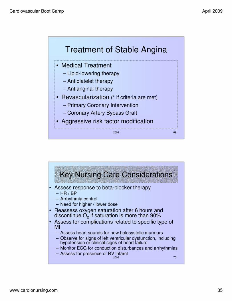
Treatment of Stable Angina
• Medical Treatment
– Lipid-lowering therapy
– Antiplatelet therapy
– Antianginal therapy
• Revascularization (* if criteria are met)
– Primary Coronary Intervention
– Coronary Artery Bypass Graft
• Aggressive risk factor modification
2009 70
Key Nursing Care Considerations
• Assess response to beta-blocker therapy– HR / BP
– Arrhythmia control– Need for higher / lower dose
• Reassess oxygen saturation after 6 hours and discontinue O2 if saturation is more than 90%
• Assess for complications related to specific type of MI– Assess heart sounds for new holosystolic murmurs
– Observe for signs of left ventricular dysfunction, including hypotension or clinical signs of heart failure.
– Monitor ECG for conduction disturbances and arrhythmias
– Assess for presence of RV infarct

Cardiovascular Boot Camp April 2009
www.cardionursing.com 36
2009 71
Key Nursing Care Considerations
• Restrict activity for at least the first 12 hours, and then begin Phase I Cardiac Rehabilitation– Referral to Phase II Cardiac Rehabilitation
• Utilize cardiac monitoring– ST-segment monitoring
– Uninterrupted monitoring for first 24-48 hours
• Focus on holistic approach to anxiety reduction– Include the family. Family visits do not have a negative impact
on vital signs or cardiac rhythm
• Address addiction to nicotine– Consideration for nicotine withdrawal
– Specific smoking cessation plan
2009 72
Complications of MI
• Hemodynamic Alterations
• Ventricular Arrhythmias
• Atrial Arrhythmias
• Pericarditis
• Ventricular Aneurysms
• Mechanical Complications– Myocardial Rupture (free wall or VSD)
– Papillary Muscle Rupture
• Long Term: Ventricular Remodeling

Cardiovascular Boot Camp April 2009
www.cardionursing.com 37
2009 73
Ventricular Aneurysm
• Persistent ST elevation after AMI (anterior)
• Anatomic LV aneurysm – Myocardial thinning and bulging
• Use of echo in the reperfusion decision
• Risk of fibrinolytics with ventricular aneurysm – Embolization of thrombus
2009 74
ST Elevation of Ventricular Aneurysm
• Most common in V1-V3
• Usually less than 3 mm elevation
• Relatively unchanged from previous ECGs
• Q waves are deep and well formed – QS pattern in V1-V3 or very minimal r
– QR pattern common in inferior aneurysm / RBBB

Cardiovascular Boot Camp April 2009
www.cardionursing.com 38
2009 75
2009 76
Myocardial Rupture
• Incidence – 10% MI deaths
• Definition – Myocardial leakage – hemipericardium – tamponade
– Perceived sudden; often slow tear
• Associated Factors – Late fibrinolytics
– Delayed hospital admission
• Septal involvement = VSD

Cardiovascular Boot Camp April 2009
www.cardionursing.com 39
2009 77
Myocardial Rupture
• Post-infarction regional pericarditis
precedes rupture
• Confirmation of rupture
T Wave Patterns in Post-infarction
Regional Pericarditis
Persistently positive T
waves 48 hours after an MI
Premature reversal of T
wave inversion to
positive ST segment
reelevation
2009 78

Cardiovascular Boot Camp April 2009
www.cardionursing.com 40
2009 79
Acute Mitral Regurgitation
• Acute event causing mitral valve
regurgitation
– Papillary muscle rupture
– Once free the attached valve leaflet will not close
– Gaping hole left for blood to eject through
2009 80
Pathophysiology
– Impairment or rupture of a papillary muscle– Damaged to myocardial wall → damage to
attachment of the papillary muscle to that ventricular wall
– Papillary muscle continues to contract with each cardiac cycle
– Attachment of papillary muscle to ventricular wall becomes weaker with each contraction
– With enough damage to the myocardial wall or papillary muscle the papillary muscle will actually disconnect from the ventricular wall
– Acute mitral regurgitation state– Emergency measures are necessary to preserve the
patient’s life

Cardiovascular Boot Camp April 2009
www.cardionursing.com 41
2009 81
Acute Mitral Regurgitation
Acute decrease in cardiac output � � SVR
�blood flow to area of least resistance (non-functional MV)
�decrease in cardiac output � � SVR
�blood flow to area of least resistance (non-functional MV)
�decrease in cardiac output � ETC.!!!!!
2009 82
Cardiovascular Boot Camp April 2009
www.cardionursing.com 42
2009 83
Diagnosis and Treatment
• Murmur
• STAT Echocardiogram
• Afterload reduction
• Emergent Surgery
Note: Antibiotics for acute endocarditis
2009 84
Final Quote:
Our grand business in life
is not to see what lies
dimly at a distance,
but to do what lies clearly at hand.
Thomas Carlyle (1795-1881)

Cardiovascular Boot Camp April 2009
www.cardionursing.com 43
2009 85
Thanks for Attending Cardiovascular Boot Camp
You may contact us at
www.cardionursing.com