Embed Size (px)
Citation preview
Acute decompensated pulmonaryhypertension
Laurent Savale1,2,3, Jason Weatherald4, Xavier Jaïs1,2,3, Constance Vuillard2,Athénaïs Boucly1,2,3, Mitja Jevnikar1,2,3, David Montani1,2,3, Olaf Mercier3,5,Gerald Simonneau1,2,3, Elie Fadel3,5, Olivier Sitbon1,2,3 and Marc Humbert1,2,3
Number 2 in the Series “Acute exacerbations in pulmonary medicine”Edited by Michael Kreuter and Vincent Cottin
Affiliations: 1Université Paris-Sud, Faculté de Médecine, Université Paris-Saclay, Le Kremlin-Bicêtre, France.2AP-HP, Service de Pneumologie, Centre de Référence de l’Hypertension Pulmonaire Sévère, Hôpital Bicêtre,Le Kremlin-Bicêtre, France. 3INSERM UMR_S 999, Hôpital Marie Lannelongue, Le Plessis Robinson, France.4University of Calgary, Dept of Medicine, Division of Respirology, Calgary, AB, Canada. 5Depts of Thoracic andVascular Surgery and Heart-Lung Transplantation, Marie Lannelongue Hospital, Le Plessis Robinson, France.
Correspondence: Laurent Savale, Université Paris-Sud, Centre de Référence de l’Hypertension PulmonaireSévère, Service de Pneumologie et Soins Intensifs Respiratoires, Hôpital Bicêtre, 78 Rue du général Leclerc,94270 Le Kremlin-Bicêtre, France. E-mail: [email protected]
@ERSpublicationsAcute decompensated PH is a life-threatening condition requiring specific management in aspecialised centre http://ow.ly/non530fkhmA
Cite this article as: Savale L, Weatherald J, Jaïs X, et al. Acute decompensated pulmonary hypertension.Eur Respir Rev 2017; 26: 170092 [https://doi.org/10.1183/16000617.0092-2017].
ABSTRACT Acute right heart failure in chronic precapillary pulmonary hypertension is characterised bya rapidly progressive syndrome with systemic congestion resulting from impaired right ventricular fillingand/or reduced right ventricular flow output. This clinical picture results from an imbalance between theafterload imposed on the right ventricle and its adaptation capacity. Acute decompensated pulmonaryhypertension is associated with a very poor prognosis in the short term. Despite its major impact onsurvival, its optimal management remains very challenging for specialised centres, without specificrecommendations. Identification of trigger factors, optimisation of fluid volume and pharmacologicalsupport to improve right ventricular function and perfusion pressure are the main therapeutic areas toconsider in order to improve clinical condition. At the same time, specific management of pulmonaryhypertension according to the aetiology is mandatory to reduce right ventricular afterload. Over the pastdecade, the development of extracorporeal life support in refractory right heart failure combined withurgent transplantation has probably contributed to a significant improvement in survival for selectedpatients. However, there remains a considerable need for further research in this field.
IntroductionPrecapillary pulmonary hypertension defines a group of disorders characterised by a progressive increasein pulmonary vascular resistance (PVR) that develops as a result of abnormal remodelling of the
Copyright ©ERS 2017. ERR articles are open access and distributed under the terms of the Creative CommonsAttribution Non-Commercial Licence 4.0.
Previous articles in this series: No. 1: Kondoh Y, Cottin V, Brown KK. Recent lessons learned in the management ofacute exacerbation of idiopathic pulmonary fibrosis. Eur Respir Rev 2017; 26: 170050.
Received: Aug 18 2017 | Accepted: Sept 17 2017
Conflict of interest: Disclosures can be found alongside this article at err.ersjournals.com
Provenance: Commissioned article, peer reviewed.
https://doi.org/10.1183/16000617.0092-2017 Eur Respir Rev 2017; 26: 170092
SERIESACUTE EXACERBATIONS
pulmonary microvasculature [1–3]. Improved understanding of the pathophysiology of precapillarypulmonary hypertension has led to the development of several medications that primarily targetendothelial dysfunction [4]. Despite these major advances, pulmonary hypertension remains a progressiveand fatal disease leading to right ventricular dysfunction and death [5, 6]. The ability of the right ventricleto adapt to the progressive increase in afterload is closely linked to the functional status and prognosis ofpatients [7]. Progressive right ventricular dysfunction plays a key role in the insidious development ofclinical symptoms. Impaired cardiac output and elevation in central venous pressure are associated withfunctional deterioration, onset of congestive signs and ultimately with mortality in patients with advancedpulmonary hypertension.
The prognostic importance of right ventricular function has been underappreciated for a long time andmost major clinical studies have focused on the pulmonary circulation as the therapeutic target inprecapillary pulmonary hypertension. However, there has been a renewed interest in recent years in theanalysis of the pathophysiology of right ventricular remodelling and dysfunction. End-stage pulmonaryhypertension is characterised by maladaptive right ventricular remodelling, leading to a fall in cardiacoutput and a right ventricle–arterial uncoupling. This condition is associated with a high risk of acuteright heart failure, which has been defined as a rapidly progressive syndrome with systemic congestionresulting from impaired right ventricular filling and/or reduced right ventricular flow output [8]. Theoccurrence of right heart failure remains the most frequent cause of death with a dismal prognosis overthe short term, highlighting the need for earlier recognition and better management options. This articleaims to provide a comprehensive review of the state of the art in acute decompensated pulmonaryhypertension.
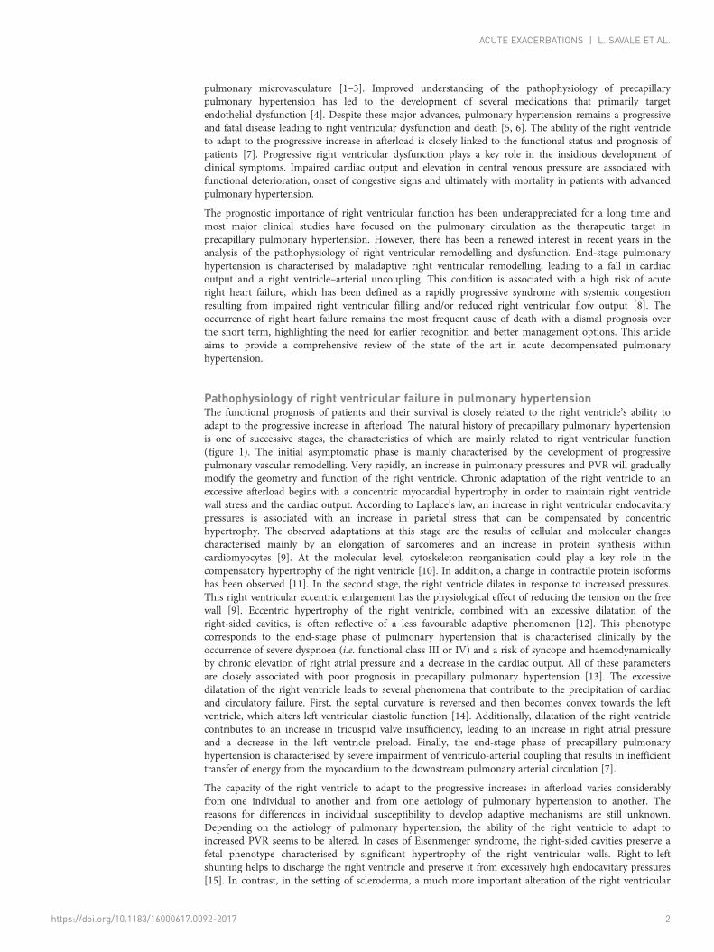
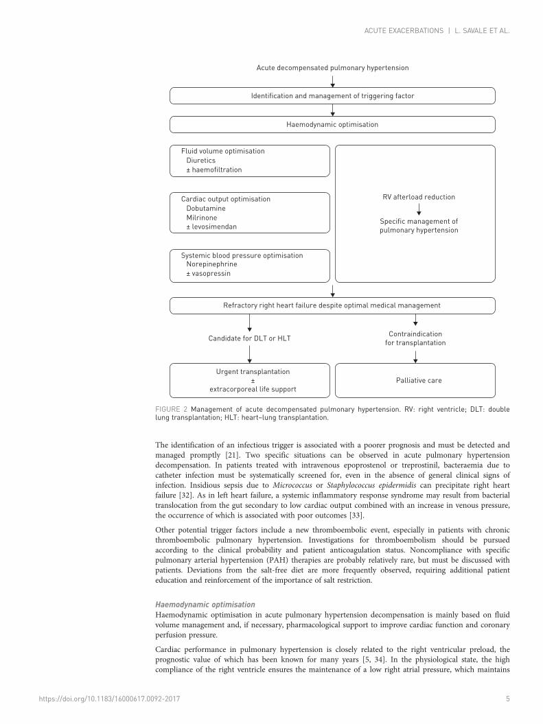
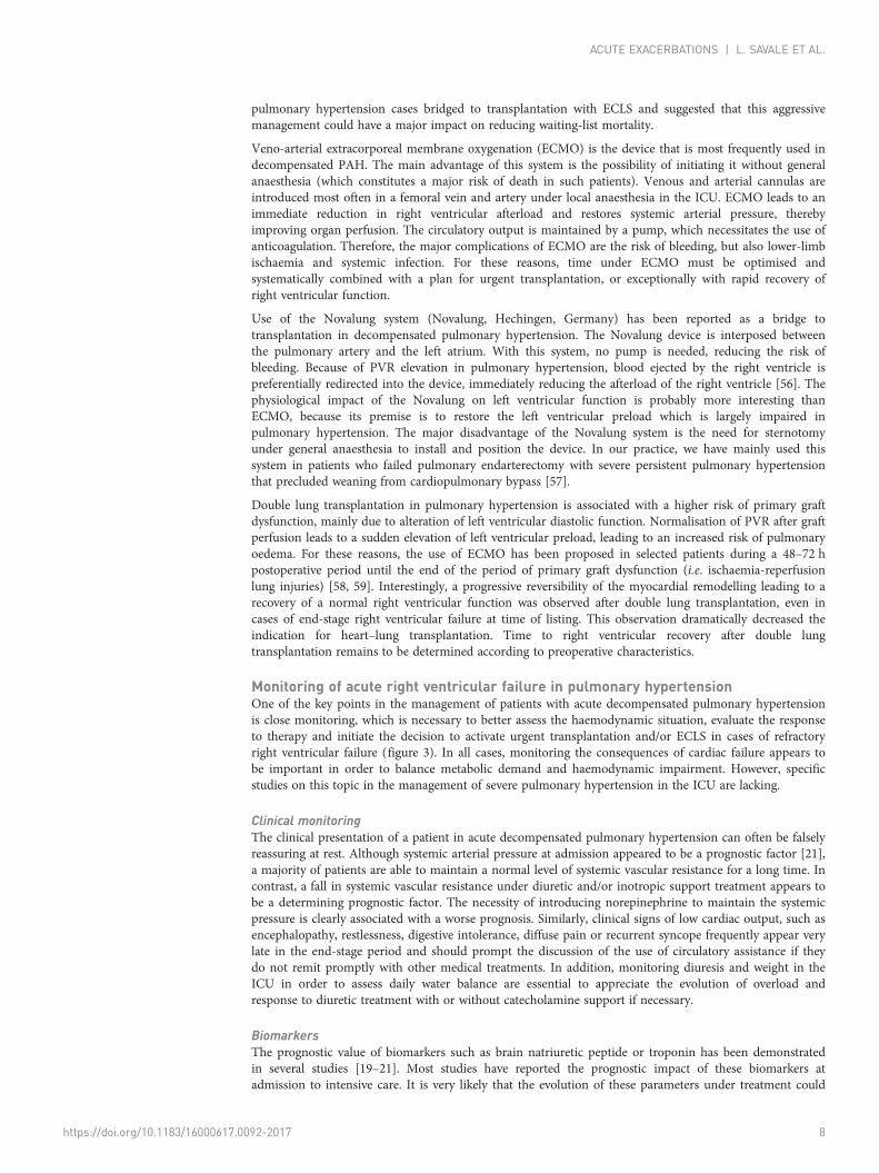
Pathophysiology of right ventricular failure in pulmonary hypertensionThe functional prognosis of patients and their survival is closely related to the right ventricle’s ability toadapt to the progressive increase in afterload. The natural history of precapillary pulmonary hypertensionis one of successive stages, the characteristics of which are mainly related to right ventricular function(figure 1). The initial asymptomatic phase is mainly characterised by the development of progressivepulmonary vascular remodelling. Very rapidly, an increase in pulmonary pressures and PVR will graduallymodify the geometry and function of the right ventricle. Chronic adaptation of the right ventricle to anexcessive afterload begins with a concentric myocardial hypertrophy in order to maintain right ventriclewall stress and the cardiac output. According to Laplace’s law, an increase in right ventricular endocavitarypressures is associated with an increase in parietal stress that can be compensated by concentrichypertrophy. The observed adaptations at this stage are the results of cellular and molecular changescharacterised mainly by an elongation of sarcomeres and an increase in protein synthesis withincardiomyocytes [9]. At the molecular level, cytoskeleton reorganisation could play a key role in thecompensatory hypertrophy of the right ventricle [10]. In addition, a change in contractile protein isoformshas been observed [11]. In the second stage, the right ventricle dilates in response to increased pressures.This right ventricular eccentric enlargement has the physiological effect of reducing the tension on the freewall [9]. Eccentric hypertrophy of the right ventricle, combined with an excessive dilatation of theright-sided cavities, is often reflective of a less favourable adaptive phenomenon [12]. This phenotypecorresponds to the end-stage phase of pulmonary hypertension that is characterised clinically by theoccurrence of severe dyspnoea (i.e. functional class III or IV) and a risk of syncope and haemodynamicallyby chronic elevation of right atrial pressure and a decrease in the cardiac output. All of these parametersare closely associated with poor prognosis in precapillary pulmonary hypertension [13]. The excessivedilatation of the right ventricle leads to several phenomena that contribute to the precipitation of cardiacand circulatory failure. First, the septal curvature is reversed and then becomes convex towards the leftventricle, which alters left ventricular diastolic function [14]. Additionally, dilatation of the right ventriclecontributes to an increase in tricuspid valve insufficiency, leading to an increase in right atrial pressureand a decrease in the left ventricle preload. Finally, the end-stage phase of precapillary pulmonaryhypertension is characterised by severe impairment of ventriculo-arterial coupling that results in inefficienttransfer of energy from the myocardium to the downstream pulmonary arterial circulation [7].
The capacity of the right ventricle to adapt to the progressive increases in afterload varies considerablyfrom one individual to another and from one aetiology of pulmonary hypertension to another. Thereasons for differences in individual susceptibility to develop adaptive mechanisms are still unknown.Depending on the aetiology of pulmonary hypertension, the ability of the right ventricle to adapt toincreased PVR seems to be altered. In cases of Eisenmenger syndrome, the right-sided cavities preserve afetal phenotype characterised by significant hypertrophy of the right ventricular walls. Right-to-leftshunting helps to discharge the right ventricle and preserve it from excessively high endocavitary pressures[15]. In contrast, in the setting of scleroderma, a much more important alteration of the right ventricular
https://doi.org/10.1183/16000617.0092-2017 2
ACUTE EXACERBATIONS | L. SAVALE ET AL.
function can be observed for lower levels of PVR. Primary myocardial involvement may participate inearly impairment of the right ventricle in these patients [16].
Definition and characteristics of acute decompensated pulmonary hypertensionAcute decompensated pulmonary hypertension is characterised by sudden worsening of clinical signs ofright heart failure with subsequent systemic circulatory insufficiency and multisystem organ failure. Thisclinical picture results from an imbalance between the afterload imposed on the right ventricle and itscapacity for compensation. Acute right heart failure can be precipitated by an external trigger factor or canbe a manifestation of disease worsening. Whatever the aetiology, the short-term prognosis is very poor[17–24].
Acute decompensation of pulmonary hypertension may combine a sudden aggravation of both diastolicand systolic right ventricular insufficiency. Diastolic failure promotes worsening congestive signs that maylead to anasarca. Right ventricular systolic failure results in a sharp decrease in left ventricular preload anda decrease in cardiac output followed by low peripheral perfusion pressure. The combination of systolic anddiastolic right ventricular failure in most severe patients promotes the sudden installation of multiple organfailure, foremost of which are kidney, hepatic and gut dysfunction. The pathophysiological mechanisms ofacute renal dysfunction during acute decompensation of pulmonary hypertension are similar to thoseobserved in type I cardiorenal syndrome [25]. Acute worsening of renal function in the context ofdecompensated pulmonary hypertension has been strongly related to mortality in numerous studies [17, 21,22, 26]. As in left heart failure, the renin-angiotensin-aldosterone and the sympathetic nervous systems arestimulated, leading to a worsening of peripheral vascular resistance and sodium and water retention [27]. Intype I cardiorenal syndrome, significant stimulation of antidiuretic hormone promotes excessive retentionof free water, leading to volume overload and hyponatraemia. These combined phenomena contribute to adecrease in renal perfusion due to both arterial vasoconstriction and venous congestion.
In the physiological state, the right ventricular coronary perfusion is systo-diastolic, the aortic pressurebeing higher than the systolic pressure of the right ventricle. In the case of acute right ventriculardecompensation, the fall in cardiac output and the resulting acute circulatory failure may favour ischaemiaof the right ventricle by compromising the systolic component of coronary perfusion.
Survival and prognostic factors of acute decompensated pulmonary hypertensionClinical studies have demonstrated the very strong impact of acute pulmonary hypertensiondecompensation on short-term prognosis (table 1). Depending on the type of population studied,
Increase in
RV wall stress
Myocardial
remodelling
RV dilatation
and failure
Acute right heart
failure
Asymptomatic phase Compensated RV dysfunction End-stage disease
Increase in
RV afterload
∙ Progressive
increase in PVR
∙ Normal CI (>2.5 L·min–1·m–2)
∙ Normal RAP (<8 mmHg)
∙ RV–arterial coupling
impairment
∙ Low CI (<2.5 L·min–1·m–2)
∙ High RAP (>8 mmHg)
∙ CI <2 L·min–1·m–2
∙ RAP >12 mmHg
∙ Asymptomatic ∙ NYHA-FC I or II ∙ NYHA-FC III or IV
∙ No signs of RVF
∙ No syncope
∙ Fluid retention
∙ Syncope
∙ Acute worsening of
RVF symptoms
∙ NYHA-FC IV
∙ Multisystem organ
failure
Haemodynamics
Clinical symptoms
FIGURE 1 Gradual evolution towards end-stage pulmonary hypertension. RV: right ventricle; PVR: pulmonary vascular resistance; CI: cardiacindex; RAP: right atrial pressure; NYHA-FC: New York Heart Association functional class; RVF: right ventricular failure.
https://doi.org/10.1183/16000617.0092-2017 3
ACUTE EXACERBATIONS | L. SAVALE ET AL.
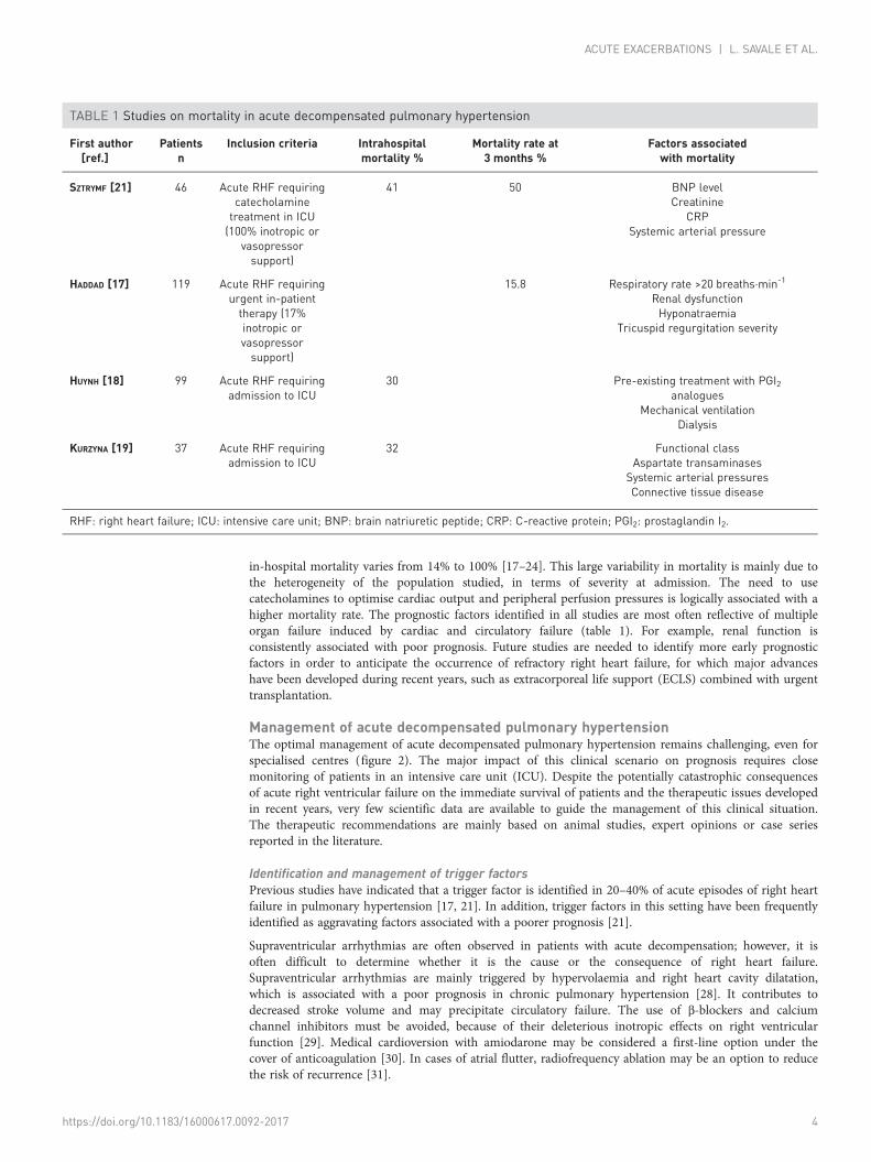
in-hospital mortality varies from 14% to 100% [17–24]. This large variability in mortality is mainly due tothe heterogeneity of the population studied, in terms of severity at admission. The need to usecatecholamines to optimise cardiac output and peripheral perfusion pressures is logically associated with ahigher mortality rate. The prognostic factors identified in all studies are most often reflective of multipleorgan failure induced by cardiac and circulatory failure (table 1). For example, renal function isconsistently associated with poor prognosis. Future studies are needed to identify more early prognosticfactors in order to anticipate the occurrence of refractory right heart failure, for which major advanceshave been developed during recent years, such as extracorporeal life support (ECLS) combined with urgenttransplantation.
Management of acute decompensated pulmonary hypertensionThe optimal management of acute decompensated pulmonary hypertension remains challenging, even forspecialised centres (figure 2). The major impact of this clinical scenario on prognosis requires closemonitoring of patients in an intensive care unit (ICU). Despite the potentially catastrophic consequencesof acute right ventricular failure on the immediate survival of patients and the therapeutic issues developedin recent years, very few scientific data are available to guide the management of this clinical situation.The therapeutic recommendations are mainly based on animal studies, expert opinions or case seriesreported in the literature.
Identification and management of trigger factorsPrevious studies have indicated that a trigger factor is identified in 20–40% of acute episodes of right heartfailure in pulmonary hypertension [17, 21]. In addition, trigger factors in this setting have been frequentlyidentified as aggravating factors associated with a poorer prognosis [21].
Supraventricular arrhythmias are often observed in patients with acute decompensation; however, it isoften difficult to determine whether it is the cause or the consequence of right heart failure.Supraventricular arrhythmias are mainly triggered by hypervolaemia and right heart cavity dilatation,which is associated with a poor prognosis in chronic pulmonary hypertension [28]. It contributes todecreased stroke volume and may precipitate circulatory failure. The use of β-blockers and calciumchannel inhibitors must be avoided, because of their deleterious inotropic effects on right ventricularfunction [29]. Medical cardioversion with amiodarone may be considered a first-line option under thecover of anticoagulation [30]. In cases of atrial flutter, radiofrequency ablation may be an option to reducethe risk of recurrence [31].
TABLE 1 Studies on mortality in acute decompensated pulmonary hypertension
First author[ref.]
Patientsn
Inclusion criteria Intrahospitalmortality %
Mortality rate at3 months %
Factors associatedwith mortality
SZTRYMF [21] 46 Acute RHF requiringcatecholaminetreatment in ICU(100% inotropic or
vasopressorsupport)
41 50 BNP levelCreatinine
CRPSystemic arterial pressure
HADDAD [17] 119 Acute RHF requiringurgent in-patienttherapy (17%inotropic orvasopressorsupport)
15.8 Respiratory rate >20 breaths·min-1
Renal dysfunctionHyponatraemia
Tricuspid regurgitation severity
HUYNH [18] 99 Acute RHF requiringadmission to ICU
30 Pre-existing treatment with PGI2analogues
Mechanical ventilationDialysis
KURZYNA [19] 37 Acute RHF requiringadmission to ICU
32 Functional classAspartate transaminases
Systemic arterial pressuresConnective tissue disease
RHF: right heart failure; ICU: intensive care unit; BNP: brain natriuretic peptide; CRP: C-reactive protein; PGI2: prostaglandin I2.
https://doi.org/10.1183/16000617.0092-2017 4
ACUTE EXACERBATIONS | L. SAVALE ET AL.
The identification of an infectious trigger is associated with a poorer prognosis and must be detected andmanaged promptly [21]. Two specific situations can be observed in acute pulmonary hypertensiondecompensation. In patients treated with intravenous epoprostenol or treprostinil, bacteraemia due tocatheter infection must be systematically screened for, even in the absence of general clinical signs ofinfection. Insidious sepsis due to Micrococcus or Staphylococcus epidermidis can precipitate right heartfailure [32]. As in left heart failure, a systemic inflammatory response syndrome may result from bacterialtranslocation from the gut secondary to low cardiac output combined with an increase in venous pressure,the occurrence of which is associated with poor outcomes [33].
Other potential trigger factors include a new thromboembolic event, especially in patients with chronicthromboembolic pulmonary hypertension. Investigations for thromboembolism should be pursuedaccording to the clinical probability and patient anticoagulation status. Noncompliance with specificpulmonary arterial hypertension (PAH) therapies are probably relatively rare, but must be discussed withpatients. Deviations from the salt-free diet are more frequently observed, requiring additional patienteducation and reinforcement of the importance of salt restriction.
Haemodynamic optimisationHaemodynamic optimisation in acute pulmonary hypertension decompensation is mainly based on fluidvolume management and, if necessary, pharmacological support to improve cardiac function and coronaryperfusion pressure.
Cardiac performance in pulmonary hypertension is closely related to the right ventricular preload, theprognostic value of which has been known for many years [5, 34]. In the physiological state, the highcompliance of the right ventricle ensures the maintenance of a low right atrial pressure, which maintains
Acute decompensated pulmonary hypertension
Identification and management of triggering factor
Haemodynamic optimisation
Fluid volume optimisation
Diuretics
± haemofiltration
Cardiac output optimisation
Dobutamine
Milrinone
± levosimendan
Systemic blood pressure optimisationNorepinephrine
± vasopressin
Refractory right heart failure despite optimal medical management
Candidate for DLT or HLTContraindication
for transplantation
Urgent transplantation
±
extracorporeal life support
Palliative care
RV afterload reduction
Specific management of
pulmonary hypertension
FIGURE 2 Management of acute decompensated pulmonary hypertension. RV: right ventricle; DLT: doublelung transplantation; HLT: heart–lung transplantation.
https://doi.org/10.1183/16000617.0092-2017 5
ACUTE EXACERBATIONS | L. SAVALE ET AL.
systemic venous return and thus preserves the cardiac output. In pulmonary hypertension, rightventricular diastolic dysfunction promotes sodium and water retention, which may be considerable in thesetting of acute decompensated pulmonary hypertension. Intravenous diuretics are used as a first-linetreatment in order to obtain a negative fluid balance and to optimise blood volume, and consequentlyreduce right ventricular preload. The aim of reducing right ventricular preload is to diminish right–leftventricular interdependence and thus improve left ventricular diastolic function, reduce tricuspidinsufficiency and reduce organ congestion, all of which contribute to the development of the cardiorenalsyndrome. A major challenge is to accurately assess and monitor right ventricle preload in these patientsin order to determine the optimal filling volume for each of them, as both hypovolaemia andhypervolaemia are deleterious to cardiac function.
Diuretic treatment alone may be insufficient to regain a state of equilibrium and to emerge from theepisode of acute decompensation. In cases of severe right ventricular systolic dysfunction, a severelyimpaired cardiac output may require the use of inotropic agents. The main goal of inotrope use is toimprove cardiac output and ventriculo-arterial coupling without increasing right ventricular afterload [35,36]. There are few specific clinical trials evaluating inotropic agents in the setting of acute decompensatedpulmonary hypertension. β1-adrenergic agonists remain the inotropic agents of choice, even thoughimpaired inotropic responses to β1-adrenergic receptor agonists have been observed in preclinical studiesdue to downregulation and desensitisation of β1-receptors in right ventricular hypertrophy [37].Dobutamine is most often initiated at low doses (2.5 µg·kg−1·min−1) and progressively uptitrated inpatients with persistent signs of low cardiac output. Dobutamine was effective in improvingventriculo-arterial coupling in an experimental model of acute occlusion of the pulmonary artery.However, the use of doses >10 µg·kg−1·min−1 can be deleterious by precipitating a decrease in systemicarterial resistance without improving cardiac output. Alternative options to dobutamine include newerinotrope/vasodilators such as milrinone or levosimendan. Milrinone is a phosphodiesterase-3 inhibitor,which increases myocardial contractility and reduces left ventriclar afterload. In some studies, milrinonehas been shown to reduce PVR [38]. Data on levosimendan are entirely experimental; however, it has beenshown to be more effective than dobutamine for improving ventriculo-arterial coupling in preclinicalstudies [39, 40].
In the most severely affected patients, systemic vascular resistance is low at admission or decreases afterthe initiation of diuretics with or without inotropic agents. This condition is generally associated withmultiorgan failure and right ventricular ischaemia. The use of vasopressors such as norepinephrine isnecessary in this situation to restore a normal perfusion pressure. In addition to its effect on systemicblood pressure, norepinephrine could help to improve right ventricular function and right ventricle–pulmonary artery coupling [40]. At low doses, vasopressin may have vasodilatory effects on the pulmonarycirculation, and some clinical cases of vasopressin use as rescue therapy in pulmonary hypertension havebeen published. At higher doses, vasopressin may paradoxically increase PVR and cause adversemyocardial effects [36].
Specific treatment of pulmonary hypertensionReduction of right ventricular afterload is a key point in the management of decompensated pulmonaryhypertension. Escalating or initiating targeted management of the underlying chronic disease is the onlyway to definitively emerge from a situation of acute right heart decompensation and avoid recurrences inthe long term. For cases of acute right ventricular insufficiency as the first presentation of chronicpulmonary hypertension, the aetiological assessment must be performed urgently to adapt specificmanagement. For PAH, all targeted therapies can be considered in combination, including preferential useof an intravenous prostaglandin analogue. Upfront triple combination therapy has been shown todramatically improve the long-term prognosis of patients with severe PAH with low cardiac index atbaseline [41]. In patients already undergoing treatment before the onset of an acute decompensation, thepossibility of treatment reinforcement must be considered in each case. Because of their vasodilatoryeffects, these drugs can contribute to a decrease in the systemic arterial resistance. It is probably better tointroduce them gradually with progressive increases in doses and after restoring haemodynamics. Inspecific cases of right heart failure due to PAH associated with systemic lupus erythematosus or mixedconnective tissue disease, dramatic clinical and haemodynamic improvement has been reported with theuse of corticosteroid and cyclophosphamide in combination with specific PAH therapies [42, 43].
Pulmonary veno-occlusive disease is a condition characterised by a dismal prognosis, which maydeteriorate dramatically when treated with PAH-targeted therapies. In this setting, the possibility of doublelung transplantation must be considered early in management [44]. The possibility of urgent surgicalendarterectomy must be considered carefully in cases of chronic thromboembolic pulmonaryhypertension. When lesions are too distal, the risk of surgery is elevated, especially in patients with right
https://doi.org/10.1183/16000617.0092-2017 6
ACUTE EXACERBATIONS | L. SAVALE ET AL.
heart failure, and medical treatment and/or balloon pulmonary angioplasty must be considered instead[45]. In cases of group 5 pulmonary hypertension, the specific management of pulmonary hypertensionmust be discussed on a case-by-case basis according to the associated disease.
Management of refractory right ventricular failureDespite maximal management in the ICU, acute decompensation can be irreversible, with persistent heartand circulatory failure. Very important advances have been developed in the management of right heartfailure refractory to optimal medical therapies. This condition was systematically fatal, until thedevelopment of rescue therapies combining ECLS devices and urgent transplantation.
Urgent lung and heart–lung transplantation in pulmonary hypertensionTransplantation remains the single option in patients with precapillary pulmonary hypertension and severeright heart dysfunction refractory to optimal medical therapy. ECLS is essential to perform lung or heart–lung transplantation in pulmonary hypertension because of the pre-existing right heart failure, in contrastto other indications for lung transplantation. The frequent occurrence of acute right heart decompensationin patients with PAH translates to a trend of a higher death rate on the transplant waiting list. Moreover, ithas been reported that primary graft dysfunction and 1-year mortality rates after transplantation arehigher in PAH than in other indications for transplantation.
To decrease waiting-list deaths, most countries have revised transplant allocation rules during the pastdecade. In the USA, an algorithm based on a lung allocation score determined at list registration wasimplemented in May 2005 [46], which was followed by improved outcomes in patients with idiopathicPAH [47]. Nevertheless, these patients remained at higher risk of death compared with other patients onthe transplantation waiting list [48]. The main factors used in the lung allocation score do not correlatewith the severity of right ventricular dysfunction. Recently, a modified lung allocation score was proposed,including a mean right atrial pressure ⩾14 mmHg and a 6-min walking distance ⩽300 m, in order toimprove the relative weighting of PAH severity at time of listing [49]. The impact of this revised score willbe evaluated in the future. A system of allocation score at time of listing has been also adopted in someEuropean countries such as Germany. In addition, an exceptional Eurotransplant lung allocation score hasbeen proposed for patients with severe right heart failure, in order to reduce the time and risk of mortalityon the waiting list [50, 51].
In other countries, such as France, another system based on a high-priority allocation programme hasbeen developed, rather than an initial score at time of listing. In cases of life-threatening conditions suchas acute decompensated pulmonary hypertension requiring the use of catecholamines, a national priorityaccess to transplantation can be obtained for a period of 8 days, which is renewable once if necessary. Theoverall survival of double lung or heart–lung transplantation candidates was significantly better afterimplementation of the high-priority listing, owing to a large increase in cumulative transplantationincidence combined with a significant decrease in waiting list deaths, with no worsening of postoperativeoutcomes [52].
Each of these systems has advantages and disadvantages. Lung allocation scores must be revisited andimproved to minimise the risk of acute decompensation on the waiting list. The high-priority allocationprogramme necessitates scrupulous follow-up of the indications for national priority access totransplantation in order to avoid a progressive increase of waiting times on the conventional list, especiallyin other indications for lung transplantation that cannot benefit from this system, such as chronicobstructive pulmonary disease. Importantly, the availability of a high-priority listing should not delayregistration on the conventional transplant waiting list for patients with pulmonary hypertension.
Extracorporeal life support in acute decompensated pulmonary hypertensionECLS is another major advance in the management of refractory right heart failure due to decompensatedpulmonary hypertension [53]. In a very large majority of cases, ECLS is introduced as bridge to urgenttransplantation. Exceptional cases of ECLS used as a bridge to recovery have been reported [54].
The indications for ECLS in the context of severe PAH should be discussed on a case-by-case basis,preferably in an expert centre with experience in the management of severe pulmonary hypertension.Because of potentially serious complications, the use of circulatory support must be reserved only forselected patients at high risk of imminent death with a plan for lung transplantation. In practice, thesedevices must be initiated only in cases of right heart failure refractory to optimal medical managementincluding catecholamine support and optimal specific medical treatment of PAH, and after havingapproved a project of lung or heart–lung transplantation. The impact of ECLS devices as bridge totransplantation on survival in the specific indication of PAH has been reported poorly in the literature,despite the growth of this management approach in most countries. DE PERROT et al. [55] reported six
https://doi.org/10.1183/16000617.0092-2017 7
ACUTE EXACERBATIONS | L. SAVALE ET AL.
pulmonary hypertension cases bridged to transplantation with ECLS and suggested that this aggressivemanagement could have a major impact on reducing waiting-list mortality.
Veno-arterial extracorporeal membrane oxygenation (ECMO) is the device that is most frequently used indecompensated PAH. The main advantage of this system is the possibility of initiating it without generalanaesthesia (which constitutes a major risk of death in such patients). Venous and arterial cannulas areintroduced most often in a femoral vein and artery under local anaesthesia in the ICU. ECMO leads to animmediate reduction in right ventricular afterload and restores systemic arterial pressure, therebyimproving organ perfusion. The circulatory output is maintained by a pump, which necessitates the use ofanticoagulation. Therefore, the major complications of ECMO are the risk of bleeding, but also lower-limbischaemia and systemic infection. For these reasons, time under ECMO must be optimised andsystematically combined with a plan for urgent transplantation, or exceptionally with rapid recovery ofright ventricular function.
Use of the Novalung system (Novalung, Hechingen, Germany) has been reported as a bridge totransplantation in decompensated pulmonary hypertension. The Novalung device is interposed betweenthe pulmonary artery and the left atrium. With this system, no pump is needed, reducing the risk ofbleeding. Because of PVR elevation in pulmonary hypertension, blood ejected by the right ventricle ispreferentially redirected into the device, immediately reducing the afterload of the right ventricle [56]. Thephysiological impact of the Novalung on left ventricular function is probably more interesting thanECMO, because its premise is to restore the left ventricular preload which is largely impaired inpulmonary hypertension. The major disadvantage of the Novalung system is the need for sternotomyunder general anaesthesia to install and position the device. In our practice, we have mainly used thissystem in patients who failed pulmonary endarterectomy with severe persistent pulmonary hypertensionthat precluded weaning from cardiopulmonary bypass [57].
Double lung transplantation in pulmonary hypertension is associated with a higher risk of primary graftdysfunction, mainly due to alteration of left ventricular diastolic function. Normalisation of PVR after graftperfusion leads to a sudden elevation of left ventricular preload, leading to an increased risk of pulmonaryoedema. For these reasons, the use of ECMO has been proposed in selected patients during a 48–72 hpostoperative period until the end of the period of primary graft dysfunction (i.e. ischaemia-reperfusionlung injuries) [58, 59]. Interestingly, a progressive reversibility of the myocardial remodelling leading to arecovery of a normal right ventricular function was observed after double lung transplantation, even incases of end-stage right ventricular failure at time of listing. This observation dramatically decreased theindication for heart–lung transplantation. Time to right ventricular recovery after double lungtransplantation remains to be determined according to preoperative characteristics.
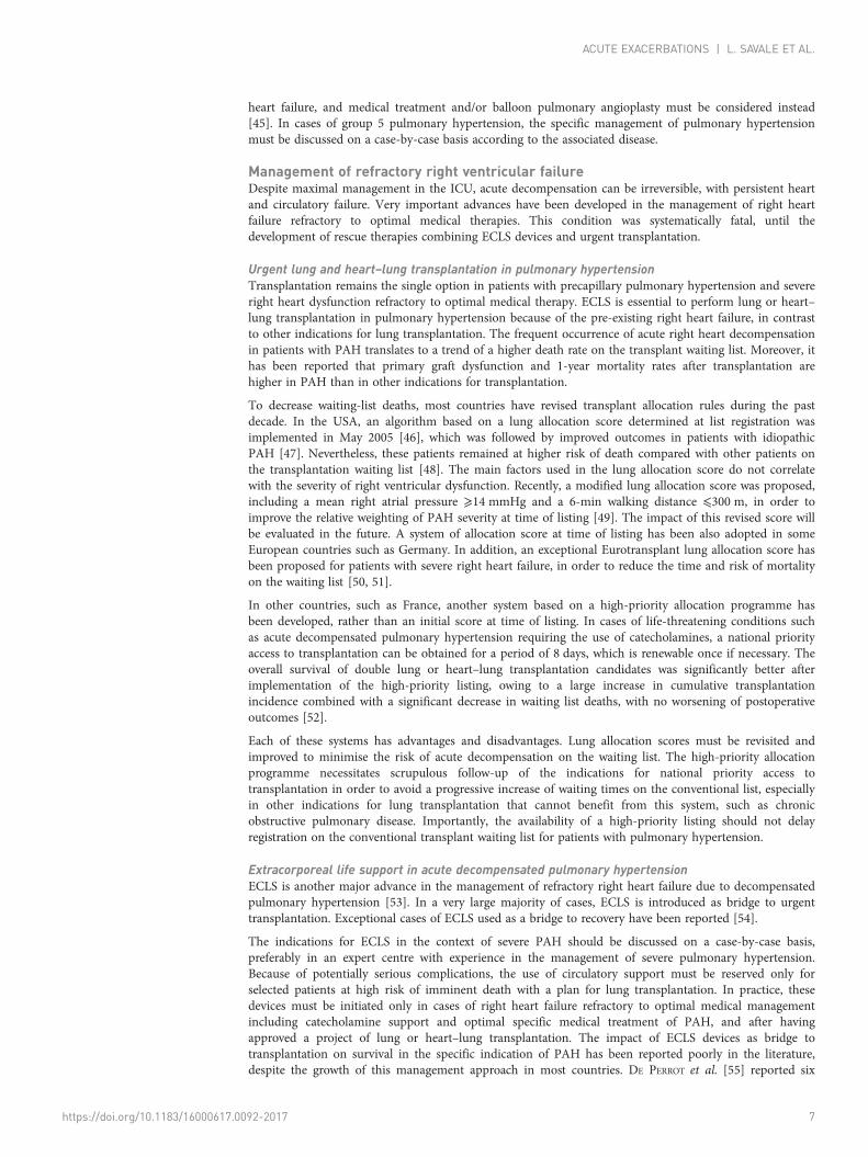
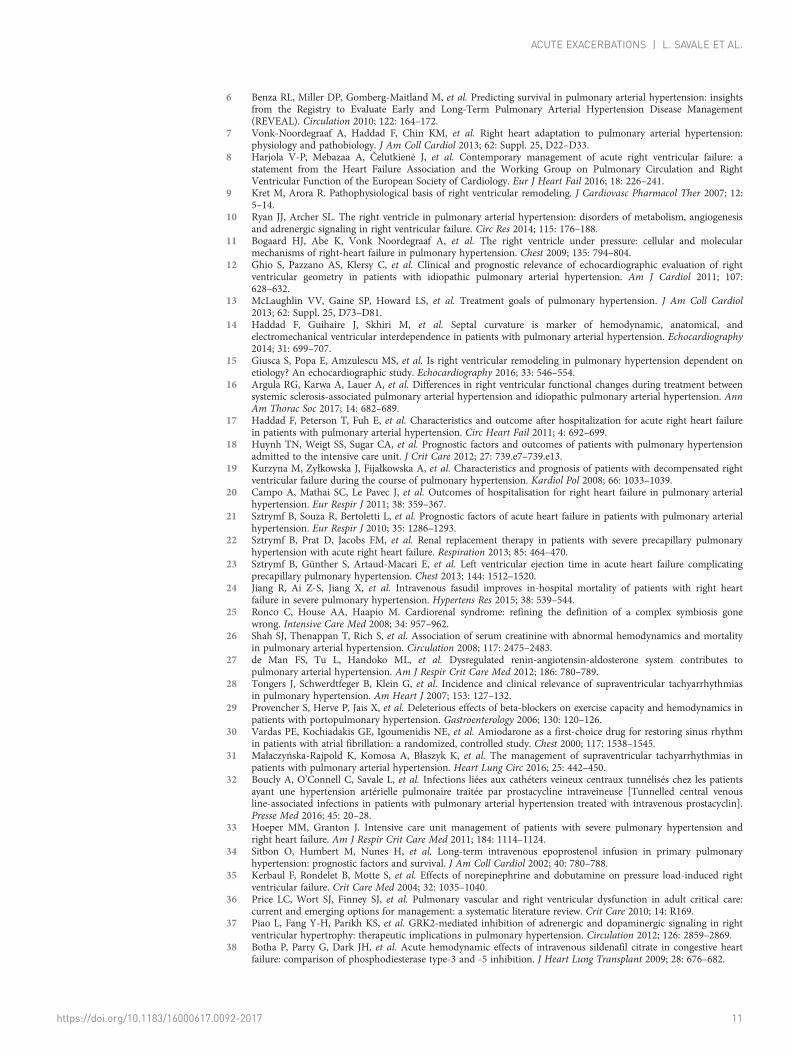
Monitoring of acute right ventricular failure in pulmonary hypertensionOne of the key points in the management of patients with acute decompensated pulmonary hypertensionis close monitoring, which is necessary to better assess the haemodynamic situation, evaluate the responseto therapy and initiate the decision to activate urgent transplantation and/or ECLS in cases of refractoryright ventricular failure (figure 3). In all cases, monitoring the consequences of cardiac failure appears tobe important in order to balance metabolic demand and haemodynamic impairment. However, specificstudies on this topic in the management of severe pulmonary hypertension in the ICU are lacking.
Clinical monitoringThe clinical presentation of a patient in acute decompensated pulmonary hypertension can often be falselyreassuring at rest. Although systemic arterial pressure at admission appeared to be a prognostic factor [21],a majority of patients are able to maintain a normal level of systemic vascular resistance for a long time. Incontrast, a fall in systemic vascular resistance under diuretic and/or inotropic support treatment appears tobe a determining prognostic factor. The necessity of introducing norepinephrine to maintain the systemicpressure is clearly associated with a worse prognosis. Similarly, clinical signs of low cardiac output, such asencephalopathy, restlessness, digestive intolerance, diffuse pain or recurrent syncope frequently appear verylate in the end-stage period and should prompt the discussion of the use of circulatory assistance if theydo not remit promptly with other medical treatments. In addition, monitoring diuresis and weight in theICU in order to assess daily water balance are essential to appreciate the evolution of overload andresponse to diuretic treatment with or without catecholamine support if necessary.
BiomarkersThe prognostic value of biomarkers such as brain natriuretic peptide or troponin has been demonstratedin several studies [19–21]. Most studies have reported the prognostic impact of these biomarkers atadmission to intensive care. It is very likely that the evolution of these parameters under treatment could
https://doi.org/10.1183/16000617.0092-2017 8
ACUTE EXACERBATIONS | L. SAVALE ET AL.
be more informative, and therefore needs to be evaluated prospectively. Plasma levels of C-reactive protein(CRP) are useful to screen for an infectious process in patients with acute decompensated pulmonaryhypertension [21]. However, moderate elevation of CRP associated with fever can be observed in theabsence of acute infection, possibly reflecting a systemic inflammatory response syndrome due to acuteright cardiac decompensation. Although few studies have assessed sodium and water regulation imbalancein right heart failure, it has been documented that hyponatraemia is linked to survival in stable PAHpatients [26, 60]. In addition, a link between hyponatraemia and survival has been demonstrated in acutedecompensated pulmonary hypertension [21].
Other biomarkers are used to detect and assess peripheral organ involvement due to the combination ofdecreased cardiac output and congestion. Indeed, creatinine level appears to be a major prognostic factorin patients with acute decompensated pulmonary hypertension. Transaminase and bilirubin levels areuseful to detect acute congestive hepatopathy. Finally, most of these biomarkers reflect advanced rightventricular failure, right ventricular ischaemia or multiorgan failure. Future biological and clinical studiesare needed to research and develop more informative and better-performing biomarkers in order topredict outcomes of patients with end-stage disease earlier and more precisely.
Haemodynamic monitoringInvasive haemodynamic monitoringAlthough the use of right haemodynamic monitoring by right cardiac catheterisation has been questionedin the management of shock in intensive care, it remains the most effective tool in the evaluation of rightventricular preload, right ventricle afterload and cardiac function in pulmonary hypertension. Right atrialpressure and cardiac output are the main altered parameters in acute decompensation, and have beendemonstrated to be major prognostic factors in PAH [5]. However, invasive haemodynamic monitoring inthe ICU is not widely used in routine practice, due to a risk of infection and arrhythmia, which may beconsidered too great in these fragile patients. Ultimately, right heart catheterisation is considered in severeand complex cases after assessing the benefit-to-risk ratio.
In patients with a central venous line, it can be useful to monitor the central venous pressure in order toassess evolution of right ventricular preload and improve management of fluid balance with diuretics. Inaddition, measurement of central venous oxygen saturation is recommended to appreciate tissue oxygenation.This measure is inversely correlated with cardiac function in most cases; however, it must be interpretedwith caution because of many other potential factors that can influence it, such as sepsis or anaemia.
Transthoracic echocardiographyEchocardiography is an interesting alternative to right cardiac catheterisation. Many echocardiographicparameters are useful to monitor right ventricular preload, right and left ventricular function and right
Clinical signs
∙ Arterial pressure
∙ Diuresis
∙ Weight
∙ Arterial oxygen saturation
∙ Temperature
∙ Respiratory rate
Biomarkers
∙ BNP/pro-BNP
∙ Troponin
∙ Lactate
∙ Glomerular filtration rate
∙ Natraemia
∙ Transaminases/bilirubin
∙ Right atrial pressure
∙ Cardiac index
∙ Central venous oxygen
saturation
Right cardiac
catheterisation
EchocardiographyNovel haemodynamic
monitoring techniques? ∙ Pericardial effusion
∙ Inferior vena cava size
∙ Tricuspid regurgitation severity
∙ Parameters of RV function (TAPSE, Tei index)
∙ Parameters of LV function
∙ Right atrial size∙ Noninvasive continuous arterial
pulse contour analysis
∙ Calibrated arterial pulse analysis with
transpulmonary thermodilution
FIGURE 3 Monitoring tools in acute decompensated pulmonary hypertension. BNP: brain natriuretic peptide;RV: right ventricle; TAPSE: tricuspid annular plane systolic excursion; LV: left ventricle.
https://doi.org/10.1183/16000617.0092-2017 9
ACUTE EXACERBATIONS | L. SAVALE ET AL.
ventricular afterload. Some variables evaluating right ventricular contractile function and right atrialfunction have shown to be prognostic factors in PAH in the stable clinical scenario [61–63]. Nevertheless,precise assessment of right ventricular function remains a challenge and very few data support itsreliability in the setting of acute right heart failure. In the study published by HADDAD et al. [17], tricuspidregurgitation severity was the only parameter associated with a worse prognosis in this population.Additional studies are necessary to evaluate precisely the accuracy of each echocardiographic parameter inthis specific setting and their impact on prognosis.
Novel haemodynamic monitoring techniques in PAHOver the past decade, haemodynamic monitoring has evolved considerably in the ICU. Novel noninvasiveor minimally invasive techniques have been developed in order to reduce adverse events and providecontinuous cardiac output and fluid responsiveness variables in real time [64]. However, these techniqueshave been studied primarily in sedated patients under mechanical ventilation. In contrast, very few dataare available on the performance of these haemodynamic monitoring tools in awake patients hospitalisedin an ICU for acute decompensated pulmonary hypertension.
Novel devices that use transpulmonary thermodilution-based methods to determine cardiac output havegenerated great interest in the general ICU population. This technology provides information regardingnumerous physiological variables including systemic arterial pressure, cardiac output or stroke volume.This method has never been formally validated in pulmonary hypertension patients to date, and rightventricular dilatation and tricuspid regurgitation are supposed to be major limitations in the reliability ofthe device. Nevertheless, preliminary data supports its reliability in cardiac output estimation [65].
Fully noninvasive techniques providing cardiac output estimation have been introduced recently. Manynew systems have been developed to monitor the cardiac output indirectly, with a continuous analysis ofthe arterial pressure waveform [66]. These techniques, combined with right ventricular preload estimationwith central venous pressure, could be an interesting option in patients with acute decompensatedpulmonary hypertension, but remain to be prospectively evaluated.
On the basis of cardiac magnetic resonance studies showing reductions in time to peak of shortening ofthe left ventricular free wall and time to aortic valve closure [67], our group tested the hypothesis that leftventricular ejection time (LVET) could be reduced as a result of the right-to-left dysynchrony. We usedapplanation tonometry, a simple, quick, painless and safe noninvasive method for estimating central aorticpressure from a peripheral recording of pulse waves at the radial or carotid artery level. We demonstratedthat LVET was associated with cardiac index in stable pulmonary hypertension patients [68]. Furthermore,in a prospective study including pulmonary hypertension patients admitted to the ICU for right heartfailure, our group showed that shortening of LVET was associated with a worse outcome [23].
ConclusionThe occurrence of acute right heart failure in patients with precapillary pulmonary hypertension is themost frequent cause of death in this population. Because pulmonary hypertension remains a progressiveand fatal disease due to irreversible pulmonary vascular remodelling, right heart remodelling and failuremust be considered important targets for future therapies. Over the past decade, the development of ECLSin refractory right heart failure combined with urgent transplantation has probably contributed to asignificant improvement in survival for selected patients. However, there remains a considerable need forfurther research in this field. Specific clinical studies are needed to improve right heart failureunderstanding and management in the ICU. Pathophysiological mechanisms of right ventricularremodelling must be better understood in order to develop future targeted therapies. Finally, thedevelopment of new ECLS techniques as a long-term treatment option for right heart failure, and not onlyas bridge to transplantation, must be considered a very interesting challenge for the future.
References1 Humbert M, Sitbon O, Simonneau G. Treatment of pulmonary arterial hypertension. N Engl J Med 2004; 351:
1425–1436.2 Galiè N, Humbert M, Vachiery J-L, et al. 2015 ESC/ERS Guidelines for the diagnosis and treatment of pulmonary
hypertension: The Joint Task Force for the Diagnosis and Treatment of Pulmonary Hypertension of the EuropeanSociety of Cardiology (ESC) and the European Respiratory Society (ERS). Eur Heart J 2016; 37: 67–119.
3 Galiè N, Humbert M, Vachiery J-L, et al. 2015 ESC/ERS Guidelines for the diagnosis and treatment of pulmonaryhypertension. The Joint Task Force for the Diagnosis and Treatment of Pulmonary Hypertension of the EuropeanSociety of Cardiology (ESC) and the European Respiratory Society (ERS). Eur Respir J 2015; 46: 903–975.
4 Humbert M, Lau EMT, Montani D, et al. Advances in therapeutic interventions for patients with pulmonaryarterial hypertension. Circulation 2014; 130: 2189–2208.
5 Humbert M, Sitbon O, Chaouat A, et al. Survival in patients with idiopathic, familial, and anorexigen-associatedpulmonary arterial hypertension in the modern management era. Circulation 2010; 122: 156–163.
https://doi.org/10.1183/16000617.0092-2017 10
ACUTE EXACERBATIONS | L. SAVALE ET AL.
6 Benza RL, Miller DP, Gomberg-Maitland M, et al. Predicting survival in pulmonary arterial hypertension: insightsfrom the Registry to Evaluate Early and Long-Term Pulmonary Arterial Hypertension Disease Management(REVEAL). Circulation 2010; 122: 164–172.
7 Vonk-Noordegraaf A, Haddad F, Chin KM, et al. Right heart adaptation to pulmonary arterial hypertension:physiology and pathobiology. J Am Coll Cardiol 2013; 62: Suppl. 25, D22–D33.
8 Harjola V-P, Mebazaa A, Čelutkienė J, et al. Contemporary management of acute right ventricular failure: astatement from the Heart Failure Association and the Working Group on Pulmonary Circulation and RightVentricular Function of the European Society of Cardiology. Eur J Heart Fail 2016; 18: 226–241.
9 Kret M, Arora R. Pathophysiological basis of right ventricular remodeling. J Cardiovasc Pharmacol Ther 2007; 12:5–14.
10 Ryan JJ, Archer SL. The right ventricle in pulmonary arterial hypertension: disorders of metabolism, angiogenesisand adrenergic signaling in right ventricular failure. Circ Res 2014; 115: 176–188.
11 Bogaard HJ, Abe K, Vonk Noordegraaf A, et al. The right ventricle under pressure: cellular and molecularmechanisms of right-heart failure in pulmonary hypertension. Chest 2009; 135: 794–804.
12 Ghio S, Pazzano AS, Klersy C, et al. Clinical and prognostic relevance of echocardiographic evaluation of rightventricular geometry in patients with idiopathic pulmonary arterial hypertension. Am J Cardiol 2011; 107:628–632.
13 McLaughlin VV, Gaine SP, Howard LS, et al. Treatment goals of pulmonary hypertension. J Am Coll Cardiol2013; 62: Suppl. 25, D73–D81.
14 Haddad F, Guihaire J, Skhiri M, et al. Septal curvature is marker of hemodynamic, anatomical, andelectromechanical ventricular interdependence in patients with pulmonary arterial hypertension. Echocardiography2014; 31: 699–707.
15 Giusca S, Popa E, Amzulescu MS, et al. Is right ventricular remodeling in pulmonary hypertension dependent onetiology? An echocardiographic study. Echocardiography 2016; 33: 546–554.
16 Argula RG, Karwa A, Lauer A, et al. Differences in right ventricular functional changes during treatment betweensystemic sclerosis-associated pulmonary arterial hypertension and idiopathic pulmonary arterial hypertension. AnnAm Thorac Soc 2017; 14: 682–689.
17 Haddad F, Peterson T, Fuh E, et al. Characteristics and outcome after hospitalization for acute right heart failurein patients with pulmonary arterial hypertension. Circ Heart Fail 2011; 4: 692–699.
18 Huynh TN, Weigt SS, Sugar CA, et al. Prognostic factors and outcomes of patients with pulmonary hypertensionadmitted to the intensive care unit. J Crit Care 2012; 27: 739.e7–739.e13.
19 Kurzyna M, Zyłkowska J, Fijałkowska A, et al. Characteristics and prognosis of patients with decompensated rightventricular failure during the course of pulmonary hypertension. Kardiol Pol 2008; 66: 1033–1039.
20 Campo A, Mathai SC, Le Pavec J, et al. Outcomes of hospitalisation for right heart failure in pulmonary arterialhypertension. Eur Respir J 2011; 38: 359–367.
21 Sztrymf B, Souza R, Bertoletti L, et al. Prognostic factors of acute heart failure in patients with pulmonary arterialhypertension. Eur Respir J 2010; 35: 1286–1293.
22 Sztrymf B, Prat D, Jacobs FM, et al. Renal replacement therapy in patients with severe precapillary pulmonaryhypertension with acute right heart failure. Respiration 2013; 85: 464–470.
23 Sztrymf B, Günther S, Artaud-Macari E, et al. Left ventricular ejection time in acute heart failure complicatingprecapillary pulmonary hypertension. Chest 2013; 144: 1512–1520.
24 Jiang R, Ai Z-S, Jiang X, et al. Intravenous fasudil improves in-hospital mortality of patients with right heartfailure in severe pulmonary hypertension. Hypertens Res 2015; 38: 539–544.
25 Ronco C, House AA, Haapio M. Cardiorenal syndrome: refining the definition of a complex symbiosis gonewrong. Intensive Care Med 2008; 34: 957–962.
26 Shah SJ, Thenappan T, Rich S, et al. Association of serum creatinine with abnormal hemodynamics and mortalityin pulmonary arterial hypertension. Circulation 2008; 117: 2475–2483.
27 de Man FS, Tu L, Handoko ML, et al. Dysregulated renin-angiotensin-aldosterone system contributes topulmonary arterial hypertension. Am J Respir Crit Care Med 2012; 186: 780–789.
28 Tongers J, Schwerdtfeger B, Klein G, et al. Incidence and clinical relevance of supraventricular tachyarrhythmiasin pulmonary hypertension. Am Heart J 2007; 153: 127–132.
29 Provencher S, Herve P, Jais X, et al. Deleterious effects of beta-blockers on exercise capacity and hemodynamics inpatients with portopulmonary hypertension. Gastroenterology 2006; 130: 120–126.
30 Vardas PE, Kochiadakis GE, Igoumenidis NE, et al. Amiodarone as a first-choice drug for restoring sinus rhythmin patients with atrial fibrillation: a randomized, controlled study. Chest 2000; 117: 1538–1545.
31 Małaczyńska-Rajpold K, Komosa A, Błaszyk K, et al. The management of supraventricular tachyarrhythmias inpatients with pulmonary arterial hypertension. Heart Lung Circ 2016; 25: 442–450.
32 Boucly A, O’Connell C, Savale L, et al. Infections liées aux cathéters veineux centraux tunnélisés chez les patientsayant une hypertension artérielle pulmonaire traitée par prostacycline intraveineuse [Tunnelled central venousline-associated infections in patients with pulmonary arterial hypertension treated with intravenous prostacyclin].Presse Med 2016; 45: 20–28.
33 Hoeper MM, Granton J. Intensive care unit management of patients with severe pulmonary hypertension andright heart failure. Am J Respir Crit Care Med 2011; 184: 1114–1124.
34 Sitbon O, Humbert M, Nunes H, et al. Long-term intravenous epoprostenol infusion in primary pulmonaryhypertension: prognostic factors and survival. J Am Coll Cardiol 2002; 40: 780–788.
35 Kerbaul F, Rondelet B, Motte S, et al. Effects of norepinephrine and dobutamine on pressure load-induced rightventricular failure. Crit Care Med 2004; 32: 1035–1040.
36 Price LC, Wort SJ, Finney SJ, et al. Pulmonary vascular and right ventricular dysfunction in adult critical care:current and emerging options for management: a systematic literature review. Crit Care 2010; 14: R169.
37 Piao L, Fang Y-H, Parikh KS, et al. GRK2-mediated inhibition of adrenergic and dopaminergic signaling in rightventricular hypertrophy: therapeutic implications in pulmonary hypertension. Circulation 2012; 126: 2859–2869.
38 Botha P, Parry G, Dark JH, et al. Acute hemodynamic effects of intravenous sildenafil citrate in congestive heartfailure: comparison of phosphodiesterase type-3 and -5 inhibition. J Heart Lung Transplant 2009; 28: 676–682.
https://doi.org/10.1183/16000617.0092-2017 11
ACUTE EXACERBATIONS | L. SAVALE ET AL.
39 Vildbrad MD, Andersen A, Holmboe S, et al. Acute effects of levosimendan in experimental models of rightventricular hypertrophy and failure. Pulm Circ 2014; 4: 511–519.
40 Kerbaul F, Rondelet B, Demester J-P, et al. Effects of levosimendan versus dobutamine on pressure load-inducedright ventricular failure. Crit Care Med 2006; 34: 2814–2819.
41 Sitbon O, Jaïs X, Savale L, et al. Upfront triple combination therapy in pulmonary arterial hypertension: a pilotstudy. Eur Respir J 2014; 43: 1691–1697.
42 Jais X, Launay D, Yaici A, et al. Immunosuppressive therapy in lupus- and mixed connective tissuedisease-associated pulmonary arterial hypertension: a retrospective analysis of twenty-three cases. Arthritis Rheum2008; 58: 521–531.
43 Chen Y, Guo L, Li Y, et al. Severe pulmonary arterial hypertension secondary to lupus in the emergencydepartment: proactive intense care associated with a better short-term survival. Int J Rheum Dis 2015; 18: 331–335.
44 Montani D, Lau EM, Dorfmüller P, et al. Pulmonary veno-occlusive disease. Eur Respir J 2016; 47: 1518–1534.45 Kim NH, Delcroix M, Jenkins DP, et al. Chronic thromboembolic pulmonary hypertension. J Am Coll Cardiol
2013; 62: Suppl. 25, D92–D99.46 Davis SQ, Garrity ER Jr. Organ allocation in lung transplant. Chest 2007; 132: 1646–1651.47 Schaffer JM, Singh SK, Joyce DL, et al. Transplantation for idiopathic pulmonary arterial hypertension:
improvement in the lung allocation score era. Circulation 2013; 127: 2503–2513.48 Chen H, Shiboski SC, Golden JA, et al. Impact of the lung allocation score on lung transplantation for pulmonary
arterial hypertension. Am J Respir Crit Care Med 2009; 180: 468–474.49 Benza RL, Gomberg-Maitland M, Miller DP, et al. The REVEAL Registry risk score calculator in patients newly
diagnosed with pulmonary arterial hypertension. Chest 2012; 141: 354–362.50 Gottlieb J, Greer M, Sommerwerck U, et al. Introduction of the lung allocation score in Germany. Am J
Transplant 2014; 14: 1318–1327.51 Gottlieb J, Smits J, Schramm R, et al. Lung transplantation in Germany since the introduction of the lung
allocation score. Dtsch Arztebl Int 2017; 114: 179–185.52 Savale L, Le Pavec J, Mercier O, et al. Impact of high-priority allocation on lung and heart-lung transplantation
for pulmonary hypertension. Ann Thorac Surg 2017; 104: 404–411.53 Pereszlenyi A, Lang G, Steltzer H, et al. Bilateral lung transplantation with intra- and postoperatively prolonged
ECMO support in patients with pulmonary hypertension. Eur J Cardiothorac Surg 2002; 21: 858–863.54 Javidfar J, Brodie D, Iribarne A, et al. Extracorporeal membrane oxygenation as a bridge to lung transplantation
and recovery. J Thorac Cardiovasc Surg 2012; 144: 716–721.55 de Perrot M, Granton JT, McRae K, et al. Impact of extracorporeal life support on outcome in patients with
idiopathic pulmonary arterial hypertension awaiting lung transplantation. J Heart Lung Transplant 2011; 30:997–1002.
56 Strueber M, Hoeper MM, Fischer S, et al. Bridge to thoracic organ transplantation in patients with pulmonaryarterial hypertension using a pumpless lung assist device. Am J Transplant 2009; 9: 853–857.
57 Boulate D, Mercier O, Mussot S, et al. Extracorporeal life support after pulmonary endarterectomy as a bridge torecovery or transplantation: lessons from 31 consecutive patients. Ann Thorac Surg 2016; 102: 260–268.
58 Kortchinsky T, Mussot S, Rezaiguia S, et al. Extracorporeal life support in lung and heart-lung transplantation forpulmonary hypertension in adults. Clin Transplant 2016; 30: 1152–1158.
59 Tudorache I, Sommer W, Kühn C, et al. Lung transplantation for severe pulmonary hypertension – awakeextracorporeal membrane oxygenation for postoperative left ventricular remodelling. Transplantation 2015; 99:451–458.
60 Forfia PR, Mathai SC, Fisher MR, et al. Hyponatremia predicts right heart failure and poor survival in pulmonaryarterial hypertension. Am J Respir Crit Care Med 2008; 177: 1364–1369.
61 Forfia PR, Fisher MR, Mathai SC, et al. Tricuspid annular displacement predicts survival in pulmonaryhypertension. Am J Respir Crit Care Med 2006; 174: 1034–1041.
62 Raymond RJ, Hinderliter AL, Willis PW, et al. Echocardiographic predictors of adverse outcomes in primarypulmonary hypertension. J Am Coll Cardiol 2002; 39: 1214–1219.
63 Yeo TC, Dujardin KS, Tei C, et al. Value of a Doppler-derived index combining systolic and diastolic timeintervals in predicting outcome in primary pulmonary hypertension. Am J Cardiol 1998; 81: 1157–1161.
64 Teboul J-L, Saugel B, Cecconi M, et al. Less invasive hemodynamic monitoring in critically ill patients. IntensiveCare Med 2016; 42: 1350–1359.
65 Artaud-Macari E, Gilbert M, Persichini R, et al. Accuracy of transpulmonary thermodilution cardiac outputmeasurement (PiCCO) in patients with right heart failure (RHF) due to precapillary pulmonary hypertension(PH). Eur Respir J 2013; 42: Suppl. 57, P3402.
66 Marik PE. Noninvasive cardiac output monitors: a state-of the-art review. J Cardiothorac Vasc Anesth 2013; 27:121–134.
67 Marcus JT, Gan CT-J, Zwanenburg JJM, et al. Interventricular mechanical asynchrony in pulmonary arterialhypertension: left-to-right delay in peak shortening is related to right ventricular overload and left ventricularunderfilling. J Am Coll Cardiol 2008; 51: 750–757.
68 Günther S, Sztrymf B, Savale L, et al. Relation between left ventricular ejection time and pulmonaryhemodynamics in pulmonary hypertension. Int J Cardiol 2015; 184: 763–765.
https://doi.org/10.1183/16000617.0092-2017 12
ACUTE EXACERBATIONS | L. SAVALE ET AL.