Embed Size (px)
Citation preview
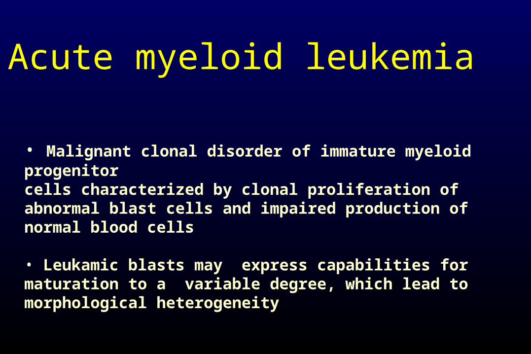
Acute myeloid leukemia
• Malignant clonal disorder of immature myeloid progenitor cells characterized by clonal proliferation of abnormal blast cells and impaired production of normal blood cells • Leukamic blasts may express capabilities for maturation to a variable degree, which lead to morphological heterogeneity

Acute leukemias
•Adults: - acute myeloid leukemia (AML) 80%- acute lymphoblastic leukemia (ALL) 20%
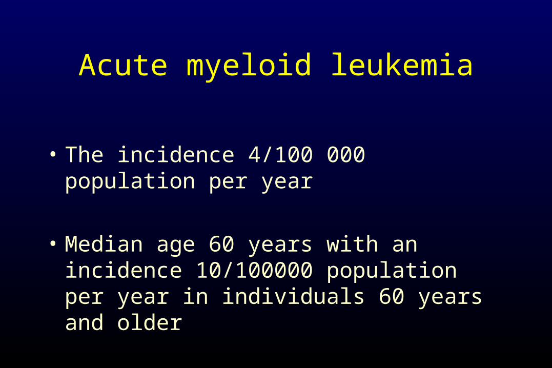
Acute myeloid leukemia
• The incidence 4/100 000 population per year
• Median age 60 years with an incidence 10/100000 population per year in individuals 60 years and older
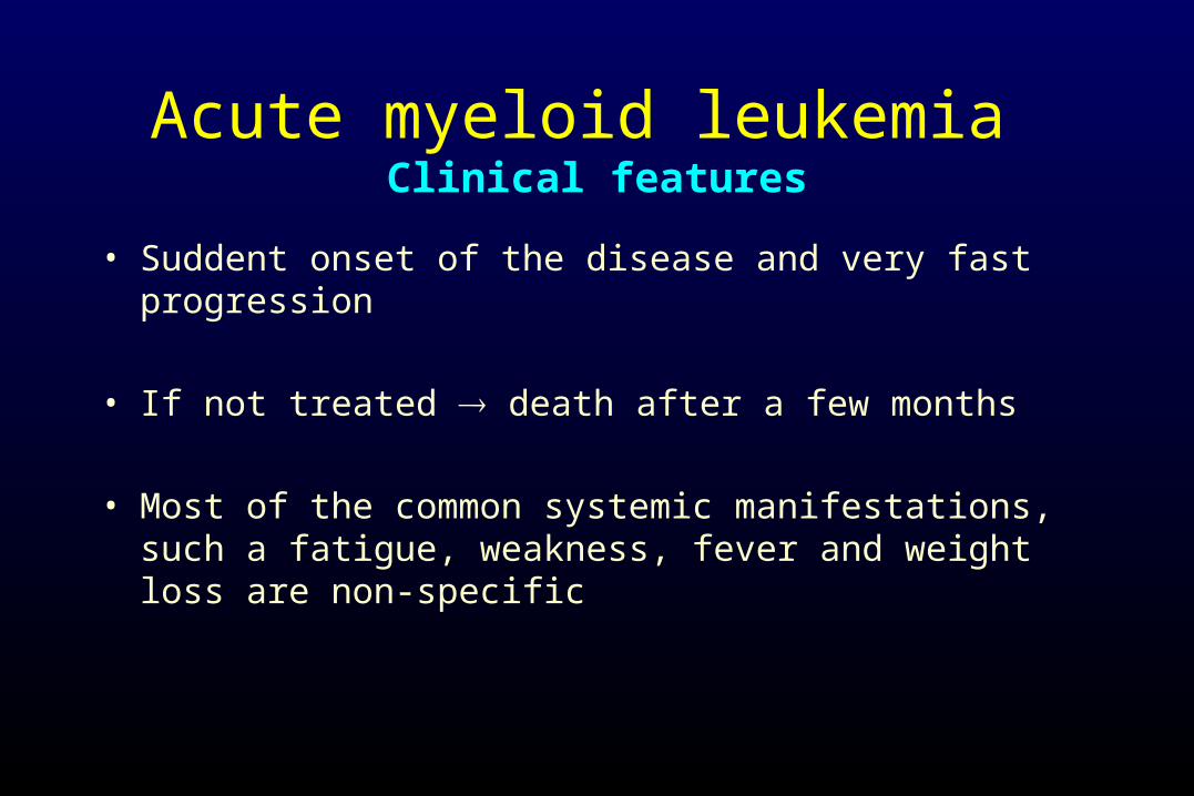
Acute myeloid leukemia Clinical features
• Suddent onset of the disease and very fast progression
• If not treated death after a few months
• Most of the common systemic manifestations, such a fatigue, weakness, fever and weight loss are non-specific
Acute myeloid leukemia Clinical features

• The prevalence and degree of organ infiltration vary somewhat with the different types of leukemia
– abdominal fullness (enlargement of the liver and spleen)
– bone and join pain and tenderness
– gum hypertrophy (AML-M4 and M5)
– neurological symptoms: headache, nausea, vomiting, blurred vision, cranial nerve dysfunction (AML-M4 and M5)
– DIC (AML-M3)
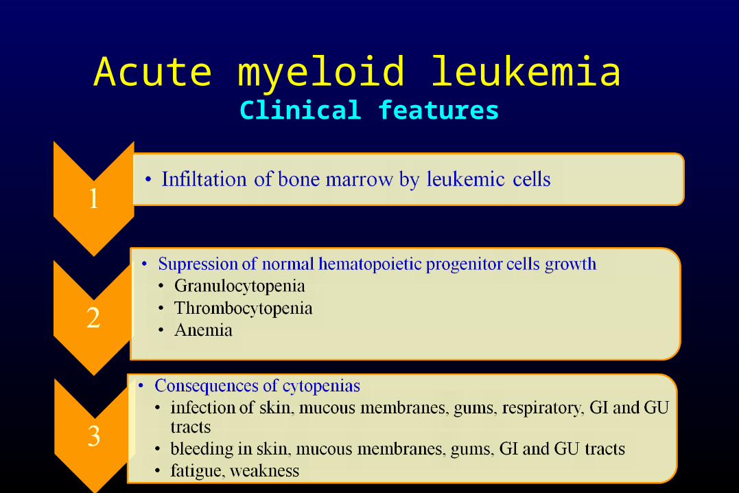
Acute myeloid leukemia Clinical features
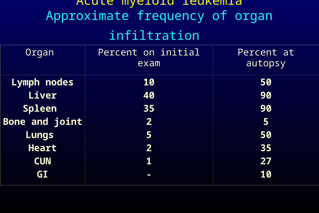
Acute myeloid leukemia
Approximate frequency of organ infiltration Organ Percent on initial exam Percent at autopsy
Lymph nodes
Liver
Spleen
Bone and joint
Lungs
Heart
CUN
GI
10
40
35
2
5
2
1
-
50
90
90
5
50
35
27
10
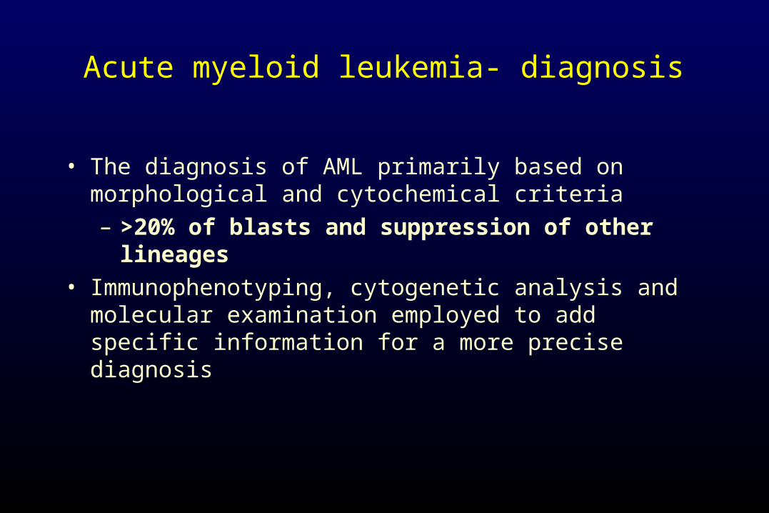
Acute myeloid leukemia- diagnosis
• The diagnosis of AML primarily based on morphological and cytochemical criteria
– >20% of blasts and suppression of other lineages
• Immunophenotyping, cytogenetic analysis and molecular examination employed to add specific information for a more precise diagnosis
Cytological criteria for the diagnosis of acute myeloid leukaemia:
French-American-British (FAB) classification
• Eight morphologic subtypes (M0-M7) are distinguished according to FAB classification system based on the morphologic features of the blasts and histochemical staining
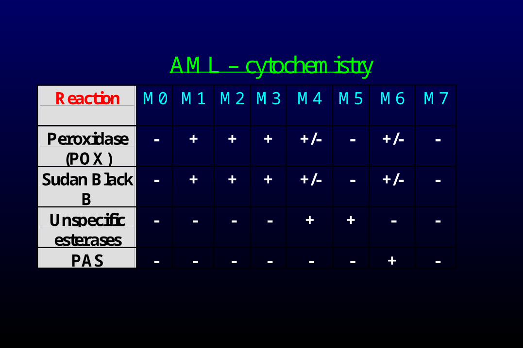
AML – cytochemistry
Reaction
M0
M1 M2 M3 M4 M5 M6 M7
Peroxidase (POX)
- + + + +/- - +/- -
Sudan Black B
- + + + +/- - +/- -
Unspecific esterases
- - - - + + - -
PAS - - - - - - + -
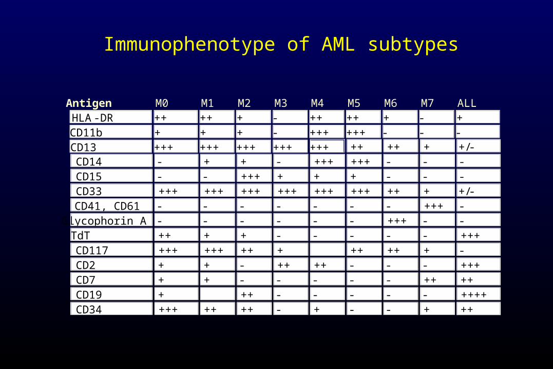
Immunophenotype of AML subtypes
Antigen M0 M1 M2 M3 M4 M5 M6 M7 ALL
HLA -DR ++ ++ + - ++ ++ + - +
CD11b + + + - +++ +++ - - -
CD13 +++ +++ +++ +++ +++ ++ ++ + +/-
CD14 - + + - +++ +++ - - -
CD15 - - +++ + + + - - -
CD33 +++ +++ +++ +++ +++ +++ ++ + +/-
CD41, CD61 - - - - - - - +++ -
Glycophorin A - - - - - - +++ - -
TdT ++ + + - - - - - +++
CD117 +++ +++ ++ + ++ ++ + -
CD2 + + - ++ ++ - - - +++
CD7 + + - - - - - ++ ++
CD19 + ++ - - - - - ++++
CD34 +++ ++ ++ - + - - + ++

AML M1 ³90% Blasts, Granulocytic component <10%. Monocytic component <10% SBB/MPO ³3%
AML M2 >30% Blasts Granulocytic component >10% , monocytic component < 20%MPO ³3%, CAE >10% , NSE < 20%
AML M3 Hypergranular PromyelocytesMultiple Auer rodsStrong positivity for MPO/SBB and CAE. NSE +/-
AML M3v Deeply notched nuclei. Fine dust granules. (Multiple) Auer rods +/-Cytochemical features like the hypergranular variant
AML M4 > 30% Blasts. Monocytic component > 20%, granulocytic component >20%MPO ³3%, CAE > 20%, NSE > 20%.A distinctive subtype, M4Eos- a variable increase of abnormal eosinophils with basophilic granules.
AML M5a >30% Blasts. Granulocytic component < 20%. Monocytic component ³80%. Monoblasts ³80% of monocytic component. MPO may be < 3%, NSE > 80% inhibited by fluoride
AML M5b ³30% Blasts. Granulocytic component < 20%. Monocytic component ³80%. Monoblasts < 80% of monocytic component. MPO may be <3%, NSE >80% inhibited by fluoride
AML M6 ³30% Blasts (nonerythroid population)³50% Erythroid precursors (total marrow cells) MPO³3% in blasts. PAS, Acid phosphatase and NSE may be positive in erithroblasts
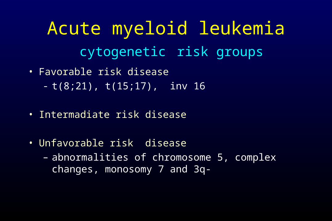
Acute myeloid leukemia cytogenetic risk groups
• Favorable risk disease
- t(8;21), t(15;17), inv 16
• Intermadiate risk disease
• Unfavorable risk disease
– abnormalities of chromosome 5, complex changes, monosomy 7 and 3q-
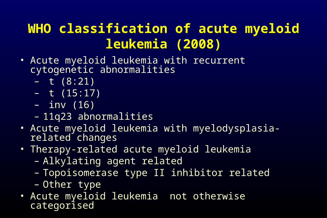
WHO classification of acute myeloid leukemia (2008)
• Acute myeloid leukemia with recurrent cytogenetic abnormalities– t (8:21)– t (15:17)– inv (16)– 11q23 abnormalities
• Acute myeloid leukemia with myelodysplasia-related changes• Therapy-related acute myeloid leukemia
– Alkylating agent related– Topoisomerase type II inhibitor related– Other type
• Acute myeloid leukemia not otherwise categorised
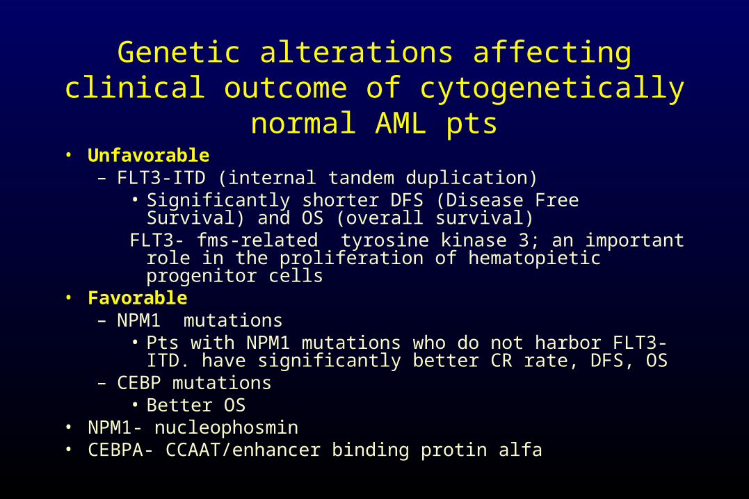
Genetic alterations affecting clinical outcome of cytogenetically normal AML pts
• Unfavorable– FLT3-ITD (internal tandem duplication)
• Significantly shorter DFS (Disease Free Survival) and OS (overall survival)
FLT3- fms-related tyrosine kinase 3; an important role in the proliferation of hematopietic progenitor cells
• Favorable– NPM1 mutations
• Pts with NPM1 mutations who do not harbor FLT3-ITD. have significantly better CR rate, DFS, OS
– CEBP mutations• Better OS
• NPM1- nucleophosmin• CEBPA- CCAAT/enhancer binding protin alfa
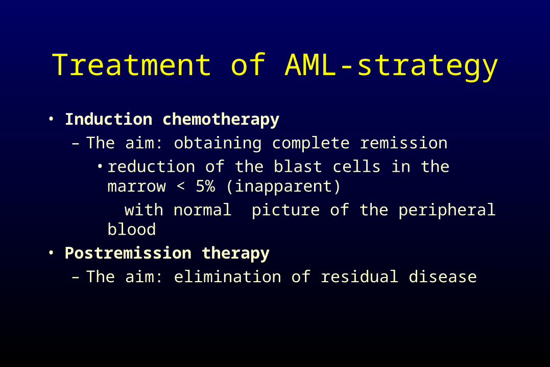
Treatment of AML-strategy
• Induction chemotherapy
– The aim: obtaining complete remission
• reduction of the blast cells in the marrow < 5% (inapparent)
with normal picture of the peripheral blood
• Postremission therapy
– The aim: elimination of residual disease

Induction chemotherapy
• Gold standard „3+7”– The anthracyclin drug for 3 days – Cytarabine for 7 days
• Complete remission- 60-70%
• Modification of standard chemotherapy– High doses of Ara-C– Purine analoges (fludarabina, 2-CDA)– 6-TG– etoposide

Postremission therapy
– Intensification of remission
• High-dose cytarabine based regimens with anthracycline drug
– Allogeneic HSCT
– Autologous HSCT
– Maintance chemotherapy
• Low-dose Ara-C, 6-TG, anthracycline drug
Acute myeloid leukemia
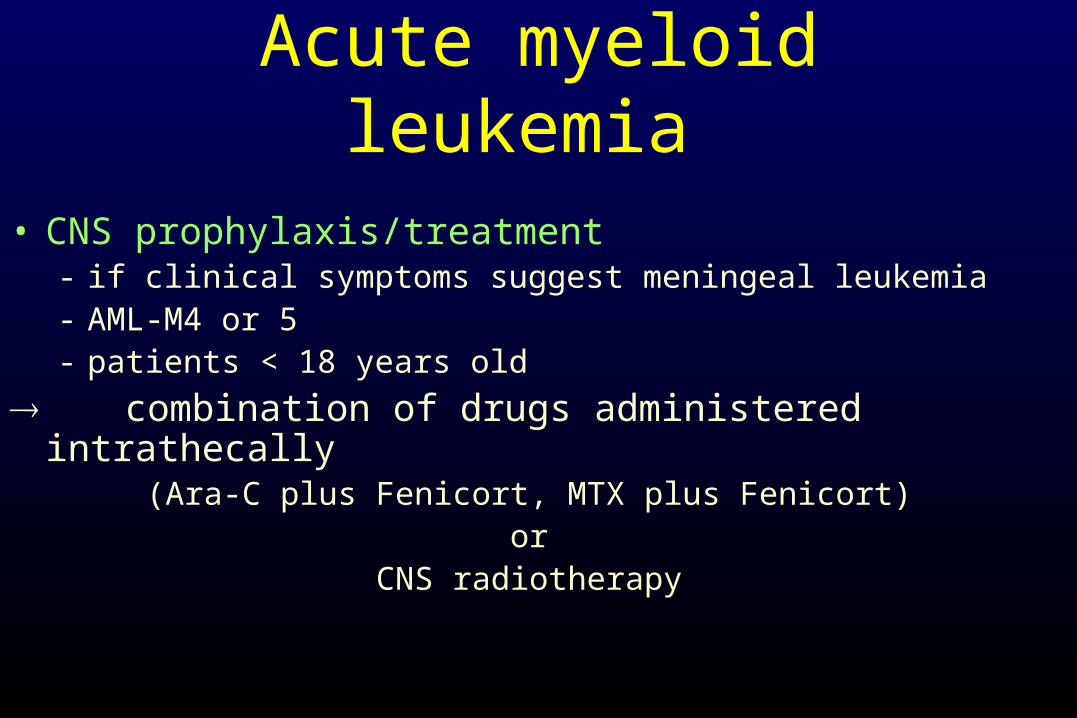
• CNS prophylaxis/treatment - if clinical symptoms suggest meningeal leukemia- AML-M4 or 5- patients < 18 years old
combination of drugs administered intrathecally (Ara-C plus Fenicort, MTX plus Fenicort)
or CNS radiotherapy
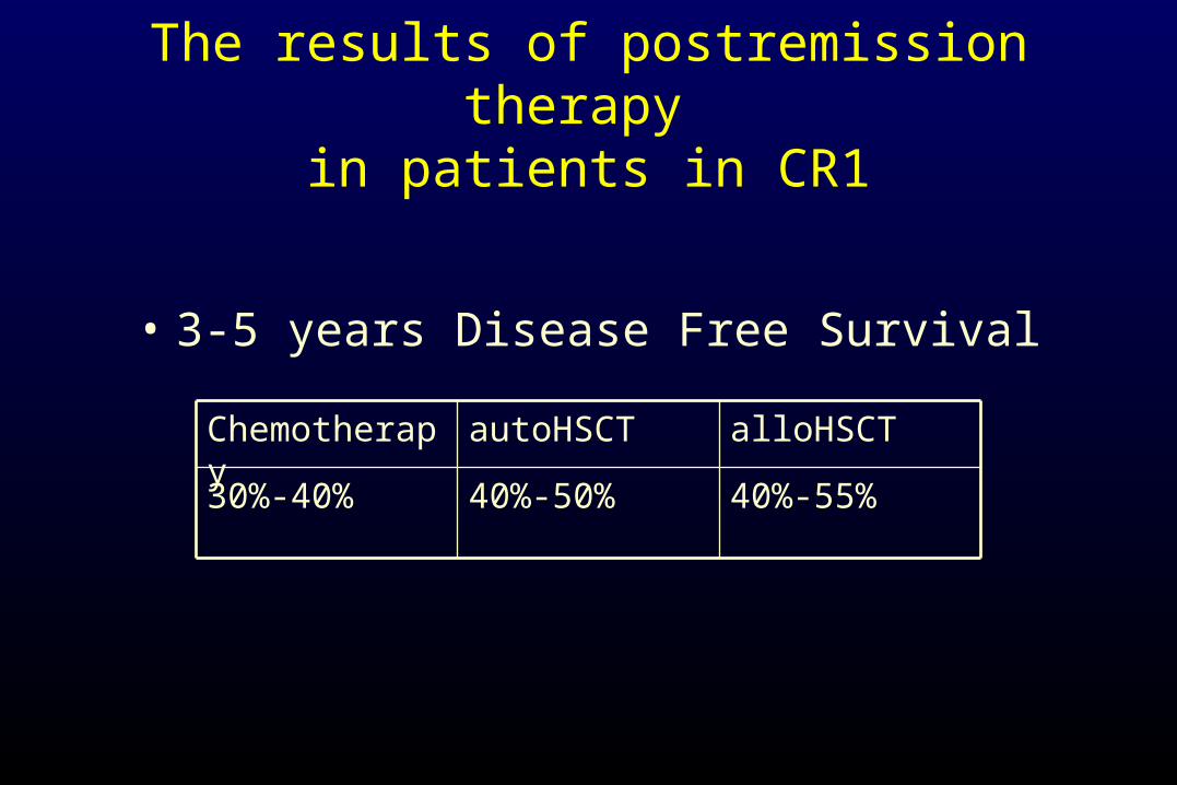
The results of postremission therapy in patients in CR1
• 3-5 years Disease Free Survival
40%-55%40%-50%30%-40%
alloHSCTautoHSCTChemotherapy
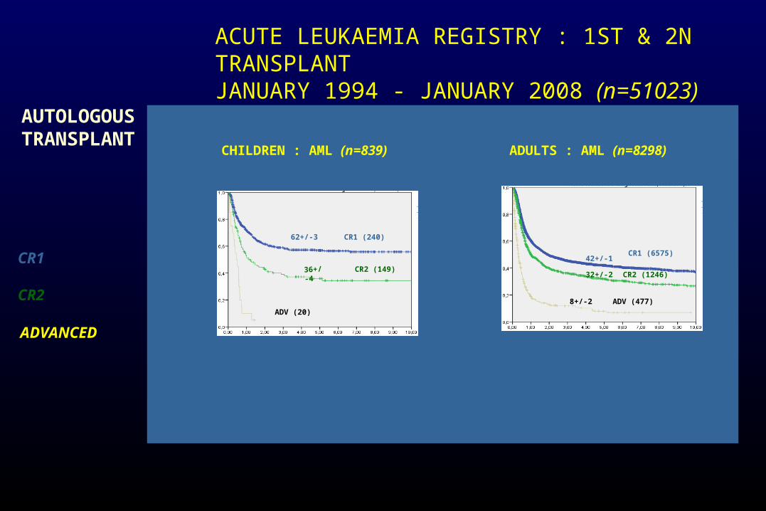
ACUTE LEUKAEMIA REGISTRY : 1ST & 2N TRANSPLANT JANUARY 1994 - JANUARY 2008 (n=51023)
CR1
CR2
ADVANCED
ADULTS : AML (n=8298)CHILDREN : AML (n=839)
AUTOLOGOUSTRANSPLANT
CR1 (670)57+/-2
CR2 (149)36+/-4
ADV (20)
CR1 (6575)42+/-1
CR2 (1246)32+/-2
ADV (477)8+/-2
CR1 (240)62+/-3
CR1 (1705)38+/-1

ACUTE LEUKAEMIA REGISTRY : 1ST & 2N TRANSPLANT JANUARY 1994 - JANUARY 2008 (n=51023)
CR1
CR2
ADVANCED
ADULTS : AML (n=10191)CHILDREN : AML (n=1146)
HLA IDENTICALTRANSPLANT
CR1 (789)61+/-2
CR2 (208)45+/-4
ADV (149)18+/-4
CR1 (6499)55+/-1
CR2 (1444)41+/-2
ADV (2248)15+/-1
CR1 (756)64+/-2
CR1 (2739)45+/-1
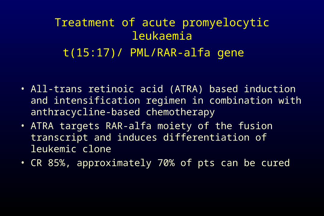
Treatment of acute promyelocytic leukaemia
t(15:17)/ PML/RAR-alfa gene
• All-trans retinoic acid (ATRA) based induction and intensification regimen in combination with anthracycline-based chemotherapy
• ATRA targets RAR-alfa moiety of the fusion transcript and induces differentiation of leukemic clone
• CR 85%, approximately 70% of pts can be cured


![· 1 Introduction Chronic Myeloid Leukemia (CML) is a hematological cancer characterized by the clonal expansion of myeloid cells in the bone marrow [Hoffbrand et al., 2001]. In](https://img.pdfslide.net/doc/110x75/60b53bf85804b54eba2e9afd/1-introduction-chronic-myeloid-leukemia-cml-is-a-hematological-cancer-characterized.jpg)