Embed Size (px)
Citation preview

Acute Renal Endothelial Injury DuringMarrow Recovery in a Cohort of Combined
Kidney and Bone Marrow AllograftsThe Harvard community has made this
article openly available. Please share howthis access benefits you. Your story matters
Citation Farris, A.B., D. Taheri, T. Kawai, L. Fazlollahi, W. Wong, N. Tolkoff-Rubin, T. R. Spitzer, et al. 2011. “Acute Renal Endothelial InjuryDuring Marrow Recovery in a Cohort of Combined Kidney and BoneMarrow Allografts.” American Journal of Transplantation 11, no. 7:1464–1477. doi:10.1111/j.1600-6143.2011.03572.x.
Published Version doi:10.1111/j.1600-6143.2011.03572.x
Citable link http://nrs.harvard.edu/urn-3:HUL.InstRepos:27526762
Terms of Use This article was downloaded from Harvard University’s DASHrepository, and is made available under the terms and conditionsapplicable to Other Posted Material, as set forth at http://nrs.harvard.edu/urn-3:HUL.InstRepos:dash.current.terms-of-use#LAA

Acute renal endothelial injury during marrow recovery in acohort of combined kidney and bone marrow allografts
AB Farris1,5,7, D Taheri1,7, T Kawai2,7, L Fazlollahi1,7, W. Wong3,7, N Tolkoff-Rubin3,7, TRSpitzer3,7, AJ Iafrate1,7, FI Preffer1,7, SA LoCascio4,6, B Sprangers6, S Saidman1,7, RNSmith1,7, AB Cosimi2,7, M Sykes4,6,7, DH Sachs4,7, and RB Colvin1,7
1 Pathology Service, Massachusetts General Hospital (MGH), Boston, Massachusetts, UnitedStates2 Transplantation Unit, MGH, Boston3 Medical Service, MGH, Boston4 Transplantation Biology Research Center, MGH, Boston5 Pathology Department and Laboratory Medicine, Emory University, Atlanta, Georgia, UnitedStates6 Department of Medicine, Surgery, and Microbiology & Immunology, Columbia Center forTranslational Immunology, Columbia University, New York City, New York, United States7 Harvard Medical School, Boston
AbstractAn idiopathic capillary leak syndrome (“engraftment syndrome”) often occurs in recipients ofhematopoietic cells, manifested clinically by transient azotemia and sometimes fever and fluidretention. Here we report the renal pathology in 10 recipients of combined bone marrow andkidney allografts. Nine developed graft dysfunction on day 10–16 and renal biopsies showedmarked acute tubular injury, with interstitial edema, hemorrhage and capillary congestion, withlittle or no interstitial infiltrate (≤10%) and marked glomerular and peritubular capillary (PTC)endothelial injury and loss by electron microscopy. Two had transient arterial endothelialinflammation; and 2 had C4d deposition. The cells in capillaries were primarily CD68+MPO+
mononuclear cells and CD3+CD8+ T cells, the latter with a high proliferative index (Ki67+). Bcells (CD20+) and CD4+ T cells were not detectable, and NK cells were rare. XY FISH showedthat CD45+ cells in PTCs were of recipient origin. Optimal treatment remains to be defined; tworecovered without additional therapy, six were treated with anti-rejection regimens. Except for onepatient, who later developed thrombotic microangiopathy and one with acute humoral rejection, allfully recovered within 2–4 weeks. Graft endothelium is the primary target of this process,attributable to as yet obscure mechanisms, arising during leukocyte recovery.
Please direct correspondence to: Alton B. Farris, MD, 1364 Clifton Road NE, Room H-188, Atlanta, GA 30322, [email protected],Phone: 601 573 - 3192, Fax: 404 - 727 - 3133.ABF and DT contributed equally as first authors of this paper during both data collection, analysis and manuscript preparation. TK,WW, N T-R, TRS, ABC, MS, DHS devised and supervised the clinical protocols and care of the patients in this study. LF and AJIperformed the fluorescence in situ hybridization (FISH) studies. FIP, SLC, BS, SS, and RNS provided laboratory services to thepatients in this study and made invaluable contributions to the assays performed. RBC supervised data collection, pathologyinterpretation and extensively edited the manuscript.Disclosures: The authors of this manuscript have no conflicts of interest to disclose as described by the American Journal ofTransplantation.
NIH Public AccessAuthor ManuscriptAm J Transplant. Author manuscript; available in PMC 2012 July 1.
Published in final edited form as:Am J Transplant. 2011 July ; 11(7): 1464–1477. doi:10.1111/j.1600-6143.2011.03572.x.
NIH
-PA Author Manuscript
NIH
-PA Author Manuscript
NIH
-PA Author Manuscript

Keywordsrenal transplant; bone marrow transplant; engraftment syndrome
IntroductionHLA-mismatched kidney transplantation without maintenance immunosuppression hasrecently been described in a cohort of 5 patients from our institution (1). This clinicalprotocol was based on non-human primate studies demonstrating that allograft kidneys canbe accepted following induction of transient mixed chimerism in which donor and recipienthematopoiesis coexist (2–9). During these studies, and in follow-up studies in five additionalpatients, transient acute kidney injury (AKI) accompanied by a capillary leak syndrome(CLS) beginning about the 10th post-transplant day was observed in nine patients, aphenomenon not observed in preclinical trials.
CLS or engraftment syndrome (ES) has been reported in patients receiving autologous stemcell or allogeneic bone marrow transplants (BMTs), a condition occurring during neutrophilrecovery, as described by Spitzer and colleagues (10, 11). Varied manifestations includefever, rash, pulmonary infiltrates, fluid retention, diarrhea, and hepatic injury and AKI (10–16). ES may be independent of graft versus host disease and a host-versus-graft reaction (10,11, 17, 18), but its pathogenesis is unknown. AKI in the first 3 months after allogeneic orautologous hematopoietic cell transplant has been attributed to drug toxicity, infection orischemia. However, the pathologic changes in kidney biopsies have not been extensivelystudied in hematopoietic cell transplantation recipients (19, 20). In particular, we wereunable to find reports of autologous renal pathology in association with ES.
Since the previous report of patients from our institution (1), five additional patients havebeen enrolled into a similar kidney/BMT regimen. All experienced a condition resemblingES. Here, we report comprehensive studies characterizing the pathologic processes in kidneybiopsies obtained during this syndrome
Materials and MethodsPatients
Ten allograft recipients in two sequential Immune Tolerance Network (ITN) sponsored,Institutional Review Board (IRB) approved trials underwent simultaneous kidney/BMTsfrom parent or sibling donors mismatched for one human leukocyte antigen (HLA)haplotype. The five patients in the first trial (1) received a conditioning regimen withcyclophosphamide (60 mg/kg on day -5 and -4), anti-CD2 monoclonal antibody (MEDI 507,MedImmune, Gaithersburg, MD), rituximab (patients 4–5) (Biogen Idec, Cambridge, MA,and Genentech, South San Francisco, CA), thymic irradiation and post transplantcyclosporine [previously described (1)]. The subsequent 5 recipients received more intensiverituximab treatment and tacrolimus instead of cyclosporine. Donor/recipient chimerism wasmonitored with flow cytometry.(1, 21)
Histologic assessmentKidney allograft tissue obtained by percutaneous core or open wedge biopsy was processedfor light (LM), immunofluorescence (IF), and electron microscopy (EM) by standardtechniques. C4d deposition was evaluated using a monoclonal antibody to C4d (clone 10–11, Biogenesis, Sandown, NH) (22, 23). The results were compared with kidney allograftsfrom recipients on conventional regimens at the same time post-transplant (10–12 days),
Farris et al. Page 2
Am J Transplant. Author manuscript; available in PMC 2012 July 1.
NIH
-PA Author Manuscript
NIH
-PA Author Manuscript
NIH
-PA Author Manuscript

with normal transplant function (n=6) or acute tubular injury (ATI) (n=5), and biopsies withacute cellular rejection, Banff type 2 (ACR2) (n=5), obtained from our biorepository.Immunohistochemistry (IHC) was performed using Ki-67 (MIB-1, DAKO, Carpinteria, CA)CD68 (KP1, DAKO), CD3 (A0452, DAKO), CD4 (1F6, Biocare Medical, Concord, CA),CD8 (SP16, Biocare), CD20 (L26, DAKO), NKG2D (3.1.1.1, Millipore, Billerica, MA),CD34 (My10, Becton-Dickinson, San Jose, California), CD31 (JC70A, DAKO),myeloperoxidase (MPO, ab15484, Abcam, Cambridge, MA) and C4d (12–500, AmericanResearch Products, Belmont, MA). XY fluorescence in situ hybridization (FISH) wasperformed on gender mismatched cases to determine leukocyte and endothelial cell origin(detailed methods in supplemental material).
Statistical analysisStatistical analysis was performed in Microsoft Excel and SAS JMP version 8.0 (SASInstitute, Cary, NC). A p value of <0.05 was accepted as a significant using two-tailed t-tests.
ResultsNine of 10 patients developed AKI in the second week after kidney/BMT, manifested by acreatinine (Cr) rise and sometimes fever and fluid retention, features compatible with ES(Table 1, Figure 1). This occurred in association with a circulating leukocyte count nadir,when donor hematopoietic cells were disappearing and recipient cells were recovering(Figure 2). Donor cell chimerism in the blood peaked at day 7 and was undetectable after 14days by flow cytometry(24). Flow cytometry showed a transient upregulation of CD25+
among CD8+ T cells during the peri-transplant period (Figure 2).
Histology and Immunofluorescence (Table 1)Renal biopsies were obtained 10–16 days post-transplant (11.9±2.0 days) when the Cr was1.4–9.0 mg/dl (3.4±2.4 mg/dl). Routine LM (Table 1, Figure 3) showed marked ATI withflattened tubular epithelial cells, vacuolization, nuclear and brush border loss, andoccasional tubular mitoses in patients diagnosed with ES. ATI was often accompanied byinterstitial edema and hemorrhage. Peritubular and glomerular capillary congestion weremost prominently seen as a red blood cell “stasis” or “sludging” in PTCs. Occasionalmononuclear and polymorphonuclear cells could be appreciated in peritubular andglomerular capillaries. In contrast to typical findings in ACR2 controls, these biopsies hadminimal or no interstitial mononuclear infiltrate (0% in all except one case, which had 10%)or tubulitis (none in all except two cases, having ≤2 cells/tubule). Focal endothelialitis waspresent in one of two biopsies from each of the two recipients 1 and 3 days apart. Patient #7had arterial endothelial injury and mononuclear inflammation in the subendothelium in oneartery on the first biopsy but not in a second biopsy 24 hours later. Patient #10 had similarlesions in two arteries only in the second biopsy. Patient #1 had a protocol biopsy taken onday 24, which was within normal limits, aside from mild donor arteriosclerosis.
C4d was negative in 5 and rarely detected in 3 (<1% of PTC in two and 3–5% in one) of the10 recipients by IF and IHC. Two cases (#3,5) had widespread C4d in PTC (>50% by IF) inconjunction with newly detectable DSA in their sera. The C4d pattern often had a web-likeor granular quality, in contrast to the usual sharp ring-like staining seen in acute antibody-mediated rejection (AHR). These biopsies both had more prominent neutrophils in PTC thanthe C4d negative cases and were interpreted as primarily AHR.
Farris et al. Page 3
Am J Transplant. Author manuscript; available in PMC 2012 July 1.
NIH
-PA Author Manuscript
NIH
-PA Author Manuscript
NIH
-PA Author Manuscript

Mesangial IgM was present in 4 recipients (1–2+) occasionally with trace C3. Fibrin wasnoted in the glomeruli, interstitium, and PTCs in 2, 3, and 1 case, respectively (9). Otherstains were negative, aside from two cases with trace IgA also detected in the donor biopsy.
Electron microscopy (Tables 1 and 3, Figure 4)EM (Table 1, Figure 4) showed endothelial activation in PTCs and glomeruli, determined byloss of fenestrations, increased cytoplasmic volume, and increased ribosomes. In 5/8 cases,focal loss of PTC endothelium was accompanied by intravascular accumulation of plateletsand fibrin. Erythrocytes were frequently found in PTCs, and extravasated into theinterstitium. Glomerular endothelial cells were segmentally lost in 5 of the 7 cases withglomeruli. Subendothelial lucencies were present in glomerular capillary loops withoutelectron dense deposits. Foot process effacement was segmentally present in 5 cases, andextensive in one. The GBM was normal in 6/7 cases. In case #7, the GBM was thinned withconcurrent prominent erythrocyte stasis. The pretransplant donor biopsy and later sampleshad a normal GBM thickness, suggesting that thinning was due to acute distention.
Immunohistochemistry (Figures 5–8)In contrast to ACR2, the panel of lineage markers revealed a sparse, diffuse accumulation ofCD3+ T cells and CD68+ or MPO+ cells in the PTC and glomeruli, with little or noinfiltration of the interstitium, not appreciably different from ATI or normal biopsies(Figures 5,6). CD8+ but no CD4+ or CD20+ cells were detected. MPO appeared to be mainlyin mononuclear cells, rather than granulocytes. There were rare interstitial NKG2D+ cells inES biopsies, not appreciably different from ACR2 or ATI (Figure 8).
Quantitative studies were performed to compare the pathologic process during ES with acutecellular rejection and ATI (Figure 7). ES biopsies had no more CD3+ T cells in theinterstitium/PTC (77.9±47.4/mm2) than ATI (79.5±98.4) or normal transplant biopsies(32.3±22.8) (all p>0.3), and significantly less than in ACR2 (341.9±87.5; p<0.0001). Countsof mononuclear cells in the interstitium included those in PTCs, since it was difficult todistinguish their location in ACR2. Similarly, intratubular CD3+ or CD68+ cells in ES didnot differ from ATI or normal biopsies and were significantly less than in ACR2 (p=0.0001and 0.0002, respectively; Figure 7). In contrast, glomerular CD3+ cells were higher in ESbiopsies (4.8±2.3/glomerulus) than in ACR2 (2.8±1.8), ATI (0.1±0.3), or normal controls(0.1±0.1) (p=0.05, p<0.0001, and p<0.0001, respectively) (Figure 7). The density of CD68+
in the interstitium/PTC in ES was highly variable (174.3±166.0/mm2), but overall wasgreater than in ATI (97.1±90.1) and normal biopsies (30.4±27.7), a difference that wassignificant compared to normal biopsies (p=0.02) but not ATI (p=0.36). ACR2 had a tighterdistribution of CD68+ cell density (145.8±20.1), but overall was not statistically differentfrom ES.
One of the most striking differences between the ES and the control biopsies was revealedby Ki67 (Figures 6,7). The ES group showed the highest average number of Ki67+ cells inglomeruli (5.8±4.5 cells/glomerulus), which was significantly greater than all othercompared groups (ACR2, ATI or normal, p=0.05,0.0088,0.0034, respectively). ES biopsiesalso had a greater interstitium/PTC Ki67+ cell density (387.6±151.9/mm2) than either ATIor normal groups (23.5±15.6/mm2 and 23.8±8.2/mm2, p=0.0025 and 0.0017, respectively).This finding did not reach statistical significance compared with ACR2 (174.9+52.7/mm2).In contrast, ES had lower numbers of Ki67+ cells in tubules than the ACR2 group(22.4±28.4 vs. 72.8±38.0 cells/100 tubules, p=0.0012). Tubular Ki67 levels in ES were notsignificantly different from ATI (15.8 nuclei±5.7/100 tubules) or normal (8.8±9.1). The twoC4d+ cases had primarily Ki67+ cells in PTCs, similar to the other cases.
Farris et al. Page 4
Am J Transplant. Author manuscript; available in PMC 2012 July 1.
NIH
-PA Author Manuscript
NIH
-PA Author Manuscript
NIH
-PA Author Manuscript
Attempts were made to identify the proliferating intravascular cells. CD31 and Ki67 doublestaining showed that only occasional glomerular and PTC endothelial cells wereCD31+Ki67+. However, CD3/Ki67 double staining showed that most of the Ki67+ cells inthe interstitium/PTC in either ES or ACR2 were CD3+ (71±18% and 82±10%, respectively)as well as in glomeruli (82±14% and 76±9%, respectively). A higher percentage of CD3+cells in ES expressed Ki67 (78±13%), compared with ACR2 (43±16%, p < 0.01) (Figure 8).ES CD3+ glomerular cells also had a high level of Ki67 expression (70±14%), although thisdid not reach statistical significance compared with ACR2 (53±16%). In ATI and normalbiopsies, Ki67+ cells expressing either CD3 or CD31 were rare.
Antibodies to antigens expressed by endothelial cells (CD34 and CD31) revealed focal PTCand glomerular endothelial cell loss in both ES and in ACR2 cases, sometimes to a strikingdegree (Figure 6,8), confirming the EM studies. PTCs in normal biopsies showed a densering of staining for CD34 or CD31. In the ES cases, several abnormal patterns were found,ranging from segmental to total loss of CD34, and staining of clumped luminal materialbelieved to be sloughed endothelial cells. CD31 also stained granular aggregates, interpretedas platelets.
Fluorescence In Situ Hybridization (FISH) (Table 2, Figure 9)Two ES samples were studied by XY FISH combined with CD45. The CD45+ cells were allof recipient genotype. In the 7 cases studied in later biopsies, all capillary CD34+ cells wereof donor genotype.
Treatment and OutcomeBased on initial biopsy interpretation and severity of the clinical symptoms and signs, duringthe period of neutrophil recovery, the nine patients with early graft dysfunction were giveneither no additional treatment (3), steroid pulses (2), or thymoglobulin ± plasma exchange(4) (Table 1). The Cr peaked at 1.4–15.4 mg/dl (mean 7.6±4.4 mg/dl) on the 13th–21st day.As reported previously, one case of C4d+ AHR (#3) was irreversible and the patientunderwent successful retransplantion with standard immunosuppression (1). Recipient #8initially improved over 2 weeks, but then developed severe arterial injury and inflammation,associated with elevated tacrolimus levels which peaked at 21.1 ng/ml 17 days posttransplant, which was interpreted at thrombotic microangiopathy (TMA) and/or ACR2.Despite ATG and discontinuance of CNI, allograft failure developed over 6 monthsrequiring reinstitution of dialysis. Recovery was complete in the remaining patients, whocurrently have a mean Cr of 1.5±0.3 mg/dl at 9 months to 7 years follow-up. Plannedwithdrawal of maintenance immunosuppressive therapy was completed in all of these 8patients. To date, only one of these 8 patients (#10) has developed an episode of cellularrejection requiring reinstitution of immunosuppression.
Follow-up Biopsies—Protocol biopsies taken at 2 months-7 years post-transplant showedno residual of the previous ES findings (Table 3). There was no infiltrate, hemorrhage, ortubular injury by LM and no acute endothelial injury by EM. C4d was negative in all follow-up biopsies, except for recipient #4 with late onset Class II donor specific antibodies andtransplant glomerulopathy(1). One patient (#10) developed an acute cellular rejection at 10months post-transplant after an episode of pyelonephritis and is currently on anti-rejectiontherapy. Two patients had protocol biopsies at 7 years: recipient #2 had subclinical recurrentMPGN, type I, and recipient #1 had a normal biopsy, aside from donor-derived mildarteriosclerosis, which had not progressed.
Farris et al. Page 5
Am J Transplant. Author manuscript; available in PMC 2012 July 1.
NIH
-PA Author Manuscript
NIH
-PA Author Manuscript
NIH
-PA Author Manuscript

DiscussionThis study describes for the first time the renal pathology associated with post-BMT AKI,which develops as part of what has been termed the “engraftment syndrome.” The mostconsistent pathological feature was acute injury of glomerular and PTC endothelium. Thisoccurred in the presence of an accumulation of dividing cells in glomerular and PTCspredominately T cells (and exclusively CD8+), accompanied by macrophages andneutrophils. Other changes, considered secondary were ATI and interstitial hemorrhage.
Reversible AKI (doubling of serum Cr) has been reported as a feature of the ES in about20% of recipients of either autologous stem cells or allogeneic marrow (22–24). To ourknowledge no renal biopsies have been reported on these patients. Although the exactpathophysiology of this syndrome remains to be defined, some patients have been reportedto exhibit signs and symptoms consistent with CLS, hypothesized to be mediated by variouscytokines (10, 25, 26), including TNFα (27, 28), IL-1, IL-2 (25), IL-8 (29), and interferonγ(30) and associated with elevated blood levels of soluble vascular cell adhesion molecule-1(31). This condition has been termed “engraftment syndrome”, since it occurs around thenadir of the leukocyte count, when the donor marrow is beginning to recover function.However, in the present situation, in which an allogeneic stem cell transplant has beenconducted, the marrow that is beginning to function includes the recipient marrow, so that amore general term might be “marrow recovery syndrome (MRS).”
Our patients differ in from the published ES series, in that they also received an allograftkidney and nephrotoxic drugs. The allograft kidney necessarily suffers some degree ofischemic injury during transplantation, and alloantigens are presented by the kidney. Any ofthese factors may contribute to the more severe AKI observed in the present series. Theseconsiderations make it difficult to extrapolate to the pathology in autologous kidneys inisolated hematopoietic transplantation, but it is likely that fundamental similarities exist,which are augmented in our patients.
Drugs with potential nephrotoxicity were given to these patients before (cyclophosphamide)(32, 33) or after transplantation (cyclosporine or tacrolimus). CNIs cause renal failure byvascular toxicity, leading to vasoconstriction and tubular injury. We did not see isometricvacuolization, a characteristic feature of acute CNI tubular toxicity, in these patients. TMAis a rare but well documented complication of CNIs, as well as chemotherapeutic orimmunosuppressive regimens that include cyclophosphamide (34–36). One patient (#8)developed severe TMA after the ES episode, attributed to increased tacrolimus dosages.However, the biopsies in other patients lacked diagnostic features of TMA, such as arterialor glomerular thrombi. Furthermore, features were present which are not typical of TMA,such as interstitial hemorrhage and PTC endothelial injury. Thus, while an element of drug-related endothelial toxicity may contribute to ES, that cannot explain all the featuresobserved.
In autologous stem cell recipients, immune dysregulation and “autoaggression” have beensuggested as the basis for the ES.(37, 38) Autoimmunity has been documented as a latecomplication of autologous stem cell transplantation (39, 40). Usually the autoimmunity ismanifested only by detection of autoantibodies, but occasionally autoimmune disease hasbeen reported, including thyroiditis, autoimmune thrombocytopenia and myasthenia gravis(41). The mechanism is not clear, but protracted deficiency of putative Treg cells(CD4+CD25+) cells has been described(42). In the present series, a striking paucity of CD4cells (which would include CD4+Foxp3+ Treg) was evident in the tissue. Few CD4 cellswere present in the peripheral blood at this time (<50/mm2)(24), although the Treg% wasenriched (T. Morokata, B. Sprangers and M. Sykes, manuscript in preparation). Similar
Farris et al. Page 6
Am J Transplant. Author manuscript; available in PMC 2012 July 1.
NIH
-PA Author Manuscript
NIH
-PA Author Manuscript
NIH
-PA Author Manuscript
transient acute graft dysfunction with a hypocellular infiltrate were observed 2 weeks afterautologous bone marrow-derived mesenchymal stem cells transplants in recipient ofallogeneic kidneys (G. Remuzzi, personal communication, 2010).
We demonstrated that proliferating T cells in the graft are a prominent feature of ES. MostKi67+ cells in the glomeruli and PTCs/interstitium are CD3+ cells, and the Ki67+ T cellconcentration is higher than that detected in ACR. This is probably a reflection of theintense expansion occurring as the lymphocytes recover. CD8 cells recover more quicklyafter lymphoid depletion by radiation or anti-T cell antibody,(43) and in these patients (1).The disparate recovery can be appreciated in the renal biopsies, in that no CD4+ cells werepresent in the day 10–16 samples. It is of interest that peripheral blood CD8+ cells showedan activated (CD25+) phenotype on Day 0, suggesting that the conditioning regimen maypromote CD8+ cell activation and contribute to the observed pathology. Lymphoidproliferation occurs in immunodeficient animals given mature T cells, a process termedhomeostatic proliferation, a normal physiological process triggered by lymphopenia.(44)Adoptively transferred T cells that undergo homeostatic proliferation in syngeneicimmunodeficient recipients trigger acute graft injury of MHC class II or class I mismatchedkidney allografts in 9–11 days, which survive for 7–10 weeks in wild type recipients (44).The graft pathology after transfer of homeostatically expanded T cells resembled ACR withCD4+ cells in the infiltrate. While the pathology differs in the present patients, wedocumented a higher level of T cell proliferation and profound lymphopenia, consistent withhomeostatic proliferation. We suspect that, although the mechanism remains unclear, theability to cause transient graft injury may be an additional property of T cells that undergohomeostatic proliferation in this protocol.
Allograft rejection must be included among possible causes of renal allograft dysfunction inthese patients. AKI occurs just when the donor marrow-derived elements are disappearingfrom the circulation and the host leukocytes are returning. Thus the host may be reacting todonor antigen shared by the marrow and kidney. In support of this possibility is the presencein 2 patients of acute arterial inflammation meeting the criteria of Banff for ACR2.However, other features of ACR were absent: little or no interstitial infiltrate or tubulitis wasevident and there were no more T cells in the interstitium/PTC and tubules than in ATI ornormal transplant biopsies. Since both of these patients were treated with ATG (andrecovered), we are unable to determine whether this was indeed a manifestation of ACR oran extension of the ES endothelial injury to arteries. The strongest argument against ACR isthe spontaneous recovery without additional immunosuppressive therapy in 2 of the patientsdiagnosed with ES, which would be exceedingly rare in ACR and more compatible with aresolution of a transient state of hyperreactivity.
Monocytes/macrophages were present in the interstitium/PTC at levels similar to that inpatients on conventional therapy with ACR2, and may be part of the mechanism of injury.Macrophages release a variety of cytotoxic molecules in the course of activation, such asTNFα and reactive oxygen species, which could contribute to endothelial injury. Inimmunodeficient mice, monocytes can recognize non-MHC antigens and promote aninflammatory response (45). Transient increased cytotoxicity of monocyte/macrophagesafter cyclophosphamide treatment has also been documented in experimental animals andpatients. For example, an increased level of cytotoxic activity (presumably TNFα) by bloodmonocytes has been described in patients treated with cyclophosphamide (46). In mice,cyclophosphamide increases macrophage production of IL-6 and reactive oxygenintermediates and decreases TGFβ production (47). Increased numbers of cytocidalimmature macrophages accumulate in the spleen after cyclophosphamide treatment (48) andincreased MPO positive immature macrophages with enhanced oxidant generative capacitycan be detected by alveolar lavage(49).
Farris et al. Page 7
Am J Transplant. Author manuscript; available in PMC 2012 July 1.
NIH
-PA Author Manuscript
NIH
-PA Author Manuscript
NIH
-PA Author Manuscript

Two cases had strongly positive and widespread C4d in the PTC but were otherwise similarto the C4d− cases, so are probably part of the spectrum of the marrow ES. However, bothpatients had the new appearance of anti-donor HLA antibodies, which were not detected inthe other cases, and we have to conclude that these have AHR (with or without an EScomponent). The regimen in the last five patients includes rituximab and tacrolimus and noAHR or C4d+ biopsies have occurred.
Because of the severe endothelial injury, we postulated that recipient replacement of graftendothelium might occur, as documented in renal allografts after ACR2 (50). However,FISH studies argue that no donor replacement occurs, or is so minimal as to be undetectable.Thus the long-term graft survival off all immunosuppression cannot be attributed toendothelial repopulation.
The optimal therapeutic strategy for ES remains to be defined. Five patients lacked featuressuggestive of acute rejection (no interstitial infiltrate, endarteritis or C4d). Two recoveredwithout any change in therapy (#6,9) and two recovered after pulse steroids (#2,4). The fifth(#8) started to improve but then developed vascular lesions variously interpreted as ACR orTMA due to high levels of tacrolimus; despite later ATG therapy, the graft was lost. Two inthe first cohort had positive C4d (#3,5) and were treated for AHR, with plasmapheresis,rituxan, and ATG); one recovered and one did not. Two had endarteritis at the time ofpresentation with ES with negative C4d (#7,10), both were treated for ACR with ATG, onewith plasmapheresis (#7) and one without (#10), the latter had the tacrolimus discontinued;both recovered fully. Based on this limited experience, we believe that pulse steroids or notherapy is usually sufficient for those without endarteritis or C4d+. Those with endarteritismay benefit from ATG and minimization or discontinuance of CNI, to avoid compoundingthe vascular injury.
We conclude that a clinical “engraftment syndrome” occurs commonly in the combinedbone marrow/kidney transplant protocol, manifested by acute renal dysfunction andsometimes fever and fluid retention that developed characteristically at the time of recipientmarrow recovery. The common pathology in all of the allograft biopsies at the time of ESwas acute allograft endothelial injury with little or no cellular infiltrate. Some patients hadadditional manifestations that suggested acute cellular (endarteritis) or humoral (C4d+)rejection. We believe that ES is caused by an attack on the graft endothelium related toimmune dysregulation or the innate immune system during recovery from the profoundleukocyte depletion, which leads to transient auto- or allo-reactivity by CD8+ T cells (and insome cases B cells) and accumulation of macrophages. CNI toxicity may also contribute tothe endothelial injury. While the molecular mediators and optimal therapy are notestablished, 80% of patients have had a full recovery from the acute vascular injury asjudged by renal function and protocol biopsies at 6–12 months.
Supplementary MaterialRefer to Web version on PubMed Central for supplementary material.
AcknowledgmentsThe authors are grateful for the excellent technical assistance of A Ackerman, C Adams, N Brousaides, AB Collins,P Della Pelle, D Sebastian, and M Selig.
The authors are grateful for the review of the pathology of this study and manuscript by Charles E. Alpers, MD,Anthony J. Demetris, MD, and Lorraine C. Racusen, MD, done as a part of the NIH/ITN sponsored clinical trial.
Funding Sources:
Farris et al. Page 8
Am J Transplant. Author manuscript; available in PMC 2012 July 1.
NIH
-PA Author Manuscript
NIH
-PA Author Manuscript
NIH
-PA Author Manuscript

Supported by a grants from the Immune Tolerance Network (N01-AI-15416), a collaborative clinical researchproject supported by the National Institute of Allergy and Infectious Diseases, the National Institute of Diabetes andDigestive and Kidney Diseases, and the Juvenile Diabetes Research Foundation, and the National Institutes ofHealth (RO1-AI-084074)
Abbreviations
ACR2 Acute cellular rejection, type 2
AHR Acute humoral rejection
AKI Acute kidney injury
ATI Acute tubular injury
BMT Bone marrow transplant
CLS Capillary leak syndrome
CNI Calcineurin inhibitor
Cr Creatinine
ES Engraftment syndrome
FISH Fluorescence in situ hybridization
GBM Glomerular basement membrane
GVHD Graft-versus-host-disease
H&E Hematoxylin and eosin
HLA Human leukocyte antigen
IHC Immunohistochemistry
LM Light microscopy
ITN Immune Tolerance Network
MPO Myeloperoxidase
MRS Marrow recovery syndrome
PAS Periodic acid-Schiff
PTC Peritubular capillary
TGF Transforming growth factor
TMA Thrombotic microangiopathy
TNF Tumor necrosis factor
References1. Kawai T, Cosimi AB, Spitzer TR, Tolkoff-Rubin N, Suthanthiran M, Saidman SL, et al. HLA-
mismatched renal transplantation without maintenance immunosuppression. N Engl J Med. 2008;358(4):353–361. [PubMed: 18216355]
2. Fuchimoto Y, Huang CA, Yamada K, Shimizu A, Kitamura H, Colvin RB, et al. Mixed chimerismand tolerance without whole body irradiation in a large animal model. J Clin Invest. 2000; 105(12):1779–1789. [PubMed: 10862793]
3. Sharabi Y, Sachs DH. Mixed chimerism and permanent specific transplantation tolerance inducedby a nonlethal preparative regimen. J Exp Med. 1989; 169(2):493–502. [PubMed: 2562984]
Farris et al. Page 9
Am J Transplant. Author manuscript; available in PMC 2012 July 1.
NIH
-PA Author Manuscript
NIH
-PA Author Manuscript
NIH
-PA Author Manuscript

4. Kawai T, Cosimi AB, Colvin RB, Powelson J, Eason J, Kozlowski T, et al. Mixed allogeneicchimerism and renal allograft tolerance in cynomolgus monkeys. Transplantation. 1995; 59(2):256–262. [PubMed: 7839449]
5. Kimikawa M, Sachs DH, Colvin RB, Bartholomew A, Kawai T, Cosimi AB. Modifications of theconditioning regimen for achieving mixed chimerism and donor-specific tolerance in cynomolgusmonkeys. Transplantation. 1997; 64(5):709–716. [PubMed: 9311707]
6. Kimikawa M, Kawai T, Sachs DH, Colvin RB, Bartholomew A, Cosimi AB. Mixed chimerism andtransplantation tolerance induced by a nonlethal preparative regimen in cynomolgus monkeys.Transplant Proc. 1997; 29(1–2):1218. [PubMed: 9123281]
7. Kawai T, Poncelet A, Sachs DH, Mauiyyedi S, Boskovic S, Wee SL, et al. Long-term outcome andalloantibody production in a non-myeloablative regimen for induction of renal allograft tolerance.Transplantation. 1999; 68(11):1767–1775. [PubMed: 10609955]
8. Kawai T, Sogawa H, Boskovic S, Abrahamian G, Smith RN, Wee SL, et al. CD154 blockade forinduction of mixed chimerism and prolonged renal allograft survival in nonhuman primates. Am JTransplant. 2004; 4(9):1391–1398. [PubMed: 15307826]
9. Pilat N, Wekerle T. Transplantation tolerance through mixed chimerism. Nat Rev Nephrol. 2010;6(10):594–605. [PubMed: 20808286]
10. Spitzer TR. Engraftment syndrome following hematopoietic stem cell transplantation. Bonemarrow transplantation. 2001; 27(9):893–898. [PubMed: 11436099]
11. Cahill RA, Spitzer TR, Mazumder A. Marrow engraftment and clinical manifestations of capillaryleak syndrome. Bone Marrow Transplant. 1996; 18(1):177–184. [PubMed: 8832012]
12. Sreedharan A, Bowyer S, Wallace CA, Robertson MJ, Schmidt K, Woolfrey AE, et al.Macrophage activation syndrome and other systemic inflammatory conditions after BMT. BoneMarrow Transplant. 2006; 37(7):629–634. [PubMed: 16501594]
13. Marin D, Berrade J, Ferra C, Mateu A, Berlanga J, Salar A, et al. Engraftment syndrome andsurvival after respiratory failure post-bone marrow transplantation. Intensive Care Med. 1998;24(7):732–735. [PubMed: 9722046]
14. Marin D, Gonzalez-Barca E, Domingo E, Berlanga J, Granena A. Noninvasive mechanicalventilation in a patient with respiratory failure after hematopoietic progenitor transplantation. BoneMarrow Transplant. 1998; 22(11):1123–1124. [PubMed: 9877278]
15. Nellen RG, van Marion AM, Frank J, Poblete-Gutierrez P, Steijlen PM. Eruption of lymphocyterecovery or autologous graft-versus-host disease? Int J Dermatol. 2008; 47 (Suppl 1):32–34.[PubMed: 18986483]
16. Inaba H, Hale G, Leung W, Woodard P, Burnette K, Handgretinger R, et al. Diagnostic challengein recurrent skin rash after autologous bone marrow transplantation. J Pediatr Hematol Oncol.2006; 28(8):525–528. [PubMed: 16912592]
17. Tichelli A, Gratwohl A. Vascular endothelium as ‘novel’ target of graft-versus-host disease. BestPract Res Clin Haematol. 2008; 21(2):139–148. [PubMed: 18503982]
18. Miano M, Faraci M, Dini G, Bordigoni P. Early complications following haematopoietic SCT inchildren. Bone Marrow Transplant. 2008; 41 (Suppl 2):S39–42. [PubMed: 18545243]
19. Troxell ML, Pilapil M, Miklos DB, Higgins JP, Kambham N. Renal pathology in hematopoieticcell transplantation recipients. Mod Pathol. 2008; 21(4):396–406. [PubMed: 18223556]
20. Chang A, Hingorani S, Kowalewska J, Flowers ME, Aneja T, Smith KD, et al. Spectrum of renalpathology in hematopoietic cell transplantation: a series of 20 patients and review of the literature.Clin J Am Soc Nephrol. 2007; 2(5):1014–1023. [PubMed: 17702721]
21. Preffer F, Dombkowski D. Advances in complex multiparameter flow cytometry technology:Applications in stem cell research. Cytometry B Clin Cytom. 2009; 76(5):295–314. [PubMed:19492350]
22. Collins AB, Schneeberger EE, Pascual MA, Saidman SL, Williams WW, Tolkoff-Rubin N, et al.Complement activation in acute humoral renal allograft rejection: diagnostic significance of C4ddeposits in peritubular capillaries. J Am Soc Nephrol. 1999; 10(10):2208–2214. [PubMed:10505698]
Farris et al. Page 10
Am J Transplant. Author manuscript; available in PMC 2012 July 1.
NIH
-PA Author Manuscript
NIH
-PA Author Manuscript
NIH
-PA Author Manuscript
23. Nadasdy GM, Bott C, Cowden D, Pelletier R, Ferguson R, Nadasdy T. Comparative study for thedetection of peritubular capillary C4d deposition in human renal allografts using differentmethodologies. Hum Pathol. 2005; 36(11):1178–1185. [PubMed: 16260271]
24. LoCascio SA, Morokata T, Chittenden M, Preffer F, Dombkowski D, Adnreola G, et al. Mixedchimerism, lymphocyte recovery, and evidence for early donor-specific unresponsiveness inpatients receiving combined kidney and bone marrow transplantation to induce tolerance.Transplantation. 2010; 90(12):1607–15. [PubMed: 21085064]
25. Jadus MR, Wepsic HT. The role of cytokines in graft-versus-host reactions and disease. BoneMarrow Transplant. 1992; 10(1):1–14. [PubMed: 1515873]
26. Rabinowitz J, Petros WP, Stuart AR, Peters WP. Characterization of endogenous cytokineconcentrations after high-dose chemotherapy with autologous bone marrow support. Blood. 1993;81(9):2452–2459. [PubMed: 7683220]
27. Holler E, Kolb HJ, Moller A, Kempeni J, Liesenfeld S, Pechumer H, et al. Increased serum levelsof tumor necrosis factor alpha precede major complications of bone marrow transplantation.Blood. 1990; 75(4):1011–1016. [PubMed: 2405918]
28. Kitko CL, Paczesny S, Yanik G, Braun T, Jones D, Whitfield J, et al. Plasma elevations of tumornecrosis factor-receptor-1 at day 7 postallogeneic transplant correlate with graft-versus-hostdisease severity and overall survival in pediatric patients. Biol Blood Marrow Transplant. 2008;14(7):759–765. [PubMed: 18541194]
29. Huber AR, Kunkel SL, Todd RF 3rd, Weiss SJ. Regulation of transendothelial neutrophilmigration by endogenous interleukin-8. Science. 1991; 254(5028):99–102. [PubMed: 1718038]
30. Ishikawa J, Maeda T, Miyazaki T, Manabe N, Honda S, Nishiura T, et al. Early onset ofhemophagocytic syndrome following allogeneic bone marrow transplantation. Int J Hematol.2000; 72(2):243–246. [PubMed: 11039676]
31. Palomo M, Diaz-Ricart M, Carbo C, Rovira M, Fernandez-Aviles F, Martinez C, et al. EndothelialDysfunction After Hematopoietic Stem Cell Transplantation: Role Of The Conditioning RegimenAnd The Type Of Transplantation. Biol Blood Marrow Transplant. 2010; 16(7):985–93. [PubMed:20167280]
32. Ayhanci A, Gunes S, Sahinturk V, Appak S, Uyar R, Cengiz M, et al. Seleno L-methionine acts oncyclophosphamide-induced kidney toxicity. Biol Trace Elem Res. 2010; 136(2):171–179.[PubMed: 19826776]
33. Sayed-Ahmed MM. Progression of cyclophosphamide-induced acute renal metabolic damage incarnitine-depleted rat model. Clinical and experimental nephrology. 2010; 14(5):418–26.[PubMed: 20652348]
34. Fisher DC, Sherrill GB, Hussein A, Rubin P, Vredenburgh JJ, Elkordy M, et al. Thromboticmicroangiopathy as a complication of high-dose chemotherapy for breast cancer. Bone marrowtransplantation. 1996; 18(1):193–198. [PubMed: 8832014]
35. Vantelon JM, Munck JN, Bourhis JH, Pico JL, Fadel C, Ulusakarya A, et al. Thromboticmicroangiopathy: a new dose-limiting toxicity of high-dose sequential chemotherapy. Bonemarrow transplantation. 2001; 27(5):531–536. [PubMed: 11313688]
36. Miyata R, Shimazu M, Tanabe M, Kawachi S, Hoshino K, Wakabayashi G, et al. Clinicalcharacteristics of thrombotic microangiopathy following ABO incompatible living donor livertransplantation. Liver Transpl. 2007; 13(10):1455–1462. [PubMed: 17902122]
37. Moreb JS, Kubilis PS, Mullins DL, Myers L, Youngblood M, Hutcheson C. Increased frequency ofautoaggression syndrome associated with autologous stem cell transplantation in breast cancerpatients. Bone marrow transplantation. 1997; 19(2):101–106. [PubMed: 9116605]
38. Carreras E, Fernandez-Aviles F, Silva L, Guerrero M, de Larrea CF, Martinez C, et al. Engraftmentsyndrome after auto-SCT: analysis of diagnostic criteria and risk factors in a large series from asingle center. Bone marrow transplantation. 2010; 45(9):1417–22. [PubMed: 20062097]
39. Daikeler T, Tyndall A. Autoimmunity following haematopoietic stem-cell transplantation. BestPract Res Clin Haematol. 2007; 20(2):349–360. [PubMed: 17448966]
40. Bohgaki T, Atsumi T, Koike T. Autoimmune disease after autologous hematopoietic stem celltransplantation. Autoimmunity reviews. 2008; 7(3):198–203. [PubMed: 18190878]
Farris et al. Page 11
Am J Transplant. Author manuscript; available in PMC 2012 July 1.
NIH
-PA Author Manuscript
NIH
-PA Author Manuscript
NIH
-PA Author Manuscript

41. Deligny C, Clave E, Sibon D, Daikeler T, Keshmandt H, Carmagnat M, et al. New onset ofmyasthenia gravis after treatment of systemic sclerosis by autologous hematopoietic stem celltransplantation: sustained autoimmunity or inadequate reset of tolerance? Human immunology.2010; 71(4):363–365. [PubMed: 20085795]
42. Bohgaki T, Atsumi T, Koike T. Multiple autoimmune diseases after autologous stem-celltransplantation. The New England journal of medicine. 2007; 357(26):2734–2736. [PubMed:18160698]
43. Haas G, Halperin E, Doseretz D, Linggood R, Russell P, Colvin R, et al. Differential recovery ofcirculating T-cell subsets after nodal irradiation for Hodgkin’s disease. J Immunol. 1984;132:1026–1030. [PubMed: 6361130]
44. Moxham VF, Karegli J, Phillips RE, Brown KL, Tapmeier TT, Hangartner R, et al. Homeostaticproliferation of lymphocytes results in augmented memory-like function and accelerated allograftrejection. J Immunol. 2008; 180(6):3910–3918. [PubMed: 18322199]
45. Zecher D, van Rooijen N, Rothstein DM, Shlomchik WD, Lakkis FG. An innate response toallogeneic nonself mediated by monocytes. J Immunol. 2009; 183(12):7810–7816. [PubMed:19923456]
46. McBride WH, Hoon DB, Jung T, Naungayan J, Nizze A, Morton DL. Cyclophosphamide-inducedalterations in human monocyte functions. J Leukoc Biol. 1987; 42(6):659–666. [PubMed:3500254]
47. Bryniarski K, Szczepanik M, Ptak M, Zemelka M, Ptak W. Influence of cyclophosphamide and itsmetabolic products on the activity of peritoneal macrophages in mice. Pharmacol Rep. 2009;61(3):550–557. [PubMed: 19605955]
48. Baccarini M, Bistoni F, Lohmann-Matthes ML. Organ-associated macrophage precursor activity:isolation of candidacidal and tumoricidal effectors from the spleens of cyclophosphamide-treatedmice. J Immunol. 1986; 136(3):837–843. [PubMed: 3079800]
49. Cooper JA Jr, Merrill WW, Reynolds HY. Cyclophosphamide modulation of bronchoalveolarcellular populations and macrophage oxidative metabolism. Possible mechanisms of pulmonarypharmacotoxicity. Am Rev Respir Dis. 1986; 134(1):108–114. [PubMed: 3014931]
50. Lagaaij EL, Cramer-Knijnenburg GF, van Kemenade FJ, van Es LA, Bruijn JA, van Krieken JH.Endothelial cell chimerism after renal transplantation and vascular rejection. Lancet. 2001;357(9249):33–37. [PubMed: 11197359]
Farris et al. Page 12
Am J Transplant. Author manuscript; available in PMC 2012 July 1.
NIH
-PA Author Manuscript
NIH
-PA Author Manuscript
NIH
-PA Author Manuscript

Figure 1.Longitudinal plot of serum Cr in the 10 recipients of combined kidney/bone marrowtransplantation. Each one except #1 experienced a transient episode of renal dysfunction inthe second to third week, here termed the engraftment syndrome (ES). Patient #3 lost thegraft due to acute humoral rejection and #8 developed thrombotic microangiopathy.
Farris et al. Page 13
Am J Transplant. Author manuscript; available in PMC 2012 July 1.
NIH
-PA Author Manuscript
NIH
-PA Author Manuscript
NIH
-PA Author Manuscript

Figure 2.(A) A plot of the mean levels of circulating blood leukocytes (overall white blood cells[WBC], lymphocytes, granulocytes, and monocytes) after combined bone marrow andkidney transplantation shows that a nadir of the circulating cells occurs at 7–10 days, just atthe onset the engraftment syndrome. Granulocytes and monocytes recover by day 21, a timewhen the kidney function is also improving. Plotted are means from patients #2, 4, 6, 7, 8, 9,and 10 and the standard error of the mean. (B) Flow cytometry shows that the mean % donorcirculating CD3+CD4+ and CD3+CD8+ cells vary in the post-transplantation period, peakingaround day 7. Data is given as an average of patients 1–7 and 9–10, with box plots depictingthe distribution of the data. Individual data points on each day are depicted. (C) For patientswith available data, flow cytometry shows a transient upregulation of CD25+ among CD8+
T cells during the peri-transplant period. The percentage of CD25+CD8+ cells in gatedCD3+CD8+ cells was determined in all patients on days 7, 10 and 14 post-transplant.Additional determinations were available pre-transplant (day -7) in patients 7 to 10, and atday 0 and 2 post-transplant in patients 6 and 7.
Farris et al. Page 14
Am J Transplant. Author manuscript; available in PMC 2012 July 1.
NIH
-PA Author Manuscript
NIH
-PA Author Manuscript
NIH
-PA Author Manuscript

Figure 3.Light microscopy of representative engraftment syndrome cases: Interstitial hemorrhage andcongestion are prominent in these cases, as illustrated in A, C and F. Intracapillarymononuclear and polymorphonuclear cells are present in peritubular and glomerularcapillaries (B green arrow and D red arrows), a hallmark of this condition. Tubular injury isalso characteristically present, as shown by loss of brush border and tubular epithelialmitoses (black arrow B). Two cases had transient arterial endothelial inflammation, asshown in E (arrow); some cells were neutrophils, in contrast to the usual T cell mediatedrejection. Little or no interstitial inflammation or tubulitis was evident in contrast to cellmediated rejection. A, C, D, and E, H&E; B, PAS; F, trichrome.
Farris et al. Page 15
Am J Transplant. Author manuscript; available in PMC 2012 July 1.
NIH
-PA Author Manuscript
NIH
-PA Author Manuscript
NIH
-PA Author Manuscript

Figure 4.EM of representative engraftment syndrome cases illustrate the widespread andcharacteristic peritubular capillary endothelial injury that occurs in this condition. (A and C)Peritubular capillary (PTC) channels no longer lined with endothelium have red blood cellstasis. The bare basement membrane is indicated by black arrows. A capillary with residual,but activated endothelium has fibrin in the lumen (B). A ghost capillary is shown in (D) inwhich only the basement membrane remains [arrowheads]; extravasated red blood cells arein the renal interstitium [black arrows].
Farris et al. Page 16
Am J Transplant. Author manuscript; available in PMC 2012 July 1.
NIH
-PA Author Manuscript
NIH
-PA Author Manuscript
NIH
-PA Author Manuscript

Figure 5.Immunohistochemistry of ES shows the sparse number of T cells, primarily in peritubularand glomerular capillaries. The T cells are almost exclusively CD8+, with rare or no CD4+
cells. The other major cell type is MPO+ and CD68+. B-cells (CD20+) are almost absent.
Farris et al. Page 17
Am J Transplant. Author manuscript; available in PMC 2012 July 1.
NIH
-PA Author Manuscript
NIH
-PA Author Manuscript
NIH
-PA Author Manuscript

Figure 6.Comparison of Ki67 staining in (A) the engraftment syndrome (ES) with (B) acute cellularrejection, type II (ACR2). Ki67 stains most of the nuclei of intracapillary cells (peritubularcapillaries and glomeruli) in ES, but in ACR2, Ki67 is mostly present in the cells infiltratingthe interstitium and tubules. (C) Loss of CD34 staining of the peritubular capillary (PTC)endothelium in the ES.
Farris et al. Page 18
Am J Transplant. Author manuscript; available in PMC 2012 July 1.
NIH
-PA Author Manuscript
NIH
-PA Author Manuscript
NIH
-PA Author Manuscript

Figure 7.Cell quantitation statistics for the engraftment syndrome (ES) and the following controls:acute cellular rejection, type II (ACR2), acute tubular injury (ATI), and normal (Nml), withrespect to the number of cells positive for Ki67, CD68, and CD3 in the glomeruli, tubules,and interstitium, respectively. Lines and numbers denote statistically significantrelationships and the corresponding p value. ES is distinguished from ACR2 by increasedCD3+, Ki67+ and CD68+ cells in glomeruli and decreased CD3+, Ki67+ and CD68+ cells intubules, as well as decreased numbers of CD3 cells in PTC/interstitium. ES is distinguishedfrom ATI by increased CD3+, Ki67+ cells in glomeruli, and from normal kidneys by thesame as well as CD68+ cells in the PTC/interstitium.
Farris et al. Page 19
Am J Transplant. Author manuscript; available in PMC 2012 July 1.
NIH
-PA Author Manuscript
NIH
-PA Author Manuscript
NIH
-PA Author Manuscript

Figure 8.(A) CD3/Ki67 double staining in the engraftment syndrome (ES) showing a CD3+ cell alsopositive for Ki67 in a glomerulus [arrow], indicating that T-cells are proliferating in theallograft; (B) CD3/Ki67 double staining in the ES showing CD3+Ki67+ double positive cellsin the interstitium [arrows], in the location of peritubular capillaries, and a Ki67 singlepositive cell in the flattened tubular epithelium [arrowhead], indicative of proliferation of theinjured tubular epithelium; and (C) CD31+Ki67+ double staining showing Ki67+ cells inPTCs [green arrows] and tubules [black arrow] and areas with loss of PTC endothelialstaining.
Farris et al. Page 20
Am J Transplant. Author manuscript; available in PMC 2012 July 1.
NIH
-PA Author Manuscript
NIH
-PA Author Manuscript
NIH
-PA Author Manuscript
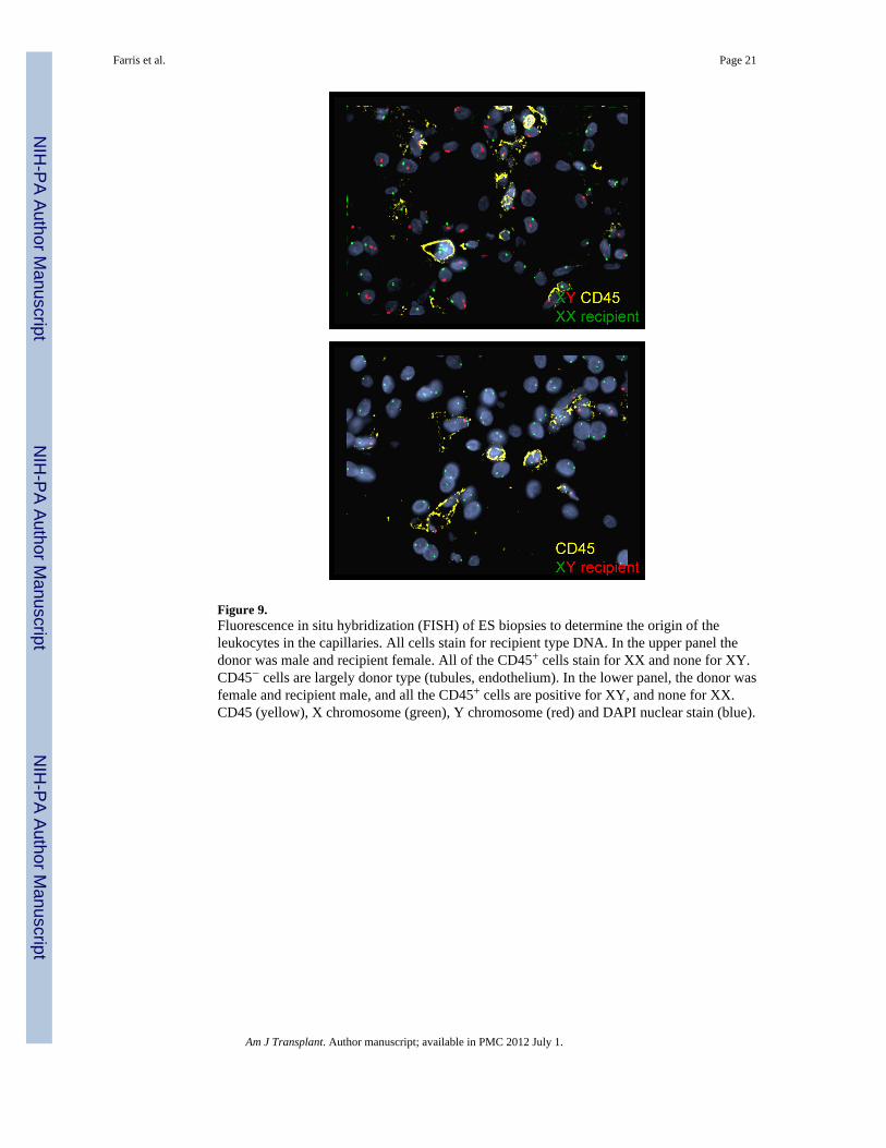
Figure 9.Fluorescence in situ hybridization (FISH) of ES biopsies to determine the origin of theleukocytes in the capillaries. All cells stain for recipient type DNA. In the upper panel thedonor was male and recipient female. All of the CD45+ cells stain for XX and none for XY.CD45− cells are largely donor type (tubules, endothelium). In the lower panel, the donor wasfemale and recipient male, and all the CD45+ cells are positive for XY, and none for XX.CD45 (yellow), X chromosome (green), Y chromosome (red) and DAPI nuclear stain (blue).
Farris et al. Page 21
Am J Transplant. Author manuscript; available in PMC 2012 July 1.
NIH
-PA Author Manuscript
NIH
-PA Author Manuscript
NIH
-PA Author Manuscript

NIH
-PA Author Manuscript
NIH
-PA Author Manuscript
NIH
-PA Author Manuscript
Farris et al. Page 22
Tabl
e 1
Clin
ical
and
Pat
holo
gic
Feat
ures
Rec
ipie
nt1
23
45
67
89
10
Bx
11
11
11
12
11
12
Day
Pos
t-tra
nspl
ant
2410
1012
1311
1213
1010
1316
Seru
m C
r
At B
x1.
44.
84.
21.
84.
52.
52.
14.
12.
31.
62.
09.
0
Peak
1.4
11.9
8.3
3.7
7.2
5.8
6.7
4.8
4.6
15.4
Last
1.3
2.0
21.
82.
01.
60.
73
1.1
1.7
Tim
e7
y6.
52
m3
y4
y1
y1
y6
mo
6 m
o6
mo
Sym
ptom
sN
one
Feve
r, flu
id re
tent
ion
Feve
r, flu
id re
tent
ion
Min
or fe
ver,
fluid
rete
ntio
nM
inor
feve
r, flu
idre
tent
ion
Min
or fl
uid
rete
ntio
nM
inor
flui
d re
tent
ion
No
sym
ptom
sM
inor
flui
d re
tent
ion
Min
or fe
ver,
min
or fl
uid
rete
ntio
n
Trea
tmen
tN
o ad
ditio
nal t
reat
men
tSt
eroi
d pu
lse
ATG
, PE,
Ritu
xan,
IVIG
Ster
oid
puls
e x3
ATG
, PE,
Ritu
xan
No
addi
tiona
l tre
atm
ent
ATG
, PE
x3N
o ad
ditio
nal
treat
men
t(A
TG la
ter)
No
addi
tiona
l tre
atm
ent
ATG
; sto
pped
tacr
olim
us
Ban
ff A
cute
Rej
ectio
n C
ateg
ory
Non
eN
one
AH
RN
one
AH
RN
one
AC
R2
Non
eN
one
Non
eN
one
AC
R2
Ligh
t Mic
rosc
opy
PTC
Con
gest
ion
-++
+++
++
+++
++++
+++
++++
+++
+
Hem
orrh
age
-++
+++
+++
+++
+++
+++
--
+++
Edem
a-
+++
++
++
+++
+++
--
++
Tubu
lar I
njur
y-
+++
+++
++
+++
++
+-
+++
+
Tubu
litis
4-
--
--
-+
--
--
+
Inte
rstit
ial I
nfilt
rate
5-
--
--
--
--
-+
Glo
mer
uliti
s-
-+
++
-++
-+
--
++
Enda
rterit
is6
--
--
--
+-
--
-++
Imm
unof
luor
esce
nce
C4d
PTC
--
>50%
->5
0%-
--
-3–
5%-
-
Elec
tron
mic
rosc
opy
Perit
ubul
ar C
apill
arie
sEn
doth
eliu
mLo
ss++
+++
--
++-
+++
Act
ivat
ion
++++
++
+++
+++
Fibr
in a
nd/o
r pla
tele
ts++
++-
-+
-+
+++
Cel
lsR
BC
RB
C-
RB
CR
BC
--
RB
C, N
Glo
mer
uli
Endo
thel
ium
Loss
-++
++N
A+
++
+
Act
ivat
ion
++
++++
+++
++
Foot
pro
cess
eff
acem
ent
0+
+++
++
++
Am J Transplant. Author manuscript; available in PMC 2012 July 1.

NIH
-PA Author Manuscript
NIH
-PA Author Manuscript
NIH
-PA Author Manuscript
Farris et al. Page 231 N
o ac
ute
graf
t dys
func
tion.
2 Ret
rans
plan
ted
unde
r con
vent
iona
l the
rapy
and
stab
le a
t 6 y
rs.
3 Dev
elop
ed T
MA
attr
ibut
ed to
cal
cine
urin
inhi
bito
r tox
icity
and
cur
rent
ly o
n di
alys
is.
4 Tubu
litis
: -, n
one;
+ u
p to
2 ly
mph
ocyt
es/tu
bule
;
5 Inte
rstit
ial i
nfla
mm
atio
n, -,
non
e, +
, up
to 1
0%
6 Arte
ritis
: +, o
ne a
rtery
, ++
mor
e th
an o
ne a
rtery
.
Abb
revi
atio
ns: A
TI, a
cute
tubu
lar i
njur
y; A
CR
2, a
cute
cel
lula
r rej
ectio
n, B
anff
type
2; A
HR
, acu
te h
umor
al re
ject
ion;
Bx:
bio
psy;
PTC
: per
itubu
lar c
apill
ary,
TM
A, t
hrom
botic
mic
roan
giop
athy
; RB
C re
dbl
ood
cells
, NA
, not
app
licab
le, A
TG, a
nti-t
hym
ocyt
e gl
obul
in; P
E, p
lasm
aphe
resi
s; N
, neu
troph
ils.
Am J Transplant. Author manuscript; available in PMC 2012 July 1.

NIH
-PA Author Manuscript
NIH
-PA Author Manuscript
NIH
-PA Author Manuscript
Farris et al. Page 24
Tabl
e 2
Fluo
resc
ence
In S
itu H
ybrid
izat
ion
(FIS
H)
Patie
nt #
Day
s Pos
t-tra
nspl
ant
Don
or/R
ec g
ende
rR
enal
Tub
ular
cel
ls g
enot
ype
End
othe
lial c
ells
(CD
34+)
geno
type
CD
45+
cells
gen
otyp
e(E
xtra
vesc
.)C
D45
+ ce
lls G
enot
ype
(Int
rava
sc.)
473
1Fe
mal
e/M
ale
XX
XX
(>10
0)X
Y (>
100)
XY
(>10
0)
454
3Fe
mal
e/M
ale
XX
XX
(48)
XY
(>10
0)X
Y (1
6)
410
87Fe
mal
e/M
ale
XX
XX
(67)
XY
(34)
XY
(27)
599
1Fe
mal
e/M
ale
XX
XX
(>10
0)X
Y (>
100)
XY
(>10
0)
566
7Fe
mal
e/M
ale
XX
XX
(58)
XY
(>10
0)X
Y (>
100)
612
Fem
ale/
Mal
eX
XX
Y
618
3Fe
mal
e/M
ale
XX
XX
(76)
XY
(23)
XY
(few
)
713
Mal
e/Fe
mal
eX
YX
X
759
Mal
e/Fe
mal
eX
YX
Y (6
8)X
X (>
100)
XX
(32)
Am J Transplant. Author manuscript; available in PMC 2012 July 1.

NIH
-PA Author Manuscript
NIH
-PA Author Manuscript
NIH
-PA Author Manuscript
Farris et al. Page 25
Tabl
e 3
Follo
w-u
p Pr
otoc
ol B
iops
ies
Rec
ipie
ntD
ays P
ost-T
xL
ight
Mic
rosc
opy
C4d
EM
127
16N
orm
al1
−N
orm
al
210
87N
orm
al2
−N
ot d
one
410
88M
ild tr
ansp
lant
glo
mer
ulop
athy
3+
cg1
510
00N
orm
al−
Nor
mal
637
4N
orm
al1
−N
orm
al
736
4N
orm
al−
Nor
mal
944
0N
orm
al−
Nor
mal
1017
7N
orm
al4
−N
orm
al
1 Mild
arte
riosc
lero
sis (
dono
r dis
ease
)
2 Prot
ocol
bio
psy
at 7
yea
rs sh
owed
subc
linic
al re
curr
ent M
PGN
, typ
e I.
3 Prot
ocol
bio
psy
at 5
.4 y
ears
show
ed c
hron
ic h
umor
al re
ject
ion
(cg3
)
4 Indi
catio
n bi
opsy
at d
ay 3
11 sh
owed
acu
te c
ellu
lar r
ejec
tion
(C4d−
)
Am J Transplant. Author manuscript; available in PMC 2012 July 1.