Embed Size (px)
Citation preview
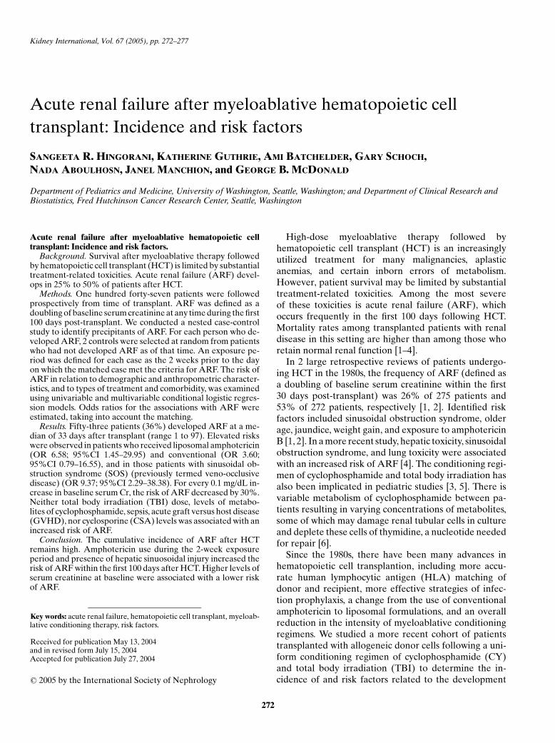
Kidney International, Vol. 67 (2005), pp. 272–277
Acute renal failure after myeloablative hematopoietic celltransplant: Incidence and risk factors
SANGEETA R. HINGORANI, KATHERINE GUTHRIE, AMI BATCHELDER, GARY SCHOCH,NADA ABOULHOSN, JANEL MANCHION, and GEORGE B. MCDONALD
Department of Pediatrics and Medicine, University of Washington, Seattle, Washington; and Department of Clinical Research andBiostatistics, Fred Hutchinson Cancer Research Center, Seattle, Washington
Acute renal failure after myeloablative hematopoietic celltransplant: Incidence and risk factors.
Background. Survival after myeloablative therapy followedby hematopoietic cell transplant (HCT) is limited by substantialtreatment-related toxicities. Acute renal failure (ARF) devel-ops in 25% to 50% of patients after HCT.
Methods. One hundred forty-seven patients were followedprospectively from time of transplant. ARF was defined as adoubling of baseline serum creatinine at any time during the first100 days post-transplant. We conducted a nested case-controlstudy to identify precipitants of ARF. For each person who de-veloped ARF, 2 controls were selected at random from patientswho had not developed ARF as of that time. An exposure pe-riod was defined for each case as the 2 weeks prior to the dayon which the matched case met the criteria for ARF. The risk ofARF in relation to demographic and anthropometric character-istics, and to types of treatment and comorbidity, was examinedusing univariable and multivariable conditional logistic regres-sion models. Odds ratios for the associations with ARF wereestimated, taking into account the matching.
Results. Fifty-three patients (36%) developed ARF at a me-dian of 33 days after transplant (range 1 to 97). Elevated riskswere observed in patients who received liposomal amphotericin(OR 6.58; 95%CI 1.45–29.95) and conventional (OR 3.60;95%CI 0.79–16.55), and in those patients with sinusoidal ob-struction syndrome (SOS) (previously termed veno-occlusivedisease) (OR 9.37; 95%CI 2.29–38.38). For every 0.1 mg/dL in-crease in baseline serum Cr, the risk of ARF decreased by 30%.Neither total body irradiation (TBI) dose, levels of metabo-lites of cyclophosphamide, sepsis, acute graft versus host disease(GVHD), nor cyclosporine (CSA) levels was associated with anincreased risk of ARF.
Conclusion. The cumulative incidence of ARF after HCTremains high. Amphotericin use during the 2-week exposureperiod and presence of hepatic sinuosoidal injury increased therisk of ARF within the first 100 days after HCT. Higher levels ofserum creatinine at baseline were associated with a lower riskof ARF.
Key words: acute renal failure, hematopoietic cell transplant, myeloab-lative conditioning therapy, risk factors.
Received for publication May 13, 2004and in revised form July 15, 2004Accepted for publication July 27, 2004
C© 2005 by the International Society of Nephrology
High-dose myeloablative therapy followed byhematopoietic cell transplant (HCT) is an increasinglyutilized treatment for many malignancies, aplasticanemias, and certain inborn errors of metabolism.However, patient survival may be limited by substantialtreatment-related toxicities. Among the most severeof these toxicities is acute renal failure (ARF), whichoccurs frequently in the first 100 days following HCT.Mortality rates among transplanted patients with renaldisease in this setting are higher than among those whoretain normal renal function [1–4].
In 2 large retrospective reviews of patients undergo-ing HCT in the 1980s, the frequency of ARF (defined asa doubling of baseline serum creatinine within the first30 days post-transplant) was 26% of 275 patients and53% of 272 patients, respectively [1, 2]. Identified riskfactors included sinusoidal obstruction syndrome, olderage, jaundice, weight gain, and exposure to amphotericinB [1, 2]. In a more recent study, hepatic toxicity, sinusoidalobstruction syndrome, and lung toxicity were associatedwith an increased risk of ARF [4]. The conditioning regi-men of cyclophosphamide and total body irradiation hasalso been implicated in pediatric studies [3, 5]. There isvariable metabolism of cyclophosphamide between pa-tients resulting in varying concentrations of metabolites,some of which may damage renal tubular cells in cultureand deplete these cells of thymidine, a nucleotide neededfor repair [6].
Since the 1980s, there have been many advances inhematopoietic cell transplantion, including more accu-rate human lymphocytic antigen (HLA) matching ofdonor and recipient, more effective strategies of infec-tion prophylaxis, a change from the use of conventionalamphotericin to liposomal formulations, and an overallreduction in the intensity of myeloablative conditioningregimens. We studied a more recent cohort of patientstransplanted with allogeneic donor cells following a uni-form conditioning regimen of cyclophosphamide (CY)and total body irradiation (TBI) to determine the in-cidence of and risk factors related to the development
272

Hingorani et al: Acute renal failure after myeloablative hematopoietic cell transplant 273
of ARF. We used a nested case-control study design, inwhich the 14 days prior to the development of ARF wasdefined as the exposure period in which clinical data andtime-related risk factors were ascertained.
METHODS
Between April 1997 and January 2000, all patientsat the Fred Hutchinson Cancer Research Center witha hematologic malignancy undergoing allogeneic trans-plantation after a conditioning regimen of CY and TBIwere invited to participate in this study. One hundredforty-seven patients agreed to participate, and signed in-formed consent forms were approved by the InstitutionalReview Board. The patients included in this nested case-control study were selected from the cohort of 147 pa-tients, as described below.
Technique of hematopoietic cell transplantation
Seven days prior to the infusion of stem cells, 60 mg/kgbody weight of CY was given through a central venousaccess catheter over 1 to 2 hours. On the following day,a second infusion of the same dose of CY was given.The pharmacokinetics of CY in this cohort of patientshas been published [7]. After a day of rest, TBI (9–14.2 Gy) was administered in hyperfractionated dosesfrom opposing cobalt sources on each of the 3 or 4 sub-sequent days. The kidneys were not shielded. Prophy-laxis regimens against graft-versus-host disease includedcyclosporine or tacrolimus and methotrexate in the ma-jority of patients, or variations on the above, includingprednisolone and BC3; BC3 is a murine antibody specificfor human CD3. Donor hematopoietic cells were infusedon day “zero;” by convention, all subsequent days arenumbered from this day. Infection prophylaxis includedfluconazole or itraconazole, acyclovir, and trimethoprim-sulfamethoxazole.
Study design
We used a nested case-control study design to explorerisk factors for ARF, defined as a doubling of baselineserum creatinine within the first 100 days after transplant.Baseline serum creatinine was the value obtained priorto the start of myeloablative conditioning therapy. Twocontrols were selected at random from among patients inthe study cohort who were event-free for at least as longas the time preceding the onset of ARF in the matchedcase. An exposure period of 2 weeks prior to the onsetof ARF was defined, in which potential time-varying riskfactors were examined. The exposure period for a controlpatient was defined as the 2 weeks prior to the study dayon which the matched case developed ARF. Thus, 53 casesand 106 control observation periods comprised the studypopulation.
The following clinical data were collected during the14 day exposure periods: daily weight, first morning pulse
and blood pressure, maximum daily temperature, dailymedications, total serum bilirubin, the presence of bac-teremia or fungemia, and cyclosporine blood levels. Inaddition to time-dependent factors, we examined the fol-lowing pretransplant patient characteristics for their asso-ciation with ARF: age, gender, baseline serum creatinine(Cr), weight, and serum albumin, as well as transplant-related factors such as TBI dose and cyclophosphamide(CY) metabolite exposure. The occurrence of acute graftversus host disease (GVHD) and sinusoidal obstructionsyndrome (SOS) developing any time prior to the onsetof ARF in the index case were also examined as potentialrisk factors.
Variations in the metabolism of CY were analyzed asareas under the curve (AUCs) as previously described [7].AUCs for each of the metabolites of CY [O-carboxyethylphosphoramide mustard (CEPM), deschloroethyl-cyclo-phosphamide (DCCY), hydroxypropyl-phosphoramidemustard (HPPM), 4-ketocyclophosphamide (KetoCY),and 4-hydroxy cyclophosphamide (HCY) [7]] were cal-culated based on their plasma levels. TBI was analyzedby total dose: 9 Gy, 12 Gy, and ≥13.2 Gy. During the 2-week exposure period, the mean pulse, systolic and dias-tolic blood pressure measurements, and change in weightfrom baseline to time of ARF were evaluated as predic-tors. Hypertension was defined in adults as 3 instancesof either a systolic measurement ≥140 mm Hg or a di-astolic measurement ≥90 mm Hg in the exposure pe-riod. In pediatric patients, hypertension was defined as3 measurements ≥95th percentile for age and height asstandardized and described elsewhere [8]. Fever was de-fined as any daily temperature ≥38 degrees Celsius. Bac-teremia/fungemia was defined as the presence of at least 1positive blood culture during the exposure period. Med-ications taken during the exposure period were dividedinto the broad categories of immunosuppression, antibi-otics, diuretics, and antihypertensives, and these cate-gories were analyzed separately. Nephrotoxic antibioticsanalyzed individually included amphotericin, liposomalamphotericin (Abelcept; The Liposome Co., Princeton,NJ, USA), vancomycin, and gentamicin. Sinusoidal ob-struction syndrome was defined based on the triad of hep-atomegaly, weight gain, and jaundice [9]. The severity ofSOS was classified as mild, moderate, or severe, based onsymptom resolution and treatment required [10]. AcuteGVHD was graded I-IV based on the Glucksberg classi-fication as modified by a working group [11].
Statistical methods
The distributions of the continuous covariates in thecases and controls were compared using Wilcoxon ranksum tests. The odds ratios (OR) were calculated us-ing conditional logistic regression models, which takeinto account the matching. All potential predictors werefirst evaluated in univariable conditional logistic regres-sion models. Those parameters reaching a univariable

274 Hingorani et al: Acute renal failure after myeloablative hematopoietic cell transplant
significance level of P ≤ 0.1 were assessed for significancein multiple conditional logistic models. The P values cor-responding to the multiple regression model are basedon the Wald test. All analyses were performed using SASstatistical software (Cary, NC, USA).
RESULTS
The demographic characteristics of the study cohorthave been described previously [7]. All patients receivedallografts, the majority (70%) for CML. One hundredthirty-nine patients (95%) received hematopoietic cellsderived from HLA-matched unrelated donors. The me-dian age at transplant was 37 years with a range of 3–58 years of age. Of 147 patients, 53 (36%) developedARF before day +100, at median day +33 (range day+1 to +97) after transplant.
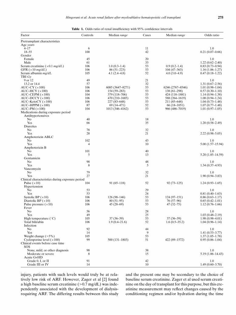
Table 1 lists the median and ranges for the risk fac-tors analyzed and modeled as continuous variables con-sidered among cases and controls, and also shows theunivariable associations between ARF and all the po-tential risk factors. The largest ORs were observed inpatients who received either conventional or liposomalamphotericin (OR 5.87, 95% CI 2.33–14.79) in the 14-dayexposure period prior to development of ARF, and inthose who had a clinical diagnosis of moderate or se-vere SOS prior to the date of onset of ARF (OR 5.19,95% CI 1.86–14.43). There was no difference in size ofthe increased risk of ARF associated with the receipt ofconventional versus liposomal amphotericin (OR 5.0 vs.5.2, respectively). The risk of ARF appeared to be sub-stantially lower in adults than children (OR 0.21, 95% CI0.07–0.66). The risk of ARF was also associated with a5% greater gain in weight from baseline (initial weight atclinic visit prior to the start of cyclophosphamide) to theend of the exposure period (OR 1.37, 95% CI 1.05–1.78).Higher levels of baseline serum creatinine were associ-ated with a reduced risk of ARF (0.83 per 0.1 mg/dL,95% CI 0.73–0.94). Exposure to metabolites of CY dur-ing conditioning therapy was not associated with an in-creased risk of ARF. Similarly, higher doses of TBI andhigher CSA levels did not appear to increase the risk ofARF in these patients.
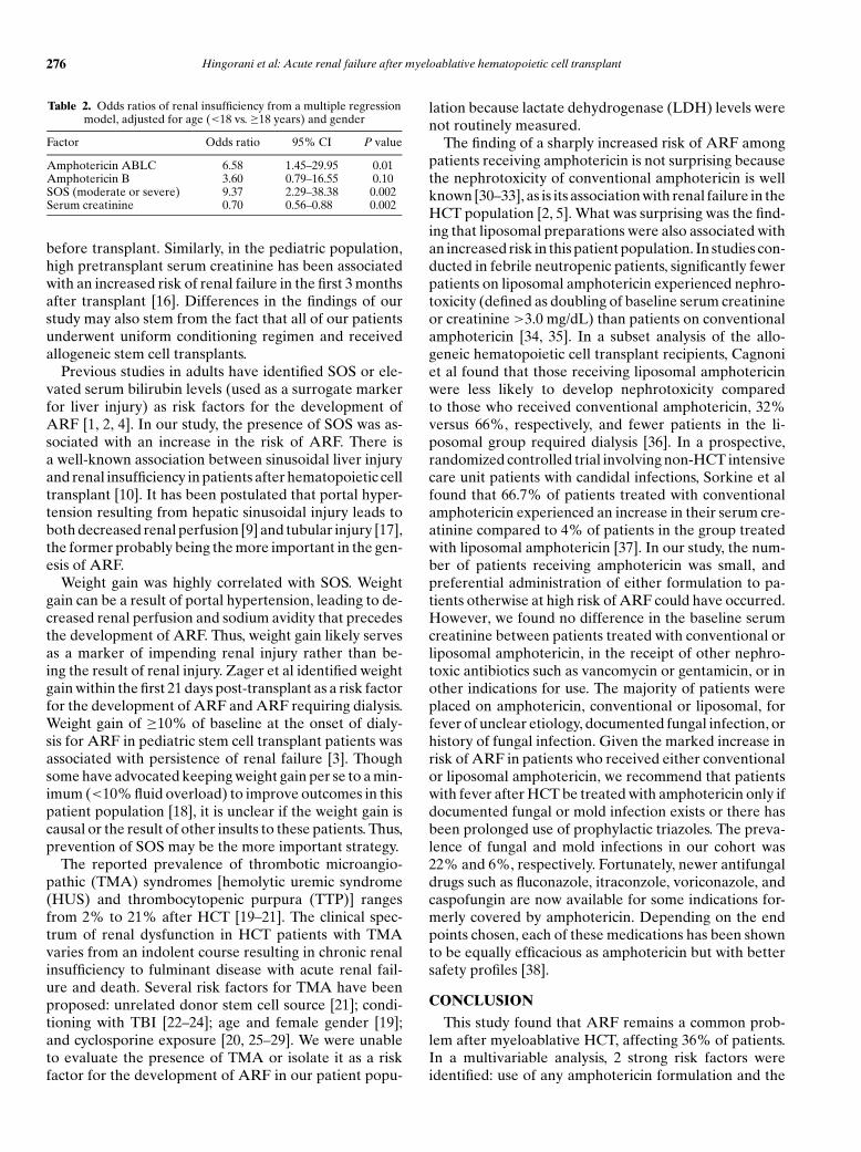
Many of these factors were inter-related. For exam-ple, younger patients were more likely to receive ampho-tericin, and were less likely to have developed moderateor severe SOS. Those patients with the largest weightgain from baseline to the onset of ARF were more likelyto have SOS and to have received amphotericin. In amultiple regression model adjusted for age and gender(Table 2), the OR for ARF associated with having re-ceived liposomal amphotericin during the 2-week expo-sure period was 6.5 (95% CI 1.45–29.95). Patients who re-ceived conventional amphotericin also had an increasedrisk of ARF (OR 3.6, 95% CI 0.79–16.55). Similarly, pa-tients with moderate or severe SOS were more likelyto develop ARF than patients without SOS or those
with mild SOS (OR 9.37, 95% CI 2.29–38.38). For every0.1 mg/dL increment in baseline serum creatinine, the ORfor ARF was reduced by approximately 30% (95% CI0.56–0.88). There was little difference in the risk of ARFbetween adults and children after adjusting for ampho-tericin use, SOS, and baseline serum creatinine (OR 0.79;95% CI 0.14–4.40). The association between a weight gainof >5% and ARF was confounded by amphotericin use,and SOS and was not a predictor of ARF after adjust-ing for these factors (OR 1.13, 95% CI 0.83–1.55). Ad-justed ORs for the remaining variables in Table 1 werecalculated and were not appreciably different from theunivariable ORs (data not shown).
DISCUSSION
Acute renal failure, defined as a doubling of baselineserum creatinine, remains common after HCT. In spite ofrecent advances in the care of patients undergoing HCT,the cumulative incidence of ARF was 36% in this cohort.Two major factors associated with an increased risk of thedevelopment of ARF were identified by multivariableanalysis: amphotericin use and the presence of moder-ate or severe SOS. There was no evidence in the data thatthe incidence of ARF was related to TBI dose, cyclophos-phamide metabolite exposure levels, sepsis, GVHD, cy-closporine levels, weight gain, and older age.
Unlike previous studies of ARF after HCT, all of ourpatients received the same conditioning regimen, allow-ing us to cleanly evaluate dosage of TBI as a risk fac-tor. This is also the first study to investigate levels ofmetabolites of cyclophosphamide as potential media-tors of renal injury. Though other studies have lookedat any cyclosporine use, we investigated blood levelsof cyclosporine in relation to the occurrence of ARF.Although much of the renal toxicity of cyclosporine isthought to be dose dependent [12], we found that higherlevels of cyclosporine were not associated with an in-creased risk of ARF in this patient population. However,in children, cyclosporine levels above 200 lg/L have beenassociated with an increased risk of ARF [13].
Our data suggest that an elevated baseline serum crea-tinine was associated with a reduced risk. It is conceivablethat this association is in part an artifact of our definitionof ARF (at least a doubling of baseline serum creatininelevel) because the absolute change required to meet thecriterion is less for a person with a low baseline level thana higher one. However, a possible basis for this reducedrisk being genuine is suggested by experimental animaldata showing increased cholesterol in renal tubular cellsat times of systemic stress or direct tubular injury [14,15]. Increased levels of cholesterol in renal tubular cellsconfer a “cytoresistant” state potentially protecting thekidney from further injury and the development of ARF[14, 15]. This cytoresistant state can persist for a variablelength of time after the initial injury [14]. Thus, to the ex-tent that a high baseline serum creatinine reflects earlier
Hingorani et al: Acute renal failure after myeloablative hematopoietic cell transplant 275
Table 1. Odds ratio of renal insufficiency with 95% confidence intervals
Factor Controls Median range Cases Median range Odds ratio
Pretransplant characteristicsAge years
4–17 6 11 1.018–55 100 42 0.21 (0.07–0.66)
GenderFemale 45 20 1.0Male 61 33 1.22 (0.62–2.40)
Serum creatinine (×0.1 mg/dL) 106 1.0 (0.3–1.4) 53 0.9 (0.2–1.4) 0.83 (0.73–0.94)GFR (×10 mg/dL) 106 86 (51–223) 53 104 (47–363) 1.16 (1.06–1.27)Serum albumin mg/dL 105 4.1 (2.4–4.8) 52 4.0 (3.0–4.9) 0.47 (0.18–1.22)TBI Gy
9 or 12 49 21 1.013.2 or 14.4 57 32 1.31 (0.67–2.56)
AUC–CY (×100) 106 6085 (3647–8271) 53 6246 (2767–8546) 1.01 (0.98–1.04)AUC–HCY (×100) 106 154 (59–283) 53 138 (61–298) 0.57 (0.30–1.10)AUC–CEPM (×100) 104 379 (118–706) 53 424 (118–1881) 1.14 (0.94–1.38)AUC–DCCY (×100) 106 470 (210–1685) 53 588 (264–1619) 1.08 (0.94–1.24)AUC–KetoCY (×100) 106 227 (83–648) 53 211 (85–648) 1.04 (0.73–1.48)AUC–HPPM (×100) 87 69 (14–471) 52 66 (18–1051) 1.07 (0.77–1.48)AUC–PM (×100) 104 1013 (346–4162) 53 966 (486–7019) 1.01 (0.97–1.05)Medications during exposure period
AntihypertensivesNo 40 18 1.0Yes 66 35 1.20 (0.58–2.49)
DiureticsNo 78 32 1.0Yes 28 21 2.22 (0.98–5.05)
Amphotericin ABLCNo 102 43 1.0Yes 4 10 5.00 (1.57–15.94)
Amphotericin BNo 101 40 1.0Yes 5 13 5.20 (1.85–14.59)
GentamicinNo 98 48 1.0Yes 8 5 1.34 (0.37–4.93)
VancomycinNo 79 32 1.0Yes 27 21 1.90 (0.94–3.83)
Clinical characteristics during exposure periodPulse (×10) 104 91 (65–118) 52 92 (73–125) 1.24 (0.93–1.65)Hypertension
No 53 29 1.0Yes 53 24 0.81 (0.40–1.63)
Systolic BP (×10) 106 126 (98–146) 53 124 (97–151) 0.86 (0.63–1.17)Diastolic BP (×10) 106 80 (51–95) 53 76 (57–96) 0.65 (0.42–1.01)Pulse pressure (×10) 106 45 (28–69) 53 47 (32–75) 1.12 (0.76–1.66)Fever
No 56 28 1.0Yes 49 25 1.03 (0.48–2.19)
High temperature (◦C) 105 37 (36–39) 53 37 (36–39) 1.98 (0.98–4.01)Total bilirubin 106 1.9 (0.4–21.6) 52 1.6 (0.5–35.2) 1.04 (0.96–1.14)Infection
No 92 44 1.0Yes 14 9 1.41 (0.53–3.77)
Weight change (>5%) 105 53 1.37 (1.05–1.78)Cyclosporine level (×100) 99 500 (131–1803) 51 422 (89–1572) 0.95 (0.86–1.04)
Clinical events before case timeSOS
None, mild, or other diagnosis 98 38 1.0Moderate or severe 8 15 5.19 (1.86–14.43)
Acute GvHDGrade 0, I, or II 91 42 1.0Grade III or IV 14 10 1.49 (0.60–3.70)
injury, patients with such levels would truly be at rela-tively low risk of ARF. However, Zager et al [2] founda high baseline serum creatinine (>0.7 mg/dL) was inde-pendently associated with the development of dialysis-requiring ARF. The differing results between this study
and the present one may be secondary to the choice ofbaseline serum creatinine. Zager et al used serum creati-nine on the day of transplant for this purpose, but this cre-atinine measurement may reflect changes caused by theconditioning regimen and/or hydration during the time
276 Hingorani et al: Acute renal failure after myeloablative hematopoietic cell transplant
Table 2. Odds ratios of renal insufficiency from a multiple regressionmodel, adjusted for age (<18 vs. ≥18 years) and gender
Factor Odds ratio 95% CI P value
Amphotericin ABLC 6.58 1.45–29.95 0.01Amphotericin B 3.60 0.79–16.55 0.10SOS (moderate or severe) 9.37 2.29–38.38 0.002Serum creatinine 0.70 0.56–0.88 0.002
before transplant. Similarly, in the pediatric population,high pretransplant serum creatinine has been associatedwith an increased risk of renal failure in the first 3 monthsafter transplant [16]. Differences in the findings of ourstudy may also stem from the fact that all of our patientsunderwent uniform conditioning regimen and receivedallogeneic stem cell transplants.
Previous studies in adults have identified SOS or ele-vated serum bilirubin levels (used as a surrogate markerfor liver injury) as risk factors for the development ofARF [1, 2, 4]. In our study, the presence of SOS was as-sociated with an increase in the risk of ARF. There isa well-known association between sinusoidal liver injuryand renal insufficiency in patients after hematopoietic celltransplant [10]. It has been postulated that portal hyper-tension resulting from hepatic sinusoidal injury leads toboth decreased renal perfusion [9] and tubular injury [17],the former probably being the more important in the gen-esis of ARF.
Weight gain was highly correlated with SOS. Weightgain can be a result of portal hypertension, leading to de-creased renal perfusion and sodium avidity that precedesthe development of ARF. Thus, weight gain likely servesas a marker of impending renal injury rather than be-ing the result of renal injury. Zager et al identified weightgain within the first 21 days post-transplant as a risk factorfor the development of ARF and ARF requiring dialysis.Weight gain of ≥10% of baseline at the onset of dialy-sis for ARF in pediatric stem cell transplant patients wasassociated with persistence of renal failure [3]. Thoughsome have advocated keeping weight gain per se to a min-imum (<10% fluid overload) to improve outcomes in thispatient population [18], it is unclear if the weight gain iscausal or the result of other insults to these patients. Thus,prevention of SOS may be the more important strategy.
The reported prevalence of thrombotic microangio-pathic (TMA) syndromes [hemolytic uremic syndrome(HUS) and thrombocytopenic purpura (TTP)] rangesfrom 2% to 21% after HCT [19–21]. The clinical spec-trum of renal dysfunction in HCT patients with TMAvaries from an indolent course resulting in chronic renalinsufficiency to fulminant disease with acute renal fail-ure and death. Several risk factors for TMA have beenproposed: unrelated donor stem cell source [21]; condi-tioning with TBI [22–24]; age and female gender [19];and cyclosporine exposure [20, 25–29]. We were unableto evaluate the presence of TMA or isolate it as a riskfactor for the development of ARF in our patient popu-
lation because lactate dehydrogenase (LDH) levels werenot routinely measured.
The finding of a sharply increased risk of ARF amongpatients receiving amphotericin is not surprising becausethe nephrotoxicity of conventional amphotericin is wellknown [30–33], as is its association with renal failure in theHCT population [2, 5]. What was surprising was the find-ing that liposomal preparations were also associated withan increased risk in this patient population. In studies con-ducted in febrile neutropenic patients, significantly fewerpatients on liposomal amphotericin experienced nephro-toxicity (defined as doubling of baseline serum creatinineor creatinine >3.0 mg/dL) than patients on conventionalamphotericin [34, 35]. In a subset analysis of the allo-geneic hematopoietic cell transplant recipients, Cagnoniet al found that those receiving liposomal amphotericinwere less likely to develop nephrotoxicity comparedto those who received conventional amphotericin, 32%versus 66%, respectively, and fewer patients in the li-posomal group required dialysis [36]. In a prospective,randomized controlled trial involving non-HCT intensivecare unit patients with candidal infections, Sorkine et alfound that 66.7% of patients treated with conventionalamphotericin experienced an increase in their serum cre-atinine compared to 4% of patients in the group treatedwith liposomal amphotericin [37]. In our study, the num-ber of patients receiving amphotericin was small, andpreferential administration of either formulation to pa-tients otherwise at high risk of ARF could have occurred.However, we found no difference in the baseline serumcreatinine between patients treated with conventional orliposomal amphotericin, in the receipt of other nephro-toxic antibiotics such as vancomycin or gentamicin, or inother indications for use. The majority of patients wereplaced on amphotericin, conventional or liposomal, forfever of unclear etiology, documented fungal infection, orhistory of fungal infection. Given the marked increase inrisk of ARF in patients who received either conventionalor liposomal amphotericin, we recommend that patientswith fever after HCT be treated with amphotericin only ifdocumented fungal or mold infection exists or there hasbeen prolonged use of prophylactic triazoles. The preva-lence of fungal and mold infections in our cohort was22% and 6%, respectively. Fortunately, newer antifungaldrugs such as fluconazole, itraconzole, voriconazole, andcaspofungin are now available for some indications for-merly covered by amphotericin. Depending on the endpoints chosen, each of these medications has been shownto be equally efficacious as amphotericin but with bettersafety profiles [38].
CONCLUSION
This study found that ARF remains a common prob-lem after myeloablative HCT, affecting 36% of patients.In a multivariable analysis, 2 strong risk factors wereidentified: use of any amphotericin formulation and the
Hingorani et al: Acute renal failure after myeloablative hematopoietic cell transplant 277
presence of moderate or severe SOS caused by the condi-tioning regimen. Prevention, early recognition, and treat-ment of SOS, and limiting exposure to amphotericin willoffer the best chance to preserve renal function in patientsafter HCT.
ACKNOWLEDGMENTS
We would like to thank Noel Weiss for his review of the manuscript.This research was supported by the following grants received by Dr.Hingorani: K23 NIH DK 63038 from the NIDDK at the NIH, ASN/RPAHealth Scholars Grant and the NKF Young Investigators Grant and Dr.McDonald’s grants: CA18029 & CA15704.
Reprint requests to Sangeeta Hingorani, M.D., MPH, Children’s Hos-pital & Regional Medical Center, 4800 Sandpoint Way NE, PediatricNephrology, M1-5, Seattle, WA 98105.E-mail: [email protected]
REFERENCES
1. GRUSS E, BERNIS C, TOMAS JF, et al: Acute renal failure in patientsfollowing bone marrow transplantation: Prevalence, risk factors andoutcome. Am J Nephrol 15:473–479, 1995
2. ZAGER RA, O’QUIGLEY J, ZAGER B, et al: Acute renal failure fol-lowing bone marrow transplantation: A retrospective study of 272patients. Am J Kidney Dis 13:210–216, 1989
3. LANE PH, MAUER SM, BLAZAR BR, et al: Outcome of dialysis foracute renal failure in pediatric bone marrow transplant patients.Bone Marrow Transplant 13:613–617, 1994
4. PARIKH CR, MCSWEENEY PA, KORULAR D, et al: Renal dysfunctionin allogeneic hematopoietic cell transplantation. Kidney Int 62:566–573, 2002
5. VAN WHY S, FRIEDMAN A, WEI L, et al: Renal insufficiency afterbone marrow transplantation in children. Bone Marrow Transplant7:383–388, 1991
6. MOHRMANN M, ANSORGE S, SCHMICH U, et al: Toxicity of ifosfamide,cyclophosphamide and their metabolites in renal tubular cells inculture. Pediatr Nephrol 8:157–163, 1994
7. MCDONALD GB, SLATTERY JT, BOUVIER ME, et al: Cyclophos-phamide metabolism, liver toxicity, and mortality followinghematopoietic stem cell transplantation. Blood 101:2043–2048, 2003
8. BONILLA-FELIX M, YETMAN R, PORTMAN R: Epidemiology of hyper-tension, in Pediatric Nephrology, edited by Barrett T, Avner E,Harmon W, Baltimore, Lippincott, Williams & Wilkins, 1999, pp959–985
9. DELEVE LD, SHULMAN HM, MCDONALD GB: Toxic injury to hepaticsinusoids: Sinusoidal obstruction syndrome (venocclusive disease).Semin Liver Dis 22:27–41, 2002
10. MCDONALD GB, HINDS MS, FISHER LB, et al: Venocclusive disease ofthe liver and multiorgan failure after bone marrow transplantation:A cohort study of 355 patients. Ann Intern Med 118:255–267, 1993
11. PRZEPIORKA D, WEISDORF D, MARTIN P, et al: 1994 consensus confer-ence on acute GVHD grading. Bone Marrow Transplant 15:825–828,1995
12. ATKINSON K, DOWNS K, ASHBY M: Clinical correlations with bloodlevels after allogeneic bone marrow transplantation: An analysis offour different assays. Transplant Proceedings 22:1331–1334, 1990
13. KIST-VAN HOLTHE J, GOEDVOLK CA, BRAND R, et al: Prospectivestudy of renal insufficiency after bone marrow transplantation. Pe-diatr Nephrol 17:1032–1037, 2002
14. ZAGER RA, ANDOH T, BENNETT W: Renal cholesterol accumulation:A durable response after acute and subacute renal insults. Am JPathol 159:743–752, 2001
15. ZAGER RA, SHAH V, SHAH H, et al: The mevalonate pathway duringacute tubular injury. Am J Pathol 161:681–692, 2002
16. KIST-VAN HOLTHE J, VAN ZWET J, BRAND R, et al: Bone marrowtransplantation in children: Consequences for renal function shortlyafter and 1 year post-BMT. Bone Marrow Transplant 22:559–564,1998
17. FINK J, COOPER M, BURKHART K, et al: Marked enzymuria follow-ing bone marrow transplantation: A correlate of veno-occlusivedisease-induced ‘hepatorenal syndrome.’ J Am Soc Nephrol 6:1655–1660, 1995
18. MICHAEL M, KUEHNLE I, GOLDSTEIN S: Fluid overload and acute renalfailure in pediatric stem cell transplant patients. Pediatr Nephrol19:91–95, 2004
19. FUGE R, BIRD J, FRASER A, et al: The clinical features, risk fac-tors and outcome of thrombotic thrombocytopenic purpura occur-ring after bone marrow transplantation. Br J Hematol 113:58–64,2001
20. PAQUETTE R, TRAN L, LANDAW E: Thrombotic microangiopathy fol-lowing allogeneic bone marrow transplantation is associated with in-tensive graft-versus-host disease prophylaxis. Bone Marrow Trans-plant 22:351–357, 1998
21. UDERZO C, FUMAGALLI M, DE LORENZO P, et al: Impact of throm-botic thrombocytopenic purpura on leukemic children undergoingbone marrow transplantation. Bone Marrow Transplant 26:1005–1009, 2000
22. LONNERHOLM G, CARLSON K, BRATTEBY L, et al: Renal function afterautologous bone marrow transplantation. Bone Marrow Transplant8:129–134, 1991
23. MARSHALL R, SWENY P: Haemolytic-uraemic syndrome in recipi-ents of bone marrow transplants not treated with cyclosporin A.Histopathology 10:953–962, 1986
24. ANTIGNAC C, GUBLER M-C, LEVERGER G, et al: Delayed renal failurewith extensive mesangiolysis following bone marrow transplanta-tion. Kidney Int 35:1336–1344, 1989
25. SCHRIBER J, HERZIG G: Transplantation-associated thromboticthrombocytopenic purpura and hemolytic uremic syndrome. SeminHematol 42:126–133, 1997
26. ATKINSON K, BIGGS J, HAYES J, et al: Cyclosporin A associatednephrotoxicity in the first 100 days after allogeneic bone marrowtransplantation: Three distinct syndromes. Br J Hematol 54:59–67,1983
27. ZEIGLER Z, ROSENFELD C, ANDREWS D III, et al: Plasma von Wille-brand factor antigen (vWF:AG) and thrombomodulin (TM) lev-els in adult thrombotic thrombocytopenic purpura/hemolytic ure-mic syndromes (TTP/HUS) and bone marrow transplant-associatedthrombotic microangiopathy (BMT-TM). Am J Hematol 52:213–220, 1996
28. CHAPPELL M, KEELING D, PRENTICE H, et al: Haemolytic uraemicsyndrome after bone marrow transplantation: An adverse effect oftotal body irradiation? Bone Marrow Transplant 3:339–347, 1988
29. COHEN E, LAWTON C, MOULDER J: Bone marrow transplantnephropathy: Radiation nephritis revisited. Nephron 70:217–222,1995
30. BUTLER W, BENNETT J, ALLING D: Nephrotoxicity of amphotericinB, early and late effects in 81 patients. Ann Int Med 61:175–187, 1964
31. HARBARTH S, BURKE J, LLOYD J, et al: Clinical and economic out-comes of conventional amphotericin B-associated nephrotoxicity.Clin Infect Dis 35:120–127, 2002
32. BATES D, SU L, YU D, et al: Mortality and costs of acute renal failureassociated with amphotericin B therapy. Clin Infect Dis 32:686–693,2001
33. WINGARD J, KUBILIS PS, LEE L, et al: Clinical significance of nephro-toxicity in patients treated with amphotericin B for suspected orproven aspergillosis. Clin Infect Dis 29:1402–1407, 1999
34. WALSH T, FINBERG R, ARNDT C, et al: Liposomal amphotericin B forempirical therapy in patients with persistent fever and neutropenia.N Engl J Med 340:764–771, 1999
35. PRENTICE H, HANN I, HERBRECHT R, et al: A randomized comparisonof liposomal versus conventional amphotericin B for the treatmentof pyrexia of unknown origin in neutropenic patients. Br J Hematol98:711–718, 1997
36. CAGNONI PJ: Liposomal amphotericin B versus conventional am-photericin B in the empirical treatment of persistently febrile neu-tropenic patients. J Antimicrob Chemother 49:81–86, 2002
37. SORKINE P, NAGAR H, WEINBROUM A, et al: Administration of am-photericin B in lipid emulsion decreases nephrotoxicity. Crit CareMed 24:1311–1315, 1996
38. MARR K: Empirical antifungal therapy: New options, new tradeoffs.N Engl J Med 346:278–280, 2002