Embed Size (px)
Citation preview
08.05.18
1
Acute vestibular syndrome– stroke vs. neuritis
PD Dr. med. Alexander A. TarnutzerDepartment of NeurologyUniversity Hospital Zurich
EAN Spring School 2018Staré Splavy, Czech Republic
Background
• Acute vertigo or dizziness
– 3.3 to 4.4% of all ED consultations à ~4.3 millionconsultations per year in the US
– Annual costs of about 4 billion USD
– Broad differential diagnosis across manyspecialities. No single diagnosis makes up morethan 5-10% of all cases.
– Isolated vertigo/dizziness à complicates DDx
– Often imaging (CCT, CTA, MRI) with low diagnosticimpact ordered.
à Systematic approach is essential!
Misdiagnosis of stroke on ED• The ED is a high-risk site for preventable errors.1
• Among adverse events in the ED deemed negligent, most are diagnostic failures.2
• Studies suggest that ED misdiagnoses may be unevenly distributed and disproportionate for neurologic conditions (deaths due to cerebrovascular events vs. myocardial infarction (45% vs. 1%, p<0.001).3,4
• Among major diagnostic errors reported by physicians, stroke is the fourth most common.5
1 Vinen. Incident monitoring in emergency departments: an Australian model. Acad Emerg Med. 2000;7(11):1290-12972 Thomas et al. Incidence and types of adverse events and negligent care in Utah and Colorado. Med Care. 2000;38(3):261-271.3 Dubois and Brook . Preventable deaths: who, how often, and why? Ann Intern Med. 1988;109(7):582-5894 Tarnutzer et al. ED misdiagnosis of cerebrovascular events in the era of modern neuroimaging: A meta-analysis. Neurology. 2017;88(15):1468-1477. 5 Schiff et al. Diagnostic error in medicine: analysis of 583 physician-reported errors. Arch Intern Med. 2009;169(20):1881-1887.
Misdiagnosis of stroke on ED II• Estimated prevalence of an initial misdiagnosis in the
preceding 30 days: 1.2% to 12.7% of all hospital
stroke admissions.1
• Disproportionally higher odds for patients presenting
with dizziness (OR=1.99, 95%-CI=1.03-3.84).2
• Posterior-circulation strokes are missed more often
than anterior-circulation strokes (37% vs. 16%,
p<0.001).2
à presenting symptoms may be an important
predictor of misdiagnosis risk.
1Newman-Toker et al. Missed diagnosis of stroke in the ED: cross-sectional analysis of a large population-based sample. Diagnosis. 2014;1:155-66.
2Arch et al. Missed Ischemic Stroke Diagnosis in the ED by Emergency Medicine and Neurology Services. Stroke. 2016;47(3):668-673.
The dizzy patient – differential diagnosison the ED
• Most frequent cases (based on severalstudies)– Neuro-otological diagnoses (peripheral and
central)(13-34%)– Other neurological disorders (5-11%)– Cardiovascular disease including arrhythmia (4-
21%)– Psychiatric disorders (2-14%)– Non-cardiovascular, internal-medicine related
causes (8-28%)
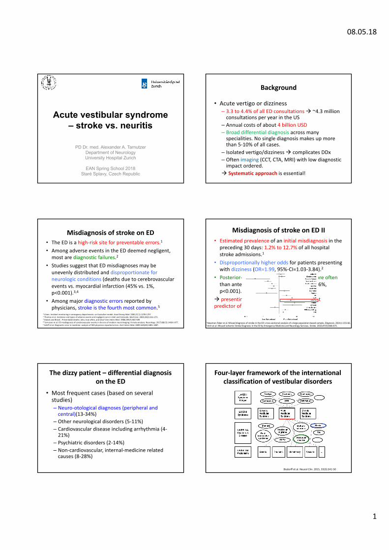
Four-layer framework of the international classification of vestibular disorders
Bisdorff et al. Neurol Clin. 2015, 33(3):541-50

08.05.18
2
Six categories of vestibular syndromes
Newman-Toker and Edlow, Neurol Clin 33 (2015) 577–599
A systematic approach to acute vertigo anddizziness
Structured history taking
• Duration and frequency of the single attacks?
Duration and frequency of attacks
seconds minutes hours days
Typical duration of a single episode
weeksor longer
vestibular migraine
single episodeepisodic-recurrentchronic-persistent
VP
acute vestibular neuritis
vertebrobasilar strokevertebrobasilar TIA
cardiac arrhythmia
episodic ataxia 2
SSCDS / PLF
CPV
panic attack
labyrinthitis
mal de debarquement
psychophysical dizziness
orthostatic hypotens.
vest. schwannomaMenière‘s disease
epileptic vertigo
intoxication
electrolyt imbalance
anemia
BPPV
hypoglycemia
myocardial infarction
centr
al
perip
heral
vesti
bular
psyc
h.
cardi
o-
vasc
ular
intern
al
medicin
e
cerebellar degen.
traumat. vestibulopathy
endocrine disorders
abbreviations: BPPV = benign paroxysmal positional vertigo; CPV = central positional vertigo;PFL = perilymph fistula; SSCDS = superior semicircular canal dehiscence syndrome; VP =vestibular paroxysmia.
Structured history taking
• Duration and frequency of the single attacks?• Onset of the attacks (abrupt vs. slowly)?
Structured history taking
• Duration and frequency of the single attacks?• Onset of the attacks (abrupt vs. slowly)?• Provocation factors?

08.05.18
3
Provocation factors
• Head inclination or reclination, turning over in bed, standing up / lyingdown à benign paroxysmal postional vertigo (BPPV)
• (fast) standing upà orthostatic hypotension
• Valsalva maneuver, acoustic stimulià superior canal dehiscence syndrome
• Busy places (shopping centers, railway stations...) à functional dizziness („psychogenic dizziness“)
• Noneà Menière‘s disease, cardiac arrhythmia, epileptogenic vertigo, (migraine)
Structured history taking
• Duration and frequency of the single attacks?• Onset of the attacks (abrupt vs. slowly)?• Provocation factors?• Focal-neurologic signs, hearing loss, tinnitus?
Structured history taking
• Duration and frequency of the single attacks?• Onset of the attacks (abrupt vs. slowly)?• Provocation factors?• Focal-neurologic signs, hearing loss, tinnitus?• Prodromi and accompanying symptoms?
Structured history taking
• Duration and frequency of the single attacks?• Onset of the attacks (abrupt vs. slowly)?• Provocation factors?• Focal-neurologic signs, hearing loss, tinnitus?• Prodromi and accompanying symptoms?• Drug history (antidepressants, AED, sedatives, diuretics,
antihypertensive drugs)?
Structured history taking
• Duration and frequency of the single attacks?• Onset of the attacks (abrupt vs. slowly)?• Provocation factors?• Focal-neurologic signs, hearing loss, tinnitus?• Prodromi and accompanying symptoms?• Drug history (antidepressants, AED, sedatives, diuretics,
antihypertensive drugs)?• Accompanying diseases (z.B. neoplasms, multiple
sclerosis, diabetes mellitus, depression)?
• Duration and frequency of the single attacks?• Onset of the attacks (abrupt vs. slowly)?• Provocation factors?• Focal-neurologic signs, hearing loss, tinnitus?• Prodromi and accompanying symptoms?• Drug history (antidepressants, AED, sedatives, diuretics,
antihypertensive drugs)?• Accompanying diseases (z.B. neoplasms, multiple
sclerosis, diabetes mellitus, depression)?• head or neck trauma?
Structured history taking
08.05.18
4
Acute and persistent vertigo ordizziness
Acute vestibular syndrome (AVS)1
• Vertigo or dizziness for more than 24 hoursaccompanied by
• Nausea / Vomitus• (head) motion intolerance• Nystagmus• Gait imbalance
• ~250’000 – 500’000 patients with AVS per year in US emergency departments
• Vertebrobasilar ischemia in ~25 ±15%.
1 Tarnutzer et al. Does my dizzy patient have a stroke? A systematic review of bedside diagnosis in acute vestibular syndrome. CMAJ 2011;183(9):E571-92
AVS - Differentialdiagnose
Modified after: Tarnutzer et al. CMAJ 2011;183(9):E571-92
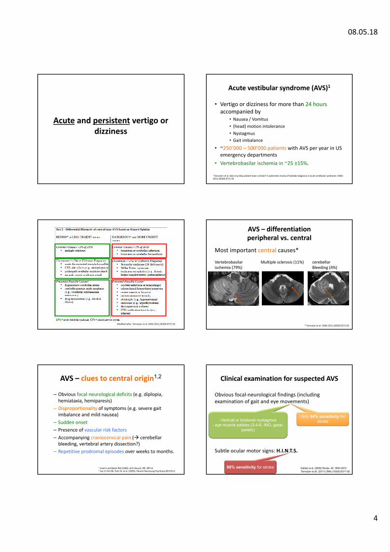
AVS – differentiationperipheral vs. central
Most important central causes*
* Tarnutzer et al. CMAJ 2011;183(9):E571-92
Multiple sclerosis (11%) cerebellarBleeding (4%)
Vertebrobasilarischemia (79%)
AVS – clues to central origin1,2
– Obvious focal-neurological deficits (e.g. diplopia, hemiataxia, hemiparesis)
– Disproportionality of symptoms (e.g. severe gaitimbalance and mild nausea)
– Sudden onset– Presence of vascular risk factors– Accompanying craniocervical pain (à cerebellar
bleeding, vertebral artery dissection?) – Repetitive prodromal episodes over weeks to months.
1 Grad A and Baloh RW (1989). Arch Neurol; 46: 281-4.2 Lee H, Kim BK, Park HJ, et al. (2009) J Neurol Neurosurg Psychiatry;80:355-6.
Clinical examination for suspected AVS
Obvious focal-neurological findings (includingexamination of gait and eye movements)
Subtle ocular motor signs: H.I.N.T.S.
Kattah et al. (2009) Stroke. 40, 3504-3510Tarnutzer et al. (2011) CMAJ;183(9):E571-92
Only 64% sensitivity forstroke-Vertical or torsional nystagmus
- eye muscle palsies (3-4-6, INO, gaze-paretic)
98% sensitivity for stroke
08.05.18
5
Subtle ocular motor signs:H.I.N.T.S. to I.N.F.A.R.C.T.
• 3 components “H.I.N.T.S.” battery– horizontal Head Impulse Test (h-HIT)– Nystagmus– Test of Skew
• Dangerous H.I.N.T.S. to “I.N.F.A.R.C.T.”– Impulse Normal– Fast-phase Alternating– Refixation on Cover Test
Any of these signs suggests a central (ischemic) origin in AVS!
2525Kattah et al. Stroke. 2009;40:3504-3510
H.I.N.T.S: head-impulse test
Dangerous H.I.N.T.S. to “I.N.F.A.R.C.T.”:Impulse Normal
Courtesy of Prof. D. Straumann
Fascicular and nuclear lesions of thevestibular nerve
Strupp und Magnusson Neurol Clin. 2015; 33:669–685
H.I.N.T.S: gaze-evoked nystagmus
Dangerous H.I.N.T.S. to “I.N.F.A.R.C.T.”:Fast-phase Alternating
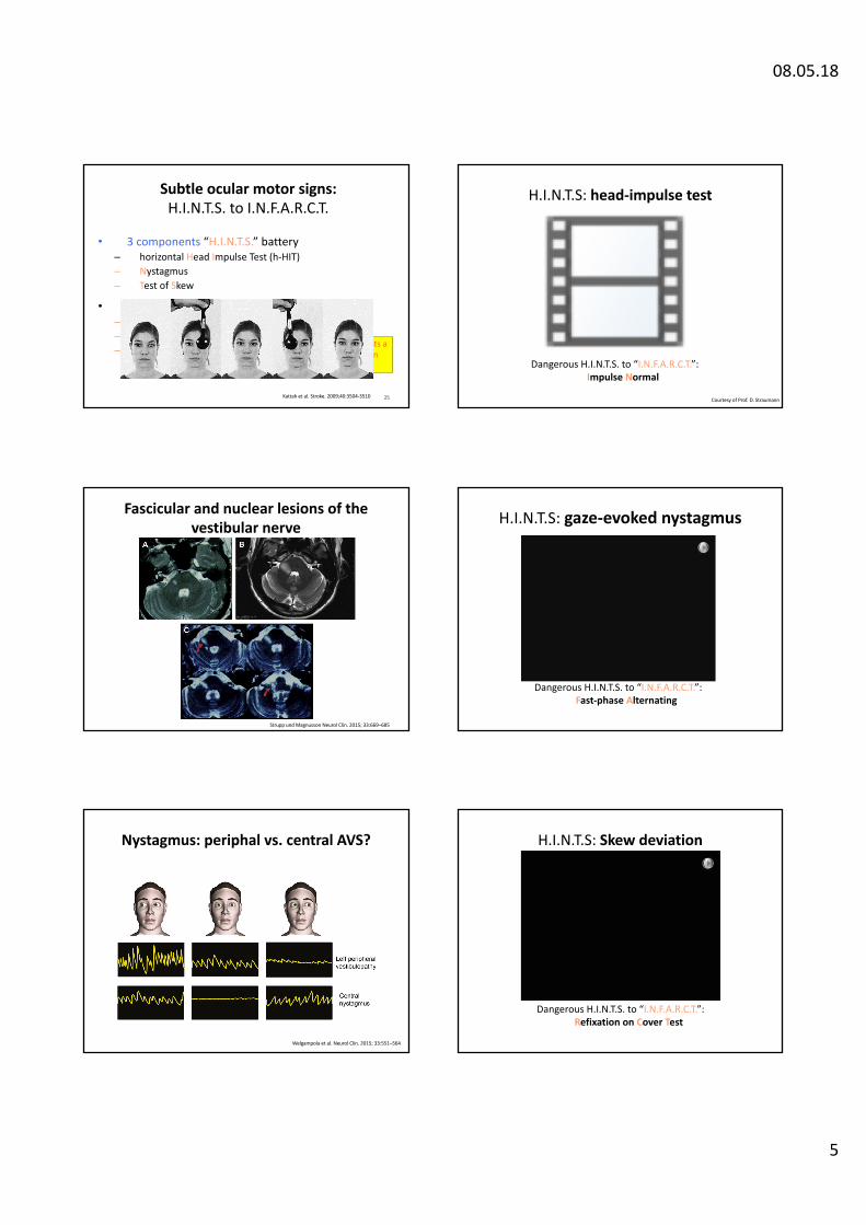
Nystagmus: periphal vs. central AVS?
Welgampola et al. Neurol Clin. 2015; 33:551–564
H.I.N.T.S: Skew deviation
Dangerous H.I.N.T.S. to “I.N.F.A.R.C.T.”:Refixation on Cover Test

08.05.18
6
Lacunar strokes – H.I.N.T.S. vs. MRI
Saber Tehrani et al. (2014) Neurology; 83:169-173
ABCD2-risk statification for patients withacute dizziness/vertigo
Navi et al. Stroke. 2012; 43:1484–9.Kerber et al. Neurology. 2015;85(21):1869-78.
H.I.N.T.S. vs. ABCD2
Newman-Toker et al. Acad Emerg Med. 2013;20(10):986-96
Quantification of ataxia – a valuable alternative to the H.I.N.T.S.?
• Grading truncal ataxia (n=114, 72 pAVS, 42 cAVS)– Grade 1 à mild to moderate imbalance with walking
independently– Grade 2 à severe imbalance with standing, but cannot
walk without support– Grade 3 à falling at upright posture
• Grade 3 found only with central AVS• Grade 2 or 3 à 92.9% sensitivity and 61.1%
specificity for AICA/PICA stroke
Carmona et al. (2016) Front. Neurol.; 7:125
The video-head-impulse test: useful in the ED?
Mantokoudis et al. Otol Neurotol. 2015;36(3):457-65
Newman-Toker et al. Stroke. 2013;44:1158-1161
Büki and Tarnutzer 2013. Vertigo anddizziness. ONL, Oxford University Press
Newman-Toker and Edlow, Neurol Clin 33 (2015) 577–599
08.05.18
7
Summary AVSpredictors for central origin
• Normal head-impulse test (HIT)
à central (ischemic) origin (PICA, less often AICA)
à CAVE: HIT “false” positive in AICA / lateral pontine stroke
• Testing for gaze-evoked nystagmus and skew deviation àincreases sensitivity of the HIT to 98%.
• H.I.N.T.S. have higher sensitivity to exclude stroke than early (first 24-48h) MRI with diffusion weighted imaging (DWI)
• MRI (including DWI) may be negative in first 24-48h in up to 20% and up to 50% for small (lacunar) strokes.
AVS cases
Interaction appreciated!
44-year-old male patientCurrent medical history: • Acute vertigo accompanied by nausea, vomiting, gait imbalance and
intense sweating since this morning.
Relevant findings from clinical examination:• Neurologic examination:
• No obvious focal neurologic deficits (no eye muscle palsies, no limbpalsies, no sensory loss, no aphasia)
• Targeted neuro-otolotic examination:• Torsional-horizontal spontaneous nystagmus to the left (Alexander
grade II) without increase during fixation suppression• Bedside head impulse test to the right with very few catch-up
saccades, normal on repetition.• No skew deviation, no gaze-evoked nystagmus, no hearing loss• Examination of stance and gait not possible due to his overall medical
condition.
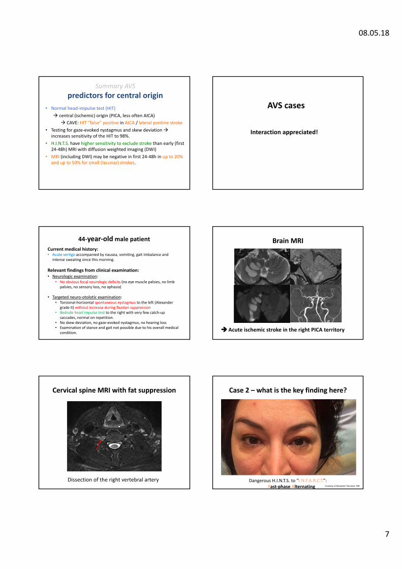
Brain MRI
à Acute ischemic stroke in the right PICA territory
Cervical spine MRI with fat suppression
Dissection of the right vertebral artery
Case 2 – what is the key finding here?
Dangerous H.I.N.T.S. to “I.N.F.A.R.C.T.”:Fast-phase Alternating Courtesy of Alexander Tarnutzer, MD
08.05.18
8
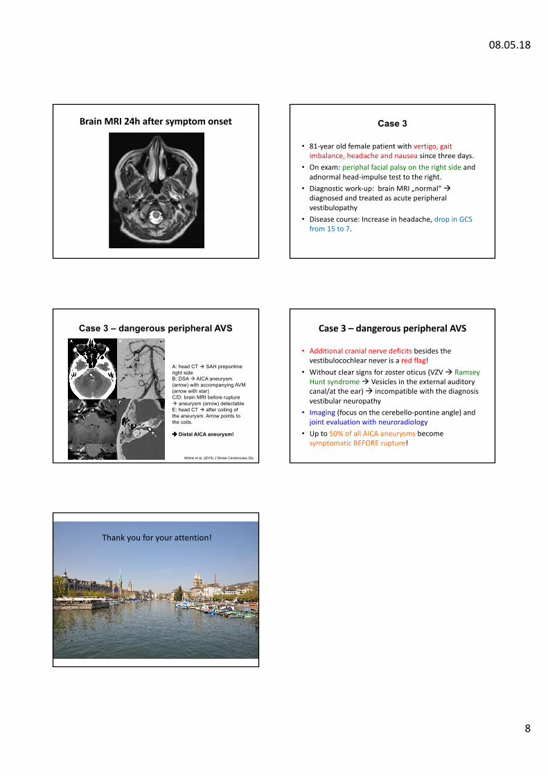
Brain MRI 24h after symptom onset
• 81-year old female patient with vertigo, gaitimbalance, headache and nausea since three days.
• On exam: periphal facial palsy on the right side andadnormal head-impulse test to the right.
• Diagnostic work-up: brain MRI „normal“ àdiagnosed and treated as acute peripheralvestibulopathy
• Disease course: Increase in headache, drop in GCS from 15 to 7.
Case 3
Case 3 – dangerous peripheral AVS
A: head CT à SAH prepontineright sideB: DSA à AICA aneurysm(arrow) with accompanying AVM (arrow with star)C/D: brain MRI before ruptureà aneurysm (arrow) detectableE: head CT à after coiling ofthe aneurysm. Arrow points tothe coils.
à Distal AICA aneurysm!
Willms et al. (2016) J Stroke Cerebrovasc Dis.
• Additional cranial nerve deficits besides thevestibulocochlear never is a red flag!
• Without clear signs for zoster oticus (VZV à Ramsey Hunt syndrome à Vesicles in the external auditorycanal/at the ear) à incompatible with the diagnosisvestibular neuropathy
• Imaging (focus on the cerebello-pontine angle) andjoint evaluation with neuroradiology
• Up to 50% of all AICA aneurysms becomesymptomatic BEFORE rupture!
Case 3 – dangerous peripheral AVS
Thank you for your attention!


![) [Kompatibilit si m d] - vizsgazas.etovabbkepzes.huvizsgazas.etovabbkepzes.hu/Files/...a...agnes).pdf · Pyramiscsont-törés Contusio cerebri Gyulladások Labyrinthitis, Neuronitis](https://img.pdfslide.net/doc/110x75/5cfe885e88c99367218c8d0b/-kompatibilit-si-m-d-pdf-pyramiscsont-toeres-contusio-cerebri-gyulladasok.jpg)