Embed Size (px)
Citation preview
AD
CONTRACT NO: DAMDl7-88-C-8069 AD-A228 158
TITLE: DEVELOPMENT OF AN ASSAY TO DETECT ANTIBODIES TO HIV-2USING RECOMBINANT DNA DERIVED ANTIGENS
PRINCIPAL INVESTIGATOR: Gerald A. Beltz, Ph.D.
CONTRACTING ORGANIZATION: Cambridge BioScience Corporation365 Plantation StreetWorcester, MA 01605
~TC
REPORT DATE: September 11, 1990 1.EC I ET16 19903
TYPE OF REPORT: Final Report -
PREPARED FOR: U.S. ARMY MEDICAL RESEARCH AND DEVELOPMENT COMMANDFort Detrick, Frederick, Maryland 21702-5012
DISTRIBUTION STATEMENT: Approved for public release;distribution unlimited
The findings in this report are not to be construed as anofficial Department of the Army position unless so designated byother authorized documents.
* %
SECURITY CLASS;F;CA-T:ON OF 7 'S PAGEForm Approved
REPORT DOCUMENTATION PAGE 0MBNo. 0704-0 88
la. REPORT SECURITY CLASSFCATION lb. RESTRICT'IVE MARK:NGS
Unclassif led2a. SECURITY CLASSIFiCATiON AUTHORITY 3. OISTRIBUTION/AVAILABILITY OF REPORT
Approved for public release;2b. DECLASSiFICATION/DOWNGRADING SCHEDULE distribution unlimited
4 PERFORMING ORGANIZA7TON REPORT NUMBER(S) 5. MONITORING ORGANiZATiON REPORT NUMBER(S)
6a. NAME OF PERFORMING ORGANIZATION 6b. OFFICE SYMBOL 7a. NAME OF ,MONITORING ORGANIZATION(If applicaole)
Cambridge BioScience Corporatio I
6c. ADDRESS (City, State, and ZIP Code) 7b. ADDRESS (City, State, and ZIP Code)
365 Plantation Street
Worcester, MA 01605
8a. NAM4E OF FUNDING , SPONSORING 8b. OFFICE SYMBOL 9. PROCUREMENT iNSTRuMENT tDENTIFICATION NUMBERORGANIZATION U.S. Army Medical (If applicable)
Research & Development Command Contract No. DAMDI7-88-C-8069
8c. ADDRESS (City, State, and ZIP Code) 10. SOURCE OF FUNDING NUMBERS
PROGRAM PROJECT TASK [WORK UNITFort Detrick ELEMENT NO. NO. 3M2 NO. ACCESSION NO.Frederick, Maryland 21702-5012 63105A 63105DH29 AB 064
11. TITLE (Include Securrty Classificarton)
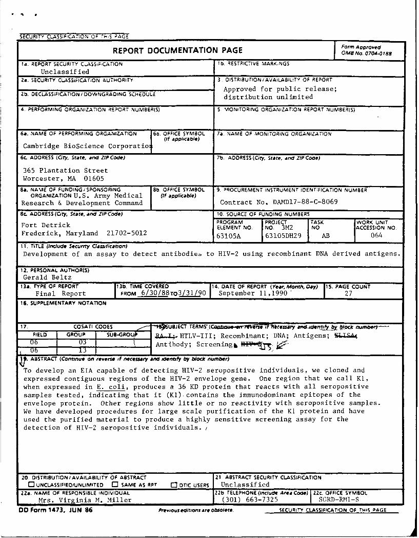
Development of an assay to detect antibodieb to HIV-2 using recombinant DNA derived antigens.
12. PERSONAL AUTHOR(S)
Gerald Beltz
13a. TYPE OF REPORT 13b. TIME COVERED 14. DATE OF REPORT (Year, Month, Oay) 15. PAGE COUNT
Final Report FROM 6/30/88TO3/31/90 September 11,1990" 2716. SUPPLEMENTARY NOTATION
17. COSATI CODES -tPUBJECT TERMS'. (Caau.Wrtr1Yjvi-sT7)'eesUy an44.dentafy O bock numbert--FIELD GROUP SUBGROU ZA-.L;.HTLV-III; Recombinant; DNA; Antigens; I.SA406 03 Antibody; Screeningb06 13
ASS RACT (Contnue on revere of necessary and idenfy by block number)
To develop an EIA capable of detecting HIV-2 seropositive individuals, we cloned andexpressed contiguous regions of the HIV-2 envelope gene. One region that we call K1,when expressed in E. coli, produces a 36 KD protein that reacts with all seropositive
samples tested, indicating that it (KI) contains the immunodominant epitopes of the
envelope protein. Other regions show little or no reactivity with seropositive samples.
We have developed procedures for large scale purification of the KI protein and haveused the purified material to produce a highly sensitive screening assay for the
detection of HIV-2 seropositive individuals. /
20 DISTRIBUTION /AVAILABILITY OF ABSTRACT 21 ABSTRACT SECURITY CLASSIFICATION
rC]UNCLASSIFIEOIIJNLIMITED ' SAME AS RPT C3 DTIC USERS Unclassified22a. NAME OF RESPONSIBLE INDIVIDUAL 22b TELEPHONE (Include Area Code) ZZc. OFFICE SYMBOL
Mrs. Virginia M. Miller (301) 663-7325 SGRD-RMI-S
DO Form 1473, JUN 86 Previous editions are obsolete. SECURITY CLASSIFICATION OF THIS PAGE
FOREWORD
Opinions, interpretations, conclusions and recommendations are those of theauthor and are not necessarily endorsed by the U.S. Army.
Where copyrighted material is quoted, permission has been obtained touse such material.
Where material from documents designated for limited distribution isquoted, permission has been obtained to use the material.
.._4 itations of commercial organizations and trade names in this report donot constitute an official Department of the -Army endorsement or approval ofthe products or services of these organizations.
___ In conducting research using animals, the investigator(s) adhered to the"Guide for the Care and Use of Laboratory Animals," prepared by the Committeeon Care and Use of Laboratory Animals of the Institute of Laboratory AnimalResources, National Research Council (NIH Publication No. 86-23, Revised 1985).
For the protection of human subjects, the investigator(s) have adheredto policies of applicable Federal Law 45CFR46.
,7_j conducting research utilizing recombinant DNA technology, theinvestigator(s) adhered to current guidelines promulgated by the NationalInstitutes of Health.
P1 Signature Date
I LI.IJ '"-%. 2DT IC ' I
Unannc.-u,:' ::d [
A.
*By_______ -
V!2~t ribut on/-?.,~lb~ltT"Codes
r, x i/cr
!
A-l
TABLE OF CONTENTS
INTRODUCTION ......................................... 2
MATERIALS AND METHODS ................................ 3
RESULTS .............................................. 5
SUMMARY .............................................. 15
REFERENCES ........................................... 17
FIGURES AND TABLES ................................... 18
1
INTRODUCTION
HIV-2 was initially identified in 2 patients from Western
Africa who developed AIDS-like symptoms but were seronegative for
HIV-1 by viral lysate immunoassays (1). The HIV-2 virus cultured
from PBL's of these patients was morphologically similar to HIV-1
and exhibited similar cell tropism for CD4+ cells. Preliminary
serological studies indicated that while HIV-2 was related to
HIV-I, it was more closely aligned with SIVma.
Subsequent cloning and sequencing of HIV-2 confirmed these
results (2,3). While the overall genetic organization of HIV-2
is similar to HIV-I, there is considerable sequence diversity
between the two. The degree of sequence divergence varies
between coding regions. While corresponding gag and pol
sequences show 50-60% homology at the amino acid level, the
envelope gene shows less sequence conservation, only 30-40%. The
lower degree of homology extends to the short open reading
frames, rev and sor (2,3).
HIV-2 infected individuals have been identified in Africa,
Europe, North and South America (4,5) and epidemiological studies
show that HIV-2 is associated with AIDS in Western Africa (6,7).
Since HIV-2 exhibits limited cross reactivity with HIV-l, it is
important to develop an assay capable of detecting HIV-2.
This report describes the development of an HIV-2 ELISA
2
using recombinant DNA derived antigens expressed in E. coli.
Recombinant derived antigens offer several advantages in
immunoassays. Thorn et al. (12) have developed a protein CBre3,
for use in an HIV-l EIA. The protein, also expressed in E. coli,
contains the major immunodominant epitopes of the HIV-l envelope
protein. Because it is expressed at high levels, large amounts
of the protein can be used in individual assays, and the protein
can be purified free of contaminants. A CBre3 EIA is sensitive
enough to detect envelope antibodies in seropositive patients
before the antibodies are detected in a viral lysate Western blot
(12).
Our strategy was to identify immunodominant regions of the
HIV-2 envelope protein by (i) cloning and expressing in E. coli
non-overlapping subfragments of the HIV-2 envelope protein and
(ii) testing the expressed proteins for reactivity with sera from
HIV-2 seropositive individuals.
By cloning regions of the HIV-2 envelope gene, we have
identified and expressed a protein called Ki, that contains the
major immunodominant epitopes of the HIV-2 envelope gene.
We have developed a purification protocol for K1 and used
the purified protein in an HIV-2 ELISA. Our results indicate
that KI can detect all HIV-2 seropositive samples thaV. we have
tested.
MATERIALS AND METHODS
HIV-2 Envelope Gene.
The initial plasmid, K3D, used for cloning, was kindly
3
provided by Dr. Genoveffa Franchini (8). The plasmid consists of
a 2.7 KB KpnI fragment containing the entire HIV-2 envelope gene,
cloned into SP65. The 2.7 KB fragment was derived from an
infectious HIV-2 clone, HIV-2SLIISY, isolated from a genomic
library of HUT 78 cells infected with the HIV-2 strain SBL6669.
The restriction map of the infectious clone, and the location of
the 2.7 KB KpnI fragment is shown in Figure 1.
Cloning and Expression of the Envelope Genes.
The precise location of the subgenomic envelope proteins are
described in the results section. DNA fragments coding for K13
and K3 were generated by Sau3AI digestion of K3D DNA. The
appropriate fragments were isolated from agarose gels by
electroelution and cloned into the Bam HI site of the expression
vector pCBC (see below). K18 was obtained by digesting K3D with
PvuII, adding Bam HI linkers and digesting with Sau3AI. The
fragment was cloned into pCBC. Cloning of the K1 coding fragment
was done by digesting K3D with EcoRI and treating with Klenow
polymerase to fill in the ends. BglII linkers were added to the
fragment and the K1 coding fragment was isolated following
digestion with Sau3AI. K2 was cloned by digesting K3D with StuI,
adding Bam HI linkers and digesting with Sau3AI. K1 and K2
coding fragments were also inserted into the expression vector
pCBC. All DNA manipulations were according to Maniatis (10).
The E. coli expression vector was pCBC. The vector, similar
to pJL6 described by Lautenberger et al. (9) contains the
bacteriophage lambda pL promotor, synthetic ribosome binding
4
site, and the first 13 amino acids of the lambda CII gene. DNA
fragments are cloned into a Bam HI site distal to the CII coding
region resulting in a fusion protein containing a 13 amino acid
leader of CII.
Envelope constructs in pCBC were used to transform E. coli
MZ-1. The MZ-l strain contains a temperature sensitive lambda
CI857 gene (9). At 320C the repressor binds to the pL promotor
inhibiting transcription. Induction by temperature shift from
320C to 420C allows transcription of pL distal genes. MZ-I cells
are grown at 320C to OD550 of 0.500-1.000 and then induced at 420C
for one hour.
Analysis of Envelope Proteins
Proteins from uninduced and induced E. coli were
electrophoresed in polyacrylamide gels and analyzed by Coomassie
blue staining and Western blotting (10). Blots were blocked in 1
X TBS, 0.1% Brij for 60' and incubated for 60' with sera diluted
1:100 in blocking buffer. Blots were washed 3 X with 1 X TBS and
incubated with HRP conjugated goat anti-human antibody for 60'.
Blots were washed as above and then incubated with the HRP
substrate 4 Chloro/Napthol.
Purification of K1 Protein and RIA Development
Purification of the recombinant protein and development of
an HIV-2 EIA are described in text.
RESULTS
The starting material for cloning was an HIV-2 subgenomic
fragment containing the complete HIV-2 envelope gene. The
5
.9
fragment, kindly provided by Dr. Genoveffa Franchini, was
isolated from a genomic HIV-2 clone and is shown in Figure 1.
Figure 2 shows a hydropathy plot of the HIV-2 envelope
protein. The regions represented beneath the plot, K18, K13, K3,
K1 and K2 represent the individual subfragments that were cloned
and expressed.
The first clone that we tested was K1. Based on our work
with HIV-1, we predicted that this region of the viral genome
would code for an immunodominant envelope antigen. E. coli
strain MZ-1, containing a thermolabile cI repressor, was
transformed with a plasmid containing the K1 region of the HIV-2
envelope gene downstream of the lambda pL promotor. As
described, proteins from uninduced and induced E. coli containing
the K1 coding fragment were analyzed.
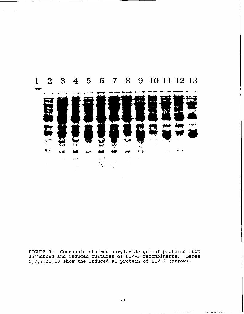
Figure 3 shows a picture of a Coomassie stained acrylamide
gel. The samples in each lane are as follows. Lane 1 has
prestained molecular weight markers. Lanes 2 and 3 are uninduced
and induced extracts of bacteria expressing the comparable region
from the envelope gene of Simian Immunodeficiency Virus (SIV).
Prior to beginning work on the HIV-2 clone, we had expressed a
protein from the envelope region of SIV containing SIV env amino
acids 365 to 646, that reacted strongly with HIV-2 samples. This
SIV protein is Accl. Accl is 80% homologous in amino acid
sequence to the comparable HIV-2 protein. Since it had been
cloned and expressed, and had been shown to be useful in
detecting HIV-2 seropositive patients, it served as a positive
6
control in the assays. The pairs of lanes, 4,5; 6,7; 8,9; 10,11;
and 12,13, are uninduced and induced cultures (respectively) of 5
independently isolated clones all expressing the recombinant HIV-
2 K1 protein. The arrow identifies the protein present in the
induced, but not in the uninduced, cultures of the HIV-2
recombinants.
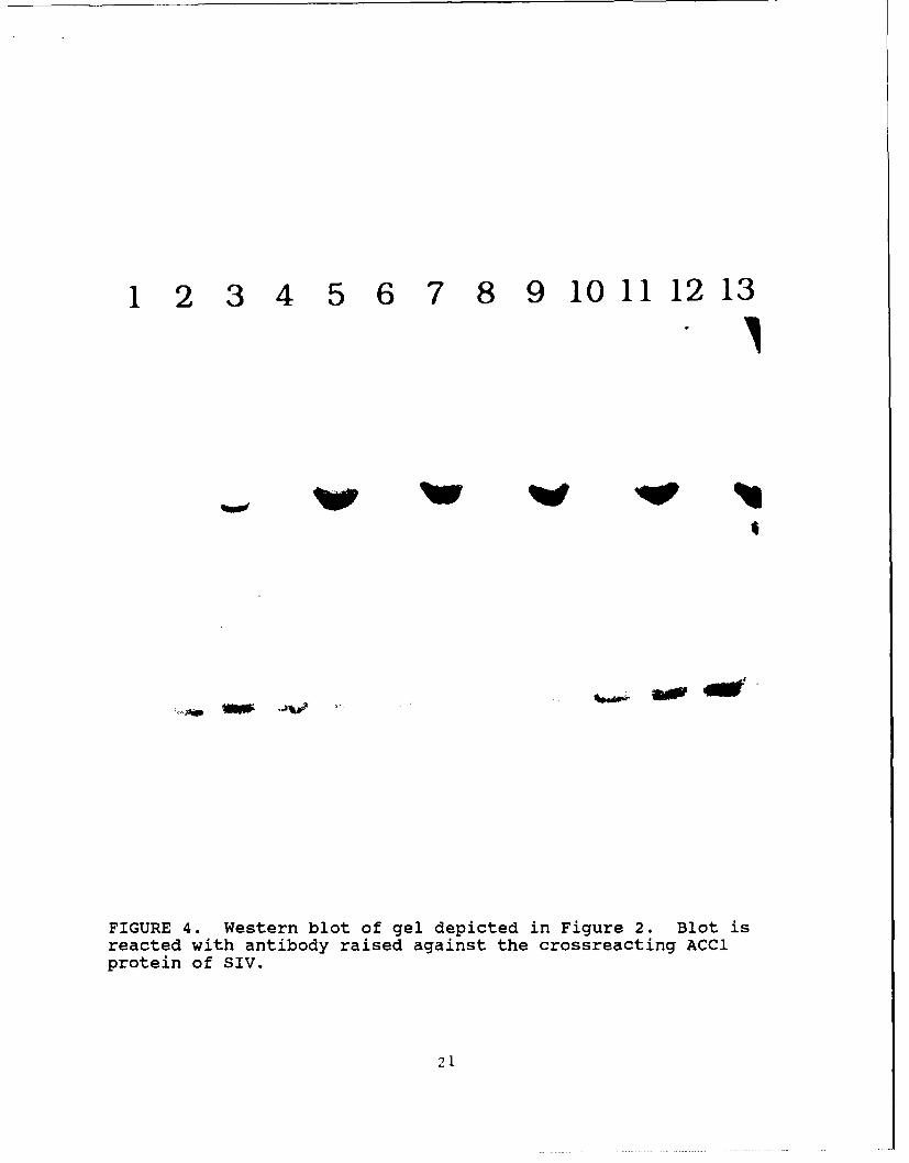
Western blot analysis was used to show that the induced
protein is coded for by the HIV-2 DNA. A duplicate gel, as shown
in Figure 3, was blotted and reacted with antibody raised in
rabbits against the SIV envelope protein Accl. Since Accl and
the HIV-2 envelope protein K1 are approximately 80% homologous,
we predicted that polyclonal antisera raised against Accl should
react with K1. Figure 4 shows the Western blot. The band in
lane 3 represents anti-Accl antibody reacting with the Accl
protein. Reactivity in lanes 5, 7, 9, 11 and 13 represent
antibody recognizing the induced HIV-2 protein, K1. The stronger
reactivity of anti-Accl antibody to K1 reflects the higher
expression level of the HIV-2 protein. We estimate that
expression of the K1 protein represents at least 5% of the total
protein.
Reactivity of K1 with Human Sera
Western blots of proteins from K1 expressing cultures
(comparable to material shown in Figure 2 and 3) were used to
assay serum samples from HIV-2 infected individuals collected in
West Africa, where there is a high incidence of HIV-2. Samples
were screened by commercial EIAs and provided by Dr. M'Boup of
7
the University ot Senegal. Figure 5 shows the Western results
from 47 diff,.ent serum samples. The majority of samples react
strongly with the K1 protein. The strips which are unreactive by
Western blot (3, 18, and 44), are negative by viral lysate
Western blots (Genetic Systems) and by radioimmune precipitations
(data not shown). We tested a second set of seropositive samples
by Western blot with comparable results (data not shown). In
all, we have tested 80 West African samples by Western blot for
reactivity to the subunit HIV-2 K1 protein. Our results continue
to show that the Ki protein detects all HIV-2 seropositive
samples.
Reactivity of K3, K18, K13, and K2 with HIV-2 Seropositive
Recombinant HIV-2 envelope proteins from 4 additional clones
were also analyzed for reactivity with sera from seropositive
individuals. All constructs were cloned into the expression
vector pCBC. Inductions were done as described for the K1
protein. The K3 protein is slightly larger than K1 because it
contains an additional 64 amino acids at the N-terminus.
Reactivity of sera to both K3 and Ki was compared. In terms of
specificity, sensitivity, and signal observed with positive
samples, there is no significant difference between the two
proteins.
The other HIV-2 envelope proteins that we have expressed are
as follows (see Figure 2):
K18. A 15 KD protein that begins at amino acid 7 and ends
at amino acid 135. The Pvu II site at amino acid 7 was converted
8
to a Bam HI site. The 3' end of the DNA fragment is a Sau 3AI
site which is compatible with the Bam HI site in the vector.
K13. The amino terminus of K13 begins at amino acid 136
which is just distal to the carboxy terminus of the K18 clone.
The 3' end of the gene is at amino acid 320 which is also a Bam
HI compatible Sau 3AI site. K13 is expected to code for a
protein of 18 KD.
K2. This is a small protein coded for by sequences just
proximal to the second major hydrophobic domain of HIV-2 (figure
2). The protein extends from amino acid 700-797.
Western blots were made for each of the proteins, K18, K13
and K2 and the blots were reacted with HIV-2 seropositive
samples. The results are shown in Table I. All three of the
constructs react with only a minority of the samples or with none
at all.
Because of the low reactivity of the proteins with sera that
had been shown to react strongly with the K1 protein, we tested
the K18, K13, and K2 Western blots to ensure that the proteins
were expressed and transferred during blotting. Since all three
proteins contain a 13 amino acid leader sequence coded for by the
CII gene of lambda, the Western blots containing these proteins
were reacted with antibodies previously shown to recognize the
CII leader sequence. The results (data not shown) indicate that
all three proteins are being expressed and are of the predicted
molecular weight. The results show that the absence of
reactivity with seropositive sera is not a technical artifact.
9
In summary, we have systematically cloned the contiguous
regions of the HIV-2 envelope protein starting at the amino
terminus and covering over 85% of the molecule. The results show
that K1 reacts with all HIV-2 positive samples that we have
tested. Other regions show little reactivity with these samples.
There is no data to correlate the serology with patient health
status.
PURIFICATION OF K1 PROTEIN
Fermentation Procedure
E. coli containing the plasmid coding for K1 protein were
grown in LB media with 0.1 mg/ml ampicillin at 320C overnight.
The overnight culture was added to a 15 L fermenter with DO and
pH control containing LB media supplemented with 1% glucose, 50
mM phosphate and 0.1 mg/ml ampicillin. Fermentation was carried
out at 320C until the culture reached an OD 550 of 1.0-1..
Expression of K1 was then induced by increasing the temperature
to 420C and the cultures maintained under these conditions for 2
hours. At the end of the induction, cells were harvested by
centrifugation at 3,000 x g for 30 minutes. The pellets were
frozen at -700C until use.
K1 protein is found in insoluble inclusion bodies within the
host cell, representing 5-10% of total E. coli protein content.
E. coli cells were suspended in 50 mM Tris HCl, pH 7.5,
containing 2mM PMSF, 50 ug/ml aprotinin and lysed with lysozyme
to release inclusion bodies. The inclusion bodies were then
subjected to a series of washings that included two washes with
10
10% Triton X-100, and one wash each with 0.5% Zwittergent 3-14, 1
M NaCI, 4M urea and 8M urea. The inclusion body preparation was
then solubilized with 6M guanidine HCI, 50mM Tris HCl, pH 10.0,
0.5% beta-mercaptoethanol. The soluble fraction, obtained by
centrifugation at 23,000 X g for 30 minutes was adjusted to pH
8.5 and alkylated with iodoacetic acid at 1.1 molar excess over
beta-mercaptoethanol. After alkylation, the sample was dialyzed
against 50mM borate, pH 9.0 to remove urea. After dialysis the
K1 protein precipitates. The K1 precipitates were redissolved in
8M urea, 50mM borate, pH 9.0, and acylated with citraconic
anhydride at 50-fold molar excess over the number of lysyl
residues on the K1 protein.
The protein was then dialyzed against the same buffer with
no urea, remaining soluble in the absence of denaturant. After
dialysis, the Kl protein was purified on a DEAE-TSK column
eluted with 0.1 M NaCl gradient. The citraconylated K1 protein
was then deacylated under acidic conditions. The purified
protein, at 0.4 mg/ml in 50 mM sodium borate, pH 9.0 was adjusted
to pH 5.0 with 1.5 M sodium citrate and incubated at room
temperature. Deacylation of K1 protein was complete after 20
hours of incubation.
Figure 6 shows a copy of an acrylamide gel of the purified
K1 protein. K1 protein migrated at approximately 36 KD. Minor
bands migrating faster and slower than the 36 KD major protein
were observed. These minor proteins were related to the K1
protein because of their immunoreactivity with sera from HIV-2
11
seropositive individuals and reactivity with rabbit antisera
raised against K1 protein. These bands do not react with rabbit
sera raised against E. coli protein (data not shown).
The purified K1 protein was then tested by Western blotting
for reactivity with sera from HIV-2 seropositive individuals. As
seen with previous Western blot strips, the K1 protein identified
all samples from HIV-2 seropositive individuals (data not shown).
This further supports the conclusion that Ki represents the
immunodominant region of the HIV-2 envelope protein. The
purified K1 protein was then used to develop an HIV-2 EIA.
HIV-2 EIA
The experiments outlined above support the conclusion that
K1 represents the immunodominant region of the HIV-2 envelope
protein. The purified K1 protein was used to develop an HIV-2
EIA.
K1 antigen was diluted in 50 mM sodium bicarbonate, pH 9.6,
to a concentration of 0.5 ug/ml. 200 uL aliquots were added to
each well of a 96 well polystyrene plate. After incubation at
room temperature for 24 hours, excess antigen was removed and the
wells were blocked at room temperature with 200 uL of a 4% BSA
solution prepared in 0.1 M sodium citrate, pH 5.0. After
blocking, plates were washed with PBS to remove residual citrate
buffer.
EIA's were performed by incubating antigen coated wells with
200 uL of test sera prediluted 1:20 in sample diluent. Samples
were incubated at 370C for 1 hour. After incubation, wells were
12
washed 6 times with 0.05% detergent in distilled water. A 200 uL
volume of HRP conjugated goat anti-human IgG peroxidase (HRP) was
then added. Plates were incubated at 370C for 30 minutes,
followed by 6 washes with the same washing solution. After
washing, 200 uL of TMB substrate was added and color was allowed
to develop in each well for 15 minutes at room temperature. The
reaction was stopped by the addition of 100 uL of 1 M sulfuric
acid. OD 450 was read in a microtiter plate reader.
Figure 7 shows the distribution of ELISA signals of HIV-2
positives (71 tested) and negatives (278 tested) using the K1
protein on plates. There is a good distribution between the
positives and negatives among the samples tested. We have
compared our HIV-2 EIA results with viral lysate Western blots
using commercially available kits. The results shown in Table 2
show the correlation between EIA and Western blot.
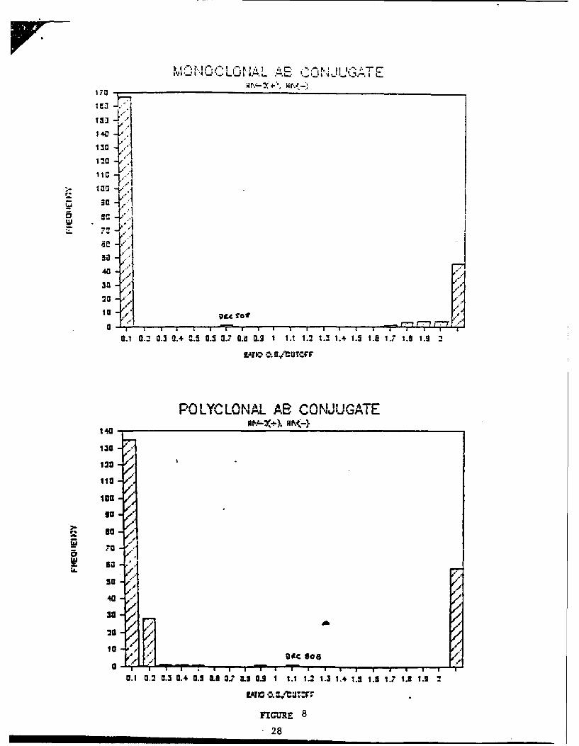
Optimization of HIV-2 EIA Comparison of Monoclonal vs. Polyclonal
Antibody Coniugates
Initial results were based on EIAs utilizing goat polyclonal
anti-human IgG labeled with HRP. For technical reasons (see
below) EIAs are being converted to using HRP conjugated anti-
human monoclonal antibodies to replace the goat polyclonal
conjugate. The monoclonals were generated by immunizing Balb/c
mice with either human IgM or IgG Fc. Spleens from immunized
mice were fused with SP2/0 cells and hybrids screened on the
immunizing Fc protein. We have tested the anti-human IgG and IgM
monoclonals in EIAs using K1 protein. The results are shown in
13
Figure 8. The data indicate that the monoclonal conjugates show
less background than the polyclonal antibody for the negative
samples that we have tested. There is a negligible loss of
signal from the seropositive samples. Our conclusion, based on
these results, is that the monoclonal conjugate is superior to
the polyclonal because of (a) the lower background with negative
samples due to a decrease in non-specific binding, (b) the
absence of effect on seropositive samples, and (c) increased
uniformity in conjugate preparation. The tests that we have
provided utilize the monoclonal conjugates.
Other Optimizations
During the course of optimization, we have tested:
Antigen Concentration - Antigen was titered on the plates in
the range of 0.1 to 1.0 ug/ml. Optimum titration, based on
positive to negative signal ratio, was found to be 0.5 ug/ml
equivalent to 100 ng/well.
Stability of Kit Reagents - Stability testing of the kit is
ongoing. To date we have data on plates and conjugate for 6
months at 40C. All other reagents have a two year shelf life.
Interfering Factors - Samples from individuals that have
tested positive for hepatitis, herpes, CMV, RF and other
interfering agents, but negative for HIV-1, have been tested in
the HIV-2 assay and no interference has been found.
Conjugate Validation - Monoclonal antibodies were conjugated
three separate times and all three lots were validated in the
HIV-2 assay using a series of positive and negative samples.
14
Assay Kinetics - Kinetics were tested by varying incubation
times and temperatures for all incubation steps. Ratios of
positive to negative samples were evaluated to ensure that
equilibrium has been reached at all steps.
Our HIV-2 EIA has been optimized for all of the above
parameters.
Reactivity of Samples from HIV-2 Seropositive Individuals in an
HIV-1 env & QaQ assay.
We have tested HIV-2 samples from Africa on our HIV-1 env &
gag EIA to determine the percentage of samples that cross
reacted. Out of 55 samples, 40 (72%) were positive using the
cutoff calculation of Positive Control X 0.4 for the env & gjg
assay. If the cutoff was decreased 20% (to approximately 0.32 X
Positive Control), the number of samples reading positive goes to
43 (78.2%). In the complementary experiment, we tested HIV-I
seropositives in the HIV-2 EIA. Of the 54 samples tested, 15
(28%) were positive while the remainder were negativ (38,72%).
SUMMARY
Using recombinant DNA techniques, we expressed in E. coli a
protein called KI that contains an immunodominant epitope of the
HIV-2 envelope protein. The location of K1 within HIV-2 is
similar to that of CBre3, a protein derived from the HIV-l
envelope. CBre3 has been cloned and expressed, and its utility
as a diagnostic for HIV-l infection has been established (11).
In this report, we show that Kl serves a similar role for
HIV-2. K1 has been expressed at high levels and techniques
15
developed for large scale purification. The purified protein has
been used in an EIA to identify HIV-2 seropositive samples of
West Africa origin. While only a limited number of HIV-2
seropositive samples have been tested (approximately 80), the
HIV-2 EIA using K1 has high specificity and sensitivity.
Finally, we have expressed other regions of the HIV-2
envelope gene and tested for reactivity with sera from HIV-2
infected individuals. Results show that these other coding
regions are poorly reactive. We have no information to correlate
reactivity with health status of infected individuals.
16
REFERENCES
1. Clavel, F., Guetard, D., Brun-Vezinet, F., Chamaret, S.,Rey, M-A, Santos-Ferreira, Mo., Laurent, A.G., Dauguet, C.,Katlama, C., Rouzioux, C., Klatzmann, D., Champalimaud,J.L., and Montagnier, L. (1986) Science, 233:343-346.
2. Guyader, M., Emerman, M., Sonigo, P., Clavel, F.,Montagnier, L., and Alizon, N. (1987) Nature, 326:662-669.
3. Zagury, J.F., Franchini, G., et al., (1988) Proc. Natl.Acad. Sci., (USA) 85:5941-5945.
4. Horsburgh, C.R., Jr. and Holmberg, S.D. (1988) Transfusion,28:192-195.
5. Cortes, E., Detels, R., et al. (1989) N. Enql. N. Med.,320:953-959.
6. Brun-Vezinet, F., Katlama, C., et al. (1987) Lancet, 1:128-132.
7. Clavel, F., Mansinho, K. et al. (1987) N. Enql. J. Med.,316:1180-1185.
8. Franchini, G., Fargnoli, K.A., et al. (1989) Proc. Natl.
Acad. Sci. (USA) 86:2433-2437.
9. Lautenberger,et al. (1984) Gene Anal. Tech., 1:63.
10. Sambrook, J., Fritsch, E.F., and Maniatis, T. (1989)Molecular Cloning, Cold Spring Harbor Laboratory Press.
11. Marciani, D.J., Thorn, R.M., Riggin, C.H, Beltz, G.A.,a ndHung, C-H, (1989) Recent Advances in Pharmacology andTherapeutics. Valesco et al. eds.
12. Thorn, et al. (1987) J. Clin. Microb., 25:1207-1212.
17
2500 5000 7500 10,000
P x x x P P PK PBH B E B )K HS E/ N)BK je//
FIGURE 1. Location of the K3D DNA fragment within the HIV-2SBL/ISY
clone. K3D is a 2.7 KB KPNI fragment containing the entire envelope
gene and flanking viral DNA. K3D is derived from the cloned virus
HIV-2SBL/ISy and was kindly provided by Dr. G. Franchini.
18
4-.. J . .i. p p m.
cl
* ..• . . ,
'-_4..... ,. ........ -
*--.".n"... .........
.s .. .. . . aa -I , .. .•" -... - ' - - . . . " . "
* - - '..'..
.... . -. . . . . .W
... . .... .. .-.-
~= .. ~..........- ......I....... 0"
. . . . . . . . .. . . .N . -
) ~ ~ ~ l mIS I l
".. .').., .. .
4-4
...........
s-Wt..-. ... ... ... -. 0
... .......... .....
.0
-- bfl. = - b
-- somiDA .1 1 0..
- I *.... 0..9
I Illlllllll~ )0
* M I i w'
* IIIIIIIIIIIII...
* ,nfZ I
--..V . ,4 ,-
o "0
5. i o .a
SV -
t" ffl."t..44 0::," I."' U .0
* .-- :-.--:. .--. ....
.. ...., .. -.-
• . • . ... '...
SI 'VN II i=Q
-9
1 2 3 4 5 6 7 8 9 10 11 12 13
FIGURE 3. Coomassie stained acrylamide gel of proteins fromuninduced and induced cultures of HIV-2 recombinants. Lanes5,7,9,11,13 show the induced K1 protein of HIV-2 (arrow).
20
1 2 3 4 5 6 7 8 9 10 11 12 13
FIGURE 4. Western blot of gel depicted in Figure 2. Blot isreacted with antibody raised against the crossreacting ACCiprotein of SIV.
21
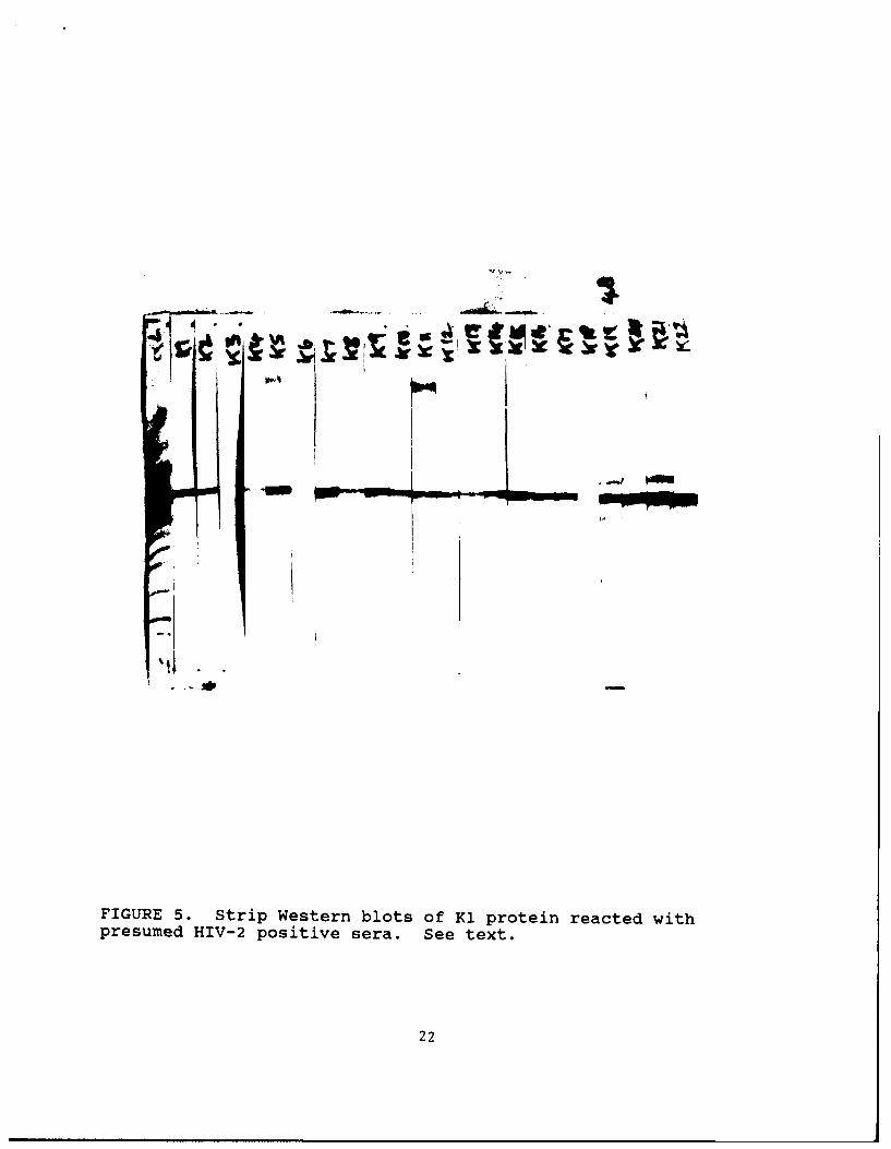
FIGURE 5. Strip Western blots of K1 protein reacted withpresumed HIV-2 positive sera. See text.
22
4 1
FIGURE 5. (continued)
23
TABLE I
REACTIVITY OF RECOMBINANT HIV-2 PROTEINS
CONSTRUCT NO. TESTED POSITIVE
118 23 4
K13 28 0
12 is 2
24
TABLE II
COMPARISON OF RECOMBINANT HIV-2 env
AND HIV-2 VIRAL LYSATE WESTERN BLOTS
Viral LysateWestern Blot
Pos. Neg.
Pos. 46 0HIV-2 env
EIANeg. 0 24
25
U a
Figure 6. Coomassie Stained Acrylamide Gel of Purified KI Protein
Lane 1 Molecular Weight StandardLane 2 Whole E.coli lysateLane 3 BlankLane 4 0.4 ug Purified KiLane 5 1.0 " itLane 6 2.0 " ifLane 7 4.0 " itLane 8 Molecular Weight Standard
26
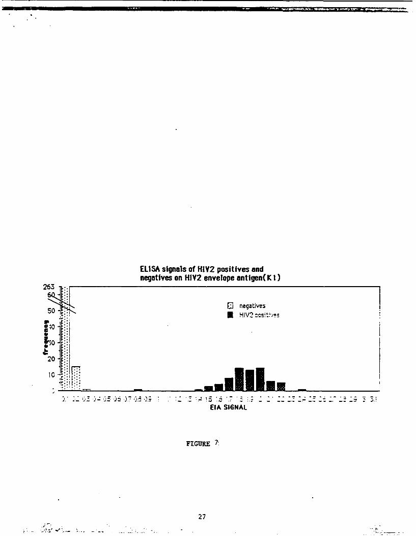
ELISA signals of HIY2 positives andnegatives on HIY2 envelope antigen(K I)
263..
50 -,. 1 negatives
: HIV2 :csvtP/
30S ic
EIA SI6NAL
FIGURE
27
itliA
130
40
13-
I cl-P~ a
0.1 0.2 0.3 Q.* 0W.5 0.5 0.7 CA~ 0.3 1 1.1 1.2 L.T 1.4 1.5 1.5 1.1 1.5 1.9 7
P0 LY-C LONAJ,*L AS C,^,NJ IGATE14U-
120/
lie
In -
50so
30
20
r A
0.1 0.2 5.3 0.4 0.5 ILI 3.7 ILS 9. 1 1.1 1.2 1.3 1.4 1.! 1.5 1.7 IJ 1.2 7
Pro
FIGUE 8
28