Embed Size (px)
Citation preview
AD_________________ Award Number: W81XWH-04-1-0141 TITLE: An Analysis of Rho-PKN Signaling in Prostate Cancer Using Drosophila Genetics PRINCIPAL INVESTIGATOR: Martha E. Betson, Ph.D. CONTRACTING ORGANIZATION: Massachusetts General Hospital Boston, MA 02114-2698 REPORT DATE: January 2006 TYPE OF REPORT: Final PREPARED FOR: U.S. Army Medical Research and Materiel Command Fort Detrick, Maryland 21702-5012 DISTRIBUTION STATEMENT: Approved for Public Release; Distribution Unlimited The views, opinions and/or findings contained in this report are those of the author(s) and should not be construed as an official Department of the Army position, policy or decision unless so designated by other documentation.
REPORT DOCUMENTATION PAGE Form Approved
OMB No. 074-0188 Public reporting burden for this collection of information is estimated to average 1 hour per response, including the time for reviewing instructions, searching existing data sources, gathering and maintaining the data needed, and completing and reviewing this collection of information. Send comments regarding this burden estimate or any other aspect of this collection of information, including suggestions for reducing this burden to Washington Headquarters Services, Directorate for Information Operations and Reports, 1215 Jefferson Davis Highway, Suite 1204, Arlington, VA 22202-4302, and to the Office of Management and Budget, Paperwork Reduction Project (0704-0188), Washington, DC 20503
2. REPORT DATE 3. REPORT TYPE AND DATES COVERED 1. AGENCY USE ONLY (Leave blank) January 2006 Final (1 Jan 2004 – 31 Dec 2005) 4. TITLE AND SUBTITLE 5. FUNDING NUMBERS An Analysis of Rho-PKN Signaling in Prostate Cancer Using Drosophila Genetics
6. AUTHOR(S) Martha E. Betson, Ph.D.
W81XWH-04-1-0141
7. PERFORMING ORGANIZATION NAME(S) AND ADDRESS(ES) Massachusetts General Hospital Boston, MA 02114-2698
8. PERFORMING ORGANIZATION REPORT NUMBER
9. SPONSORING / MONITORING AGENCY NAME(S) AND ADDRESS(ES)
10. SPONSORING / MONITORING AGENCY REPORT NUMBER
U.S. Army Medical Research and Materiel Command Fort Detrick, Maryland 21702-5012
11. SUPPLEMENTARY NOTES Original contains colored plates: ALL DTIC reproductions will be in black and white
12a. DISTRIBUTION / AVAILABILITY STATEMENT Approved for Public Release; Distribution Unlimited
12b. DISTRIBUTION CODE
13. ABSTRACT (Maximum 200 Words) The Rho effector protein kinase N (PKN) has been implicated in prostate cancer. To study the role of PKN andclosely related PRK2 in prostate cancer progression, lentiviral small hairpin RNA constructs have been obtained andwhich knock down expression of PKN and PRK2 in human cells. The constructs will be introduced into prostatecancer cells to study the role of PKN and PRK2 in cellular processes related to tumorigenesis. To identify novelcomponents of the PKN signaling pathway, a genetic screen has been undertaken in the fruit fly Drosophilamelanogaster, which has a well-conserved Pkn gene. So far two potential genetic interactors for Pkn have beenidentified: CKIIa-i3 and RHOGAP71E. Screening is still ongoing so more interactors may be discovered. For anyinteractors identified, the mechanism of interaction with Pkn and the conservation of the pathway in humans the willbe investigated. Taken together these studies should lead to an increased understanding of a poorly characterizedsignaling pathway in flies and humans which may play a role in prostate cancer progression.
14. SUBJECT TERMS 15. NUMBER OF PAGES 17 Drosophila genetics, cell biology, Rho GTPases, protein kinase N
16. PRICE CODE
17. SECURITY CLASSIFICATION OF REPORT
Unclassified
18. SECURITY CLASSIFICATION OF THIS PAGE
Unclassified
19. SECURITY CLASSIFICATION OF ABSTRACT
Unclassified
20. LIMITATION OF ABSTRACT
UnlimitedNSN 7540-01-280-5500 Standard Form 298 (Rev. 2-89)
Prescribed by ANSI Std. Z39-18 298-102

3
Table of Contents
Cover…………………………………………………………………………….…1
SF 298……………………………………………………………………………...2
Introduction…………………………………………………………….………....4
Body………………………………………………………………………………..5
Key Research Accomplishments………………………………………….……..15
Reportable Outcomes…………………………………………………………….15
Conclusions………………………………………………………………………..16
References…………………………………………………………………………17
4
Introduction
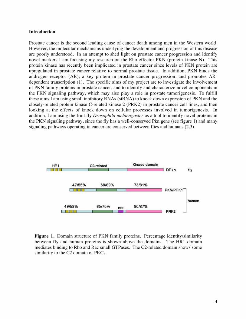
Prostate cancer is the second leading cause of cancer death among men in the Western world.However, the molecular mechanisms underlying the development and progression of this diseaseare poorly understood. In an attempt to shed light on prostate cancer progression and identifynovel markers I am focusing my research on the Rho effector PKN (protein kinase N). Thisprotein kinase has recently been implicated in prostate cancer since levels of PKN protein areupregulated in prostate cancer relative to normal prostate tissue. In addition, PKN binds theandrogen receptor (AR), a key protein in prostate cancer progression, and promotes AR-dependent transcription (1). The specific aims of my project are to investigate the involvementof PKN family proteins in prostate cancer, and to identify and characterize novel components inthe PKN signaling pathway, which may also play a role in prostate tumorigenesis. To fulfillthese aims I am using small inhibitory RNAs (siRNA) to knock down expression of PKN and theclosely-related protein kinase C-related kinase 2 (PRK2) in prostate cancer cell lines, and thenlooking at the effects of knock down on cellular processes involved in tumorigenesis. Inaddition, I am using the fruit fly Drosophila melanogaster as a tool to identify novel proteins inthe PKN signaling pathway, since the fly has a well-conserved Pkn gene (see figure 1) and manysignaling pathways operating in cancer are conserved between flies and humans (2,3). human
Figure 1. Domain structure of PKN family proteins. Percentage identity/similaritybetween fly and human proteins is shown above the domains. The HR1 domainmediates binding to Rho and Rac small GTPases. The C2-related domain shows somesimilarity to the C2 domain of PKCs.
5
Body
Task 1. To characterize the role of PKN family proteins in prostate cancerPrevious studies have indicated that PKN is expressed in prostate cancer cells (1). To confirmthis and check that PRK2 is also expressed, I obtained two commercially available antibodies: amouse anti-PKN antibody (Transduction Labs) and a rabbit anti-PRK2 antibody (Cell Signaling).Both antibodies recognized single bands of the expected sizes in PC3 cells, confirmingexpression of PKN and PRK2 in these cells (data not shown).
As a first attempt to knock down PKN and PRK2 expression in prostate cancer cell lines Idesigned 21-base-pair siRNA oligonucleotides (oligos) targeting two different regions in thehuman PKN transcript/mRNA sequence and two regions in the PRK2 sequence. Whendesigning these oligos, ensured that the sequences used were not identical to any sequences inthe human genome apart from PKN and PRK2. Oligos were transfected into PC3 cells andknockdown of PKN and PRK2 was assessed by western blotting. However, I failed to obtainstrong and consistent knock down, despite trying a variety of different transfection conditions. Itis unclear whether the problem was inefficient knock down per se or inefficient transfection ofthe oligos into the PC3 cells.
At this time the possibility arose of establishing a collaboration with the laboratory of WilliamHahn at the Dana Faber Cancer Institute to generate small hairpin RNAs (shRNA) against PKNand PRK2 in a lentiviral vector. The advantages of using the lentiviral system to introduceshRNAs into prostate cancer cell lines are that the efficiency of DNA transduction is muchhigher than when using standard transfection techniques and that stable cell lines expressing theshRNAs can be generated. In addition, once the virus has been produced in can be used totransfect multiple different prostate cancer cell lines. The Hahn lab cloned five 21-base-pairsequences from human PKN and five from human PRK2 into the lentiviral pLKO.1ps vector(see Table 1 for sequences used).
Construct SequencePKN
G9G10G11G12H1
cgaattccggcccagtggggactgctcggccaccaacctgagcccctccctccgcgcggggactccgaattccggcccagtgggccacctgctcggccaccaacc
PRK2C2C3C4C5C6
gccaccaatagcttctgagttgcaggaattaaatgcacatatccaccatttatacctaccatagcacattcatactgatgtcttgcagcagaaattggatgatat
Table 1. Sequences used to generate PKN and PRK2 shRNA constructs in pLKO.1ps
6
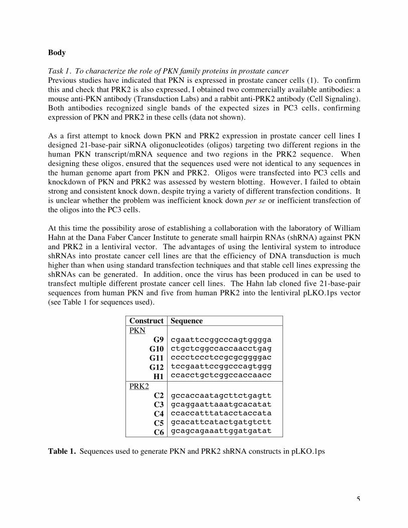
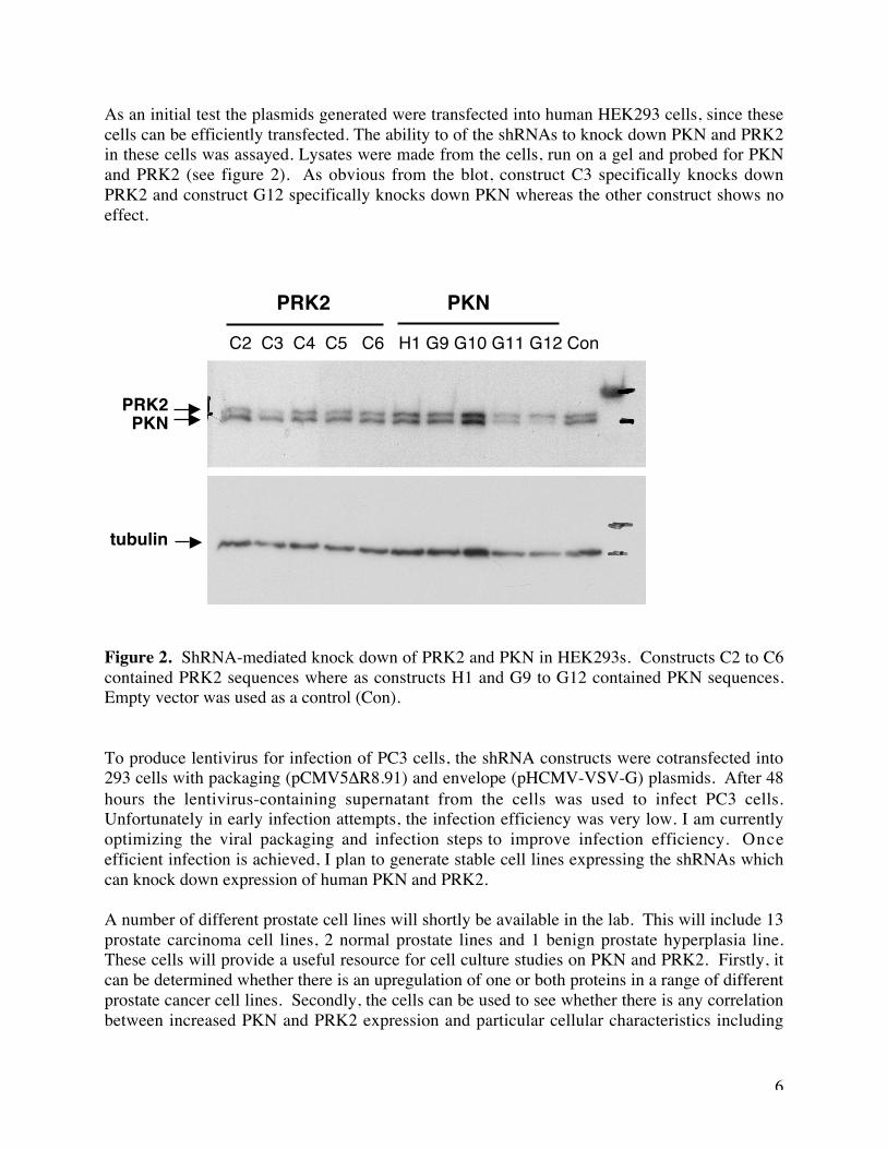
As an initial test the plasmids generated were transfected into human HEK293 cells, since thesecells can be efficiently transfected. The ability to of the shRNAs to knock down PKN and PRK2in these cells was assayed. Lysates were made from the cells, run on a gel and probed for PKNand PRK2 (see figure 2). As obvious from the blot, construct C3 specifically knocks downPRK2 and construct G12 specifically knocks down PKN whereas the other construct shows noeffect.
Figure 2. ShRNA-mediated knock down of PRK2 and PKN in HEK293s. Constructs C2 to C6contained PRK2 sequences where as constructs H1 and G9 to G12 contained PKN sequences.Empty vector was used as a control (Con).
To produce lentivirus for infection of PC3 cells, the shRNA constructs were cotransfected into293 cells with packaging (pCMV5ΔR8.91) and envelope (pHCMV-VSV-G) plasmids. After 48hours the lentivirus-containing supernatant from the cells was used to infect PC3 cells.Unfortunately in early infection attempts, the infection efficiency was very low. I am currentlyoptimizing the viral packaging and infection steps to improve infection efficiency. Onceefficient infection is achieved, I plan to generate stable cell lines expressing the shRNAs whichcan knock down expression of human PKN and PRK2.
A number of different prostate cell lines will shortly be available in the lab. This will include 13prostate carcinoma cell lines, 2 normal prostate lines and 1 benign prostate hyperplasia line.These cells will provide a useful resource for cell culture studies on PKN and PRK2. Firstly, itcan be determined whether there is an upregulation of one or both proteins in a range of differentprostate cancer cell lines. Secondly, the cells can be used to see whether there is any correlationbetween increased PKN and PRK2 expression and particular cellular characteristics including
C2 C3 C4 C5 C6 H1 G9 G10 G11 G12 Con
PKNPRK2
tubulin
PRK2 PKN

7
invasiveness and androgen-independence. Such information can be used to direct future shRNAexperiments on PKN and PRK2.
8
Task 2. To identify novel components of the Rho-Pkn signaling pathway by undertaking agenetic screen in Drosophila
I proposed to undertake a dominant modifier screen in Drosophila to identify novel componentsof the Rho-Pkn signaling pathway. In brief, this involves overexpressing a gene of interest (inthis case Pkn) in a tissue (such as the wing or eye) where it produces a visible, non-lethalphenotype, crossing these flies to flies carrying different mutations and then screening in the nextgeneration for those mutations which suppress or enhance the phenotype. The mutations whichmodify the phenotype are likely to occur in genes which function in the same signaling pathwayas the gene of interest. This technique has been used successfully in the lab to identify novelcomponents of signaling pathways (4, 5).
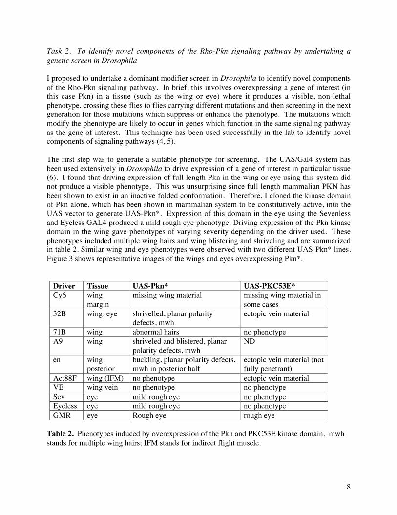
The first step was to generate a suitable phenotype for screening. The UAS/Gal4 system hasbeen used extensively in Drosophila to drive expression of a gene of interest in particular tissue(6). I found that driving expression of full length Pkn in the wing or eye using this system didnot produce a visible phenotype. This was unsurprising since full length mammalian PKN hasbeen shown to exist in an inactive folded conformation. Therefore, I cloned the kinase domainof Pkn alone, which has been shown in mammalian system to be constitutively active, into theUAS vector to generate UAS-Pkn*. Expression of this domain in the eye using the Sevenlessand Eyeless GAL4 produced a mild rough eye phenotype. Driving expression of the Pkn kinasedomain in the wing gave phenotypes of varying severity depending on the driver used. Thesephenotypes included multiple wing hairs and wing blistering and shriveling and are summarizedin table 2. Similar wing and eye phenotypes were observed with two different UAS-Pkn* lines.Figure 3 shows representative images of the wings and eyes overexpressing Pkn*.
Driver Tissue UAS-Pkn* UAS-PKC53E*Cy6 wing
marginmissing wing material missing wing material in
some cases32B wing, eye shrivelled, planar polarity
defects, mwhectopic vein material
71B wing abnormal hairs no phenotypeA9 wing shriveled and blistered, planar
polarity defects, mwhND
en wingposterior
buckling, planar polarity defects,mwh in posterior half
ectopic vein material (notfully penetrant)
Act88F wing (IFM) no phenotype ectopic vein materialVE wing vein no phenotype no phenotypeSev eye mild rough eye no phenotypeEyeless eye mild rough eye no phenotypeGMR eye Rough eye rough eye
Table 2. Phenotypes induced by overexpression of the Pkn and PKC53E kinase domain. mwhstands for multiple wing hairs; IFM stands for indirect flight muscle.
9
Figure 3. A) Rough eye phenotypes induced by driving expression of UAS-Pkn* in the eye. B)Phenotypes induced by driving expression of UAS-Pkn* in the wing.
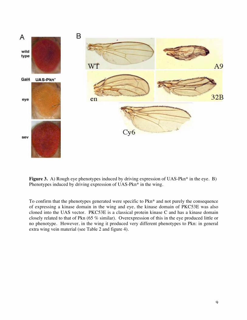
To confirm that the phenotypes generated were specific to Pkn* and not purely the consequenceof expressing a kinase domain in the wing and eye, the kinase domain of PKC53E was alsocloned into the UAS vector. PKC53E is a classical protein kinase C and has a kinase domainclosely related to that of Pkn (65 % similar). Overexpression of this in the eye produced little orno phenotype. However, in the wing it produced very different phenotypes to Pkn: in generalextra wing vein material (see Table 2 and figure 4).
10
Figure 4. Phenotypes induced by driving expression of UAS-PKC53E* in the wing.
The phenotype generated using the Engrailed-Gal4 (en-GAL4) driver seemed the most consistentof the Pkn* phenotypes observed. I recombined en-Gal4 and UAS-Pkn* onto the samechromosome and made a stock. Unfortunately I discovered that this UAS/Gal4 combinationcaused substantial pupal lethality and so was not suitable for generating the large number ofadult flies that are necessary for screening. In parallel I had also generated lines expressing thePkn kinase domain directly under control of the GMR promoter, which drives expression of thePkn protein in the eye. I found that one copy of GMR-Pkn* gave no phenotype but two copies incis gave a mild rough eye which was enhanced with three copies (Figure 5). The rough eyephenotype is particularly marked at the anterior part of the eye and is stronger in females thanmales. Stocks for recombinants carrying three copies of GMR-Pkn* were viable and fertile andso I decided to use them for screening. Since the rough eye phenotype results fromoverexpression of the kinase domain of Pkn without the amino terminal regulatory region, Iwould expect to pull out downstream components of the Pkn signaling pathway rather thanupstream components in the screen.
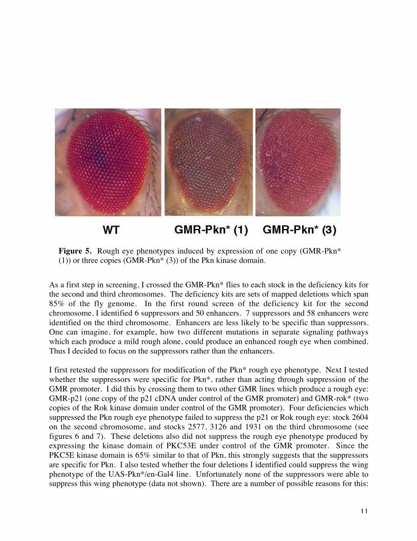
11
As a first step in screening, I crossed the GMR-Pkn* flies to each stock in the deficiency kits forthe second and third chromosomes. The deficiency kits are sets of mapped deletions which span85% of the fly genome. In the first round screen of the deficiency kit for the secondchromosome, I identified 6 suppressors and 50 enhancers. 7 suppressors and 58 enhancers wereidentified on the third chromosome. Enhancers are less likely to be specific than suppressors.One can imagine, for example, how two different mutations in separate signaling pathwayswhich each produce a mild rough alone, could produce an enhanced rough eye when combined.Thus I decided to focus on the suppressors rather than the enhancers.
I first retested the suppressors for modification of the Pkn* rough eye phenotype. Next I testedwhether the suppressors were specific for Pkn*, rather than acting through suppression of theGMR promoter. I did this by crossing them to two other GMR lines which produce a rough eye:GMR-p21 (one copy of the p21 cDNA under control of the GMR promoter) and GMR-rok* (twocopies of the Rok kinase domain under control of the GMR promoter). Four deficiencies whichsuppressed the Pkn rough eye phenotype failed to suppress the p21 or Rok rough eye: stock 2604on the second chromosome, and stocks 2577, 3126 and 1931 on the third chromosome (seefigures 6 and 7). These deletions also did not suppress the rough eye phenotype produced byexpressing the kinase domain of PKC53E under control of the GMR promoter. Since thePKC5E kinase domain is 65% similar to that of Pkn, this strongly suggests that the suppressorsare specific for Pkn. I also tested whether the four deletions I identified could suppress the wingphenotype of the UAS-Pkn*/en-Gal4 line. Unfortunately none of the suppressors were able tosuppress this wing phenotype (data not shown). There are a number of possible reasons for this:
Figure 5. Rough eye phenotypes induced by expression of one copy (GMR-Pkn*(1)) or three copies (GMR-Pkn* (3)) of the Pkn kinase domain.
12
the wing phenotype may be too strong to be modified through removing only one copy of a gene;the suppressors may only participate in Pkn signaling in the eye, not in the wing; or maybe thedeletions identified are not true suppressors of Pkn. Since it is difficult to distinguish betweenthese possibilities at this stage, I decided to continue work on the suppressors I had identified.
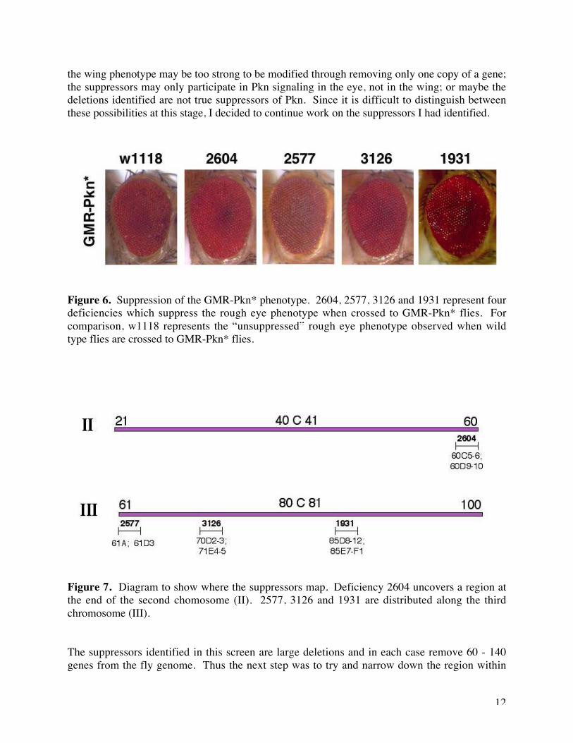
Figure 6. Suppression of the GMR-Pkn* phenotype. 2604, 2577, 3126 and 1931 represent fourdeficiencies which suppress the rough eye phenotype when crossed to GMR-Pkn* flies. Forcomparison, w1118 represents the “unsuppressed” rough eye phenotype observed when wildtype flies are crossed to GMR-Pkn* flies.
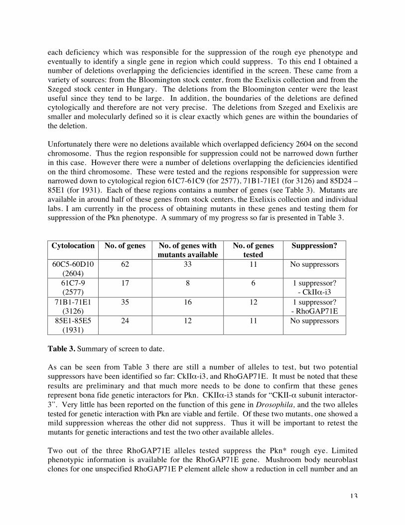
Figure 7. Diagram to show where the suppressors map. Deficiency 2604 uncovers a region atthe end of the second chomosome (II). 2577, 3126 and 1931 are distributed along the thirdchromosome (III).
The suppressors identified in this screen are large deletions and in each case remove 60 - 140genes from the fly genome. Thus the next step was to try and narrow down the region within
13
each deficiency which was responsible for the suppression of the rough eye phenotype andeventually to identify a single gene in region which could suppress. To this end I obtained anumber of deletions overlapping the deficiencies identified in the screen. These came from avariety of sources: from the Bloomington stock center, from the Exelixis collection and from theSzeged stock center in Hungary. The deletions from the Bloomington center were the leastuseful since they tend to be large. In addition, the boundaries of the deletions are definedcytologically and therefore are not very precise. The deletions from Szeged and Exelixis aresmaller and molecularly defined so it is clear exactly which genes are within the boundaries ofthe deletion.
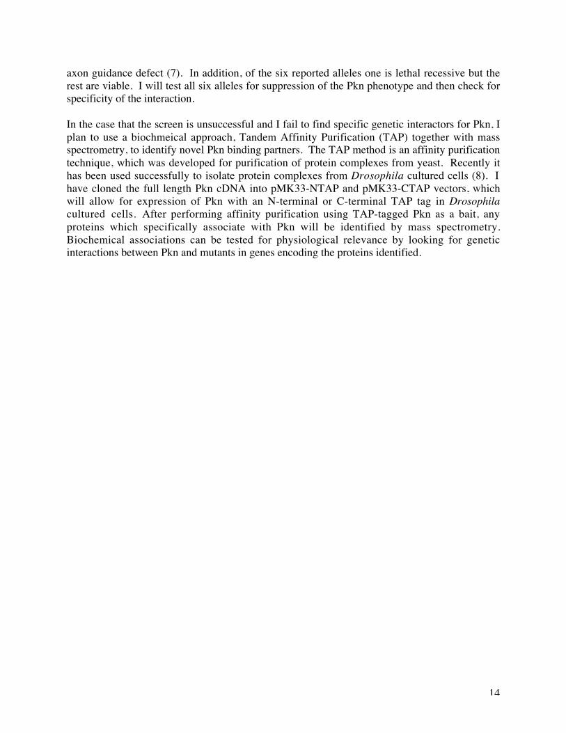
Unfortunately there were no deletions available which overlapped deficiency 2604 on the secondchromosome. Thus the region responsible for suppression could not be narrowed down furtherin this case. However there were a number of deletions overlapping the deficiencies identifiedon the third chromosome. These were tested and the regions responsible for suppression werenarrowed down to cytological region 61C7-61C9 (for 2577), 71B1-71E1 (for 3126) and 85D24 –85E1 (for 1931). Each of these regions contains a number of genes (see Table 3). Mutants areavailable in around half of these genes from stock centers, the Exelixis collection and individuallabs. I am currently in the process of obtaining mutants in these genes and testing them forsuppression of the Pkn phenotype. A summary of my progress so far is presented in Table 3.
Cytolocation No. of genes No. of genes withmutants available
No. of genestested
Suppression?
60C5-60D10(2604)
62 33 11 No suppressors
61C7-9(2577)
17 8 6 1 suppressor?- CkIIα-i3
71B1-71E1(3126)
35 16 12 1 suppressor?- RhoGAP71E
85E1-85E5(1931)
24 12 11 No suppressors
Table 3. Summary of screen to date.
As can be seen from Table 3 there are still a number of alleles to test, but two potentialsuppressors have been identified so far: CkIIα-i3, and RhoGAP71E. It must be noted that theseresults are preliminary and that much more needs to be done to confirm that these genesrepresent bona fide genetic interactors for Pkn. CKIIα-i3 stands for “CKII-α subunit interactor-3”. Very little has been reported on the function of this gene in Drosophila, and the two allelestested for genetic interaction with Pkn are viable and fertile. Of these two mutants, one showed amild suppression whereas the other did not suppress. Thus it will be important to retest themutants for genetic interactions and test the two other available alleles.
Two out of the three RhoGAP71E alleles tested suppress the Pkn* rough eye. Limitedphenotypic information is available for the RhoGAP71E gene. Mushroom body neuroblastclones for one unspecified RhoGAP71E P element allele show a reduction in cell number and an
14
axon guidance defect (7). In addition, of the six reported alleles one is lethal recessive but therest are viable. I will test all six alleles for suppression of the Pkn phenotype and then check forspecificity of the interaction.
In the case that the screen is unsuccessful and I fail to find specific genetic interactors for Pkn, Iplan to use a biochmeical approach, Tandem Affinity Purification (TAP) together with massspectrometry, to identify novel Pkn binding partners. The TAP method is an affinity purificationtechnique, which was developed for purification of protein complexes from yeast. Recently ithas been used successfully to isolate protein complexes from Drosophila cultured cells (8). Ihave cloned the full length Pkn cDNA into pMK33-NTAP and pMK33-CTAP vectors, whichwill allow for expression of Pkn with an N-terminal or C-terminal TAP tag in Drosophilacultured cells. After performing affinity purification using TAP-tagged Pkn as a bait, anyproteins which specifically associate with Pkn will be identified by mass spectrometry.Biochemical associations can be tested for physiological relevance by looking for geneticinteractions between Pkn and mutants in genes encoding the proteins identified.
15
Key Research Accomplishments
• Lentiviral shRNA constructs generated which knock down levels of PKN and PRK2protein when introduced into HEK293
• Transgenic flies generated carrying UAS-Pkn*, UAS-PKC53E*, GMR-Pkn* and GMR-PKC53E*
• Overexpression of the Pkn kinase domain in wing and eye using the UAS/Gal4 system orthe GMR promoter shown to produce phenotypes which can be used for dominantmodifier screen
• Phenotypes have been demonstrated to be specific for Pkn since overexpression of thePKC53 kinase domain produces different phenotypes
• Screen of the second and third chromosome deficiency kits with GMR-Pkn* completed
• 4 deletions identified which specifically suppress the GMR-Pkn* rough eye phenotypeand do not suppress GMR-p21, GMR-rok*, or GMR-PKC53E*
• Narrowed down the regions of suppression to 60C5-60D10, 61C7-61C9, 71B1-71E1 and85D24-85E5.
• In the process of the testing mutants in regions for suppression. Two potentialsuppressors identified so far: CkIIα-i3 and RhoGAP71E
• Generated TAP-tagged Pkn constructs for expression in Drosophila tissue culture cells
Reportable Outcomes
Abstracts: “Genetic analysis of Rho effector kinase, PKN, in Drosophila” in Program andAbstracts for 45th Annual Drosophila Research Conference, Washington DC, March 24-28, 2004
Presentations: Posters at 45th Annual Drosophila Research Conference, Washington DC, March24-28, 2004 and MGH Cancer Center Retreat and Scientific Advisory Board meeting, WoodsHole, October 14th-15th, 2004: “Genetic analysis of Rho effector kinase, PKN, in Drosophila”,and at MGH Cancer Center Retreat and Scientific Advisory Board meeting, Woods Hole,September 30th – October 1st, 2005: “Genetic studies of Drosophila Pkn”.
Infomatics: transgenic flies: UAS-Pkn*, GMR-Pkn*, UAS-PKC53E*, GMR-PKC53E*
16
Conclusions
I have generated and obtained many reagents which I can use for the study of PKN familyproteins in prostate cancer cells. I have acquired antibodies which recognize PKN and PRK2 inPC3 cells, and shRNA lentiviral vectors that specifically knock down human PKN and PRK2 inhuman cells. I am currently optimizing the lentiviral production and infection techniques, andwill soon be able to generate stable prostate cancer cell lines expressing the PKN and PRK2-shRNAs. Then I can analyze the effects of knock down of PKN and PRK2 in these cells withparticular reference to cellular processes involved in tumorigenesis. This may provide novelinsights into the mechanisms underlying prostate cancer development.
In terms of identifying new components of the Pkn signaling pathway, I have generatedtransgenic fly lines overexpressing Pkn with wing and eye phenotypes which can be used forgenetic screens. I have screened the deficiency kits for the 2nd and 3rd chromosomes andidentified 4 deficiencies which specifically suppress the rough eye induced by expression ofPkn*. Using smaller deletions overlapping the regions uncovered by the deficiency, I havenarrowed down the regions responsible for suppression. By testing mutants in genes in thisregion I have identified two potential suppressors: CkIIα-i3 and RhoGAP71E. The most closelyrelated human protein to CkIIα-i3 is intersectin 2. CkIIα-i3 shows 21% identity and 42%similarity to intersectin 2 along most of its length, but intersectin 2 is a much larger protein andcontains multiple domains including a Dbl homology domain (9). The region of homologybetween CkIIα-i3 and intersectin 2 does not lie in any of these recognizable domains, however.
The potential interaction between Pkn and RhoGAP71E is intriguing, given that Pkn is reportedto be an effector of Rho1 and that RhoGAPs (Rho GTPase activator proteins) are upstreamregulators of Rho proteins. Since RhoGAPs generally function to downmodulate Rho activity itmight be anticipated that mutations in a RhoGAP would enhance rather than suppress the Pknmutant phenotype. However, the overexpressed Pkn* lacks the Rho binding domain and so isunlikely to be under Rho1 control. Perhaps a feedback loop is in operation whereby Pkn activatesRhoGAP71E and thereby downmodulates Rho activity. The most closely related human proteinto RhoGAP71E is ARHGAP20, which shows 32% identity and 51% similarity to RhoGAP71Ealong the length of, and a bit beyond, the RhoGAP domain. ARHGAP20-1 is poorlycharacterized but the gene is disrupted in a translocation associated with one case of B-cellchronic lymphocytic leukemia. ARHGAP20 expression is also upregulated in 22 cases of B-cellchronic lymphocytic leukemia (10).
The screening strategy I have adopted to look for novel components of the Pkn signalingpathway looks promising, since two potential genetic interactors for Pkn have already beenidentified, and there are still more genes to test. The challenges now will be to confirm that thegenetic interactors identified represent bona fide components of the Pkn signaling pathway inDrosophila and to determine their mechanism of interaction with Pkn. Ultimately the goal is todetermine whether the signaling pathways characterized in Drosophila are conserved in humansand what role they play in prostate cancer initiation and progression.
17
References
1. Metzger, E., Müller, J.M., Ferrari, S., Buettner, R., and Schüle, R. (2003). A novel inducibletransactivation domain in the androgen receptor: implications for PRK in prostate cancer. EMBOJ. 22, 270-280.
2. Lu, Y., and Settleman, J. (1999). The Drosophila Pkn protein kinase is a Rho/Rac effectortarget required for dorsal closure during embryogenesis. Genes Dev. 13, 1168-1180.
3. Potter, C.J., Turenchalk, G.S., and Xu, T. (2000). Drosophila in cancer research, anexpanding role. Trends Genet. 16, 33-39.
4. Barrett, K., Leptin, M., and Settleman, J. (1997). The Rho GTPase and a putative RhoGEFmediate a signaling pathway for the cell shape changes in Drosophila gastrulation. Cell 91, 905-915.
5. Nolan, K.M., Barrett, K., Lu, Y., Hu, K.Q., Vincent, S., and Settleman, J. (1998). Myoblastcity, the Drosophila homolog of DOCK180/CED-5, is required in a rac signaling pathwayutilized for multiple developmental processes. Genes and Development 12, 3337-3342.
6. Brand, A.H. and Perrimon, N. (1993). Targeted gene expression as a means of altering cellfates and generating dominant phenotypes. Development 118, 401-15.
7. Billuart, P., Winter, C.G., Maresh, A., Zhao, X., and Luo, L. (2001). Regulating axon branchstability: the role of p190 RhoGAP in repressing a retraction signaling pathway. Cell 107, 195-207.
8. Veraksa, A., Bauer, A., and Artavanis-Tsakonas, S. (2005) Analyzing protein complexes inDrosophila with tandem affinity purification-mass spectrometry. Dev. Dyn. 232, 827-834.
9. Pucharcos, C., Estivill, X., de la Luna, S. (2000) Intersectin 2, a new multimodular proteininvolved in clathrin-mediated endocytosis. FEBS Lett. 478, 43-51.
10. Kalla, C., Nentwich, H., Schlotter, M., Mertens, D., Wildenberger, K., Dohner, H.,Stilgenbauer, S. and Lichter, P. (2005) Translocation t(X;11)(q13;q23) in B-cell chroniclymphocytic leukemia disrupts two novel genes. Genes Chromosomes Cancer 42, 128-143.