Embed Size (px)
Citation preview

1
SPROUTY 2: A NOVEL ATTENUATOR OF B-CELL RECEPTOR AND MAPK-Erk
SIGNALING IN CLL
Ashima Shukla1, Karan Rai1, Vipul Shukla1, Nagendra K Chaturvedi1, R. Gregory Bociek2,
Samuel J. Pirruccello3, Hamid Band1,3,4, Runqing Lu1 and Shantaram S. Joshi1*
1Department of Genetics, Cell Biology and Anatomy, 2Department of Internal Medicine, Section of Oncology and Hematology,
3Department of Pathology and Microbiology, College of Medicine, 4Eppley Institute for Research in Cancer and Allied Disease,
University of Nebraska Medical Center, Omaha, NE 68198-6395
*Address Correspondence to: Shantaram S. Joshi, Ph.D. Department of Genetics, Cell Biology and Anatomy University of Nebraska Medical Center 986395 Nebraska Medical Center Omaha, Nebraska 68198-6395 Phone: 402-559-4165 Fax: 402-559-3400 Email: [email protected]
Abstract word count: 239
Text word count: 4,198
Numbers of Figures: 7 + 3 Supplemental
Numbers of Tables: 2 Supplemental
Number of References: 43
Blood First Edition Paper, prepublished online January 25, 2016; DOI 10.1182/blood-2015-09-669317
Copyright © 2016 American Society of Hematology
For personal use only.on April 2, 2018. by guest www.bloodjournal.orgFrom
2
Abstract
Clinical heterogeneity is a major barrier to effective treatment of CLL. Emerging evidence
suggests that constitutive activation of various signaling pathways plays a role in the
heterogeneous clinical outcome of CLL patients. MAPK-Erk signaling represents one such
pathway with a demonstrated role in CLL pathogenesis. In this study, we have investigated the
role of SPRY2 as a negative regulator of receptor and non-receptor tyrosine kinase signaling in
the pathogenesis of CLL. We show that SPRY2 expression is significantly decreased in CLL-
cells, particularly from poor-prognosis patients compared to those from good-prognosis patients.
Over-expression of SPRY2 in CLL-cells from poor-prognosis patients decreased their
proliferation, while increasing their apoptosis. Conversely, down-regulation of SPRY2 in CLL-
cells from good-prognosis patients resulted in increased proliferation. Furthermore, CLL-cells
with low SPRY2 expression grew more rapidly in a xenograft model of CLL. Strikingly, B-cell
specific transgenic over-expression of spry2 in mice led to a decrease in the frequency of B1-
cells, the precursors of CLL-cells in rodents. Mechanistically, we show that SPRY2 attenuates
the BCR and MAPK-Erk signaling by binding to and antagonizing the activities of RAF1, BRAF
and SYK in normal B-cells and CLL-cells. We also show that SPRY2 is targeted by miR-21,
which in turn leads to increased activity of Syk and Erk in CLL-cells. Taken together, these
results establish SPRY2 as a critical negative regulator of BCR-mediated MAPK-Erk signaling in
CLL, thereby providing one of the molecular mechanisms to explain the clinical heterogeneity of
CLL.
Key Points:
1. SPRY2 is down-regulated in CLL-cells from patients with poor-prognosis.
2. SPRY2 is negative regulator of Syk mediated BCR and MAPK-Erk signaling in CLL.
For personal use only.on April 2, 2018. by guest www.bloodjournal.orgFrom
3
Introduction:
Chronic Lymphocytic Leukemia (CLL) is a clinically heterogeneous B-cell neoplasm that
represents the most common form of adult leukemia in the United States1. Based on the
Immunoglobulin variable heavy chain (IgVH) mutational status, chromosomal abnormalities and
cell surface markers, CLL patients are categorized into good or poor-prognosis groups. Recent
studies have identified a small actively-proliferating population of CLL-cells that resides in micro-
anatomical sites known as the proliferation centers (PCs)2. CLL-cells receive diverse stimuli
promoting their proliferation and survival in these PCs3-5. We have previously used Gene
Expression Profiling to decipher the diverse signaling that regulates the survival and
proliferation of CLL-cells in PCs. These studies revealed a critical role for B-cell receptor (BCR)
and MAPK-Erk signaling in the survival and proliferation of CLL-cells5. Furthermore Gardener et
al, have recently reported that 36% of CLL patients possess mutations associated with
activation of MAPK-Erk signaling pathways6. Similarly, BCR-signaling is upregulated in CLL,
providing a chronic stimulus for their proliferation3-5.
Precise regulation of cellular processes, such as those mediated by B-cells, requires
homeostatic integration between intrinsic and extrinsic factors7,8. Deregulation of such
homeostatic mechanisms in CLL-cells can lead to aberrant activation of MAPK-Erk and BCR-
signaling. Constitutive activation of BCR and MAPK-Erk signaling promotes CLL-cell survival
and proliferation9-14. However, the molecular mechanisms that lead to the constitutive activation
of these pathways have not been fully explored. Identifying novel regulators of these pathways
in CLL is crucial for understanding the disease biology and for the eventual development of
targeted therapies.
To identify potential regulators of BCR and MAPK-Erk signaling in CLL, we performed a
transcriptome analysis for genes that are differentially expressed in CLL patients with good vs.
For personal use only.on April 2, 2018. by guest www.bloodjournal.orgFrom
4
poor-prognosis. Of interest in relationship to MAPK-Erk signaling, we observed that expression
of Sprouty-2 (SPRY2), a member of the sprouty protein family to be significantly down-regulated
in CLL-cells from poor-prognosis patients compared to those from good-prognosis patients.
Sprouty proteins play key roles in maintaining cellular homeostasis by attenuating signaling,
downstream to several ligand induced receptor tyrosine kinases (RTKs)7-10. Hence, we
reasoned that SPRY2 might act as a negative regulator of BCR-signaling to inhibit the survival
and proliferation of CLL-cells. Therefore, we hypothesized that low levels of SPRY2 lead to a
state of constitutive activation of BCR and MAPK-Erk signaling in poor-prognosis CLL patients.
Consistent with such a possibility, a recent study demonstrated the induction of SPRY2, but not
SPRY1, downstream of BCR-signaling in mouse B-cells15. This study also showed that SPRY2
levels negatively correlate with Erk signaling in mouse B-cells, a finding similar to that described
in other cellular systems9,10,15. However, the molecular mechanism by which SPRY2 functions
as a negative regulator of BCR-signaling has not been deciphered. Moreover, the role of
SPRY2 in B-cell development and function are unknown. SPRY2 was previously shown to be
down-regulated in Diffuse Large B-cell Lymphoma but the functional significance of this down-
regulation remains ambiguous15.
In the present study, we identify SPRY2 down-regulation as a marker of poor-prognosis in CLL
and demonstrate that loss of SPRY2 provides a novel mechanism to constitutively activate BCR
and MAPK-Erk signaling in CLL through Spleen Tyrosine Kinase (Syk). Finally, we show that
SPRY2 is targeted by miR-21 in poor-prognosis CLL that leads to a constitutively activated state
of BCR and MAPK-Erk signaling in CLL-cells.
Methods:
Isolation of CLL-cells from patients and nB-cells from healthy donors: PB from CLL
patients/healthy donors was obtained under an approved IRB protocol. Mononuclear cells were
For personal use only.on April 2, 2018. by guest www.bloodjournal.orgFrom
5
isolated using Lymphoprep (StemCell-Technologies) following manufacturers instruction.
CLL/normal B-cells (nB-cells) were isolated by negative selection using CLL-cells/B-cell
isolation kit Milteny Biotech. Purity of the isolated CLL/nB-cells were tested by flow cytometry
using CD19+CD5+/only CD19+ cell surface markers. When the CLL-cell number was more than
90% in the peripheral blood from patients, the cells purification step was not performed.
Calcium mobilization assay: Calcium influx assay of human nB-cells from PB and murine
splenic B-cells was performed and analyzed as described.16
Animal studies: CD19-cre animals were kindly provided by Dr. Runqing Lu, UNMC. Spry2
conditional transgenic mouse17 was purchased from MMRRC, UNC. To generate CD19-
cre;Spry2(tg) mice we crossed CD19-cre and spry2(tg)flox/flox. Overexpression of spry2 in B-cells
was tested by western blot. Expression of spry2 was tracked by GFP+ cells in flow cytometry
experiments. NOD-SCID-Gamma chain deficient (NSG) mice were purchased from Jackson
Laboratory. All animal experiments were conducted under the IACUC approved protocols.
Nucleofection: Primary B-cells/CLL-cells and Mec-1 were nucleofected with Spry2-cDNA or
Empty plasmid using primary B-cells and V kit (Lonza), program U-16 and X-00119.
Western blot analysis: As previously described, western blotting was performed to analyze
protein expression20. (Continued supplementary-methods).
Results:
Spry2 expression is lower in CLL-cells from poor-prognosis patients: CLL is clinically
heterogeneous, with varying clinical outcomes and possibly distinct molecular pathogenesis. In
order to identify molecular mechanisms associated with the poor-prognosis CLL, we performed
RNA-sequencing based transcriptome analysis of 15 CLL samples: 7 from good-prognosis and
8 from poor-prognosis patients. Poor-prognosis patients were defined by un-mutated IgVH-
For personal use only.on April 2, 2018. by guest www.bloodjournal.orgFrom
6
segments, Ch11q22 deletion, Ch17p deletion, trisomy Ch12 and/or high CD38 expression.
Good-prognosis patients were defined as having mutated IgVH, Ch13q14 deletions and/or low
CD38 expression (criteria used for Figure 1A-C). Using these approaches, the expression levels
of 146 genes were found to be significantly different between good vs. poor-prognosis CLL
samples. We further selected genes based on their roles in B-cell biology, or their putative
tumor suppressor or pro-oncogenic functions. Interestingly, spry2 was found to be significantly
(3.4 log2-fold) down-regulated in the CLL-cells from poor-prognosis patients compared to those
from good-prognosis (Figure 1A). The differential expression was confirmed using western
blotting and real-time PCR of CLL-cells from good vs. poor-prognosis patients (Figure 1B&C).
High CD38 positivity of CLL-cells is associated with poor patient outcomes and constitutive
activation of MAPK-Erk signaling21. Therefore, we further compared spry2 expression in a larger
cohort of 38 patients. Spry2 was significantly down-regulated (~2.8 folds) in CLL-cells of
patients with high CD38 expression compared to those with low CD38 expression (Figure 1D).
Examination of the expression of other sprouty family members (SPRY1, 3 and 4) in our data-
set showed that none of them were significantly differentially expressed between the good and
poor-prognosis CLL-cells (data not shown). Thus, our results show that the expression of
SPRY2 is significantly down-regulated in CLL-cells from patients with poorer outcomes.
SPRY2 is induced upon BCR cross-linking of nB-cells and CLL-cells, and functions as a
negative-regulator of BCR-signaling: It is well established that BCR-signaling is critical for
survival and proliferation of CLL-cells; however, no known recurrent mutations have so far been
identified in this pathway among CLL patients. Moreover, it is known that SPRY2 functions as a
negative-feedback regulator of RTKs22-25. To investigate if SPRY2 is regulated by BCR-
signaling, we stimulated nB-cells and CLL-cells using BCR-crosslinking with 5µg of anti-IgM/IgD
antibody. Cells were collected at 0, 6, 12, 24 and 48 hours after stimulation and the expression
of SPRY2 was determined by western blotting. We observed a gradual increase over time in
For personal use only.on April 2, 2018. by guest www.bloodjournal.orgFrom
7
SPRY2 expression in BCR stimulated nB-cells (Figure 2A upper-panel). On the other hand,
primary CLL-cells and Mec-1 CLL-cell-line exhibited a biphasic induction of SPRY2 (Figure 2A
middle&lower-panels). In CLL-cells, SPRY2 expression peaked at 12 hours upon stimulation
and declined at later time points. Thus, we conclude that SPRY2 expression induced by BCR-
crosslinking is sustained in nB-cells but follows a cyclical expression pattern in CLL-cells and is
not sustained.
We next studied the effect of down-regulating SPRY2 expression on BCR-signaling in nB-cells.
Compared to the scramble siRNA control, both SPRY2 siRNAs led to a decrease in the SPRY2
protein levels (Figure 2B). Next, we transfected nB-cells and compared their anti-IgM-induced
calcium influx with that of unperturbed control following with the Indo-1 dye. Compared to un-
transfected and scrambled siRNA controls, B-cells with SPRY2 knockdown exhibited elevated
calcium influx (Figure 2C). Even the basal levels of calcium were increased in B-cells upon
SPRY2 knockdown. Taken together, these results establish that SPRY2 functions as a negative
regulator of BCR-signaling.
SPRY2 depletion in human CLL-cells from Good-prognosis patients lead to increased
proliferation. To assess if the down-regulation of SPRY2 expression in poor vs. good-
prognosis CLL-cells is of functional consequence, we examined the impact of SPRY2 depletion
in good-prognosis CLL-cells. For this purpose, we isolated CLL-cells from newly-diagnosed
patients with good-prognosis CLL. We also used Mec-1 human CLL-cell-line, a widely-used
cultured CLL-cell model, for these studies. Mec-1 cells have an intermediate level of SPRY2
relative to primary good and poor-prognosis CLL-cells (Figure 2A). As SPRY2 expression is
induced in CLL-cells upon BCR stimulation (Figure 2A), we assessed the impact of SPRY2
depletion on anti-IgM/anti-IgD induced Mec-1 cell proliferation, by analyzing changes in Ki-67+
fraction in FACS analyses. SPRY2 knockdown led to an increase in proliferation of Mec-1 cells
relative to scrambled siRNA control under both anti-IgM and anti-IgD cross-linking condition
For personal use only.on April 2, 2018. by guest www.bloodjournal.orgFrom
8
(Figure 3A). Next, we transfected primary human CLL-cells isolated from PB of 7 different good-
prognosis patients expressing higher levels of SPRY2 with siRNAs against SPRY2. 24 hours
post-transfection, the cells were subjected to BCR cross-linking and proliferation was assessed
48 hours later. Knockdown of SPRY2 led to an increase in the number of proliferating cells
compared to the scrambled siRNA transfected cells (Figure 3B-D). We observed an increase in
proliferation upon SPRY2 depletion compared to scrambled controls, with increased
proliferation seen in 6 out of 7 with siRNA-B and in 5 out of 7 cases with siRNA-A mediated
knockdown. The effects on proliferation were consistent with a more robust SPRY2 knockdown
with siRNA-B than siRNA-A (Figure 3A). SPRY2 knockdown in these cells also showed
decrease in survival however; the values did not reach statistical significance. This might be due
to use of anti-IgM antibodies for induction of SPRY2 expression as anti-IgM-stimulation itself
have positive effect on CLL-cells survival. Therefore, we reason that differences in the effects
on survival may have been masked by anti-IgM stimulation. However, more CLL samples are
needed to be analyzed to confirm these findings. These findings indicate that SPRY2 functions
as a key negative feedback regulator of BCR-signaling in CLL-cells, thus limiting their BCR
induced proliferation.
SPRY2 expression induces apoptosis in human CLL-cells from poor-prognosis patients:
To further elucidate the underlying basis for the down-regulation of SPRY2 expression in CLL-
cells from poor-prognosis patients, we also examined the impact of ectopic SPRY2 expression
on CLL-cells with low SPRY2 expression. First, we examined the impact of increasing the
expression of SPRY2 by transfecting Mec-1 cells with a SPRY2 construct co-expressing GFP.
At 48 and 72 hours post-transfection, we observed an average of 58% and 18% of SPRY2-
GFP+ expressing Mec-1 cells to be AnnexinV+, respectively, compared to 36% and 11% (Figure
4A&B). Next, we isolated CLL-cells from five different poor-prognosis patients, ectopically
expressed Spry2 to measure the proportion of AnnexinV+ cells after 48 hours (Figure 4C). An
For personal use only.on April 2, 2018. by guest www.bloodjournal.orgFrom
9
increase in apoptosis was observed in CLL-cells from all patients (Figure 4C&D). Moreover, we
did not observe a significant impact on the proliferation of these cells compared to controls
probably because these cells were not stimulated with anti-IgM antibodies. We choose not to
stimulate these cells with anti-IgM because BCR stimulation through anti-IgM would have further
induce SPRY2 levels. Thus, our results demonstrate that low levels of SPRY2 in poor-prognosis
patients contribute to a survival advantage for CLL-cells.
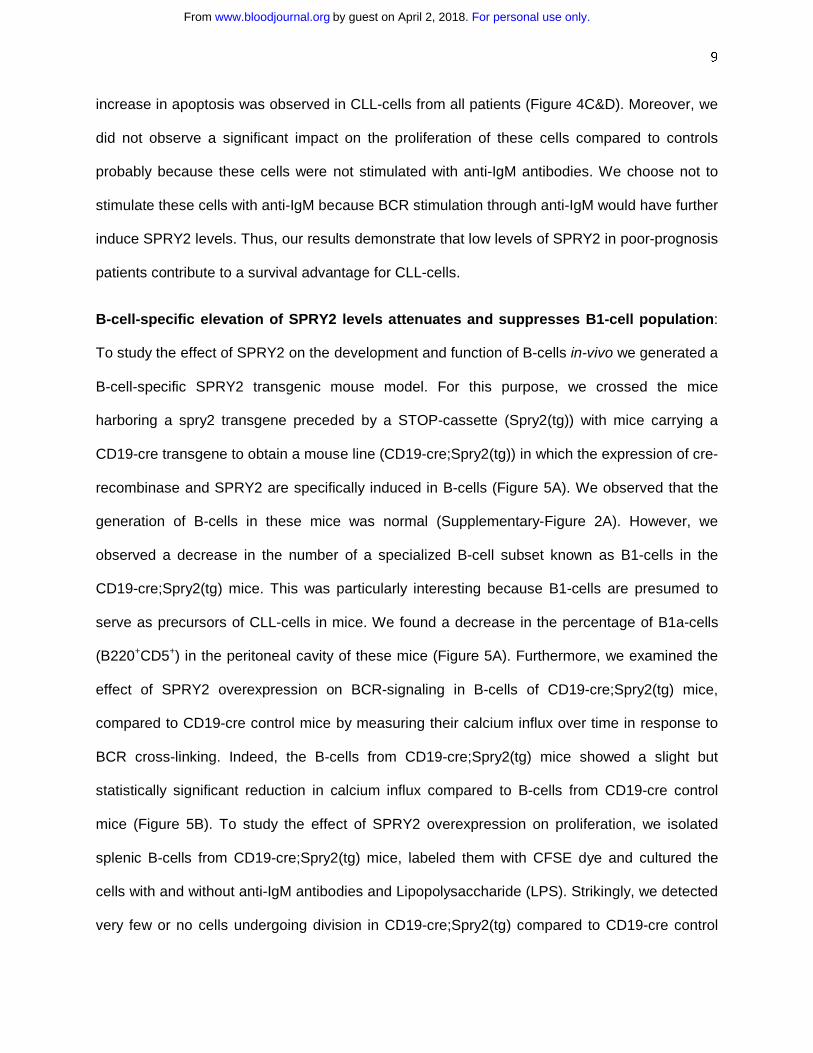
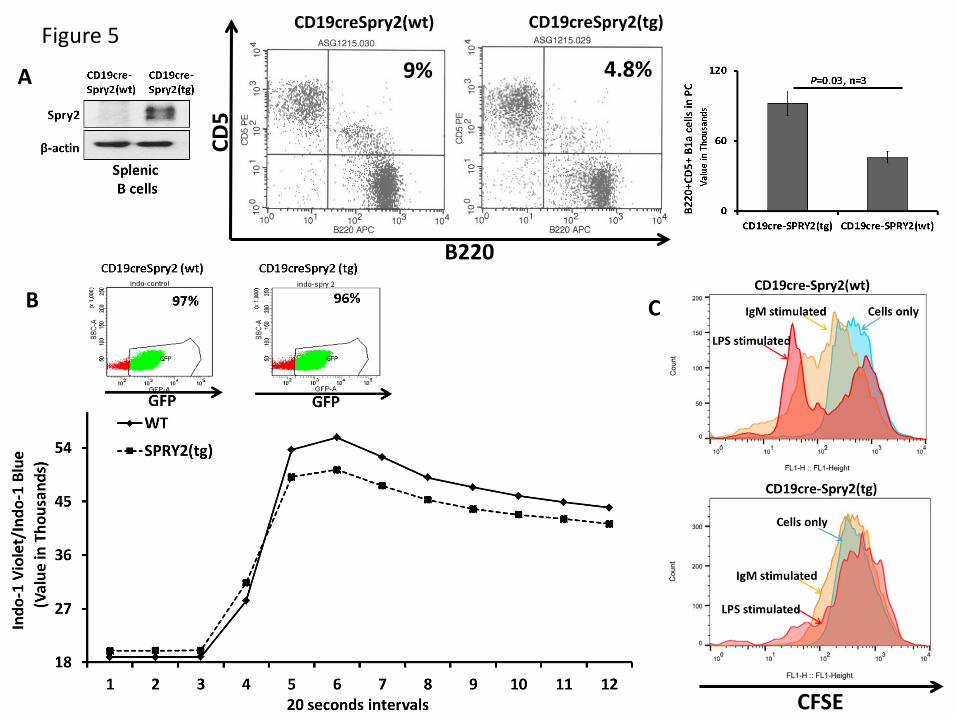
B-cell-specific elevation of SPRY2 levels attenuates and suppresses B1-cell population:
To study the effect of SPRY2 on the development and function of B-cells in-vivo we generated a
B-cell-specific SPRY2 transgenic mouse model. For this purpose, we crossed the mice
harboring a spry2 transgene preceded by a STOP-cassette (Spry2(tg)) with mice carrying a
CD19-cre transgene to obtain a mouse line (CD19-cre;Spry2(tg)) in which the expression of cre-
recombinase and SPRY2 are specifically induced in B-cells (Figure 5A). We observed that the
generation of B-cells in these mice was normal (Supplementary-Figure 2A). However, we
observed a decrease in the number of a specialized B-cell subset known as B1-cells in the
CD19-cre;Spry2(tg) mice. This was particularly interesting because B1-cells are presumed to
serve as precursors of CLL-cells in mice. We found a decrease in the percentage of B1a-cells
(B220+CD5+) in the peritoneal cavity of these mice (Figure 5A). Furthermore, we examined the
effect of SPRY2 overexpression on BCR-signaling in B-cells of CD19-cre;Spry2(tg) mice,
compared to CD19-cre control mice by measuring their calcium influx over time in response to
BCR cross-linking. Indeed, the B-cells from CD19-cre;Spry2(tg) mice showed a slight but
statistically significant reduction in calcium influx compared to B-cells from CD19-cre control
mice (Figure 5B). To study the effect of SPRY2 overexpression on proliferation, we isolated
splenic B-cells from CD19-cre;Spry2(tg) mice, labeled them with CFSE dye and cultured the
cells with and without anti-IgM antibodies and Lipopolysaccharide (LPS). Strikingly, we detected
very few or no cells undergoing division in CD19-cre;Spry2(tg) compared to CD19-cre control
For personal use only.on April 2, 2018. by guest www.bloodjournal.orgFrom
10
cells with or without BCR stimulation (Figure 5C). Taken together, these results show that Spry2
functions to limit the numbers of B1-cells (CLL-precursors) in mice and negatively regulates
BCR-signaling and associated proliferation in mouse B-cells.
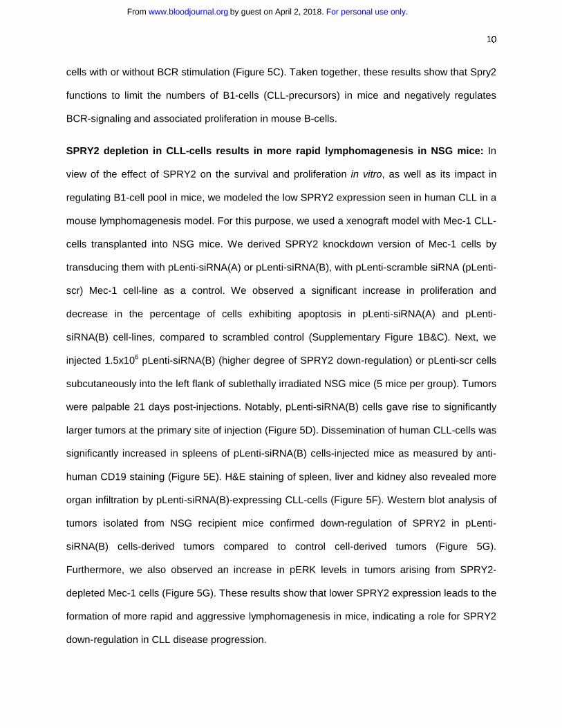
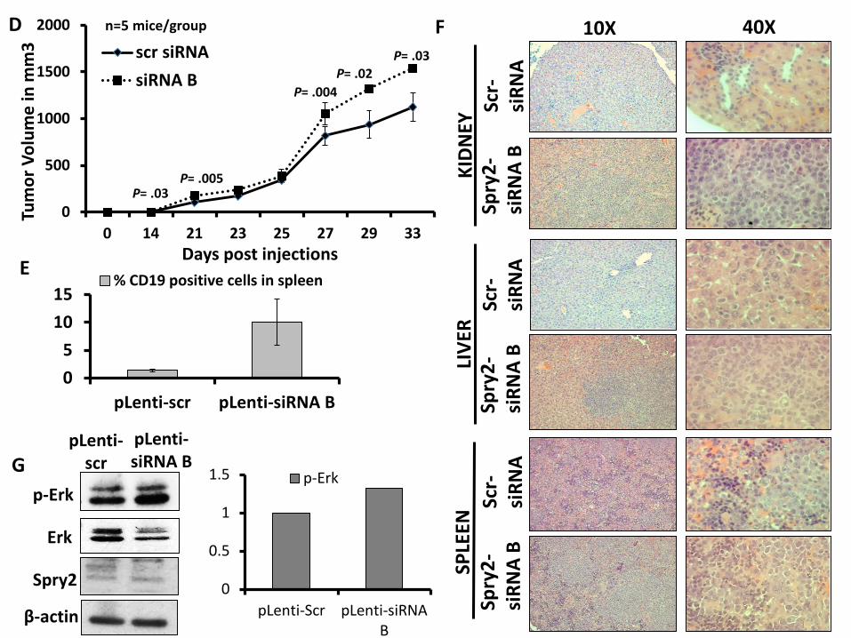
SPRY2 depletion in CLL-cells results in more rapid lymphomagenesis in NSG mice: In
view of the effect of SPRY2 on the survival and proliferation in vitro, as well as its impact in
regulating B1-cell pool in mice, we modeled the low SPRY2 expression seen in human CLL in a
mouse lymphomagenesis model. For this purpose, we used a xenograft model with Mec-1 CLL-
cells transplanted into NSG mice. We derived SPRY2 knockdown version of Mec-1 cells by
transducing them with pLenti-siRNA(A) or pLenti-siRNA(B), with pLenti-scramble siRNA (pLenti-
scr) Mec-1 cell-line as a control. We observed a significant increase in proliferation and
decrease in the percentage of cells exhibiting apoptosis in pLenti-siRNA(A) and pLenti-
siRNA(B) cell-lines, compared to scrambled control (Supplementary Figure 1B&C). Next, we
injected 1.5x10⁶ pLenti-siRNA(B) (higher degree of SPRY2 down-regulation) or pLenti-scr cells
subcutaneously into the left flank of sublethally irradiated NSG mice (5 mice per group). Tumors
were palpable 21 days post-injections. Notably, pLenti-siRNA(B) cells gave rise to significantly
larger tumors at the primary site of injection (Figure 5D). Dissemination of human CLL-cells was
significantly increased in spleens of pLenti-siRNA(B) cells-injected mice as measured by anti-
human CD19 staining (Figure 5E). H&E staining of spleen, liver and kidney also revealed more
organ infiltration by pLenti-siRNA(B)-expressing CLL-cells (Figure 5F). Western blot analysis of
tumors isolated from NSG recipient mice confirmed down-regulation of SPRY2 in pLenti-
siRNA(B) cells-derived tumors compared to control cell-derived tumors (Figure 5G).
Furthermore, we also observed an increase in pERK levels in tumors arising from SPRY2-
depleted Mec-1 cells (Figure 5G). These results show that lower SPRY2 expression leads to the
formation of more rapid and aggressive lymphomagenesis in mice, indicating a role for SPRY2
down-regulation in CLL disease progression.
For personal use only.on April 2, 2018. by guest www.bloodjournal.orgFrom
11
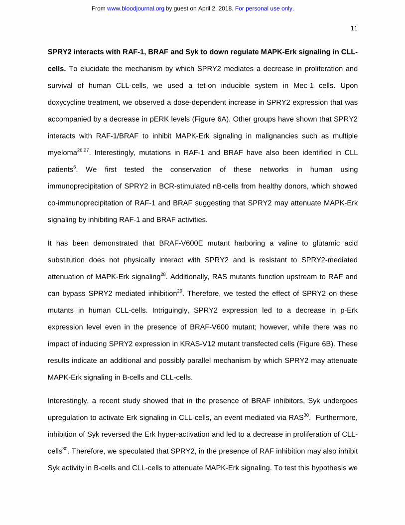
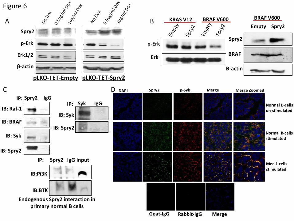
SPRY2 interacts with RAF-1, BRAF and Syk to down regulate MAPK-Erk signaling in CLL-
cells. To elucidate the mechanism by which SPRY2 mediates a decrease in proliferation and
survival of human CLL-cells, we used a tet-on inducible system in Mec-1 cells. Upon
doxycycline treatment, we observed a dose-dependent increase in SPRY2 expression that was
accompanied by a decrease in pERK levels (Figure 6A). Other groups have shown that SPRY2
interacts with RAF-1/BRAF to inhibit MAPK-Erk signaling in malignancies such as multiple
myeloma26,27. Interestingly, mutations in RAF-1 and BRAF have also been identified in CLL
patients6. We first tested the conservation of these networks in human using
immunoprecipitation of SPRY2 in BCR-stimulated nB-cells from healthy donors, which showed
co-immunoprecipitation of RAF-1 and BRAF suggesting that SPRY2 may attenuate MAPK-Erk
signaling by inhibiting RAF-1 and BRAF activities.
It has been demonstrated that BRAF-V600E mutant harboring a valine to glutamic acid
substitution does not physically interact with SPRY2 and is resistant to SPRY2-mediated
attenuation of MAPK-Erk signaling28. Additionally, RAS mutants function upstream to RAF and
can bypass SPRY2 mediated inhibition29. Therefore, we tested the effect of SPRY2 on these
mutants in human CLL-cells. Intriguingly, SPRY2 expression led to a decrease in p-Erk
expression level even in the presence of BRAF-V600 mutant; however, while there was no
impact of inducing SPRY2 expression in KRAS-V12 mutant transfected cells (Figure 6B). These
results indicate an additional and possibly parallel mechanism by which SPRY2 may attenuate
MAPK-Erk signaling in B-cells and CLL-cells.
Interestingly, a recent study showed that in the presence of BRAF inhibitors, Syk undergoes
upregulation to activate Erk signaling in CLL-cells, an event mediated via RAS30. Furthermore,
inhibition of Syk reversed the Erk hyper-activation and led to a decrease in proliferation of CLL-
cells30. Therefore, we speculated that SPRY2, in the presence of RAF inhibition may also inhibit
Syk activity in B-cells and CLL-cells to attenuate MAPK-Erk signaling. To test this hypothesis we
For personal use only.on April 2, 2018. by guest www.bloodjournal.orgFrom
12
performed immuno-precipitation assays as described in Supplementary-Data. Intriguingly,
SPRY2 was found to physically interact with Syk but not with Bruton’s Tyrosine kinase (BTK)
and PI3K in B-cells as determined by reciprocal immuno-precipitation experiments (Figure 6C).
Also, using immunofluorescence analysis, we observed a co-localization of SPRY2 with p-Syk
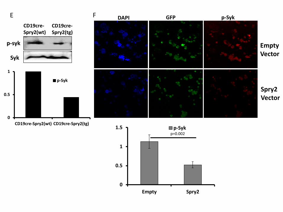
in nB-cells and Mec-1 CLL-cells (Figure 6D). We next tested the effect of SPRY2
overexpression on the activated form of Syk by measuring the levels of p-Syk in splenic-B-cells
from CD19-cre;Spry2(tg) mice and SPRY2 overexpressing Mec-1 cells by immunofluorescence.
Notably, we observed a decrease in p-Syk expression in B-cells of CD19-cre;Spry2(tg) mice
and Mec-1 cells overexpressing SPRY2 compared to B-cells from CD19-cre mice and Empty
vector Mec-1 cells, respectively (Figure 6E&F). We also observed a synergistic effect of SPRY2
with BRAF and Syk inhibitors in the presence of BRAF-V600 mutant, again highlighting the dual
mechanism through with SPRY2 downregulates MAPK-Erk signaling in CLL-cells
(Supplementary Fig3). Thus, our results demonstrate that SPRY2 attenuates BCR mediated
MAPK-Erk signaling by simultaneous inhibition of RAF and Syk activity in B-cells and CLL-cells.
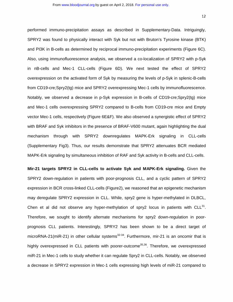
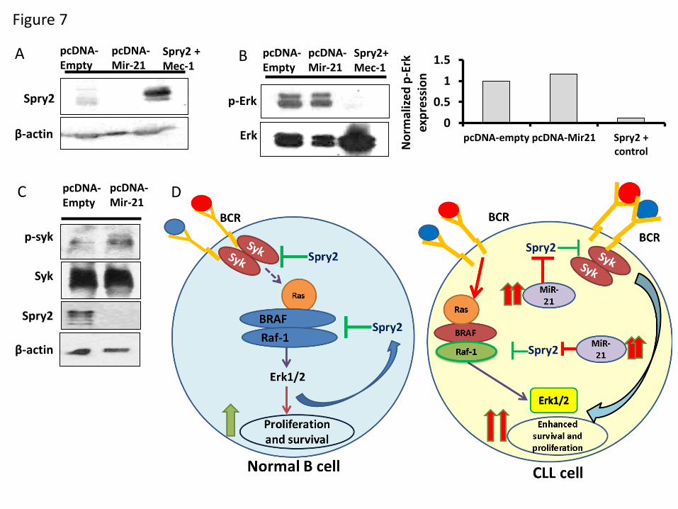
Mir-21 targets SPRY2 in CLL-cells to activate Syk and MAPK-Erk signaling. Given the
SPRY2 down-regulation in patients with poor-prognosis CLL, and a cyclic pattern of SPRY2
expression in BCR cross-linked CLL-cells (Figure2), we reasoned that an epigenetic mechanism
may deregulate SPRY2 expression in CLL. While, spry2 gene is hyper-methylated in DLBCL,
Chen et al did not observe any hyper-methylation of spry2 locus in patients with CLL31.
Therefore, we sought to identify alternate mechanisms for spry2 down-regulation in poor-
prognosis CLL patients. Interestingly, SPRY2 has been shown to be a direct target of
microRNA-21(miR-21) in other cellular systems32-34. Furthermore, mir-21 is an oncomir that is
highly overexpressed in CLL patients with poorer-outcome35,36. Therefore, we overexpressed
miR-21 in Mec-1 cells to study whether it can regulate Spry2 in CLL-cells. Notably, we observed
a decrease in SPRY2 expression in Mec-1 cells expressing high levels of miR-21 compared to
For personal use only.on April 2, 2018. by guest www.bloodjournal.orgFrom

13
those with an empty-vector control (Figure 7A). Intriguingly, concurrent with SPRY2 down-
regulation in miR-21 overexpressing Mec-1 cells, we also observed an increase in the levels of
p-Syk and p-Erk (Figure 7B-D). In summary, our results here demonstrate that Spry2 is
deregulated by miR-21 in CLL-cells. The results here also, provide a mechanism by which miR-
21 promotes CLL progression via down-regulation of SPRY2.
Discussion:
Clinical heterogeneity is a major problem in the management of CLL. The heterogeneous
clinical outcome in patients appears to be the results of interactions between several molecules
and cellular pathways. Consequently, in order to treat CLL effectively, a better understanding of
the molecules and cellular pathways that contribute to such heterogeneous clinical outcome is
needed. In the present study we have elucidated the molecular basis for aberrant BCR and
MAPK-Erk signaling, where SPRY2 acts as a negative regulator for survival and proliferation of
CLL-cells. Moreover, SPRY2 may represent a molecule responsible for maintaining the clinical
heterogeneity in CLL.
Notably, our findings identify SPRY2 as a negative-feedback-regulator downstream to BCR
stimulation that is critical for attenuation of MAPK-Erk signaling. Moreover, SPRY2 may function
as an attenuator of tonic BCR-signaling in CLL-cells and B-cells as basal levels of signaling are
elevated upon SPRY2 knockdown. While, SPRY2 over-expression in CD19-cre;Spry2(tg) mice
led to impaired BCR-signaling in B-cells, we did not observe an apparent defect in the overall
generation of B-cells. Notably though, and directly relevant to role of SPRY2 down-regulation in
CLL, we observed a decrease in the B1-cells. These results indicate a potential role for SPRY2
in the development of B1-cells and hence, possibly in the initiation of CLL37. It will be of
considerable interest to breed the CD19-cre;Spry2(tg) mice to established models of CLL such
as Eµ-Tcl1 or IRF4-/-Vh11 mice to directly study the role of SPRY2 in development of CLL37.
For personal use only.on April 2, 2018. by guest www.bloodjournal.orgFrom
14
Interestingly, SPRY2 levels are down-regulated in CLL-cells isolated from IRF4-/-Vh11 mouse
model (data not shown). Even though no other defects in B-cell development are apparent in
CD19-cre;Spry2(tg) mice, it will be interesting to further study the induction of functional humoral
responses in these mice given our findings that aberration of SPRY2 expression deregulate
BCR signaling.
Functionally, our studies have shown that SPRY2 mediated regulation of BCR-signaling is
important for survival and proliferation of CLL-cells. Moreover, SPRY2 plays a role in controlling
the disease aggressiveness as knockdown of SPRY2 in Mec-1 CLL-cells resulted in more
aggressive disease in NSG mice. We show that SPRY2 expression in nB-cells and CLL-cells
leads to decreased p-Syk levels. Mechanistically, BCR-signaling in CLL-cells constitutes a
signaling axis whereby Syk can also function to regulate the activation of MAPK-Erk signaling
(Figure 7D). Of significant interest in this regard, we observed that SPRY2 not only interacts
with and antagonizes RAF/BRAF activities but that it also interacts and co-localizes with Syk
near the plasma membrane to disrupt the MAPK-Erk signaling axis. Therefore, we propose a
model in which SPRY2 functions to regulate two different nodes of an overlapping signaling axis
by attenuating Syk as well as RAF/BRAF activity (Figure 7D). Importantly, our studies indicate
that SPRY2 functions as a broad attenuator of BCR-signaling as evidenced by a decrease in
calcium influx upon BCR-stimulation, a process mediated by PLCγ2-signaling downstream of
Syk activation.
SPRY2 has been shown to interact with RTKs and the associated adaptor molecules through its
SH2-domain-binding motifs generated upon its phosphorylation10. Syk is a non-receptor tyrosine
kinase harboring multiple SH2-domains. Hence, it is tempting to speculate that SPRY2 might
interact with Syk using one or more of its SH2-domain binding motifs. However, the precise
domain(s) required for the interaction of SPRY2 with Syk is currently unknown. Additionally, how
the functional inhibition of Syk is brought about by its interaction with Spry2 is an open question.
For personal use only.on April 2, 2018. by guest www.bloodjournal.orgFrom
15
Furthermore, SPRY2 overexpression in CLL-cells induces an anti-survival effect that functions
synergistically with Syk and BRAF inhibitors. Collectively, these studies highlight the presence
of dual mechanisms through which SPRY2 regulates BCR-induced MAPK-Erk signaling in CLL
and B-cells (Figure 7D). Thus, our findings provide a strong rationale for targeting of these
pathways in the treatment of CLL patients, in particular those with MAPK pathway associated
mutations. A recently study has identified a small subset of CLL patients that do not respond to
the Btk inhibitor Ibrutinib38. It will be of interest to evaluate the therapeutic potential of a
combinatorial Syk and MAPK-Erk inhibition in such patients.
Spry2 is either epigenetically silenced or repressed by miR-21 in several cancers including
breast, prostrate, lungs, liver and lymphoma34,39-43,15. Chen et al have demonstrated that the
promoter region of spry2 was only hypermethylated in a small fraction of the 55 CLL patients
that were profiled, signifying alternate mechanisms that lead to Spry2 down-regulation in CLL31.
Interestingly, we identify SPRY2 as a direct target of miR-21 in human CLL-cells. Several
studies have correlated high miR-21 expression in CLL patients with poorer outcomes35,36. High
MiR-21 expression has been shown to activate MAPK-Erk signaling in several malignancies by
suppressing SPRY2 levels33,41. However, in this report we have shown the molecular
mechanism through which miR-21 leads to disease advancement. We observed elevated p-Syk
and p-Erk levels and low levels of SPRY2 in high miR-21 expressing CLL-cells. Together, these
findings suggest that miR-21 targets SPRY2 to activate Syk-mediated BCR and MAPK-Erk
signaling in poor-prognosis CLL. Also, miR-21 overexpression may be responsible for the
biphasic expression of SPRY2 observed in CLL-cells. Further studies are required to establish
the robustness of biphasic cyclical expression pattern of SPRY2 at later time points in CLL-cells.
To elucidate this further, the kinetics of miR-21 induction should be carefully monitored along
with SPRY2 expression in CLL-cells. Nevertheless, the biphasic expression of SPRY2 may
contribute to the sustained BCR-signaling in CLL-cells leading to their enhanced survival and
For personal use only.on April 2, 2018. by guest www.bloodjournal.orgFrom
16
proliferation. Moreover, the molecular mechanisms leading to the upregulation of miR-21 in
poor-prognosis CLL is still unknown.
Our studies show that SPRY2 functions as a molecular rheostat important for fine-tuning the
signaling cascades critical for survival and proliferation of CLL. By investigating the relevance
and mechanism of SPRY2 down-regulation in human CLL-cells and mouse models, our studies
here identify and validate key molecular networks that can be therapeutically targeted in the
treatment of CLL.
Acknowledgement: We would like to thank Department of Genetics, Cell Biology and
Anatomy, UNMC and Dr. Vimla Band for providing pilot grant funds for this
project. We also thank Dr, Robert T. Binhammer for providing funds for this
project. We thank Dr. Gail R. Martin, for donating Spry2(tg) mouse strain to
MMRRC. We thank NIH grants CA87986 and CA105489 to H.B., UNMC graduate research
assistance to A.S. and UNMC Flow core facility.
Conflict-of-interest disclosure: The authors declare no conflicts of interest.
Author Contributions: A.S. designed, performed experiments, analyzed data and wrote the
paper with the help of H.B., R.L. and S.S.J.; K.R., V.S., N.C. performed experiments; G.B.
provided CLL patients samples; S.P. provided and analyzed CLL samples; H.B. provided
SPRY2 constructs and supervised experiments; R.L. provided Mec-1, CD19-cre mouse model
and supervised experiments; S.S.J. designed, analyzed data and supervised experiments.
For personal use only.on April 2, 2018. by guest www.bloodjournal.orgFrom
17
REFERENCES:
1. Chiorazzi N, Rai KR, Ferrarini M. Chronic lymphocytic leukemia. N Engl J Med. 2005;352(8):804-815.
2. Messmer BT, Messmer D, Allen SL, et al. In vivo measurements document the dynamic cellular kinetics of chronic lymphocytic leukemia B cells. J Clin Invest. 2005;115(3):755-764.
3. Herishanu Y, Pérez-Galán P, Liu D, et al. The lymph node microenvironment promotes B-cell receptor signaling, NF-κB activation, and tumor proliferation in chronic lymphocytic leukemia. Blood. 2011;117(2):563-574.
4. Burger JA, Kipps TJ. CXCR4: a key receptor in the crosstalk between tumor cells and their microenvironment. Blood. 2006;107(5)1761-1767.
5. Mittal AK, Chaturvedi NK, Rai KJ, et al. Chronic Lymphocytic Leukemia Cells in Lymph Node Microenvironment Depict Molecular Signature Associated with an Aggressive Disease. Mol Med. 2014;20(1):290.
6. Gardner JR, Nahas MK, He J, et al. Extensive High-Depth Sequencing Of Longitudinal CLL Samples Identifies Frequent Mutations In MAP Kinase Signaling and Novel Mutations Activating Notch and Beta-Catenin [abstract]. Blood. 2013;122(21):641. Abstract 2858.
7. Mason JM, Morrison DJ, Albert Basson M, Licht J D. Sprouty proteins: multifaceted negative-feedback regulators of receptor tyrosine kinase signaling. Trends Cell Biol. 2006;16(1):45-54.
8. Hanafusa H, Torii S, Yasunaga T, et al. Sprouty1 and Sprouty2 provide a control mechanism for the Ras/MAPK signaling pathway. Nat Cell Biol. 2002;4(11):850-858.
9. Guy GR, Wong ES, Yusoff P, et al. Sprouty: how does the branch manager work? J Cell Sci. 2003;116(15):3061-3068.
10. Martínez N, García-Domínguez CA, Domingo B, et al. Sprouty2 binds Grb2 at two different proline-rich regions, and the mechanism of ERK inhibition is independent of this interaction. Cell Signal. 2007;19(11):2277-2285.
11. Petlickovski A, Laurenti L, Li X, et al. Sustained signaling through the B-cell receptor induces Mcl-1 and promotes survival of chronic lymphocytic leukemia B cells. Blood. 2005;105(12):4820-4827.
12. Muzio M, Apollonio B, Scielzo C, et al. Constitutive activation of distinct BCR-signaling pathways in a subset of CLL patients: a molecular signature of anergy. Blood. 2008;112(1):188-195.
13. Chen L, Widhopf G, Huynh L, et al. Expression of ZAP-70 is associated with increased B-cell receptor signaling in chronic lymphocytic leukemia. Blood. 2002;100(13):4609-4614.
14. Gobessi S, Laurenti L, Longo PG, et al. Inhibition of constitutive and BCR-induced Syk activation downregulates Mcl-1 and induces apoptosis in chronic lymphocytic leukemia B cells. Leukemia. 2009;23(4):686-697.
15. Frank MJ, Dawson DW, Bensinger SJ, et al. Expression of sprouty2 inhibits B-cell proliferation and is epigenetically silenced in mouse and human B-cell lymphomas. Blood. 2009;113(11):2478-2487.
For personal use only.on April 2, 2018. by guest www.bloodjournal.orgFrom
18
16. Ma S, Shukla V, Fang L, Gould KA, Joshi SS, Lu R. Accelerated development of chronic lymphocytic leukemia in New Zealand Black mice expressing a low level of interferon regulatory factor 4. J Biol Chem. 2013;288(37):26430-26440.
17. Calmont A, Wandzioch E, Tremblay KD, et al. An FGF response pathway that mediates hepatic gene induction in embryonic endoderm cells. Dev Cell. 2006;11(3):339-348.
18. Hwang HW, Wentzel EA, Mendell JT. Cell–cell contact globally activates microRNA biogenesis. Proc Natl Acad Sci U S A. 2009;106(17):7016-7021.
19. Seiffert M, Stilgenbauer S, Döhner H, Lichter P. Efficient nucleofection of primary human B cells and B-CLL cells induces apoptosis, which depends on the microenvironment and on the structure of transfected nucleic acids. Leukemia. 2007;21(9):1977-1983.
20. Chaturvedi NK, Rajule RN, Shukla A, et al. Novel treatment for mantle cell lymphoma including therapy-resistant tumor by NF-κB and mTOR dual-targeting approach. Mol Cancer Ther. 2013;12(10):2006-2017.
21. Patten PE, Buggins AG, Richards J, et al. CD38 expression in chronic lymphocytic leukemia is regulated by the tumor microenvironment. Blood. 2008;111(10):5173-5181.
22. Hacohen N, Kramer S, Sutherland D, Hiromi Y, Krasnow MA. Sprouty Encodes a Novel Antagonist of FGF Signaling that Patterns Apical Branching of the Drosophila Airways. Cell. 1998;92(2):253-263.
23. Minowada G, Jarvis LA, Chi CL, et al. Vertebrate Sprouty genes are induced by FGF signaling and can cause chondrodysplasia when overexpressed. Development. 1999;126(20):4465-4475.
24. Kramer S, Okabe M, Hacohen N, et al. Sprouty: a common antagonist of FGF and EGF signaling pathways in Drosophila. Development. 1999;126(11):2515-2525.
25. Impagnatiello MA, Weitzer S, Gannon G, et al. Mammalian sprouty-1 and-2 are membrane-anchored phosphoprotein inhibitors of growth factor signaling in endothelial cells. J Cell Biol. 2001;152(5):1087-1098.
26. Tsavachidou D, Coleman ML, Athanasiadis G, et al. SPRY2 is an inhibitor of the ras/extracellular signal-regulated kinase pathway in melanocytes and melanoma cells with wild-type BRAF but not with the V599E mutant. Cancer Res. 2004;64(16):5556-5559.
27. Joseph EW, Pratilas CA, Poulikakos PI, et al. The RAF inhibitor PLX4032 inhibits ERK signaling and tumor cell proliferation in a V600E BRAF-selective manner. Proc Natl Acad Sci U S A. 2010;107(33):14903-14908.
28. Friday BB, Yu C, Dy GK, et al. BRAF V600E disrupts AZD6244-induced abrogation of negative feedback pathways between extracellular signal-regulated kinase and Raf proteins. Cancer Res. 2008;68(15):6145-6153.
29. Shaw AT, Meissner A, Dowdle JA, et al. Sprouty-2 regulates oncogenic K-ras in lung development and tumorigenesis. Genes Dev. 2007;21(6):694-707.
30. Yaktapour N, Meiss F, Mastroianni J, et al. BRAF inhibitor–associated ERK activation drives development of chronic lymphocytic leukemia. J Clin Invest. 2014;124(11):5074.
31. Chen SS, Raval A, Johnson AJ, et al. Epigenetic changes during disease progression in a murine model of human chronic lymphocytic leukemia. Proc Natl Acad Sci U S A. 2009;106(32):13433-13438.
32. Kwak HJ, Kim YJ, Chun KR, et al. Downregulation of Spry2 by miR-21 triggers malignancy in human gliomas. Oncogene. 2011;30(21):2433-2442.
33. Mei Y, Bian C, Li J, et al. miR‐21 modulates the ERK–MAPK signaling pathway by regulating SPRY2 expression during human mesenchymal stem cell differentiation. J Cell Biochem. 2013;114(6):1374-1384.
34. Qian B, Katsaros D, Lu L, et al. High miR-21 expression in breast cancer associated with poor disease-free survival in early stage disease and high TGF-β1. Breast Cancer Res Treat. 2009;117(1):131-140.
For personal use only.on April 2, 2018. by guest www.bloodjournal.orgFrom
19
35. Fulci V, Chiaretti S, Goldoni M, et al. Quantitative technologies establish a novel microRNA profile of chronic lymphocytic leukemia. Blood. 2007;109(11):4944-4951.
36. Rossi S, Shimizu M, Barbarotto E, et al. microRNA fingerprinting of CLL patients with chromosome 17p deletion identify a miR-21 score that stratifies early survival. Blood. 2010;116(6):945-952.
37. Shukla V, Ma S, Hardy RR, Joshi SS & Lu, R. A role for IRF4 in the development of CLL. Blood. 2013;122(16):2848-2855.
38. Furman RR, Cheng S, Lu P, et al. Ibrutinib resistance in chronic lymphocytic leukemia. N Engl J Med. 2014;370(24):2352-2354
39. McKie AB, Douglas DA, Olijslagers S, et al. Epigenetic inactivation of the human sprouty2 (hSPRY2) homologue in prostate cancer. Oncogene. 2005;24(13):2166-2174.
40. Gao M, Patel R, Ahmad I, et al. SPRY2 loss enhances ErbB trafficking and PI3K/AKT signalling to drive human and mouse prostate carcinogenesis. EMBO Mol Med. 2012;4(8):776-790.
41. Hatley ME, Patrick DM, Garcia MR, et al. Modulation of K-Ras-dependent lung tumorigenesis by MicroRNA-21. Cancer cell. 2010;18(3):282-293.
42. Calvisi DF, Ladu S, Gorden A, et al. Mechanistic and prognostic significance of aberrant methylation in the molecular pathogenesis of human hepatocellular carcinoma. J Clin Invest. 2007;117(9):2713.
43. Sanchez A, Setien F, Martinez N, et al. Epigenetic inactivation of the ERK inhibitor Spry2 in B-cell diffuse lymphomas. Oncogene. 2008;27(36):4969-4972.
For personal use only.on April 2, 2018. by guest www.bloodjournal.orgFrom
20
FIGURES LEGENDS:
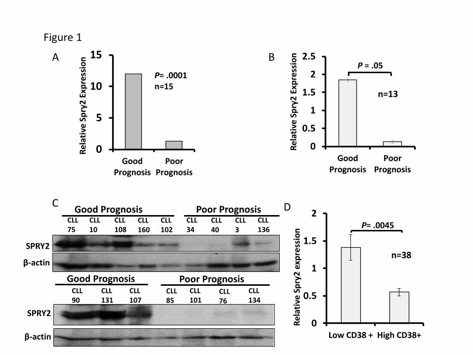
FIGURE 1: Comparison of SPRY2 levels in CLL-cells from Good and Poor-Prognosis
patients. To compare the expression of SPRY2, CLL-cells were isolated from PB of good and
poor-prognosis patients. (A) Relative mRNA level of spry2 from transcriptome analyses of 7
good-prognosis and 8 poor-prognosis CLL patients (n=15). RNA isolated from PB CLL-cells was
used for sequencing. The expression was normalized with GAPDH and genomic reference DNA
was used as control for transcriptome analysis. (B) Real-time PCR measurement of relative
expression of spry2 in CLL-cells from good and poor-prognosis patients, normalized with GAPH.
(C) Levels of SPRY2 protein expression was measured in good-prognosis patients (n=8) and
poor-prognosis patients (n=8). Displayed is a scanned western blot showing reduced protein
levels of SPRY2 in poor-prognosis CLL patients. Mononuclear cells from healthy donor were
used as positive control for antibody. 50µg of protein was loaded on 10%SDS gel. Beta actin is
used as loading control. (D) Microarray data showing low relative expression of SPRY2 in
patients with high CD38 expression. Patients with more than 30% of CD38 positive cells were
considered CD38 High (n=15) and patients with less than 30% were considered Low CD38
(n=23) GAPDH was used to normalize the value, * p value=0.0045.
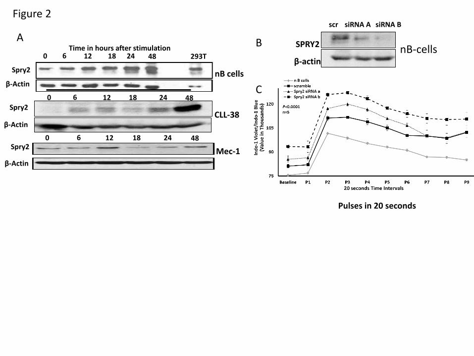
FIGURE 2: Effect of SPRY2 on BCR signaling. To study the role of SPRY2 in CLL and nB-
cells, we isolated CLL and nB-cells from patients and healthy donors respectively. (A) NB-cells,
primary human CLL-cells and Mec-1 CLL-cells were stimulated by BCR crosslinking for 0, 6, 12,
18, 24 and 48 hours. Cells were washed and protein lysate was prepared. Protein level of
SPRY2 was determined by western blotting. First panel shows SPRY2 levels in nB-cells,
Second Panel shows SPRY2 levels in primary CLL-cells from patient and third panel shows
SPRY2 levels in Mec-1 CLL-cells. (B) To test the efficacy of siRNAs against SPRY2 nB-cells
For personal use only.on April 2, 2018. by guest www.bloodjournal.orgFrom
21
were transfected with siRNAa and siRNAb after 48 hours of transfection cells were washed and
lysate was prepared. Equal amount of protein was loaded in each well, Scramble siRNA and β-
actin was used as control. Displayed is scanned western blot showing decrease in SPRY2
levels after siRNAs treatment. (C) NB-cells were isolated from healthy donors and nucleofected
with scramble, siRNA A and siRNA B. After 48 hours of calcium influx assay was performed
using Indo-1 dye dots represent 20 second time intervals. Displayed is mean graph of Indo-1
violet/Indo-1 Blue ratio, n=5 nB-cells samples from different healthy donors and p=0.0001.
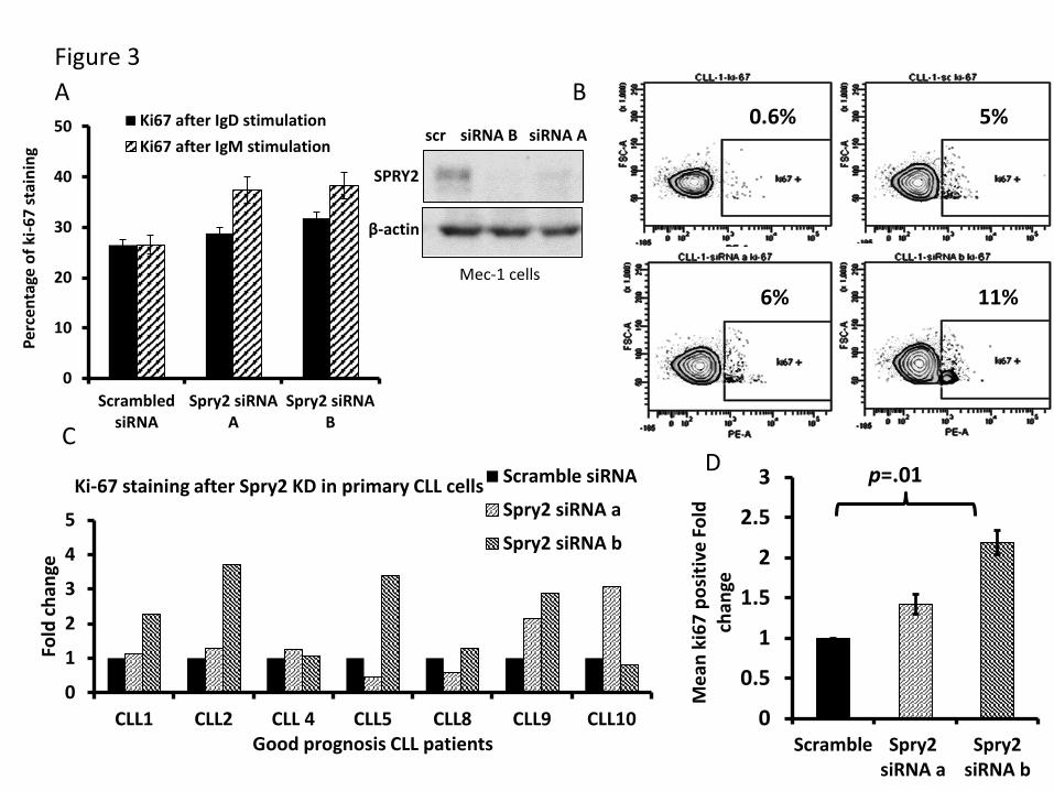
FIGURE 3: Knockdown of SPRY2 increases proliferation of CLL-cells from Good-
prognosis patients. To determine the effect of SPRY2 knockdown on human CLL-cells we
used Mec-1 cells and primary CLL-cells from Good-Prognosis CLL patients. (A) Displays mean
bar graph of 3 repeats showing Ki-67 staining of Mec-1 cells after SPRY2 knockdown with anti-
IgM and anti-IgD antibody stimulation and scanned western blot showing decrease in SPRY2
protein levels after siRNAs treatment (B) Dot plot of patient’s sample CLL-1 showing increase in
proliferation after SPRY2 knockdown using two different siRNA. (C) CLL-cells were isolated
from peripheral blood of n=7 different good prognosis CLL patients. CLL-cells were
nucleofected with scramble, siRNA A and siRNA B and co-culture on S-17 stromal layer. After
48 hours CLL-cells were stained with Ki-67 stain and proliferation was measured. Displayed bar
graph is fold in the rate of proliferation of CLL-cells. (D) Displays the mean fold change of (C)
showing significant p=0.01 increase in proliferation after SPRY2 knockdown with siRNA B.
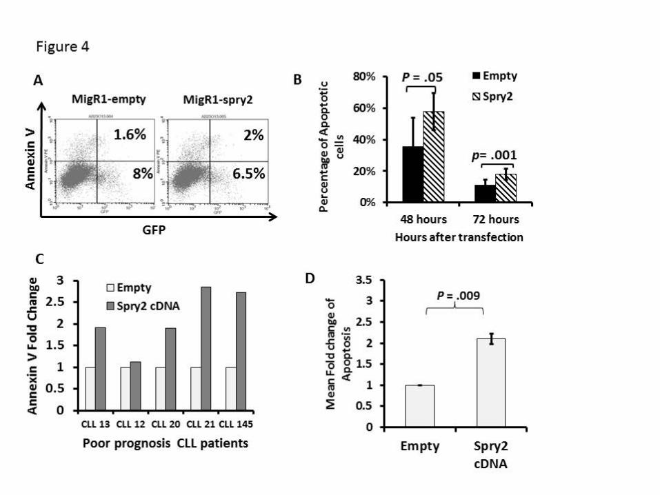
FIGURE 4: Expression of SPRY2 induces spontaneous apoptosis in CLL-cells. (A) Mec-1
cells were transfected with Empty and spry2-cDNA GFP co-expressing vectors. (B) This figure
shows Annexin-V staining done after 48 and 72 hours. Only live cells were gated for the
analysis. The numbers represent the percentage of GFP positive cells also positive for Annexin-
V. (C) Primary CLL-cells from n=5 different Poor-Prognosis CLL patients were nucleofected with
For personal use only.on April 2, 2018. by guest www.bloodjournal.orgFrom
22
spry2-cDNA and Empty vector. Bar graph represents fold in percentage of apoptotic cells. (D)
Mean Fold change of spry2-cDNA and Empty vector transfected cells from Figure (C) p=0.009.
FIGURE 5: Effect of SPRY2 on B1-cell development and BCR signaling in murine model
and SPRY2 knockdown leads to disease progression in NSG mice. (A) Displayed is a
western blot showing overexpression of spry2 in splenic B-cells of CD19-cre;Spry2(tg). B1-cells
were isolated from peritoneal cavity of CD19-cre;Spry2(wt) and CD19-cre;Spry2(tg) mice. B1-
cells were stained with CD5 and B220 dye to determine the frequency of B1-cells using Flow
cytometric analysis. Displayed is dot plot of showing B1a-cells frequency in CD19-cre;Spry2(wt)
and CD19-cre;Spry2(tg) mice. Bar graph of absolute number of B1a-cells in peritoneal cavity of
CD19-cre;Spry2(wt) and CD19-cre;Spry2(tg). (B) Splenic B-cells were isolated by negative
selection from rTTA positive CD19-cre;Spry2(wt) and CD19-cre;Spry2(tg) mice. GFP+
population displays CD19-cre expressed cells and displayed is calcium mobilization assay of
splenic B-cells from above described mice n=3; p=0.0002. (C) Splenic B-cells were isolated
from CD19-cre;Spry2(wt) and CD19-cre;Spry2(tg) stained with CFSE dye and cultured in vitro
for six days with and without anti-IgM antibody and LPS. Displayed is the CFSE labeling of
splenic B-cells of CD19-cre;Spry2(wt) and CD19-cre;Spry2(tg) mice. SPRY2 was stably
knockdown in Mec-1 CLL-cells using pLenti-siRNA A and pLenti-siRNA B, pLenti-scramble was
used as control. (D) Displayed average of tumor volume in n=5 mice in each group were
transplanted with 1.5 million pLenti-scramble and pLenti-siRNA B Mec-1 CLL-cells. Tumor
volume was measured, using digital caliper we determine the length and width of the tumor.
Tumor volume was calculated by V = (L x W x W)/2 where V is the tumor volume, L is the tumor
length and W is the width of the tumor. (E) Represents the frequency of Human CD19+ cells in
spleen of pLenti-scramble and pLenti-siRNA B CLL-cells transplanted mice. (F) Shows the
hemotoxilin and eosin staining with tissue section of Kidney, Liver and Spleen to observe the
number of CLL-cells infiltration in pLenti-scramble and pLenti-siRNA B CLL-cells transplanted
For personal use only.on April 2, 2018. by guest www.bloodjournal.orgFrom
23
mice. (G) Protein lysates were prepared from tumors; equal amount of protein was loaded in
each lane of 10%SDS gel. Shown is scanned western blot to determine the protein levels of p-
Erk and SPRY2 in tumors from pLenti-scramble and pLenti-siRNA B CLL-cells transplanted
mice. Total Erk and β-actin were used as control. Densitometric measurements showing
elevated of p-Erk normalized by total Erk in SPRY2 knockdown tumors.
FIGURE 6: SPRY2 downregulate MAPK-ERK signaling by interacting with RAF-1, Syk and
BRAF. (A) Mec-1 cells were transfected with pLKO-TET-Empty and pLKO-TET-Spry2. Cells
were treated with doxycycline for four days to induce the expression of SPRY2. Displayed is a
scanned western blot of CLL-cells transfected with Empty and spry2 cDNA to measure the
protein levels of SPRY2 and p-Erk upon doxycycline treatment. β-actin and PAN Erk1/2 were
used as controls. (B) KRAS-V12 and BRAF-V600 mutants were co-transfected with Empty and
Spry2 vectors in Mec-1 cells. Scanned western blot showing the p-Erk and Erk levels in these
cells. Second panel shows scanned western blot of SPRY2 and BRAF protein levels in cells
described in (B), β-actin was used as loading control. (C) Human nB-cells were isolated from
healthy donors, cells were stimulated by BCR crosslinking for 24 hours. Cells were lysed to
prepare lysate for immunoprecipitation. Displayed is a scanned western blot of
immunoprecipitation with SPRY2 and Syk showing pull down of RAF-1, Syk, BRAF and SPRY2.
IgG, Pi3K and BTK were used as negative controls. (D) NB-cells and Mec-1 cells were
stimulated as described in (C) cyotospin were prepared using these cells. Cells were stained
with SPRY2, p-Syk and DAPI fluorescence antibody. Displayed are immunofluorescence
images showing co-localization of SPRY2 and p-Syk in normal B-cells and Mec-1 CLL-cells.
Unstimulated cell, Goat-IgG and Rabbit-IgG were used as control. (E) NB-cells were isolated
from spleen of CD19-cre;Spry2(wt) and CD19-cre;Spry2(tg) mice and were stimulated for 30
mins with anti-IgM antibody. Displayed is scanned western blot of p-Syk and Syk and
densitometric measurements showing normalized decreased levels of p-Syk. (F) Mec-1 CLL-
For personal use only.on April 2, 2018. by guest www.bloodjournal.orgFrom
24
cells were overexpressed with Spry2 cDNA and Empty vector co-expressing GFP, cytospins
were prepared of these cells. Slides were stained with p-Syk (red) antibody. Displayed are
immunofluorescence images and Densitometric measurements showing decreased levels of
normalized p-Syk in SPRY2 co-expressing GFP positive cells.
FIGURE 7: MiR-21 targets SPRY2 in human CLL-cells to activate BCR mediated MAPK-
Erk signaling. (A) Displayed is scanned western blot showing SPRY2 protein levels in miR-21
overexpressing CLL-cells. β-actin was used as loading control. (B) Shows protein levels of p-Erk
and Erk in miR-21 overexpressing cells and Densitometric measurements showing elevated
levels of p-Erk in miR-21 overexpressing CLL-cells. (C) Displayed is scanned western blot
showing the protein levels of p-Syk, Syk and SPRY2 in miR-21 overexpressing Mec-1 CLL-
cells. (D) In the case of nB-cells, BCR stimulation leads to activation of MAPK-Erk signaling
which results in their proliferation and survival. As a homeostasis response SPRY2 gets induced
to regulate signaling in activated B-cell. Whereas in case of CLL-cells, elevated level of miR-21
leads to decreased SPRY2 which results constitutive activation of BCR and MAPK-Erk
signaling. This in turn increases the proliferation and survival of CLL-cells.
For personal use only.on April 2, 2018. by guest www.bloodjournal.orgFrom
0
0.5
1
1.5
2
2.5
GoodPrognosis
PoorPrognosis
Rela
tive
Spry
2 Ex
pres
sion P = .05
n=13
0
0.5
1
1.5
2
Low CD38 + High CD38+
Rela
tive
Spry
2 ex
pres
sion
n=38
P= .0045
0
5
10
15
GoodPrognosis
PoorPrognosis
Rela
tive
Spry
2 Ex
pres
sion
P= .0001n=15
A B
C D
Figure 1
Good Prognosis Poor Prognosis
β-actin
SPRY2
CLL75
CLL10
CLL108
CLL160
CLL34
CLL40
CLL136
CLL102
CLL3
Good Prognosis Poor PrognosisCLL90
CLL131
CLL107
CLL85
CLL101
CLL76
CLL134
β-actin
SPRY2
For personal use only.on April 2, 2018. by guest www.bloodjournal.orgFrom
A B
C
Pulses in 20 seconds
Figure 2
0 6 12 18 24 48Spry2
β-Actin
0 6 12 18 24 48 293TTime in hours after stimulation
Spry2
β-Actin
0 6 12 18 24 48Spry2
β-Actin
nB cells
CLL-38
Mec-1
SPRY2
β-actinnB-cells
scr siRNA A siRNA B
For personal use only.on April 2, 2018. by guest www.bloodjournal.orgFrom
0
10
20
30
40
50
ScrambledsiRNA
Spry2 siRNAA
Spry2 siRNAB
Perc
enta
ge o
f ki-6
7 st
aini
ng
Ki67 after IgD stimulationKi67 after IgM stimulation
0
0.5
1
1.5
2
2.5
3
Scramble Spry2siRNA a
Spry2siRNA b
Mea
n ki
67 p
ositi
ve F
old
chan
ge
p=.01
0
1
2
3
4
5
CLL1 CLL2 CLL 4 CLL5 CLL8 CLL9 CLL10
Fold
chan
ge
Good prognosis CLL patients
Ki-67 staining after Spry2 KD in primary CLL cells Scramble siRNA
Spry2 siRNA a
Spry2 siRNA b
0.6% 5%
6% 11%
A B
CD
Figure 3
SPRY2
β-actin
Mec-1 cells
scr siRNA B siRNA A
For personal use only.on April 2, 2018. by guest www.bloodjournal.orgFrom
For personal use only.on April 2, 2018. by guest www.bloodjournal.orgFrom
9% 4.8%
CD5
B220
CD19creSpry2(wt) CD19creSpry2(tg)
A
B C
Figure 5
CFSE
For personal use only.on April 2, 2018. by guest www.bloodjournal.orgFrom
D F
p-Erk
Spry2
pLenti-scr
pLenti-siRNA B
Erk
β-actin
KID
NEY
LIVE
RSP
LEEN
Scr-
siRN
ASp
ry2-
siRN
A B
Scr-
siRN
ASp
ry2-
siRN
A B
Scr-
siRN
ASp
ry2-
siRN
A B
10X 40X
G
0
500
1000
1500
2000
0 14 21 23 25 27 29 33
Tum
or V
olum
e in
mm
3
Days post injections
scr siRNAsiRNA B
P= .03P= .005
P= .004P= .02
P= .03
n=5 mice/group
0
0.5
1
1.5
pLenti-Scr pLenti-siRNAB
p-Erk
05
1015
pLenti-scr pLenti-siRNA B
% CD19 positive cells in spleenE
For personal use only.on April 2, 2018. by guest www.bloodjournal.orgFrom
Spry2
β-actin
pLKO-TET-Empty pLKO-TET-Spry2
p-Erk
Erk1/2
p-Erk
KRAS V12 BRAF V600
Erk
Empty Spry2
B-actin
Spry2
BRAF
BRAF V600A B
Spry2 IgG inputIP:
IB:Pi3K
IB: Raf-1
Spry2 IgG
IB: Syk
IB: Spry2
IP:
IB: BRAF
Syk IgGIP:
IB: Syk
IB: Spry2
Endogenous Spry2 interaction in primary normal B cells
IB:BTK
C D
Figure 6
For personal use only.on April 2, 2018. by guest www.bloodjournal.orgFrom
p-syk
CD19cre-Spry2(wt)
CD19cre-Spry2(tg)
Syk
DAPI GFP p-Syk
Empty Vector
Spry2 Vector
E F
0
0.5
1
CD19cre-Spry2(wt) CD19cre-Spry2(tg)
p-Syk
0
0.5
1
1.5
Empty Spry2
p-Sykp=0.002
For personal use only.on April 2, 2018. by guest www.bloodjournal.orgFrom
0
0.5
1
1.5
pcDNA-empty pcDNA-Mir21 Spry2 +controlN
orm
aliz
ed p
-Erk
ex
pres
sion
Spry2
β-actin
pcDNA-Empty
pcDNA-Mir-21
Spry2 + Mec-1
p-Erk
Erk
pcDNA-Empty
pcDNA-Mir-21
Spry2+Mec-1
p-syk
Spry2
β-actin
pcDNA-Empty
pcDNA-Mir-21
Syk
A B
C D
Figure 7
For personal use only.on April 2, 2018. by guest www.bloodjournal.orgFrom
doi:10.1182/blood-2015-09-669317Prepublished online January 25, 2016;
Pirruccello, Hamid Band, Runqing Lu and Shantaram S. JoshiAshima Shukla, Karan Rai, Vipul Shukla, Nagendra K. Chaturvedi, R. Gregory Bociek, Samuel J. CLLSprouty 2: a novel attenuator of B-cell receptor and MAPK-Erk signaling in
http://www.bloodjournal.org/site/misc/rights.xhtml#repub_requestsInformation about reproducing this article in parts or in its entirety may be found online at:
http://www.bloodjournal.org/site/misc/rights.xhtml#reprintsInformation about ordering reprints may be found online at:
http://www.bloodjournal.org/site/subscriptions/index.xhtmlInformation about subscriptions and ASH membership may be found online at:
digital object identifier (DOIs) and date of initial publication. indexed by PubMed from initial publication. Citations to Advance online articles must include final publication). Advance online articles are citable and establish publication priority; they areappeared in the paper journal (edited, typeset versions may be posted when available prior to Advance online articles have been peer reviewed and accepted for publication but have not yet
Copyright 2011 by The American Society of Hematology; all rights reserved.Hematology, 2021 L St, NW, Suite 900, Washington DC 20036.Blood (print ISSN 0006-4971, online ISSN 1528-0020), is published weekly by the American Society of
For personal use only.on April 2, 2018. by guest www.bloodjournal.orgFrom


![[450] Gregory David Roberts - Shantaram vol 2.pdf](https://img.pdfslide.net/doc/110x75/563dbb94550346aa9aae6735/450-gregory-david-roberts-shantaram-vol-2pdf.jpg)


![[450] Gregory David Roberts - Shantaram vol 1.pdf](https://img.pdfslide.net/doc/110x75/563dbb94550346aa9aae6568/450-gregory-david-roberts-shantaram-vol-1pdf.jpg)