Embed Size (px)
DESCRIPTION
Shivangi Patel, Gunvanti Rathod, Purva Shinde, RK Tandan. Adenocarcinoma of urinary bladder in 55 years old male patient - A rare case report. IAIM, 2015; 2(1): 116-120.
Citation preview
Adenocarcinoma of urinary bladder
International Archives of Integrated Medicine, Vol.
Copy right © 2015, IAIM, All Rights Reserved.
Case Report
Adenocarcinoma of urinary bladder in 55
years old male patient
Shivangi Patel1, Gunvanti Rathod
1PG Student, Pathology Department, SBKS MI & RC, Sumandeep Vidyapeeth, Vadodara, India
2Assistant Professor, Pathology Department, SBKS MI & RC, Sumandeep Vidyapeeth, Vadodara, India
3Professor, Pathology Department, SBKS MI & RC, Sumandeep Vidyapeeth, Vadodara, India
*Corresponding author email:
How to cite this article: Shivangi Patel, Gunvanti Rathod, Purva Shinde, RK Tandan
of urinary bladder in 55 years old male patient
Available online at
Received on: 12-12-2014
Abstract
Adenocarcinoma of urinary bladder is rather rare and so that here we present the case of a 55 years
old male patient who presented with hematuria and diagnosed as adenocarcinoma of the urinary
bladder. Because of no specific characteristics for symptoms,
compared with common urothelial carcinoma, adenocarcinoma of urinary bladder was diagnosed
mainly on histopathology and with the help of Immunohistochemistry (IHC). For recurrent tumor
after transurethral resection of bladd
or radical surgery.
Key words
Urinary bladder, Adenocarcinoma,
Introduction
Urinary bladder cancer is the second most
frequent tumor of the genito-urinary tract [1].
causes more than 3,30,000 new cases each year
and more than 1,30,000 deaths per year. Its
generally estimated male: female incidence ratio
is 3.8: 1.0. At any point in time, 2.7 million
people have a history of urinary
[2]. The histological and pathological type of
Adenocarcinoma of urinary bladder
International Archives of Integrated Medicine, Vol. 2, Issue 1, January, 2015.
, IAIM, All Rights Reserved.
Adenocarcinoma of urinary bladder in 55
years old male patient - A rare case report
, Gunvanti Rathod2*
, Purva Shinde1, RK Tandan
PG Student, Pathology Department, SBKS MI & RC, Sumandeep Vidyapeeth, Vadodara, India
Assistant Professor, Pathology Department, SBKS MI & RC, Sumandeep Vidyapeeth, Vadodara, India
Professor, Pathology Department, SBKS MI & RC, Sumandeep Vidyapeeth, Vadodara, India
*Corresponding author email: [email protected]
Shivangi Patel, Gunvanti Rathod, Purva Shinde, RK Tandan
of urinary bladder in 55 years old male patient - A rare case report. IAIM, 2015; 2(1
Available online at www.iaimjournal.com
2014 Accepted on:
Adenocarcinoma of urinary bladder is rather rare and so that here we present the case of a 55 years
old male patient who presented with hematuria and diagnosed as adenocarcinoma of the urinary
Because of no specific characteristics for symptoms, signs and accessory examinations
compared with common urothelial carcinoma, adenocarcinoma of urinary bladder was diagnosed
mainly on histopathology and with the help of Immunohistochemistry (IHC). For recurrent tumor
after transurethral resection of bladder tissue (TUR-BT), the patient should undergo total cystectomy
Urinary bladder, Adenocarcinoma, Histopathology.
Urinary bladder cancer is the second most
urinary tract [1]. It
causes more than 3,30,000 new cases each year
and more than 1,30,000 deaths per year. Its
generally estimated male: female incidence ratio
is 3.8: 1.0. At any point in time, 2.7 million
people have a history of urinary bladder cancer
[2]. The histological and pathological type of
bladder cancer is mainly urothelial carcinoma,
also called transitional cell carcinoma,
accounting for approximately 90% [3]. Other
types including squamous cell carcinoma and
adenocarcinoma, account for 3
respectively [4]. As adenocarcinoma of bladder
is rather rare, here we present the case of a 55
years old male patient who presented with
hematuria and diagnosed as adenocarcinoma of
the urinary bladder. Main purpose to present
ISSN: 2394-0026 (P)
ISSN: 2394-0034 (O)
Page 116
Adenocarcinoma of urinary bladder in 55
A rare case report
, RK Tandan3
PG Student, Pathology Department, SBKS MI & RC, Sumandeep Vidyapeeth, Vadodara, India
Assistant Professor, Pathology Department, SBKS MI & RC, Sumandeep Vidyapeeth, Vadodara, India
Professor, Pathology Department, SBKS MI & RC, Sumandeep Vidyapeeth, Vadodara, India
Shivangi Patel, Gunvanti Rathod, Purva Shinde, RK Tandan. Adenocarcinoma
5; 2(1): 116-120.
Accepted on: 06-01-2015
Adenocarcinoma of urinary bladder is rather rare and so that here we present the case of a 55 years
old male patient who presented with hematuria and diagnosed as adenocarcinoma of the urinary
signs and accessory examinations
compared with common urothelial carcinoma, adenocarcinoma of urinary bladder was diagnosed
mainly on histopathology and with the help of Immunohistochemistry (IHC). For recurrent tumor
BT), the patient should undergo total cystectomy
bladder cancer is mainly urothelial carcinoma,
also called transitional cell carcinoma,
accounting for approximately 90% [3]. Other
types including squamous cell carcinoma and
count for 3-7% and < 2%
respectively [4]. As adenocarcinoma of bladder
is rather rare, here we present the case of a 55
years old male patient who presented with
hematuria and diagnosed as adenocarcinoma of
the urinary bladder. Main purpose to present
Adenocarcinoma of urinary bladder
International Archives of Integrated Medicine, Vol.
Copy right © 2015, IAIM, All Rights Reserved.
this case is to add an additional case to the
literature.
Case report
A 55 years old male patient from poor socio
economic class presented with pain in the right
lumbar region and burning micturition since 2
months. He was also complaining of red
discoloration of urine since 20 days which was
intermittent with clots. He had lost 3 kg weight
over last 2 months and was also having
anorexia. He denied as use of drug history. He
had past history of pyelolithotomy before 5
years. On per abdomen examination, patien
had tenderness in right lumbar region. There
was no hepatomegaly or splenomegaly. His
vitals were unremarkable. On investigations, his
haemoglobin was 13.2 gm%, white blood cell
count was 6500/cmm and platelet count was
2.17 lac/cmm. On microscopic exami
urine showed plenty of red blood cells (RBCs).
His serum for HIV, HBs Ag were negative and
renal function test, liver function test and
random blood sugar were within the reference
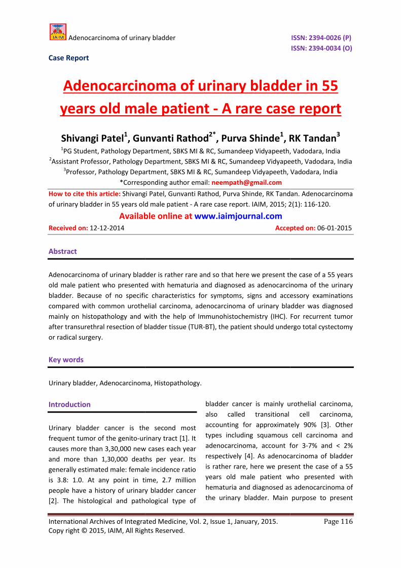
range. CT abdomen pelvis showed mass arised
from base of bladder. (Photo –
done and the tissue was sent to histopathology
department. Tumor tissue was fixed in 10%
formalin solution and routinely processed.
Sections from the paraffin block were cut with
thickness of 5 micron and stained by
hematoxylin and eosin (H & E) stain. Microscopic
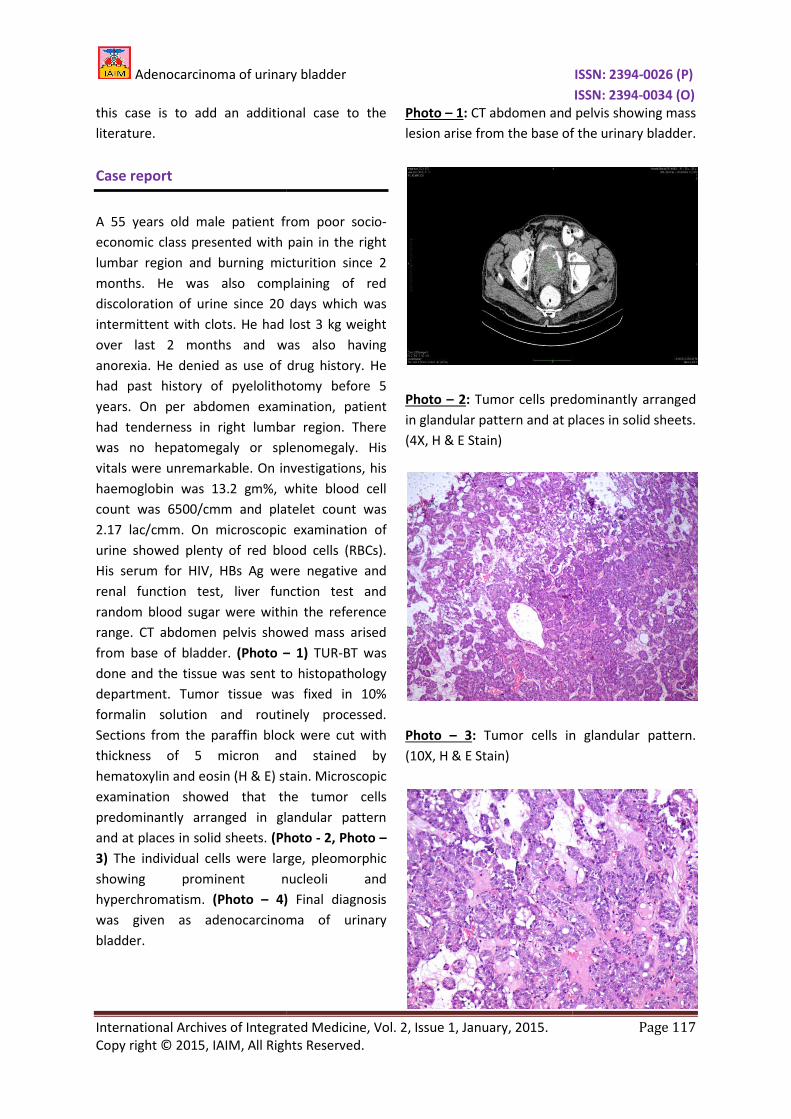
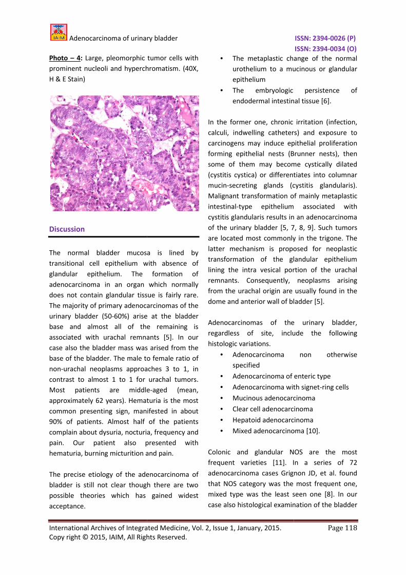
examination showed that the tumor cells
predominantly arranged in glandular pattern
and at places in solid sheets. (Photo
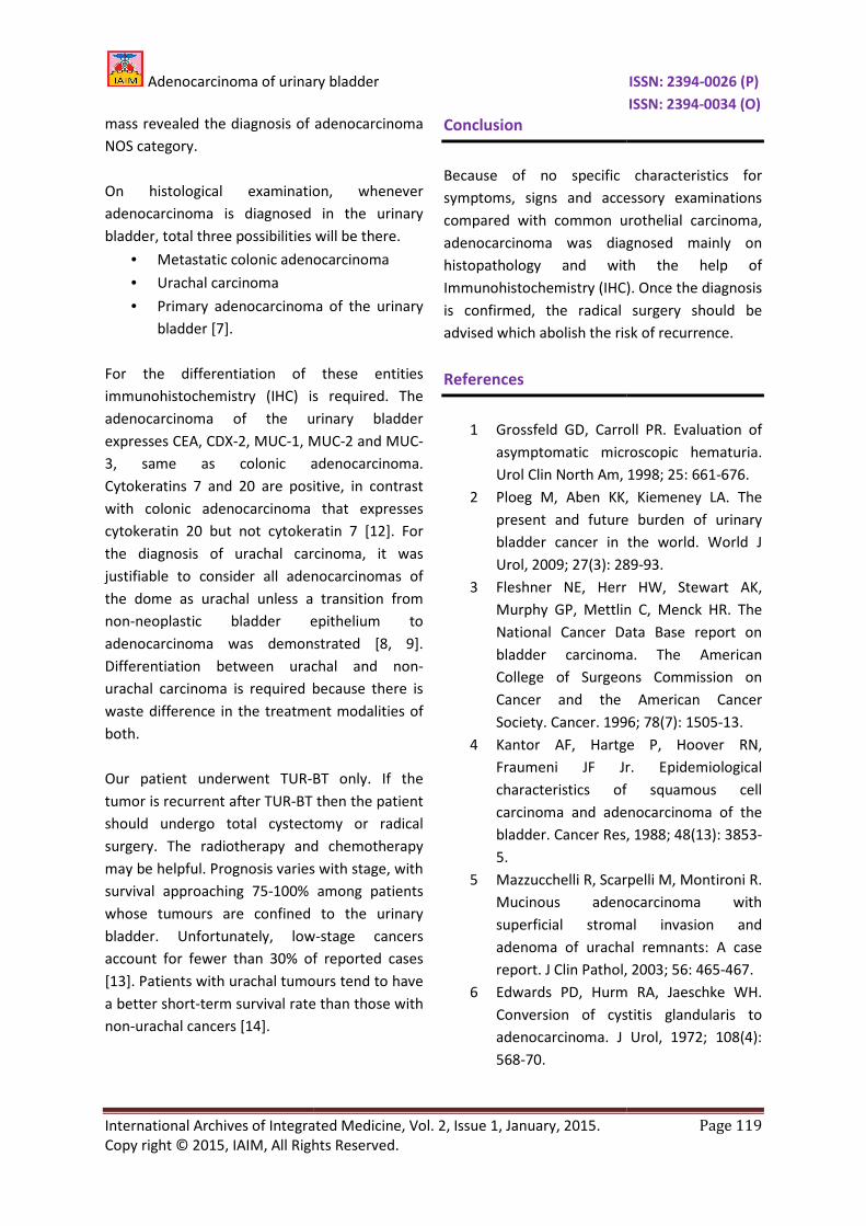
3) The individual cells were large, pleomorphic
showing prominent nucleoli and
hyperchromatism. (Photo – 4)
was given as adenocarcinoma of urinary
bladder.
Adenocarcinoma of urinary bladder
International Archives of Integrated Medicine, Vol. 2, Issue 1, January, 2015.
, IAIM, All Rights Reserved.
case is to add an additional case to the
A 55 years old male patient from poor socio-
economic class presented with pain in the right
lumbar region and burning micturition since 2
months. He was also complaining of red
on of urine since 20 days which was
intermittent with clots. He had lost 3 kg weight
over last 2 months and was also having
anorexia. He denied as use of drug history. He
had past history of pyelolithotomy before 5
years. On per abdomen examination, patient
had tenderness in right lumbar region. There
was no hepatomegaly or splenomegaly. His
vitals were unremarkable. On investigations, his
haemoglobin was 13.2 gm%, white blood cell
count was 6500/cmm and platelet count was
2.17 lac/cmm. On microscopic examination of
urine showed plenty of red blood cells (RBCs).
His serum for HIV, HBs Ag were negative and
renal function test, liver function test and
random blood sugar were within the reference
range. CT abdomen pelvis showed mass arised
– 1) TUR-BT was
done and the tissue was sent to histopathology
Tumor tissue was fixed in 10%
formalin solution and routinely processed.
Sections from the paraffin block were cut with
thickness of 5 micron and stained by
eosin (H & E) stain. Microscopic
examination showed that the tumor cells
predominantly arranged in glandular pattern
(Photo - 2, Photo –
The individual cells were large, pleomorphic
showing prominent nucleoli and
4) Final diagnosis
was given as adenocarcinoma of urinary
Photo – 1: CT abdomen and pelvis showing mass
lesion arise from the base of the urinary bladder.
Photo – 2: Tumor cells predominantly arranged
in glandular pattern and at places in solid sheets.
(4X, H & E Stain)
Photo – 3: Tumor cells in glandular pattern.
(10X, H & E Stain)
ISSN: 2394-0026 (P)
ISSN: 2394-0034 (O)
Page 117
CT abdomen and pelvis showing mass
lesion arise from the base of the urinary bladder.
cells predominantly arranged
in glandular pattern and at places in solid sheets.
Tumor cells in glandular pattern.
Adenocarcinoma of urinary bladder
International Archives of Integrated Medicine, Vol.
Copy right © 2015, IAIM, All Rights Reserved.
Photo – 4: Large, pleomorphic tumor cells with
prominent nucleoli and hyperchromatism
H & E Stain)
Discussion
The normal bladder mucosa is lined by
transitional cell epithelium with absence of
glandular epithelium. The formation of
adenocarcinoma in an organ which normally
does not contain glandular tissue is fairly rare.
The majority of primary adenocarcinomas of the
urinary bladder (50-60%) arise at the bladder
base and almost all of the remaining is
associated with urachal remnants [5]. In our
case also the bladder mass was arised from the
base of the bladder. The male to female ratio of
non-urachal neoplasms approaches 3 to 1, in
contrast to almost 1 to 1 for urachal tumors.
Most patients are middle
approximately 62 years). Hematuria is the most
common presenting sign, manifested in about
90% of patients. Almost half of the patients
complain about dysuria, nocturia, frequency and
pain. Our patient also presented with
hematuria, burning micturition and pain.
The precise etiology of the adenocarcinoma of
bladder is still not clear though there are two
possible theories which has gained widest
acceptance.
Adenocarcinoma of urinary bladder
International Archives of Integrated Medicine, Vol. 2, Issue 1, January, 2015.
, IAIM, All Rights Reserved.
Large, pleomorphic tumor cells with
prominent nucleoli and hyperchromatism. (40X,
The normal bladder mucosa is lined by
transitional cell epithelium with absence of
glandular epithelium. The formation of
adenocarcinoma in an organ which normally
does not contain glandular tissue is fairly rare.
The majority of primary adenocarcinomas of the
60%) arise at the bladder
base and almost all of the remaining is
associated with urachal remnants [5]. In our
case also the bladder mass was arised from the
o female ratio of
urachal neoplasms approaches 3 to 1, in
contrast to almost 1 to 1 for urachal tumors.
Most patients are middle-aged (mean,
approximately 62 years). Hematuria is the most
common presenting sign, manifested in about
ost half of the patients
complain about dysuria, nocturia, frequency and
pain. Our patient also presented with
hematuria, burning micturition and pain.
The precise etiology of the adenocarcinoma of
bladder is still not clear though there are two
theories which has gained widest
• The metaplastic change of the normal
urothelium to a mucinous
epithelium
• The embryologic persistence of
endodermal intestinal tissue [6].
In the former one, chronic irritation (infection,
calculi, indwelling catheters) and exposure to
carcinogens may induce epithelial proliferation
forming epithelial nests (Brunner nests), then
some of them may become cystically dilated
(cystitis cystica) or differentiates into columnar
mucin-secreting glands (cy
Malignant transformation of mainly metaplastic
intestinal-type epithelium associated with
cystitis glandularis results in an adenocarcinoma
of the urinary bladder [5, 7, 8, 9]. Such tumors
are located most commonly in the trigone. The
latter mechanism is proposed for neoplastic
transformation of the glandular epithelium
lining the intra vesical portion of the urachal
remnants. Consequently, neoplasms arising
from the urachal origin are usually found in the
dome and anterior wall of blad
Adenocarcinomas of the urinary bladder,
regardless of site, include the
histologic variations.
• Adenocarcinoma non otherwise
specified
• Adenocarcinoma of enteric type
• Adenocarcinoma with signet
• Mucinous adenocarcinoma
• Clear cell adenocarcinoma
• Hepatoid adenocarcinoma
• Mixed adenocarcinoma [10].
Colonic and glandular NOS are the most
frequent varieties [11]. In a series of 72
adenocarcinoma cases Grignon
that NOS category was the most frequent one,
mixed type was the least seen one [8]. In our
case also histological examination
ISSN: 2394-0026 (P)
ISSN: 2394-0034 (O)
Page 118
The metaplastic change of the normal
urothelium to a mucinous or glandular
The embryologic persistence of
endodermal intestinal tissue [6].
In the former one, chronic irritation (infection,
i, indwelling catheters) and exposure to
carcinogens may induce epithelial proliferation
forming epithelial nests (Brunner nests), then
some of them may become cystically dilated
(cystitis cystica) or differentiates into columnar
secreting glands (cystitis glandularis).
Malignant transformation of mainly metaplastic
type epithelium associated with
cystitis glandularis results in an adenocarcinoma
of the urinary bladder [5, 7, 8, 9]. Such tumors
are located most commonly in the trigone. The
latter mechanism is proposed for neoplastic
transformation of the glandular epithelium
vesical portion of the urachal
remnants. Consequently, neoplasms arising
from the urachal origin are usually found in the
bladder [5].
Adenocarcinomas of the urinary bladder,
regardless of site, include the following
oma non otherwise
nocarcinoma of enteric type
noma with signet-ring cells
Mucinous adenocarcinoma
cell adenocarcinoma
Hepatoid adenocarcinoma
Mixed adenocarcinoma [10].
Colonic and glandular NOS are the most
frequent varieties [11]. In a series of 72
adenocarcinoma cases Grignon JD, et al. found
that NOS category was the most frequent one,
mixed type was the least seen one [8]. In our
xamination of the bladder
Adenocarcinoma of urinary bladder
International Archives of Integrated Medicine, Vol.
Copy right © 2015, IAIM, All Rights Reserved.
mass revealed the diagnosis of
NOS category.
On histological examination, whenever
adenocarcinoma is diagnosed in the urinary
bladder, total three possibilities will be there.
• Metastatic colonic adenocarcinoma
• Urachal carcinoma
• Primary adenocarcinoma of the urinary
bladder [7].
For the differentiation of these entities
immunohistochemistry (IHC) is required. The
adenocarcinoma of the urinary bladder
expresses CEA, CDX-2, MUC-1, MUC
3, same as colonic adenocarcinoma.
Cytokeratins 7 and 20 are positive, in contrast
with colonic adenocarcinoma that expresses
cytokeratin 20 but not cytokeratin 7 [12].
the diagnosis of urachal carcinoma
justifiable to consider all adenocarcinomas of
the dome as urachal unless a transition from
non-neoplastic bladder epithelium to
adenocarcinoma was demonstrated [8, 9].
Differentiation between urachal and non
urachal carcinoma is required because there is
waste difference in the treatment modalities of
both.
Our patient underwent TUR-BT only. I
tumor is recurrent after TUR-BT then the patient
should undergo total cystectomy or radical
surgery. The radiotherapy and chemotherapy
may be helpful. Prognosis varies with stage, with
survival approaching 75-100% among patients
whose tumours are confined to the urinary
bladder. Unfortunately, low
account for fewer than 30% of reported cases
[13]. Patients with urachal tumours tend to have
a better short-term survival rate than those with
non-urachal cancers [14].
Adenocarcinoma of urinary bladder
International Archives of Integrated Medicine, Vol. 2, Issue 1, January, 2015.
, IAIM, All Rights Reserved.
adenocarcinoma
On histological examination, whenever
adenocarcinoma is diagnosed in the urinary
bladder, total three possibilities will be there.
Metastatic colonic adenocarcinoma
Primary adenocarcinoma of the urinary
tion of these entities
immunohistochemistry (IHC) is required. The
adenocarcinoma of the urinary bladder
1, MUC-2 and MUC-
3, same as colonic adenocarcinoma.
Cytokeratins 7 and 20 are positive, in contrast
ma that expresses
cytokeratin 20 but not cytokeratin 7 [12]. For
the diagnosis of urachal carcinoma, it was
justifiable to consider all adenocarcinomas of
the dome as urachal unless a transition from
neoplastic bladder epithelium to
demonstrated [8, 9].
Differentiation between urachal and non-
urachal carcinoma is required because there is
waste difference in the treatment modalities of
BT only. If the
BT then the patient
ould undergo total cystectomy or radical
surgery. The radiotherapy and chemotherapy
may be helpful. Prognosis varies with stage, with
100% among patients
whose tumours are confined to the urinary
bladder. Unfortunately, low-stage cancers
account for fewer than 30% of reported cases
[13]. Patients with urachal tumours tend to have
term survival rate than those with
Conclusion
Because of no specific characteristics for
symptoms, signs and accessory examinations
compared with common urothelial carcinoma,
adenocarcinoma was diagnosed mainly on
histopathology and with the help of
Immunohistochemistry (IHC). Once the diagnosis
is confirmed, the radical surgery should be
advised which abolish the risk of recurrence.
References
1 Grossfeld GD, Carroll PR. Evaluation of
asymptomatic microscopic hematuria.
Urol Clin North Am, 1998;
2 Ploeg M, Aben KK, Kiemeney
present and future burden of urinary
bladder cancer in the world. World J
Urol, 2009; 27(3): 289
3 Fleshner NE, Herr HW, Stewart AK,
Murphy GP, Mettlin C, Menck HR. The
National Cancer Data Base report on
bladder carcinoma. The American
College of Surgeons Commission on
Cancer and the American Cancer
Society. Cancer. 1996; 78(7): 1505
4 Kantor AF, Hartge P, Hoover RN,
Fraumeni JF Jr. Epidemiological
characteristics of squamous cell
carcinoma and adenocarcinoma of the
bladder. Cancer Res, 1988;
5.
5 Mazzucchelli R, Scarpelli M, Montironi R.
Mucinous adenocarcinoma with
superficial stromal invasion and
adenoma of urachal remnants: A case
report. J Clin Pathol,
6 Edwards PD, Hurm RA, Jaeschke WH.
Conversion of cystitis
adenocarcinoma. J Urol, 1972; 108(4):
568-70.
ISSN: 2394-0026 (P)
ISSN: 2394-0034 (O)
Page 119
Because of no specific characteristics for
essory examinations
compared with common urothelial carcinoma,
adenocarcinoma was diagnosed mainly on
histopathology and with the help of
Immunohistochemistry (IHC). Once the diagnosis
is confirmed, the radical surgery should be
isk of recurrence.
Grossfeld GD, Carroll PR. Evaluation of
asymptomatic microscopic hematuria.
Urol Clin North Am, 1998; 25: 661-676.
Ploeg M, Aben KK, Kiemeney LA. The
present and future burden of urinary
bladder cancer in the world. World J
Urol, 2009; 27(3): 289-93.
Fleshner NE, Herr HW, Stewart AK,
Murphy GP, Mettlin C, Menck HR. The
National Cancer Data Base report on
bladder carcinoma. The American
of Surgeons Commission on
Cancer and the American Cancer
Society. Cancer. 1996; 78(7): 1505-13.
Kantor AF, Hartge P, Hoover RN,
Fraumeni JF Jr. Epidemiological
characteristics of squamous cell
carcinoma and adenocarcinoma of the
bladder. Cancer Res, 1988; 48(13): 3853-
Mazzucchelli R, Scarpelli M, Montironi R.
Mucinous adenocarcinoma with
superficial stromal invasion and
adenoma of urachal remnants: A case
J Clin Pathol, 2003; 56: 465-467.
Edwards PD, Hurm RA, Jaeschke WH.
Conversion of cystitis glandularis to
adenocarcinoma. J Urol, 1972; 108(4):
Adenocarcinoma of urinary bladder
International Archives of Integrated Medicine, Vol.
Copy right © 2015, IAIM, All Rights Reserved.
7 Gill HS, Dhillon HK, Woodhouse CR.
Adenocarcinoma of the urinary bladder.
Br J Urol, 1989; 64(2): 138
8 Grignon DJ, Ro JY, Ayala AG, Johnson DE,
Ordonez NG. Primary adenocarcinoma
of the urinary bladder. A
clinicopathologic analysis of 72 cases.
Cancer, 1991; 67(8): 2165
9 Anderstrom C, Johansson SL, von Schultz
L. Primary adenocarcinoma of the
urinary bladder. A clinicopathologic and
prognostic study. Cancer, 1983; 52(7):
1273-80.
10 Eble JN, Epstein JI, Seternhenn IA.
Health Organization Classification of
Tumors. Pathology and Genetics,
Tumours of the Urinary System and
Male Genital track. Lyon: IARC Press;
2004: 128-132.
Source of support: Nil
Adenocarcinoma of urinary bladder
International Archives of Integrated Medicine, Vol. 2, Issue 1, January, 2015.
, IAIM, All Rights Reserved.
Gill HS, Dhillon HK, Woodhouse CR.
Adenocarcinoma of the urinary bladder.
Br J Urol, 1989; 64(2): 138-42.
Grignon DJ, Ro JY, Ayala AG, Johnson DE,
Ordonez NG. Primary adenocarcinoma
y bladder. A
clinicopathologic analysis of 72 cases.
Cancer, 1991; 67(8): 2165-72.
Anderstrom C, Johansson SL, von Schultz
L. Primary adenocarcinoma of the
urinary bladder. A clinicopathologic and
prognostic study. Cancer, 1983; 52(7):
stein JI, Seternhenn IA. World
Health Organization Classification of
Tumors. Pathology and Genetics,
Tumours of the Urinary System and
Lyon: IARC Press;
11 Abenoza P, Manivel C, Fraley EE. Primary
adenocarcinoma of urinary b
Clinicopathologic study of 16 cases.
Urology, 1987; 29(1): 9
12 Bostwick DG, Cheng L.
Pathology. Adenocarcinoma of the
Urinary Bladder. Elsevier, New York,
2008, p. 300-302.
13 Werling RW, Yaziji H, Bacchi CE, Gown
AM. CDX2, a highly sensitive and specific
marker of adenocarcinomas of intestinal
origin: An immunohistochemical survey
of 476 primary and metastatic
carcinomas. AJSP, 2003;
14 Stenhouse G, Mcrae D, Pollock AM.
Urachal adenocarcinoma in situ with
pseudomyxoma perit
J Clin Pathol, 2003; 56:
Nil Conflict of interest:
ISSN: 2394-0026 (P)
ISSN: 2394-0034 (O)
Page 120
Abenoza P, Manivel C, Fraley EE. Primary
adenocarcinoma of urinary bladder.
Clinicopathologic study of 16 cases.
Urology, 1987; 29(1): 9-14.
Bostwick DG, Cheng L. Urologic Surgical
Pathology. Adenocarcinoma of the
Elsevier, New York,
Werling RW, Yaziji H, Bacchi CE, Gown
ly sensitive and specific
marker of adenocarcinomas of intestinal
origin: An immunohistochemical survey
of 476 primary and metastatic
2003; 27: 303.
Stenhouse G, Mcrae D, Pollock AM.
Urachal adenocarcinoma in situ with
pseudomyxoma peritonei: A case report.
56: 152-153.
Conflict of interest: None declared.