Embed Size (px)
Citation preview
Adenoviral-mediated Gene Transfer to Fetal Pulmonary Epithelia In Vitroand In VivoPaul B. McCray, Jr.,* Kelly Armstrong,* Joseph Zabner, Daniel W. Miller,* Gary A. Koretzky, Larry Couture,§Jean E. Robillard,* Alan E. Smith,§ and Michael J. WelshtDepartment of *Pediatrics and Departments of Internal Medicine and Physiology and Biophysics, Howard Hughes Medical Institute,
University of Iowa College of Medicine, Iowa City, Iowa 52242; and § Genzyme Corporation, Framingham, Massachusetts 01701
Abstract
Vector-mediated gene transfer offers a direct method ofcorrecting genetic pulmonary diseases and might also beused to correct temporary abnormalities associated with ac-
quired, nongenetic disorders. Because the fetus or newbornmay be a more immune tolerant host for gene transfer usingviral vectors, we used replication defective recombinant ade-noviral vectors to test the feasibility of gene transfer to thefetal pulmonary epithelium in vitro and in vivo. Both proxi-mal and distal epithelial cells in cultured fetal lung tissuesfrom rodents and humans diffusely expressed the lacZtransgene 3 d after viral infection. In vivo gene deliveryexperiments were performed in fetal mice and lambs. Deliv-ery of Ad2/CMV-I8Gal to the amniotic fluid in mice pro-
duced intense transgene expression in the fetal epidermisand amniotic membranes, some gastrointestinal expression,but no significant airway epithelial expression. When we
introduced the adenoviral vector directly into the tracheaof fetal lambs, the lacZ gene was expressed in the tracheal,bronchial, and distal pulmonary epithelial cells 3 d afterviral infection. Unexpectedly, reactive hyperplasia andsquamous metaplasia were noted in epithelia expressinglacZ in the trachea, but not in the distal lung of fetal lambs.1 wk after infection, adenovirus-treated fetuses developedinflammatory cell infiltrates in the lung tissue with CD4,CD8, IgM, and granulocyte/macrophage positive immuneeffector cells. Transgene expression faded coincident withinflammation and serologic evidence of antiadenoviral anti-body production. While these studies document the feasibil-ity of viral-mediated gene transfer in the prenatal lung, theyindicate that immunologic responses to El-deleted recombi-nant adenoviruses limit the duration of transgene expres-
sion. (J. Clin. Invest. 1995. 95:2620-2632.) Key words: gene
therapy * adenovirus * fetus * CFTR * immune response
Introduction
Several recent studies support the feasibility of viral vector-mediated gene transfer to epithelial tissues as an approach to the
Address correspondence to Paul B. McCray, Jr., Department of Pediat-rics, University of Iowa College of Medicine, Iowa City, IA 52242.Phone: 319-356-4866; FAX: 319-353-6217.
Received for publication 28 March 1994 and in revised form 22February 1995.
treatment of genetic diseases. Examples include gene transfer topulmonary epithelial cells to treat cystic fibrosis (CF)' (1-3) and a-1-antitrypsin deficiency (4), and gene delivery tohepatocytes as an approach to correct CF liver disease andhemophilia (5, 6). The recombinant adenovirus Ad2/CFTR-1was successfully used to transfer CFTR cDNA to the humannasal epithelium in vivo (7). Most gene transfer studies havefocused on adult animals or humans and current trials of genetransfer for CF are being performed in adults. Recently, it hasbeen reported that immunologic responses limit the duration ofadenoviral-mediated gene expression (8, 9). Stimulation ofboth the humoral and cellular immune systems has been notedwith these vectors (8, 10). Studies of gene transfer to the liverusing MHCclass I-deficient mice suggest that cytotoxic Tlymphocyte responses to recombinant viral protein expressionmay limit the clinical utility of current El-deleted adenoviralvectors (11).
One exception to the immunologic responses noted aboveis a report of long-term transgene expression in skeletal andcardiac muscle after adenoviral-mediated gene transfer in neo-natal mice (12). These findings suggest that the fetus or new-born may be a more immune tolerant host for gene transferusing recombinant adenoviruses. In addition, there may be anumber of indications for gene transfer to the fetus or neonate.For example, if a genetic abnormality is associated with theearly onset or progression of disease, then gene transfer in theprenatal or neonatal period may be indicated. Because geneticdiagnostic testing now allows for the accurate genotyping offetuses with many inherited disorders, an increasing number ofprenatal diagnoses raise issues of prenatal treatment. In addition,gene transfer may have applications for the perinatal or neonataltreatment of acquired disorders such as neonatal respiratorydistress syndrome and surfactant deficiency syndromes. Finally,gene transfer may provide a research tool to study the develop-mental regulation of genes introduced into the fetal lung andother organs.
Recombinant adenoviruses offer an attractive means fortransferring genes into mammalian pulmonary epithelial cellsbecause of their high efficiency. Weasked whether an adenovi-rus vector might be used to introduce genes into the pulmonaryepithelium prenatally. Wespecifically tested the hypothesis thatthe fetus is an immune tolerant host for adenoviral-mediatedgene transfer to the lung and therefore unlikely to mount animmunologic response to recombinant adenoviral vectors.
MethodsAdenovirus vector. Construction, preparation, and titering of the recom-binant adenoviruses Ad2/CMV-/3Gal and Ad2/CFTR-2 have been re-
1. Abbreviations used in this paper: CF, cystic fibrosis; DiI, 1,1-diocta-decyl-3,3,3',3'-tetramethylindocarbocyanine perchlorate; PGK, phos-phoglycerate kinase; RT, reverse transcriptase; SP, surfactant protein.
2620 McCray et al.
J. Clin. Invest.© The American Society for Clinical Investigation, Inc.0021-9738/95/06/2620/13 $2.00Volume 95, June 1995, 2620-2632
ported (1, 13). In Ad2/CMV-,6Gal, the viral early region 1 codingsequences Ela and Elb are replaced with the Escherichia coli lacZ genedriven by the CMVpromoter. The vector retains the E3 region.Transgene expression is localized to the nucleus because a nuclear local-ization signal has been fused to /3-galactosidase. The phosphoglyceratekinase (PGK) promoter is used in Ad2/CFTR-2. The viral constructsare replication defective and are grown in the human embryonal kidney293 cell line that complements the viral El gene products. Cells arefreeze/thawed three times to release the virus, and the preparation isthen purified on a CsCl gradient and dialyzed against PBS and 1%sucrose to remove CsCl as described previously (1).
Viral assays. Viral cultures were performed on the permissive 293cells or on HeLa cells as described previously (1). Serial dilutions ofamniotic fluid were added in duplicate to 293 cells grown in 96-wellplates at 50% confluence. The 293 cells were incubated for 72 h at370C, then fixed with a 1:1 mixture of methanol and acetone for 10min and incubated with an FITC-labeled antiadenovirus monoclonalantibody (Chemicon International, Inc., Temecula, CA) for 30 min.Positive nuclear immunofluorescence was noted as a positive cultureindicating expression of viral proteins. To test for production of replica-tion competent virus or contamination by wild-type adenovirus, HeLacell cultures, which support the growth of wild-type, but not El-deletedadenovirus, were treated with aliquots of amniotic fluid and assayed forviral protein production.
Microscopy. Histochemistry was used to identify cells expressingthe E. coli lacZ gene as described previously (1). The left mainstembronchus was ligated and the left lung was excised and frozen at -700Cfor use in immunohistochemistry studies or for PCR. The trachea andremaining lung tissue were fixed by instillation with 2%paraformalde-hyde and 0.02% gluteraldehyde in PBS for 1 h, rinsed twice with PBS,and then incubated overnight in the X-gal reagent (5-bromo-4-chloro-3-indolyl-,i-D-galactopyranoside) at room temperature. 8-jpm-thick frozensections were prepared from tissues embedded in OCTcompound (Tis-sue-Tek; Miles Inc., Kankakee, IL). Alternatively, some fixed tissueswere embedded in glycomethacrylate using standard techniques, and 2-4-pum-thick sections were prepared. LacZ-positive cells stain blue; thepredominant staining is nuclear as the nuclear localization signal directslacZ expression to the nucleus. Sections were examined to assess thesurface epithelium cell types susceptible to adenoviral gene transfer(ciliated, nonciliated, basal) and for evidence of gene transfer to submu-cosal glands. Somespecimens were fixed and embedded for transmissionelectron microscopy using standard techniques to document the nuclearlocalization of the gene product. We also looked for evidence of in-flammation after viral infection by examining hematoxylin and eosin-stained sections for the presence of inflammatory cell infiltrates and byperforming immunohistochemistry. For each experimental or littermatecontrol animal, two to four tissue blocks from areas of the trachea(proximal, middle, and distal) and each lobe of the right lung (apical,middle, diaphragmatic, and accessory) were examined histochemically.Hematoxylin and eosin sections were scored for inflammation semiquan-titatively from 0 to 4+. Sections were also examined by two pathologistsunaware of the treatment the animals received.
To quantify transgene expression, we studied X-gal-stained, plastic-embedded sections from the trachea and lung tissue of the eight animalsthat received Ad2/CMV-f3Gal. Differential cell counts were performedat three levels of the trachea (proximal, middle, and distal) and twolobes of the right lung (apical and diaphragmatic). Sections were se-lected randomly from the same site in each animal and examined underoil immersion at a magnification of 1,000 to determine the number offiGal-positive epithelial cells. 2,000 cells were counted from each re-gion, and the percentage of f3Gal-positive cells per 2,000 cells wasdetermined. The mean percentage and range of /3Gal-positive cells be-tween the tracheal regions and lung regions were also calculated. Onlycells with visible nuclei were counted. If glycomethacrylate sectionsshowed no evidence of transgene expression, frozen sections from thesame anatomic site were examined to confirm the finding.
Immunohistochemistry. To characterize the immunologic responseto adenoviral-mediated gene transfer in the lung, we performed immuno-
histochemistry using specific monoclonal antibodies against sheep IgM,CD4, CD8, and granulocyte/macrophage markers. 1 x 1 cm pieces oflung tissue were embedded in OCTcompound and immediately frozenin liquid nitrogen. 6-pm serial cryostat sections were placed on Su-perfrost Plus microscope slides (Fisher Scientific Co., Pittsburgh, PA)and allowed to dry overnight at room temperature before being fixed inice-cold acetone for 5 min and air dried. Samples were then dehydratedat 370C for 30 min with 50 mMglycine in PBS, changing the solutionevery 10 min. Rehydrated samples were blocked with 100 ul casein(1% wt/vol in PBS, Hammersten grade; Pierce, Rockford, IL) for 1 hat 370C. 50 j1 of the primary antibody was then diluted in casein andplaced on top of the sample and incubated overnight at 40C. The follow-ing IgG isotype antibodies and dilutions were used: mouse myelomaprotein (MOPC) 1:10-1:640 (1.0 mg/ml stock; Cappel Laboratories,Cochranville, PA), mouse anti-sheep IgM at 1:100 (1.0 mg//ml stock;Veterinary Medical Research and Development, Pullman, WA) (14),mouse anti-sheep CD4 at 1:10 (hybridoma supernatant, undiluted)(15), mouse anti-sheep CD8 at 1:640 (1.0 mg/ml stock; VeterinaryMedical Research and Development) ( 16), and mouse anti-sheep gran-ulocyte macrophage at 1:100 (1.0 mg/ml stock; Veterinary MedicalResearch and Development) (14). After incubation the sample slideswere washed in PBS at 40C for 10 min. Sheep anti-mouse IgG conju-gated to Cy3 (1.5 mg/ml stock; Jackson ImmunoResearch Laboratories,Inc., West Grove, PA) was diluted at 1:400 in casein, and 50 /1l wasapplied to each sample. Samples were subsequently incubated at roomtemperature for 1 h and washed as above. Slides were then cover slippedusing Aqua-Mount (Lerner Laboratories, NewHaven, CT) and viewedunder fluorescent light between 546 and 590 nm. MOPC,a nonspecificmonoclonal antibody against mouse myeloma protein, was used as anegative control with dilutions that matched the specific antibodies.Adult sheep lymph nodes were used as a positive control.
Reverse transcriptase (RT)-PCR. Weused RT-PCR to detect vec-tor-generated mRNAin lung tissues from fetal lambs as described pre-viously (10). Primers were similar to those reported previously withthe exception of the substitution of promoter-specific sequences as fol-lows: PGK5' CGGAGCGCACGTCGGCAGTCGG 3', PGK5'GCTCCCTCGTTGACCGAATCACCGACC3' (10, 13). Lungsamples were snap frozen in liquid nitrogen and stored at -70°C untilused. Total RNAwas isolated using the single-step chloroform/phenolextraction method, and 2 ,1 of RNAwas reverse transcribed using theGeneAmpRNAPCRkit (Perkin-Elmer Corp., Norwalk, CT) ( 17). Fora negative control, reverse transcriptase was omitted from the reactionmixture. A 10-iu1 aliquot of the PCRproduct was electrophoresed on a1% agarose gel and visualized with ethidium bromide.
In vitro models. 12-13-d gestation fetal rat lung explants weregrown in culture using methods similar to those described previously(18). Timed pregnant (sperm positive = day 0) Sprague-Dawley ratswere killed with CO2inhalation, and the fetuses were removed by hyster-otomy and placed in sterile Hanks' balanced salt solution at 4°C. Fetallungs were removed and placed in 35-mm tissue culture dishes precoatedwith 0.8 ml of Waymouth's media with 1%FCS and 100 U/ml penicil-lin/100 /ig/ml streptomycin. After overnight incubation at 37°C/5%CO2 in air, during which the tissues adhered to the dishes, the sampleswere placed on a rocking platform at 3 oscillations/min. The mediawere changed daily. Similar experiments were performed with culturedexplants of midgestation human fetal lung as previously described ( 19).
The adenoviral vector (Ad2/CMV-,/Gal, 2 x 10'0 IU/ml) was intro-duced into the lumen of fetal lung tissue explants via a micropipetteand a pressure-driven apparatus (Picospritzer; General Valve Corp.,Fairfield, NJ). From 0.5 to 4 p1 of virus ( I07-8 x I07 IU) was injectedinto each explant (n = 32 rat and 48 human explants). The exact volumedelivered is approximate, as some viral suspension always leaked fromthe injection sites. 3 d after the injection, tissues were fixed and stainedwith X-gal.
In vivo models. We used techniques of in utero manipulation ofthe mouse embryo to introduce either a labeling dye or recombinantadenovirus into the amniotic fluid (20, 21). To test the feasibility ofthis method of gene delivery to the lung, 5-50 IL of 2% fast green
Gene Transfer to Fetal Pulmonary Epithelium 2621
dye (Sigma Immunochemicals, St. Louis, MO) or the fluorescent dye1,1-dioctadecyl-3,3,3 ',3 '-tetramethylindocarbocyanine perchlorate (DiI;Molecular Probes, Inc., Eugene, OR) was introduced into the amnioticcavity (22). DiI was prepared as a 0.5% solution in 100% ethanol,diluted 1:100 in 0.3 M sucrose and warmed to 370C before injection.24-72 h later, the animals were killed, and fetal tissues were examinedfor rhodamine fluorescence. Similar methods were then used to introduceAd2/CMV-f3Gal into the amniotic cavity.
Fetal lamb surgical procedures were performed under sterile condi-tions using previously described techniques (23). 18 lambs of 90-110d of gestation were used in these experiments (term = 145 d); eachexperiment used twin gestations with gestational ages determined basedon the induced ovulation technique (23). Two methods were used tointroduce the viral suspension into the lung. Briefly, the ewe was fasted24 h before surgery and anesthetized using mixtures of halothane ( 1%),oxygen (33%), and nitrous oxide (66%). Under sterile conditions, theuterus was opened over the fetal head, and the head and neck of thefetus were exteriorized. In 17 animals a tracheotomy was performed,and a 5 French balloon-tipped catheter was inserted into the tracheaand inflated. Ad2/CMV-,lBGal (six animals 1 x 10¹⁰ HU, two animals3 x 10 0 IU), Ad2/CFTR-2 (three animals 1 X 10¹⁰ IU, two animals3 x 10'° IU), or normal saline (five animals) was mixed in 2 or 20ml of normal saline and injected into the lower trachea of 17 fetusesthrough the catheter. This dose was extrapolated from a previous safetyand efficacy study in cotton rats and was expected to transduce a signifi-cant number of epithelial cells (10). The balloon catheter was deflated3 h after the virus was instilled. In one animal, the head and neck wereexteriorized, the thyroid cartilage was identified, and 1 x 10¹⁰ IU ofAd2/CMV-3Gal was injected into the tracheal lumen through a needle.The fetuses were then returned to the uterus. Animals were killed atintervals of 3 (8 animals), 4 (2 animals), 7 (2 animals), 8 (4 animals),and 14 (2 animals) d postoperatively, and the lungs were fixed andstained with X-gal. Catheters were also placed in the amniotic cavityin 5 3-and 4-d experiments to sample the amniotic fluid to test for thepresence of infectious virus during the experimental period. Samples ofamniotic fluid were collected at 0, 24, 48, 72, and 96 h after adenoviralinfection. In the 7-, 8-, and 14-d studies amniotic fluid samples wereobtained at the beginning and end of the experiments.
Results
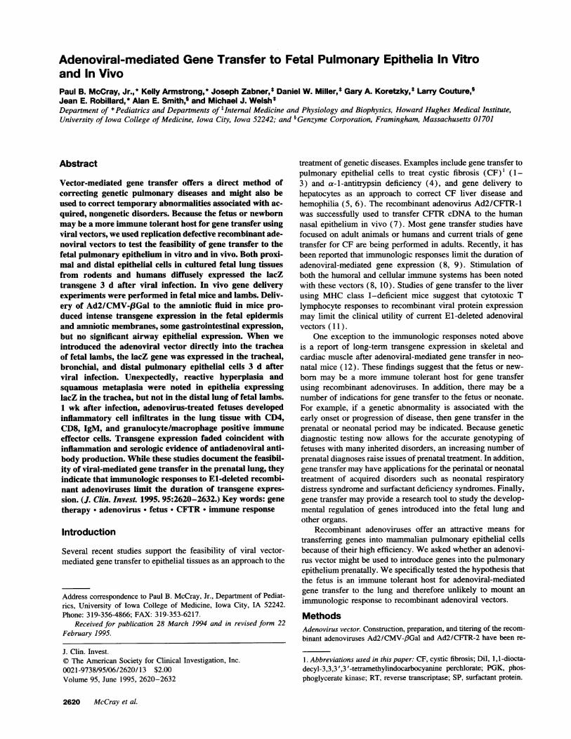
In vitro gene transfer to fetal lungsOrgan cultures have been used as model systems to study thedeveloping lung because many aspects of their morphology andfunction parallel in vivo development. Therefore we tested theability of an adenoviral vector to transfer a gene to fetal rat andhuman lung in organ culture. 3 d after injection of Ad2/CMV-3Gal, X-gal-stained fetal rat lung tissues showed evidence of
both diffuse and patchy epithelial cell lacZ gene expression(Fig. 1, A, C, and D). In some sections virtually all epitheliastained blue, whereas in other areas lacZ expression was patchy.Sites far removed from the point of injection expressed littleor no transgene. These findings probably reflect the unequaldistribution of injected virus within the fluid-filled airway lu-men. Occasionally we saw blue-stained cells in the mesen-chyme. LacZ expression in nonepithelial cells probably repre-sents areas exposed to the virus when the micropipette wasinserted, as it was not always possible to selectively inject virusinto the lumen alone. LacZ-expressing epithelial cells includedthose in the conducting airways and the distal lung. Noninjectedtissues showed no evidence of endogenous /3-galactosidase ac-tivity or blue stained nuclei (Fig. 1 B).
To determine if the adenoviral vector would also direct geneexpression in the human fetal pulmonary epithelium, we studiedsecond trimester lung tissue in explant culture. Epithelial cells in
cultured midgestation human fetal lung tissue explants showedwidespread evidence of 3-galactosidase activity 3 d after injec-tion of virus (Fig. 1, E and F). Nonciliated cuboidal epithelialcells lining the acinar tubules expressed the lacZ gene. Thesecells include type II cells and their glycogen-rich precursors(24). These in vitro studies indicate that the adenoviral vectorcan transfer a cDNA to murine and human fetal pulmonaryepithelia and direct expression of recombinant protein.
In vivo gene transfer to fetal lungsFetal mice. The most simple approach to deliver a vector tofetal pulmonary airways would be to introduce the vector intothe amniotic fluid. Wehypothesized that fetal breathing activitymight allow the vector access to the fetal lung (25). To testthis possibility, we first developed the in utero techniques ofamniotic cavity injection by introducing 2% fast green dye intothe amnions of fetal mice using a micropipette attached to asyringe. With this approach we successfully stained the amnioticcavity in 60% of the fetuses (data not shown). Therefore theamniotic cavities of 15-d gestation fetal mice were injectedwith 5-50 M1 of Dii. Examination of the fetuses 24-72 h laterrevealed diffuse epithelial cell staining of the pulmonary (Fig.2, A-D) and gastrointestinal tracts. Fluorescent staining wasalso present in the epidermis but not in other fetal organs. Thepulmonary epithelial cell staining extended from the trachea tothe distal acinar tubules (Fig. 2, A-D). These observationsindicate that the marker dye in amniotic fluid reached the pulmo-nary epithelium, possibly during episodes of fetal breathing withthe glottis open.
The preliminary experiments with dyes suggested that avector introduced into amniotic fluid might reach pulmonaryepithelial cells. In subsequent experiments 5-50 kll of viralsuspension was injected into the amniotic cavities of 15-d gesta-tion mouse embryos. 2 d later the fetuses were removed andfixed and stained with X-gal. While this approach achievedabundant lacZ gene expression in the skin, oropharynx, and, toa lesser extent, the gastrointestinal tract of the fetus, we saw nosignificant staining of the pulmonary epithelium (n = 10, Fig.2, E and F). Control, untreated animals showed no blue-stainedcells. The lack of pulmonary cell labeling in virus-treated fe-tuses suggests that diffuse infection of the fetal epidermal andamniotic membrane surfaces may have significantly reduced theamniotic fluid viral titer and thus decreased the amount of virusthat could be transferred to the lung. Alternatively, the diffusionand flow of the virus may have been more restricted than thesmaller dyes. Therefore we examined the effect of direct instilla-tion of virus into the fetal airway.
Fetal lambs. The fetal lamb was used for these in vivostudies because its large size allowed easy manipulation anddirect introduction of the vector into the fetal lung throughex utero approaches. Furthermore, submucosal glands developprenatally in the lamb, as in humans, and lung developmentproceeds at a slower rate compared with other nonprimates.Fetal lambs of 90-1 10-d gestation were studied to test thehypothesis that a vector introduced into lung fluid could infectthe epithelium in vivo. As shown in Figs. 3 and 4, 3 d aftergene transfer blue-stained cells were evident in the trachea,conducting airways, and the distal lung epithelium of the virus-treated fetuses. The distribution of transgene-expressing cellswas variable. Pronounced tracheal epithelium staining was seenin animals in which the virus was instilled with 2 ml of saline(Table I). The percentage of #3Gal-positive tracheal cells ranged
2622 McCray et al.
B
4'
to <~ ~ I ~'~:-*i W0I'%'<>e
*- eaa -,s
.9 N~~~~~~~~~~: t tI
k ~ ~ ~ ~ *
iF
i'
o'p4
_
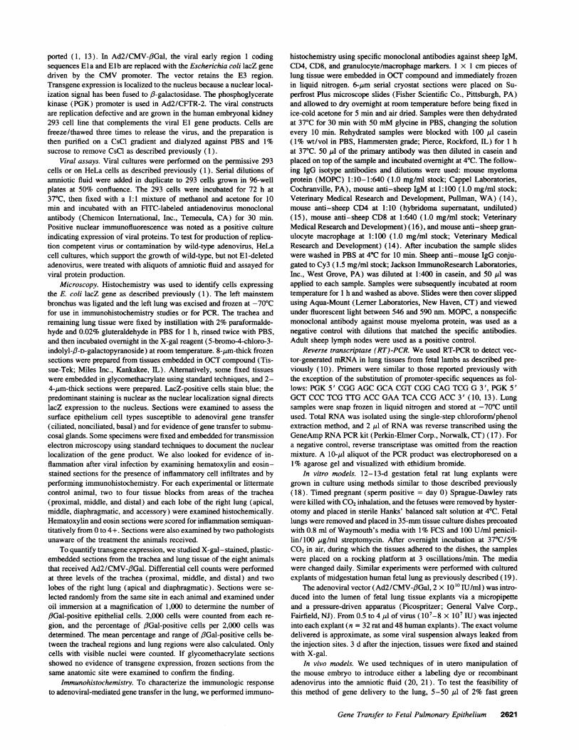
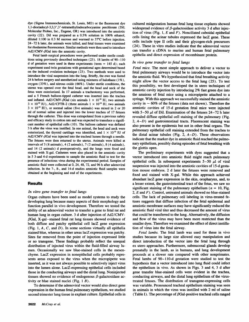
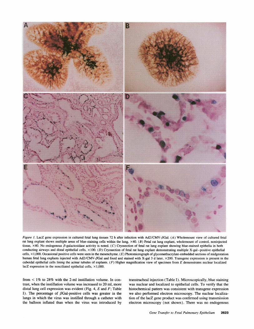
Figure 1. LacZ gene expression in cultured fetal lung tissues 72 h after infection with Ad2/CMV-,BGal. (A) Wholemount view of cultured fetalrat lung explant shows multiple areas of blue-staining cells within the lung, x40. (B) Fetal rat lung explant, wholemount of control, noninjectedtissue, x40. No endogenous /3-galactosidase activity is noted. (C) Cryosection of fetal rat lung explant showing blue-stained epithelia in bothconducting airways and distal epithelial cells, x 100. (D) Cryosection of fetal rat lung explant demonstrating multiple X-gal-positive epithelialcells, x 1,000. Occasional positive cells were seen in the mesenchyme. (E) Photomicrograph of glycomethacrylate-embedded sections of midgestationhuman fetal lung explants injected with Ad2/CMV-,6Gal and fixed and stained with X-gal 3 d later, x200. Transgene expression is present in thecuboidal epithelial cells lining the acinar tubules of explants. (F) Higher magnification view of specimen from E demonstrates nuclear localizedlacZ expression in the nonciliated epithelial cells, X 1,000.
from < 1% to 28% with the 2-ml instillation volume. In con-trast, when the instillation volume was increased to 20 ml, moredistal lung cell expression was evident (Fig. 4, E and F; TableI). The percentage of f3Gal-positive cells was greater in thelungs in which the virus was instilled through a catheter withthe balloon inflated than when the virus was introduced by
transtracheal injection (Table I). Microscopically, blue stainingwas nuclear and localized to epithelial cells. To verify that thehistochemical pattern was consistent with transgene expressionwe also performed electron microscopy. The nuclear localiza-tion of the lacZ gene product was confirmed using transmissionelectron microscopy (not shown). There was no endogenous
Gene Transfer to Fetal Pulmonary Epithelium 2623
4,j!
.1w..:!.a:F C -..AA...
to.W.M-
As
.J-1k. 1410.."VQ
W
E r...',..,i .
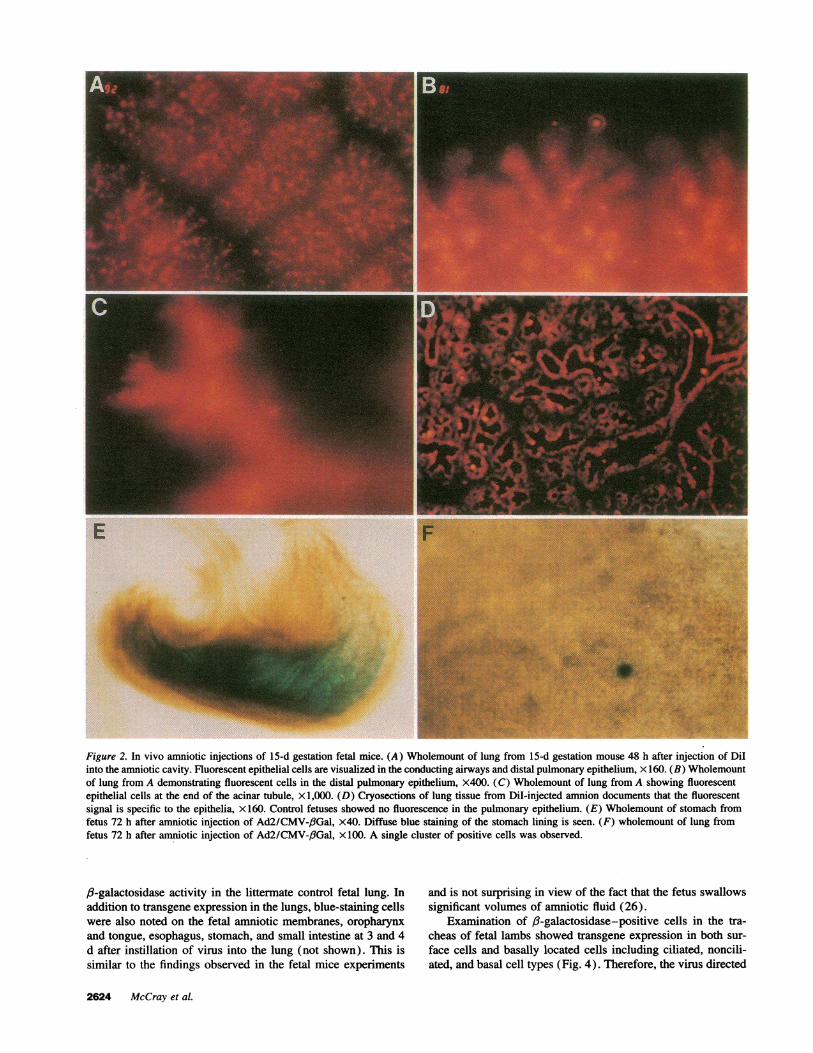
Figure 2. In vivo amniotic injections of 15-d gestation fetal mice. (A) Wholemount of lung from 15-d gestation mouse 48 h after injection of DiIinto the amniotic cavity. Fluorescent epithelial cells are visualized in the conducting airways and distal pulmonary epithelium, x 160. (B) Wholemountof lung from A demonstrating fluorescent cells in the distal pulmonary epithelium, x400. (C) Wholemount of lung from A showing fluorescentepithelial cells at the end of the acinar tubule, xl ,000. (D) Cryosections of lung tissue from Dil-injected amnion documents that the fluorescentsignal is specific to the epithelia, x 160. Control fetuses showed no fluorescence in the pulmonary epithelium. (E) Wholemount of stomach fromfetus 72 h after amniotic injection of Ad2/CMV-,fGal, x40. Diffuse blue staining of the stomach lining is seen. (F) wholemount of lung fromfetus 72 h after amniotic injection of Ad2/CMV-f3Gal, x 100. A single cluster of positive cells was observed.
f3-galactosidase activity in the littermate control fetal lung. Inaddition to transgene expression in the lungs, blue-staining cellswere also noted on the fetal amniotic membranes, oropharynxand tongue, esophagus, stomach, and small intestine at 3 and 4d after instillation of virus into the lung (not shown). This issimilar to the findings observed in the fetal mice experiments
and is not surprising in view of the fact that the fetus swallowssignificant volumes of amniotic fluid (26).
Examination of (8-galactosidase-positive cells in the tra-cheas of fetal lambs showed transgene expression in both sur-face cells and basally located cells including ciliated, noncili-ated, and basal cell types (Fig. 4). Therefore, the virus directed
2624 McCray et al.
AA
.. .- .t
N.'
D
mlt
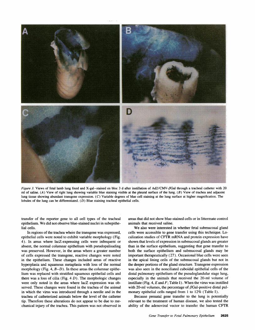
Figure 3. Views of fetal lamb lung fixed and X-gal-stained en bloc 3 d after instillation of Ad2/CMV-/3Gal through a tracheal catheter with 20ml of saline. (A) View of right lung showing variable blue staining visible at the pleural surface of the lung. (B) View of trachea and adjacentlung tissue showing abundant transgene expression. (C) Variable degrees of blue cell staining at the lung surface at higher magnification. Thelobules of the lung can be differentiated. (D) Blue staining tracheal epithelial cells.
transfer of the reporter gene to all cell types of the trachealepithelium. Wedid not observe blue-stained nuclei in subepithe-lial cells.
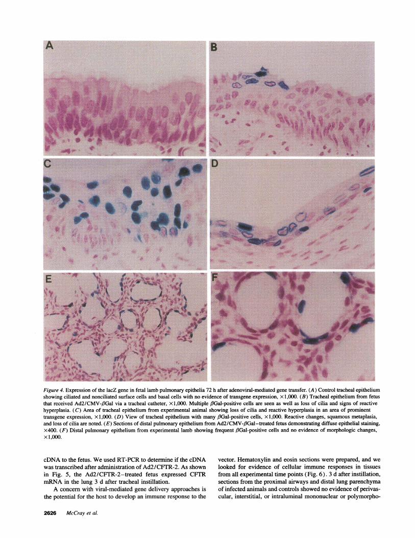
In regions of the trachea where the transgene was expressed,epithelial cells were noted to exhibit variable morphology (Fig.4). In areas where lacZ-expressing cells were infrequent orabsent, the normal columnar epithelium with pseudopalisadingwas preserved. However, in the areas where a greater numberof cells expressed the transgene, reactive changes were notedin the epithelium. These changes included areas of reactivehyperplasia and squamous metaplasia with loss of the normalmorphology (Fig. 4, B-D). In these areas the columnar epithe-lium was replaced with stratified squamous epithelial cells andthere was a loss of cilia (Fig. 4 D). The morphologic changeswere only noted in the areas where lacZ expression was ob-served. These changes were found in the trachea of the animalin which the virus was introduced through a needle and in thetrachea of catheterized animals below the level of the cathetertip. Therefore these alterations do not appear to be due to me-chanical injury of the trachea. This pattern was not observed in
areas that did not show blue-stained cells or in littermate controlanimals that received saline.
Wealso were interested in whether fetal submucosal glandcells were accessible to gene transfer using this technique. Lo-calization studies of CFTRmRNAand protein expression haveshown that levels of expression in submucosal glands are greaterthan in the surface epithelium, suggesting that gene transfer toboth the surface epithelium and submucosal glands may beimportant therapeutically (27). Occasional blue cells were seenin the apical lining cells of the submucosal glands but not inthe deeper portions of the gland structure. Transgene expressionwas also seen in the nonciliated cuboidal epithelial cells of thedistal pulmonary epithelium of the pseudoglandular stage lung,especially in the animals that received the 20-ml volume ofinstillate (Fig. 4, Eand F; Table I). Whenthe virus was instilledwith 20-ml volumes, the percentage of fiGal-positive distal pul-monary epithelial cells ranged from 1 to 12% (Table I).
Because prenatal gene transfer to the lung is potentiallyrelevant to the treatment of human disease, we also tested theability of the adenoviral vector to transfer the human CFTR
Gene Transfer to Fetal Pulmonary Epithelium 2625
Ivi I .-
A B
AP~~~~.I.,~~~~~~~~~~~~~~~~~~~~~~~~~~~~~~~~~~~~~~~~~~~~~~~~
4~~~
DC-
.
_ ..
. } ...gs.... * *_ _
e f -* as Hi: i
#
di B ifa :.
.:s E 11;
Figure 4. Expression of the lacZ gene in fetal lamb pulmonary epithelia 72 h after adenoviral-mediated gene transfer. (A) Control tracheal epitheliumshowing ciliated and nonciliated surface cells and basal cells with no evidence of transgene expression, x 1,000. (B) Tracheal epithelium from fetusthat received Ad2/CMV-fiGal via a tracheal catheter, x 1,000. Multiple f3Gal-positive cells are seen as well as loss of cilia and signs of reactivehyperplasia. (C) Area of tracheal epithelium from experimental animal showing loss of cilia and reactive hyperplasia in an area of prominenttransgene expression, xl ,000. (D) View of tracheal epithelium with many fGal-positive cells, x 1,000. Reactive changes, squamous metaplasia,and loss of cilia are noted. (E) Sections of distal pulmonary epithelium from Ad2/CMV-,/3Gal-treated fetus demonstrating diffuse epithelial staining,x400. (F) Distal pulmonary epithelium from experimental lamb showing frequent ,/Gal-positive cells and no evidence of morphologic changes,x 1,000.
cDNA to the fetus. Weused RT-PCR to determine if the cDNAwas transcribed after administration of Ad2/CFT1R-2. As shownin Fig. 5, the Ad2/CFTR-2-treated fetus expressed CFTRmRNAin the lung 3 d after tracheal instillation.
A concern with viral-mediated gene delivery approaches isthe potential for the host to develop an immune response to the
vector. Hematoxylin and eosin sections were prepared, and welooked for evidence of cellular immune responses in tissuesfrom all experimental time points (Fig. 6). 3 d after instillation,sections from the proximal airways and distal lung parenchymaof infected animals and controls showed no evidence of perivas-cular, interstitial, or intraluminal mononuclear or polymorpho-
2626 McCray et al.
i.11 p4 .0 1%
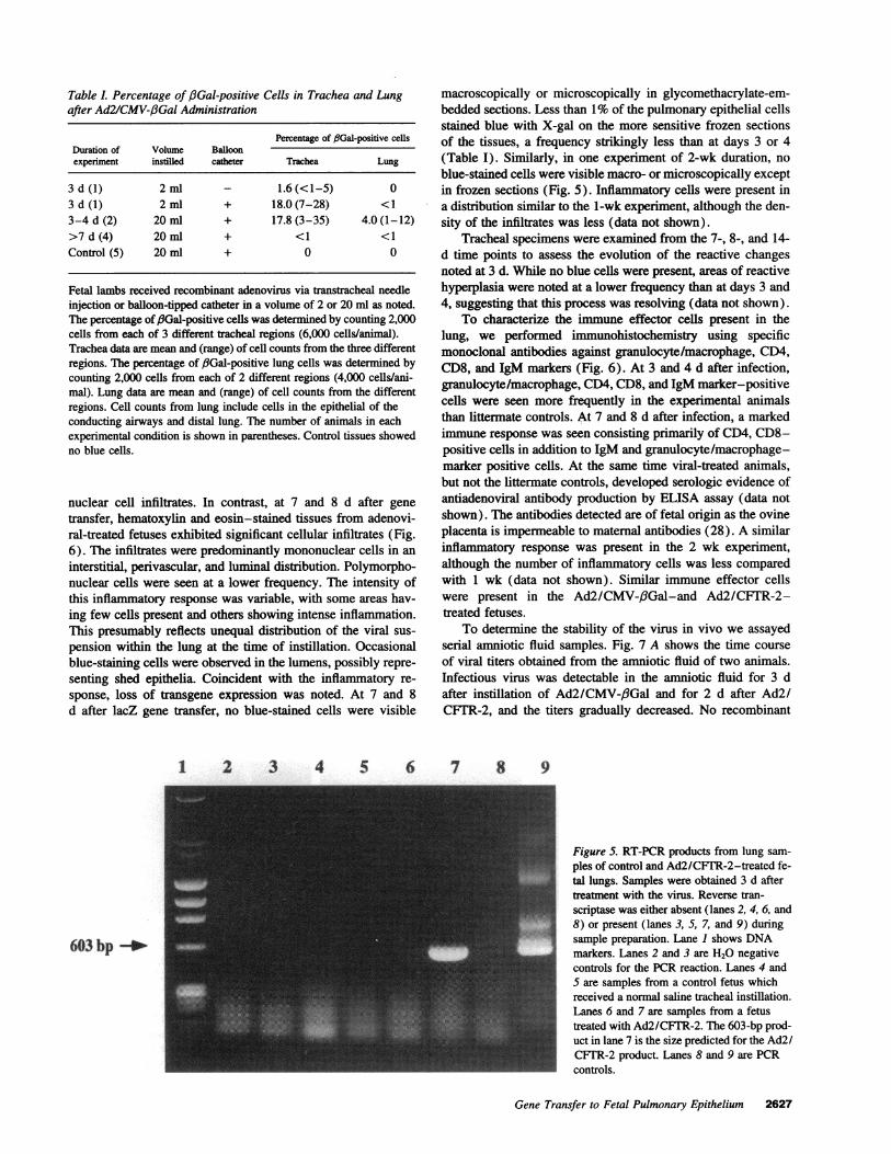
Table l. Percentage of f3Gal-positive Cells in Trachea and Lungafter Ad2/CMV-,3Gal Administration
Percentage of f3Gal-positive cellsDuration of Volume Balloonexperiment instilled catheter Trachea Lung
3d(1) 2ml - 1.6(<1-5) 03 d (1) 2 ml + 18.0 (7-28) <13-4 d (2) 20 ml + 17.8 (3-35) 4.0 (1-12)>7 d (4) 20 ml + <1 <1Control (5) 20 ml + 0 0
Fetal lambs received recombinant adenovirus via transtracheal needleinjection or balloon-tipped catheter in a volume of 2 or 20 ml as noted.The percentage of /Gal-positive cells was determined by counting 2,000cells from each of 3 different tracheal regions (6,000 cells/animal).Trachea data are mean and (range) of cell counts from the three differentregions. The percentage of /3Gal-positive lung cells was determined bycounting 2,000 cells from each of 2 different regions (4,000 cells/ani-mal). Lung data are mean and (range) of cell counts from the differentregions. Cell counts from lung include cells in the epithelial of theconducting airways and distal lung. The number of animals in eachexperimental condition is shown in parentheses. Control tissues showedno blue cells.
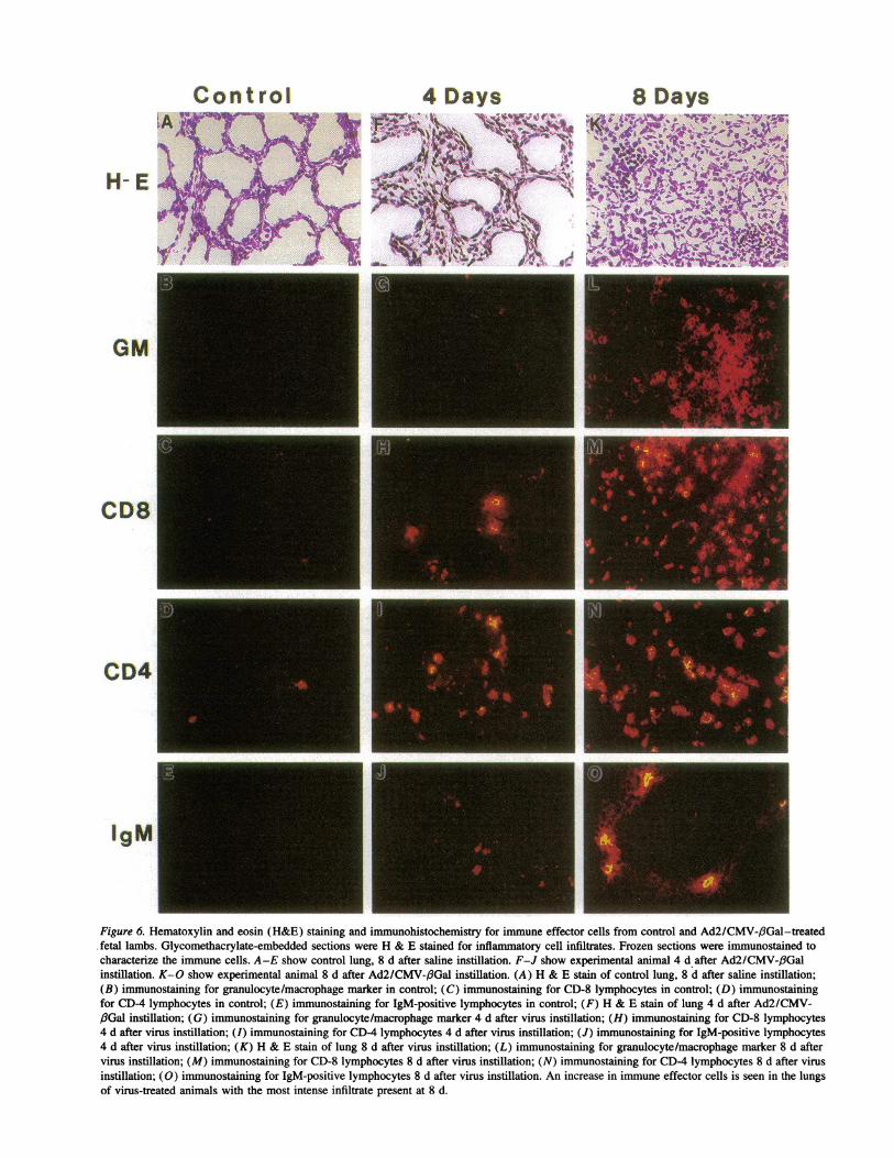
nuclear cell infiltrates. In contrast, at 7 and 8 d after genetransfer, hematoxylin and eosin-stained tissues from adenovi-ral-treated fetuses exhibited significant cellular infiltrates (Fig.6). The infiltrates were predominantly mononuclear cells in aninterstitial, perivascular, and luminal distribution. Polymorpho-nuclear cells were seen at a lower frequency. The intensity ofthis inflammatory response was variable, with some areas hav-ing few cells present and others showing intense inflammation.This presumably reflects unequal distribution of the viral sus-pension within the lung at the time of instillation. Occasionalblue-staining cells were observed in the lumens, possibly repre-senting shed epithelia. Coincident with the inflammatory re-sponse, loss of transgene expression was noted. At 7 and 8d after lacZ gene transfer, no blue-stained cells were visible
1 2 3 4 5 6
603 bp -*
macroscopically or microscopically in glycomethacrylate-em-bedded sections. Less than 1%of the pulmonary epithelial cellsstained blue with X-gal on the more sensitive frozen sectionsof the tissues, a frequency strikingly less than at days 3 or 4(Table I). Similarly, in one experiment of 2-wk duration, noblue-stained cells were visible macro- or microscopically exceptin frozen sections (Fig. 5). Inflammatory cells were present ina distribution similar to the 1-wk experiment, although the den-sity of the infiltrates was less (data not shown).
Tracheal specimens were examined from the 7-, 8-, and 14-d time points to assess the evolution of the reactive changesnoted at 3 d. While no blue cells were present, areas of reactivehyperplasia were noted at a lower frequency than at days 3 and4, suggesting that this process was resolving (data not shown).
To characterize the immune effector cells present in thelung, we performed immunohistochemistry using specificmonoclonal antibodies against granulocyte/macrophage, CD4,CD8, and IgM markers (Fig. 6). At 3 and 4 d after infection,granulocyte/macrophage, CD4, CD8, and IgM marker-positivecells were seen more frequently in the experimental animalsthan littermate controls. At 7 and 8 d after infection, a markedimmune response was seen consisting primarily of CD4, CD8-positive cells in addition to IgM and granulocyte/macrophage-marker positive cells. At the same time viral-treated animals,but not the littermate controls, developed serologic evidence ofantiadenoviral antibody production by ELISA assay (data notshown). The antibodies detected are of fetal origin as the ovineplacenta is impermeable to maternal antibodies (28). A similarinflammatory response was present in the 2 wk experiment,although the number of inflammatory cells was less comparedwith 1 wk (data not shown). Similar immune effector cellswere present in the Ad2/CMV-3Gal-and Ad2/CFTR-2-treated fetuses.
To determine the stability of the virus in vivo we assayedserial amniotic fluid samples. Fig. 7 A shows the time courseof viral titers obtained from the amniotic fluid of two animals.Infectious virus was detectable in the amniotic fluid for 3 dafter instillation of Ad2/CMV-,6Gal and for 2 d after Ad2/CFTR-2, and the titers gradually decreased. No recombinant
7 8 9
Figure 5. RT-PCR products from lung sam-ples of control and Ad2/CFTR-2-treated fe-tal lungs. Samples were obtained 3 d aftertreatment with the virus. Reverse tran-scriptase was either absent (lanes 2, 4, 6, and8) or present (lanes 3, 5, 7, and 9) duringsample preparation. Lane 1 shows DNAmarkers. Lanes 2 and 3 are H20 negativecontrols for the PCRreaction. Lanes 4 and5 are samples from a control fetus whichreceived a normal saline tracheal instillation.Lanes 6 and 7 are samples from a fetustreated with Ad2/CFTR-2. The 603-bp prod-uct in lane 7 is the size predicted for the Ad2/CFTR-2 product. Lanes 8 and 9 are PCRcontrols.
Gene Transfer to Fetal Pulmonary Epithelium 2627
Control
H- E
GM
CD8
CD4
4 Days 8 Days
IgM
Figure 6. Hematoxylin and eosin (H&E) staining and immunohistochemistry for immune effector cells from control and Ad2/CMV-(3Gal-treatedfetal lambs. Glycomethacrylate-embedded sections were H & E stained for inflammatory cell infiltrates. Frozen sections were immunostained tocharacterize the immune cells. A-E show control lung, 8 d after saline instillation. F-J show experimental animal 4 d after Ad2/CMV-/3Galinstillation. K-O show experimental animal 8 d after Ad2/CMV-f3Gal instillation. (A) H & E stain of control lung, 8 d after saline instillation;(B) immunostaining for granulocyte/macrophage marker in control; (C) immunostaining for CD-8 lymphocytes in control; (D) immunostainingfor CD-4 lymphocytes in control; (E) immunostaining for IgM-positive lymphocytes in control; (F) H & E stain of lung 4 d after Ad2/CMV-,fGal instillation; (G) immunostaining for granulocyte/macrophage marker 4 d after virus instillation; (H) immunostaining for CD-8 lymphocytes4 d after virus instillation; (I) immunostaining for CD-4 lymphocytes 4 d after virus instillation; (J) immunostaining for IgM-positive lymphocytes4 d after virus instillation; (K) H & E stain of lung 8 d after virus instillation; (L) immunostaining for granulocyte/macrophage marker 8 d aftervirus instillation; (M) immunostaining for CD-8 lymphocytes 8 d after virus instillation; (N) immunostaining for CD-4 lymphocytes 8 d after virusinstillation; (0) immunostaining for IgM-positive lymphocytes 8 d after virus instillation. An increase in immune effector cells is seen in the lungsof virus-treated animals with the most intense infiltrate present at 8 d.
AViral Titer
(Log i.uIml)
-1 24 48 72
(hr)
BViral Titer
(Log i.uIml)
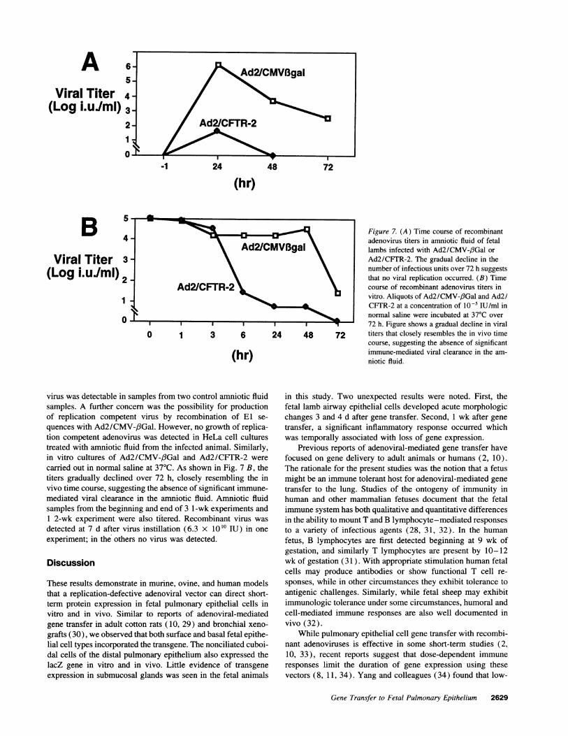
Figure 7. (A) Time course of recombinantadenovirus titers in amniotic fluid of fetalAd2/CMVBgal lambs infected with Ad2/CMV-/3Gal orAd2/CFTR-2. The gradual decline in thenumber of infectious units over 72 h suggeststhat no viral replication occurred. (B) Time
AdVCFTR-2 course of recombinant adenovirus titers invitro. Aliquots of Ad2/CMV-/3Gal and Ad2/CFTR-2 at a concentration of 10-5 IU/ml innormal saline were incubated at 370C over72 h. Figure shows a gradual decline in viral
0 1 3 6 24 48 72 titers that closely resembles the in vivo timecourse, suggesting the absence of significant
(hr) immune-mediated viral clearance in the am-niotic fluid.
virus was detectable in samples from two control amniotic fluidsamples. A further concern was the possibility for productionof replication competent virus by recombination of El se-quences with Ad2/CMV-,6Gal. However, no growth of replica-tion competent adenovirus was detected in HeLa cell culturestreated with amniotic fluid from the infected animal. Similarly,in vitro cultures of Ad2/CMV-,6Gal and Ad2/CFTR-2 werecarried out in normal saline at 37TC. As shown in Fig. 7 B, thetiters gradually declined over 72 h, closely resembling the invivo time course, suggesting the absence of significant immune-mediated viral clearance in the amniotic fluid. Amniotic fluidsamples from the beginning and end of 3 l-wk experiments and1 2-wk experiment were also titered. Recombinant virus wasdetected at 7 d after virus instillation (6.3 X 10¹⁰ IU) in oneexperiment; in the others no virus was detected.
Discussion
These results demonstrate in murine, ovine, and human modelsthat a replication-defective adenoviral vector can direct short-term protein expression in fetal pulmonary epithelial cells invitro and in vivo. Similar to reports of adenoviral-mediatedgene transfer in adult cotton rats (10, 29) and bronchial xeno-grafts (30), we observed that both surface and basal fetal epithe-lial cell types incorporated the transgene. The nonciliated cuboi-dal cells of the distal pulmonary epithelium also expressed thelacZ gene in vitro and in vivo. Little evidence of transgeneexpression in submucosal glands was seen in the fetal animals
in this study. Two unexpected results were noted. First, thefetal lamb airway epithelial cells developed acute morphologicchanges 3 and 4 d after gene transfer. Second, 1 wk after genetransfer, a significant inflammatory response occurred whichwas temporally associated with loss of gene expression.
Previous reports of adenoviral-mediated gene transfer havefocused on gene delivery to adult animals or humans (2, 10).The rationale for the present studies was the notion that a fetusmight be an immune tolerant host for adenoviral-mediated genetransfer to the lung. Studies of the ontogeny of immunity inhuman and other mammalian fetuses document that the fetalimmune system has both qualitative and quantitative differencesin the ability to mount T and B lymphocyte-mediated responsesto a variety of infectious agents (28, 31, 32). In the humanfetus, B lymphocytes are first detected beginning at 9 wk ofgestation, and similarly T lymphocytes are present by 10-12wk of gestation (3 1 ). With appropriate stimulation human fetalcells may produce antibodies or show functional T cell re-sponses, while in other circumstances they exhibit tolerance toantigenic challenges. Similarly, while fetal sheep may exhibitimmunologic tolerance under some circumstances, humoral andcell-mediated immune responses are also well documented invivo (32).
While pulmonary epithelial cell gene transfer with recombi-nant adenoviruses is effective in some short-term studies (2,10, 33), recent reports suggest that dose-dependent immuneresponses limit the duration of gene expression using thesevectors (8, 11, 34). Yang and colleagues (34) found that low-
Gene Transfer to Fetal Pulmonary Epithelium 2629
level viral gene expression occurs with liver-directed gene trans-fer using an El-deleted adenovirus. Their findings suggest thata virus-specific cellular immune response may lead to the de-struction of the genetically modified hepatocytes (34). Furtherstudies using mice deficient in CD8+ lymphocytes are consis-tent with the hypothesis that MHCclass I-restricted CD8+cytolytic T cells are the primary immune effectors causing lossof transgene expression in this model ( 11). Gene transfer withrecombinant adenoviruses has also been associated with stimu-lation of the humoral immune system and production of neu-tralizing antibodies (10).
Unexpectedly, gene transfer to the large airways in the fetallamb was associated with morphologic changes of reactive hy-perplasia and squamous metaplasia in areas of transgene expres-sion 3 and 4 d after gene transfer. To our knowledge the mor-phological changes in many tracheal epithelial cells incorporat-ing the transgene 3 d after infection have not been observedbefore. These nonspecific changes of cellular hyperplasia andsquamous metaplasia appeared reactive, presumably reflectinga cellular response to the viral infection. Similar morphologicchanges were seen in aimals that received the CFIR vectorAd2/CFTR-2, indicating that this response is unlikely to be dueto the expression of /3-galactosidase. Temporally these changespreceded the inflammatory cell infiltrates that were seen 1 wkafter gene transfer. These morphologic changes were not ob-served in cultured rat or human explants, suggesting either thatthe findings are species specific or that they are due to factorsonly present in vivo.
There are several possible explanations for this finding. Thefirst is that viral antigens or viral gene expression and proteinproduction stimulate cell growth and differentiation. Low levelsof some adenoviral early and late gene products (E4 and L5)may be expressed by the recombinant virus, and these geneproducts could modulate cell growth (1). Bronchial xenograftsinfected with an El-and E3-deleted adenovirus showed lowlevels of viral fiber protein production and E2a gene product(30). Wealso observed low levels of E2a expression by North-ern blotting in the lungs of adenoviral-treated fetuses (our un-published observation). It is also possible that host factors couldsubstitute for deleted viral products and allow viral protein pro-duction. This has been demonstrated in the case of Ela-deletedadenoviruses in HepG2 cells where NF-IL6 acts as a sequence-specific cellular nuclear factor regulating Ela-responsive genesin the absence of Ela (35, 36). Second, it is possible that hostresponses, such as the release of cytokines in responseto adenovirus administration, might produce these reactivechanges. In a mouse model of adenovirus pneumonia, infectionwith type 5 adenovirus was associated with the production oftumor necrosis factor-a and interleukins 1 and 6 (37). Respira-tory syncytial virus infection of human nasal epithelia stimulatessecretion of interleukin 8 (38). Further evaluation for the ex-pression of viral gene products and cytokines may help clarifythese findings.
Several findings show that the fetal lamb mounted bothhumoral and cellular immune responses to the recombinant ade-novirus. The fetuses developed antiadenoviral antibodies by 1wk after infection. Furthermore, they were specific to the viralinfection as they were absent in littermate controls. Of note,the antibodies detected were of fetal origin, as maternal antibod-ies do not cross the ovine placenta (28). This result contrastswith data in neonatal mice in which long-term expression hasbeen observed ( 12). Characterization of the interstitial, perivas-
cular, and intraluminal inflammatory response showed the pres-ence of CD4, CD8, IgM, and granulocyte/macrophage marker-positive immune effector cells. Similar inflammatory responseswere seen with both Ad2/CMV-/3Gal and Ad2/CFTR-2, sug-gesting that the response was not due to recombinant proteinexpression. The temporal association between the influx of im-mune effector cells and the loss of transgene expression impli-cates these cells in the process. The cellular characteristics andtiming of the inflammatory cell infiltrates in this study are con-sistent with at least part of the loss of gene expression beingdue to destruction of infected cells by cytotoxic T lymphocytesas reported recently by Wilson and colleagues ( 11). Studies ofgenetically altered sheep or specific suppression of T and Bcell lymphocyte populations would be required to prove thishypothesis.
Surprisingly, we found that although the titers declined, livevirus persisted in the amniotic fluid for as long as 7 d aftertracheal instillation. This suggests that the fluid-filled amnioticenvironment supports the persistence of the nonreplicating vi-rus. Engelhardt and colleagues reported detection of live virusfor up to 24 d in effluents from bronchial xenografts treatedwith recombinant adenovirus (30).
A potential advantage of fetal gene transfer is the possibilityof permanently correcting a genetic defect early in life, beforeirreversible organ damage occurs. Since the total number ofepithelial cells in the fetal lung is significantly less, it is possiblethat fewer cells would need to be corrected by gene transfer inthe fetus. While adenovirus can transfer genes to cells regardlessof their proliferative state, the frequency of adenoviral integra-tion into chromosomal DNAis very low, and gene expressionis expected to be episomal and transient. Gene transfer to thepulmonary epithelium of the fetus might be particularly advan-tageous using a vector that integrates into the host genome(retrovirus or adeno-associated virus). This might allow pas-sage of the transgene from progenitor to daughter cells, therebyincreasing the population of corrected cells in the lung.
There are several conditions where gene transfer to the so-matic cells of the fetal or neonatal lung might have applications.First, transfer of surfactant protein genes or antioxidant genesto the lung of the preterm infant could enhance surfactant pro-duction and protect the pulmonary epithelium from oxygen-induced lung injury associated with the treatment of neonatalrespiratory distress syndrome (39). Second, surfactant proteinB (SP-B) deficiency, a lung disease of infants associated withmutations in the SP-B gene and absence of SP-B mRNAandprotein, may be amenable to gene therapy in the fetus or neonate(40, 41). Third, the perinatal transition to air breathing is facili-tated by active absorption of sodium through amiloride-sensitiveepithelial sodium channels (42). Maladaptions of this processmay contribute to lung disease in premature and term infants(43); such problems might be addressed by introducing theepithelial sodium channel genes into the lung of the infant (44,45). Fourth, it may be advantageous to begin CF gene therapyearly in life, prenatally or neonatally, to prevent the onset andprogression of chronic lung disease. There is now evidence thatinfants with CF have pulmonary inflammation and altered lungfunction in infancy before they develop clinical symptoms ofrespiratory disease or are colonized with bacteria (46, 47).Finally, vector-mediated gene transfer may be a useful tech-nique to study lung cell lineage and the developmental regula-tion of genes introduced into the fetal lung.
In summary, while a replication-defective adenoviral vector
2630 McCray et al.
was effective for short-term gene transfer to fetal pulmonaryepithelia in vitro, gene transfer in vivo in fetal lambs was short-lived and associated with significant cellular and humoral im-mune responses. These results suggest that current El-deletedadenoviral vectors are ineffective for gene transfer in the fetus.Several strategies may be proposed to circumvent the fetal im-mune response to the vector and deserve further study. Addi-tional deletions or alterations of the viral backbone to reduceviral protein expression may blunt the fetal immune response.It may also be possible to tolerize a fetus by administeringlower doses of virus, perhaps at an earlier developmental timepoint. The fetal immune response could be minimized or pre-vented using pharmacologic immunosuppressive agents or spe-cific monoclonal antibodies against immune effector cells. Suc-cessful gene transfer to the fetal or neonatal lung will requirefurther modifications of the adenoviral vector system or alterna-tive vector approaches.
Acknowledgments
Wethank Tim Joseph, Oliva McWeeny, Jeff Otto, Aurita Puga, BruceSmith, and Kathy Walter for excellent technical assistance. Weespe-cially thank Deanna Peterson for assistance with RT-PCR and Dr. JohnHopkins for technical advice and for providing sheep anti-CD4 and-CD8 antibodies. We thank Dr. Jeanne Snyder in the Department ofAnatomy and Drs. Stephen Bonsib, Stephen Raab, and Garvan Brownein the Department of Pathology for reviewing and commenting on themorphology of the tissues.
Gary A. Koretzky is an Established Investigator of the AmericanHeart Association. This work was supported in part by the HowardHughes Medical Institute, grants from the National Institutes of Health(HL-51670 to M. J. Welsh and HL-02767), and the Cystic FibrosisFoundation (P. B. McCray).
References
1. Rich, D. P., L. A. Couture, L. M. Cardoza, V. M. Guiggio, D. Armentano,P. C. Espino, K. Hehir, M. J. Welsh, A. E. Smith, and R. J. Gregory. 1993.Development and analysis of recombinant adenovirus for gene therapy of cysticfibrosis. Hum. Gene Ther. 4:461-476.
2. Rosenfeld, M. A., K. Yoshimura, B. C. Trapnell, Y. Koichi, E. R. Rosenthal,W. Dalemans, M. Fukayama, J. Bargon, L. E. Stier, L. D. Stratford-Perricaudet, etal. 1992. In vivo transfer of the human cystic fibrosis transmembrane conductanceregulator gene to the airway epithelium. Cell. 68:143-155.
3. Engelhardt, J. F., J. R. Yankaskas, and J. M. Wilson. 1992. In vivo retroviralgene transfer into human bronchial epithelia of xenografts. .1. Clin. Invest.90:2598-2607.
4. Rosenfeld, M. A., W. Siegfried, K. Yoshimura, K. Yoneyama, M. Fukay-ama, L. E. Stier, P. K. Paakko, P. Gilardi, L. D. Stratford-Perricaudet, M. Perri-caudet, et al. 1991. Adenovirus-mediated transfer of a recombinant a-antitrypsingene to the lung epithelium in vivo. Science (Wash. DC). 252:431-434.
5. Yang, Y., S. E. Raper, J. A. Cohn, J. F. Engelhardt, and J. M. Wilson.1993. An approach for treating the hepatobiliary disease of cystic fibrosis bysomatic gene transfer. Proc. Natl. Acad. Sci. USA. 90:4601-4605.
6. Kay, M. A., S. Rothenberg, C. N. Landen, D. A. Bellinger, F. Leland,C. Toman, M. Finegold, A. R. Thompson, M. S. Read, K. M. Brinkhous, andS. L. C. Woo. 1993. In vivo gene therapy of hemophilia B: sustained partialcorrection in factor IX-deficient dogs. Science (Wash. DC). 262:117-119.
7. Zabner, J., L. A. Couture, R. J. Gregory, S. M. Graham, A. E. Smith, andM. J. Welsh. 1993. Adenovirus-mediated gene transfer transiently corrects thechloride transport defect in nasal epithelia of patients with cystic fibrosis. Cell.75:207-216.
8. Simon, R. H., J. F. Engelhardt, Y. Yang, M. Zepeda, S. Weber-Pendleton,M. Grossman, and J. M. Wilson. 1993. Adenovirus-mediated transfer of the CFTRgene to lung of nonhuman primates: toxicity study. Hum. Gene Ther. 4:771-780.
9. Yang, Y., F. A. Nunes, K. Berencsi, E. Gonczol, J. F. Engelhardt, and J.M. Wilson. 1994. Inactivation of E2a in recombinant adenoviruses improves theprospect for gene therapy in cystic fibrosis. Nature Genet. 7:362-369.
10. Zabner, J., D. M. Petersen, A. P. Puga, S. M. Graham, M. J. Welsh, L. A.Couture, L. D. Keyes, M. J. Lukason, J. A. St. George, R. J. Gregory, and A. E.Smith. 1994. Safety and efficacy of repetitive adenovirus-mediated transfer of
CFTRcDNA to airway epithelia of primates and cotton rats. Nature Genet. 6:75-83.
11. Yang, Y., H. C. J. Ertl, and J. M. Wilson. 1994. MHCclass I-restricedcytotoxic T lymphocytes to viral antigens destroy hepatocytes in mice infectedwith El-deleted recombinant adenoviruses. Immunity. 1:433-442.
12. Stratford-Perricaudet, L. D., I. Makeh, M. Perricaudet, and P. Briand.1992. Widespread long-term gene transfer to mouse skeletal muscles and heart.J. Clin. Invest. 90:626-630.
13. Armentano, D., C. Sookdeo, L. Couture, K. Vincent, L. Cardoza, V.Guiggio, D. Souza, R. Gregory, and A. Smith. 1993. Second generation adenovir-uses for gene therapy of CF. Pediatr. Pulmonol. 9 (Suppl.):143a. (Abstr.)
14. Davis, W. C., S. Marusic, H. A. Lewin, G. A. Splitter, L. E. Perryman,T. C. McGuire, and J. R. Gorham. 1987. The development and analysis of speciesspecific and cross reactive monoclonal antibodies to leukocyte differentiationantigens and antigens of the major histocompatibility complex for use in the studyof the immune system in cattle and other species. Vet. Immunol. Immunopathol.15:337-376.
15. Hopkins, J., A. Ross, and B. M. Dutia. 1993. 5.1 Summary of workshopfindings of leukocyte antigens in sheep. Vet. Immunol. Immunopathol. 39:49-59.
16. Davis, W. C., Y. H. Park, L. P. Perryman, and R. A. Larsen. 1988. NonT/nonB cells are a major antigenically distinct population of cells in ruminants.FASEB (Fed. Am. Soc. Exp. Biol.) J. 2:465a. (Abstr.)
17. Chomczynski, P., and N. Sacchi. 1987. Single-step method of RNAisola-tion by acid guanidinium thiocyanate-phenol-chloroform extraction. Anal. Bio-chem. 162:156-159.
18. Massoud, A. S., H. S. Sekhon, A. Rotschild, and W. M. Thurlbeck. 1992.The in vitro effect of triamcinolone acetonide on branching morphogenesis in thefetal rat lung. Pediatr. Pulmonol. 14:28-36.
19. McCray, P. B., Jr., J. D. Bettencourt, J. Bastacky, G. M. Denning, andM. J. Welsh. 1993. Expression of CFTRand a cAMP-stimulated chloride secretorycurrent in cultured human fetal alveolar epithelial cells. Am. J. Respir. Cell Mol.Biol. 9:578-585.
20. Papaioannou, V. 1990. In utero manipulations. In Postimplantation Mam-malian Embryos. A. J. Copp and D. L. Cockroft, editors. IRL Press, New York.61-80.
21. Trevino, C., R. Anderson, and K. Muneoka. 1993. 3T3 cell integrationand differentiative potential during limb development in the mouse. Dev. Biol.155:38-45.
22. Artinger, K. B., and M. Bronner-Fraser. 1992. Partial restriction in thedevelopmental potential of late emigrating avian neural crest cells. Dev. Biol.149:149-157.
23. Robillard, J. E., and R. E. Weitzman. 1980. Developmental aspects of thefetal renal response to exogenous arginine vasopressin. Am. J. Physiol. 238:F407-F414.
24. Snyder, J. M., J. M. Johnston, and C. R. Mendelson. 1981. Differentiationof type II cells of human fetal lung in vitro. Cell Tissue Res. 220:17-25.
25. Harding, R. 1991. Fetal breathing movements. In The Lung: ScientificFoundations. R. G. Crystal and J. B. West, editors. Raven Press Ltd., New York.1655-1663.
26. Leaver, L. T. 1991. Anatomy and embryology. In Pediatric GastrointestinalDisease. W. A. Walker, P. R. Durie, J. R. Hamilton, J. A. Walker-Smith, andJ. B. Watkins, editors. B. C. Decker, Inc., Philadelphia. 195-216.
27. Engelhardt, J. F., J. R. Yankaskas, S. A. Ernst, Y. Yang, C. R. Marino,R. C. Boucher, J. A. Cohn, and J. A. Wilson. 1992. Submucosal glands are thepredominant site of CFTR expression in the human bronchus. Nature Genetics.2:240-248.
28. Campbell, S. G., M. J. Siegel, and B. J. Knowlton. 1977. Sheep immuno-globulins and their transmission to the neonatal lamb. NZ Vet. J. 25:361-365.
29. Mastrangeli, A., C. Danel, M. A. Rosenfeld, L. D. Stratford-Perricaudet,M. Perricaudet, A. Pavirani, J.-P. Lecocq, and R. G. Crystal. 1993. Diversity ofairway epithelial cell targets for in vivo recombinant adenovirus-mediated genetransfer. J. Clin. Invest. 91:225-234.
30. Engelhardt, J. F., Y. Yang, L. D. Stratford-Perricaudet, E. D. Allen, K.Kozarsky, M. Perricaudet, J. R. Yankaskas, and J. M. Wilson. 1993. Direct genetransfer of human CFTR into human bronchial epithelia of xenografts with El-deleted adenoviruses. Nature Genet. 4:27-34.
31. Lawton, A. R., and M. D. Cooper. 1989. Ontogeny of immunity. InImmunologic Disorders in Infants and Children. R. Stiehm, editor. Saunders Pub-lishing Company, Philadelphia. 1-14.
32. Miyasaka, M., and B. Morris. 1988. The ontogeny of the lymphoid systemand immune responsiveness in sheep. Prog. Vet. Microbiol. Immunol. 4:21-55.
33. Englehardt, J. F., R. H. Simon, Y. Yang, M. Zepeda, S. Weber-Pendleton,B. Doranz, M. Grossman, and J. M. Wilson. 1993. Adenovirus-mediated transferof the CFTRgene to lung of nonhuman primates: biological efficacy study. Hum.Gene Ther. 4:759-769.
34. Yang, Y., F. A. Nunes, K. Berencsi, E. E. Furth, E. Gonczol, and J. M.Wilson. 1994. Cellular immunity to viral antigens limits El-deleted adenovirusesfor gene therapy. Proc. Natl. Acad. Sci. USA. 91:4407-4411.
35. Spergel, J. M., and S. Chen-Kiang. 1991. Interleukin-6 enhances a cellular
Gene Transfer to Fetal Pulmonary Epithelium 2631

activity that functionally substitutes for EIA protein in transactivation. Proc. Nati.Acad. Sci. USA. 88:6472-6476.
36. Spergel, J. M., W. Hsu, S. Akira, B. Thimmappaya, T. Kishimoto, and S.Chen-Kiang. 1992. NF-IL6, a member of the C/EBP family, regulates ElA-responsive promoters in the absence of EIA. J. Virol. 66:1021-1030.
37. Ginsberg, H. S., L. L. Moldawer, P. B. Sehgal, M. Redington, P. L. Kilian,R. M. Chanock, and G. A. Prince. 1991. A mouse model for investigating themolecular pathogenesis of adenovirus pneumonia. Proc. NatL. Acad. Sci. USA.88:1651-1655.
38. Becker, S., H. S. Koren, and D. C. Henke. 1993. Interleukin-8 expressionin normal nasal epithelium and its modulation by infection with respiratory syncy-tial virus and cytokines tumor necrosis factor, interleukin-1, and interleukin-6.Am. J. Respir. Cell Mol. Biol. 8:20-27.
39. Wispe, J. R., B. B. Warner, J. C. Clark, C. R. Dey, J. Neuman, S. W.Glasser, J. D. Crapo, L.-Y. Chang, and J. A. Whitsett. 1992. HumanMn-superox-ide dismutase in pulmonary epithelial cells of transgenic mice confers protectionfrom oxygen injury. J. Biol. Chem. 267:23937-23941.
40. Nogee, L. M., D. E. deMello, L. P. Dehner, and H. R. Colten. 1993.Brief report: deficiency of pulmonary surfactant protein B in congenital alveolarproteinosis. N. Engl. J. Med. 328:406-410.
41. Nogee, L. M., G. Garnier, H. C. Dietz, L. Singer, A. M. Murphy, D. E.
deMello, and H. R. Colten. 1994. A mutation in the surfactant protein B generesponsible for fatal neonatal respiratory disease in multiple kindreds. J. Clin.Invest. 93:1860-1863.
42. Strang, L. B. 1991. Fetal lung liquid: secretion and reabsorption. Physiol.Rev. 71:991-1016.
43. Gowen, C. W., Jr., E. E. Lawson, J. Gingras, R. C. Boucher, J. T. Gatzy,and M. R. Knowles. 1988. Electrical potential difference and ion transport acrossnasal epithelium of term neonates: correlation with mode of delivery, transienttachypnea of the newborn, and respiratory rate. J. Pediatr. 113:121-127.
44. Canessa, C. M., J.-D. Horisberger, and B. C. Rossier. 1993. Epithelialsodium channel related to proteins involved in neurodegeneration. Nature (Lond.).361:467-470.
45. Canessa, C. M., L. Schild, G. Buell, B. Thorens, I. Gautschi, J.-D. Horis-berger, and B. C. Rossier. 1994. Amiloride-sensitive epithelial Na+ channel ismade of three homologous subunits. Nature (Lond.). 367:463-467.
46. Balough, K., R. Fick, Jr., M. Weinberger, M. McCubbin, and R. Ahrens.1992. Inflammation in early cystic fibrosis lung lesion: lack of correlation withinfection. Am. Rev. Respir. Dis. 145:689a.(Abstr.)
47. Khan, T. Z., J. S. Wagener, D. W. H. Riches, and F. J. Accurso. 1993.Increased interleukin-8 levels and gene expression by pulmonary macrophages inbronchoalveolar lavage fluid from infants with cystic fibrosis. Clin. Res.41 :2a .(Abstr.)
2632 McCray et al.