Embed Size (px)
Citation preview

ACTAUNIVERSITATIS
UPSALIENSISUPPSALA
2013
Digital Comprehensive Summaries of Uppsala Dissertationsfrom the Faculty of Medicine 914
Adenovirus for Cancer Therapy
With a Focus on its Surface Modification
DI YU
ISSN 1651-6206ISBN 978-91-554-8700-3urn:nbn:se:uu:diva-203662

Dissertation presented at Uppsala University to be publicly examined in Rudbecksalen, TheRudbeck Laboratory C11, Dag Hammarskjölds väg 20, Uppsala, Friday, September 6, 2013 at09:15 for the degree of Doctor of Philosophy (Faculty of Medicine). The examination will beconducted in English.
AbstractYu, D. 2013. Adenovirus for Cancer Therapy: With a Focus on its Surface Modification. ActaUniversitatis Upsaliensis. Digital Comprehensive Summaries of Uppsala Dissertations fromthe Faculty of Medicine 914. 59 pp. Uppsala. ISBN 978-91-554-8700-3.
Adenovirus serotype 5 (Ad5) is widely used as an oncolytic agent for cancer therapy. However,its infectivity is highly dependent on the expression level of coxsackievirus-adenovirus receptor(CAR) on the surface of tumor cells. We engineered Ad5 virus with the protein transductiondomain (PTD) from the HIV-1 Tat protein (Tat-PTD) inserted in the hypervariable region 5(HVR5) of the hexon protein in the virus capsid. Tat-PTD-modified Ad5 shows a dramaticallyincreased transduction level of CAR-negative cells and bypassed fiber-mediated transduction.It also overcomes the fiber-masking problem, which is caused by release of excess fiber proteinsfrom infected cells. To achieve specific viral replication in neuroblastoma and neuroendocrinetumor cells, we identified the secretogranin III (SCG3) promoter and constructed an adenovirusAd5PTD(ASH1-SCG3-E1A) wherein E1A gene expression is controlled by the SCG3 promoterand the achaete-scute complex homolog 1 (ASH1) enhancer. This virus shows selective andefficient killing of neuroblastoma cell lines in vitro, and delays human neuroblastoma xenografttumor growth on nude mice. To further enhance the viral oncolytic efficacy, we also switchedthe fiber 5 to fiber 35 to generate Ad5PTDf35. This vector shows dramatically increasedtransduction capacity of primary human cell cultures including hematopoietic cells and theirderivatives, pancreatic islets and exocrine cells, mesenchymal stem cells and primary tumorcells including primary cancer initiating cells. Ad5PTDf35-based adenovirus could be a usefulplatform for gene delivery and oncolytic virus development. Viral oncolysis alone cannotcompletely eradicate tumors. Therefore, we further armed the Ad5PTDf35-D24 virus with asecreted form of Helicobacter pylori Neutrophil Activating Protein (HP-NAP). Expression ofHP-NAP recruits neutrophils to the site of infection, activates an innate immune response againsttumor cells and provokes a Th1-type adaptive immune response. Established tumor on nudemice could be completely eradicated in some cases after treatment with this virus and thesurvival of mice was significantly prolonged.
Keywords: Adenovirus, cancer, therapy, neuroblastoma, neuroendocrine, modification, Tat,PTD, cell penetrating peptide, Helicobacter pylori, NAP
Di Yu, Uppsala University, Department of Immunology, Genetics and Pathology, ClinicalImmunology, Rudbecklaboratoriet, SE-751 85 Uppsala, Sweden.
© Di Yu 2013
ISSN 1651-6206ISBN 978-91-554-8700-3urn:nbn:se:uu:diva-203662 (http://urn.kb.se/resolve?urn=urn:nbn:se:uu:diva-203662)

Stay hungry, stay foolish
Dedicated to my family献给我的家人


List of Papers
This thesis is based on the following papers, which are referred to in the text by their Roman numerals.
I Yu D, Jin C, Leja J, Majdalani N, Nilsson B, Eriksson F, Es-
sand M. Adenovirus with hexon Tat-protein transduction do-main modification exhibits increased therapeutic effect in ex-perimental neuroblastoma and neuroendocrine tumors. J Virol, 2011. 85(24): 13114-13123.
II Jin C*, Yu D*, Čančer M, Nilsson B, Leja J, Essand M. Tat-PTD-modified Oncolytic Adenovirus Driven by the SCG3 Promoter and ASH1 Enhancer for Neuroblastoma Therapy. Hum Gene Ther. 2013 Jul 26. doi:10.1089/hum.2012.132. [Epub ahead of print].
III Yu D, Jin C, Ramachandran M, Xu J, Nilsson B, Korsgren O, Le Blanc K, Uhrbom L, Forsberg-Nilsson K, Westermark B, Adamson R, Maitland N, Fan X, Essand M. Adenovirus sero-type 5 vectors with Tat-PTD modified hexon and serotype 35 fiber show greatly enhanced transduction capacity of primary cell cultures. PLoS One, 2013. 8(1): e54952.
IV Ramachandran, M, Yu, D, Wanders A, Essand M*, Eriksson, F*. An infection-enhanced oncolytic adenovirus secreting H. pylori neutrophil-activating protein with therapeutic effects on neuroendocrine tumors. Mol Ther, 2013, Jul 2. doi: 10.1038/mt.2013.153. [Epub ahead of print]
* Authors contributed equally to the work.
Reprints were made with permission from the respective publishers.
I Copyright © 2011 American Society for Microbiology. II, III Open access under the terms of the Creative Commons Attribution
3.0 Unported License (CC BY). IV Open access under the terms of the Creative Commons Attribution-
NonCommercial-NoDerivs 3.0 Unported License (CC BY-NC-ND).

Other related works
I Leja J, Nilsson B, Yu D, Gustafson E, Akerström G, Oberg K, Giandomenico V, Essand M. Double-detargeted oncolytic ad-enovirus shows replication arrest in liver cells and retains neu-roendocrine cell killing ability. PLoS One. 2010. 5(1):e8916.
II Leja J, Yu D, Nilsson B, Gedda L, Zieba A, Hakkarainen T, Åkerström G, Öberg K, Giandomenico V, Essand M. Onco-lytic adenovirus modified with somatostatin motifs for selec-tive infection of neuroendocrine tumor cells. Gene Ther. 2011. 18(11):1052-1062.
III Liljenfeldt L, Yu D, Chen LY, Essand M, Mangsbo S. A dou-ble-modified adenovirus expressing CD40L improves trans-duction efficacy of both tumor and dendritic cells leading to enhanced antigen presentation. Manuscript.
Yu D. 2013. Adenovirus for cancer therapy -with a focus on its surface modification.
Supervisor: Prof. Magnus Essand, PhD
Uppsala University, Sweden
Co-supervisor: Assoc. Prof. Valeria Giandomenico, PhD
Uppsala University Hospital, Sweden
Co-supervisor: Prof. Thomas Tötterman, MD, PhD
Uppsala University, Sweden
Opponent: Assoc. Prof. Dirk Nettelbeck, PhD
Heidelberg University Hospital, Germany
Committee members:
Prof. Göran Akusjärvi, PhD
Uppsala University, Sweden
Assoc. Prof. Torbjörn Ramqvist, PhD
Karolinska Institute, Sweden
Assoc. Prof. Daniel Öberg, PhD
Uppsala University, Sweden

Contents
Introduction ................................................................................................... 11 1. Human adenoviruses ............................................................................ 11
1.1 Adenovirus structure ...................................................................... 13 1.2 Life cycle ....................................................................................... 14
2. Adenoviruses in gene therapy .............................................................. 17 2.1 Replication defective adenovirus as a gene delivery vector .......... 17 2.2 Replication competent adenovirus as an oncolytic agent .............. 18
3. Adenovirus surface modification ......................................................... 20 4. Cell penetrating peptides ...................................................................... 23
4.1 CPPs translocation by energy-dependent endocytosis ................... 24 4.2 CPPs translocation by energy-independent direct translocation .... 25
5. Neuroendocrine tumor and treatments with Adenovirus ...................... 26 5.1 Neuroendocrine tumors ................................................................. 26 5.2 Neuroblastoma ............................................................................... 27
6. Clinical Trials with surface modified oncolytic adenoviruses ............. 28
Methods ........................................................................................................ 30 Recombineering ....................................................................................... 30 Selection and Counter-selection ............................................................... 30
Results and Discussion ................................................................................. 32 Paper I ...................................................................................................... 32 Paper II ..................................................................................................... 33 Paper III .................................................................................................... 34 Paper IV ................................................................................................... 35
Future Perspectives ....................................................................................... 36
Acknowledgements ....................................................................................... 38
References ..................................................................................................... 41

Abbreviations
a.a. amino acid Ad Adenovirus ADP Adenoviral death protein AmpR Ampicillin resistant gene CAR Coxsackievirus and adenovirus receptor CD Cluster of differentiation CMV Cytomegalovirus CPP Cell penetrating peptide CRAds Conditionally replicating adenoviruses DNA Deoxyribonucleic acid dNTP Deoxyribonucleotide mix DSG Desmoglein FX factor X GFP Green fluorescent protein GM-CSF Granulocyte-macrophage colony-stimulating factor GON Group of nine GOS Group of six HIV Human immunodeficiency virus HLA Human leukocyte antigen HP-NAP Helicobacter pylori neutrophils activating protein HSPG Heparan sulfate proteoglycans HVRs Hyper variable regions IFN Interferon IL Interleukin IPTG Isopropyl β-D-1-thiogalactopyranoside ITR Inverted terminal repeat kb kilo base pairs kDa kilo Dalton Luc Luciferase MAGE Melanoma-associated antigen MCSF Macrophage colony-stimulating factor

MHC Major histocompatibility complex miRNA micro-RNA MLP Major late promoter mRNA Messenger RNA NB Neuroblastoma NET Neuroendocrine tumor ORF Open reading frame PCR Polymerase Chain Reaction PEG Polyethylene glycol PMNs polymorphonuclear leukocytes poly I:C Polyinosinic : polycytidylic acid PTD Peptide transduction/translocation domain pTP precursor terminal protein Rb Retinoblastoma RGD Arginine-glycine aspartic acid RNA Ribonucleic acid scFV Single-chain variable fragment SCID Severe combined immunodeficiency siRNA Short interfering RNA Tat Trans-activator of transcription TNF Tumor necrosis factor UTR Un-translated region Wt Wild type X-Gal 5-bromo-4-chloro-3-indolyl-β-D-galactopyranoside


11
Introduction
1. Human adenoviruses Human adenoviruses (Ads, hereafter, the term adenovirus refers to human adenovirus if otherwise specified) are non-enveloped, medium-sized (70-90 nm in diameter) viruses. The viral particle is mainly composed of a double-stranded linear DNA molecule encapsidated in an icosahedral protein capsid with 12 protractile fibers (Figure 1). Adenovirus was first discovered by Rowe and colleagues in 1953 form adenoid cells hence the family was named adenoviridae [1]. Since then, 57 serotypes/types of human adenovi-ruses have been reported, which are subdivided into 7 species (A-G) based on immunological distinctiveness and sequence [2-10]. Species B is further divided in 2 groups (B1 and B2) based on restriction enzyme digestion pat-tern [11] and these two subgroups also show different tropisms [12]. Recent studies have suggested a new grouping strategy for species B based on their receptor usage [13, 14]. All the discovered serotypes are listed with infor-mation about fiber length and the major receptor (Table 1). It is believed there is no exclusive receptor usage for each serotype based on the published data [14-16].
Naturally adenoviral infection is spread worldwide and not restricted to geography. They mostly cause respiratory diseases with symptoms including common cold syndrome to pneumonia, croup, and bronchitis; however, de-pending on the serotype of infecting adenovirus, they may also cause other illnesses, such as gastroenteritis [17, 18], conjunctivitis, cystitis, and rash illness. Typically, Adenovirus infect the respiratory tract (species B1, C, and E), the kidney and urinary tract (species B2), the intestines (species A and F), and the eye (species D and E) [19]. Among all serotypes, Ad14 is the only reported serotype that can lead to fatal infections [20]. Immunocom-promised hosts are more susceptible to severe complications of adenovirus infection. Some serotypes are capable of establishing persistent asymptomat-ic infections in tonsils, adenoids, and intestines of infected hosts, and shed-ding can occur for months or years [21]. More than 50% of the population is naturally infected by adenovirus and the infection is usually acquired during childhood [22].

12
Table 1 Classification of human adenovirus
Subgroup Serotypes Fiber length (nm, a.a.‡)
Attachment receptors†
A 12, 18, 31 28-31, 588 CAR, FX B B1 3, 7, 16, 21, 50
9-11, 324 CD46, DSG2, CD80/86, FX B2 11, 14, 34, 35, 55*
C 1, 2, 5, 6, 57* 23-31, 583 CAR D 8-10, 13, 15, 17, 19, 20,
22-30, 32, 33, 36-39, 42-49, 51, 53*, 54*, 56*
12-13, 362 CAR, CD46, SA, GD1a glycan
E 4 17, 425 CAR F 40, 41 ~15, 388
~29, 548 CAR
G 52* ~12, 363 ~28, 560
CAR
*These adenoviruses are rather “types” than “serotype” since the descriptions are based on
bioinformatics analysis.
‡ Length are an approximate estimation. a.a.: amino acids, based on published sequences
in Genbank.
† Listed are extensively described receptors.
Figure 1. A) A schematic drawing of the adenovirus structure. B) Structure of a biological assembly of four Ad5 hexon trimers (top-view). Rainbow-colored regions represent the hypervariable regions (HVRs). C) Amino acid alignment of published adenovirus hexon sequences from >50 different serotypes. Gaps indicate the HVR regions.
A B
C

13
1.1 Adenovirus structure Adenovirus has a well-defined symmetric structure, of which 240 homotri-mer hexon proteins form the icosahedral particle. The 12 vertices of the ico-sahedral particle is occupied by penton; wherein a homopentamer named penton base is assembled on the vertices of the icosahedron with each homo-trimerized fiber molecule extended out along the 5-fold axis (Figure 1). Hex-on proteins are classified into four groups (H1-H4) based on the surface lo-cation. Sixty peripentonal hexons designate H1 hexon. Five of H1 hexons are lashed together with one penton base by pIIIa to form group of six (GOS) hexons [23, 24]; H2 hexons lie along the twofold axis, H3 hexons surround the threefold axis, and H4 hexons are at the fourth nonequivalent position. When adenovirus is dissociated under mild conditions groups of nine (GON) hexons can be isolated, each originating from the major part of an icosahe-dral face of the virus. Three of each H2, H3 and H4 hexons forms GON hex-on with the help of pIX protein, which also mediates the assembly of GONs with GONs [23, 24]. pIIIa and pVIII help the assembly of the GONs with GOSs [23]. There are up to 9 hypervariable regions (HVRs) that have been identified based on amino acid diversity of hexon protein among the sero-types. HVRs form flexible loops and are located on the outer surface of the viral particle. The HVR5 and HVR7 of Ad5 are thought to be important in coagulation factor X (FX) mediated transduction [25, 26]. The penton base has a pentameric structure with a loop consisting of Arginine-Glycine-Aspartic Acid (RGD), which is thought to be the cell surface integrin-attachment motif [27, 28]. The fiber (pIV) is a homotrimeric protein with three distinct regions: tail, shaft and knob. The fiber knob is mainly respon-sible for the viral particle attachment to the cell surface receptor. The shaft is a beta sheet stack domain consisting of 10-25 repeats, which varies between the serotypes, confers the difference in length and flexibility to the fiber molecule (Table 1). It has been a long debated on how the asymmetric as-sembly of the trimeric fiber and the pentameric penton base occurs. Recent study suggests that a hydrophobic ring on the top surface of the penton base mediates the interaction and three flexible tails inserted into three of the five grooves formed by neighboring subunits of penton base [29]. More interest-ingly, from some serotypes, the penton base alone or together with the fiber could form a dodecahedron, which is mainly thought to play an important role in viral infection [30, 31]. Five proteins (pV, pVII, Mu, Iva2 and pTP) are found mainly in the viral core and associated with genomic DNA. The precursor terminal protein (pTP) is covalently linked to the 5 prime end of each DNA molecule and function as primers in DNA replication [32].

14
1.2 Life cycle Adenovirus has the capability to infect both dividing and non-dividing cells in a wide range of cell types. The infection starts with a relatively weak docking attachment between the adenoviral fiber knob domain and the cell surface receptor. Group B1 adenovirus almost exclusively use CD46 as the main receptor; Group B2 adenovirus preferentially utilize desmoglein-2 as their main receptor [33], GD1a glycan was recently identified as the receptor for Ad37 [34] and the coxsackievirus adenovirus receptor (CAR) are consid-ered as the main receptor for other human adenoviruses [35]. Other mole-cules are also suggested as alternative initiate binding targets including hepa-ran sulfate proteoglycans (HSPG) [36], CD80 [37], CD86 [37], sialic acid (SA) [38], MHC molecule [39] and scavenging receptors [40]. After fiber docking occurs, the penton base binds to integrin molecules on the cell sur-face, wherein the RGD motif on the outer layer of penton base are recog-nized as the main interacting player. This binding stimulates actin polymeri-zation and it is followed by endocytosis of the virus particles via clathrin-coated pits [41].
Once the adenovirus has successfully entered into the host cell in the en-dosome, the pH value in endosome lowers. This leads to disassociation of the fiber and penton base from the capsid and causes endosome disruption, by which the viral particle escapes from the endosome vesicle to the cyto-plasm with the help of pVI [42]. The partially disassociated viral particle is then translocated to the nuclear pore with the help of cellular microtubules and dynein. The adenovirus particle disassembles and the viral DNA is in-jected in the nucleus [43]. The viral DNA starts binding to histone molecules and subsequently initiates viral gene expression, viral DNA replication and progeny virus production. The replication of adenovirus relies on the host cell’s replication machinery.
Figure 2. A) Schematic drawing of adenoviral transcriptome. Blue lines show the double-stranded DNA genome with the left and right inverted terminal repeat (ITR) in black at both ends. The adenoviral packaging signal (ψ) is shown in yellow. Early genes (red) and late genes (green) are symbolized as broken lines to visualize mRNA isoforms as a results of alternative splicing, wherein the promoter for each gene is indicated by pink and light green brackets.

15
The whole genome of adenovirus is about 30-38kb in size and contains 30-40 genes depending on the serotype. Adenovirus genes are assigned into two major groups according their expression pattern: early genes (E1-E4) are transcribed before and late genes (L1-L5) are transcribed after DNA replica-tion. The early mRNAs are transcribed from scattered regions of both strands, using host cell RNA polymerase, under control of multiple promot-ers, as illustrated in Figure 2, to achieve flexible expression control. The mRNAs are processed by host cellular enzymes which includes capping, methylation, polyadenylation and splicing, and then exported to the cyto-plasm for translation [44]. The early gene products are mainly non-structural and regulatory proteins. Among these the E1A gene (also refer to as “imme-diate early”) is first to be transcribed and expressed. One of the roles of the E1A proteins is forcing the host cell into S-phase of the cell cycle by binding to a variety of cellular proteins, one among them is the retinoblastoma pro-tein (pRb). The binding to pRb leads to E2F release, which is normally in complex with pRb, and this leads to activation of the adenovirus E2 gene cassette resulting in DNA replication and synthesis [45]. The multiple inter-action of E1A with cellular proteins is a pro-apoptotic event and in order to prevent the host cell from going into apoptosis the two E1B gene products 19K and 55K are expressed from the virus genome. The E1B-19K gene product is analogous to that from the anti-apoptotic cellular Bcl-2 gene and the E1B-55K gene product directly binds to and represses p53 activity. An-other important role of the E1A proteins is to initiate the transcription of the other early gene (also refer to as “delayed early”), whose products are main-ly responsible for viral DNA synthesis (E2); modulation of the host immune response and escape from host immune scavenging (E3) [46-49]; the regula-tion of DNA replication, mRNA transport and apoptosis (E4) [50]. There-fore, adenoviruses lacking the E1A gene are replication defective. Viral DNA replication and late gene transcription occurs directly after the expres-sion of early genes. Adenovirus encodes its own DNA-dependent DNA pol-ymerase for synthesizing new DNA molecules with the help of terminal protein pTP, which acts as a primer. Hence, the DNA replication is a strand displacement process and both strands are replicated in a continuous fashion without the formation of Okazaki fragment. The late genes (L1-L5) are mainly coding for structural proteins such as fiber, penton base and hexon. The action by which late transcription is switched on is not well understood, while it is clear that a single major later promoter (MLP) drives all late tran-scripts [44]. The primary transcripts are processed to generate various monocistronic mRNAs, which undergo maturation by polyadenylation and intron removal with the help of cellular enzymes [44]. Adenovirus seems to make more mRNA and proteins than what are needed for assembly and the precise control of transcription and translation are not well understood [51-53]. The excessive production of fiber molecules is reported to achieve a persistent infection in the host [54-57].

16
Two viral-associated (VA) RNA called VAI and VAII are also encoded from the adenovirus genome of which VAI is the major species. It is a non-coding RNA, which plays a role in regulating translation of both early and late gene and can act as siRNA and miRNA [58].
New viral particles are assembled in the host cell nucleus. The newly translated structural proteins are transported to the nucleus where they as-semble and package the newly replicated viral DNA [59]. There is an encap-sidation sequence at the beginning of the viral DNA, which allows the recognition of viral DNA for packaging. At the end of the viral life cycle, the virus is released from the cell as a result of virally induced cell lysis (Figure 3). Group C adenovirus accumulates adenoviral death protein (ADP) at the end of life cycle, which is thought to mediate the cytolysis [60]. However, group B adenovirus do not encode ADP and the exact mechanism of viral induced cytolysis remains unclear [61, 62].
Figure 3. Adenovirus Life Cycle. Cellular infection is initiated by the attachment of the fiber molecule of the virus to a cell surface receptor (eg. CAR for Ad5). Virus is then internalized, partly degraded and translocated to nucleus, where the viral DNA is imported. Early gene expression starts with help of the host cellular machinery immediate after the virus DNA is imported into the nucleus. The early proteins fur-ther initiate viral DNA replication and late gene expression. Structural proteins are synthesized in the cytoplasm and translocated into nucleus where progeny virus assembles. The host cell is then lysed and the newly formed viral particles are re-leased.

17
2. Adenoviruses in gene therapy At the very beginning, dietary therapy and drug therapy were used for treat-ment of human genetic disorders. For example clinical benefits were ob-tained by lowing lactose in diets for patients with galactosemia or using al-lopurinol to inhibit xanthine oxidase for patients with the Lesch-Nyhan syn-drome [63, 64]. By the development of techniques for manipulating human DNA and protein, physicians started to use exogenous enzymes/proteins to treat genetic disorders. The concept of gene therapy was formed during the 1970s when Friedmann and Roblin coauthored a paper [64] in Science refer-ring proposal [65] that “exogenous good DNA can be used to replace the defective DNA in those patients who suffer from genetic defects”. Since then, both DNA and RNA viruses have been genetically engineered for “good DNA” delivery. Adenovirus was developed as a vector for gene de-livery both for the purpose of therapy as well as a research tool [66]. To date, two classes of antitumor adenovirus agents have been developed; replica-tion-defective adenoviral vectors, which are capable of and aimed for trans-ferring and expressing therapeutic transgenes, and replication-competent oncolytic adenoviruses, which selectively kill tumor cells.
2.1 Replication defective adenovirus as a gene delivery vector Adenovirus was one of the first gene transfer vectors to be developed in the research field. The first generation of adenovirus vector had its E1 region replaced by a transgene expression cassette, which renders them replication-deficient. Second generation adenovirus vector had also its E3 region deleted for enlargement of the insertion size of the transgene expression cassette of up to 7.5kb [67, 68]. In order to reduce the immunogenicity of adenovirus vectors, the third generation of adenovirus vector was developed containing only the inverted terminal repeats (ITRs) and the packaging signal, in which the viral genes were supplied in trans by a helper-virus [69, 70].
As a research tool, adenoviral vectors were mainly used to overexpress a gene of interest in cells. As a therapeutic agent, adenoviruses were first eval-uated to deliver toxic genes to tumor with a purpose to kill cancer cells. Due to the lack of ability of the adenovirus vectors to reinfect cells within the tumor and the presence pre-existing immunity against adenovirus, the clini-cal benefits were not significant. However, the transient but strong expres-sion profile and the immunogenic nature have made adenovirus rather suita-ble for immunotherapy and vaccine development. A clinical trial of Ad(CD40L) on bladder cancer patients, conducted showed both safety and successful immune response activation in tumors in patients [71].

18
2.2 Replication competent adenovirus as an oncolytic agent Virus-based therapy of cancer, where viruses are modified and/or selected to replicate in and destroy cancer cells, is an emerging therapeutic approach for cancer. Conditionally replicating adenoviruses (CRAds) are developed based on this rationale. CRAds contain all the adenoviral replication/propagation essential genes including the early genes E1A, E2, E4 (in many cases also E1B and E3) and all late genes. They have the ability to infect cells, replicate therein, lyse the cells and release the new viral particles which yield a sec-ondary infection of neighboring cells. By combining three types of engineer-ing strategies; targeting, arming and shielding, oncolytic viruses can obtain high specificity to cancer cells without losing its lytic capacity. Targeting improves the safety, arming through the expression of therapeutic genes improves the therapeutic efficacy and coating with low immunogenic mole-cules provides shielding from the host immune response with increased cir-culation time and as a consequence improved targeting [72-75].
Adenovirus targeting Viral targeting can be achieved at different levels. Transductional selective targeting is achieved by viral surface modifications, which restrict/skew the viral entry into cancer cells. This is further discussed below. A second tar-geting strategy is based on mutations or deletions of certain viral genes, which could be complemented in tumor cells but not in normal cells. The earliest and most well-known example is the dl1520 virus, also called ON-YX-015, in which the E1B-55kD gene is deleted. The E1B-55kD gene prod-uct has the function of p53 degradation, which led to the prediction that ONYX-015 should selectively replicate in and kill p53-deficient cancer cells [76]. However, the selectivity seems to be more dependent on normal versus tumor cells and not dependent of the p53 status of the tumor cells, since normal cells fail trans-providing the late viral RNA export function of E1B-55kD, which are required for ONYX-015 replication [77, 78]. Other exam-ples of adenoviruses with genetic mutations are the Δ24 strain (also referred as “D24”) [79] and the dl922-947 strain [80]. They both have the same 24bp deletions in conserved region 2 of E1A that mediate interaction with the pRb family of proteins, which regulate the G1- to S-phase cell cycle checkpoint. These viruses confer selective replication in cells with a disrupted pRb pathway. A third strategy for selective replication is based on transcriptional using a tumor/tissue specific promoter to control the E1A gene expression falls within this category. Many promoter-based adenoviruses have been developed and examined [81]. One example is by Cheng et. al., who report-ed the results on an adenovirus controlled by a prostate-specific promoter (PPT), which had restricted replication in and specific killing of prostate cancer cells [82, 83]. The reverse transcriptase (hTERT) promoter is also used by several groups to control viral replication, since the hTERT promot-
19
er is active in most cancer cells, but not in normal cells [84]. A forth, rather new strategy is post-transcriptional targeting where the E1A RNA stability and thereby the translation of E1A proteins is modulated in critical target organs. The general idea is to insert a microRNA (miRNA) target sequence in to the 3’ UTR of the viral E1A transcript. The viral transcript is then de-graded in the cells that express the corresponding miRNA [85]. Others and we have recently reported results on adenovirus with target sequences for the liver specific miR122 inserted in the 3’ UTR of E1A gene. This virus showed decreased toxicity in liver cell [86-89].
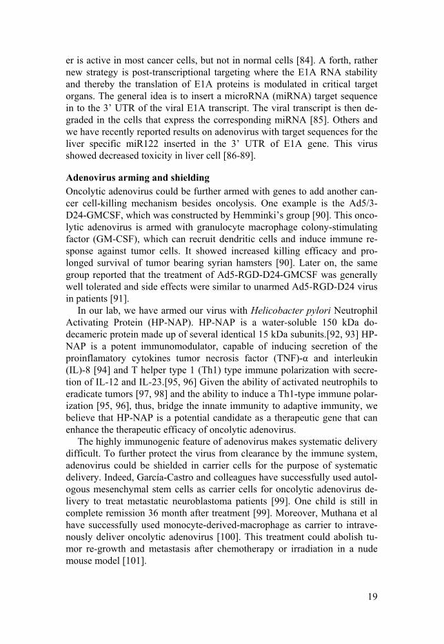
Adenovirus arming and shielding Oncolytic adenovirus could be further armed with genes to add another can-cer cell-killing mechanism besides oncolysis. One example is the Ad5/3-D24-GMCSF, which was constructed by Hemminki’s group [90]. This onco-lytic adenovirus is armed with granulocyte macrophage colony-stimulating factor (GM-CSF), which can recruit dendritic cells and induce immune re-sponse against tumor cells. It showed increased killing efficacy and pro-longed survival of tumor bearing syrian hamsters [90]. Later on, the same group reported that the treatment of Ad5-RGD-D24-GMCSF was generally well tolerated and side effects were similar to unarmed Ad5-RGD-D24 virus in patients [91].
In our lab, we have armed our virus with Helicobacter pylori Neutrophil Activating Protein (HP-NAP). HP-NAP is a water-soluble 150 kDa do-decameric protein made up of several identical 15 kDa subunits.[92, 93] HP-NAP is a potent immunomodulator, capable of inducing secretion of the proinflamatory cytokines tumor necrosis factor (TNF)-α and interleukin (IL)-8 [94] and T helper type 1 (Th1) type immune polarization with secre-tion of IL-12 and IL-23.[95, 96] Given the ability of activated neutrophils to eradicate tumors [97, 98] and the ability to induce a Th1-type immune polar-ization [95, 96], thus, bridge the innate immunity to adaptive immunity, we believe that HP-NAP is a potential candidate as a therapeutic gene that can enhance the therapeutic efficacy of oncolytic adenovirus.
The highly immunogenic feature of adenovirus makes systematic delivery difficult. To further protect the virus from clearance by the immune system, adenovirus could be shielded in carrier cells for the purpose of systematic delivery. Indeed, García-Castro and colleagues have successfully used autol-ogous mesenchymal stem cells as carrier cells for oncolytic adenovirus de-livery to treat metastatic neuroblastoma patients [99]. One child is still in complete remission 36 month after treatment [99]. Moreover, Muthana et al have successfully used monocyte-derived-macrophage as carrier to intrave-nously deliver oncolytic adenovirus [100]. This treatment could abolish tu-mor re-growth and metastasis after chemotherapy or irradiation in a nude mouse model [101].

20
3. Adenovirus surface modification Since adenovirus is engineered for cancer therapy, researchers have devoted huge efforts on viral surface modification to achieve i) retargeting to achieve higher transductional specificity, and ii) lower toxicity and/or immune re-sponse against viral particles, and iii) imaging and other purposes. A sum-mary of surface modified adenovirus is listed in Table 2.
In order to selectively target certain cell types at the transductional level, many researchers have attempted to modify the viral surface proteins. Most studies have focused on the modification of fiber molecules by either replac-ing the fiber or fiber knob with one from another serotype or inserting a short peptide in the HI loop or the C-terminus of the adenovirus fiber mole-cule. For instance, an adenovirus with RGD motif inserted into the HI-loop of the fiber selectively infects cancer cells with high integrin expression [102-105]. Leja et. al., reported a novel adenovirus with a cyclic FWKT motif from somatostatin in the HI-loop, where, the virus showed selective infection of neuroendocrine cancer cells through interaction with a somato-statin receptor [106]. The cement protein pIX is also reported to have toler-ance to modification. By inserting a single-chain T-cell receptor in pIX, the adenovirus selectively targeted HLA-A1/MAGE-A1-positive tumor cells [107]. Adenovirus has also been reported for use in imaging by incorporat-ing GFP on its surface proteins [108, 109]. Researchers have identified that the exposed HVRs contain key adenovirus-specific neutralizing antibody epitopes [110]. In order to prevent neutralization by pre-existing immunity, researchers have tried to pseudotype the hexon HVRs (exposed on surface) with the ones from other serotype, which has less prevalence of the neutrali-zation antibody. Roberts and colleagues successfully circumvent pre-existing anti-vector immunity, by replacing the Ad5 HVRs with corresponding HVRs from Ad48 (Ad5HVR48), [110]. However, it was later shown that Ad5 to Ad48 HVR substitution lead to toxicity and increased inflammatory respons-es following intravenous delivery [111]. In another example to lower the immune response the surface of adenovirus was chemically conjugated to PEG molecules [112].
Waddington and colleagues showed that liver sequestration of Ad5, at least in mouse model, is mediated by binding of FX to the hexon [26]. This phenomenon could be abolished by pseudotyping the HVRs from serotype 48, which did not bind to FX [26], or by point mutation in HVR5 and/or HVR7 to disrupt the FX-hexon interaction. It has also been reported recently that the so called fiber-masking problem could be rate limiting in oncolytic adenoviral therapy. This is caused by overproduction of adenovirus fiber proteins [51, 57, 141], which are released from the infected cell before cell lysis. The released fibers bind to CAR on non-infected neighboring cells, thereby limiting infection efficiency of progeny virus [57]. The fiber-masking problem is not limited to the Ad5 fiber but also observed for the

21
Ad35 fiber, which binds to CD46 [57]. These limitations must be overcome to develop successful oncolytic adenovirus agents.
Table 2 A list of adenoviral surface modification for different purpose.
Aim Sites Modification Types
Retargeting
hexon
Cysteine, chemical conjugation of ligands [113] Peptide ligand (RGD [114, 115], Tat-PTD [115, 116], BAP [117]) Metabolic N3 labeling, chemical conjugation [118]
pIX
Peptide ligand (RGD[114, 115], BAP [117]) Cysteine, chemical conjugation of ligands [119] scFv or other antibody-based molecules [120, 121] Single chain T-cell receptor [107]
fiber
Pseudotyping, knob/shaft/tail [122-127] Antibody-based molecules [128-130] Peptide ligand (RGD [102], Tat-PTD [131], BAP [117]) Metabolic N3 labeling, chemical conjugation [118]
Detargeting hexon
HVRs pseudotyping / mutation [132] Chimeric hexon [25, 26] Cysteine, chemical conjugation of ligands [113]
fiber Shaft KKTK mutation / point mutation [133]
Immune Response
hexon
HVRs pseudotyping / mutation [127, 134] Chimeric hexon [110, 111] Epitope [135, 136] PEGylation [112, 137, 138]
fiber Epitope [139] Imaging pIX GFP/eGFP [108, 109] Other pIX Therapeutic genes [140]

22
Figure 4. Schematic drawing of the surface modified viruses used in our studies. Ad5: Natural Adenovirus serotype 5 based virus. Ad5PTD: Adenovirus serotype 5 based virus with hexon PTD modification. Ad5f35: Adenovirus serotype 5 based virus with fiber molecules from serotype 35. Ad5PTDf35: Adenovirus serotype 5 based virus harboring both the above mentioned PTD and f35 modifications.
In our studies, both hexon HVR5 modification and fiber pseudotyping have been accomplished to address these problems (Figure 4). Especially, when the Ad5 hexon HVR5 was modified to incorporate the Tat-PTD motif (pro-tein transduction domain from the HIV-1 Tat protein), the FX-mediated cell binding was significantly reduced, which may theoretically decrease liver transduction. In addition, Tat-PTD-mediated receptor-independent transduc-tion led the virus to overcome the fiber-masking problem. By switching the fiber 5 molecules to fiber 35 viral transduction in most of the human primary cells were increased. Moreover, the combination of fiber 35 and PTD modi-fication depicted a synergetic effect on transduction efficiency, which en-large the utility of adenovirus as gene delivery vector.

23
4. Cell penetrating peptides Cell penetrating peptides (CPPs) are short peptides (usually <30 a.a.) with the ability to penetrate tissues or enter cells at a relatively high efficiency. In some cases, members of linear CPPs have the “carrier” features to transport conjugated “cargos” (from small molecules to large DNA complexes) into cells. The first insight into cellular uptake of CPPs dates back to 1965, when researchers reported that histones and basic polyamino acids stimulate the uptake of albumin by tumor cells in culture [142]. The transactivator of tran-scription (Tat) from the human immunodeficiency virus (HIV)-1 virus was found to have carrier/penetrating properties in 1994 [143]. Further studies by Lebleu’s group showed that the ability of Tat to penetrate plasma mem-branes was associated with certain domains of the Tat peptide, designated protein transduction domains (PTD) [144]. From that time an increasing number of new CPPs have been found and characterized [145]. These CPPs have been successfully used, both in vitro and in vivo, for the intracellular delivery of different cargoes [146-152], including nanoparticles, proteins, liposomes and nucleic acids. CPPs are classified in different groups based on their properties as listed in Table 3. Among all CPPs described to date, the Tat-PTD and Penetratin (the homoeodomain of the Antennapedia protein of Drosophila [153]) are among the best characterized.
Table 3 Examples of cell-penetrating peptides, their origins, structures, and mecha-nisms
Name Origin/design Structure Proposed mechanism
Protein transduction domains Penetratin (pAntp)
Antennapedia Dro-sophila melanogaster
Amphipathic, α-helix/β-sheet
Direct penetration, endocy-tosis
Tat HIV-1 Tat Random coil/PPII helix
Direct penetration, pore formation
pVEC Murine vascular endo-thelial cadherin
Amphipathic, β-sheet
Direct penetration, trans-porter-mediated
Chimeric CPPs Transpor-tan
Galanin + Mastoparan Amphipathic, α-helical
Endocytosis, direct penetra-tion
Pep-1 Chimeric Amphipathic, α-helical
Direct penetration, pore formation
Synthetic CPPs Poly-Arg (Rn)
From the key role of Arg residues in Tat
Random coil, α-helical
Direct penetration, endocy-tosis
MAP Model amphipathic peptides
Amphipathic, α-helical
Multiple mechanisms

24
Cell surface heparan sulfate proteoglycans (HSPG) is thought to play an important role in the CPP internalization process. Any of the possible uptake mechanisms discussed so far is consistent with the contribution of proteo-glycan to CPP internalization [154]. However, the mechanism of each exact CPP uptake is still not fully elucidated, which is a limitation in clinical us-age. Different models have been proposed to illustrate the penetration, which can be mainly categorized into two groups as i) energy-dependent endocyto-sis and ii) direct translocation via the lipid bilayer [155]. Another report also suggests that CPPs may only play a role in “adherence” or “docking” to the cell surface while endocytosis mediates the actual cellular uptake [156]. Moreover, the secondary structure was also found to be important for differ-ent classes of CPPs [157]. It should be emphasized that distinct internaliza-tion mechanisms for the same CPP exist, depending on the presence or ab-sence of cargo.
4.1 CPPs translocation by energy-dependent endocytosis The internalization of several CPPs and their conjugates has been demon-strated with involvement of endocytosis. However, the exact endocytosis pathways contributing to this process is still unclear. Endocytosis is defined as an energy dependent process by which cell absorbs biomolecules, such as proteins, toxins and even other cells. The process is subdivided mainly into 4 categories (Figure 5): i) Phagocytosis, a process occurs only in specialized cells (i.e macrophages), by which the cell engulfed relatively big particles (≥0.75μm) such as cell debris, dusts and microorganisms. ii) Macropinocyto-sis, an action in which the cell membrane extended out with support of actin filaments forms a pocket and engulfs a large volume of extracellular fluid in an un-specific manner, which then pinches off from cell membrane to form a cytosolic vesicle. iii) Clathrin-mediated endocytosis is mediated by receptor-ligand interaction where specific receptors capture the internalizing mole-cules. This leads plasma membrane to be coated inward with clathrins, which then buds off from the membrane by the help of dynamin to form a cytosolic vesicle. iv) Caveolin-mediated endocytosis is a receptor-mediated endocytosis process that increases membrane oligomerization of caveolin and cause plasma membrane invagination, which then fizzed off as cytosolic vesicles with the help of dynamin.
By using different approaches, such as drug inhibition of a specific path-way, different studies revealed that CPPs simultaneously use all known en-docytic pathways in cellular internalization. However, depending on the class of CPP, different CPPs may have a favorable pathway. For example, the fusion protein GST-Tat-GFP was found to enter cells mainly by caveo-lae-mediated endocytosis [158, 159], while the Tat peptide and the Tat-HA2 fusion peptide were described to be internalized mainly through macropino-cytosis [160-163] and clathrin-mediated pathway [164]. Peneratin, a proto-

25
type in another class of CPPs, mainly utilizes clathrin-mediated endocytosis for cellular uptake. In comparison, cationic conjugated CPPs rely more on macropinocytosis for internalization.
Figure 5. Proposed hypotheses for cellular uptake of cell penetrating peptides. CPPs uptake are classified in two major categories: energy-dependent endocytosis and energy-independent direct translocation. Energy-dependent endocytosis includes macropinocytosis, caveolae- or clathrin- mediated pathway and other pathways (Left panel). Energy-independent direct translocation of CPPs includes inverted micelle model, barrel stave pore formation model, toroidal pore formation model and carpet model.
4.2 CPPs translocation by energy-independent direct translocation The Tat peptide cellular uptake was also found to be endocytosis-independent by using genetically engineered cells that lack endocytosis me-diators [165]. Several models have been proposed to explain the direct trans-location of CPPs, including the “inverted micelle model”, the “barrel stave pore formation model” the “toroidal pore formation model” and the “carpet model” (Figure 5).
According to the “inverted micelle model” [166], the interaction of CPPs with phospholipids membrane would recruit negatively charged phospholip-ids and induces the formation of an inverted micelle, which subsequently is disrupted and release the CPPs in the other side of the membrane due to the
Caveolae/ClathrinIndependent Endocytosis
Caveolae-mediatedEndocytosis
Clathrin-mediatedEndocytosis
MacropinocytosisToroidal Pore
Barrel Stave Pore
Inverted Micelle Carpet
Dynamin
CPP±Cargo
ClathrinCaveolinActin
Nucleus
Cytoplasma
ENDOCYTOSIS DIRECT TRANSLOCATION

26
hydrophilic cavity of the inverted micelle [167]. However, this model can hardly explain the translocation of CPPs-cargo, where it is not likely to form the inverted micelle containing high weight or big size molecules in their hydrophilic core.
According to either the “barrel stave”- or the “toroidal”- pore formation models, the translocation of peptides and their conjugates across phospholip-id bilayer membranes would result from the formation of transient pores, produced upon peptide insertion into the membrane, and oligomerization of the inserted peptides in a ring-shape structure. Both pore formation models are similar. In the “barrel stave pore” model, the hydrophobic faces of the CPPs interact with hydrophobic lipid tail of the bilayer phospholipid and the hydrophilic face would form the interior of the pore [168-171]. On the other hand, in the “toroidal pores” model, the CPPs exclusively interact with only the hydrophilic phosphor head of the phospholipids by rearranging the con-formation of the bilayer [168, 172].
In the “carpet model”, translocation of CPPs and their conjugates would occur as a consequence of a transient rearrangement of the lipid bilayer, induced by the extensive association of the peptide to its surface [168-171].
An adenovirus particle is a huge macromolecule compared to the cargos that are normally carried by CPPs. It is currently not known how CPP modi-fication of the adenovirus HVR-5 with Tat-PTD leads to increased transduc-tion of cells. However, since neither macropinocytosis blockage nor energy starvation at 4°C could completely abolish the transduction (our unpublished data), we can speculate that the viral penetration may rely on several differ-ent pathways including both energy-dependent and independent transloca-tion.
5. Neuroendocrine tumor and treatments with Adenovirus 5.1 Neuroendocrine tumors Neuroendocrine tumors (NETs) are neoplasms arising from endocrine cells and the nerve system [173] [174]. The endocrine cells are distributed all over the body, abundantly found in the respiratory tract and intestinal mucosa and also found as clusters in certain organs such as thyroid (e.g. C-cells), pancre-as (e.g. islets) and adrenal glands. Therefore, NETs can occur all over the body. However it is a relatively rare disease. All NETs cells highly express chromogranin A, which is the most important general marker [175]. The largest group is the gastroenteropancreatic NETs (GEP-NETs), which in-cludes gastrointestinal NETs (GI-NETs, also referred as carcinoids) and pancreatic NETs (PNETs). They share similarity in their cell features [176]. The majority of PNETs are potentially malignant with an expected median

27
survival of metastatic disease of less than 30 months [177]. The GI-NETs are often small in size but all malignant. They often develop liver and local lymph nodes metastases. The median survival for patients with metastatic small intestinal NETs in most studies are 6-7 years, but can be considerably prolonged when treated at a Center of Excellence (e.g. Uppsala, Sweden) with a median survival of more than 12 years [173]. Other types of NETs involves lung NETs, parafollicular cell cancer and Neuroblastoma. There is no clinical data regarding oncolytic adenovirus treatment of NETs. However, we have systemically developed and pre-clinically evaluated ade-noviruses optimized for treatment of GI-NETs liver metastases. The adeno-virus is based on serotype 5, wherein E1A gene is transcriptional controlled by CgA promoter and post-transcriptional regulated by miRNA targeting sequence miR122. The viral particles are either hexon PTD modified to by-pass the CAR-dependent cellular transduciton or fiber FWKT modified to enhance the transduction via somatostatin receptor 2 and reduce the hepato-toxicity [86, 106].
5.2 Neuroblastoma Neuroblastoma (NB) is one type of neuroendocrine tumor and is one of the most common extracranial solid tumors in early childhood, accounting for 8-10% of all childhood cancer. NB is a tumor of the sympathetic nervous sys-tem and originates from immature nerve cells of the neural crest. The prima-ry location is along the migration path of neural crest-derived cells, where most primary tumors (65%) occur within the abdomen, of which at least half occur in the adrenal medulla. Other common sites of disease include the neck, chest, and pelvis [178]. NB is often classified in 4 broad categories depending on the International Neuroblastoma Risk Group (INRG) classifi-cation system: very low risk, low risk, intermediate risk, and high risk. The proposed 5-year event-free survival rates for the risk groups are >85%, >75 to ≤85%, ≥50 to ≤75%, and <50%, respectively [178]. Very low risk group NB patients include infants younger than 1 year, who can recover with care-ful daily observation, even with the presence of metastatic disease, whereas older patients with metastatic disease have poor prognosis. Most children with low or intermediate risk neuroblastoma achieve remission via a combi-nation of surgery, radiation and chemotherapy, and some children with high-risk disease also benefit from mega-dose chemotherapy with autologous hematopoietic stem cell rescue [179]. However, more than half the patients with high-risk neuroblastoma relapse after standard treatment and to date no curative treatment exists [178].

28
6. Clinical Trials with surface modified oncolytic adenoviruses Adenoviruses have been extensively used in clinical trials. Most registered studies so far are replication-deficient adenoviruses with the purpose of vac-cination/gene delivery. Oncolytic adenovirus has also been widely used for treatment of various cancer types. Most of the trials are phase I dose escala-tion studies while some oncolytic adenovirus entered phase II/III. In general, the clinical outcome of oncolytic adenovirus is positive and relatively safe with only mild adverse events such as local injection site pain, fatigue, nau-sea, and temporary fever. It has been reported that adenovirus treatment me-diated high neutralizing antibody production within 3 weeks, while the virus could persist in circulation for up to 5 weeks [180].
Surface-modified adenoviruses have also been tested in clinical trials such as RGD fiber modification and fiber pseudotyping. Most viruses used by different groups have the D24 deletion of the E1A gene, which render selec-tive viral replication in pRb-deficient cancer cells. Ad5-D24-RGD and Ad5-D24-RGD-GMCSF both contains D24-E1A and the later one is further armed with GM-CSF. Patients (N=21) treated with Ad5-D24-RGD (a single 3-day cycle of 109–1012 VP/day) showed relatively high safety aspects with 14 cases of stable disease (SD) and 7 cases of progressed disease (PD). The adverse events include fever, fatigue and abdominal pain [181]. In another study all patients (N=9) who were treated with the same virus showed dis-ease progression in radiological analysis [182]. While in the same study as comparison, 3 evaluable patients with previously progressing disease stabi-lized after a single treatment with Ad5-D24-RGD-GMCSF and two thirds of the patients had stabilization or reduction in tumor marker levels [182]. In yet another study, patients received a single injection of Ad5E2F-E1AD24-RGD, a similar virus wherein the E2F promoter controls the D24-E1A gene, was also well tolerated by patients in response to the treatments [183]. Ad-verse events were also mild like fever and fatigue. Ad5/3-D24-GMCSF is a fiber pseudotyped virus that re-target the adenovirus to DSG-2. Patients (8/12) treated with Ad5/3-D24-GMCSF show objective clinical benefit as evaluated by radiology and 13/21 patients have biological activity of the virus. Moreover, the level of survivin-specific CD8+ lymphocytes increased after treatment in 8/14 patients, which indicated an elicited anti-tumor im-mune response [90].
Two surface-modified oncolytic adenoviruses have been used to treat neuroblastoma. The first report is from Hemminki’s group [184] in 2010. A 6-year-old boy with Stage 4 disease (lymph nodes and bone marrow metas-tases) failed to respond to three different chemotherapy regimens (vincristine + cisplatin / carboplatin + etoposide + cyclophosphamide, doxorubicin + etoposide + iphosphamide, high-dose cyclophosphamide) including a high-dose intensive regimen with an autologous stem cell transplant. The patient

29
was treated with an ultrasound-guided injection of in total 1×10¹¹ adenoviral particles of the Ad5/3-Cox2L-D24 oncolytic adenovirus, one-third into the primary tumor near the left kidney, one-third into adjacent lymph nodes, and one-third intravenously. The adenovirus is serotype 5-based and pseudo-typed with the serotype 3 fiber to alter the transduction via DSG2. D24-E1A expression is under control of the cyclooxygenase-2 promoter (Cox2L). The treatment was found to be safe with mild adverse events including grade 1 fever (max 37.1⁰C), diarrhea, stomach pains and grade 2 liver enzyme eleva-tions for two weeks. A bone marrow biopsy was found to be free of disease and the primary tumor had regressed by 71% one month after treatment. Increased CD3+CD8+ lymphocytes in the circulation indicate an immunlogi-cal response. The same virus was also used to treat other types of solid tumor. Anti-tumor activity was seen in 11/18 of patients [180]. In yet another report published by Alemany’s group in 2010, where 4 patients with metastatic NB refractory to front-line therapies received intravenously autologous mesen-chymal stem cells loaded with oncolytic adenovirus ICOVIR-5 [185], a E2F promoter-controlled D24-E1A gene with fiber RGD modified adenovirus backbone. The tolerance to the treatment was excellent. A complete clinical response was documented in one case, and the child is in complete remission 3 years after this therapy. Taken together these two studies suggest that on-colytic viruses can substantially affect treatment of therapy-resistant neuro-blastoma.
Up to date, one oncolytic adenovirus based anti-cancer agent (H101, brand name Oncorine, an Ad5 based virus with E1B-55k gene deletion) was approved by China State Food & Drug Administration on November 2005 to be used in combination with chemotherapy as a treatment for patients with late stage refractory nasopharyngeal cancer, which later on expanded for treatment of hepatocellular-, pancreatic- and lung- carcinoma as well as ma-lignant pleural effusion.
In conclusion, adenovirus mediated cancer therapy is safe and has poten-tial clinical benefits.

30
Methods
Details can be found in each paper. However, two of the most important techniques, used in this study are listed below.
Recombineering Recombination-mediated genetic engineering, termed recombineering, is a method either to construct or modify DNA vectors based on homologous recombination in an E. coli strain using the Red recombinase from the λ bacteriophage [186]. This method is powerful for fast and efficient construc-tion and modification of DNA vectors. Episomes in the bacteria can be mod-ified using linear PCR products (double-stranded) or synthetic oligonucleo-tides (single-stranded) as substrate, with as short as 35-40 base pair homolo-gies. The bacterial strains, used for recombineering should be able to express the bacteriophage recombination system. This system contains the genes: gam, exo and beta. gam prevents degradation of the linear DNA introduced into the host bacteria. exo has 5'-3' exonuclease activity, which creates sin-gle-stranded overhangs on introduced linear DNA. beta protects these over-hangs and helps the subsequent recombination process. Warming and col-leagues developed an E.coli strain, SW102, containing the λ recombination system in its genome [187]. These genes are transcribed by a λPL promoter and tightly controlled by a temperature-sensitive cI857 repressor. When the bacteria are cultured at low temperature (30-32°C), expression of the recom-bination proteins are repressed. However, after about 15 minutes incubation at 42°C, the promoter is activated and a sufficient amount of recombinase is produced for the recombination process. The linear DNA substrate, with sufficient homologous ends at both 5’ and 3’ of the site of recombination in the target vector, which is already present in the bacteria, can be easily intro-duced into the bacteria by electroporation. Recombinants are obtained by selection against antibiotic markers.
Selection and Counter-selection As the name implies this process of selection is based on two steps, a posi-tive and a negative selection. The positive selection is based on introducing a

31
gene into E. coli, which makes the bacteria resistant to antibiotics, while the negative selection is based on introducing a gene, which makes the bacteria sensitive to a substrate. In the present study, we used the bla gene for ampi-cillin resistance as a positive selection marker (AmpR), and the sacB gene, which makes the E. coli sensitive to sucrose, as a negative counter-selection marker. lacZ is a gene involved in the lac operon for lactose catabolism, which is naturally controlled by the lacI repressor. In this work, IPTG was used to induce the expression of the lacZ gene and the gene product β-galactosidase could digest the substrate X-Gal, which changes the color of the colony to blue.
Figure 6. Recombineering (A) A schematic view of the molecular mechanistic steps in DNA recombination using the λ Red system. The gam protein prevents the degradation of dsDNA. The exo protein is a 5’-3’ double-stranded DNA (dsDNA) specific exonuclease and is only required for dsDNA recombination. The beta pro-tein, a single-stranded DNA (ssDNA) annealing protein, is a central player in re-combineering and is required for both dsDNA and ssDNA-mediated recombination. (B) An illustration of selection and counter-selection steps in adenoviral genome engineering. The example shows insertion of the PTD motif in the HVR5 region.

32
Results and Discussion
Paper I The main aim of this study was to increase viral transduction efficiency and to overcome the fiber-masking problem caused by excessive fiber proteins release from infected cells that blocks CAR on non-infected neighboring cells and thereby preventing progeny virus entry [57]. To achieve this, we decided to keep the targeting agent away from the fiber and to put it on the virus capsid. Several groups have verified that the HVR5 site is tolerant for foreign peptide insertion [188-191]. Moreover, given the fact that there are 240 hexon trimers expressed on the adenoviral surface versus only 12 fiber trimer molecules and that hexon modification would not affect the native fiber binding, we decided to modify the hexon HVR5 site.
The Tat-PTD sequence that we used here is flanked by a short α-helix spacer at both ends and is inserted into the hexon HVR5 region. We hypoth-esized that the short α-helix spacer would expose the Tat-PTD motif, thereby increasing the virus-cell interactions, thus improving the transduction effi-ciency. In vitro experiments demonstrate the Tat-PTD modified viruses have increased transduction efficiency and improved cell-killing efficacy in a CAR-independent manner. Mice xenografted with SK-N-SH tumor when treated with Tat-PTD modified viruses had delayed tumor growth and pro-longed survival.
The over-production of fiber molecules during the first round of viral in-fection leads to a release of fiber molecules prior to cell lysis which mask the receptor on adjacent uninfected cells and therefore inhibit the subsequent rounds of infection [57]. This phenomenon limits the usage of replicating oncolytic adenoviruses as anti-cancer agents. In contrast to chemically con-jugated Tat-PTD-modified virus [192] or HI-loop/C-terminus Tat-PTD-modified virus [193], which would only enhance the first round of infection, we show that our Tat-PTD-modified virus, which utilizes a CAR-independent cellular transduction pathway, can overcome this problem. The plaque formation assay confirmed that Ad5PTD(wt) spreads faster than Ad5(wt) in a 2-dimensional model and implicates that the Tat-PTD-modified virus should spread faster also in 3-dimentionally structured tumors.
Different strategies have been proposed to enhance the adenovirus trans-duction capacity. One example is AdΔ24-425S11, by expressing a bispecific scFv to link adenovirus to the epidermal growth factor receptor for a higher

33
transduction level on CAR-low neuroblastoma cells [194]. However, the infectivity-enhancement of that virus still relies on uptake via CAR at the first round of viral infection. In contrast, the infectivity of our Tat-PTD-modified viruses is guaranteed also on CAR-low cells at the first viral infec-tion step and will be carried on to viral progeny. Parikh et. al. claimed that treatment of neuroblastoma by wild-type Ad5 was not as efficient as by on-colytic herpes simplex virus due to the lack of Ad5 transduction [195]. We show in this study that by enhancing Ad5 transduction, therapeutic effect could be achieved for neuroblastoma.
In conclusion, we have developed Tat-PTD-modified oncolytic Ad5-based viruses with elevated infectivity. The viruses circumvent problems caused by excessive production and secretion of virus fiber protein in the first round of infection, fibers that could block receptors on neighboring non-infected cells and slow down subsequent replication rounds. They are partic-ularly promising for the treatment of tumors with low CAR expression as demonstrated herein for experimental neuroblastoma and neuroendocrine tumors.
Paper II In paper I, we have shown that the hexon Tat-PTD modification could un-specifically increase the viral transduction, while the transcriptional specific-ity still relies on the selectively expression of E1A, which could be achieved by and as mentioned above, using a specific promoter. In this study we aimed at evaluating an oncolytic adenovirus wherein neuroblastoma cell-selectivity is obtained from an artificially constructed promoter controlling E1A expression. We have identified a 0.5 kb region upstream of the secre-togranin III (SCG3) gene with promoter activity, which selectively drives gene expression in neuroblastoma cells. A 0.2 kb enhancer of Achaete-scute-related 1 (ASH1) gene [196] was coupled in front of the SCG3 promoter to enhance both the selectivity and the transcription activity in neuroblastoma cells. We then evaluated the Tat-PTD surface modified, promoter-controlled oncolytic adenovirus Ad5PTD(ASH1-SCG3-E1A) in the treatment of exper-imental neuroblastoma. The ASH1-SCG3 promoter-controlled virus selec-tively replicates and kills neuroblastoma cancer cells in vitro. Mice harbor-ing SK-N-FI xenograft tumor treated with Ad5PTD(ASH1-SCG3-E1A) virus showed a delayed tumor growth and prolonged survival. Chemothera-py is used as a standard method for neuroblastoma treatment. However, treatment induced drug-resistance is a major problem in this approach and eventually the cancer initiating cells are thought to become refractory to chemotherapeutic drugs. In our study, we observed different killing pattern of our oncolytic virus on different drug-sensitive and drug-insensitive cancer cells. Our experimental data suggests that patients treated with cisplatin

34
would have a beneficial effect from subsequent oncolytic adenovirus treat-ment, while patients treated with etoposide or doxorubicin may have a mod-erate beneficial effect and patients treated with vincristine may have less beneficial effect from oncolytic adenovirus treatment. However, the detailed mechanisms need to be further studied. In conclusion, oncolytic Ad5PTD(ASH1-SCG3-E1A) virus could be used to treat neuroblastoma in conjunction with standard therapy.
Paper III In paper I we had shown that a Tat-PTD modification of the Ad5 hexon pro-tein improves Ad5 infection of tumor cells. In the third paper, we pseudo-typed the Tat-PTD modified adenovirus with fiber from serotype 35 to fur-ther enhance the transduction on difficult-to-transduce cells. This viral vec-tor shows a remarkable increased transduction of human primary cell cul-tures including: T cells, monocytes, macrophages, dendritic cells, pancreatic islets and exocrine cells, mesenchymal stem cells and cancer initiating cells. To demonstrate the beneficial effect of using an Ad5PTDf35-based vector for gene delivery, we constructed Ad5(pp65) and Ad5PTDf35(pp65), which both express the full-length cytomegalovirus (CMV) pp65 antigen. We have previously shown that a population of CMV pp65495-503-specific T cells can be significantly enriched if T cells from a CMV seropositive, HLA-A2-positive blood donor are stimulated by Ad5(pp65)-transduced autologous dendritic cells (DCs) [197, 198]. However, large amounts of viral vector need to be used. Since monocytes and DCs are far more efficiently trans-duced with the Ad5PTDf35(GFP) than the Ad5(GFP) vector, we argued that we should have the same effect in expanding specific T cells ex vivo by us-ing far less Ad5PTDf35-based vector than Ad5-based vector. We therefore compared the T cell expansion ability of DCs either transduced with Ad5(pp65) or Ad5PTDf35(pp65) at a relatively low amount of vector. The Ad5PTDf35(pp65)/DC stimulation increased the pp65-reactive T cells popu-lation for all donors, approximately 50-100 fold, while Ad5(pp65)/DC stimulation only increased the pp65-reactive T cell population 2-8 fold. These data clearly show that Ad5PTDf35(pp65) would be highly efficient for DC modification, to expand T cells ex vivo for adoptive transfer to im-munocompromised patients with CMV complications. Last but not least, user-friendly backbone plasmids containing these modifications were devel-oped for compatibility to the AdEasy-system to facilitate the development of surface-modified adenoviruses for gene delivery to difficult-to-transduce cells in basic, pre-clinical and clinical research.

35
Paper IV In paper I, II and III, we have shown that adenoviruses can be surface modi-fied to enhance the transduction efficiency and that replication can be-controlled through transcriptional targeting. However, limited success has been reported in treatment of advanced cancers when relying on the onco-lytic virus effect alone. In this study, we armed the Ad5PTDf35-based, repli-cation-restricted adenovirus with the Helicobacter pylori Neutrophil Activat-ing Protein (HP-NAP) as an immune-modulating agent, to further enhance the potency of oncolytic adenoviral therapy.
In vitro, the Ad5PTDf35-[Δ24-sNAP] virus retains the specificity and has equal potency in tumor cell killing as the same virus without transgene. In addition, the expressed and secreted NAP could activate immune cells and attract polymorphonuclear leukocytes (PMNs) in migration assays.
In vivo, Ad5PTDf35-[Δ24-sNAP] virus treatment eradicated BON (neu-roendocrine cancer cell) xenograft tumor on nude mice and the survival was significantly prolonged. The secreted HP-NAP could recruit neutrophils to the site of infection and randomly kill the surrounding cells and cause necro-sis, which mediated release of Reactive oxygen intermediates (ROIs) like O2− and hypochlorous acid and secrete granule enzymes like myeloperoxi-dase and metalloproteinase [199]. Importantly, HP-NAP also induces im-mune cells to secrete TNF-α, which in turns prime PMNs to produce more ROIs [200] and activate endothelial cells to produce adhesion molecules like VCAM-1, ICAM-1, and E-selectin [201] and also IL-8 (the mouse analog is MIP2-α) to promote PMN trans-endothelial migration [202, 203]. In addi-tion, the expressed viral E1A proteins can sensitize the infected cells to TNF-α exposure. HP-NAP is potent in inducing Th1-type immune polariza-tion [204] and suppressing Th2-type immune polarization[205]. They are also known to enhance infiltration of CD4+ and CD8+ IFN-γ secreting T cells within the tumor microenvironment[204]. In summary, this study describes that treatment with oncolytic adenovirus armed with HP-NAP could prolong the survival of tumor-bearing mice and potentially cure a proportion of them. Furthermore, HP-NAP is a potent immunomodulating agent that polarizes the otherwise immunosuppressive tumor microenvironment in favor of anti-tumor immunity and potentially evokes the adaptive immunity against tu-mor.

36
Future Perspectives
1) We demonstrated that hexon Tat-PTD modification led to higher viral transduction and thus better gene delivery and therapeutic outcomes. How-ever, the exact mechanism was not fully illustrated. As DNA virus, adenovi-ral vector mediated gene delivery needs the transgene cassette to be deliv-ered into nucleus; wherein the viral particle needs penetrates both the cell membrane and the nuclear membrane. Viral hexon has its own ability in nuclear translocation while the mechanism of Tat-PTD modification would be hypothesized through helping either the interaction between viral particle and cell membrane and/or the process inside the cell including endosome escape and nuclear translocation. We are currently investigating the virus-cell membrane interaction. Preliminary data suggests macropinocytosis is partially associated with increased Tat-PTD viral transduction. However, the detail experiments need to be performed to fully illustrate the mechanism.
2) Monotherapy using oncolytic adenovirus will most likely not be sufficient to cure cancer. However, combinatorial approaches involving viro- and im-muno-therapy are emerging in both experimental models as well as in clini-cal trials. In paper IV, we armed an oncolytic virus with an immune-modulator gene HP-NAP, by which evoke the innate immune response against tumors. Our future goals will focus on combining viral-mediated oncolysis and viral-induced immune response against a variety of tumors. Human adenovirus does not replicate in mice and fully immunocompetent wild type mice should be used in order to study adequate immune responses. As we are interested in neuroblastoma, A/J mice and the syngeneic neuro-blastoma cell line NXS2 would be used as a model. Therefore, adenovirus will be used as a gene delivery vector of a tumor antigen mimotope and used together with oncolytic vaccinia virus (WR strain) and/or Semlikie forest virus, which is an ongoing project.
3) A clinical trial is being planned with a Tat-PTD-engineered virus. The E1A gene expression is under control of the human chomogranin A (CgA) promoter for selective expression in neuroendocrine cells. It is also post-transcriptionally regulated by target sequences for the liver-specific miRNA miR122 for selective blockage of translation in hepatocytes, in case the CgA promoter is leaky in normal liver cells. The virus will be used to treat pa-tients with liver metastases from neuroendocrine cancer originating from the

37
small intestine or pancreas. The characteristics of this virus are currently being evaluated in human primary cancer cells, normal hepatocytes and normal human blood.

38
Acknowledgements
All the work was performed at Department of Immunology, Genetics and Pathology at Rudbeck Laboratory, Uppsala University, Uppsala. The Swe-dish Cancer Society, the Swedish Research Council, the Swedish Children Cancer Foundation and Gunnar Nilsson’s Cancer Foundation supported the work.
I would like to express my greatest sincere thank you to the people who contributed to this work. Many thanks to:
My supervisor, Professor Magnus Essand, thank you for all your inspira-tion and marvelous supervision. I learned a lot from you both in sciences and the attitude to life. Moreover, you allowed me to pursue the Tat-PTD study since the time I was a master student in your lab.
My co-supervisor, Associate Professor Valeria Giandomenico, thank you for your guidance in the projects, and the happiness you transmit during discussions.
My co-supervisor, Professor Thomas Tötterman, thank you for creative talks and for sharing your immunological knowledge with me.
My “lab mummy”, Berith Nilsson, thank you for all your kindness and endless support with daily lab work, especially on molecular cloning and virus production.
Dr. Angelika Danielsson, thank you for opening the door at Clinical Im-munology on 2007-Oct-19 and let me in.
Dr. Nadim Majdalani, thank you for patiently teaching me about recom-bineering via email and Skype.
Dr. Xiaolong Fan, thank you for sharing the fiber 35 modification with us and for the creative discussions.
Dr. Fredrik Eriksson, thank you for all the discussion about viral therapy and sharing immunological knowledge with me.
Dr. Justyna Leja, thank you for the introduction to the lab and various techniques, your great enormous help and supports in my project.
MSc. Mohanraj Ramachandran, thank you for all your helps in my pro-jects, for sharing your delicious Indian food, and most important, you prom-ised to let me attend your wedding.
MSc. Matko Cancer, thank you for your works on vaccinia virus and lunch gossip.
Min Ma, thank you for the help in viral construction and for letting me stay in your apartment when I was in Switzerland.

39
Jin Xu, thank you for the help in double-modified virus testing in primary cell cultures.
Dr. Katarina Le Blanc, Dr. Lene Uhrbom, Dr. Karin Forsberg-Nilsson, Dr. Bengt Westermark, Dr. Norman Maitland, thank you for your collaborative work on the double-modified viruses. Especially, thanks to Dr. Rachel Ad-amson who helped to perform the transductional test on primary prostate cancer cells.
Ann-Charlotte Hellström, thank you for teaching me how to handle mice. Liye Chen, for your help in CD40L testing and all the other time spend with Chuan and Di. Lu Zhang, for your help in plasmid cloning and all the other time spend with Di.
Dr. Alkwin Wanders, thank you for warm-hearted and professional help with immunohistochemisty.
Linda Sandin, thank you my first immunology teacher, who answers all my stupid questions, made me start spinning classes and most importantly, let me understood the importance of the lunar phase in experimental design.
Dr. Sara Mangsbo, thank you for the collaboration, discussion and an-swering my immunological questions.
Lina Liljenfeldt, thank you for testing the surface modified viruses in combination with CD40L.
All other current and former GIG members including Dr. Angelica Los-kog Dr. Lisa Christiansson, Emma Svensson, Hannah Karlsson, Dr. Joachim Burman, Erika Gustafsson, Victoria Hillerdal. Gabriella Paul-Wetterberg, Dr. Christian Molnar, Dr. Arian Sadeghi, Dr. Ole Forsberg, Dr. Moa Fransson, Dr. Wing-Sheng Cheng, Dr. Björn Carlsson, Dr. Camilla Lindqvist for all your help in scientific work such as teaching me FACS staining, T cell han-dling as well as in my own daily life regarding translation of Swedish. All of you create a nice working atmosphere.
Warm thanks to Dr. Andrew Forsberg, Mahesh Anagandula, Magnus Ståhle, Dr. Olle Korsgren, Sofia Nordling, Dr. Peetra Magnusson and Mar-gareta “Bumsan” Engkvist for helping me with everything about pancreatic islets and exocrine tissues.
Dirk Pacholsky, thank you for the helps in imaging and cell sorting. Dr. Karl Andersson and Dr. Hanna Björkelund, thank you for the helps in
measuring the virus-cell interaction by LigandTracer. Dongyan Song and Linghua Xu, for accompanying me on my first flight
to Sweden. Carolina Yaping Wang, Dan Li, Ling Wang, Henrik Cam, for the happy times spent together when we were living in Sernandersväg.
Dr. Yanling Cai, Fang Mao, Leo Shangfeng Liu, Haisha Ma, Bo Xu, Yang Sun, Yani Zhao, Li Li, Jinzhi Hu, Yu Sun, Xi Fu, for being together in the master program and a lot of fun we shared.
Thanks to Dr. Xiang Jiao, Dr, Yiwen Jiang, Dr. Gucci Jijuan Gu, Dr. Ra-chel Nong, Dr. Rongqin Ke, Tao Cui, Su-chen Li, Lei Zhang, Lei Chen, Hua Huang, Yuan Xie, Kun Wei, Anqi Xiong, Di Wu, Junhong Yan, Shujing Shi,

40
Jin Zhao, for creating a nice Chinese network at the Rudbeck Laboratory and Academic hospital.
Yinghua Zha, thank you for your encouragements and accompany in gym and opening my knowledge in gut flora enterotypes.
Rui Miao, thank you for your introduction on Gibson assembly and shar-ing the initial reagents.
Staff in IGP administration and animal facility, thank you for the helps during my PhD life.
Dr. Ulrika Gunnarsson, Dr. Malin Graffner Nordberg, Gerald Pettersson, Dr. Lars Jonsson, Hampus Rystedt and Henrik Steinrud, warm thanks to all of you for the help with patent application and ideas for commercialization.
All my friends in Sweden, in China, and elsewhere, thank all of you for
supporting and caring about me.
我的爸爸妈妈,感谢你们对我的支持,鼓励以及信任,让我自由的选
择自己想要的生活。 Last but not least to my beloved wife Chuan Jin, who make viruses for
Di’s projects and food for Di. Thank you for your support and great lovely companionship! 执子之手,与君偕老!
/Di Yu (郁笛) on July 25, 2013

41
References
1. Rowe, W.P., Huebner, R.J., Gilmore, L.K., Parrott, R.H., and Ward, T.G., Isolation of a cytopathogenic agent from human adenoids undergoing spontaneous degeneration in tissue culture. Proceedings of the Society for Experimental Biology and Medicine. Society for Experimental Biology and Medicine, 1953. 84(3): 570-573. PMID: 13134217
2. Jones, M.S., 2nd, Harrach, B., Ganac, R.D., Gozum, M.M., Dela Cruz, W.P., Riedel, B., Pan, C., Delwart, E.L., and Schnurr, D.P., New adenovirus species found in a patient presenting with gastroenteritis. J. Virol., 2007. 81(11): 5978-5984. PMID: 17360747
3. Kaneko, H., Aoki, K., Ishida, S., Ohno, S., Kitaichi, N., Ishiko, H., Fujimoto, T., Ikeda, Y., Nakamura, M., Gonzalez, G., Koyanagi, K.O., Watanabe, H., and Suzutani, T., Recombination analysis of intermediate human adenovirus type 53 in Japan by complete genome sequence. The Journal of general virology, 2011. 92(Pt 6): 1251-1259. PMID: 21402595
4. Kaneko, H., Aoki, K., Ohno, S., Ishiko, H., Fujimoto, T., Kikuchi, M., Harada, S., Gonzalez, G., Koyanagi, K.O., Watanabe, H., and Suzutani, T., Complete genome analysis of a novel intertypic recombinant human adenovirus causing epidemic keratoconjunctivitis in Japan. J. Clin. Microbiol., 2011. 49(2): 484-490. PMID: 21147954
5. Kaneko, H., Suzutani, T., Aoki, K., Kitaichi, N., Ishida, S., Ishiko, H., Ohashi, T., Okamoto, S., Nakagawa, H., Hinokuma, R., Asato, Y., Oniki, S., Hashimoto, T., Iida, T., and Ohno, S., Epidemiological and virological features of epidemic keratoconjunctivitis due to new human adenovirus type 54 in Japan. The British journal of ophthalmology, 2011. 95(1): 32-36. PMID: 20530657
6. Robinson, C.M., Singh, G., Henquell, C., Walsh, M.P., Peigue-Lafeuille, H., Seto, D., Jones, M.S., Dyer, D.W., and Chodosh, J., Computational analysis and identification of an emergent human adenovirus pathogen implicated in a respiratory fatality. Virology, 2011. 409(2): 141-147. PMID: 21056888
7. Seto, D., Chodosh, J., Brister, J.R., and Jones, M.S., Using the whole-genome sequence to characterize and name human adenoviruses. J. Virol., 2011. 85(11): 5701-5702. PMID: 21450823
8. Walsh, M.P., Chintakuntlawar, A., Robinson, C.M., Madisch, I., Harrach, B., Hudson, N.R., Schnurr, D., Heim, A., Chodosh, J., Seto, D., and Jones, M.S., Evidence of molecular evolution driven by recombination events influencing tropism in a novel human adenovirus that causes epidemic keratoconjunctivitis. PLoS One, 2009. 4(6): e5635. PMID: 19492050

42
9. Walsh, M.P., Seto, J., Jones, M.S., Chodosh, J., Xu, W., and Seto, D., Computational analysis identifies human adenovirus type 55 as a re-emergent acute respiratory disease pathogen. J. Clin. Microbiol., 2010. 48(3): 991-993. PMID: 20042633
10. de Jong, J.C., Osterhaus, A.D., Jones, M.S., and Harrach, B., Human adenovirus type 52: a type 41 in disguise? J. Virol., 2008. 82(7): 3809; author reply 3809-3810. PMID: 18334604
11. Wadell, G., Hammarskjold, M.L., Winberg, G., Varsanyi, T.M., and Sundell, G., Genetic variability of adenoviruses. Ann. N. Y. Acad. Sci., 1980. 354: 16-42. PMID: 6261642
12. Wadell, G., Molecular epidemiology of human adenoviruses. Curr. Top. Microbiol. Immunol., 1984. 110: 191-220. PMID: 6090058
13. Tuve, S., Wang, H., Ware, C., Liu, Y., Gaggar, A., Bernt, K., Shayakhmetov, D., Li, Z., Strauss, R., Stone, D., and Lieber, A., A new group B adenovirus receptor is expressed at high levels on human stem and tumor cells. J. Virol., 2006. 80(24): 12109-12120. PMID: 17020944
14. Wang, H., Li, Z.Y., Liu, Y., Persson, J., Beyer, I., Moller, T., Koyuncu, D., Drescher, M.R., Strauss, R., Zhang, X.B., Wahl, J.K., 3rd, Urban, N., Drescher, C., Hemminki, A., Fender, P., and Lieber, A., Desmoglein 2 is a receptor for adenovirus serotypes 3, 7, 11 and 14. Nat. Med., 2011. 17(1): 96-104. PMID: 21151137
15. Fleischli, C., Sirena, D., Lesage, G., Havenga, M.J., Cattaneo, R., Greber, U.F., and Hemmi, S., Species B adenovirus serotypes 3, 7, 11 and 35 share similar binding sites on the membrane cofactor protein CD46 receptor. The Journal of general virology, 2007. 88(Pt 11): 2925-2934. PMID: 17947513
16. Segerman, A., Arnberg, N., Erikson, A., Lindman, K., and Wadell, G., There are two different species B adenovirus receptors: sBAR, common to species B1 and B2 adenoviruses, and sB2AR, exclusively used by species B2 adenoviruses. J. Virol., 2003. 77(2): 1157-1162. PMID: 12502832
17. Robinson, C. and Echavarria, M., Adenoviruses, in Manual of Clinical Microbiology, Versalovic, J., et al., Editors, 2011, ASM Press. Volume: 2, Section: IV, Chapter: 101.
18. Jones, M.S., 2nd, Harrach, B., Ganac, R.D., Gozum, M.M., Dela Cruz, W.P., Riedel, B., Pan, C., Delwart, E.L., and Schnurr, D.P., New adenovirus species found in a patient presenting with gastroenteritis. J. Virol., 2007. 81(11): 5978-5984. PMID: 17360747
19. Wadell, G.r., ADENOVIRUSES(ADENOVIRIDAE) | General Features, in Encyclopedia of Virology (Second Edition), Editors-in-Chief:   Allan, G. and Robert, G.W., Editors, 1999, Elsevier: Oxford. Volume: Section: Chapter: pp. 1-7.
20. Centers for Disease Control and Prevention (CDC), Acute respiratory disease associated with adenovirus serotype 14--four states, 2006-2007. Morb. Mortal. Weekly Rep., 2007. 56(45): 1181-1184. PMID: 18004235
21. Echavarria, M., Adenoviruses in immunocompromised hosts. Clin. Microbiol. Rev., 2008. 21(4): 704-715. PMID: 18854488

43
22. Foy, H.M. and Grayston, J.T., Adenoviruses, in Viral infections of humans: epidemiology and control, Evans, A.S., Editor, 1976, Plenum Medical Book Company: New York and London. Volume: Section: Chapter: pp. 53-69.
23. Liu, H., Jin, L., Koh, S.B., Atanasov, I., Schein, S., Wu, L., and Zhou, Z.H., Atomic structure of human adenovirus by cryo-EM reveals interactions among protein networks. Science, 2010. 329(5995): 1038-1043. PMID: 20798312
24. Reddy, V.S., Natchiar, S.K., Stewart, P.L., and Nemerow, G.R., Crystal structure of human adenovirus at 3.5 A resolution. Science, 2010. 329(5995): 1071-1075. PMID: 20798318
25. Alba, R., Bradshaw, A.C., Parker, A.L., Bhella, D., Waddington, S.N., Nicklin, S.A., van Rooijen, N., Custers, J., Goudsmit, J., Barouch, D.H., McVey, J.H., and Baker, A.H., Identification of coagulation factor (F)X binding sites on the adenovirus serotype 5 hexon: effect of mutagenesis on FX interactions and gene transfer. Blood, 2009. 114(5): 965-971. PMID: 19429866
26. Waddington, S.N., McVey, J.H., Bhella, D., Parker, A.L., Barker, K., Atoda, H., Pink, R., Buckley, S.M., Greig, J.A., Denby, L., Custers, J., Morita, T., Francischetti, I.M., Monteiro, R.Q., Barouch, D.H., van Rooijen, N., Napoli, C., Havenga, M.J., Nicklin, S.A., and Baker, A.H., Adenovirus serotype 5 hexon mediates liver gene transfer. Cell, 2008. 132(3): 397-409. PMID: 18267072
27. Wickham, T.J., Carrion, M.E., and Kovesdi, I., Targeting of adenovirus penton base to new receptors through replacement of its RGD motif with other receptor-specific peptide motifs. Gene Ther., 1995. 2(10): 750-756. PMID: 8750015
28. Wickham, T.J., Filardo, E.J., Cheresh, D.A., and Nemerow, G.R., Integrin alpha v beta 5 selectively promotes adenovirus mediated cell membrane permeabilization. The Journal of cell biology, 1994. 127(1): 257-264. PMID: 7523420
29. Liu, H., Wu, L., and Zhou, Z.H., Model of the trimeric fiber and its interactions with the pentameric penton base of human adenovirus by cryo-electron microscopy. J. Mol. Biol., 2011. 406(5): 764-774. PMID: 21146538
30. Fender, P., Ruigrok, R.W., Gout, E., Buffet, S., and Chroboczek, J., Adenovirus dodecahedron, a new vector for human gene transfer. Nat. Biotechnol., 1997. 15(1): 52-56. PMID: 9035106
31. Szolajska, E., Burmeister, W.P., Zochowska, M., Nerlo, B., Andreev, I., Schoehn, G., Andrieu, J.P., Fender, P., Naskalska, A., Zubieta, C., Cusack, S., and Chroboczek, J., The structural basis for the integrity of adenovirus Ad3 dodecahedron. PLoS One, 2012. 7(9): e46075. PMID: 23049939
32. Kaplan, L.M., Ariga, H., Hurwitz, J., and Horwitz, M.S., Complementation of the temperature-sensitive defect in H5ts125 adenovirus DNA replication in vitro. Proc. Natl. Acad. Sci. U. S. A., 1979. 76(11): 5534-5538. PMID: 230480

44
33. Wang, H., Li, Z.Y., Liu, Y., Persson, J., Beyer, I., Moller, T., Koyuncu, D., Drescher, M.R., Strauss, R., Zhang, X.B., Wahl, J.K., 3rd, Urban, N., Drescher, C., Hemminki, A., Fender, P., and Lieber, A., Desmoglein 2 is a receptor for adenovirus serotypes 3, 7, 11 and 14. Nat. Med., 2011. 17(1): 96-104. PMID: 21151137
34. Nilsson, E.C., Storm, R.J., Bauer, J., Johansson, S.M., Lookene, A., Angstrom, J., Hedenstrom, M., Eriksson, T.L., Frangsmyr, L., Rinaldi, S., Willison, H.J., Pedrosa Domellof, F., Stehle, T., and Arnberg, N., The GD1a glycan is a cellular receptor for adenoviruses causing epidemic keratoconjunctivitis. Nat. Med., 2011. 17(1): 105-109. PMID: 21151139
35. Bergelson, J.M., Cunningham, J.A., Droguett, G., Kurt-Jones, E.A., Krithivas, A., Hong, J.S., Horwitz, M.S., Crowell, R.L., and Finberg, R.W., Isolation of a common receptor for Coxsackie B viruses and adenoviruses 2 and 5. Science, 1997. 275(5304): 1320-1323. PMID: 9036860
36. Dechecchi, M.C., Melotti, P., Bonizzato, A., Santacatterina, M., Chilosi, M., and Cabrini, G., Heparan sulfate glycosaminoglycans are receptors sufficient to mediate the initial binding of adenovirus types 2 and 5. J. Virol., 2001. 75(18): 8772-8780. PMID: 11507222
37. Short, J.J., Pereboev, A.V., Kawakami, Y., Vasu, C., Holterman, M.J., and Curiel, D.T., Adenovirus serotype 3 utilizes CD80 (B7.1) and CD86 (B7.2) as cellular attachment receptors. Virology, 2004. 322(2): 349-359. PMID: 15110532
38. Arnberg, N., Edlund, K., Kidd, A.H., and Wadell, G., Adenovirus type 37 uses sialic acid as a cellular receptor. J. Virol., 2000. 74(1): 42-48. PMID: 10590089
39. Hierholzer, J.C., Guyer, B., O'Day, D., and Schaffner, W., Letter: Adenovirus type 19 keratoconjunctivitis. N. Engl. J. Med., 1974. 290(25): 1436. PMID: 4364287
40. Haisma, H.J., Boesjes, M., Beerens, A.M., van der Strate, B.W., Curiel, D.T., Pluddemann, A., Gordon, S., and Bellu, A.R., Scavenger receptor A: a new route for adenovirus 5. Mol Pharm, 2009. 6(2): 366-374. PMID: 19227971
41. Wu, E. and Nemerow, G.R., Virus yoga: the role of flexibility in virus host cell recognition. Trends Microbiol., 2004. 12(4): 162-169. PMID: 15051066
42. Henaff, D., Salinas, S., and Kremer, E.J., An adenovirus traffic update: from receptor engagement to the nuclear pore. Future Microbiol, 2011. 6(2): 179-192. PMID: 21366418
43. Meier, O. and Greber, U.F., Adenovirus endocytosis. The journal of gene medicine, 2003. 5(6): 451-462. PMID: 12797110
44. Akusjarvi, G., Temporal regulation of adenovirus major late alternative RNA splicing. Frontiers in bioscience : a journal and virtual library, 2008. 13: 5006-5015. PMID: 18508565
45. Gallimore, P.H. and Turnell, A.S., Adenovirus E1A: remodelling the host cell, a life or death experience. Oncogene, 2001. 20(54): 7824-7835. PMID: 11753665

45
46. Horwitz, M.S., Function of adenovirus E3 proteins and their interactions with immunoregulatory cell proteins. The journal of gene medicine, 2004. 6 Suppl 1: S172-183. PMID: 14978760
47. Lichtenstein, D.L., Toth, K., Doronin, K., Tollefson, A.E., and Wold, W.S., Functions and mechanisms of action of the adenovirus E3 proteins. Int. Rev. Immunol., 2004. 23(1-2): 75-111. PMID: 14690856
48. Fessler, S.P., Delgado-Lopez, F., and Horwitz, M.S., Mechanisms of E3 modulation of immune and inflammatory responses. Curr. Top. Microbiol. Immunol., 2004. 273: 113-135. PMID: 14674600
49. Windheim, M., Hilgendorf, A., and Burgert, H.G., Immune evasion by adenovirus E3 proteins: exploitation of intracellular trafficking pathways. Curr. Top. Microbiol. Immunol., 2004. 273: 29-85. PMID: 14674598
50. Weitzman, M.D., Functions of the adenovirus E4 proteins and their impact on viral vectors. Frontiers in bioscience : a journal and virtual library, 2005. 10: 1106-1117. PMID: 15769610
51. Franqueville, L., Henning, P., Magnusson, M., Vigne, E., Schoehn, G., Blair-Zajdel, M.E., Habib, N., Lindholm, L., Blair, G.E., Hong, S.S., and Boulanger, P., Protein crystals in Adenovirus type 5-infected cells: requirements for intranuclear crystallogenesis, structural and functional analysis. PLoS One, 2008. 3(8): e2894. PMID: 18682854
52. Boulanger, P.A. and Puvion, F., Large-scale preparation of soluble adenovirus hexon, penton and fiber antigens in highly purified form. European journal of biochemistry / FEBS, 1973. 39(1): 37-42. PMID: 4203745
53. Norrby, E., The structural and functional diversity of Adenovirus capsid components. The Journal of general virology, 1969. 5(2): 221-236. PMID: 4899805
54. Kaye, S.B., Lloyd, M., Williams, H., Yuen, C., Scott, J.A., O'Donnell, N., Batterbury, M., Hiscott, P., and Hart, C.A., Evidence for persistence of adenovirus in the tear film a decade following conjunctivitis. J. Med. Virol., 2005. 77(2): 227-231. PMID: 16121360
55. Shields, A.F., Hackman, R.C., Fife, K.H., Corey, L., and Meyers, J.D., Adenovirus infections in patients undergoing bone-marrow transplantation. The New England journal of medicine, 1985. 312(9): 529-533. PMID: 2982098
56. Numazaki, Y., Shigeta, S., Kumasaka, T., Miyazawa, T., Yamanaka, M., Yano, N., Takai, S., and Ishida, N., Acute hemorrhagic cystitis in children. Isolation of adenovirus type II. The New England journal of medicine, 1968. 278(13): 700-704. PMID: 4295621
57. Rebetz, J., Na, M., Su, C., Holmqvist, B., Edqvist, A., Nyberg, C., Widegren, B., Salford, L.G., Sjogren, H.O., Arnberg, N., Qian, Q., and Fan, X., Fiber mediated receptor masking in non-infected bystander cells restricts adenovirus cell killing effect but promotes adenovirus host co-existence. PLoS One, 2009. 4(12): e8484. PMID: 20041185

46
58. Kamel, W., Segerman, B., Oberg, D., Punga, T., and Akusjarvi, G., The adenovirus VA RNA-derived miRNAs are not essential for lytic virus growth in tissue culture cells. Nucleic Acids Res, 2013. 41(9): 4802-4812. PMID: 23525465
59. Wodrich, H., Guan, T., Cingolani, G., Von Seggern, D., Nemerow, G., and Gerace, L., Switch from capsid protein import to adenovirus assembly by cleavage of nuclear transport signals. EMBO J., 2003. 22(23): 6245-6255. PMID: 14633984
60. Tollefson, A.E., Scaria, A., Hermiston, T.W., Ryerse, J.S., Wold, L.J., and Wold, W.S., The adenovirus death protein (E3-11.6K) is required at very late stages of infection for efficient cell lysis and release of adenovirus from infected cells. J. Virol., 1996. 70(4): 2296-2306. PMID: 8642656
61. Mei, Y.F., Skog, J., Lindman, K., and Wadell, G., Comparative analysis of the genome organization of human adenovirus 11, a member of the human adenovirus species B, and the commonly used human adenovirus 5 vector, a member of species C. The Journal of general virology, 2003. 84(Pt 8): 2061-2071. PMID: 12867636
62. Hong, J.S., Mullis, K.G., and Engler, J.A., Characterization of the early region 3 and fiber genes of Ad7. Virology, 1988. 167(2): 545-553. PMID: 2849239
63. Holtzman, N.A., Dietary treatment of inborn errors of metabolism. Annu. Rev. Med., 1970. 21: 335-356. PMID: 4193735
64. Friedmann, T. and Roblin, R., Gene therapy for human genetic disease? Science, 1972. 175(4025): 949-955. PMID: 5061866
65. Aposhian, H.V., The use of DNA for gene therapy--the need, experimental approach, and implications. Perspect. Biol. Med., 1970. 14(1): 98-108. PMID: 4321470
66. Yaniv, M., Construction of hybrid viruses in vitro and their possible use for the production of specific viral or cellular proteins. Dev. Biol. Stand., 1979. 42: 87-91. PMID: 223926
67. Russell, W.C., Update on adenovirus and its vectors. J. Gen. Virol., 2000. 81(Pt 11): 2573-2604. PMID: 11038369
68. Volpers, C. and Kochanek, S., Adenoviral vectors for gene transfer and therapy. The journal of gene medicine, 2004. 6 Suppl 1: S164-171. PMID: 14978759
69. Palmer, D.J. and Ng, P., Helper-dependent adenoviral vectors for gene therapy. Hum. Gene Ther., 2005. 16(1): 1-16. PMID: 15703484
70. Jozkowicz, A. and Dulak, J., Helper-dependent adenoviral vectors in experimental gene therapy. Acta Biochim. Pol., 2005. 52(3): 589-599. PMID: 16082408
71. Malmstrom, P.U., Loskog, A.S., Lindqvist, C.A., Mangsbo, S.M., Fransson, M., Wanders, A., Gardmark, T., and Totterman, T.H., AdCD40L immunogene therapy for bladder carcinoma--the first phase I/IIa trial. Clin. Cancer Res., 2010. 16(12): 3279-3287. PMID: 20448220
72. Cattaneo, R., Miest, T., Shashkova, E.V., and Barry, M.A., Reprogrammed viruses as cancer therapeutics: targeted, armed and shielded. Nat Rev Microbiol, 2008. 6(7): 529-540. PMID: 18552863

47
73. Mathis, J.M., Stoff-Khalili, M.A., and Curiel, D.T., Oncolytic adenoviruses - selective retargeting to tumor cells. Oncogene, 2005. 24(52): 7775-7791. PMID: 16299537
74. Rein, D.T., Breidenbach, M., and Curiel, D.T., Current developments in adenovirus-based cancer gene therapy. Future Oncol, 2006. 2(1): 137-143. PMID: 16556080
75. Waehler, R., Russell, S.J., and Curiel, D.T., Engineering targeted viral vectors for gene therapy. Nat Rev Genet, 2007. 8(8): 573-587. PMID: 17607305
76. Bischoff, J.R., Kirn, D.H., Williams, A., Heise, C., Horn, S., Muna, M., Ng, L., Nye, J.A., Sampson-Johannes, A., Fattaey, A., and McCormick, F., An adenovirus mutant that replicates selectively in p53-deficient human tumor cells. Science, 1996. 274(5286): 373-376. PMID: 8832876
77. O'Shea, C.C., Soria, C., Bagus, B., and McCormick, F., Heat shock phenocopies E1B-55K late functions and selectively sensitizes refractory tumor cells to ONYX-015 oncolytic viral therapy. Cancer Cell, 2005. 8(1): 61-74. PMID: 16023599
78. O'Shea, C.C., Johnson, L., Bagus, B., Choi, S., Nicholas, C., Shen, A., Boyle, L., Pandey, K., Soria, C., Kunich, J., Shen, Y., Habets, G., Ginzinger, D., and McCormick, F., Late viral RNA export, rather than p53 inactivation, determines ONYX-015 tumor selectivity. Cancer Cell, 2004. 6(6): 611-623. PMID: 15607965
79. Fueyo, J., Gomez-Manzano, C., Alemany, R., Lee, P.S., McDonnell, T.J., Mitlianga, P., Shi, Y.X., Levin, V.A., Yung, W.K., and Kyritsis, A.P., A mutant oncolytic adenovirus targeting the Rb pathway produces anti-glioma effect in vivo. Oncogene, 2000. 19(1): 2-12. PMID: 10644974
80. Heise, C., Hermiston, T., Johnson, L., Brooks, G., Sampson-Johannes, A., Williams, A., Hawkins, L., and Kirn, D., An adenovirus E1A mutant that demonstrates potent and selective systemic anti-tumoral efficacy. Nat. Med., 2000. 6(10): 1134-1139. PMID: 11017145
81. Dorer, D.E. and Nettelbeck, D.M., Targeting cancer by transcriptional control in cancer gene therapy and viral oncolysis. Adv Drug Deliv Rev, 2009. 61(7-8): 554-571. PMID: 19394376
82. Cheng, W.S., Kraaij, R., Nilsson, B., van der Weel, L., de Ridder, C.M., Totterman, T.H., and Essand, M., A novel TARP-promoter-based adenovirus against hormone-dependent and hormone-refractory prostate cancer. Molecular therapy : the journal of the American Society of Gene Therapy, 2004. 10(2): 355-364. PMID: 15294182
83. Cheng, W.S., Dzojic, H., Nilsson, B., Totterman, T.H., and Essand, M., An oncolytic conditionally replicating adenovirus for hormone-dependent and hormone-independent prostate cancer. Cancer Gene Ther., 2006. 13(1): 13-20. PMID: 16052227
84. Doloff, J.C., Jounaidi, Y., and Waxman, D.J., Dual E1A oncolytic adenovirus: targeting tumor heterogeneity with two independent cancer-specific promoter elements, DF3/MUC1 and hTERT. Cancer Gene Ther., 2011. 18(3): 153-166. PMID: 20865021

48
85. Edge, R.E., Falls, T.J., Brown, C.W., Lichty, B.D., Atkins, H., and Bell, J.C., A let-7 MicroRNA-sensitive vesicular stomatitis virus demonstrates tumor-specific replication. Molecular therapy : the journal of the American Society of Gene Therapy, 2008. 16(8): 1437-1443. PMID: 18560417
86. Leja, J., Nilsson, B., Yu, D., Gustafson, E., Akerstrom, G., Oberg, K., Giandomenico, V., and Essand, M., Double-detargeted oncolytic adenovirus shows replication arrest in liver cells and retains neuroendocrine cell killing ability. PLoS One, 2010. 5(1): e8916. PMID: 20111709
87. Ylosmaki, E., Lavilla-Alonso, S., Jaamaa, S., Vaha-Koskela, M., af Hallstrom, T., Hemminki, A., Arola, J., Makisalo, H., and Saksela, K., MicroRNA-mediated suppression of oncolytic adenovirus replication in human liver. PLoS One, 2013. 8(1): e54506. PMID: 23349911
88. Cawood, R., Chen, H.H., Carroll, F., Bazan-Peregrino, M., van Rooijen, N., and Seymour, L.W., Use of tissue-specific microRNA to control pathology of wild-type adenovirus without attenuation of its ability to kill cancer cells. PLoS Pathog, 2009. 5(5): e1000440. PMID: 19461878
89. Ylosmaki, E., Hakkarainen, T., Hemminki, A., Visakorpi, T., Andino, R., and Saksela, K., Generation of a conditionally replicating adenovirus based on targeted destruction of E1A mRNA by a cell type-specific MicroRNA. J. Virol., 2008. 82(22): 11009-11015. PMID: 18799589
90. Koski, A., Kangasniemi, L., Escutenaire, S., Pesonen, S., Cerullo, V., Diaconu, I., Nokisalmi, P., Raki, M., Rajecki, M., Guse, K., Ranki, T., Oksanen, M., Holm, S.L., Haavisto, E., Karioja-Kallio, A., Laasonen, L., Partanen, K., Ugolini, M., Helminen, A., Karli, E., Hannuksela, P., Joensuu, T., Kanerva, A., and Hemminki, A., Treatment of cancer patients with a serotype 5/3 chimeric oncolytic adenovirus expressing GMCSF. Molecular therapy : the journal of the American Society of Gene Therapy, 2010. 18(10): 1874-1884. PMID: 20664527
91. Pesonen, S., Diaconu, I., Cerullo, V., Escutenaire, S., Raki, M., Kangasniemi, L., Nokisalmi, P., Dotti, G., Guse, K., Laasonen, L., Partanen, K., Karli, E., Haavisto, E., Oksanen, M., Karioja-Kallio, A., Hannuksela, P., Holm, S.L., Kauppinen, S., Joensuu, T., Kanerva, A., and Hemminki, A., Integrin targeted oncolytic adenoviruses Ad5-D24-RGD and Ad5-RGD-D24-GMCSF for treatment of patients with advanced chemotherapy refractory solid tumors. Int. J. Cancer, 2011. PMID: 21630267
92. Evans, D.J., Jr., Evans, D.G., Takemura, T., Nakano, H., Lampert, H.C., Graham, D.Y., Granger, D.N., and Kvietys, P.R., Characterization of a Helicobacter pylori neutrophil-activating protein. Infect Immun, 1995. 63(6): 2213-2220. PMID: 7768601
93. Zanotti, G., Papinutto, E., Dundon, W., Battistutta, R., Seveso, M., Giudice, G., Rappuoli, R., and Montecucco, C., Structure of the neutrophil-activating protein from Helicobacter pylori. J Mol Biol, 2002. 323(1): 125-130. PMID: 12368104

49
94. Iankov, I.D., Haralambieva, I.H., and Galanis, E., Immunogenicity of attenuated measles virus engineered to express Helicobacter pylori neutrophil-activating protein. Vaccine, 2011. 29(8): 1710-1720. PMID: 21182995
95. D'Elios, M.M., Amedei, A., Cappon, A., Del Prete, G., and de Bernard, M., The neutrophil-activating protein of Helicobacter pylori (HP-NAP) as an immune modulating agent. FEMS Immunol Med Microbiol, 2007. 50(2): 157-164. PMID: 17521355
96. Amedei, A., Cappon, A., Codolo, G., Cabrelle, A., Polenghi, A., Benagiano, M., Tasca, E., Azzurri, A., D'Elios, M.M., Del Prete, G., and de Bernard, M., The neutrophil-activating protein of Helicobacter pylori promotes Th1 immune responses. J Clin Invest, 2006. 116(4): 1092-1101. PMID: 16543949
97. Eriksson, F., Tsagozis, P., Lundberg, K., Parsa, R., Mangsbo, S.M., Persson, M.A.A., Harris, R.A., and Pisa, P., Tumor-Specific Bacteriophages Induce Tumor Destruction through Activation of Tumor-Associated Macrophages. The Journal of Immunology, 2009. 182(5): 3105-3111. PMID:
98. Eriksson, F., Culp, W.D., Massey, R., Egevad, L., Garland, D., Persson, M.A., and Pisa, P., Tumor specific phage particles promote tumor regression in a mouse melanoma model. Cancer Immunol Immunother, 2007. 56(5): 677-687. PMID: 16967280
99. Garcia-Castro, J., Alemany, R., Cascallo, M., Martinez-Quintanilla, J., Arriero Mdel, M., Lassaletta, A., Madero, L., and Ramirez, M., Treatment of metastatic neuroblastoma with systemic oncolytic virotherapy delivered by autologous mesenchymal stem cells: an exploratory study. Cancer Gene Ther., 2010. 17(7): 476-483. PMID: 20168350
100. Muthana, M., Giannoudis, A., Scott, S.D., Fang, H.Y., Coffelt, S.B., Morrow, F.J., Murdoch, C., Burton, J., Cross, N., Burke, B., Mistry, R., Hamdy, F., Brown, N.J., Georgopoulos, L., Hoskin, P., Essand, M., Lewis, C.E., and Maitland, N.J., Use of macrophages to target therapeutic adenovirus to human prostate tumors. Cancer Res., 2011. 71(5): 1805-1815. PMID: 21233334
101. Muthana, M., Rodrigues, S., Chen, Y.Y., Welford, A., Hughes, R., Tazzyman, S., Essand, M., Morrow, F., and Lewis, C.E., Macrophage delivery of an oncolytic virus abolishes tumor regrowth and metastasis after chemotherapy or irradiation. Cancer Res., 2013. 73(2): 490-495. PMID: 23172310
102. Sengupta, S., Ulasov, I.V., Thaci, B., Ahmed, A.U., and Lesniak, M.S., Enhanced transduction and replication of RGD-fiber modified adenovirus in primary T cells. PLoS One, 2011. 6(3): e18091. PMID: 21464908
103. Sandovici, M., Deelman, L.E., Smit-van Oosten, A., van Goor, H., Rots, M.G., de Zeeuw, D., and Henning, R.H., Enhanced transduction of fibroblasts in transplanted kidney with an adenovirus having an RGD motif in the HI loop. Kidney Int., 2006. 69(1): 45-52. PMID: 16374422

50
104. Tyler, M.A., Ulasov, I.V., Borovjagin, A., Sonabend, A.M., Khramtsov, A., Han, Y., Dent, P., Fisher, P.B., Curiel, D.T., and Lesniak, M.S., Enhanced transduction of malignant glioma with a double targeted Ad5/3-RGD fiber-modified adenovirus. Mol Cancer Ther, 2006. 5(9): 2408-2416. PMID: 16985075
105. Reynolds, P., Dmitriev, I., and Curiel, D., Insertion of an RGD motif into the HI loop of adenovirus fiber protein alters the distribution of transgene expression of the systemically administered vector. Gene Ther., 1999. 6(7): 1336-1339. PMID: 10455445
106. Leja, J., Yu, D., Nilsson, B., Gedda, L., Zieba, A., Hakkarainen, T., Akerstrom, G., Oberg, K., Giandomenico, V., and Essand, M., Oncolytic adenovirus modified with somatostatin motifs for selective infection of neuroendocrine tumor cells. Gene Ther., 2011. 18(11): 1052-1062. PMID: 21490682
107. de Vrij, J., Uil, T.G., van den Hengel, S.K., Cramer, S.J., Koppers-Lalic, D., Verweij, M.C., Wiertz, E.J., Vellinga, J., Willemsen, R.A., and Hoeben, R.C., Adenovirus targeting to HLA-A1/MAGE-A1-positive tumor cells by fusing a single-chain T-cell receptor with minor capsid protein IX. Gene Ther., 2008. 15(13): 978-989. PMID: 18323790
108. Le, L.P., Everts, M., Dmitriev, I.P., Davydova, J.G., Yamamoto, M., and Curiel, D.T., Fluorescently labeled adenovirus with pIX-EGFP for vector detection. Molecular imaging, 2004. 3(2): 105-116. PMID: 15296675
109. Meulenbroek, R.A., Sargent, K.L., Lunde, J., Jasmin, B.J., and Parks, R.J., Use of adenovirus protein IX (pIX) to display large polypeptides on the virion--generation of fluorescent virus through the incorporation of pIX-GFP. Molecular therapy : the journal of the American Society of Gene Therapy, 2004. 9(4): 617-624. PMID: 15093192
110. Roberts, D.M., Nanda, A., Havenga, M.J., Abbink, P., Lynch, D.M., Ewald, B.A., Liu, J., Thorner, A.R., Swanson, P.E., Gorgone, D.A., Lifton, M.A., Lemckert, A.A., Holterman, L., Chen, B., Dilraj, A., Carville, A., Mansfield, K.G., Goudsmit, J., and Barouch, D.H., Hexon-chimaeric adenovirus serotype 5 vectors circumvent pre-existing anti-vector immunity. Nature, 2006. 441(7090): 239-243. PMID: 16625206
111. Coughlan, L., Bradshaw, A.C., Parker, A.L., Robinson, H., White, K., Custers, J., Goudsmit, J., Van Roijen, N., Barouch, D.H., Nicklin, S.A., and Baker, A.H., Ad5:Ad48 hexon hypervariable region substitutions lead to toxicity and increased inflammatory responses following intravenous delivery. Molecular therapy : the journal of the American Society of Gene Therapy, 2012. 20(12): 2268-2281. PMID: 22929662
112. Danielsson, A., Elgue, G., Nilsson, B.M., Nilsson, B., Lambris, J.D., Totterman, T.H., Kochanek, S., Kreppel, F., and Essand, M., An ex vivo loop system models the toxicity and efficacy of PEGylated and unmodified adenovirus serotype 5 in whole human blood. Gene Ther., 2010. 17(6): 752-762. PMID: 20220781

51
113. Laakkonen, J.P., Engler, T., Romero, I.A., Weksler, B., Couraud, P.O., Kreppel, F., and Kochanek, S., Transcellular targeting of fiber- and hexon-modified adenovirus vectors across the brain microvascular endothelial cells in vitro. PLoS One, 2012. 7(9): e45977. PMID: 23029348
114. Kurachi, S., Koizumi, N., Sakurai, F., Kawabata, K., Sakurai, H., Nakagawa, S., Hayakawa, T., and Mizuguchi, H., Characterization of capsid-modified adenovirus vectors containing heterologous peptides in the fiber knob, protein IX, or hexon. Gene Ther., 2007. 14(3): 266-274. PMID: 17006548
115. Di, B., Mao, Q., Zhao, J., Li, X., Wang, D., and Xia, H., A rapid generation of adenovirus vector with a genetic modification in hexon protein. J. Biotechnol., 2012. 157(3): 373-378. PMID: 22226912
116. Yu, D., Jin, C., Leja, J., Majdalani, N., Nilsson, B., Eriksson, F., and Essand, M., Adenovirus with hexon Tat-protein transduction domain modification exhibits increased therapeutic effect in experimental neuroblastoma and neuroendocrine tumors. J. Virol., 2011. 85(24): 13114-13123. PMID: 21957304
117. Campos, S.K. and Barry, M.A., Comparison of adenovirus fiber, protein IX, and hexon capsomeres as scaffolds for vector purification and cell targeting. Virology, 2006. 349(2): 453-462. PMID: 16504233
118. Rubino, F.A., Oum, Y.H., Rajaram, L., Chu, Y., and Carrico, I.S., Chemoselective modification of viral surfaces via bioorthogonal click chemistry. Journal of visualized experiments : JoVE, 2012(66): e4246. PMID: 22929552
119. Corjon, S., Wortmann, A., Engler, T., van Rooijen, N., Kochanek, S., and Kreppel, F., Targeting of adenovirus vectors to the LRP receptor family with the high-affinity ligand RAP via combined genetic and chemical modification of the pIX capsomere. Molecular therapy : the journal of the American Society of Gene Therapy, 2008. 16(11): 1813-1824. PMID: 18714309
120. Poulin, K.L., Lanthier, R.M., Smith, A.C., Christou, C., Risco Quiroz, M., Powell, K.L., O'Meara, R.W., Kothary, R., Lorimer, I.A., and Parks, R.J., Retargeting of adenovirus vectors through genetic fusion of a single-chain or single-domain antibody to capsid protein IX. J. Virol., 2010. 84(19): 10074-10086. PMID: 20631131
121. de Vrij, J., Dautzenberg, I.J., van den Hengel, S.K., Magnusson, M.K., Uil, T.G., Cramer, S.J., Vellinga, J., Verissimo, C.S., Lindholm, L., Koppers-Lalic, D., and Hoeben, R.C., A cathepsin-cleavage site between the adenovirus capsid protein IX and a tumor-targeting ligand improves targeted transduction. Gene Ther., 2012. 19(9): 899-906. PMID: 22011643
122. Yu, D., Jin, C., Ramachandran, M., Xu, J., Nilsson, B., Korsgren, O., Le Blanc, K., Uhrbom, L., Forsberg-Nilsson, K., Westermark, B., Adamson, R., Maitland, N., Fan, X., and Essand, M., Adenovirus serotype 5 vectors with Tat-PTD modified hexon and serotype 35 fiber show greatly enhanced transduction capacity of primary cell cultures. PLoS One, 2013. 8(1): e54952. PMID: 23372800

52
123. Viale, D.L., Cafferata, E.G., Gould, D., Rotondaro, C., Chernajovsky, Y., Curiel, D.T., Podhajcer, O.L., and Lopez, M.V., Therapeutic Improvement of a Stroma-Targeted CRAd by Incorporating Motives Responsive to the Melanoma Microenvironment. J. Invest. Dermatol., 2013. PMID: 23604101
124. Gotoh, A., Nagaya, H., Kanno, T., Tagawa, M., and Nishizaki, T., Fiber-substituted conditionally replicating adenovirus Ad5F35 induces oncolysis of human bladder cancer cells in in vitro analysis. Urology, 2013. 81(4): 920 e927-911. PMID: 23394881
125. Kim, J.W., Glasgow, J.N., Nakayama, M., Ak, F., Ugai, H., and Curiel, D.T., An adenovirus vector incorporating carbohydrate binding domains utilizes glycans for gene transfer. PLoS One, 2013. 8(2): e55533. PMID: 23383334
126. Li, X., Mao, Q., Wang, D., and Xia, H., A novel Ad5/11 chimeric oncolytic adenovirus for improved glioma therapy. Int. J. Oncol., 2012. 41(6): 2159-2165. PMID: 23117867
127. Tian, X., Su, X., Li, H., Li, X., Zhou, Z., Liu, W., and Zhou, R., Construction and characterization of human adenovirus serotype 3 packaged by serotype 7 hexon. Virus Res., 2011. 160(1-2): 214-220. PMID: 21740937
128. Matsui, H., Sakurai, F., Katayama, K., Abe, Y., Machitani, M., Kurachi, S., Tachibana, M., and Mizuguchi, H., A targeted adenovirus vector displaying a human fibronectin type III domain-based monobody in a fiber protein. Biomaterials, 2013. 34(16): 4191-4201. PMID: 23473963
129. Magnusson, M.K., Kraaij, R., Leadley, R.M., De Ridder, C.M., van Weerden, W.M., Van Schie, K.A., Van der Kroeg, M., Hoeben, R.C., Maitland, N.J., and Lindholm, L., A transductionally retargeted adenoviral vector for virotherapy of Her2/neu-expressing prostate cancer. Hum. Gene Ther., 2012. 23(1): 70-82. PMID: 21875358
130. Magnusson, M.K., Henning, P., Myhre, S., Wikman, M., Uil, T.G., Friedman, M., Andersson, K.M., Hong, S.S., Hoeben, R.C., Habib, N.A., Stahl, S., Boulanger, P., and Lindholm, L., Adenovirus 5 vector genetically re-targeted by an Affibody molecule with specificity for tumor antigen HER2/neu. Cancer Gene Ther., 2007. 14(5): 468-479. PMID: 17273181
131. Wang, D., Liu, S., Mao, Q., Zhao, J., and Xia, H., A novel vector for a rapid generation of fiber-mutant adenovirus based on one step ligation and quick screening of positive clones. J. Biotechnol., 2011. 152(3): 72-76. PMID: 21320554
132. Yu, B., Wang, C., Dong, J., Zhang, M., Zhang, H., Wu, J., Wu, Y., Kong, W., and Yu, X., Chimeric hexon HVRs protein reflects partial function of adenovirus. Biochem. Biophys. Res. Commun., 2012. 421(2): 170-176. PMID: 22490664
133. Koski, A., Karli, E., Kipar, A., Escutenaire, S., Kanerva, A., and Hemminki, A., Mutation of the fiber shaft heparan sulphate binding site of a 5/3 chimeric adenovirus reduces liver tropism. PLoS One, 2013. 8(4): e60032. PMID: 23585829

53
134. Bruder, J.T., Semenova, E., Chen, P., Limbach, K., Patterson, N.B., Stefaniak, M.E., Konovalova, S., Thomas, C., Hamilton, M., King, C.R., Richie, T.L., and Doolan, D.L., Modification of Ad5 hexon hypervariable regions circumvents pre-existing Ad5 neutralizing antibodies and induces protective immune responses. PLoS One, 2012. 7(4): e33920. PMID: 22496772
135. Flatt, J.W., Fox, T.L., Makarova, N., Blackwell, J.L., Dmitriev, I.P., Kashentseva, E.A., Curiel, D.T., and Stewart, P.L., CryoEM visualization of an adenovirus capsid-incorporated HIV antigen. PLoS One, 2012. 7(11): e49607. PMID: 23166728
136. Tian, X., Su, X., Li, X., Li, H., Li, T., Zhou, Z., Zhong, T., and Zhou, R., Protection against enterovirus 71 with neutralizing epitope incorporation within adenovirus type 3 hexon. PLoS One, 2012. 7(7): e41381. PMID: 22848478
137. Matsui, H., Sakurai, F., Katayama, K., Yamaguchi, T., Okamoto, S., Takahira, K., Tachibana, M., Nakagawa, S., and Mizuguchi, H., A hexon-specific PEGylated adenovirus vector utilizing blood coagulation factor X. Biomaterials, 2012. 33(14): 3743-3755. PMID: 22357151
138. Suzuki-Kouyama, E., Katayama, K., Sakurai, F., Yamaguchi, T., Kurachi, S., Kawabata, K., Nakagawa, S., and Mizuguchi, H., Hexon-specific PEGylated adenovirus vectors utilizing avidin-biotin interaction. Biomaterials, 2011. 32(6): 1724-1730. PMID: 21106235
139. Sharma, A., Krause, A., Xu, Y., Sung, B., Wu, W., and Worgall, S., Adenovirus-based vaccine with epitopes incorporated in novel fiber sites to induce protective immunity against Pseudomonas aeruginosa. PLoS One, 2013. 8(2): e56996. PMID: 23437292
140. Li, J., Le, L., Sibley, D.A., Mathis, J.M., and Curiel, D.T., Genetic incorporation of HSV-1 thymidine kinase into the adenovirus protein IX for functional display on the virion. Virology, 2005. 338(2): 247-258. PMID: 15996701
141. Boulanger, P.A. and Puvion, F., Large-scale preparation of soluble adenovirus hexon, penton and fiber antigens in highly purified form. Eur. J. Biochem., 1973. 39(1): 37-42. PMID: 4203745
142. Ryser, H.J. and Hancock, R., Histones and basic polyamino acids stimulate the uptake of albumin by tumor cells in culture. Science, 1965. 150(3695): 501-503. PMID: 5835786
143. Fawell, S., Seery, J., Daikh, Y., Moore, C., Chen, L.L., Pepinsky, B., and Barsoum, J., Tat-mediated delivery of heterologous proteins into cells. Proc. Natl. Acad. Sci. U. S. A., 1994. 91(2): 664-668. PMID: 8290579
144. Vives, E., Brodin, P., and Lebleu, B., A truncated HIV-1 Tat protein basic domain rapidly translocates through the plasma membrane and accumulates in the cell nucleus. J. Biol. Chem., 1997. 272(25): 16010-16017. PMID: 9188504
145. Johnson, R.M., Harrison, S.D., and Maclean, D., Therapeutic applications of cell-penetrating peptides. Methods Mol. Biol., 2011. 683: 535-551. PMID: 21053155

54
146. Blumling Iii, J.P. and Silva, G.A., Targeting the brain: advances in drug delivery. Curr. Pharm. Biotechnol., 2012. 13(12): 2417-2426. PMID: 23016646
147. Nasrollahi, S.A., Taghibiglou, C., Azizi, E., and Farboud, E.S., Cell-penetrating peptides as a novel transdermal drug delivery system. Chem Biol Drug Des, 2012. 80(5): 639-646. PMID: 22846609
148. Heitz, F., Morris, M.C., and Divita, G., Twenty years of cell-penetrating peptides: from molecular mechanisms to therapeutics. Br. J. Pharmacol., 2009. 157(2): 195-206. PMID: 19309362
149. Morris, M.C., Deshayes, S., Heitz, F., and Divita, G., Cell-penetrating peptides: from molecular mechanisms to therapeutics. Biology of the cell / under the auspices of the European Cell Biology Organization, 2008. 100(4): 201-217. PMID: 18341479
150. Abes, R., Arzumanov, A.A., Moulton, H.M., Abes, S., Ivanova, G.D., Iversen, P.L., Gait, M.J., and Lebleu, B., Cell-penetrating-peptide-based delivery of oligonucleotides: an overview. Biochem. Soc. Trans., 2007. 35(Pt 4): 775-779. PMID: 17635146
151. Meade, B.R. and Dowdy, S.F., Exogenous siRNA delivery using peptide transduction domains/cell penetrating peptides. Adv. Drug Del. Rev., 2007. 59(2-3): 134-140. PMID: 17451840
152. Dietz, G.P. and Bahr, M., Delivery of bioactive molecules into the cell: the Trojan horse approach. Mol. Cell. Neurosci., 2004. 27(2): 85-131. PMID: 15485768
153. Derossi, D., Joliot, A.H., Chassaing, G., and Prochiantz, A., The third helix of the Antennapedia homeodomain translocates through biological membranes. The Journal of biological chemistry, 1994. 269(14): 10444-10450. PMID: 8144628
154. Belting, M., Heparan sulfate proteoglycan as a plasma membrane carrier. Trends Biochem. Sci., 2003. 28(3): 145-151. PMID: 12633994
155. Sawant, R. and Torchilin, V., Intracellular transduction using cell-penetrating peptides. Mol Biosyst, 2010. 6(4): 628-640. PMID: 20237640
156. Leifert, J.A., Harkins, S., and Whitton, J.L., Full-length proteins attached to the HIV tat protein transduction domain are neither transduced between cells, nor exhibit enhanced immunogenicity. Gene Ther., 2002. 9(21): 1422-1428. PMID: 12378404
157. Eiriksdottir, E., Konate, K., Langel, U., Divita, G., and Deshayes, S., Secondary structure of cell-penetrating peptides controls membrane interaction and insertion. Biochim. Biophys. Acta, 2010. 1798(6): 1119-1128. PMID: 20214875
158. Ferrari, A., Pellegrini, V., Arcangeli, C., Fittipaldi, A., Giacca, M., and Beltram, F., Caveolae-mediated internalization of extracellular HIV-1 tat fusion proteins visualized in real time. Molecular therapy : the journal of the American Society of Gene Therapy, 2003. 8(2): 284-294. PMID: 12907151

55
159. Fittipaldi, A., Ferrari, A., Zoppe, M., Arcangeli, C., Pellegrini, V., Beltram, F., and Giacca, M., Cell membrane lipid rafts mediate caveolar endocytosis of HIV-1 Tat fusion proteins. The Journal of biological chemistry, 2003. 278(36): 34141-34149. PMID: 12773529
160. Gump, J.M., June, R.K., and Dowdy, S.F., Revised role of glycosaminoglycans in TAT protein transduction domain-mediated cellular transduction. The Journal of biological chemistry, 2010. 285(2): 1500-1507. PMID: 19858185
161. Kaplan, I.M., Wadia, J.S., and Dowdy, S.F., Cationic TAT peptide transduction domain enters cells by macropinocytosis. Journal of controlled release : official journal of the Controlled Release Society, 2005. 102(1): 247-253. PMID: 15653149
162. Nakase, I., Niwa, M., Takeuchi, T., Sonomura, K., Kawabata, N., Koike, Y., Takehashi, M., Tanaka, S., Ueda, K., Simpson, J.C., Jones, A.T., Sugiura, Y., and Futaki, S., Cellular uptake of arginine-rich peptides: roles for macropinocytosis and actin rearrangement. Molecular therapy : the journal of the American Society of Gene Therapy, 2004. 10(6): 1011-1022. PMID: 15564133
163. Wadia, J.S., Stan, R.V., and Dowdy, S.F., Transducible TAT-HA fusogenic peptide enhances escape of TAT-fusion proteins after lipid raft macropinocytosis. Nat. Med., 2004. 10(3): 310-315. PMID: 14770178
164. Richard, J.P., Melikov, K., Brooks, H., Prevot, P., Lebleu, B., and Chernomordik, L.V., Cellular uptake of unconjugated TAT peptide involves clathrin-dependent endocytosis and heparan sulfate receptors. The Journal of biological chemistry, 2005. 280(15): 15300-15306. PMID: 15687490
165. Ter-Avetisyan, G., Tunnemann, G., Nowak, D., Nitschke, M., Herrmann, A., Drab, M., and Cardoso, M.C., Cell entry of arginine-rich peptides is independent of endocytosis. The Journal of biological chemistry, 2009. 284(6): 3370-3378. PMID: 19047062
166. Derossi, D., Calvet, S., Trembleau, A., Brunissen, A., Chassaing, G., and Prochiantz, A., Cell internalization of the third helix of the Antennapedia homeodomain is receptor-independent. The Journal of biological chemistry, 1996. 271(30): 18188-18193. PMID: 8663410
167. Berlose, J.P., Convert, O., Derossi, D., Brunissen, A., and Chassaing, G., Conformational and associative behaviours of the third helix of antennapedia homeodomain in membrane-mimetic environments. European journal of biochemistry / FEBS, 1996. 242(2): 372-386. PMID: 8973656
168. Lundberg, P. and Langel, U., A brief introduction to cell-penetrating peptides. Journal of molecular recognition : JMR, 2003. 16(5): 227-233. PMID: 14523933
169. Shai, Y., Mechanism of the binding, insertion and destabilization of phospholipid bilayer membranes by alpha-helical antimicrobial and cell non-selective membrane-lytic peptides. Biochim. Biophys. Acta, 1999. 1462(1-2): 55-70. PMID: 10590302

56
170. Matsuzaki, K., Sugishita, K., and Miyajima, K., Interactions of an antimicrobial peptide, magainin 2, with lipopolysaccharide-containing liposomes as a model for outer membranes of gram-negative bacteria. FEBS Lett., 1999. 449(2-3): 221-224. PMID: 10338136
171. Pouny, Y., Rapaport, D., Mor, A., Nicolas, P., and Shai, Y., Interaction of antimicrobial dermaseptin and its fluorescently labeled analogues with phospholipid membranes. Biochemistry (Mosc). 1992. 31(49): 12416-12423. PMID: 1463728
172. Yang, L., Harroun, T.A., Weiss, T.M., Ding, L., and Huang, H.W., Barrel-stave model or toroidal model? A case study on melittin pores. Biophys. J., 2001. 81(3): 1475-1485. PMID: 11509361
173. Essand, M., Virotherapy of neuroendocrine tumors. Neuroendocrinology, 2013. 97(1): 26-34. PMID: 22378183
174. Oberg, K., Genetics and molecular pathology of neuroendocrine gastrointestinal and pancreatic tumors (gastroenteropancreatic neuroendocrine tumors). Curr Opin Endocrinol Diabetes Obes, 2009. 16(1): 72-78. PMID: 19115524
175. Welin, S., Stridsberg, M., Cunningham, J., Granberg, D., Skogseid, B., Oberg, K., Eriksson, B., and Janson, E.T., Elevated plasma chromogranin A is the first indication of recurrence in radically operated midgut carcinoid tumors. Neuroendocrinology, 2009. 89(3): 302-307. PMID: 19176944
176. Rindi, G., Capella, C., and Solcia, E., Cell biology, clinicopathological profile, and classification of gastro-enteropancreatic endocrine tumors. J. Mol. Med., 1998. 76(6): 413-420. PMID: 9625298
177. Modlin, I.M., Oberg, K., Chung, D.C., Jensen, R.T., de Herder, W.W., Thakker, R.V., Caplin, M., Delle Fave, G., Kaltsas, G.A., Krenning, E.P., Moss, S.F., Nilsson, O., Rindi, G., Salazar, R., Ruszniewski, P., and Sundin, A., Gastroenteropancreatic neuroendocrine tumours. Lancet Oncol, 2008. 9(1): 61-72. PMID: 18177818
178. Maris, J.M., Recent advances in neuroblastoma. The New England journal of medicine, 2010. 362(23): 2202-2211. PMID: 20558371
179. George, R.E., Li, S., Medeiros-Nancarrow, C., Neuberg, D., Marcus, K., Shamberger, R.C., Pulsipher, M., Grupp, S.A., and Diller, L., High-risk neuroblastoma treated with tandem autologous peripheral-blood stem cell-supported transplantation: long-term survival update. Journal of clinical oncology : official journal of the American Society of Clinical Oncology, 2006. 24(18): 2891-2896. PMID: 16782928
180. Pesonen, S., Nokisalmi, P., Escutenaire, S., Sarkioja, M., Raki, M., Cerullo, V., Kangasniemi, L., Laasonen, L., Ribacka, C., Guse, K., Haavisto, E., Oksanen, M., Rajecki, M., Helminen, A., Ristimaki, A., Karioja-Kallio, A., Karli, E., Kantola, T., Bauerschmitz, G., Kanerva, A., Joensuu, T., and Hemminki, A., Prolonged systemic circulation of chimeric oncolytic adenovirus Ad5/3-Cox2L-D24 in patients with metastatic and refractory solid tumors. Gene Ther., 2010. 17(7): 892-904. PMID: 20237509

57
181. Kimball, K.J., Preuss, M.A., Barnes, M.N., Wang, M., Siegal, G.P., Wan, W., Kuo, H., Saddekni, S., Stockard, C.R., Grizzle, W.E., Harris, R.D., Aurigemma, R., Curiel, D.T., and Alvarez, R.D., A phase I study of a tropism-modified conditionally replicative adenovirus for recurrent malignant gynecologic diseases. Clinical cancer research : an official journal of the American Association for Cancer Research, 2010. 16(21): 5277-5287. PMID: 20978148
182. Pesonen, S., Diaconu, I., Cerullo, V., Escutenaire, S., Raki, M., Kangasniemi, L., Nokisalmi, P., Dotti, G., Guse, K., Laasonen, L., Partanen, K., Karli, E., Haavisto, E., Oksanen, M., Karioja-Kallio, A., Hannuksela, P., Holm, S.L., Kauppinen, S., Joensuu, T., Kanerva, A., and Hemminki, A., Integrin targeted oncolytic adenoviruses Ad5-D24-RGD and Ad5-RGD-D24-GMCSF for treatment of patients with advanced chemotherapy refractory solid tumors. International journal of cancer. Journal international du cancer, 2012. 130(8): 1937-1947. PMID: 21630267
183. Nokisalmi, P., Pesonen, S., Escutenaire, S., Sarkioja, M., Raki, M., Cerullo, V., Laasonen, L., Alemany, R., Rojas, J., Cascallo, M., Guse, K., Rajecki, M., Kangasniemi, L., Haavisto, E., Karioja-Kallio, A., Hannuksela, P., Oksanen, M., Kanerva, A., Joensuu, T., Ahtiainen, L., and Hemminki, A., Oncolytic adenovirus ICOVIR-7 in patients with advanced and refractory solid tumors. Clinical cancer research : an official journal of the American Association for Cancer Research, 2010. 16(11): 3035-3043. PMID: 20501623
184. Pesonen, S., Helin, H., Nokisalmi, P., Escutenaire, S., Ribacka, C., Sarkioja, M., Cerullo, V., Guse, K., Bauerschmitz, G., Laasonen, L., Kantola, T., Ristimaki, A., Rajecki, M., Oksanen, M., Haavisto, E., Kanerva, A., Joensuu, T., and Hemminki, A., Oncolytic adenovirus treatment of a patient with refractory neuroblastoma. Acta Oncol., 2010. 49(1): 117-119. PMID: 19735000
185. Alonso, M.M., Cascallo, M., Gomez-Manzano, C., Jiang, H., Bekele, B.N., Perez-Gimenez, A., Lang, F.F., Piao, Y., Alemany, R., and Fueyo, J., ICOVIR-5 shows E2F1 addiction and potent antiglioma effect in vivo. Cancer Res., 2007. 67(17): 8255-8263. PMID: 17804740
186. Thomason, L., Court, D.L., Bubunenko, M., Costantino, N., Wilson, H., Datta, S., and Oppenheim, A., Recombineering: genetic engineering in bacteria using homologous recombination. Curr Protoc Mol Biol, 2007. Chapter 1: Unit 1 16. PMID: 18265390
187. Warming, S., Costantino, N., Court, D.L., Jenkins, N.A., and Copeland, N.G., Simple and highly efficient BAC recombineering using galK selection. Nucleic Acids Res, 2005. 33(4): e36. PMID: 15731329
188. Wu, H., Han, T., Belousova, N., Krasnykh, V., Kashentseva, E., Dmitriev, I., Kataram, M., Mahasreshti, P.J., and Curiel, D.T., Identification of sites in adenovirus hexon for foreign peptide incorporation. J. Virol., 2005. 79(6): 3382-3390. PMID: 15731232

58
189. Shashkova, E.V., May, S.M., Doronin, K., and Barry, M.A., Expanded anticancer therapeutic window of hexon-modified oncolytic adenovirus. Molecular therapy : the journal of the American Society of Gene Therapy, 2009. 17(12): 2121-2130. PMID: 19755961
190. Vigant, F., Descamps, D., Jullienne, B., Esselin, S., Connault, E., Opolon, P., Tordjmann, T., Vigne, E., Perricaudet, M., and Benihoud, K., Substitution of hexon hypervariable region 5 of adenovirus serotype 5 abrogates blood factor binding and limits gene transfer to liver. Molecular therapy : the journal of the American Society of Gene Therapy, 2008. 16(8): 1474-1480. PMID: 18560416
191. Vigne, E., Mahfouz, I., Dedieu, J.F., Brie, A., Perricaudet, M., and Yeh, P., RGD inclusion in the hexon monomer provides adenovirus type 5-based vectors with a fiber knob-independent pathway for infection. J. Virol., 1999. 73(6): 5156-5161. PMID: 10233980
192. Eto, Y., Yoshioka, Y., Asavatanabodee, R., Kida, S., Maeda, M., Mukai, Y., Mizuguchi, H., Kawasaki, K., Okada, N., and Nakagawa, S., Transduction of adenovirus vectors modified with cell-penetrating peptides. Peptides, 2009. 30(8): 1548-1552. PMID: 19467282
193. Kurachi, S., Tashiro, K., Sakurai, F., Sakurai, H., Kawabata, K., Yayama, K., Okamoto, H., Nakagawa, S., and Mizuguchi, H., Fiber-modified adenovirus vectors containing the TAT peptide derived from HIV-1 in the fiber knob have efficient gene transfer activity. Gene Ther., 2007. 14(15): 1160-1165. PMID: 17508008
194. Geoerger, B., van Beusechem, V.W., Opolon, P., Morizet, J., Laudani, L., Lecluse, Y., Barrois, M., Idema, S., Grill, J., Gerritsen, W.R., and Vassal, G., Expression of p53, or targeting towards EGFR, enhances the oncolytic potency of conditionally replicative adenovirus against neuroblastoma. The journal of gene medicine, 2005. 7(5): 584-594. PMID: 15651070
195. Parikh, N.S., Currier, M.A., Mahller, Y.Y., Adams, L.C., Di Pasquale, B., Collins, M.H., and Cripe, T.P., Oncolytic herpes simplex virus mutants are more efficacious than wild-type adenovirus Type 5 for the treatment of high-risk neuroblastomas in preclinical models. Pediatr Blood Cancer, 2005. 44(5): 469-478. PMID: 15570577
196. Watt, F., Watanabe, R., Yang, W., Agren, N., Arvidsson, Y., and Funa, K., A novel MASH1 enhancer with N-myc and CREB-binding sites is active in neuroblastoma. Cancer Gene Ther., 2007. 14(3): 287-296. PMID: 17124508
197. Carlsson, B., Cheng, W.S., Totterman, T.H., and Essand, M., Ex vivo stimulation of cytomegalovirus (CMV)-specific T cells using CMV pp65-modified dendritic cells as stimulators. Br J Haematol, 2003. 121(3): 428-438. PMID: 12716365
198. Forsberg, O., Carlsson, B., Totterman, T.H., and Essand, M., Strategic use of an adenoviral vector for rapid and efficient ex vivo-generation of cytomegalovirus pp65-reactive cytolytic and helper T cells. Br. J. Haematol., 2008. 141(2): 188-199. PMID: 18307565

59
199. Bauerova, K. and Bezek, A., Role of reactive oxygen and nitrogen species in etiopathogenesis of rheumatoid arthritis. Gen Physiol Biophys, 1999. 18 Spec No: 15-20. PMID: 10703714
200. Satin, B., Del Giudice, G., Della Bianca, V., Dusi, S., Laudanna, C., Tonello, F., Kelleher, D., Rappuoli, R., Montecucco, C., and Rossi, F., The neutrophil-activating protein (HP-NAP) of Helicobacter pylori is a protective antigen and a major virulence factor. J Exp Med, 2000. 191(9): 1467-1476. PMID: 10790422
201. Brisslert, M., Enarsson, K., Lundin, S., Karlsson, A., Kusters, J.G., Svennerholm, A.M., Backert, S., and Quiding-Jarbrink, M., Helicobacter pylori induce neutrophil transendothelial migration: role of the bacterial HP-NAP. FEMS Microbiol Lett, 2005. 249(1): 95-103. PMID: 16000239
202. Smart, S.J. and Casale, T.B., TNF-alpha-induced transendothelial neutrophil migration is IL-8 dependent. Am J Physiol, 1994. 266(3 Pt 1): L238-245. PMID: 8166294
203. de Jong, A.L., Green, D.M., Trial, J.A., and Birdsall, H.H., Focal effects of mononuclear leukocyte transendothelial migration: TNF-alpha production by migrating monocytes promotes subsequent migration of lymphocytes. J Leukoc Biol, 1996. 60(1): 129-136. PMID: 8699117
204. Codolo, G., Fassan, M., Munari, F., Volpe, A., Bassi, P., Rugge, M., Pagano, F., D'Elios, M.M., and de Bernard, M., HP-NAP inhibits the growth of bladder cancer in mice by activating a cytotoxic Th1 response. Cancer Immunol Immunother, 2012. 61(1): 31-40. PMID: 21833592
205. Del Prete, G., Chiumiento, L., Amedei, A., Piazza, M., D'Elios, M.M., Codolo, G., de Bernard, M., Masetti, M., and Bruschi, F., Immunosuppression of TH2 responses in Trichinella spiralis infection by Helicobacter pylori neutrophil-activating protein. J Allergy Clin Immunol, 2008. 122(5): 908-913 e905. PMID: 18804852
Acta Universitatis UpsaliensisDigital Comprehensive Summaries of Uppsala Dissertationsfrom the Faculty of Medicine 914
Editor: The Dean of the Faculty of Medicine
A doctoral dissertation from the Faculty of Medicine, UppsalaUniversity, is usually a summary of a number of papers. A fewcopies of the complete dissertation are kept at major Swedishresearch libraries, while the summary alone is distributedinternationally through the series Digital ComprehensiveSummaries of Uppsala Dissertations from the Faculty ofMedicine.
Distribution: publications.uu.seurn:nbn:se:uu:diva-203662
ACTAUNIVERSITATIS
UPSALIENSISUPPSALA
2013