Embed Size (px)
Citation preview

Adhesive Properties of YapV and Paralogous Autotransporter Proteinsof Yersinia pestis
Manoj K. M. Nair,* Leon De Masi, Min Yue, Estela M. Galván,* Huaiqing Chen,* Fang Wang,* Dieter M. Schifferli
Department of Pathobiology, University of Pennsylvania, School of Veterinary Medicine, Philadelphia, Pennsylvania, USA
Yersinia pestis is the causative agent of plague. This bacterium evolved from an ancestral enteroinvasive Yersinia pseudotubercu-losis strain by gene loss and acquisition of new genes, allowing it to use fleas as transmission vectors. Infection frequently leads toa rapidly lethal outcome in humans, a variety of rodents, and cats. This study focuses on the Y. pestis KIM yapV gene and itsproduct, recognized as an autotransporter protein by its typical sequence, outer membrane localization, and amino-terminalsurface exposure. Comparison of Yersinia genomes revealed that DNA encoding YapV or each of three individual paralogousproteins (YapK, YapJ, and YapX) was present as a gene or pseudogene in a strain-specific manner and only in Y. pestis and Y.pseudotuberculosis. YapV acted as an adhesin for alveolar epithelial cells and specific extracellular matrix (ECM) proteins, asshown with recombinant Escherichia coli, Y. pestis, or purified passenger domains. Like YapV, YapK and YapJ demonstratedadhesive properties, suggesting that their previously related in vivo activity is due to their capacity to modulate binding proper-ties of Y. pestis in its hosts, in conjunction with other adhesins. A differential host-specific type of binding to ECM proteins byYapV, YapK, and YapJ suggested that these proteins participate in broadening the host range of Y. pestis. A phylogenic tree in-cluding 36 Y. pestis strains highlighted an association between the gene profile for the four paralogous proteins and the geo-graphic location of the corresponding isolated strains, suggesting an evolutionary adaption of Y. pestis to specific local animalhosts or reservoirs.
Yersinia pestis is a Gram-negative, facultative intracellular bac-terium responsible for bubonic, systemic, or pneumonic
plague in humans. Y. pestis enters mammalian hosts by one ofthree methods. When an infected flea injects Y. pestis into a host’sskin, the bacteria use the lymphatic system to reach a local lymphnode, possibly hitchhiking with polymorphonuclear leukocytes ordendritic cells (1, 2). Local multiplication with the ensuing in-flammatory response leads to the typical swollen lymph node orbubo that characterizes bubonic plague. Unconstrained bacteriacan cross into the blood, leading to a more deadly bacteremic formof plague, whereby the bacteria colonize the lungs, causing sec-ondary pneumonic plague, or disseminate to further organs, re-sulting in septicemic plague. More rarely, fleas deliver the patho-gen directly into a blood capillary, consistent with cases ofsepticemic plague in patients lacking a bubo (3). When systemicspreading of the bacteria leads to colonization of the lungs, aerosoltransmission to new hosts can result in cases of primary pneu-monic plague.
Various bacterial surface molecules are involved in the adher-ence and colonization of Y. pestis in the lungs. Work in our labo-ratory has revealed that the Psa fimbria is a dominant Y. pestisadhesin that mediates binding of bacteria to pulmonary epithelialcells even in the presence of the capsular antigen F1 (4). Mutantslacking Psa, F1, and Pla, the cell surface plasminogen activatorprotease that was reported to have adhesive and invasive proper-ties (5, 6), still bound to and invaded pulmonary epithelial cells,hinting at the existence of additional Y. pestis adhesins and in-vasins.
Although the yadA and inv genes of enteropathogenic Yersiniaexpress invasins, the corresponding orthologs are pseudogenes inY. pestis. Information on the genomic sequences of several Y. pestisstrains highlighted the presence of potential new adhesins andinvasins, particularly by targeting predicted surface proteins (7).In addition to the identification of several fimbriae with known or
potentially relevant adhesive functions (4, 8, 9), adhesive and in-vasive properties have been characterized for a variety of predictednonfimbrial outer membrane proteins. The Y. pestis Ail proteinwas identified as another major adhesin (10–13), whereas severalautotransporter proteins (14), such as YapC (15), YapE (16, 17),and the YadA-like oligomeric autotransporter proteins (18, 19),were also found to have adhesive properties.
The “autotransporter” designation was given to specific outermembrane proteins based on the early assumption that they ex-trude their N-terminal end or passenger domain through a chan-nel formed by their membrane-embedded C-terminal �-barreldomain (20). More recent work indicates that the Bam proteinsand possibly TAM (translocation assembly module) proteins par-ticipate in this process (21–23). Even though the translocated pas-
Received 6 February 2015 Accepted 10 February 2015
Accepted manuscript posted online 17 February 2015
Citation Nair MKM, De Masi L, Yue M, Galván EM, Chen H, Wang F, Schifferli DM.2015. Adhesive properties of YapV and paralogous autotransporter proteins ofYersinia pestis. Infect Immun 83:1809 –1819. doi:10.1128/IAI.00094-15.
Editor: A. J. Bäumler
Address correspondence to Dieter M. Schifferli, [email protected].
* Present address: Manoj K. M. Nair, Roche Molecular Systems, Marlborough,Massachusetts, USA; Estela M. Galván, Laboratorio de Genetica Bacteriana,Fundacion Instituto Leloir, Buenos Aires, Argentina; Huaiqing Chen, Departmentof Medicine, University of Massachusetts Medical School, Worcester,Massachusetts, USA; Fang Wang, Institute of Veterinary Medicine, JiangsuAcademy of Agricultural Sciences, Nanjing, Jiangsu, China.
M.K.M.N., L.D.M., M.Y., E.M.G., and H.C. contributed equally to this work.
Supplemental material for this article may be found at http://dx.doi.org/10.1128/IAI.00094-15.
Copyright © 2015, American Society for Microbiology. All Rights Reserved.
doi:10.1128/IAI.00094-15
May 2015 Volume 83 Number 5 iai.asm.org 1809Infection and Immunity
on July 19, 2018 by guesthttp://iai.asm
.org/D
ownloaded from

senger domain of some autotransporter proteins is cleaved off (17,24), a defining characteristic of the type V protein secretion system(T5SS), a number of them remain surface associated by noncova-lent bonds (25). Passenger domains typically endow the bacteriawith new virulence properties by serving as adhesins, invasins,proteases, or toxins. Surface exposure (or secretion of the passen-ger domain) of several autotransporter proteins of Y. pestis strainCO92 was confirmed in vitro, and transcription of their genes wasdetected in the lymph nodes and lungs of mice using models ofbubonic or pneumonic plague (26). Interestingly, two of theseproteins, YapK and YapJ, were shown in Y. pestis strain CO92 toshare a high level of sequence identity that was extended to thecorresponding autotransporter proteins in Yersinia pseudotuber-culosis.
A recent study described a variety of structural and exportproperties of a Y. pestis KIM strain-specific autotransporter pro-tein, designated YapV, including its capacity to recruit mamma-lian neural Wiskott-Aldrich syndrome protein (N-WASP) (27).Here, we characterized new adhesive properties of YapV and an-alyzed them in the context of its paralogous proteins YapK andYapJ.
MATERIALS AND METHODSBacterial strains and plasmids. Bacterial strains and plasmids used in thisstudy are listed in Table 1. E. coli was routinely grown at 37°C in Luria-Bertani (LB) medium (Difco, BD Diagnostics, NJ). Y. pestis strains weregrown overnight in brain heart infusion (BHI) broth (Difco) at 26°C,diluted 1:20 in fresh BHI broth containing 2.5 mM CaCl2, and cultured
overnight at 37°C. Appropriate antibiotics were used when required, atthe following concentrations: 200 �g ml�1 ampicillin, 45 �g ml�1 kana-mycin, and 35 �g ml�1 chloramphenicol. Maintenance of plasmid pMT1in the mutants was checked by agarose gel electrophoresis.
Autotransporter orthology and paralogy identification. All the Yer-sinia autotransporter proteins were identified by using the OMPdb data-base (http://aias.biol.uoa.gr/OMPdb/). All the outer membrane proteinswere manually scanned for an N-terminal extracellular domain (“passen-ger domain”) and a C-terminal membrane-embedded domain (�-barreldomain) by using Conserved Domain Database v2.29 (http://www.ncbi.nlm.nih.gov/Structure/cdd/wrpsb.cgi) and the InterPro release v18.0 da-tabase (http://www.ebi.ac.uk/Tools/pfa/iprscan/), respectively. The nu-cleotide sequences of all the hits were further analyzed by using a BLASTNsearch with a cutoff value of 0.1 to identify potentially missed genes andpseudogenes for autotransporter proteins within 55 representative Yer-sinia genomes (see Table S2 in the supplemental material).
Phylogenomic tree. A phylogenomic tree was prepared from 40 Y.pestis and Y. pseudotuberculosis genomes, which were obtained fromthe NCBI (http://www.ncbi.nlm.nih.gov/bioproject/12302) and PATRIC(Pathosystems Resource Integration Center) (http://patricbrc.vbi.vt.edu/portal/portal/patric/Taxon?cType�taxon&cId�629). To identify all ho-mologous (orthologous and paralogous) genes for full or partial auto-transporter proteins shared by Yersinia spp., the predicted encodedprotein sequences were retrieved from the GenBank and RAST databases(28). Pairs of proteins with �45% identity, 70% alignment coverage, andan E value of �1 � 10�20 were considered homologous. To construct thephylogenomic tree, 35 highly conserved orthologous housekeeping genes(with most being involved in gene regulation and transcription) werechosen (atpD, cinA, cpdP, dnaN, engD, ftsX, glpB, hflX, kdpA, lpxH, metK,modE, mreC, mukF, mutH, nicO, nlpE, nqrD, nuoB, ompW, pcbC, ptsP,rarA, recO, rexX, rph, rplS, rsmB, tyrR, vacJ, via, ydgD, yibK, yraN, andystK). For this, the 35 genes were concatenated into a nucleotide sequenceof 30 kb. The 44 concatenated sequences were aligned by using Clust-alW with default parameters to produce an alignment in MEGA format(29). Phylogenomic tree construction was done by using the maximumlikelihood method with a bootstrap value of 1,000 (30).
Phylogenetic tree for the Yersinia autotransporter proteins. All 73identified homologous autotransporter proteins in the 40 Y. pestis and Y.pseudotuberculosis strains with an intact reading frame, and the correctedy3427/y3428 gene, were used to perform multiple-sequence alignmentswith ClustalW (default parameters) and to construct a phylogenetic tree(neighbor-joining method with 1,000 bootstrap replicates; Jones-Taylor-Thornton model). Representatives of the 13 known autotransporter pro-teins YapA (YPO2886), YapB (y1345), YapC (YPO2796), YapE(YPO3984), YapF (YPO0606), YapG (YPO0587), YapH (YPO1004), YapJ(YPO1672), YapK (YPO0309), YapL (YPO1672), YapM (YPO0823),YapV (y3429), and YapX (YPK_0763) were analyzed by the same methodto produce a Yersinia-specific autotransporter protein phylogenetic tree.
Construction of Y. pestis mutants. Mutants and plasmids are listed inTable 1. Strain DSY50 [KIM6 pPCP1� (pla) caf psa] was describedpreviously (4). Mutant strains DSY53, DSY51, and DSY52 were con-structed by using the lambda red recombination method (31). Briefly, tocreate strain DSY53, the kanamycin resistance marker of the red templateplasmid pKD4 was PCR amplified by using primers y3428-29-RED (seeTable S1 in the supplemental material), which target DNA flanking they3429 gene (yapV) with the upstream pseudogene y3428 on the annotatedKIM genome sequence (32). The PCR product was introduced into elec-trocompetent DSY50 cells expressing � red recombinase from pKD46.Kanamycin-resistant colonies were selected, and the deletion of yapV wasconfirmed by PCR. The recombinant strain was cured of pKD46 bygrowth of the bacteria overnight at 37°C. The same approach was used todelete the y0567 gene (yapK) from strain DSY50 and to generate strainDSY51, using primers y0567-RED (Table 1) (32). Similarly, to generateyapV yapK mutant strain DSY52, primers designed for y3428-29 andpKD3 were used to amplify a chloramphenicol resistance marker, which
TABLE 1 Strains and plasmids
Strain orplasmid Genotype or characteristic(s)a
Referenceor source
StrainsE. coli
SE5000 MC4100 recA56 (Fim�) 59BL21(DE3) F� ompT hsdSB(rB
� mB�) dcm gal (DE3) Novagen
Y. pestisKIM5 pgm 60KIM6 KIM5 pCD1� 60DSY50 KIM6 pPCP1� (pla) caf psa 4DSY51 KIM6 pPCP1� (pla) yapK::aphA This studyDSY52 KIM6 pPCP1� (pla) yapK::aphA
yapV::catThis study
DSY53 KIM6 pPCP1� (pla) yapV::aphA This study
PlasmidspMAL-p2X ori pMB1; Apr NEBpET22b(�) ori pMB1; Apr NovagenpCS319 pMAL-p2X-yapK This studypCS320 pMAL-p2X-yapV This studyp1741 pMAL-p2X-yapJ This studypCS326 pCS320-yapV(Ile145-Ala602) This studypCS327 pCS320-yapV(Asp277-Ile317) This studypCS328 pCS320-yapV(Arg517-Thr1023) This studypCS329 pET22b-yapK(Cys56-Ala759)-6�His This studypCS330 pET22b-yapV(Asn53-Ala749)-6�His This studyp1779 pET22b-yapJ(Pro59-Leu753)-6�His This studypKD3 Template plasmid; cat flanked by FRT sites 31pKD4 Template plasmid; aphA flanked by FRT sites 31pKD46 Red recombinase expression plasmid 31
a FRT, FLP recombination target.
Nair et al.
1810 iai.asm.org May 2015 Volume 83 Number 5Infection and Immunity
on July 19, 2018 by guesthttp://iai.asm
.org/D
ownloaded from

was introduced into strain DSY51. In vitro growth curves of mutants (orconstructs) and their parental strains were not significantly different.
Cloning of yapV, yapK, and yapJ. The genes encoding YapV (y3429),YapK (y0567), and YapJ (y1833-y1834; one open reading frame [ORF] inKIM5) (33) were amplified by PCR using genomic DNA from strainKIM5 as the template and primers designed from the genome of strainKIM (32) and flanked by NdeI and XbaI restriction sites. Our gene desig-nations are from the GenBank sequence of parental strain KIM, whichcontains an erroneous frameshift in the yapJ DNA (32). Our KIM5 yapJgene lacked the frameshift, as determined by sequencing, and corre-sponded to the one in strain KIM D27 (33). The three genes were insertedinto the corresponding sites of pMAL-p2x (New England BioLabs) tocreate pCS219, pCS320, and p1741, respectively. The three constructswere sequenced to confirm PCR accuracy. Three different in-frame dele-tions were constructed in yapV by digesting pCS320 with MfeI, ClaI, orSmaI. For each digest, the longer fragment was isolated by agarose gelelectrophoresis and religated, resulting in plasmids pCS326, pCS327, andpCS328, respectively.
Purification of His-tagged YapV, YapK, and YapJ passenger do-mains and antibody production. y3429-His, y0567-His, and y1833-y1834-His primers (see Table S1 in the supplemental material) were usedto amplify DNA fragments from strain KIM5 encoding the passengerdomains of YapV (amino acids [aa] 53 to 748), YapK (aa 56 to 761), andYapJ (aa 59 to 753), respectively. The PCR products were digested withNdeI and XhoI and inserted into the corresponding sites of pET22b (No-vagen, Gibbstown, NJ), resulting in plasmids pCS330, pCS329, andp1779, respectively. The inserted DNA was sequenced to confirm PCRaccuracy. The recombinant proteins were expressed by induction withisopropyl-�-D-thiogalactopyranoside (IPTG) in E. coli BL21(DE3) andpurified by metal chelation chromatography, as described previously (34).The resulting proteins did not have their signal sequences and contained6-histidine tags at their C-terminal ends. Specific polyclonal antiseraagainst YapV-His and YapK-His were prepared in rabbits by using a con-ventional immunization protocol (Cocalico Biologicals Inc., Reamstown,PA). Both antisera reacted strongly against the three His-tagged Yap pro-teins but not against His-tagged Psa and bovine serum albumin (BSA), asshown by enzyme-linked immunosorbent assays (ELISAs), indicatingcomparable antibody affinities for the three Yap proteins (see Fig. S1 inthe supplemental material). For fluorescence microscopy and quantita-tive Western blot analyses, the antisera were adsorbed twice with E. coliBL21(DE3)/pMAL-p2X. Briefly, 1 ml antisera and 0.06% sodium azidewere incubated with bacterial pellets from 10-ml cultures grown over-night for 18 h at 4°C; the adsorbed sera were filtered (0.02-�m-pore-sizefilter) before use.
Analysis of YapV, YapK, and YapJ expression. To examine YapV,YapK, and YapJ expression, cells of E. coli SE5000 carrying plasmidpCS320, pCS319, p1741, pCS326, pCS327, or pCS328 were grown over-night in LB broth at 37°C; diluted 1:100 in fresh medium; and grown for 4h at 37°C to an optical density at 600 nm (OD600) of 0.6. Expression ofrecombinant proteins was induced by the addition of 0.5 mM IPTG, andcultures were grown for an additional 2 h. Outer membrane fractions wereprepared as described previously (35). Samples from induced cultures(total bacteria or outer membranes) were resolved by SDS-PAGE on 12%polyacrylamide gels and visualized by Coomassie blue staining or trans-ferred onto nitrocellulose membranes for Western blot analysis. Blotswere probed with rabbit anti-YapV antiserum (1:1,000), followed by anti-rabbit horseradish peroxidase (HRP) secondary antibodies (Cappel; MPBiomedicals, Aurora, OH), prior to development with ECL substrate(Pierce), using several phosphate-buffered saline (PBS) washing cyclesbetween each step. Expression levels of the three Yap proteins were com-pared by analyzing Western blots of 3-fold serial culture dilutions of E. colistrain SE5000 carrying plasmid pCS320, pCS319, or p1741, using a mix-ture of adsorbed anti-YapV and anti-YapK antibodies (1:500) and ananti-rabbit polyclonal antibody conjugated to an IRDye 800CW fluoro-phore (1:5,000; Li-Cor Biosciences Inc., Lincoln, NE). All images were
taken by using an Odyssey Fc imaging system and its imager software(Li-Cor) for densitometric analysis. For fluorescence microscopy, cells ofE. coli strain BL21(DE3) carrying plasmid pCS320, pCS319, p1741, orpMAL-p2X were grown and induced as described above for YapV andYapJ. For YapK, bacteria were grown for 1 h after IPTG induction. Thebacterial cells were deposited onto slides and dried for 20 min. The slideswere washed once with PBS, and the bacteria were fixed with 4% parafor-maldehyde in PBS (pH 7.4) and then labeled with adsorbed anti-YapVand/or anti-YapK antiserum (1:500), followed by anti-rabbit Alexa Fluor480 (1,1000; Invitrogen, Lifetechnologies, Grand Island, NY). Imageswere captured with a Coolsnap digital camera (Photometrics, Tucson,AZ) mounted onto a Nikon Eclipse E600 microscope with Coolsnap ver-sion 1.2.0 software.
Adhesion of bacteria to cells. Human cell lines used in this studyincluded the type II alveolar epithelial cell line A549 (ATCC CCL-185)and the epithelial-like cell line WI26 VA4 (ATCC CCL-95.1) from embry-onic lung tissue, a simian virus 40 (SV40)-transformed derivative ofWI-26 that was later recognized as a diploid fibroblast cell line. Adhesionassays were done as described previously, by determining CFU counts ofbound bacteria (4). Binding of bacteria to cells was also examined afterGiemsa staining by using bright-field microscopy. For the Y. pestis bindingstudies, the bacterial strains were grown overnight at 26°C in RPMI buff-ered to pH 7.2 with 0.1 M MOPS (3-morpholinopropanesulfonic acid).Adherence percentages were calculated as the number of cell-associatedbacteria divided by the total number of inoculated bacteria � 100.
Binding to extracellular matrix molecules. To analyze the ability ofYapV, YapK, and YapJ to interact with extracellular matrix (ECM) pro-teins, 96-well Immuno Maxisorb plates (Nunc; Thermo Fisher Scientific,Rochester, NY) were coated with 10 �g/ml BSA (control); collagen type I,II, III, IV, or VI; laminin; or fibronectin in PBS at 4°C overnight and thenwashed with PBS and blocked with PBS plus 3% BSA (Sigma) for 1 h. Allof the ECM proteins were obtained from Sigma-Aldrich Corp. (St. Louis,MO), with the exception of rat collagen type I (Corning, NY), humanfibronectin (BD Biosciences, San Jose, CA), and human laminin (Trevi-gen, Gaithersburg, MD). Binding of His-tagged YapV, YapK, and YapJ toECM proteins was assayed as described previously (36). Briefly, 96-wellplates coated with ECM proteins were incubated with the purified YapV,YapK, or YapJ protein for 2 h. After washing, the corresponding anti-YapV or anti-YapK antiserum (1:2,000), or both antibodies together forYapJ, was added, followed by wash cycles and incubation with goat anti-rabbit HRP-conjugated antibody (1:2,000). After several wash cycles,bound antibodies were detected by using the 1-Step Turbo TMB ELISAsubstrate (Thermo Fisher Scientific) followed by 2 M sulfuric acid andmeasuring the absorbance at 450 nm. Binding of bacteria to the ECMproteins on plates was studied by using YapV-, YapK-, or YapJ-expressingE. coli BL21(DE3). Induced cultures of YapV-, YapK-, or YapJ-expressingbacteria were washed with PBS, and equal amounts (107 CFU) were addedto wells coated with ECM protein. E. coli harboring vector pMal-p2x wasused as a control. After incubation for 2 h, unbound bacteria were re-moved, plates were washed once with PBS, and adherent bacteria werefixed with 4% paraformaldehyde for 20 min. Plates were washed with PBSagain and stained with crystal violet for 20 min. After washing, the dye wassolubilized with 80% ethanol plus 20% acetone, and the absorbance wasmeasured at 570 nm.
Statistical analysis. Student’s t test (two tailed) was used to calculatestatistical significance for the binding assays.
RESULTSAutotransporter protein paralogs in Yersinia spp. Based on thedeciphered Y. pestis KIM genome (32), 10 different ORFs for pre-dicted autotransporter proteins, designated YapA, YapC, YapE toYapH, and YapK to YapN, were originally described. Several ofthese proteins were investigated in more detail in the same strain(14) and in strain CO92 (16, 17, 24, 26, 37). An originally unrec-ognized ORF for a predicted autotransporter protein present in
Paralogous Autotransporter Adhesins of Yersinia pestis
May 2015 Volume 83 Number 5 iai.asm.org 1811Infection and Immunity
on July 19, 2018 by guesthttp://iai.asm
.org/D
ownloaded from
KIM5 but absent in strain CO92 was later designated yapV(y3429) (27). YapV shares 86% amino acid identity with YapK(y0567). Comparisons of Y. pestis and Y. pseudotuberculosis ge-nomes (38–42) revealed that most of the genomes carried a yapVgene as well as up to three paralogous genes or pseudogenes, des-ignated yapK, yapJ, and yapX (see Fig. S2 and Table S2 in thesupplemental material). Pairwise alignments of the four paralo-gous KIM proteins, using a patched ORF for the frameshifted gene(yapX), showed 79 to 86% amino acid identity, with 96 to 98%identity for the carboxy-terminal halves (660 amino acids).These autotransporter proteins determined a phylogenetic groupthat was clearly separated from the other Y. pestis or Y. pseudotu-berculosis Yap proteins (see Fig. S3 in the supplemental material).A phylogenetic tree based on Yersinia species housekeeping genesillustrated the sudden appearance of these paralogous genes/pseu-dogenes only in Y. pseudotuberculosis, considered ancestral to Y.pestis (Fig. 1; see also Table S2 in the supplemental material). All Y.pestis strains lacked or had a mutated yapX gene, highlighting areductive evolutionary process that is typical for this species (39).Interestingly, all 13 North American strains of Y. pestis were bio-type Orientalis strains and had lost yapV, even though the biotypeOrientalis isolates from Asia and South America still carried yapV(43, 44). The three African isolates also lacked yapV. With theexception of strain FV-1, all Y. pestis strains carried the yapJ gene,in contrast to all the Y. pseudotuberculosis strains, which did notcarry this gene. Taken together, specific reductive evolution pat-terns appeared to link geographic and phylogenic groups (Fig. 1;see also Table S2 in the supplemental material).
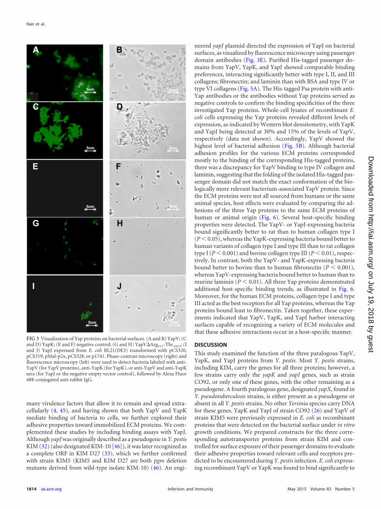
Y. pestis KIM5 recombinant YapV and YapK proteins. Con-sistent with data from previous transcription studies of yap genesin Y. pestis grown in vitro (14, 26), we failed to detect YapV orYapK proteins in Y. pestis KIM5 and KIM6 by Western blot anal-ysis under various in vitro growth conditions, including the use ofBHI and heart infusion broths, at 26°C or 37°C (data not shown).Recent studies with a recombinant YapV protein showed that thisprotein is an autotransporter protein by localizing in the outermembrane and exposing part of itself on the bacterial surface (27).Earlier studies with the orthologous YapK and YapJ autotrans-porter proteins of strain CO92 used murine models of plague thathighlighted their additive effect on increasing bacterial dissemina-tion (26, 37). However, the mechanism responsible for the latterphenotype was not investigated further. Some earlier work onautotransporter proteins of Y. pestis had suggested that YapKmight have adhesive properties based on a weak hemagglutinatingreaction (14), and recent data highlighted the interaction of YapVwith a host protein, albeit unexpectedly a cytoplasmic protein,neuronal Wiskott-Aldrich syndrome protein (N-WASP) (27).Since Y. pestis enters the respiratory tract to cause primary pneu-monic plague, the yapV and yapK genes were cloned to study theadhesive properties of their products toward relevant cells andmolecules encountered by Y. pestis when it invades lung tissue.Recombinant proteins of 130 kDa were expressed by E. coli, inagreement with the calculated molecular masses of both proteins(Fig. 2A). YapV and YapK were shown by fluorescence micros-copy to expose their passenger domain on the bacterial surface(Fig. 3). Some of the bacteria expressing a YapV construct that has
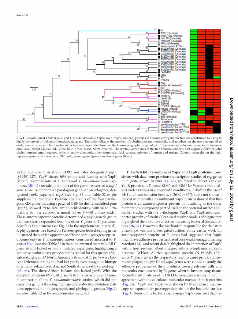
FIG 1 Coevolution of Yersinia pestis and Y. pseudotuberculosis YapX, YapK, YapV, and YapJ proteins. A Yersinia phylogenomic tree was constructed by using 35highly conserved orthologous housekeeping genes. The scale indicates the number of substitutions per nucleotide, and numbers on the tree correspond toevolutionary distances. The branches of the tree are color-coded based on the known geographic origin of each Y. pestis isolate as follows: cyan, South America;pink, Asia (except China); red, China; blue, Africa; black, North America. The symbols at the ends of the tree branches indicate host origins as follows: solidcircles, human; empty squares, rodents; empty diamonds, other mammals; black squares, mixture of human and rodent. Colored rectangles on the rightrepresent genes with a complete ORF (red), pseudogenes (green), or absent genes (black).
Nair et al.
1812 iai.asm.org May 2015 Volume 83 Number 5Infection and Immunity
on July 19, 2018 by guesthttp://iai.asm
.org/D
ownloaded from

a portion of its C-terminal �-barrel domain deleted, YapV(567–1023), were labeled in a punctate way (Fig. 3), despite the fact thatit had been removed from the outer membrane fraction preparedwith Sarkosyl (Fig. 2B and C). It is likely that the latter detergentremoved YapV(567–1023) from the outer membrane becausethe protein’s insertion into the outer membrane was unstable butstill partially detectable by fluorescence microscopy. In contrast,two constructs lacking only portions of the YapV passenger do-main were clearly detected in the bacterial outer membrane frac-tions (Fig. 2B and C). Taken together, these results confirmed thatthe cloned yapV and yapK gene constructs were expressed in E. coliand, in agreement with data from previous studies, demonstratedattributes of autotransporter proteins (14, 26, 27, 37).
YapV- and YapK-expressing bacteria bind to human respira-tory tract epithelial cells. Although many autotransporter genesof Y. pestis CO92 were transcribed only at very low levels in mu-rine models of plague (26), some were shown to be significantlymore expressed, including yapK and yapJ, with both of their prod-ucts improving bacterial dissemination in murine models ofplague (15, 16, 26, 37). The virulence properties that were respon-sible for this phenotype remained unknown. Since many auto-transporter proteins have adhesive functions, we investigatedwhether YapV and YapK could mediate adhesion of bacteria torespiratory tract epithelial cells. The binding of E. coli SE5000carrying pCS320 (YapV) or pCS319 (YapK) was studied with thetype II alveolar epithelial cell line A549 and the lung epithelial-likecell line WI26 VA4. E. coli cells expressing recombinant YapV orYapK bound well to A549 cells, whereas E. coli cells carrying theempty vector did not bind significantly, as visualized by micros-copy (Fig. 4A to C). Quantitative analysis of adherence of bacteriato A549 cells by colony counts determined that YapV- and YapK-expressing E. coli cells bound 7 and 8 times better, respectively,than did the E. coli control strain (Table 2). Comparable results
were observed for WI26 VA4 cells. None of the E. coli strainsexpressing any of the three in-frame deletion constructs in YapVbound to A549 cells, indicating the importance of the full-lengthprotein as a requirement for efficient binding (data not shown).When Y. pestis was grown in vitro at 37°C, YapK and YapJ couldnot be detected by Western blot analysis, even though yapK andyapJ had higher levels of transcripts at this temperature than at26°C (26), leaving it possible that some proteins are expressed atlevels below the level of detection. Since recombinant YapV andYapK were shown to have adhesive properties, we investigatedwhether Y. pestis yapV and yapK mutants would show reducedadhesive properties. Previous work done by using Y. pestis strainKIM6 demonstrated that the removal of the surface structures Psa,F1, and Pla (pPCP1� strain) improved the detection of other sur-face proteins with adhesive properties (4) and that the absence ofthe type III secretion system in KIM6 prevented eukaryotic celldeath (12). Thus, to optimize the binding assays, the yapV andyapK mutations were engineered in the KIM6 pPCP1� caf psastrain, and the adhesive properties of the mutants were studiedwith A549 cells. The lack of both the yapV and yapK genes causeda significant decrease in the adherence of Y. pestis to A549 cells(Fig. 4D). The binding of the yapV single mutant to A549 cells wasdiminished significantly compared with that of the nonmutatedstrain. The yapK mutant also showed reduced, albeit not statisticallysignificant, adhesion, suggesting a lower binding affinity or expres-sion level of YapK than of YapV. When complemented in trans withyapV-containing plasmid pCS320, the Y. pestis yapV yapK doublemutant regained the adhesive properties of the yapK mutantstrain. These results indicated that YapV, and possibly YapK,might participate in primary pneumonic plague for the initiationof contact of bacteria with respiratory tract epithelial cells.
Binding of YapV-, YapK-, and YapJ-expressing bacteria orpassenger domains to ECM proteins. Since Y. pestis expresses
FIG 2 Characterization of YapK and YapV. (A) Expression of YapK and YapV in Escherichia coli. Cells of E. coli SE5000 transformed with pMal-p2X (emptyvector), pCS319 (containing the yapK gene [y0567]), or pCS320 (containing the yapV gene [y3429]) were grown at 37°C, and protein expression was inducedwith IPTG. Whole cells were solubilized and analyzed by SDS-PAGE and Coomassie blue staining. Solid arrowheads indicate monomeric forms of YapK andYapV. The positions of molecular mass markers (in kDa) are indicated on the left. (B) DNA segments of the yapV gene included in each construct. The full-lengthyapV gene can be divided into DNA segments predicted to encode a typically long signal sequence (gray), a surface-exposed passenger domain (light gray), alinker domain (black), and the outer membrane-embedded �-barrel domain (white). The numbers correspond to the amino acid residues that flank theevaluated YapV regions. (C) Western blot analysis of whole cells and outer membrane fractions of E. coli SE5000 expressing full-length or truncated forms(in-frame deletions) of YapV detected with anti-YapV antiserum.
Paralogous Autotransporter Adhesins of Yersinia pestis
May 2015 Volume 83 Number 5 iai.asm.org 1813Infection and Immunity
on July 19, 2018 by guesthttp://iai.asm
.org/D
ownloaded from
many virulence factors that allow it to remain and spread extra-cellularly (4, 45), and having shown that both YapV and YapKmediate binding of bacteria to cells, we further explored theiradhesive properties toward immobilized ECM proteins. We com-plemented these studies by including binding assays with YapJ.Although yapJ was originally described as a pseudogene in Y. pestisKIM (32) (also designated KIM-10 [46]), it was later recognized asa complete ORF in KIM D27 (33), which we further confirmedwith strain KIM5 (KIM5 and KIM D27 are both pgm deletionmutants derived from wild-type isolate KIM-10) (46). An engi-
neered yapJ plasmid directed the expression of YapJ on bacterialsurfaces, as visualized by fluorescence microscopy using passengerdomain antibodies (Fig. 3E). Purified His-tagged passenger do-mains from YapV, YapK, and YapJ showed comparable bindingpreferences, interacting significantly better with type I, II, and IIIcollagens; fibronectin; and laminin than with BSA and type IV ortype VI collagens (Fig. 5A). The His-tagged Psa protein with anti-Yap antibodies or the antibodies without Yap proteins served asnegative controls to confirm the binding specificities of the threeinvestigated Yap proteins. Whole-cell lysates of recombinant E.coli cells expressing the Yap proteins revealed different levels ofexpression, as indicated by Western blot densitometry, with YapKand YapJ being detected at 30% and 15% of the levels of YapV,respectively (data not shown). Accordingly, YapV showed thehighest level of bacterial adhesion (Fig. 5B). Although bacterialadhesion profiles for the various ECM proteins correspondedmostly to the binding of the corresponding His-tagged proteins,there was a discrepancy for YapV binding to type IV collagen andlaminin, suggesting that the folding of the isolated His-tagged pas-senger domain did not match the exact conformation of the bio-logically more relevant bacterium-associated YapV protein. Sincethe ECM proteins were not all sourced from humans or the sameanimal species, host effects were evaluated by comparing the ad-hesions of the three Yap proteins to the same ECM proteins ofhuman or animal origin (Fig. 6). Several host-specific bindingproperties were detected. The YapV- or YapJ-expressing bacteriabound significantly better to rat than to human collagen type I(P � 0.05), whereas the YapK-expressing bacteria bound better tohuman variants of collagen type I and type III than to rat collagentype I (P � 0.001) and bovine collagen type III (P � 0.01), respec-tively. In contrast, both the YapV- and YapK-expressing bacteriabound better to bovine than to human fibronectin (P � 0.001),whereas YapV-expressing bacteria bound better to human than tomurine laminin (P � 0.01). All three Yap proteins demonstratedadditional host-specific binding trends, as illustrated in Fig. 6.Moreover, for the human ECM proteins, collagen type I and typeIII acted as the best receptors for all Yap proteins, whereas the Yapproteins bound least to fibronectin. Taken together, these exper-iments indicated that YapV, YapK, and YapJ harbor interactingsurfaces capable of recognizing a variety of ECM molecules andthat these adhesive interactions occur in a host-specific manner.
DISCUSSION
This study examined the function of the three paralogous YapV,YapK, and YapJ proteins from Y. pestis. Most Y. pestis strains,including KIM, carry the genes for all three proteins; however, afew strains carry only the yapK and yapJ genes, such as strainCO92, or only one of these genes, with the other remaining as apseudogene. A fourth paralogous gene, designated yapX, found inY. pseudotuberculosis strains, is either present as a pseudogene orabsent in all Y. pestis strains. No other Yersinia species carry DNAfor these genes. YapK and YapJ of strain CO92 (26) and YapV ofstrain KIM5 were previously expressed in E. coli as recombinantproteins that were detected on the bacterial surface under in vitrogrowth conditions. We prepared constructs for the three corre-sponding autotransporter proteins from strain KIM and con-trolled for surface exposure of their passenger domains to evaluatetheir adhesive properties toward relevant cells and receptors pre-dicted to be encountered during Y. pestis infection. E. coli express-ing recombinant YapV or YapK was found to bind significantly to
FIG 3 Visualization of Yap proteins on bacterial surfaces. (A and B) YapV; (Cand D) YapK; (E and F) negative control; (G and H) YapVArg517-Thr1023; (Iand J) YapJ expressed from E. coli BL21(DE3) transformed with pCS320,pCS319, pMal-p2x, pCS328, or p1741. Phase-contrast microscopy (right) andfluorescence microscopy (left) were used to detect bacteria labeled with anti-YapV (for YapV proteins), anti-YapK (for YapK), or anti-YapV and anti-YapKsera (for YapJ or the negative empty vector control), followed by Alexa Fluor488-conjugated anti-rabbit IgG.
Nair et al.
1814 iai.asm.org May 2015 Volume 83 Number 5Infection and Immunity
on July 19, 2018 by guesthttp://iai.asm
.org/D
ownloaded from

alveolar A549 epithelial and WI26 VA4 epithelial-like cells. The Y.pestis yapV yapK double mutant bound significantly less to A549cells than did the parental nonmutated strain, and the phenotypewas significantly complemented with a plasmid expressing YapV.Interestingly, reduced binding was detected despite the presenceof Ail in the mutants, which probably explains some of the back-ground adhesion of the double yap mutant. Binding differences ofthe mutants also attested to the expression of the proteins, despitethe proteins being undetectable by Western blotting. This inter-pretation is consistent with the previously reported detection ofautotransporter transcripts from in vitro-grown Y. pestis CO92,with yapK and yapJ being among the three yap genes that weresignificantly more expressed than the other ones (26). Since Y. pestisis an invasive pathogen that uses several antiphagocytic mecha-
nisms to remain extracellular and uses its Pla protease to dissem-inate through intercellular compartments, where it encountersECM proteins, we investigated whether the paralogous Yap pro-teins of Y. pestis KIM interact with these host proteins. We dem-onstrated that not only YapV but also YapK and YapJ acted asadhesins recognizing specific ECM proteins. Most interestingly,the adhesive interactions demonstrated host-specific affinities.YapV was previously reported to bind to N-WASP (27); however,the biological relevance of this property remains unclear consid-ering the expected separate compartmentalization of YapV andN-WASP. Even though Y. pestis can be detected in macrophages,particularly early during infection (47, 48), Y. pestis is not knownto reach the host cytosol, where N-WASP is located. In addition,whether the passenger domain of an autotransporter protein canbe translocated or injected into the host cytosol remains to bedemonstrated. In contrast, the adhesive properties of the threeYap proteins described here provide a reasonable explanation forthe reduced dissemination of the corresponding Y. pestis mutantsin murine models of plague (37). It remains to be examinedwhether these paralogous adhesins and the previously described Y.pestis adhesins are each temporally and spatially regulated in aparticular sequential manner so as to modulate the attachmentand spreading properties of this pathogen in different hosts andhost tissues (49–51).
Comparative genomics has led to the consideration of Y. pestis
FIG 4 Yap protein-mediated binding of bacteria to A549 human type II alveolar epithelial cells. (A to C) Light microscopy of Giemsa-stained A549 cells and E.coli SE5000 transformed with the empty vector pMal-p2X (A), YapK-expressing plasmid pCS319 (B), or YapV-expressing plasmid pCS320 (C). Bacteria weregrown at 37°C, and protein expression was induced by IPTG. Incubation of A549 cells with bacteria was done at a multiplicity of infection of 50. (D) Adherenceof Y. pestis mutants to A549 cells. Cells of Y. pestis strains DSY50 (YapK� YapV�), DSY52 (YapK� YapV�), DSY51 (YapK� YapV�), DSY53 (YapK� YapV�),and DSY52 transformed with pCS320 were incubated at a multiplicity of infection of 10 for binding to A549 cells grown in 24-well plates. Percentages ofcell-associated bacteria were determined by CFU counts. Data represent the means standard errors from three independent experiments done in duplicate. *,P � 0.05 compared to DSY50 binding.
TABLE 2 Binding of recombinant E. coli strains expressing YapK orYapV to human respiratory tract cells
Strain
Mean % adherence to cell line SD
A549 WI26 VA4
SE5000 empty vector 1.34 0.10 1.38 0.06SE5000 YapK� 10.86 0.85a 7.99 1.20a
SE5000 YapV� 9.29 1.76a 10.17 0.68a
a Statistically significant difference from the E. coli SE5000 empty vector control(P � 0.05).
Paralogous Autotransporter Adhesins of Yersinia pestis
May 2015 Volume 83 Number 5 iai.asm.org 1815Infection and Immunity
on July 19, 2018 by guesthttp://iai.asm
.org/D
ownloaded from

as an evolutionary descendant of an ancestral Y. pseudotuberculosisstrain. Reductive evolution as well as the acquisition of two plas-mids, each with a major virulence factor (Pla and F1), have playedan integral part in the conversion to a more lethal pathogen withdifferent transmission mechanisms, a narrow host range, and im-mune evasion factors that bypass innate immune defenses (38, 42,52). Massive evolutionary gene loss and pseudogenization have
been linked to the generation of specialized life-styles in otherpathogens, such as Shigella, Salmonella enterica serovar Typhi, andMycobacterium leprae (53–55). Evolving to a new life cycle couldexplain how many Y. pestis genes, such as the inv and yad genes,became pseudogenes by neutral genetic drift (55). Alternatively,the rscA biofilm repressor gene of Y. pseudotuberculosis mutated inY. pestis is a typical example of selection by adaptive pseudogeni-
FIG 5 Binding of YapV, YapK, and YapJ to ECM proteins. Plastic wells were coated with human collagen type I, chicken collagen type II, bovine collagen typeIII, human collagen type IV, human collagen type VI, murine laminin, bovine fibronectin, and BSA. (A) Purified His-tagged passenger domains of YapV, YapK,and YapJ were added to wells coated with ECM proteins and detected with polyclonal anti-YapV and/or anti-YapK serum, HRP-conjugated secondary antibody,and a chromogenic substrate. Negative controls included anti-YapV or anti-YapK antibody used without any Yap proteins and the two antibodies used with aHis-tagged PsaA protein. (B) Binding of YapV-, YapK-, or YapJ-expressing E. coli SE5000 cells to wells coated with ECM proteins. Bound bacteria were stainedwith crystal violet, and the absorbance was measured at 570 nm. Data represent the means standard errors from �3 independent experiments.
Nair et al.
1816 iai.asm.org May 2015 Volume 83 Number 5Infection and Immunity
on July 19, 2018 by guesthttp://iai.asm
.org/D
ownloaded from

zation, as biofilm formation in fleas benefits bacterial transmis-sion (56, 57). The pseudogenization or absence of yapX in full Y.pestis genomes confirms the pattern of reductive loss in this bac-terium compared to Y. pseudotuberculosis. However, additionalevolutionary processes must have directed the presence or absenceof the four paralogous autotransporter genes in the 40 Yersiniagenomes examined. From an evolutionary standpoint, the earliestpresence of these genes was detected in Y. pseudotuberculosis. It islikely that yapV was acquired by horizontal gene transfer (HGT),considering that phage DNA is present downstream of this gene.The origins of yapK and yapX might be the same, or these genesmight have evolved from a duplication(s) of yapV or from anancestral gene of yapE (see Fig. S3 in the supplemental mate-rial). Alternatively, a Yersinia frederiksenii gene with a similarcarboxy-terminal third (ATCC 33641; GenBank accession num-ber ZP_04633941) might have served as an ancestral gene for geneduplication, and genomic rearrangements and drift would haveresulted in the creation of yapK and yapJ. The strain-specific palletof yap genes might illustrate the definitions of inparalogs (i.e., alineage-specific duplication of yapX and yapJ) (see Fig. S2 in thesupplemental material), outparalogs (i.e., a duplication precedingspeciation for yapK and yapV), or pseudoparalogs (i.e., HGT ac-quisition for yapJ) (58). The reductive loss of similar genes sug-gests either redundant functions or the removal of genes that nolonger serve Yersinia pestis maintenance in its hosts or environ-ments (39). The acquisition of a new gene, yapJ (Fig. 1), and themaintenance of at least one of the four paralogous yap genes in anyY. pestis genome suggest a beneficial role for such genes for theperpetuation of Y. pestis.
Most intriguing is the significant relationship between the pro-file of the four paralogous genes/pseudogenes in Y. pestis strains
and the geographic location of their isolation. Since the reservoirsof host species vary geographically, this suggests an evolutionaryadaption of Y. pestis to a specific local host(s). YapV and its paralo-gous proteins could play a role in the maintenance of a reservoir,thus benefiting the life cycle of the pathogen. Whether the panoplyof paralogous yap genes in the different Y. pestis strains representseffective functional adaption or only random gene losses geo-graphically grouped because of time-dependent evolutionarysteps remains to be investigated. Finally, the observed associationbetween geographic origin and strain signatures for the yapK,yapV, yapX, and yapJ genes or pseudogenes could be useful totrack strain origins and thus serve diagnostic and epidemiologicalpurposes.
ACKNOWLEDGMENTS
This work was supported by NIH grant AI076695, a University of Penn-sylvania Research Foundation grant, and research initiative funds fromthe University of Pennsylvania Veterinary Center for Infectious Disease.F.W. was sponsored by the Jiangsu Scholarship Council, China.
REFERENCES1. Spinner JL, Cundiff JA, Kobayashi SD. 2008. Yersinia pestis type III
secretion system-dependent inhibition of human polymorphonuclearleukocyte function. Infect Immun 76:3754 –3760. http://dx.doi.org/10.1128/IAI.00385-08.
2. Zhang P, Skurnik M, Zhang SS, Schwartz O, Kalyanasundaram R,Bulgheresi S, He JJ, Klena JD, Hinnebusch BJ, Chen T. 2008. HumanDC-SIGN (CD209) is a receptor for Yersinia pestis that promotes phago-cytosis by dendritic cells. Infect Immun 76:2070 –2079. http://dx.doi.org/10.1128/IAI.01246-07.
3. Sebbane F, Jarrett CO, Gardner D, Long D, Hinnebusch BJ. 2006. Roleof the Yersinia pestis plasminogen activator in the incidence of distinctsepticemic and bubonic forms of flea-borne plague. Proc Natl Acad SciU S A 103:5526 –5530. http://dx.doi.org/10.1073/pnas.0509544103.
FIG 6 Comparisons of binding of YapV-, YapK-, and YapJ-expressing E. coli SE5000 cells to plastic wells coated with rat (R), human (H), bovine (B), or mouse(M) ECM proteins. The assays were done as described in the legend of Fig. 5B.
Paralogous Autotransporter Adhesins of Yersinia pestis
May 2015 Volume 83 Number 5 iai.asm.org 1817Infection and Immunity
on July 19, 2018 by guesthttp://iai.asm
.org/D
ownloaded from

4. Liu F, Chen H, Galván EM, Lasaro MA, Schifferli DM. 2006. Effects ofPsa and F1 on the adhesive and invasive interactions of Yersinia pestis withhuman respiratory tract epithelial cells. Infect Immun 74:5636 –5644.http://dx.doi.org/10.1128/IAI.00612-06.
5. Lahteenmaki K, Kukkonen M, Korhonen TK. 2001. The Pla surfaceprotease/adhesin of Yersinia pestis mediates bacterial invasion into humanendothelial cells. FEBS Lett 504:69 –72. http://dx.doi.org/10.1016/S0014-5793(01)02775-2.
6. Lahteenmaki K, Virkola R, Saren A, Emody L, Korhonen TK. 1998.Expression of plasminogen activator pla of Yersinia pestis enhances bacte-rial attachment to the mammalian extracellular matrix. Infect Immun66:5755–5762.
7. Yen YT, Bhattacharya M, Stathopoulos C. 2008. Genome-wide insilico mapping of the secretome in pathogenic Yersinia pestis KIM.FEMS Microbiol Lett 279:56 – 63. http://dx.doi.org/10.1111/j.1574-6968.2007.01008.x.
8. Runco LM, Myrczek S, Bliska JB, Thanassi DG. 2008. Biogenesis of thefraction 1 capsule and analysis of the ultrastructure of Yersinia pestis. JBacteriol 190:3381–3385. http://dx.doi.org/10.1128/JB.01840-07.
9. Felek S, Jeong JJ, Runco LM, Murray S, Thanassi DG, Krukonis ES.2011. Contributions of chaperone/usher systems to cell binding, biofilmformation and Yersinia pestis virulence. Microbiology 157:805– 818. http://dx.doi.org/10.1099/mic.0.044826-0.
10. Kolodziejek AM, Sinclair DJ, Seo KS, Schnider DR, Deobald CF,Rohde HN, Viall AK, Minnich SS, Hovde CJ, Minnich SA, BohachGA. 2007. Phenotypic characterization of OmpX, an Ail homologue ofYersinia pestis KIM. Microbiology 153:2941–2951. http://dx.doi.org/10.1099/mic.0.2006/005694-0.
11. Bartra SS, Styer KL, O’Bryant DM, Nilles ML, Hinnebusch BJ, AballayA, Plano GV. 2008. Resistance of Yersinia pestis to complement-dependent killing is mediated by the Ail outer membrane protein. InfectImmun 76:612– 622. http://dx.doi.org/10.1128/IAI.01125-07.
12. Felek S, Krukonis ES. 2009. The Yersinia pestis Ail protein mediatesbinding and Yop delivery to host cells required for plague virulence. InfectImmun 77:825– 836. http://dx.doi.org/10.1128/IAI.00913-08.
13. Felek S, Tsang TM, Krukonis ES. 2010. Three Yersinia pestis adhesinsfacilitate Yop delivery to eukaryotic cells and contribute to plaguevirulence. Infect Immun 78:4134 – 4150. http://dx.doi.org/10.1128/IAI.00167-10.
14. Yen YT, Karkal A, Bhattacharya M, Fernandez RC, Stathopoulos C.2007. Identification and characterization of autotransporter proteins ofYersinia pestis KIM. Mol Membr Biol 24:28 – 40. http://dx.doi.org/10.1080/09687860600927626.
15. Felek S, Lawrenz MB, Krukonis ES. 2008. The Yersinia pestis autotrans-porter YapC mediates host cell binding, autoaggregation and biofilm for-mation. Microbiology 154:1802–1812. http://dx.doi.org/10.1099/mic.0.2007/010918-0.
16. Lawrenz MB, Lenz JD, Miller VL. 2009. A novel autotransporter adhesinis required for efficient colonization during bubonic plague. Infect Im-mun 77:317–326. http://dx.doi.org/10.1128/IAI.01206-08.
17. Lawrenz MB, Pennington J, Miller VL. 2013. Acquisition of omptinreveals cryptic virulence function of autotransporter YapE in Yersinia pes-tis. Mol Microbiol 89:276 –287. http://dx.doi.org/10.1111/mmi.12273.
18. Forman S, Wulff CR, Myers-Morales T, Cowan C, Perry RD, StraleySC. 2008. yadBC of Yersinia pestis, a new virulence determinant for bu-bonic plague. Infect Immun 76:578 –587. http://dx.doi.org/10.1128/IAI.00219-07.
19. Murphy BS, Wulff CR, Garvy BA, Straley SC. 2007. Yersinia pestis YadC:a novel vaccine candidate against plague. Adv Exp Med Biol 603:400 – 414.http://dx.doi.org/10.1007/978-0-387-72124-8_37.
20. Leyton DL, Rossiter AE, Henderson IR. 2012. From self sufficiency todependence: mechanisms and factors important for autotransporterbiogenesis. Nat Rev Microbiol 10:213–225. http://dx.doi.org/10.1038/nrmicro2733.
21. Hagan CL, Silhavy TJ, Kahne D. 2011. Beta-barrel membrane proteinassembly by the Bam complex. Annu Rev Biochem 80:189 –210. http://dx.doi.org/10.1146/annurev-biochem-061408-144611.
22. Rossiter AE, Leyton DL, Tveen-Jensen K, Browning DF, Sevastsyanov-ich Y, Knowles TJ, Nichols KB, Cunningham AF, Overduin M, Schem-bri MA, Henderson IR. 2011. The essential beta-barrel assembly machin-ery complex components BamD and BamA are required forautotransporter biogenesis. J Bacteriol 193:4250 – 4253. http://dx.doi.org/10.1128/JB.00192-11.
23. Grijpstra J, Arenas J, Rutten L, Tommassen J. 2013. Autotransportersecretion: varying on a theme. Res Microbiol 164:562–582. http://dx.doi.org/10.1016/j.resmic.2013.03.010.
24. Lane MC, Lenz JD, Miller VL. 2013. Proteolytic processing of the Yersiniapestis YapG autotransporter by the omptin protease Pla and the contribu-tion of YapG to murine plague pathogenesis. J Med Microbiol 62:1124 –1134. http://dx.doi.org/10.1099/jmm.0.056275-0.
25. Charbonneau ME, Janvore J, Mourez M. 2009. Autoprocessing of theEscherichia coli AIDA-I autotransporter: a new mechanism involvingacidic residues in the junction region. J Biol Chem 284:17340 –17351. http://dx.doi.org/10.1074/jbc.M109.010108.
26. Lenz JD, Lawrenz MB, Cotter DG, Lane MC, Gonzalez RJ, Palacios M,Miller VL. 2011. Expression during host infection and localization ofYersinia pestis autotransporter proteins. J Bacteriol 193:5936 –5949. http://dx.doi.org/10.1128/JB.05877-11.
27. Besingi RN, Chaney JL, Clark PL. 2013. An alternative outer membranesecretion mechanism for an autotransporter protein lacking a C-terminalstable core. Mol Microbiol 90:1028 –1045. http://dx.doi.org/10.1111/mmi.12414.
28. Aziz RK, Bartels D, Best AA, DeJongh M, Disz T, Edwards RA,Formsma K, Gerdes S, Glass EM, Kubal M, Meyer F, Olsen GJ, OlsonR, Osterman AL, Overbeek RA, McNeil LK, Paarmann D, Paczian T,Parrello B, Pusch GD, Reich C, Stevens R, Vassieva O, Vonstein V,Wilke A, Zagnitko O. 2008. The RAST server: rapid annotations usingsubsystems technology. BMC Genomics 9:75. http://dx.doi.org/10.1186/1471-2164-9-75.
29. Tamura K, Peterson D, Peterson N, Stecher G, Nei M, Kumar S. 2011.MEGA5: molecular evolutionary genetics analysis using maximum likeli-hood, evolutionary distance, and maximum parsimony methods. MolBiol Evol 28:2731–2739. http://dx.doi.org/10.1093/molbev/msr121.
30. Delsuc F, Brinkmann H, Philippe H. 2005. Phylogenomics and thereconstruction of the tree of life. Nat Rev Genet 6:361–375. http://dx.doi.org/10.1038/nrg1603.
31. Datsenko KA, Wanner BL. 2000. One-step inactivation of chromosomalgenes in Escherichia coli K-12 using PCR products. Proc Natl Acad SciU S A 97:6640 – 6645. http://dx.doi.org/10.1073/pnas.120163297.
32. Deng W, Burland V, Plunkett G, III, Boutin A, Mayhew GF, Liss P,Perna NT, Rose DJ, Mau B, Zhou S, Schwartz DC, Fetherston JD,Lindler LE, Brubaker RR, Plano GV, Straley SC, McDonough KA, NillesML, Matson JS, Blattner FR, Perry RD. 2002. Genome sequence ofYersinia pestis KIM. J Bacteriol 184:4601– 4611. http://dx.doi.org/10.1128/JB.184.16.4601-4611.2002.
33. Losada L, Varga JJ, Hostetler J, Radune D, Kim M, Durkin S, Schnee-wind O, Nierman WC. 2011. Genome sequencing and analysis of Yersinapestis KIM D27, an avirulent strain exempt from select agent regulation.PLoS One 6:e19054. http://dx.doi.org/10.1371/journal.pone.0019054.
34. Cao J, Khan AS, Bayer ME, Schifferli DM. 1995. Ordered translocationof 987P fimbrial subunits through the outer membrane of Escherichia coli.J Bacteriol 177:3704 –3713.
35. Schifferli DM, Alrutz M. 1994. Permissive linker insertion sites in theouter membrane protein of 987P fimbriae of Escherichia coli. J Bacteriol176:1099 –1110.
36. Heise T, Dersch P. 2006. Identification of a domain in Yersinia virulencefactor YadA that is crucial for extracellular matrix-specific cell adhesionand uptake. Proc Natl Acad Sci U S A 103:3375–3380. http://dx.doi.org/10.1073/pnas.0507749103.
37. Lenz JD, Temple BR, Miller VL. 2012. Evolution and virulence contri-butions of the autotransporter proteins YapJ and YapK of Yersinia pestisCO92 and their homologs in Y. pseudotuberculosis IP32953. Infect Immun80:3693–3705. http://dx.doi.org/10.1128/IAI.00529-12.
38. Parkhill J, Wren BW, Thomson NR, Titball RW, Holden MT, PrenticeMB, Sebaihia M, James KD, Churcher C, Mungall KL, Baker S, Basham D,Bentley SD, Brooks K, Cerdeno-Tarraga AM, Chillingworth T, Cronin A,Davies RM, Davis P, Dougan G, Feltwell T, Hamlin N, Holroyd S, JagelsK, Karlyshev AV, Leather S, Moule S, Oyston PC, Quail M, Rutherford K,Simmonds M, Skelton J, Stevens K, Whitehead S, Barrell BG. 2001.Genome sequence of Yersinia pestis, the causative agent of plague. Nature413:523–527. http://dx.doi.org/10.1038/35097083.
39. Chain PS, Hu P, Malfatti SA, Radnedge L, Larimer F, Vergez LM,Worsham P, Chu MC, Andersen GL. 2006. Complete genome sequenceof Yersinia pestis strains Antiqua and Nepal516: evidence of gene reduc-tion in an emerging pathogen. J Bacteriol 188:4453– 4463. http://dx.doi.org/10.1128/JB.00124-06.
Nair et al.
1818 iai.asm.org May 2015 Volume 83 Number 5Infection and Immunity
on July 19, 2018 by guesthttp://iai.asm
.org/D
ownloaded from

40. Song Y, Tong Z, Wang J, Wang L, Guo Z, Han Y, Zhang J, Pei D,Zhou D, Qin H, Pang X, Zhai J, Li M, Cui B, Qi Z, Jin L, Dai R, ChenF, Li S, Ye C, Du Z, Lin W, Yu J, Yang H, Huang P, Yang R. 2004.Complete genome sequence of Yersinia pestis strain 91001, an isolateavirulent to humans. DNA Res 11:179 –197. http://dx.doi.org/10.1093/dnares/11.3.179.
41. Eppinger M, Rosovitz MJ, Fricke WF, Rasko DA, Kokorina G, FayolleC, Lindler LE, Carniel E, Ravel J. 2007. The complete genome sequenceof Yersinia pseudotuberculosis IP31758, the causative agent of Far East scar-let-like fever. PLoS Genet 3:e142. http://dx.doi.org/10.1371/journal.pgen.0030142.
42. Chain PS, Carniel E, Larimer FW, Lamerdin J, Stoutland PO, RegalaWM, Georgescu AM, Vergez LM, Land ML, Motin VL, Brubaker RR,Fowler J, Hinnebusch J, Marceau M, Medigue C, Simonet M, Che-nal-Francisque V, Souza B, Dacheux D, Elliott JM, Derbise A,Hauser LJ, Garcia E. 2004. Insights into the evolution of Yersinia pestisthrough whole-genome comparison with Yersinia pseudotuberculosis.Proc Natl Acad Sci U S A 101:13826 –13831. http://dx.doi.org/10.1073/pnas.0404012101.
43. Touchman JW, Wagner DM, Hao J, Mastrian SD, Shah MK, Vogler AJ,Allender CJ, Clark EA, Benitez DS, Youngkin DJ, Girard JM, AuerbachRK, Beckstrom-Sternberg SM, Keim P. 2007. A North American Yersiniapestis draft genome sequence: SNPs and phylogenetic analysis. PLoS One2:e220. http://dx.doi.org/10.1371/journal.pone.0000220.
44. Auerbach RK, Tuanyok A, Probert WS, Kenefic L, Vogler AJ, Bruce DC,Munk C, Brettin TS, Eppinger M, Ravel J, Wagner DM, Keim P. 2007.Yersinia pestis evolution on a small timescale: comparison of whole ge-nome sequences from North America. PLoS One 2:e770. http://dx.doi.org/10.1371/journal.pone.0000770.
45. Viboud GI, Bliska JB. 2005. Yersinia outer proteins: role in modulation ofhost cell signalling responses and pathogenesis. Annu Rev Microbiol 59:69 – 89. http://dx.doi.org/10.1146/annurev.micro.59.030804.121320.
46. Finegold MJ, Petery JJ, Berendt RF, Adams HR. 1968. Studies on thepathogenesis of plague. Blood coagulation and tissue responses of Macacamulatta following exposure to aerosols of Pasteurella pestis. Am J Pathol53:99 –114.
47. Finegold MJ. 1969. Pneumonic plague in monkeys. An electron micro-scopic study. Am J Pathol 54:167–185.
48. Cavanaugh DC, Randall R. 1959. The role of multiplication of Pasteurellapestis in mononuclear phagocytes in the pathogenesis of flea-borneplague. J Immunol 83:348 –363.
49. Kim TJ, Chauhan S, Motin VL, Goh EB, Igo MM, Young GM. 2007.Direct transcriptional control of the plasminogen activator gene of Yer-sinia pestis by the cyclic AMP receptor protein. J Bacteriol 189:8890 – 8900.http://dx.doi.org/10.1128/JB.00972-07.
50. Galván EM, Lasaro MA, Schifferli DM. 2008. Capsular antigen fraction
1 and Pla modulate the susceptibility of Yersinia pestis to pulmonary an-timicrobial peptides such as cathelicidin. Infect Immun 76:1456 –1464.http://dx.doi.org/10.1128/IAI.01197-07.
51. Suomalainen M, Lobo LA, Brandenburg K, Lindner B, Virkola R,Knirel YA, Anisimov AP, Holst O, Korhonen TK. 2010. Temperature-induced changes in the lipopolysaccharide of Yersinia pestis affect plas-minogen activation by the pla surface protease. Infect Immun 78:2644 –2652. http://dx.doi.org/10.1128/IAI.01329-09.
52. Achtman M, Morelli G, Zhu P, Wirth T, Diehl I, Kusecek B, Vogler AJ,Wagner DM, Allender CJ, Easterday WR, Chenal-Francisque V, Wor-sham P, Thomson NR, Parkhill J, Lindler LE, Carniel E, Keim P. 2004.Microevolution and history of the plague bacillus, Yersinia pestis. ProcNatl Acad Sci U S A 101:17837–17842. http://dx.doi.org/10.1073/pnas.0408026101.
53. Maurelli AT. 2007. Black holes, antivirulence genes, and gene inactivationin the evolution of bacterial pathogens. FEMS Microbiol Lett 267:1– 8.http://dx.doi.org/10.1111/j.1574-6968.2006.00526.x.
54. Cole ST, Eiglmeier K, Parkhill J, James KD, Thomson NR, Wheeler PR,Honore N, Garnier T, Churcher C, Harris D, Mungall K, Basham D,Brown D, Chillingworth T, Connor R, Davies RM, Devlin K, Duthoy S,Feltwell T, Fraser A, Hamlin N, Holroyd S, Hornsby T, Jagels K, LacroixC, Maclean J, Moule S, Murphy L, Oliver K, Quail MA, Rajandream MA,Rutherford KM, Rutter S, Seeger K, Simon S, Simmonds M, Skelton J,Squares R, Squares S, Stevens K, Taylor K, Whitehead S, Woodward JR,Barrell BG. 2001. Massive gene decay in the leprosy bacillus. Nature 409:1007–1011. http://dx.doi.org/10.1038/35059006.
55. Parkhill J, Thomson N. 2003. Evolutionary strategies of human patho-gens. Cold Spring Harb Symp Quant Biol 68:151–158. http://dx.doi.org/10.1101/sqb.2003.68.151.
56. Sun YC, Hinnebusch BJ, Darby C. 2008. Experimental evidence fornegative selection in the evolution of a Yersinia pestis pseudogene. ProcNatl Acad Sci U S A 105:8097– 8101. http://dx.doi.org/10.1073/pnas.0803525105.
57. Zhang J. 2008. Positive selection, not negative selection, in the pseudog-enization of rcsA in Yersinia pestis. Proc Natl Acad Sci U S A 105:E69.http://dx.doi.org/10.1073/pnas.0806419105. (Reply, 105:E70, http://dx.doi.org/10.1073/pnas.0807434105.)
58. Koonin EV. 2005. Orthologs, paralogs, and evolutionary genomics.Annu Rev Genet 39:309 –338. http://dx.doi.org/10.1146/annurev.genet.39.073003.114725.
59. Silhavy TJ, Berman ML, Enquist LW. 1984. Experiments with genefusion. Cold Spring Harbor Laboratory, Cold Spring Harbor, NY.
60. Goguen JD, Yother J, Straley SC. 1984. Genetic analysis of the lowcalcium response in Yersinia pestis mu d1(Ap lac) insertion mutants. JBacteriol 160:842– 848.
Paralogous Autotransporter Adhesins of Yersinia pestis
May 2015 Volume 83 Number 5 iai.asm.org 1819Infection and Immunity
on July 19, 2018 by guesthttp://iai.asm
.org/D
ownloaded from