Embed Size (px)
Citation preview

Adjuvant Activity of Naturally Occurring MonophosphorylLipopolysaccharide Preparations from Mucosa-Associated Bacteria
Paula M. Chilton,a,b Diana M. Hadel,a Thao T. To,c Thomas C. Mitchell,a,b Richard P. Darveauc
Institute for Cellular Therapeuticsa and Department of Microbiology and Immunology,b University of Louisville School of Medicine, Louisville, Kentucky, USA; Departmentof Periodontics, University of Washington School of Dentistry, Seattle, Washington, USAc
Natural heterogeneity in the structure of the lipid A portion of lipopolysaccharide (LPS) produces differential effects on the in-nate immune response. Gram-negative bacterial species produce LPS structures that differ from the classic endotoxic LPS struc-tures. These differences include hypoacylation and hypophosphorylation of the diglucosamine backbone, both differencesknown to decrease LPS toxicity. The effect of decreased toxicity on the adjuvant properties of many of these LPS structures hasnot been fully explored. Here we demonstrate that two naturally produced forms of monophosphorylated LPS, from the muco-sa-associated bacteria Bacteroides thetaiotaomicron and Prevotella intermedia, function as immunological adjuvants for anti-gen-specific immune responses. Each form of mucosal LPS increased vaccination-initiated antigen-specific antibody titers inboth quantity and quality when given simultaneously with vaccine antigen preparations. Interestingly, adjuvant effects on initialT cell clonal expansion were selective for CD4 T cells. No significant increase in CD8 T cell expansion was detected. MyD88/Toll-like receptor 4 (TLR4) and TRIF/TLR4 signaling pathways showed equally decreased signaling with the LPS forms studied hereas with endotoxic LPS or detoxified monophosphorylated lipid A (MPLA). Natural monophosphorylated LPS from mucosa-as-sociated bacteria functions as a weak but effective adjuvant for specific immune responses, with preferential effects on antibodyand CD4 T cell responses over CD8 T cell responses.
Lipopolysaccharide (LPS), also known as endotoxin, is a com-mon term used for the major cell wall constituent of Gram-
negative bacteria, which differs in composition depending on thebacterial species in which it is formed. For decades, endotoxicforms of LPS have been studied as natural adjuvants for specificimmune responses, especially antigen (Ag)-specific antibody andT cell responses (1–3). The toxicity associated with LPS structureshas precluded their use as effective and safe vaccine adjuvants.However, the discovery and characterization of monophosphory-lated lipid A (MPLA), a chemically degraded structure from Sal-monella enterica serovar Minnesota Re595 LPS, have yielded apreparation that is less toxic than its parent LPS molecule butsimilarly immunostimulatory. Therefore, MPLA works well as asafe and effective vaccine adjuvant. Lipid A structural featuresknown to account for the maintenance of adjuvant properties andthe loss of toxicity include the number of phosphate groups, aswell as the number, type, and location of fatty acid residues (4–6).Although the most abundant structure in the S. Minnesota MPLAused in these studies is a hexa-acylated structure (Fig. 1), the prep-aration also contains a mixture of underacylated lipid A struc-tures, including penta-, tetra-, and triacylated forms known to beless potent, less toxic, or less antagonistic of TLR4 signaling thanthe parent LPS molecule (7, 8). Further, the absence of a singlephosphate residue from the synthetic form of Escherichia coli lipidA is enough to decrease the production of proinflammatory fac-tors, with little effect on many other products associated withadaptive immunity, including type I interferon (IFN) and IFN-inducible products (9–11). Although MPLA is a partially degradedform of an endotoxic LPS produced after chemical extraction(12), low-toxicity forms of LPS (LT-LPS) are produced naturallyas cell wall constituents of bacterial species colonizing various mu-cosal linings, including the oral and intestinal cavities (13–15).These LPS forms are structurally similar to MPLA (Fig. 1). TheLPS structures from Porphyromonas, Bacteroides, and Prevotella
species are naturally monophosphorylated but penta-acylated,whereas MPLA is primarily hexa-acylated (16–18). Like MPLA,LPS obtained from these bacterial species is less toxic than E. coliLPS (17, 19). The published Bacteroides fragilis LPS structure hasbeen shown to be at least 1,000-fold less toxic than E. coli LPS invivo after D-galactosamine priming (20). The striking differencesin toxicity in response to the various Gram-negative species LPSforms has led Munford to propose that the structural features ofLPS act as guides enabling the host Toll-like receptor 4 (TLR4)/MD2 receptor system to distinguish the low-toxicity commensalGram-negative bacteria from the more highly pathogenic mem-bers of the family Enterobacteriaceae (21).
Possible mechanisms that may account for the reduced toxicityof these LPS forms include (i) decreased affinity for the TLR4/MD2 receptor, (ii) differential ability to associate at the initialsteps of TLR4 engagement, from lipoprotein binding protein(LBP) and CD14 to MD2 and TLR4 binding (22–24), and (iii)differential or preferential signaling through one or both signalingadaptor proteins associated with TLR4: MyD88 or TRIF. Coats etal. have demonstrated that the penta-acylated Porphyromonas gin-givalis LPS (Pg 1690) structure is monophosphorylated on the 4=
Received 19 February 2013 Returned for modification 15 March 2013Accepted 18 June 2013
Published ahead of print 24 June 2013
Editor: R. P. Morrison
Address correspondence to Richard P. Darveau, [email protected], orThomas C. Mitchell, [email protected].
Supplemental material for this article may be found at http://dx.doi.org/10.1128/IAI.01150-12.
Copyright © 2013, American Society for Microbiology. All Rights Reserved.
doi:10.1128/IAI.01150-12
September 2013 Volume 81 Number 9 Infection and Immunity p. 3317–3325 iai.asm.org 3317
on July 12, 2018 by guesthttp://iai.asm
.org/D
ownloaded from

carbon of the diglucosamine head group and has intermediateTLR4/MD2-stimulatory effects through NF-�B (6). Furthermore,this preparation functions as a weak TLR4 agonist in vitro, andthat weak agonism of TLR4 activates a subset of the genes acti-vated by endotoxic E. coli LPS, suggesting a hierarchy of transcrip-tional activation through TLR4 agonism based on the strength ofthe signal (18). Interestingly, the specific set of genes activatedafter partial or weak TLR4/MD2 agonism with P. gingivalis LPS invitro was focused on cellular immune responses, with the majorityof the genes associated with either immune cell adhesion or celltrafficking (18). Similarly, MPLA-stimulated cells have demon-strated a bias toward utilizing the TLR4 signaling adaptor proteinTRIF, as opposed to MyD88, in eliciting cellular responses (25),which could help explain the retention of immune-stimulatoryactivities even when toxic activities are markedly decreased(26–28).
Bacteroides thetaiotaomicron and Prevotella intermedia are twomucosa-associated Gram-negative species that produce predom-
inately low-toxicity forms of LPS identical or similar to the pub-lished LT-LPS produced by B. fragilis (16–18, 20). In this report,LPS forms obtained from these bacteria were examined for adju-vant properties in vivo. S. Minnesota LPS (SmLPS) and S. Minne-sota MPLA (SmMPLA) were used as benchmark TLR4 adjuvantsfor comparison to the adjuvant effects elicited by LPS from B.thetaiotaomicron (BtLPS) or P. intermedia (PiLPS). Adjuvant ef-fects on the antigen-specific antibody response and the initial Tcell clonal expansion were assessed. Each preparation testedinfluenced the expression of antigen-specific IgG subtypes,both the amount and the kinetics of the antigen-specific anti-body response. Additionally, these LPS preparations led topreferential antigen-specific clonal expansion of CD4 T cellsbut had no effect on CD8 T cell antigen-specific expansion.Together, these results indicate that BtLPS and PiLPS struc-tures function in vivo as immunological adjuvants; however,their effects are smaller and less broad than that of endotoxicSmLPS or its related SmMPLA form.
FIG 1 Predominant structures of the LPS preparations used as adjuvants.
Chilton et al.
3318 iai.asm.org Infection and Immunity
on July 12, 2018 by guesthttp://iai.asm
.org/D
ownloaded from

MATERIALS AND METHODSMice. BALB/cByJ and C57BL/6J mice were purchased from The JacksonLaboratory (Bar Harbor, ME). C57BL/6Nai-[Tg]OT-I-[KO]RAG1 (OTI)and C57BL/6-[Tg]OT-II-[KO]RAG1 (OTII) (29–31) mice were origi-nally purchased from Taconic Farms (Germantown, NY) and were bredto Cd45a(Ly5a)/NAi (B6.SJL) mice so that they were no longer RAG1deficient and were congenic to C57BL/6 at the Ptprca, Cd45 locus, result-ing in mice referred to as OTI.SJL and OTII.SJL. Mice were bred andhoused in a specific-pathogen-free barrier facility at the Institute for Cel-lular Therapeutics, University of Louisville, Louisville, KY, and were caredfor according to specific University of Louisville and National Institutes ofHealth animal care guidelines.
Reagents. The hepatitis B virus (HBV) vaccine Engerix-B (GSK Bio-logicals), including aluminum hydroxide (alum) as an adjuvant, was pur-chased from the University of Louisville Hospital pharmacy. Recombi-nant hepatitis B virus surface antigen (rHBsAg) subtype adw waspurchased from Aldevron. Egg white albumin was isolated from endotox-in-free embryonated eggs (Charles River) and was found to contain lessthan 0.05 endotoxin units/ml as measured by a quantitative chromogenicLimulus amebocyte lysate assay (QCL-1000; BioWhittaker, Walkersville,MD). Formalin-inactivated influenza virus A/PR/8/34 (H1N1) was pur-chased from Charles River. Ovalbumin peptides for OTII (Ova323-339;ISQAVHAAHAEINEAGR) and OTI (Ova257-264; SIINFEKL) mice weresynthesized and found to be endotoxin free by CPC Scientific (Sunnyvale,CA). Dimethyl sulfoxide (DMSO) and incomplete Freund’s adjuvantwere purchased from Sigma-Aldrich. Goat anti-mouse immunoglobulinsubtype secondary antibodies conjugated to horseradish peroxidase (IgM,total IgG, IgG1, IgG2a, IgG2b, IgG2c) were purchased from JacksonImmunoResearch Labs.
Lipid A and LPS preparations. SmLPS and SmMPLA were purchasedfrom InvivoGen for the egg white albumin immunization experiment andfrom Alexis Biochemicals for the remainder of the experiments. B.thetaiotaomicron and P. intermedia LPS preparations were obtained usinga modified Tri reagent protocol for LPS isolation, as described previously(32, 33). Dried LPS samples were suspended in 10 mM sodium acetate(pH 4.5) containing 1% SDS, heated at 100°C for 1 h, and then lyophilizedovernight. Pellets were washed in ice-cold 95% ethanol containing 0.02 NHCl, followed by three 95% ethanol washes. LPS preparations were sub-jected to a final Bligh-Dyer extraction, which consisted of 1.16 ml of achloroform-methanol-water mixture (1:1:0.9), in order to remove theresidual carbohydrate contaminants. Lyophilized preparations were sus-pended to a stock concentration of 1.0 mg/ml in DMSO. Therefore,DMSO (Sigma) was added as the vehicle control to levels consistent withthose used in the LT-LPS treatments.
Immunizations. Groups of 5 or 6 C57BL/6J mice were immunizedsubcutaneously (s.c.) in each of the two hocks, at the base of the leg pos-terior to the knee joint, with 0.2 �g Engerix-B, a commercially availableHBV vaccine (total, 0.4 �g per mouse) that already contains alum as anadjuvant. The HBV vaccine was combined with either a vehicle control,1.5 �g SmLPS (Alexis Biochemicals), 5 �g SmMPLA (Alexis Biochemi-cals), 5 �g BtLPS, or 5 �g PiLPS. A single boost immunization (at 5 weeks)was the same as the primary immunization. Serum samples were collectedby lateral tail vein bleeds 2 weeks after the primary immunization and 1week after the boost. Formalin-inactivated influenza virus (A/PR/8/34)particles (iPR8) were diluted in phosphate-buffered saline (PBS), andgroups of five C56BL/6J mice were immunized subcutaneously with 0.25�g iPR8 (total, 0.5 �g per mouse) and either a vehicle control, 1.5 �gSmLPS (Alexis), 5 �g SmMPLA (Alexis), 5 �g BtLPS, or 5 �g PiLPS. Thesecond (at 5 weeks) and third (at 20 weeks) immunizations were the samefor each group. Serum samples were taken at 4 weeks for primary immu-nization and then 1 week following each subsequent immunization. Eggwhite albumin (40 �g) was emulsified in incomplete Freund’s adjuvantalong with either a vehicle control (DMSO), 10 �g SmLPS (InvivoGen),20 �g SmMPLA (InvivoGen), 20 �g BtLPS, or 20 �g PiLPS. Groups of sixBALB/cByJ mice were immunized with 100 �l of the emulsified vaccine
preparations subcutaneously in the flanks. Second (at 4 weeks) and third(at 16 weeks) immunizations were the same for each group, except that 10�g egg white albumin was used as the antigen. Serum samples were ob-tained at 3 weeks for primary immunization and then 4 days after subse-quent immunizations. All serum samples were stored at �80°C until en-zyme-linked immunosorbent assays (ELISA) were performed todetermine the titers of antigen-specific antibody present. No animal ex-hibited signs of endotoxemia during these experiments. The amountsused (1.5 to 10 �g SmLPS or 5 to 20 �g SmMPLA, BtLPS, or PiLPS) aremuch lower than those used to study the toxic effects of LPS (typically 50to 200 �g).
Antigen-specific antibody titers determined by ELISA. An antigenspecific to the immunization protocol (rHBsAg, iPR8, or egg white albu-min) was adhered to Immulon 2 ELISA plates. Wells were blocked over-night at 4°C using 2% bovine serum albumin (BSA)-PBS. Replicate plateswere used; serial 10-fold dilutions of the serum samples were made; and100 �l of each dilution was incubated on the Ag-coated plates. Horserad-ish peroxidase-conjugated goat anti-mouse IgM, total IgG, IgG1, IgG2c(or IgG2a), and IgG2b secondary antibodies (Jackson ImmunoResearch)were used to detect specific isotypes. Titers are expressed as the inverse ofthe dilution required to reach the half-maximum value of the absorbancecurve, as determined by SoftMax Pro software.
BM-DC derivation, culture and qRT-PCR. Bone marrow-deriveddendritic cells (BM-DC) were prepared according to the protocol of Lutzet al. (34). Bone marrow cells were collected from femurs and tibiae ofC57BL/6 mice, and 2 � 106 cells were placed in bacteriological cultureplates for 10 days in a complete medium, RPMI 1640 containing 10%heat-inactivated fetal bovine serum (Valley Biomedical), 2 mM L-glu-tamine, 1 mM sodium pyruvate, 50 U/ml penicillin, 50 �g/ml streptomy-cin, 50 �m 2-mercaptoethanol, and 5 ng/ml granulocyte-macrophagecolony-stimulating factor (GM-CSF; R&D Systems). Cultures were givennew medium and GM-CSF on days 3, 6, and 8. Nonadherent cells werecollected on day 10 and were verified by flow cytometry to be 90 to 95%CD11c� CD11b� MHCII� Gr1� before use in experiments. For quanti-tative real-time PCR (qRT-PCR) analysis, 1 � 106 BM-DC were rested for2 h in 24-well polystyrene tissue culture plates (BD Biosciences) in com-plete medium. The cells were then activated with increasing concentra-tions of the test adjuvant preparations. After 2 h, total RNA was isolatedusing an RNeasy Plus minikit (Qiagen), and 0.5 �g total RNA was used tomake cDNA by using qScript cDNA synthesis kits (Quanta) according tothe manufacturer’s protocol. cDNA was diluted 2-fold and was then am-plified with 0.5 �M target (endothelin1, Ifit1, ip10, or il10) or control(gapdh) QuantiTect primer assays from Qiagen. qRT-PCR was performedon a CFX96 real-time system C1000 thermal cycler (Bio-Rad) with PowerSYBR green master mix (Applied Biosystems). Relative mRNA abun-dance was calculated by the comparative cycle method (��CT) usingvehicle control (DMSO)-treated cells for normalization.
Primary human monocyte culture. Deidentified human peripheralblood mononuclear cells (PBMC) from individual healthy donors wereobtained as a waste product of polymorphonuclear leukocyte (PMN) iso-lation from another group at the University of Louisville School of Med-icine. PBMC monocytes were enriched by plating 2 � 106 cells per well in24-well tissue culture plates for 2 h in 0.5 ml complete medium. Adherentcells were enriched by washing away the nonadherent cells with threewashes consisting of 1� Hanks balanced salt solution (HBSS; Life Tech-nologies). The resulting cells were then rested for an additional 2 h beforebeing activated by increasing concentrations of adjuvant preparations.RNA isolation and qRT-PCR were performed, and the results were ana-lyzed as described above for mouse BM-DC. PCRs used undiluted cDNAwith 0.5 �M target (human IL-6, endothelin1, or IFIT1) or control (hu-man GAPDH) QuantiTect primer assays.
Statistical analysis. Statistical analysis was performed using Prismsoftware, version 5.0 (GraphPad). A two-way analysis of variance(ANOVA) with a Bonferroni post hoc analysis was used to compare theserum antibody titers in vivo. One-way ANOVA and Newman-Keuls post
Adjuvant Effects of Natural Monophosphoryl LPS
September 2013 Volume 81 Number 9 iai.asm.org 3319
on July 12, 2018 by guesthttp://iai.asm
.org/D
ownloaded from

hoc analysis were used to compare the mean values for adjuvant-depen-dent T cell clonal expansion.
RESULTSNaturally occurring LT-LPS preparations promote a balancedisotype profile after vaccination. The abilities of the BtLPS andPiLPS forms to function as immunological adjuvants were as-sessed with three separate immunization antigens. Because a pre-liminary study detected no antigen-specific antibody whenSmLPS or SmMPLA was used as the adjuvant for soluble egg whitealbumin given subcutaneously, each of the antigens chosen wasgiven along with a known adjuvant to ensure that an antibodyresponse would be mounted in the time allotted (data not shown).The antigens were (i) a commercially available hepatitis B virus(HBV) vaccine, which contains aluminum hydroxide, (ii) forma-lin-inactivated PR8 (iPR8) influenza virus particles, which haveendogenous TLR7 agonist RNA, and (iii) egg white albuminemulsified in incomplete Freund’s adjuvant. The doses of antigenand adjuvants used in each protocol were chosen to optimize theadjuvant effects. Previous studies had also determined that 2- to3-fold more SmMPLA than SmLPS is needed to elicit similar levelsof peak T cell clonal expansion in different groups of mice (26),and the LT-LPS amounts used were matched to that of SmMPLA.Because stock LT-LPS preparations were suspended in DMSO,enough DMSO was added to each vaccine preparation to controlfor the DMSO present in the LT-LPS-adjuvanted vaccine prepa-rations.
An alum-containing HBV vaccine supplemented with the nat-ural LT-LPS preparations was given subcutaneously to groups ofC57BL/6 mice. DMSO alone was added for the baseline controlgroup, while SmLPS or SmMPLA was added for positive-controlTLR4 agonist groups. Titers of antigen-specific antibodies in se-rum were used to monitor the developing immune responses afterimmunization. Adjuvant responses would be indicated if eitherBtLPS or PiLPS was able to change or supplement the alum-di-rected Th2-like antibody response (inducing IgG1) with a Th1-like response (inducing IgG2c or IgG2b). All HBV vaccine-treatedC57BL/6 mice mounted similar IgM responses to HBsAg (Fig.2A). However, higher total-IgG titers than those for the vehiclecontrol group were observed after the first immunization for allLPS-treated groups (Fig. 2A); therefore, increased isotype switch-ing occurred in each of the LPS-treated groups. As expected, thetiters of the IgG1 subtype (Fig. 2A) were significantly increasedafter both primary and boost immunizations with only alum, andthere was no difference from alum when any of the LPS forms wasadded (35, 36). The control immunization group had increasedIgG2b titers after the boost immunization, whereas IgG2c titersremained low throughout. The IgG2b responses were nearly iden-tical in all the groups with LPS as an adjuvant. Although no in-crease in the complement-fixing IgG2c subclass was evident withany of the groups after the primary immunization, each of theLPS-treated groups had high titers after the boost immunization,which is typical after a sufficient immune response is mounted.
FIG 2 Antigen-specific antibody responses in sera of HBV vaccine-immunized C57BL/6 mice. (A) Groups of C57BL/6 mice were immunized s.c. with an HBVvaccine in the absence (�DMSO) or presence (3 �g SmLPS, 10 �g SmMPLA, 10 �g BtLPS, or 10 �g PiLPS) of LPS preparations on day zero, followed by a boostimmunization 5 weeks after the primary immunization. Serum was sampled before and after each of the immunizations, and the titers of HBsAg-specific IgM,total IgG, IgG1, IgG2c, and IgG2b were determined by ELISA by limiting dilution. Arrowheads indicate times of vaccination. (B) Antigen-specific IgG2a titersafter the third immunization with egg white albumin emulsified in incomplete Freund’s adjuvant with either a vehicle control (�DMSO) or one of the adjuvantpreparations. (C) Antigen-specific IgG2b titers after the second immunization with iPR8 influenza virus particles and either a vehicle control (�DMSO) or oneof the adjuvant preparations. Results are expressed as means � standard errors of the means of the antibody titer, calculated as the inverse of the dilution factor(df) needed to give the half-maximum value (max1/2) of the absorbance curve after 10-fold serial dilutions. Asterisks indicate significant differences from theDMSO control group at a 95% (P � 0.05) confidence level, as determined using two-way ANOVA and Bonferroni post hoc analysis.
Chilton et al.
3320 iai.asm.org Infection and Immunity
on July 12, 2018 by guesthttp://iai.asm
.org/D
ownloaded from

Both BtLPS and PiLPS increased IgG2c titers after this immuniza-tion, but titers for the BtLPS-treated group were lower than thosefor the SmLPS-treated group. No such difference was noted be-tween the PiLPS- and SmLPS-treated groups. Both the BtLPS andPiLPS groups exhibited IgG2b titers significantly higher than thatof the control vaccine group and not significantly lower than thoseelicited by SmLPS or SmMPLA after both the primary and boostimmunizations. The increase in the breadth of the IgG subtypesafter the inclusion of either BtLPS or PiLPS in these immuniza-tions indicates immune adjuvant activity in these preparations.
The antibody responses in the two other immunization proto-cols also demonstrated the adjuvant activity of the LT-LPS prep-arations (see Tables S1 to S5 in the supplemental material). In thefirst protocol, BALB/c mice were given three subcutaneous immu-nizations of egg white albumin in incomplete Freund’s adjuvant.The results from this protocol were initially hampered by rela-tively higher titers of antigen-specific antibodies, especially IgMand IgG2b isotypes, in the preimmunization serum samples (datanot shown), possibly caused by environmental exposure toovalbumin in feed or bedding. A similar polyclonal antiovalbu-min response was seen in unimmunized BALB/c mice infectedwith Leishmania infantum (37). After the third immunization,each LPS preparation tested increased the titers of the comple-ment-fixing, Th1-associated isotype IgG2a in most vaccinatedmice (Fig. 2B). The BtLPS group had several low responders (titer,�200), which increased the variability observed, thus making thedifference between the vehicle control and BtLPS groups insignif-icant (Fig. 2B).
Subcutaneous immunization of C57BL/6 mice with iPR8 virusparticles and the LPS preparations elicited increased titers in allimmunized groups, including the DMSO control group (see Ta-bles S6 to S10 in the supplemental material). Following the secondimmunization, minor differences in titers of the transforminggrowth factor (TGF-)-associated isotype IgG2b were detectedbetween the control group and the groups treated with SmLPS,BtLPS, or PiLPS, but not SmMPLA (Fig. 2C). However, the sub-sequent immunization increased all isotype titers and eliminatedthe differences between the DMSO control and LPS-treatedgroups (data not shown).
Together, the results from these three immunization protocolsclearly demonstrate adjuvant activity for BtLPS and PiLPS, mean-ing that the inclusion of either preparation leads to increased titersand/or a more diverse repertoire of antibodies to various T cell-dependent antigens, therefore indirectly providing evidence foreffects on T cell responses.
Natural LT-LPS preparations from B. thetaiotaomicron andP. intermedia serve as adjuvants for the initial clonal expansionof CD4� T cells in vivo. Adjuvant signals are required for produc-tive or sustained clonal expansion of naïve CD4 or CD8 T cells inresponse to purified antigen in vivo (1, 38). LPS is a natural adju-vant that increases the number of cell divisions and the rate of cellsurvival during and after T cell activation and clonal expansion (1,39, 40). SmMPLA also acts effectively as an adjuvant for initial Tcell clonal expansion in vivo, which we and others have shown isdependent on the TLR4 signaling adaptor TRIF (25, 26). Using astandard adoptive-transfer model of ovalbumin-specific T cell re-ceptor (TCR) transgenic (Tg) CD8 and CD4 T cells (OTI.SJL andOTII.SJL cells, respectively), we assessed the effects of LT-LPS oninitial T cell clonal expansion compared to the effects elicited withSmLPS and SmMPLA. After adoptive transfer of OTI (CD45.1)
and OTII (CD45.1) Tg-TCR cells (1 � 105 and 1.5 � 105, respec-tively), C57BL/6 (CD45.2) recipients were treated with antigenicpeptides mixed with either a vehicle control or one of the LPSforms as an adjuvant through intravenous injection. The fold in-creases in OTI and OTII cell accumulation in the spleen or per-fused lung tissue at peak clonal expansion for mice treated with anadjuvant over the levels for controls given only the peptide anti-gens with DMSO were calculated. As shown in Fig. 3, each of thepreparations tested increased the number of splenic antigen-spe-cific T cells; therefore, each of the LT-LPS preparations was able toserve as an adjuvant for initial T cell clonal expansion. BtLPS treat-ment increased the level of CD4 T cells (OTII) to approximatelyone-half or one-third of the response seen with SmLPS orSmMPLA, respectively. PiLPS increased the CD4 clonal expan-sion to a level similar to that for SmLPS. The adjuvant effects of theLT-LPS preparations on CD8 T cell numbers were markedly lowerthan that for SmLPS or SmMPLA. BtLPS increased the number ofCD8 T cells in the spleen 3-fold over that with the antigen alone,while the PiLPS increase was near 4-fold.
Because adjuvant signals are needed for activated T cells tomigrate to nonlymphoid sites after activation (41, 42), the numberof activated T cells in the lungs is an indicator of TLR-dependentadjuvant effects in vivo. Adjuvant-specific increases in the num-bers of CD4 T cells in the lungs were markedly greater than thosewith the antigen alone (averages, 16.9-fold � 3.2-fold for BtLPSand 12.2-fold � 3.5-fold for PiLPS), though less than those ob-served with SmLPS or SmMPLA. However, the increases in the
FIG 3 Short-term T cell clonal expansion is increased in the spleens of animalsimmunized with any of the LPS preparations. (A) Schematic of the adoptivetransfer and immunization protocol used on recipient C57BL/6 (CD45.2)mice. Spleen cells (105 OTI.SJL cells and 1.5 � 105 OTII.SJL [CD45.1] cells)were transferred 2 days before intravenous immunization with antigenic pep-tides in the absence (DMSO) or presence of TLR4 agonists. The numbers ofcells present in primary (spleen) and secondary (lung) organs 3.5 days afterimmunization were determined. (B) Numbers of antigen-specific CD4 (OTII)and CD8 (OTI) T cells recovered in each tissue were determined by flowcytometry. Results are expressed as fold increases, calculated as the number ofcells recovered from adjuvant-treated mice divided by the average number ofcells recovered from the group treated with the antigen only, with no adjuvant(DMSO). Data are from two independent experiments, with triplicate miceused in each. Results for the individual recipients from experiments 1 (circles)and 2 (triangles) are shown. The horizontal line indicates the average foldincrease for the combined groups. Asterisks indicate significant differencesfrom the DMSO control group as determined by one-way ANOVA and New-man-Keuls post hoc analysis (P � 0.05).
Adjuvant Effects of Natural Monophosphoryl LPS
September 2013 Volume 81 Number 9 iai.asm.org 3321
on July 12, 2018 by guesthttp://iai.asm
.org/D
ownloaded from
numbers of OTI CD8 T cells in the lung were only marginal forrecipients of either LT-LPS preparation and did not reach signif-icance. The numbers of CD8 T cells in the lungs, and the foldincreases in these numbers, for groups receiving SmLPS orSmMPLA as an adjuvant increased significantly, but the fold in-creases were lower than those observed for CD4 T cells.
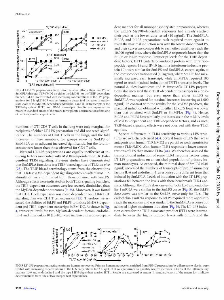
Natural LT-LPS preparations are equally ineffective at in-ducing factors associated with MyD88-dependent or TRIF-de-pendent TLR4 signaling. Previous studies have demonstratedthat SmMPLA functions as a TRIF-biased agonist of TLR4 in vivo(25). The TRIF-biased terminology stems from the observationsthat TLR4/MyD88-dependent signaling outcomes after SmMPLAstimulation were diminished from those obtained with SmLPS.Although effects were indicated on both TLR4 signaling branches,the TRIF-dependent outcomes were less severely diminished thanthe MyD88-dependent outcomes (9, 25). Moreover, it was foundthat CD8 T cell expansion was more dependent on TLR4/TRIFsignaling than was CD4 T cell expansion (25). Therefore, we as-sessed the abilities of BtLPS and PiLPS to induce MyD88-depen-dent and TRIF-dependent transcripts in BM-DC. As shown in Fig.4, transcript levels for two MyD88-dependent factors, endothe-lin-1 and interleukin 10 (IL-10), were increased in a dose-depen-
dent manner for all monophosphorylated preparations, whereasthe SmLPS MyD88-dependent responses had already reachedtheir peak at the lowest dose tested (10 ng/ml). The SmMPLA,BtLPS, and PiLPS preparations each required more agonist toreach the maximal induction seen with the lowest dose of SmLPS,and their curves are comparable to each other until they reach the10,000-ng/ml dose, where the SmMPLA response is lower than theBtLPS or PiLPS response. Transcript levels for the TRIF-depen-dent factors, IFIT1 (interferon-induced protein with tetratrico-peptide repeats 1) and IP-10 (gamma interferon-inducible pro-tein 10), were similar for SmLPS and SmMPLA, except, again, atthe lowest concentration used (10 ng/ml), where SmLPS had max-imally increased each transcript, while SmMPLA required 100ng/ml to reach maximal induction of IFIT1 transcript levels. Thenatural B. thetaiotaomicron and P. intermedia LT-LPS prepara-tions also increased these TRIF-dependent transcripts in a dose-dependent manner, but even more than either SmLPS orSmMPLA was required for maximal increases (occurring at 1,000ng/ml). In contrast with the results for the MyD88 products, themaximal induction obtained with either LT-LPS form was lowerthan that obtained with SmLPS or SmMPLA (Fig. 4). Hence,BtLPS and PiLPS have similarly low increases in the mRNA levelsof MyD88-dependent and TRIF-dependent factors, and as such,TRIF-biased signaling effects were not observed with these TLR4agonists.
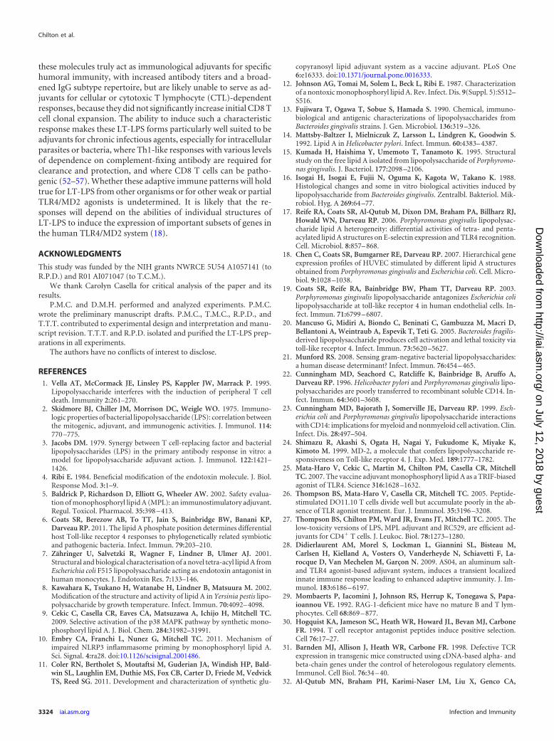
Species differences in TLR4 sensitivity to various LPS struc-tures are well characterized (43). Several forms of LPS that act asantagonists on human TLR4/MD2 are partial or weak agonists formouse TLR4/MD2. Also, human TLR4 responds to lower concen-trations of LPS than mouse TLR4 (44). We therefore assessed thetranscriptional induction of some TLR4 response factors usingLT-LPS preparations on an enriched population of primary hu-man monocytes. As expected, the minimal dose of SmLPS (0.01ng/ml) increased the numbers of transcripts of proinflammatoryfactors IL-6 and endothelin-1, a response quite different from thatinduced by SmMPLA. Levels of induction with the LT-LPS prep-arations fell between the levels with these benchmark TLR4 ago-nists. Although the PiLPS dose curves for both IL-6 and endothe-lin-1 mRNA were similar to the SmLPS curve (Fig. 5), the BtLPSdose curve was similar to the SmLPS curve only for IL-6. Theendothelin-1 mRNA response to BtLPS required more agonist toreach the maximum and was similar to the SmMPLA response butachieved higher maximum induction (Fig. 5). The LT-LPS induc-tion curves for the TRIF-associated product IFIT1 were interme-diate between the highly induced levels with SmLPS and the
FIG 4 LT-LPS preparations have lower relative effects than SmLPS orSmMPLA through TLR4/MD2 on either the MyD88- or the TRIF-dependentbranch. BM-DC were treated with increasing concentrations of the LPS prep-arations for 2 h. qRT-PCR was performed to detect fold increases in steady-state levels of the MyD88-dependent endothelin-1 and IL-10 transcripts or theTRIF-dependent IFIT1 and IP-10 transcripts. Results are expressed asmeans � standard errors of the means for triplicate determinations from oneof two independent experiments.
FIG 5 LT-LPS preparations activate primary human monocytes. Primary human monocytes, enriched from PBMC preparations by adherence to plastic, weretreated with increasing concentrations of the LPS preparations for 2 h. qRT-PCR was performed to quantify relative increases in levels of the inflammatorymarkers IL-6 and endothelin-1 and the type I IFN-dependent marker IFIT1. Results are expressed as means � standard errors of the means for triplicatedeterminations from one of two independent experiments.
Chilton et al.
3322 iai.asm.org Infection and Immunity
on July 12, 2018 by guesthttp://iai.asm
.org/D
ownloaded from

poorly induced levels with SmMPLA, which barely increasedmore than 2-fold at higher doses (1,000 ng/ml). PiLPS requiredmore agonist and reached a lower maximal induction of IFIT1mRNA than either SmLPS or BtLPS. However, BtLPS increasedIFIT1 mRNA expression in human monocytes to a maximumsimilar to that with SmLPS but required more agonist to do so(Fig. 5).
DISCUSSION
Although previous reports have routinely suggested that naturallyoccurring LT-LPS structures may represent a new class of immu-nologic adjuvants, to our knowledge these studies represent thefirst test of their ability to function as such in vivo, increasingantigen-specific immune responses. The results presented heresuggest that the TLR4 responses to these and other LT-LPS formsmay indeed be a means to fine-tune the innate immune responseso as to elicit the most desired immune effector functions againstany number of microbial infectious agents.
The LPS forms tested were obtained from the mutualistic gutbacterium B. thetaiotaomicron and the oral and systemic patho-genic bacterium P. intermedia. Structurally, these forms are verysimilar to one another and to the B. fragilis LPS, previously deter-mined to be of low toxicity (20). However, they differ from the S.Minnesota LPS and MPLA forms at key areas known to influenceTLR4 activation, mainly in the length and number of acyl chains(Fig. 1). Although previous structure/function studies suggest thatthese penta-acyl LT-LPS forms should have decreased TLR4/MD2signaling, the extra length in the acyl chains may help offset theloss of the sixth fatty acid chain within the MD2 hydrophobicpocket and allow activating interactions between these LPSforms and TLR4 (6). Each of the LT-LPS forms activated hu-man MyD88- and TRIF-associated transcripts in primary humanmonocytes (Fig. 5), indicating agonistic signaling through humanTLR4/MD2. When we included these LT-LPS forms as adjuvantsfor mice vaccinated with the alum-containing HBV vaccine, bothBtLPS and PiLPS increased the titers and broadened the IgG sub-types produced during the antigen-specific antibody response.The broadened antibody response caused by the LPS forms there-fore increased potential effector functions mediated by the in-duced HBsAg-specific antibodies. The effects on the antibody re-sponse were comparable to those obtained with SmMPLA, withno effects on the IgG1 titers (45–47). The development of IgG2bwas observed regardless of the nature of the LPS form used. There-fore, naturally produced LT-LPS preparations were nearly as ef-fective as SmMPLA at increasing the IgG differentiated classswitching within the antigen-specific B cell population. Changesin the amount and the quality of the antibody repertoire are well-characterized indicators of differentiated CD4 T cell responses,because isotype switching requires help from CD4 T cells throughcytokine production and cell surface interactions (46, 47). Thedemonstrated increase in CD4 T cell clonal expansion in the OTII/OTI adoptive-transfer model is consistent with inferred adjuvantactivity on CD4 T cells.
Increases in early T cell clonal expansion after antigen stimu-lation were also noted using LT-LPS forms as the only adjuvantcompounds present. The adjuvant-dependent increase was moreevident in antigen-specific CD4 T cells than in CD8 T cells. Theability of activated T cells to migrate from the spleen and/or lymphnodes to extralymphoid organs, an effect that is strongly corre-lated with adjuvant treatment and increased T cell memory (42,
48), was measured using the LT-LPS forms during activation. Be-cause the antigen remains in the lung after intravenous adminis-tration, the lung is an ideal extralymphoid site for this assay. Theadjuvant-dependent increases in the numbers of CD4 (OTII) Tcells in both the spleen and the lung were lower with the LT-LPSpreparations than with either SmLPS or SmMPLA, but the effectswere similar with the BtLPS and PiLPS treatments. Although theLT-LPS exerted a moderate adjuvant effect on CD4 (OTII) T cells,only a small adjuvant-dependent increase was noted for the CD8 Tcells (OTI cells) in the same recipients. In fact, there was no in-crease in the number of lung CD8 T cells with either LT-LPS formover that for recipients given antigenic peptides alone. The obser-vation of differing effects on CD4 T cells and CD8 T cells is rem-iniscent of previous results with this adoptive-transfer model,where TRIF-deficient recipients did not support TLR4 adjuvant-dependent expansion of CD8 OTI T cells, while CD4 OTII cellswere only partially affected (25). That CD8 T cell expansion ismore dependent on the set of signals provided through the TLR4/TRIF-dependent pathway than CD4 T cell expansion is supportedby other studies demonstrating greater dependence of CD8 T cellsthan CD4 T cells on TLR4/TRIF signaling, and the associated pro-duction of type I IFN, for expansion and survival (49–51).
The selective deficit in CD8 T cell expansion with the LT-LPScould suggest poorer utilization of the TRIF signaling branch fromTLR4, compared to that for classically defined TLR4/MD2 ago-nists. Decreased LPS activation of NF-�B in BtLPS-treatedHEK293 cells expressing human TLR4/MD2 has been reportedpreviously (6). Here we showed that BtLPS and PiLPS each in-creased the number of MyD88-dependent and TRIF-dependenttranscripts in mouse BM-DC in a dose-dependent manner; how-ever, the responses from both signaling branches required at least10- to 100-fold more agonist to reach maximal induction than inthe case of SmLPS. The decrease in potency for MyD88-depen-dent products with BtLPS or PiLPS was comparable to that ob-tained with SmMPLA. These differences in TLR4/MD2 potencyand efficacy (Fig. 4) may help explain the decreased in vivo adju-vant effects, especially on the CD8 T cells. Many studies haveshown that TRIF-dependent factors are required for increasedCD8 T cell survival after activation, in addition to increased pro-liferation during the initial antigen encounter (25, 38, 49, 50). Wehave recently reported that TRIF-dependent production of type IIFN and the subsequent type I IFN-dependent increase in thenumber of costimulatory molecules on certain subsets of den-dritic cells in vivo play key roles in CD8 T cell expansion, whileCD4 T cells are not as responsive to type I IFN-dependent effects(51). Although deficits in CD8 expansion could be related to dif-ferences in antigen processing, the use of antigenic peptides in thisstudy decreases the dependence of the CD8 T cell response onprocessing. Also, each of the adjuvant molecules utilizes the samereceptor system, TLR4/MD2. Accordingly, any difference in pep-tide loading into major histocompatibility complex (MHC) class Imolecules would reflect partial agonism of TLR4, and the dimin-ished effects are still appropriately attributed to partial receptoragonism by the LT-LPS forms.
In summary, two naturally produced LT-LPS structures in-creased the scope of antigen-specific antibody responses to vac-cine antigens and increased initial T cell clonal expansion. TheseLT-LPS preparations induced responses comparable to one an-other, and the antibody responses were similar to those inducedwith SmLPS and SmMPLA. The responses evoked indicate that
Adjuvant Effects of Natural Monophosphoryl LPS
September 2013 Volume 81 Number 9 iai.asm.org 3323
on July 12, 2018 by guesthttp://iai.asm
.org/D
ownloaded from
these molecules truly act as immunological adjuvants for specifichumoral immunity, with increased antibody titers and a broad-ened IgG subtype repertoire, but are likely unable to serve as ad-juvants for cellular or cytotoxic T lymphocyte (CTL)-dependentresponses, because they did not significantly increase initial CD8 Tcell clonal expansion. The ability to induce such a characteristicresponse makes these LT-LPS forms particularly well suited to beadjuvants for chronic infectious agents, especially for intracellularparasites or bacteria, where Th1-like responses with various levelsof dependence on complement-fixing antibody are required forclearance and protection, and where CD8 T cells can be patho-genic (52–57). Whether these adaptive immune patterns will holdtrue for LT-LPS from other organisms or for other weak or partialTLR4/MD2 agonists is undetermined. It is likely that the re-sponses will depend on the abilities of individual structures ofLT-LPS to induce the expression of important subsets of genes inthe human TLR4/MD2 system (18).
ACKNOWLEDGMENTS
This study was funded by the NIH grants NWRCE 5U54 A1057141 (toR.P.D.) and R01 AI071047 (to T.C.M.).
We thank Carolyn Casella for critical analysis of the paper and itsresults.
P.M.C. and D.M.H. performed and analyzed experiments. P.M.C.wrote the preliminary manuscript drafts. P.M.C., T.M.C., R.P.D., andT.T.T. contributed to experimental design and interpretation and manu-script revision. T.T.T. and R.P.D. isolated and purified the LT-LPS prep-arations in all experiments.
The authors have no conflicts of interest to disclose.
REFERENCES1. Vella AT, McCormack JE, Linsley PS, Kappler JW, Marrack P. 1995.
Lipopolysaccharide interferes with the induction of peripheral T celldeath. Immunity 2:261–270.
2. Skidmore BJ, Chiller JM, Morrison DC, Weigle WO. 1975. Immuno-logic properties of bacterial lipopolysaccharide (LPS): correlation betweenthe mitogenic, adjuvant, and immunogenic activities. J. Immunol. 114:770 –775.
3. Jacobs DM. 1979. Synergy between T cell-replacing factor and bacteriallipopolysaccharides (LPS) in the primary antibody response in vitro: amodel for lipopolysaccharide adjuvant action. J. Immunol. 122:1421–1426.
4. Ribi E. 1984. Beneficial modification of the endotoxin molecule. J. Biol.Response Mod. 3:1–9.
5. Baldrick P, Richardson D, Elliott G, Wheeler AW. 2002. Safety evalua-tion of monophosphoryl lipid A (MPL): an immunostimulatory adjuvant.Regul. Toxicol. Pharmacol. 35:398 – 413.
6. Coats SR, Berezow AB, To TT, Jain S, Bainbridge BW, Banani KP,Darveau RP. 2011. The lipid A phosphate position determines differentialhost Toll-like receptor 4 responses to phylogenetically related symbioticand pathogenic bacteria. Infect. Immun. 79:203–210.
7. Zähringer U, Salvetzki R, Wagner F, Lindner B, Ulmer AJ. 2001.Structural and biological characterisation of a novel tetra-acyl lipid A fromEscherichia coli F515 lipopolysaccharide acting as endotoxin antagonist inhuman monocytes. J. Endotoxin Res. 7:133–146.
8. Kawahara K, Tsukano H, Watanabe H, Lindner B, Matsuura M. 2002.Modification of the structure and activity of lipid A in Yersinia pestis lipo-polysaccharide by growth temperature. Infect. Immun. 70:4092– 4098.
9. Cekic C, Casella CR, Eaves CA, Matsuzawa A, Ichijo H, Mitchell TC.2009. Selective activation of the p38 MAPK pathway by synthetic mono-phosphoryl lipid A. J. Biol. Chem. 284:31982–31991.
10. Embry CA, Franchi L, Nunez G, Mitchell TC. 2011. Mechanism ofimpaired NLRP3 inflammasome priming by monophosphoryl lipid A.Sci. Signal. 4:ra28. doi:10.1126/scisignal.2001486.
11. Coler RN, Bertholet S, Moutaftsi M, Guderian JA, Windish HP, Bald-win SL, Laughlin EM, Duthie MS, Fox CB, Carter D, Friede M, VedvickTS, Reed SG. 2011. Development and characterization of synthetic glu-
copyranosyl lipid adjuvant system as a vaccine adjuvant. PLoS One6:e16333. doi:10.1371/journal.pone.0016333.
12. Johnson AG, Tomai M, Solem L, Beck L, Ribi E. 1987. Characterizationof a nontoxic monophosphoryl lipid A. Rev. Infect. Dis. 9(Suppl. 5):S512–S516.
13. Fujiwara T, Ogawa T, Sobue S, Hamada S. 1990. Chemical, immuno-biological and antigenic characterizations of lipopolysaccharides fromBacteroides gingivalis strains. J. Gen. Microbiol. 136:319 –326.
14. Mattsby-Baltzer I, Mielniczuk Z, Larsson L, Lindgren K, Goodwin S.1992. Lipid A in Helicobacter pylori. Infect. Immun. 60:4383– 4387.
15. Kumada H, Haishima Y, Umemoto T, Tanamoto K. 1995. Structuralstudy on the free lipid A isolated from lipopolysaccharide of Porphyromo-nas gingivalis. J. Bacteriol. 177:2098 –2106.
16. Isogai H, Isogai E, Fujii N, Oguma K, Kagota W, Takano K. 1988.Histological changes and some in vitro biological activities induced bylipopolysaccharide from Bacteroides gingivalis. Zentralbl. Bakteriol. Mik-robiol. Hyg. A 269:64 –77.
17. Reife RA, Coats SR, Al-Qutub M, Dixon DM, Braham PA, Billharz RJ,Howald WN, Darveau RP. 2006. Porphyromonas gingivalis lipopolysac-charide lipid A heterogeneity: differential activities of tetra- and penta-acylated lipid A structures on E-selectin expression and TLR4 recognition.Cell. Microbiol. 8:857– 868.
18. Chen C, Coats SR, Bumgarner RE, Darveau RP. 2007. Hierarchical geneexpression profiles of HUVEC stimulated by different lipid A structuresobtained from Porphyromonas gingivalis and Escherichia coli. Cell. Micro-biol. 9:1028 –1038.
19. Coats SR, Reife RA, Bainbridge BW, Pham TT, Darveau RP. 2003.Porphyromonas gingivalis lipopolysaccharide antagonizes Escherichia colilipopolysaccharide at toll-like receptor 4 in human endothelial cells. In-fect. Immun. 71:6799 – 6807.
20. Mancuso G, Midiri A, Biondo C, Beninati C, Gambuzza M, Macri D,Bellantoni A, Weintraub A, Espevik T, Teti G. 2005. Bacteroides fragilis-derived lipopolysaccharide produces cell activation and lethal toxicity viatoll-like receptor 4. Infect. Immun. 73:5620 –5627.
21. Munford RS. 2008. Sensing gram-negative bacterial lipopolysaccharides:a human disease determinant? Infect. Immun. 76:454 – 465.
22. Cunningham MD, Seachord C, Ratcliffe K, Bainbridge B, Aruffo A,Darveau RP. 1996. Helicobacter pylori and Porphyromonas gingivalis lipo-polysaccharides are poorly transferred to recombinant soluble CD14. In-fect. Immun. 64:3601–3608.
23. Cunningham MD, Bajorath J, Somerville JE, Darveau RP. 1999. Esch-erichia coli and Porphyromonas gingivalis lipopolysaccharide interactionswith CD14: implications for myeloid and nonmyeloid cell activation. Clin.Infect. Dis. 28:497–504.
24. Shimazu R, Akashi S, Ogata H, Nagai Y, Fukudome K, Miyake K,Kimoto M. 1999. MD-2, a molecule that confers lipopolysaccharide re-sponsiveness on Toll-like receptor 4. J. Exp. Med. 189:1777–1782.
25. Mata-Haro V, Cekic C, Martin M, Chilton PM, Casella CR, MitchellTC. 2007. The vaccine adjuvant monophosphoryl lipid A as a TRIF-biasedagonist of TLR4. Science 316:1628 –1632.
26. Thompson BS, Mata-Haro V, Casella CR, Mitchell TC. 2005. Peptide-stimulated DO11.10 T cells divide well but accumulate poorly in the ab-sence of TLR agonist treatment. Eur. J. Immunol. 35:3196 –3208.
27. Thompson BS, Chilton PM, Ward JR, Evans JT, Mitchell TC. 2005. Thelow-toxicity versions of LPS, MPL adjuvant and RC529, are efficient ad-juvants for CD4� T cells. J. Leukoc. Biol. 78:1273–1280.
28. Didierlaurent AM, Morel S, Lockman L, Giannini SL, Bisteau M,Carlsen H, Kielland A, Vosters O, Vanderheyde N, Schiavetti F, La-rocque D, Van Mechelen M, Garçon N. 2009. AS04, an aluminum salt-and TLR4 agonist-based adjuvant system, induces a transient localizedinnate immune response leading to enhanced adaptive immunity. J. Im-munol. 183:6186 – 6197.
29. Mombaerts P, Iacomini J, Johnson RS, Herrup K, Tonegawa S, Papa-ioannou VE. 1992. RAG-1-deficient mice have no mature B and T lym-phocytes. Cell 68:869 – 877.
30. Hogquist KA, Jameson SC, Heath WR, Howard JL, Bevan MJ, CarboneFR. 1994. T cell receptor antagonist peptides induce positive selection.Cell 76:17–27.
31. Barnden MJ, Allison J, Heath WR, Carbone FR. 1998. Defective TCRexpression in transgenic mice constructed using cDNA-based alpha- andbeta-chain genes under the control of heterologous regulatory elements.Immunol. Cell Biol. 76:34 – 40.
32. Al-Qutub MN, Braham PH, Karimi-Naser LM, Liu X, Genco CA,
Chilton et al.
3324 iai.asm.org Infection and Immunity
on July 12, 2018 by guesthttp://iai.asm
.org/D
ownloaded from

Darveau RP. 2006. Hemin-dependent modulation of the lipid A structureof Porphyromonas gingivalis lipopolysaccharide. Infect. Immun. 74:4474 –4485.
33. Coats SR, Jones JW, Do CT, Braham PH, Bainbridge BW, To TT,Goodlett DR, Ernst RK, Darveau RP. 2009. Human Toll-like receptor 4responses to P. gingivalis are regulated by lipid A 1- and 4=-phosphataseactivities. Cell. Microbiol. 11:1587–1599.
34. Lutz MB, Kukutsch N, Ogilvie AL, Rossner S, Koch F, Romani N,Schuler G. 1999. An advanced culture method for generating large quan-tities of highly pure dendritic cells from mouse bone marrow. J. Immunol.Methods 223:77–92.
35. McKee AS, Munks MW, MacLeod MK, Fleenor CJ, Van Rooijen N,Kappler JW, Marrack P. 2009. Alum induces innate immune responsesthrough macrophage and mast cell sensors, but these sensors are not re-quired for alum to act as an adjuvant for specific immunity. J. Immunol.183:4403– 4414.
36. Marrack P, McKee AS, Munks MW. 2009. Towards an understanding ofthe adjuvant action of aluminium. Nat. Rev. Immunol. 9:287–293.
37. Deak E, Jayakumar A, Cho KW, Goldsmith-Pestana K, Dondji B,Lambris JD, McMahon-Pratt D. 2010. Murine visceral leishmaniasis:IgM and polyclonal B-cell activation lead to disease exacerbation. Eur. J.Immunol. 40:1355–1368.
38. Marrack P, Kappler J, Mitchell T. 1999. Type I interferons keep activatedT cells alive. J. Exp. Med. 189:521–530.
39. Thompson BS, Mitchell TC. 2004. Measurement of daughter cell accu-mulation during lymphocyte proliferation in vivo. J. Immunol. Methods295:79 – 87.
40. Sengupta S, Jayaraman P, Chilton PM, Casella CR, Mitchell TC. 2007.Unrestrained glycogen synthase kinase-3 activity leads to activated T celldeath and can be inhibited by natural adjuvant. J. Immunol. 178:6083–6091.
41. Jenkins MK, Khoruts A, Ingulli E, Mueller DL, McSorley SJ, ReinhardtRL, Itano A, Pape KA. 2001. In vivo activation of antigen-specific CD4 Tcells. Annu. Rev. Immunol. 19:23– 45.
42. Reinhardt RL, Khoruts A, Merica R, Zell T, Jenkins MK. 2001. Visual-izing the generation of memory CD4 T cells in the whole body. Nature410:101–105.
43. Hajjar AM, Ernst RK, Tsai JH, Wilson CB, Miller SI. 2002. HumanToll-like receptor 4 recognizes host-specific LPS modifications. Nat.Immunol. 3:354 –359.
44. Bowen WS, Minns LA, Johnson DA, Mitchell TC, Hutton MM, EvansJT. 2012. Selective TRIF-dependent signaling by a synthetic toll-like re-ceptor 4 agonist. Sci. Signal. 5:ra13. doi:10.1126/scisignal.2001963.
45. Rosewich M, Schulze J, Eickmeier O, Telles T, Rose MA, Schubert R,Zielen S. 2010. Tolerance induction after specific immunotherapy with
pollen allergoids adjuvanted by monophosphoryl lipid A in children. Clin.Exp. Immunol. 160:403– 410.
46. Snapper CM, Paul WE. 1987. Interferon-gamma and B cell stimulatoryfactor-1 reciprocally regulate Ig isotype production. Science 236:944 –947.
47. Snapper CM, Peschel C, Paul WE. 1988. IFN- stimulates IgG2a secre-tion by murine B cells stimulated with bacterial lipopolysaccharide. J.Immunol. 140:2121–2127.
48. Khoruts A, Mondino A, Pape KA, Reiner SL, Jenkins MK. 1998. Anatural immunological adjuvant enhances T cell clonal expansionthrough a CD28-dependent, interleukin (IL)-2-independent mechanism.J. Exp. Med. 187:225–236.
49. Curtsinger JM, Valenzuela JO, Agarwal P, Lins D, Mescher MF. 2005.Type I IFNs provide a third signal to CD8 T cells to stimulate clonalexpansion and differentiation. J. Immunol. 174:4465– 4469.
50. Ogasawara K, Hida S, Weng Y, Saiura A, Sato K, Takayanagi H,Sakaguchi S, Yokochi T, Kodama T, Naitoh M, De Martino JA, Tani-guchi T. 2002. Requirement of the IFN-�/-induced CXCR3 chemokinesignalling for CD8� T cell activation. Genes Cells 7:309 –320.
51. Gandhapudi SK, Chilton PM, Mitchell TC. 2013. TRIF is required forTLR4 mediated adjuvant effects on T cell clonal expansion. PLoS One8:e56855. doi:10.1371/journal.pone.0056855.
52. Coban C, Ishii KJ, Uematsu S, Arisue N, Sato S, Yamamoto M, KawaiT, Takeuchi O, Hisaeda H, Horii T, Akira S. 2007. Pathological role ofToll-like receptor signaling in cerebral malaria. Int. Immunol. 19:67–79.
53. Belnoue E, Potter SM, Rosa DS, Mauduit M, Gruner AC, Kayibanda M,Mitchell AJ, Hunt NH, Renia L. 2008. Control of pathogenic CD8� T cellmigration to the brain by IFN- during experimental cerebral malaria.Parasite Immunol. 30:544 –553.
54. Brodskyn CI, Barral A, Boaventura V, Carvalho E, Barral-Netto M.1997. Parasite-driven in vitro human lymphocyte cytotoxicity againstautologous infected macrophages from mucosal leishmaniasis. J. Immu-nol. 159:4467– 4473.
55. Faria DR, Souza PE, Duraes FV, Carvalho EM, Gollob KJ, Machado PR,Dutra WO. 2009. Recruitment of CD8� T cells expressing granzyme A isassociated with lesion progression in human cutaneous leishmaniasis.Parasite Immunol. 31:432– 439.
56. Wang ZE, Reiner SL, Hatam F, Heinzel FP, Bouvier J, Turck CW,Locksley RM. 1993. Targeted activation of CD8 cells and infection of2-microglobulin-deficient mice fail to confirm a primary protective rolefor CD8 cells in experimental leishmaniasis. J. Immunol. 151:2077–2086.
57. Gibson-Corley KN, Boggiatto PM, Mukbel RM, Petersen CA, Jones DE.2010. A deficiency in the B cell response of C57BL/6 mice correlates withloss of macrophage-mediated killing of Leishmania amazonensis. Int. J.Parasitol. 40:157–161.
Adjuvant Effects of Natural Monophosphoryl LPS
September 2013 Volume 81 Number 9 iai.asm.org 3325
on July 12, 2018 by guesthttp://iai.asm
.org/D
ownloaded from