Embed Size (px)
Citation preview

Cadegiani and Kater BMC Endocrine Disorders (2016) 16:48 DOI 10.1186/s12902-016-0128-4
RESEARCH ARTICLE Open Access
Adrenal fatigue does not exist: a systematicreview
Flavio A. Cadegiani and Claudio E. Kater*Abstract
Background: The term “adrenal fatigue” (“AF”) has been used by some doctors, healthcare providers, and thegeneral media to describe an alleged condition caused by chronic exposure to stressful situations. Despite this,“AF” has not been recognized by any Endocrinology society, who claim there is no hard evidence for the existence.The aim of this systematic review is to verify whether there is substantiation for “AF”.
Methods: A systematic search was performed at PUBMED, MEDLINE (Ebsco) and Cochrane databases, from thebeginning of the data until April 22nd, 2016. Searched key words were: “adrenal” + “fatigue”, “adrenal” + “burnout”,“adrenal” + “exhaustion”, “hypoadrenia”, “burnout” + “cortisol”, “fatigue” + “cortisol”, “clinical” + “burnout”, “cortisol” +“vitalility”, “adrenal” + “vitality”, and “cortisol” + “exhaustion”. Eligibility criteria were: (1) articles written in English, (2)cortisol profile and fatigue or energy status as the primary outcome, (3) performed tests for evaluating the adrenalaxis, (4) absence of influence of corticosteroid therapy, and (5) absence of confounding diseases. Type of questionnaireto distinct fatigued subjects, population studied, tests performed of selected studies were analyzed.
Results: From 3,470 articles found, 58 studies fulfilled the criteria: 33 were carried in healthy individuals, and 25 insymptomatic patients. The most assessed exams were “Direct Awakening Cortisol” (n = 29), “Cortisol AwakeningResponse” (n = 27) and “Salivary Cortisol Rhythm” (n = 26).
Discussion: We found an almost systematic finding of conflicting results derived from most of the studies methodsutilized, regardless of the validation and the quality of performed tests. Some limitations of the review include: (1)heterogeneity of the study design; (2) the descriptive nature of most studies; (3) the poor quality assessment of fatigue;(4) the use of an unsubstantiated methodology in terms of cortisol assessment (not endorsed by endocrinologists);(5) false premises leading to an incorrect sequence of research direction; and, (6) inappropriate/invalid conclusionsregarding causality and association between different information.
Conclusion: This systematic review proves that there is no substantiation that “adrenal fatigue” is an actual medicalcondition. Therefore, adrenal fatigue is still a myth.
Keywords: Adrenal depletion, Adrenal fatigue, Cortisol, Adrenal insufficiency, Burnout, Fatigue
Abbreviations: 24 h UFC, 24-h urinary free cortisol; ACTH, Adrenocorticotropic hormone; ADAS, Abbreviateddyadic adjustment scale; AF, Adrenal fatigue; AUC, Estimated cortisol release (area under the curve); BFI, Brieffatigue inventory; CAR, Cortisol awakening response; CFQ, Chalder fatigue questionnaire; CFS, Chronic fatiguesyndrome; CST, Cosyntropin stimulation test; DAC, Direct awakening cortisol; DCSRD, Diagnosis criteria ofstress-related exhaustion disorder; DHEA-S, Dehydroepiandrosterone sulfate; DST, Dexamethasone suppression test;FAQ, Fatigue assessment questionnaire; FMG, Fibromyalgia; FSE, Fatigue severity scale; H/B, Healthy/Burnout;(Continued on next page)
* Correspondence: [email protected] the Adrenal and Hypertension Unit, Division of Endocrinology andMetabolism, Department of Medicine, Escola Paulista de Medicina,Universidade Federal de São Paulo (EPM/UNIFESP), R. Pedro de Toledo 781–13th floor, 04039-032 São Paulo, SP, Brazil
© 2016 The Author(s). Open Access This article is distributed under the terms of the Creative Commons Attribution 4.0International License (http://creativecommons.org/licenses/by/4.0/), which permits unrestricted use, distribution, andreproduction in any medium, provided you give appropriate credit to the original author(s) and the source, provide a link tothe Creative Commons license, and indicate if changes were made. The Creative Commons Public Domain Dedication waiver(http://creativecommons.org/publicdomain/zero/1.0/) applies to the data made available in this article, unless otherwise stated.

Cadegiani and Kater BMC Endocrine Disorders (2016) 16:48 Page 2 of 16
(Continued from previous page)
HRFS, HIV-related fatigue scale; Maastricht, Maastricht vital exhaustion questionnaire; Maslach, Maslach burnoutinventory; MFI, Multidimensional fatigue inventory; MP, Memory performance; MSC, morning serum cortisol(& salivary); MST, Mental stress tests; NC-WHO, Neurasthenia criteria; NRWS, Need for recovery from work scale;NSC, Night salivary cortisol; POMS, Profile of mood states; SCR, Salivary cortisol rhythm; SEQ, Exclusion withstress-energy questionnaire; SF-36, Short form health servey 36; SMBQ, Shirom-melamed burnout questionnaire;SOFI, Swedish occupational fatigue inventory; UFC, 24 h Urinary free cortisol
BackgroundThe term “adrenal fatigue” (“AF”) has been used bysome doctors, healthcare providers, and the generalmedia to describe an alleged condition caused bychronic exposure to stressful situations. According tothis theory, chronic stress could potentially lead to“overuse” of the adrenal glands, eventually resulting intheir functional failure. In a recent search on Google(April 22, 2016), “adrenal fatigue” provided 640,000 re-sults, and the association of the two words exhibited1,540,000 findings. Despite this, “adrenal fatigue” hasnot been recognized by any endocrinology societies todate, who claim there is no evidence for the existenceof this syndrome [1].Conversely, some medical societies, although unrecognized
by American Board of Medical Specialties and Associ-ation of American Medical Colleges [2, 3], claim thatadrenal fatigue is a real and underdiagnosed disease[4, 5]. According to these societies, to screen for “AF”in patients, a questionnaire developed by Dr. Wilson,who is reportedly the first person to describe this sup-posed syndrome, is recommended to be used [6]. Inaddition, patients suspected of “AF” are now beingtested for serum basal cortisol levels and salivary cor-tisol rhythm. Those who present impaired results fromthese tests are then treated with corticosteroids, re-gardless of the etiology. As a result, corticosteroids(mainly hydrocortisone) are probably being prescribedto a large number of patients, as at least 24,000 healthproviders [7] are instructed by one medical society(The American Academy of Anti-Aging Medicine – A4M)to prescribe corticosteroids in these cases.Arguments for corticosteroid use as a treatment for
“claimed AF” include: [1] the immediate and significantimprovement seen in patients who are prescribed cor-ticosteroid, and [2] the long and extensive clinical symp-tomatology of this alleged disease, which shows a slowdepletion before clinical and severe hypocortisolism en-sues [4–6]. Moreover, others claim that endocrinologistsuse much too strict diagnostic criteria before prescribingcorticosteroids, and thus, many sufferers would not bereceiving adequate treatment [4, 6]. However, there arelogical counterarguments to routine corticosteroid usein these patients. First, corticosteroids promote a senseof wellbeing (usually temporary), regardless of the
patient’s condition. Second, even at low and physio-logical doses, corticosteroids increase the risk for severaldisorders, such as psychiatric disorders [8–11], osteopor-osis [12], myopathy [13], glaucoma [14], metabolic disor-ders [14, 15], sleep disturbances [16] and cardiovasculardiseases [17, 18].Therefore, is “adrenal fatigue” an actual disorder? Is
fatigue related to depleted adrenal function? Does fa-tigued healthy subjects present relative adrenal failure?Is adrenal involved in the pathophysiology of fatigue indiseases? Which tests were performed in order to es-tablish markers or triggers? The aim of this systematicreview was to determine the correlation between ad-renal status and fatigue states, including the recentlydescribed “burnout” or “burnout syndrome”, and otherfatigue-related diseases. The primary objective was toevaluate the methodology for fatigue status assessment,including cortisol tests, and to examine the results ofstudies involving cortisol and fatigue correlation.
MethodsSearch strategiesThe PRISMA protocol for systematic reviews was uti-lized for this study design. A systematic search wasconducted through the electronic PUBMED, MEDLINE(Ebsco), and COCHRANE databases, from the begin-ning of the data until April 22, 2016. The search strat-egy included the following keywords: (1) “adrenal +fatigue”; (2) “adrenal + burnout”; (3) “adrenal + exhaus-tion”; (4) “adrenal” + “fatigue”; (5) “hypoadrenia”; (6)“cortisol” + “fatigue”; (7) “cortisol” + “burnout”; (8)“clinical” + “burnout”; (9) “cortisol” + “vitality”; (10) “ad-renal” + “vitality”; and (11) “cortisol” + “exhaustion”,where “a + b” means “a” and “b” together in the exactexpression, and “a” +”b” means that both words neededto be contained in the article, but not necessarily to-gether. Although the terms “adrenal + fatigue” and “ad-renal” + “fatigue” were searched, as articles found usingthe first criteria were also found using the second cri-teria, further analysis were performed for the exactexpression that matched with the disease. We alsoanalyzed articles mentioned within identified studieswhenever the alleged disorder, or a similar situation,were described (such as cortisol profile and exhaustionor fatigued patients).

Cadegiani and Kater BMC Endocrine Disorders (2016) 16:48 Page 3 of 16
Data extractionAll studies were evaluated by the two reviewers (F.A.C.and C.E.K.) after removal of duplicate articles, accordingto: (1) authorship, (2) journal, (3) publication date, (4)studied population, (5) definition of “fatigue”, “exhaus-tion”, and “burnout”, (6) study design and methods, (7)analysis methods to assess adrenal axis, (8) results, (9)conclusions, and (10) study variables and bias.
Inclusion and exclusion criteriaInclusion criteria were: (1) whole article written in English,(2) cortisol profile and fatigue or energy status as the pri-mary outcome, (3) specific tests performed for evaluatingthe adrenal axis, (4) absence of corticosteroid therapy, (5)absence of confounding diseases that would lead to animpaired cortisol status caused by the disorder itself(such as depression, alcoholism, and morbid obesity).Studies with a merely description about adrenal axisimpairment with no tests performed were excluded.
Quality assessmentThe abstract of each of identified study was analyzed byone of the authors (F.A.C.), and was excluded if it did notmeet the eligibility criteria. The studies that fulfilled theinclusion criteria were entirely evaluated regarding the ra-tionale, method design, primary outcome, assessment offatigue, statistical analysis, results, discussion, and conclu-sions, in order to improve data quality. Those studies thatpresented any bias in the methodology, results, or inter-pretation of the exposed data, which could be reflected inthe analysis of the study as a whole, were also excluded.
Statistical analysisEach of the studied populations, each of the question-naires and each of the tests performed were quantified,
Fig. 1 Study selection
whereas tests results were analyzed in terms of percent-age of type of responses for each of the tests performed.Results were analyzed in general and according to theunderlying disease.
ResultsStudy selectionIn total, 3,470 articles were identified. A summary of thestudy selection is shown in Fig. 1. The search for “ad-renal” + “burnout” yielded 56 studies; “adrenal” + “ex-haustion” yielded 446 articles; “adrenal” + “fatigue”yielded 1,353 articles; “fatigue” + “cortisol” yielded 1,128articles; “cortisol” + “burnout” yielded 102 articles; “corti-sol” + “vitality” yielded 37 articles; “adrenal” + “vitality”yielded 53 articles; “hypoadrenia” yielded 9 articles arti-cles (“hypoadrenocorticism” yielded 1,302 articles but isused to refer to hypocortisolism in animals, and there-fore, was not included here); and “cortisol” + “exhaus-tion” yielded 286 articles. Twelve studies were excludedbecause they were written in languages other thanEnglish, 1,989 were excluded because there was norelation with the purpose of the systematic review,whereas 905 articles of interest were duplicates. Ofthe 564 remaining studies, 504 had only descriptivecharacteristics or contained results already presentedin another study (in which tests were performed), andtherefore were excluded. Two studies were excludedbecause despite of the correlation between cortisolprofile and burnout or multiple sclerosis, they did notperform correlation between fatigue and cortisol, butother aspects, as depression and pain [19, 20]. For thesystematic review, we analyzed all the included andnot excluded studies, which represent a total of 58 ar-ticles (1.67 % of the original search) (Table 1).
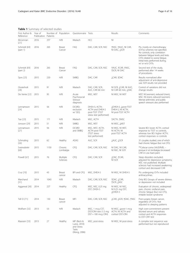
Table 1 Summary of selected studies
First Author &Reference
Year ofPublication
Number ofPatients
Population Questionnaire Tests Results Comments
McLennan[21]
2016 257 H/B Maslach HCC Nl
Schmidt [69](part 1)
2016 265 BreastCancer
FAQ DAC, CAR, SCR, NSC ↑NSC, ↑AUC, Nl CAR,Nl DAC, ↓SCR
Pts. mostly on chemotherapy(ChTx); schemes not specified.No controls, only correlationbetween fatigue levels and tests.ChTx related to worse fatigue.Initial tests performed during,w/ or w/o ChTx.
Schmidt [69](part 2)
2016 265 BreastCancer
FAQ DAC, CAR, SCR, NSC ↑AUC, ↑CAR, ↑NSC,↑SCR, Nl DAC
Second test of the study,performed after 14 weeksof procedures.
Sjors [25] 2015 220 H/B SMBQ DAC, CAR ↓CAR, ↑DAC Results normalized afteradjustment of anti-depressiveuse; SCR results not provided
Oosterholt[28]
2015 91 H/B Maslach DAC, CAR, SCR,AUC, CAR 60 min
Nl SCR, ↓CAR, Nl AUC,Nl CAR 60 min, ↓DAC
Control of variations did notchange results
De Vente [22] 2015 95 H/B AcutePsychosocialStressordiagnosis
MSC, MST Nl MSC, Nl MST MST: Nl (women), reduced (men);MSC: Nl (men), reduced (women).Mental arithmetic and publicspeech stressors also performed
Lennartsson[24]
2015 56 H/B DCSRD;exclusionw/ SEQ
DHEA-S; ACTH;ACTH and DHEA-Spost-TSST (TSSTstress test performed)
↓DHEA-S, ↓post-TSSTDHEA-S, Nl ACTH,Nl post-TSST ACTH
Tao [23] 2015 171 H/B Maslach MSC, ACTH ↑ACTH, ↑MSC
Jonsson [29] 2015 51 H/B SMBQ MSC, MST Nl MSC, ↓MST
Lennartsson[27]
2015 56 H/B DCSRDand SMBQ
MSC, MST, ACTH,ACTH post-TSST(TSST stresstest performed)
Nl MSC, Nl MST,Nl ACTH, Nlpost-TSST ACTH
Severe BO: lower ACTH, cortisolresponse to TSST vs controls,whereas low BO: higher ACTH,cortisol responses vs controls
Schmaling[26]
2015 62 Healthy ADAS AUC, SCR ↓AUC, ↓SCR 31 couples studied, one of whichhad chronic fatigue (but not CFS)
Sveinsdottir[68]
2015 1150 ChronicLombalgia
CFQ DAC, CAR, SCR, NSC Nl DAC, Nl CAR,Nl NSC, Nl SCR
**Colocar como SAUDÁVEL –porque é só lombalgia (increasedCAR in Low back pain)
Powell [67] 2015 76 MultipleSclerosis
CFQ DAC, CAR, SCR ↓DAC, ↑CAR,Nl SCR
Sleep disorders excluded;adjusted for depressive symptoms;NSC not published. Multiplesclerosis had increased awakeningcortisol and decreased CAR
Cruz [70] 2015 43 Breastcancer
BFI and CFQ MSC, DHEA-S Nl MSC, Nl DHEA-S Pts undergoing ChTx includedanthracyclines
Marchand[31]
2014 1043 H/B Maslach DAC, CAR, SCR, NSC ↑DAC, ↓CAR,↑SCR, ↓NSC
Only BO; Groups of severe distressor depression not included
Aggarwal [30] 2014 227 Healthy CFQ MSC, NSC, 0.25 mgDST, DHEA-S
Nl MSC, Nl NSC,Nl 0.25 mg DST,↓DHEA-S
Evaluation of chronic, widespreadpain, chronic orofacial pain,chronic fatigue (but not CFS),irritable bowel syndrome
Tell D [71] 2014 130 Breastcancer
MFI DAC, CAR, SCR, NSC ↓CAR, ↓SCR, ↑DAC, ↑NSC Post-surgery breast cancer,regardless of ChTx. Notadjusted to sleeping patterns
Wolfram [32] 2013 53 H/B Maslach MSC, 1 mcg CST,DST/CRH test (1.5 mgDST + 100 mcg CRH)
Nl MSC, ↓post-1 mcgACTH, Nl ACTH andcortisol DST-CRH
High over-commitment presentblunted serum and salivarycortisol and ACTH responsesto DST-CRH test
Klaassen [33] 2013 27 Healthy MP (Beck &Luine, 2010)and StressTasks(Wang, 2006)
MSC, post-stress Nl MSC, Nl post-stress A complex test sequence wasperformed but not reproduced
Cadegiani and Kater BMC Endocrine Disorders (2016) 16:48 Page 4 of 16

Table 1 Summary of selected studies (Continued)
Eek [34] 2012 581 Healthy SOFI-20 DAC, CAR, NSC,SCR, MSC
Nl DAC, Nl CAR, NlNSC, Nl SCR, Nl MSC
Women: reduced awakening,increased CAR, increased SCR;Men: increased awakening andreduced CAR – when fatigued
Sjors [35] 2012 247 H/B DCSRDand SMBQ
DAC, 15 min CAR Nl DAC, Nl 15 minCAR
Rahman [54] 2011 30 CFS PreviousDx – Noquestionnaire
MSC, SCR, NSC Nl MSC, Nl SCR,Nl NSC
Moya-Albiol[37]
2010 64 H/B Maslach DAC, CAR Nl DAC, Nl CAR
Kumari [38] 2009 4,299 Healthy SF-36 DAC, CAR, NSC, SCR ↓DAC, ↓CAR,↑NSC, ↓SCR
Adjusted for WC, BMI, sleepduration, CVD medication,depressive symptoms, smoking,alcohol intake provides Nlawakening but lower SCR
Osterberg[39]
2009 304 H/B Maslach DAC, CAR, NSC, SCR Nl DAC, Nl CAR,↓NSC, ↑SCR
0.5 mg DST was not comparedto controls
Wingenfeld[41]
2009 279 H/B Maslach andMaastricht
AUC, SCR Nl AUC, Nl SCR DAC and CAR not done;conclusions different from results.For AUC, Low BO: Nl, moderate:increased, severe: decreased
Rydstedt [40] 2009 76 Healthy NRWS DAC, NSC Nl DAC, Nl NSC
Papadopoulos[55]
2009 38 CFS CFQ andSF-36
MSC, AUC, morningAUC, 0,5 mg DST
↑MSC, ↑AUC, ↑MAUC,Nl 0.5 mg DST
Data on absolute cortisol levelsat each point not published.DST reduction evaluated bypercent reduction.
Bay [72] 2009 75 Posttraumaticbrain injury
POMS AUC Nl AUC Correlation between brain injury-related fatigue level and cortisolAUC. Basal and NSC results notreported; SCR not evaluated.
Sudhaus [73] 2009 43 ChronicLombalgia
MFI DAC, CAR, MAUC ↓CAR, Nl DAC, Nl MAUC(correlation betweenfatigue levels amonglow back pain subjects)
Lindeberg [36] 2008 78 Healthy SF-36 DAC, CAR, NSC, SCR Nl DAC, ↓CAR,Nl NSC, ↓SCR
Sertoz [42] 2008 72 H/B Maslach Basal and post1.0 mcg DST cortisol
Nl basal cortisol and1.0 mg DST
Bellingrath[43]
2008 101 H/B Maslach andMaastricht
DAC, CAR, NSC, SCR,0.25 mg DST
Nl DAC, Nl CAR, Nl NSC,Nl SCR, ↓0.25 mg DST
Nater [57] 2008 185 CFS SF-36and MFI
DAC, CAR, MAUC Nl DAC, Nl CAR, ↓MAUC
Torres-Harding[56]
2008 108 CFS FSE AUC, SCR Nl AUC, Nl SCR Multiple psychological testsperformed. Data on NSC,basal and CAR not published.
Sonnenschein[45]
2007 42 H/B Maslach CAR, 0.5 mg DST,DHEA-S
Nl CAR, Nl 0.5 mg DST,Nl DHEA-S
Adjusted for depression, sleepquality. Awakening levels andeach level graphics not available
Harris [44] 2007 44 Healthy SF-36 DAC, CAR, NSC, SCR Nl DAC, Nl CAR, Nl NSC,Nl SCR
Other aspects also correlated:complains, job stress anddemand, QOL and coping.Adjusted for coffee and tobacco.
Langelaan [46] 2006 55 H/B Maslach DAC, CAR, 0.5 mgDST, DHEA-S
Nl DAC, Nl CAR, Nl 0.5 mgDST, Nl DHEA-S
Engaged work also compared andhad stronger suppression in DST
Mommersteeg[47]
2006 109 Healthy NC-WHO DAC, CAR, 0.5 mgDST, SCR, AUC, NSC
Nl DAC, Nl CAR, Nl NSC, NlSCR, Nl 0.5 mg DST, Nl AUC
Barroso [74] 2006 40 HIV HRFS MSC, NSC ↓MSC, ↑NSC
Jerjes [58] 2006 80 CFS CFQ UFC, TCM ↓UFC, Nl TCM
Grossi [48] 2005 64 H/B SMBQ DAC, CAR ↓DAC, ↑CAR Groups were high x moderate x lowBO score; correlation was significant
Cadegiani and Kater BMC Endocrine Disorders (2016) 16:48 Page 5 of 16

Table 1 Summary of selected studies (Continued)
Segal [59] 2005 40 CFS Noquestionnaire
MSC, 1 mcg CST ↓ MSC, ↓1 mcg CST DHEA-S collected only in CFS.No questionnaires used.
Jerjes [60] 2005 35 CFS CFQ MSC, SCR, NSC, AUC ↓MSC, ↓SCR, ↓AUC ↓,Nl NSC
Bower [75] 2005 29 Breastcancer
SF-36 DAC, AUC, SCR, NSC ↑AUC, ↓SCR, Nl DAC,↑NSC
Post-ChTx (regardless of time)complete cancer remission andexclusion of other disorders
McLean [76] 2005 55 Fybro-mialgia
SF-36 DAC, 60 min CAR,SCR, AUC, NSC
Nl DAC, Nl 60 min CAR,Nl SCR, Nl AUC, Nl NSC(correlation betweenfatigue levels amongFMG subjects)
FMG subjects presented NlDAC and CAR, as controls.
Roberts [62] 2004 92 CFS CFQ andSF-36
DAC, CAR, MAUC Nl DAC, ↓CAR, ↓MAUC
Crofford [61] 2004 72 CFS/FMG POMS ACTH, MSC, SCR,NSC, AUC
Nl ACTH, Nl SCR,Nl NSC, ↓AUC, Nl MSC
Tests performed in: CFS, FMG andCFS + FMG; FMG w/o fatigue hadNl AUC and increased BMC levels
Moch [50] 2003 16 H/B Maslach UFC, DHEA-S,ACTH, MSC
↓UFC, Nl DHEA-S,Nl ACTH, ↓MSC
Only women; longitudinalevaluation – Nl initial cortisol.
De Vente [49] 2003 45 H/B Maslach DAC, MSC, post-TSST ↑DAC, ↑MSC, Nl post-TSST
Gaab [63] 2002 42 CFS MFI DAC, CAR, SCR,0.5 mg DST
↓0.5 mg DST, Nl AUC,Nl CAR, Nl DAC, Nl SCR,Nl NSC
CAR also performed at 15,45 and 60 min.
Dekkers [77] 2000 53 RheumatoidArthritis
MFI DAC, CAR, SCR, AUC Nl AUC, Nl SCR Nl,↓DAC, ↑CAR
5/25 subjects with RA takingprednisone (5–10 mg/d); RSsubjects had smaller SCR,increased AM cortisol anddecreased CAR. 15 and 45 minCAR also performed.
Melamed [51] 1999 111 H/B SMBQ andMaastricht
MSC and 4 PMcortisol
↑MSC, Nl 4 PM cortisol
Pruessner [52] 1999 66 Healthy Maslach DAC, CAR,0.5 mg DST
↓DAC, ↓CAR,↓0.5 mg DST
15 min and 60 min CARalso performed
Strickland [65] 1998 74 CFS Notspecified/detailed
MSC, NSC ↓NSC, Nl MSC Adjusted for depression
Young [66] 1998 45 CFS NC-WHO UFC, SCR, MSC, AUC Nl UFC, Nl SCR,Nl MSC, Nl AUC
Scott [64] 1998 28 CFS Not specified(not detailed)
MSC, ACTH, 100 mcgCRH cortisolstimulation
Nl MSC, Nl ACTH, CRHstim test: ↓cortisol, ↓ACTH
Raikkonen [53] 1996 22 Healthy Not assessed 1 mcg CST, DST(non specified),OGTT, MSC, ACTH,cortisol/ACTH ratio
↑Cortisol/ACTH ratio;↑CST, Nl DST, Nl OGTT,Nl MSC, Nl ACTH
Full article not assessed – notin PUBMED or other database
Questionnaires: SMBQ shirom-melamed burnout questionnaire, BFI brief fatigue inventory, CFQ chalder fatigue questionnaire, Maslach maslach burnout inventory,SF-36 short form health survey 36, NC-WHO neurasthenia criteria, DCSRD diagnosis criteria of stress-related exhaustion disorder, SEQ stress-energy questionnaire,ADAS, abbreviated dyadic adjustment scale, MFI multidimensional fatigue inventory, FAQ fatigue assessment questionnaire, MP memory performance,POMS profile of mood states, Stress Tasks, FSE fatigue severity scale, SOFI Swedish occupational fatigue inventory, Maastricht Maastricht vital exhaustionquestionnaire, NRWS need for recovery from work scale, HRFS HIV-related fatigue scale, WC waist circumferenceOther abbreviations: CFS chronic fatigue syndrome, H/B healthy/burnout, 24 h-UFC 24-h urinary free cortisol, FMG fibromyalgia; ↑: Increased or elevated;↓: Decreased or reduced; →: Unchanged; Nl: Normal
Cadegiani and Kater BMC Endocrine Disorders (2016) 16:48 Page 6 of 16
Study characteristicsAmong the 58 studies included, 33 (56.9 % of the selectedstudies) were performed in healthy subjects [21–53], sincewe considered “burnout” not an actual disorder but in-stead a stressful condition presented by some groups ofhealth workers. Despite the several studies describing cor-tisol impairment in Chronic Fatigue Syndrome (CFS), only13 (22.4 %) studies performed an actual assessment of
the hypothalamic–pituitary–adrenal (HPA) axis [54–66].Twelve studies (20.7 %) were found in which tests for cor-tisol profiling were performed for other diseases [67–77].However, for analysis purposes, one study [69] was dividedinto two studies as it performed two distinct protocols atdifferent moments. Among these, five were done per-formed in patients with a diagnosis of breast cancer whohad undergone or were undergoing chemotherapy. One

Cadegiani and Kater BMC Endocrine Disorders (2016) 16:48 Page 7 of 16
study tested patients with fibromyalgia, two studiescompared patients with chronic lower pain, one withrheumatoid arthritis, one with post brain injury, twowith multiple sclerosis, and one involved patients withhuman immunodeficiency virus (HIV) and CFS. Onestudy evaluated both patients with fibromyalgia and pa-tients with CFS in different groups.The median number of tested subjects in the 58 studies
was 72 (range: 16–4,299). The median numbers ofparticipants in articles involving healthy individuals,patients with CFS, or patients with other diseaseswere 76 (16–4,299), 45 (28–185), and 65 (29–1150),respectively. The largest number of healthy subjectsincluded groups of workers whose cortisol resultswere compared to exhaustion and fatigue status, in anattempt to discriminate correlations between cortisoland energy levels. One study involving 4,299 individ-uals was responsible for more subjects than the sumof all the other studies.
Methods used to evaluate fatigue in the general studypopulationSome authors utilized more than one method to com-pare the different patients and were included in multiplegroups. A summary of all the methods used to assess fa-tigue, and their results, is shown in Table 2. Among the58 studies, 27 (46.6 %) utilized the Cortisol AwakeningResponse (CAR) to assess the HPA axis. This method isbased on previous studies [77–81] that indicate cortisollevels rise by 50 % on average within 30 min of wakingas a physiological response to stay alert, with a bluntedCAR resulting in fatigue symptoms. For the CAR, saliv-ary cortisol is collected immediately on waking (t = 0)and again 30 min later (t = 30), and the difference (deltacortisol) between the two measurements are analyzed.Among the 27 studies that employed CAR, fourteen(51.9 %) showed a normal response, nine (23.3 %) had adiminished delta cortisol, and four (14.8 %) demon-strated an increased delta cortisol.Another method that became widely used to evaluate
exhaustion/burnout/fatigue states is the salivary cortisolrhythm (SCR), which evaluates the change in cortisollevels between morning, afternoon, and late night. Atotal of 26 studies evaluated SCR (44.8 %). Some hetero-geneity in the method was found between studies, but ingeneral, salivary cortisol was collected at 8 AM, 4 PM,and 10–11 PM. While the SCR is considered as anotherfatigue marker [82, 83], like the CAR, there is no justifi-cation for considering this as an etiology for “adrenal fa-tigue”. Sixteen (61.5 %) studies showed no differencebetween fatigued and control patients, whereas seven(26.9 %) demonstrated an impaired decrease in the circa-dian SCR. The remaining three (11.6 %) studies dis-closed a more pronounced decrease in cortisol level.
The direct awakening cortisol (DAC) level, collected atthe exact moment of waking, was used in 29 studies(50.0 %). Unlike CAR, DAC reflects sleep quality ra-ther than being a possible identifying factor of fatigue[84–86], even though a poor quality sleep plays an im-portant role in the fatigue process [87–89]. In studiesthat employed DAC, inconsistent results were ob-served: normal results were found in nineteen (65.5 %)studies, elevated levels were shown in four (13.8 %),and reduced levels in six (20.7 %).The DAC, CAR and SCR methods were by far the
most commonly elected ones for examining the cor-relation between cortisol profile and fatigue status.However, a few other studies analyzed other aspects ofcortisol release.The dexamethasone (Dex) suppression test (DST) was
also used in nine (15.3 %) studies. The DST identifiesautonomous hypercortisolism, as cortisol production isnormally suppressed by Dex. DSTs have also been usedto investigate hypocortisolism, based on the supposedassumption that it promotes “oversuppression” of corti-sol in low cortisol states, indicating that lower levels ofcortisol would disclose a more prolonged suppressionthan controls [55, 90–93], although many studies do notshow correlation between DST and fatigue [47, 55, 94, 95].In six studies, a lower Dex dose (0.5 mg) was used in anattempt to improve the test sensitivity. Among these, fourstudies (66.7 %) showed the same results for both groups,whereas in two others (33.3 %), the test resulted in lowerand prolonged suppression of cortisol levels in fatiguedsubjects. Moreover, an even lower Dex dose (0.25 mg) wasperformed in two studies and resulted in reduced cortisolin one study and normal levels in the group with exhaus-tion. In one study, Dex dose was not specified, but levelswere not different among exhausted and control groups.As a whole, the DST was used in nine studies, and nosignificant differences were observed between fatiguedand non-fatigued groups in six of these studies(66.7 %), whereas reduced levels were observed inthree studies (33.3 %).Adrenocorticotropic hormone (ACTH) is a pituitary
peptide hormone that stimulates cortisol productionby the adrenocortical zona fasciculata. Elevated ACTHoccurs early in primary adrenal insufficiency, whereasinappropriate (normal) ACTH levels in the presence oflow serum cortisol are found in secondary adrenal fail-ure. Although, normal ACTH levels with normal corti-sol levels does not exclude the possibility of relativeadrenocortical failure. Six (10.3 %) studies employedthe morning ACTH levels to compare fatigued andnon-fatigued patients; no significant differences forACTH, as well as for cortisol, were found in five stud-ies (83.3 %), meanwhile one showed elevated ACTHlevels in burnout patients (16.7 %).

Table 2 Assessed methods and results of all selected studies (N = 58)
Procedure (*) Number of studies (% of total) Not different (%) Decreased (%) Increased (%)
DAC 29 (50.0 %) 19 (65.5 %) 6 (20.7 %) 4 (13.8 %)
CAR 27 (46.6 %) 14 (51.9 %) 9 (33.3 %) 4 (14.8 %)
SCR 26 (44.8 %) 16 (61.5 %) 7 (26.9 %) 3 (11.5 %)
MSC 22 (37.9 %) 14 (63.6 %) 4 (18.2 %) 4 (18.2 %)
NSC 22 (37.9 %) 13 (59.1 %) 3 (13.6 %) 6 (27.3 %)
AUC 13 (22.4 %) 8 (61.5 %) 3 (23.1 %) 2 (15.4 %)
DST 9 (15.5 %) 6 (66.7 %) 3 (33.3 %) -
DHEA-S 6 (10.3 %) 4 (66.7 %) 2 (33.3 %) -
ACTH 6 (10.3 %) 5 (83.3 %) - 1 (16.7 %)
MST 5 (8.6 %) 4 (80.0 %) 1 (20.0 %) -
UFC 3 (5.2 %) 1 (33.3 %) 2 (66.7 %) -
CST 3 (5.2 %) - 2 (66.7 %) 1 (33.3 %)
MAUC 3 (5.2 %) - 2 (66.7 %) 1 (33.3 %)
CAR 60 min 2 (3.4 %) 2 (100 %) - -
ACTH MST 2 (3.4 %) 2 (100 %) - -
4 PM cortisol 1 1 - -
DST + CRH cortisol 1 1 - -
DST + CRH ACTH 1 1 - -
CAR 15 min 1 1 - -
TCM 1 1 - -
DHEA-S MST 1 - 1 -
CRST cortisol 1 - 1 -
CRST ACTH 1 - 1 -
OGTT cortisol 1 - - 1
Cortisol/ACTH ratio 1 - - 1
Legends: (*): DAC direct awakening cortisol, CAR cortisol awakening response, SCR salivary cortisol rhythm, MSC morning serum (& salivary) cortisol, NSC nightsalivary cortisol, AUC area under-the-curve (Estimated Cortisol Release), DST dexamethasone suppression test, DHEA-S dehydroepiandrosterone sulfate, ACTHadrenocorticotropic hormone, MST mental stress test, UFC 24 h-urinary free cortisol, CST cosyntropin stimulation test, MAUC morning area under-the-curve(morning estimated cortisol release), CRH corticotropin releasing hormone, TCM total urinary cortisol metabolites, CRST corticotropin releasing stimulationtest (?), OGTT oral glucose tolerance test
Cadegiani and Kater BMC Endocrine Disorders (2016) 16:48 Page 8 of 16
On the other hand, three studies (5.2 %) used the low-dose cosyntropin (a synthetic 1-24ACTH) stimulation test(CST), in which 1 μg of cosyntropin is used instead ofthe classic 250 μg dose, based on the premise that theCST is more accurate and sensitive for verifying theadrenocortical cortisol reserve [96], even though mostfindings indicate that both doses have similar accuracy[97, 98]. Surprisingly, one of three (33.3 %) studies dis-closed a paradoxically higher cortisol increase comparedto controls, while in two (66.7 %) lower levels were ob-served. Conversely, impaired cortisol and ACTH re-sponses was observed in the fatigued group in a singlestudy in which corticotropin-releasing hormone (CRH)was used to stimulate the HPA axis.Three (5.2 %) studies measured 24 h-urinary free cor-
tisol (UFC) in an attempt to correlate cortisol excretionrates with intensity of fatigue. Although the 24 h-UFCreflects the total cortisol produced per day, it was
initially conceived to investigate cortisol excess syn-dromes, although diminished levels could hypothetic-ally imply subnormal adrenal function, despite of lackof any evidence. One of these studies (33.3 %) found nocorrelation between 24 h-UFC and energy status,whereas two studies (66.7 %) showed reduced values infatigued patients.Thirteen studies (22.4 %) estimated total cortisol re-
lease (AUC) by calculating the areas under the curvesfor the whole day salivary cortisol collection by usingthree or more daily salivary cortisol levels over four ormore days. Assessment of the total 24 h cortisol releaseby this method would complement the SCR, since thelack of the expected decrease throughout the day ob-served in some studies can be due either to a non-elevated morning serum cortisol (MSC) level or to a fullday elevated cortisol, although three daily levels of corti-sol is probably too few for a minimally precise AUC;

Cadegiani and Kater BMC Endocrine Disorders (2016) 16:48 Page 9 of 16
herein, findings are conflicting. AUC was elevated in two(15.4 %) studies, normal in eight (61.5 %), and reducedin another three (23.1 %).Twenty-two studies (37.9 %) compared baseline MSC
between controls and fatigued patients; traditionally[98], this is the initial cortisol assessment to investigatepossible hypocortisolism. Basal MSC was not differentbetween individuals in fourteen (63.6 %) of these studies,was significantly reduced in fatigued patients in three(23.1 %), and was elevated in two (15.4 %).Twenty-two articles (37.9 %) correlated late night sal-
ivary cortisol (11 PM NSC) and fatigue status. TheNSC was initially validated to assess cortisol excess, asphysiologically, one expects lower cortisol levels at theend of the day; although, NSC has been extended to in-vestigate hypocortisolism in these studies, despite oflack of validation. Three studies (13.6 %) showed alower cortisol level in fatigued subjects compared tocontrols, thirteen (59.1 %) found no differences, and six(27.3 %) showed increased levels in fatigued subjects.Six studies (10.3 %) investigated the correlation be-
tween dehydroepiandrosterone sulfate (DHEA-S) levelsand fatigue status. Reduced DHEA-S levels are usuallyfound in hypocortisolism and are a potential marker offatigue, although there is still not enough evidence tocorroborate this affirmation. Four studies (66.7 %) foundno correlation with DHEA-S, whereas two (33.3 %)found lower levels in chronic exhausted patients.The morning estimated total cortisol release (MAUC)
is obtained by calculating the area under the curves forthe period between the awakening moment and 1 hourlater, and is based on determining three or more salivarycortisol levels during this period of the day, althoughthis method has also not been validated by any indexedstudy. A total of four studies (6.9 %) among the selectedstudies reported the MAUC. Two of these studies(50.0 %) showed reduced MAUC levels in fatigued sub-jects, one demonstrated increased results (25.0 %), andone demonstrated no differences (25.0 %).Mental stress tests (MST) have been performed in
some studies in order to identify possible differences incortisol and ACTH release between fatigued and non-fatigued individuals. The most employed test was the TrierSocial Stress Test (TSST), which has been already vali-dated as a stress trigger test [99–102], and requirescomplete HPA axis integrity for a proper response. Othertypes of MSTs have also been proposed and validated[103, 104]. MSTs were performed in five different studiesin order to correlate cortisol and ACTH responses andburnout status. No difference was seen in four studies(80.0 %), whereas in one (20.0 %), cortisol and ACTHresponses were impaired in exhausted individuals.Some other tests were performed in a smaller number
of the selected studies, as follows: two studies performed
a 60 min CAR (both showed normal results among fa-tigued and non-fatigued subjects); one study performed a15 min CAR (and showed normal results); two studiesperformed the ACTH MST (both used the TSST andfound normal results); one study performed the DHEA-SMST (which also used the TSST and demonstrated normalresults); one study performed the cortisol post Oral Glu-cose Tolerance Test (OGTT) (and found no differencesamong fatigued and non-fatigued subjects); one study cal-culated cortisol/ACTH ratio (and found an increased ratioamong exhausted subjects); one study evaluated the 4 PMcortisol level (and found no significant differences betweenexhausted subjects and controls); one study used 1.5 mg-Dex followed by 0.1 mg-CRH to stimulate cortisol andACTH (and showed normal responses); one study stimu-lated ACTH and cortisol with 0.1 mg of CRH (and foundreduced levels of both hormones in fatigued subjects com-pared to controls); and finally, one study evaluated themultiple urinary cortisol metabolites and calculated theTotal Cortisol Metabolites (TCM) (and found no differ-ences between fatigued subjects and controls).Finally, we were not able to find studies in which the
gold standard test for assessing the integrity and func-tionality of the HPA axis—the insulin tolerance test(ITT)—were performed. The same was true for the lipo-polysaccharides (LPS) stimulation test. Both tests stimu-late hypothalamic CRH secretion, leading to a completeevaluation of the HPA axis.
Fatigue in burnout syndromeBurnout syndrome or clinical burnout, or simply “burn-out”, refers to a decrease in the cognitive functions, emo-tional exhaustion, and physical fatigue that is triggered bystressful situations associated with excessive working[105]. However, there is no pathognomonic marker forburnout [105]. For practical purposes, we considered non-CFS burnout patients as “healthy”, as burnout is yet to beconsidered a disease and its characterization is still hetero-geneous. A summary of the performed methods and theirrespective results in non-CFS burnout/healthy patients[21–53] are shown in Table 3. Assessment of the HPA axisintegrity in burnout patients (at the pituitary and hypo-thalamic levels) has not been determined.
Fatigue in chronic fatigue syndromeCFS is a diagnosis used for patients who present severefatigue for more than six months, not explained by anyhormonal, metabolic, inflammatory, or other disorders.Correlations between CFS and the HPA axis have beenstudied [54–66] and the results are shown in Table 4.
Fatigue in other disordersComplaints regarding fatigue not entirely explained bythe underlying pathophysiology of the disease have been

Table 3 Studies in Burnout syndrome and healthy subjects (N = 33): Methods of assessment and respective results
Procedure Number of studies (% of total) Not different (%) Decreased (%) Increased (%)
DAC 17 (51.5 %) 10 (58.8 %) 4 (23.5 %) 3 (17.7 %)
CAR 16 (48.5 %) 9 (56.2 %) 6 (37.5 %) 1 (6.3 %)
MSC 12 (36.4 %) 8 (66.7 %) 1 (8.3 %) 3 (25.0 %)
SCR 12 (36.4 %) 7 (58.3 %) 3 (25.0 %) 2 (16.7 %)
NSC 10 (30.3 %) 7 (70.0 %) 2 (20.0 %) 1 (10.0 %)
DST 7 (21.2 %) 5 (71.4 %) 2 (28.6 %) -
MST 5 (15.2 %) 4 (80.0 %) 1 (20.0 %) -
DHEA-S 5 (15.2 %) 3 (60.0 %) 2 (40.0 %) -
ACTH 4 (10.1 %) 3 (75.0 %) - 1 (25.0 %)
AUC 3 (9.1 %) 2 (66.7 %) 1 (33.3 %) -
ACTH MST 2 (6.1 %) 2 (100 %) - -
CST 2 (6.1 %) - 1 (50 %) 1 (50 %)
4 PM cortisol 1 1 - -
DST + CRH cortisol 1 1 - -
DST + CRH ACTH 1 1 - -
CAR 15 min 1 1 - -
CAR 60 min 1 1 - -
DHEA-S MST 1 - 1 -
UFC 1 - 1 -
OGTT cortisol 1 - - 1
Cortisol/ACTH ratio 1 - - 1
Legends: (*): DAC direct awakening cortisol, CAR cortisol awakening response, MSC morning serum (& salivary) cortisol, SCR salivary cortisol rhythm, NSC nightsalivary cortisol, DST dexamethasone suppression test, MST mental stress test, DHEA-S dehydroepiandrosterone sulfate, ACTH adrenocorticotropic hormone,AUC area under-the-curve (estimated cortisol release), CST cosyntropin stimulation test, CRH corticotropin releasing hormone, UFC 24 h-urinary free cortisol,OGTT oral glucose tolerance test
Cadegiani and Kater BMC Endocrine Disorders (2016) 16:48 Page 10 of 16
observed in patients suffering from other disorders,such as chronic low back pain [106, 107], breast cancersurvivors [108–110], and HIV [111, 112]. Therefore,the role of the HPA axis in the etiology of fatigue inthese subjects has been analyzed [67–77] and the find-ings are presented in Table 5.
Table 4 Studies in Chronic Fatigue Syndrome (N = 13): Methods of
Procedure Number of studies (% of total) Not
MSC 8 (61.5 %) 5 (6
SCR 6 (46.2 %) 5 (8
AUC 6 (46.2 %) 3 (5
NSC 5 (38.5 %) 4 (8
DAC 3 (23.1 %) 3 (1
CAR 3 (23.1 %) 2 (6
ACTH 2 (15.4 %) 2 (1
DST 2 (15.4 %) 1 (5
UFC 2 (15.4 %) 1 (5
CST 1 (7.7 %) -
Legends: (*): MSC morning serum (& salivary) cortisol, SCR salivary cortisol rhythm, ADAC direct awakening cortisol, CAR cortisol awakening response, ACTH adrenocorticcortisol, CST cosyntropin stimulation test
Questionnaires for fatigue assessmentAmong all studies included in this review, nineteendifferent types of questionnaires and scores were re-ported. The most commonly used were: the MaslachBurnout Inventory (MBI, n = 15), SF-36 (n = 9), theChalder Fatigue Scale (CFS, n = 8), the General
assessment and respective results
different (%) Decreased (%) Increased (%)
2.5 %) 2 (25.0 %) 1 (12.5 %)
3.3 %) 1 (16.7 %) -
0.0 %) 2 (33.3 %) 1 (16.7 %)
0.0 %) 1 (20.0 %)
00.0 %) - -
6.7 %) 1 (33.3 %) -
00.0 %) - -
0.0 %) 1 (50.0 %) -
0.0 %) 1 (50.0 %) -
1 (100.0 %) -
UC area under-the-curve (estimated cortisol release), NSC night salivary cortisol,otropic hormone, DST dexamethasone suppression test, UFC 24 h-urinary free

Table 5 Studies in Other Disorders (N = 12): Methods of and respective results
Procedure Number of studies (% of total studies) Not different (%) Decreased (%) Increased (%)
DAC 9 (75.0 %) 6 (66.7 %) 2 (22.2 %) 1 (11.1 %)
SCR 8 (66.7 %) 4 (50.0 %) 3 (37.5 %) 1 (12.5 %)
CAR 8 (66.7 %) 3 (37.5 %) 2 (25.0 %) 3 (37.5 %)
NSC 7 (58.3 %) 2 (28.6 %) - 5 (71.4 %)
AUC 4 (33.3 %) 3 (75.0 %) - 1 (25.0 %)
MSC 2 (16.7 %) 1 (50.0 %) 1 (50.0 %) -
DHEA-S 1 (8.3 %) 1 (100.0 %) - -
Legends: (*): DAC direct awakening cortisol, SCR salivary cortisol rhythm, CAR cortisol awakening response, NSC night salivary cortisol, AUC area under-the-curve(estimated cortisol release), MSC morning serum (& salivary) cortisol, DHEA-S Dehydroepiandrosterone sulfate
Cadegiani and Kater BMC Endocrine Disorders (2016) 16:48 Page 11 of 16
Fatigue Scale of the Multidimensional Fatigue Inven-tory (MFI, n = 6) and the Shirom Melamed BurnoutQuestionnaire (n = 6). In ten studies, more than onetype of survey was performed. In four studies, themethods to assess fatigue were not specified orassessed. A summary of the assessed questionnaires isshown in Table 6.
Table 6 Assessed questionnaires employed in the selected studies (
Questionnaire Gene
Maslach Burnout Inventory 15 (2
SF-36-Short Form Health Survey 36 9 (15
CFQ-Chalder Fatigue Questionnaire 8 (13
SMBQ - Shirom-Melamed Burnout Questionnaire 6 (10
MFI-Multidimensional Fatigue Inventory 6 (10
DCSRD: Diagnosis criteria of stress-related exhaustion disorder 3 (5.
Maastricht Vital Exhaustion Questionnaire 2 (3.
FAQ-Fatigue Assessment Questionnaire 2 (3.
NC-WHO-Neurasthenia Criteria 2 (3.
POMS-Profile of Mood States 2 (3.
SOFI-Swedish Occupational Fatigue Inventory 1
ADAS-Abbreviated Dyadic Adjustment Scale 1
SEQ-Exclusion with Stress-Energy Questionnaire 1
MP-Memory performance 1
Stress Tasks 1
NRWS - Need for Recovery from Work Scale 1
FSE-Fatigue Severity Scale 1
HRFS-HIV-related Fatigue Scale 1
BFI: Brief Fatigue Inventory 1
More than one questionnaire 10 (1
Stress tests 5 (8.
Not specified 4 (6.
Legends: Maslach maslach burnout inventory, SF-36 short form health survey 36, CFQ cMFI multidimensional fatigue inventory, DCSRD diagnosis criteria of stress-relatedFAQ fatigue assessment questionnaire, NC-WHO neurasthenia criteria, POMS profile ofdyadic adjustment scale, SEQ stress-energy questionnaire, MP memory performance, SHRFS HIV-related fatigue scale, BFI brief fatigue inventory
DiscussionTheories on adrenal impairment as the genesis forfatigue are tempting, as they allow for a treatable condi-tion. Despite the widespread use of the term “adrenalfatigue” by the general media and certain health practi-tioner groups, in this systematic review, only ten cita-tions [113–122] were found with this exact expression,
N = 58)
ral Healthy/Burnout CFS Other diseases
5.7 %) 15 - -
.5 %) 4 3 2
.8 %) 1 4 3
.3 %) 6 - -
.3 %) - 2 4
2 %) 3 - -
4 %) 2 - -
4 %) - - 2
4 %) 1 1 -
4 %) - 1 1
1 - -
1 - -
1 - -
1 - -
1 - -
1 - -
- 1 -
- - 1
- - 1
7.2 %) 6 3 1
6 %) 5 - -
9 %) - 4
halder fatigue questionnaire, SMBQ shirom-melamed burnout questionnaire,exhaustion disorder, Maastricht Maastricht vital exhaustion questionnaire,mood states, SOFI Swedish occupational fatigue inventory, ADAS abbreviatedtress Tasks; NRWS need for recovery from work scale, FSE fatigue severity scale,

Cadegiani and Kater BMC Endocrine Disorders (2016) 16:48 Page 12 of 16
and they were all only descriptive and did not performany test regarding the HPA axis and “adrenal fatigue”.Studies that tried to correlate the HPA axis and fatiguestates used the term “burnout” instead of “adrenal fatigue”to denote adrenal depletion. Therefore, a distinctionbetween the “general information” and the actual scien-tific literature regarding this condition is evident. First,this suggests that the terminology of a hypotheticaladrenal depletion should be normalized, with a suitablename given for the purported condition, as “adrenalfatigue” has been already been stigmatized and lacksproper scientific support. Second, methodology employedto evaluate the proposed correlation between fatigue andadrenal function should be standardized among physiciansand medical associations that claim for the existence ofadrenal impairment in patients with fatigue before evidentclinical hypocortisolism manifests, in order to strengtheventual evidence, in case one finds actual and propercausal correlation.No confirmed methods of clinical screening for AF are
available. Indeed, the popular questionnaire developedby Dr. Wilson and published in the first book exclusivelydedicated to the description of this supposedly disease[6] has not been cited in any indexed databases. Anothertheory, the “Thompson cortisol hypothesis” [123], sug-gests that cortisol is responsible for yawning and fatigue;however, again, no studies that tested this theory havebeen published in indexed journals. Validated surveyshave been used in studies that investigate fatigue states,but they were not correlated with proper cortisol assess-ment methods. The TSST is the only survey to haveenough credibility to be officially tested and standardizedas a trigger of stress [99–102].Functional tests are the only methods to assess adrenal
cortisol production endorsed by endocrinology societies[97]. Although, the ITT is considered the gold standardtest to evaluate the entire HPA axis, neither the ITT (orthe similar LPS stimulation test) was performed in anystudies investigating the correlation between fatiguestates and adrenocortical function. Moreover, we gener-ally found conflicting data using most of the functionaltests when trying to differentiate exhausted, fatigued,and burnout individuals from healthy patients. For ex-ample, using the low-dose CST, we found an unexpectedincrease in cortisol levels in fatigued subjects in theselected studies. This may have been perhaps the resultof a relative secondary adrenal insufficiency, which leadsto an amplified adrenal cortisol response due to an up-regulation of ACTH receptors, but this sounds unjustifi-able since the lack of continuous stimulation of theadrenal cortices would cause atrophy, rendering themnon-responsive to a low- (and even high) dose of cosyn-tropin stimulation in the long run. Regardless of the the-oretical explanation, CST has shown to be not a good
marker of fatigue. Similarly, ACTH levels were alsopoorly studied and did not show significant correlationsin most fatigued subjects. In addition, despite its lack ofstandardization, the DST was performed in nine studies,but conflicting results invalidated attempts to establishthis as a new marker for fatigue states. Moreover, the24 h-UFC has been shown to be so far inaccurate forinvestigation of adrenal impairment. Findings were alsocontradictory in the six studies that calculated cortisolAUC as well as in the four studies that performedMAUC. Therefore, the above methods cannot be used todifferentiate fatigued from non-fatigued individuals.In this review, we also examined whether cortisol
markers can be used to assess cortisol impairment. Theresults of our review indicate that the three major tests(CAR, DAC and SCR) used to identify the underlyingcauses of the fatigue/exhaustion state failed to do so, sincethey were unable to demonstrate significant differences orproper causality. CAR and DAC frequently showed incon-sistent results in studies that used heterogeneous groupsof subjects. CAR and DAC are not necessarily indicativesof the etiology and pathogenesis of the fatigue status, sinceboth can be consequences of other disorders, such assleep disturbances. Indeed, a recent study [124] was thefirst to use CAR as a marker of improvement of burnoutsyndrome, which reinforces the use of this method formonitoring the consequences of fatigue states, but not forits etiology [77–81].With regards to the SCR, the results may be misleading
if they are not analyzed together with the total 24 h corti-sol release. This is because a non-physiological bluntedrhythm can be due either to an impairment of the lower-ing cortisol trend throughout the day or due to a lowermorning cortisol level. Despite this, studies that evaluatedtotal 24 h cortisol by measuring serial salivary cortisollevels also showed conflicting findings. Our systematicreview corroborates another systematic review [83] thatshows inconsistency regarding measuring methods amongacross different randomized controlled trials. Similarly,baseline MSC and NSC were poor markers of fatigue sta-tus as it failed to reveal any differences in burnout/exhaus-tion/fatigue patients compared to healthy subjects.Adrenal size could be considered another marker of
adrenal activity, as hypertrophic/hyperplastic adrenalglands could be the result of an ACTH over-stimulationby the pituitary, as seen in subjects exposed to chronicstress [125, 126], whereas a diminished or atrophic glandmay reflect adrenal insufficiency at any level of the HPAaxis [98]. However, not a single study could be identifiedin which the adrenal size has been checked in fatiguedor exhausted patients. Similarly, although DHEA-Scould also be a potential marker for adrenal atrophy ordysfunction, is still uncertain whether it plays anypathophysiological role in fatigue. Finally, none of the

Cadegiani and Kater BMC Endocrine Disorders (2016) 16:48 Page 13 of 16
abovementioned methods were accurate markers of fa-tigue, nor could they be correlated with the HPA axisdysfunction as an etiology of fatigue.It is also important to note that once adrenal impair-
ment is confirmed using any of these tests, the etiologyshould also be elucidated. As the HPA axis can beaffected by several chronic and/or metabolic disorders,other primary conditions must be excluded beforeintrinsic disorders of the HPA axis are deemed respon-sible. Typical differential diagnosis of “adrenal fatigue”and related states are: (1) sleep obstructive apnea syn-drome; (2) adrenal insufficiency; (3) mental illnesses; (4)excessive working (overwork); (5) night-shift workers; (6)other hormonal deficiencies; (7) liver and kidney dys-functions; (8) heart conditions; (9) chronic pulmonaryobstructive disease; (10) autoimmune diseases.Although conflicting data were reported, patients with
CFS tend to have a normal cortisol profile, and the ab-normalities found can be typically be explained by apoor quality sleeping patterns. Therefore, health pro-viders should not be concerned about adrenal functionin CFS subjects once they had been already excluded toother conditions prior to the diagnosis of CFS. Similarly,studies investigating patients with the burnout syndromewere greatly inconsistent So far, HPA axis tests shouldnot be used as markers for burnout syndrome by healthpractitioners. Similar conclusions can be drawn for theuse of HPA axis tests as markers for fibromyalgia andother chronic diseases, which tend to demonstrate in-consistent findings, whereas studies that were performedin breast cancer subjects tended to show depletion ofcortisol levels; however, studies in breast cancer wereperformed while administering chemotherapy, whichcan introduce a confounding bias.Therefore, based on our current knowledge, cortisol
tests should not yet be used in clinical practice forexamining any condition, except if adrenal impairmentis suspected. Moreover, glucocorticoid therapy should beavoided in patients, as it can increase the risk of cardio-vascular disease or osteoporosis, even in low doses.
LimitationsSome limitations of this review include: (1) our inabilityto perform a meta-analysis due to heterogeneity of thestudy design; (2) the descriptive nature of most studies,and the reporting of a condition that has not been scientif-ically proven without adding new data nor providing solidarguments; (3) the fact that most studies were publishedin low impact journals; (4) the inadequate and poor qual-ity assessment of fatigue; (5) the use of an unsubstantiatedmethodology in terms of cortisol assessment; (6) the lackof concern regarding validated adrenal assessment (as en-dorsed by endocrinologists); (7) false premises leading toan incorrect sequence of thinking and research direction;
and, (8) inappropriate/invalid conclusions regarding caus-ality and association between different information, inparticular, whether any abnormalities would be amarker or a potential target for treatment.
Final discussionsOur results corroborate an Endocrine Society warningstatement regarding adrenal fatigue (1), as saying that“adrenal fatigue is not a real medical condition”. While arecent systematic review on burnout was published(109) that implicated some HPA dysfunctions as markersor triggers of burnout, there were important bias selectionregarding the articles chosen. Therefore, we recommendthat for further prospective studies aiming to correlate fa-tigue, exhaustion, or burnout status with impairment of theHPA axis, an ITT or a 250 μg CST should be performed toevaluate the adrenocortical ability to release cortisol,measurements of ACTH, DHEA-S, and corticosterone (anintermediate steroid product that is impaired earlier thancortisol [127]), the adoption of the most validated question-naires, particularly Maslach Burnout Inventory, the ChalderFatigue Scale, SF-36 or the General Fatigue Scale of theMultidimensional Fatigue Inventor, and considering differ-ent study populations, including: (a) healthy subjects; (b)burnout healthy subjects; (c) subjects with overtrainingsyndrome; (d) subjects post-chemotherapy; (e) subjectswith CFS; and (f) subjects with fibromyalgia.In addition, we do not recommend the use of the
many methods reported in the articles evaluated in thissystematic review, as they are not accurate to determinewhether a patient has or has not adrenal failure.The answer to whether “adrenal fatigue” or depletion
exists or not may not be simple, but different answerscan be offered according to the presence of an under-lying disease. However, so far, there is no substantiationto show its existence.
ConclusionTo our knowledge, this is the first systematic reviewmade by endocrinologists to examine a possible correl-ation between the HPA axis and a purported “adrenalfatigue” and other conditions associated with fatigue,exhaustion or burnout. So far, there is no proof or dem-onstration of the existence of “AF”. While a significantnumber of the reported studies showed differencesbetween the healthy and fatigued groups, importantmethodological issues and confounding factors wereapparent. Two concluding remarks emerge from thissystematic review: (1) the results of previous studieswere contradictory using all the methods for assessingfatigue and the HPA axis, and (2) the most appropriatemethods to assess the HPA axis were not used to evaluatefatigue. Therefore, “AF” requires further investigationby those who claim for its existence.

Cadegiani and Kater BMC Endocrine Disorders (2016) 16:48 Page 14 of 16
AcknowledgementsWe acknowledge the support of the adrenal team of Endocrinology Unit ofFederal University of Sao Paulo: Flavia Barbosa, MD, PhD; Regina do Carmo,MD, PhD; Marcelo Vieira, MD; Rafaela Fontenele, MD and Denise Farinelle,MD, who helped giving some ideas regarding the most important aspects tobe discussed in the systematic review. All them are aware of the inclusion oftheir names in this section.
FundingNo funding was obtained for the research for the design strategy, forcollection, analysis and interpretation of data and for writing the manuscript.
Availability of data and materialsSearched studies that were not open access were assessed using CAPES/CNPqand Federal University of Sao Paulo bases, which were responsible for providingall the supplied data to all MEDLINE (Ebsco), PubMed and Cochrane databases.
Authors’ contributionsFAC performed the research and selection of the studies, analyzed theresults, detailed each selected study in the Table 1, calculated the results andperformed part of the discussions and conclusions. CEK participated in thedesign of the systematic review, including selection of the searchedexpressions and the databases, including and excluding criteria, qualityassessmentand statistical analysis; CEK also performed part of the discussion,the conclusions and helped to organize the sequence of the systematicreview. All authors read and approved the final manuscript.
Competing interestsThe authors declare that they have no competing interests regarding financialand non-financial aspects.
Consent for publicationWe declare that the consent for publication is not applicable to this study.
Ethics approval and consent to participateThis systematic review did not include direct human material or data, butindirectly used human subjects from the selected studies. All the selectedstudies were supposed to have an explicit statement of approval of anappropriate ethics committee in order to be included, as required fromthe ethics committee of the Federal University of Sao Paulo. Therefore,we declare that ethics approval and consent to participate is not applicableto this study.
Received: 25 June 2016 Accepted: 11 August 2016
References1. The Endocrine Society. Hormone Health Netowrk. Hormones and Health.
Myth vs Fact. Adrenal Fatigue. Available from : http://www.hormone.org/hormones-and-health/myth-vs-fact/adrenal-fatigue. Accessed 23 Aug 2016.
2. Association of American Medical Colleges. Careers in Medicine. List ofSpecialities. Avaliable from: https://www.aamc.org/cim/specialty/list/us/.Accessed 23 Aug 2016.
3. American Board of Medical Specialities. Avaliable fromL http://www.abms.org/.Accessed 23 Aug 2016.
4. The American Academy of Anti-Aging Medicine. Beyond Adrenal Fatigue:From Anedecdotal to Evidence Based Medicine. Avaliable from:http://www.a4m.com/assets/pdf/medical-news/A4M_Hypocortisolism_paper_draft_3-Final.pdf. Accessed 23 Aug 2016.
5. Functional Medicine Institute. Adrenal Fatigue. https://www.functionalmedicine.org/content_management/files/AFMCPSeptember2011/9_Onsite%20Course%20Materials/Thursday/Lukaczer%20Panico_Thyroid%20Dysfunction%201%20per%20page.pdf. Accessed 23 Aug 2016.
6. Wilson JL. Adrenal Fatigue the 21st Century Stress Syndrome. 1st ed. 2001.7. The American Academy of Anti-Aging Medicine. Overview. Available from:
http://www.a4m.com/about-a4m-overview.html. Accessed 23 Aug 2016.8. Lotan I, Fireman L, Benninger F, Weizman A, Steiner I. Psychiatric side
effects of acute high-dose corticosteroid therapy in neurological conditions.Int Clin Psychopharmacol. 2016;31(4):224–31.
9. Cerullo MA. Expect psychiatric side effects from corticosteroid use in theelderly. Geriatrics. 2008;63(1):15–8.
10. Kenna HA, Poon AW, Angeles CP D l, Koran LM. Psychiatric complicationsof treatment with corticosteroids: review with case report. Psychiatry ClinNeurosci. 2011;65(6):549–60.
11. Drozdowicz LB, Bostwick JM. Psychiatric adverse effects of pediatriccorticosteroid use. Mayo Clin Proc. 2014;89(6):817–34.
12. Whittier X, Saag KG. Glucocorticoid-induced Osteoporosis. Rheum Dis ClinNorth Am. 2016;42(1):177–89.
13. Gupta A, Gupta Y. Glucocorticoid-induced myopathy: Pathophysiology,diagnosis, and treatment. Indian J Endocrinol Metab. 2013;17(5):913–6.
14. Huscher D, Thiele K, Gromnica-Ihle E, Hein G, Demary W, Dreher R, Zink A,Buttgereit F. Dose-related patterns of glucocorticoid-induced side effects.Ann Rheum Dis. 2009;68(7):1119–24.
15. Filipsson H, Monson JP, Koltowska-Haggstrom M, Mattsson A, Johannsson G.The impact of glucocorticoid replacement regimens on metabolic outcomeand comorbidity in hypopituitary patients. J Clin Endocrinol and Metab.2006;91:3954–61.
16. McDonough AK, Curtis JR, Saag KG. The epidemiology of glucocorticoid-associated adverse events. Curr Opin Rheumatol. 2008;20(2):131–7.
17. Oray M, Abu Samra K, Ebrahimiadib N, Meese H, Foster CS. Long-term sideeffects of glucocorticoids. Expert Opin Drug Saf. 2016;15(4):457–65.
18. Wei L, MacDonald TM, Walker BR. Taking glucocorticoids by prescriptionis associated with subsequent cardiovascular disease. Ann Intern Med.2004;141:764–70.
19. Rystedt LW, Cropley M, Devereux JJ, Michalianou G. The relationshipbetween long-term job strain and morning and evening saliva cortisolsecretion among white-collar workers. J Occup Health Psychol. 2008;13(2):105–13.
20. Gold SM, Kruger S, Ziegler KJ, Krieger T, Schulz KH, Otte C, Heesen C.Endocrine and immune substrates of depressive symptoms and fatigue inmultiple sclerosis patients with comorbid major depression. J NeurosurgPsychiatry. 2011;82:814–8.
21. McLennan SN, Ihle A, Steudte-Schmiedgen S, Kirschbaum C, Kliegel M.Hair cortisol and cognitive performance in working age adults.Psychoneuroendocrinology. 2016;67:100–3.
22. De Vente W, van Amsterdam JG, OIff M, Kamphuis JH, Emmelkamp PM.Burnout is associated with reduced parasympathetic activity and reducedHPA axis responsiveness, predominantly in males. Biomed Res Int. 2015;431725.
23. Tao N, Zhang J, Song Z, Tang J, Liu J. Relationship between job burnoutand neuroendocrine indicators in soldiers in the Xinjiang arid desert: across-sectional study. Int J Environ Res Public Health. 2015;12(12):15154–61.
24. Lennartsson AK, Sjörs A, Jonsdottir IH. Indication of attenuated DHEA-Sresponse during acute psychosocial stress in patients with clinical burnout.J Psychosom Res. 2015;79(2):107–11.
25. Sjörs A, Jonsdottir IH. No alterations in diurnal cortisol profiles before andduring the treatment in patients with stress-related exhaustion. Int J OccupMed Environ Health. 2015;28(1):120–9.
26. Schmaling KB, Romano JM, Jensen MP, Wilkinson CW, McPherson S. Salivarycortisol responses to household tasks among couples with unexplainedchronic fatigue. J Fam Psychol. 2015;29(2):296–301.
27. Lennartsson AK, Sjörs A, Währborg P, Ljung T, Jonsdottir IH. Burnout andhypocortisolism - A matter of severity? A study on ACTH and cortisolresponses to acute psychosocial stress. Front Psychiatry. 2015;2:6–8.
28. Oosterholt BG, Maes JH, Van der Linden D, Verbraak MJ, Kompier MA.Burnout and cortisol: Evidence for a lower cortisol awakening response inboth clinical and non-clinical burnout. J Psychosom Res. 2015;78(5):445–51.
29. Jonsson P, Osterberg K, Wallergard M, et al. Exaustion-related changes incardiovascular and cortisol reactivity to acute psychosocial stress. PhysiolBehav. 2015;151:327–37.
30. Aggarwal VR, Macfarlane GJ, Tajar A, Mulvey MR, Power A, Ray D, McBeth J.Functioning of the hypothalamic-pituitary-adrenal and growth hormoneaxes in frequently unexplained disorders: results of a population study.Eur J Pain. 2014;18(3):447–54.
31. Marchand A, Durand P, Juster RP, Lupien SJ. Workers psychological distress,depression, and burnout symptoms: associations with diurnal cortisolprofiles. Scand J Work Environ Health. 2014;40(3):305–14.
32. Wolfram M, Bellingratin S, Feuerhahn N, Kudielka BM. Emotional exhaustionand over commitment to work are differentially associated withhypothalamus-pituitary-adrenal (HPA) axis responses to a low-doseACTH1-24 (Synacthen) and dexamethasone-CRH test in healthy schoolteachers. Stress. 2013;16(1):54–64.

Cadegiani and Kater BMC Endocrine Disorders (2016) 16:48 Page 15 of 16
33. Klaassen EB, de Groot RH, Evers EA, Nicolson NA, Veltman DJ, Jolles J.Cortisol and induced cognitive fatigue: effects on memory activation inhealthy males. Biol Psychol. 2013;94(1):167–74.
34. Eek F, Karlson B, Garde AH, Hansen AM, Orbaek P. Cortisol, sleep, andrecovery – Some gender differences but no straight associations.Psychoneuroendocrinology. 2012;17(1):56–64.
35. Sjörs A, Ljung T, Jonsdottir IH. Long-term follow-up of cortisol awakeningresponse in patients treated for stress-related exhaustion. BMJ Open.2012;2:e001091.
36. Lindeberg SI, Eek F, Lindbladh E, Ostergren PO, Hansen AM, Karlson B.Exhaustion measured by the SF-36 vitality scale is associated with aflattened diurnal cortisol profile. Neurosci Biohav Rev. 2010;35:97–100.
37. Moya-Albiol L, Serrano MA, Salvador A. Job satisfaction and cortisolawakening response in teachers scoring high and low on burnout.Span J Psychol. 2010;13(2):629–36.
38. Kumari M. Cortisol secretion and fatigue: Associations in a communitybased cohort. Psychoneuroendocrinology. 2009;34(10):1423–36.
39. Osterberg K, Karlson B, Hansen AM. Cognitive performance in patients withburnout, in relation to diurnal salivary cortisol. Stress. 2009;12(1):70–81.
40. Rydstedt LW, Cropley M, Devereux JJ, Michalianou G. The effects of gender,long-term need for recovery and trait inhibition-rumination on morningand evening saliva cortisol secretion. Stress Coping. 2009;22(4):465–74.
41. Wingenfeld K, Schulz M, Damkroeger A, Rose M, Driessen M. Elevateddiurnal salivary cortisol in nurses is associated with burnout but not withvital exhaustion. Psychoneuroendocrinology. 2009;34(8):1144–51.
42. Sertoz O, Tolga Binbay I, Koylu E, Noyan A, Yildirim E, Elbi MH. The roleof BDNF and HPA axis in the neurobiology of burnout syndrome. ProgNeuropsychopharmacol Biol Psychiatry. 2008;32(6):1459–65.
43. Bellingrath S, Weigl T, Kudielka BM. Cortisol dysregulation in school teachersin relation to burnout, vital exhaustion, and effort-reward-imbalance. BiolPsychol. 2008;78(1):104–13.
44. Harris A, Ursin H, Murison R, Eriksen HR. Coffee, stress and cortisol in nursingstaff. Psychoneuroendocrinology. 2007;32(4):322–30.
45. Sonnenschein M, Mommersteeg PM, Houtveen JH, Sorbi MJ, Schaufeli WB,van Doornen LJ. Exhaustion and endocrine functioning in clinical burnout:an in-depth study using the experience sampling method. Biol Psychol.2007;75(2):176–84.
46. Langelaan S, Bakker AB, Schaufeli WB, van Rhenen W, van Doornen LJ. Doburned-out and work-engaged employees differ in the functioning of thehypothalamic-pituitary-adrenal axis? Scand J Work Environ Health. 2006;32(5):339–48.
47. Mommersteeg PM, Heijnen CJ, Verbraak MJ, van Doornen LJ. Clinicalburnout is not reflected in the cortisol awakening response, the day-curve or the response to a low-dose dexamethasone suppression test.Psychoneuroendocrinology. 2006;31(2):216–25.
48. Grossi G, Perski A, Ekstedt M, Johansson T, Lindström M, Holm K. Themorning salivary cortisol response in burnout. J Psychosom Res. 2005;59(2):103–11.
49. De Vente W, Olff M, Van Amsterdam JG, Kamphuis JH, Emmelkamp PM.Physiological differences between burnout patients and healthy controls:blood pressure, heart rate, and cortisol responses. Occup Environ Med.2003;60 Suppl 1:i54–61.
50. Moch SL, Panz VR, Joffe BI, Havlik I, Moch JD. Longitudinal changes inpituitary-adrenal hormones in South African women with burnout.Endocrine. 2003;21(3):267–72.
51. Melamed S, Ugarten U, Shirom A, Kahana L, Lerman Y, Froom PJ. Chronicburnout, somatic arousal and elevated salivary cortisol levels. J PsychosomRes. 1999;46(6):591–8.
52. Pruessner JC, Hellhammer DH, Kirschbaum C. Burnout, perceived stress,and cortisol responses to awakening. Psychosom Med. 1999;61(2):197–204.
53. Räikkönen K, Hautanen A, Keltikangas-Järvinen L. Feelings of exhaustion,emotional distress, and pituitary and adrenocortical hormones in borderlinehypertension. J Hypertens. 1996;14(6):713–8.
54. Rahman K, Burton A, Galbraith S, Lloyd A, Vollmer-Conna U. Sleep-wakebehavior in chronic fatigue syndrome. Sleep. 2011;34(5):671–8.
55. Papadopoulos A, Ebrecht M, Roberts AD, Poon L, Rohleder N, Cleare AJ.Glucocorticoid receptor mediated negative feedback in chronic fatiguesyndrome using the low dose (0.5 mg) dexamethasone suppression test.J Affect Disord. 2009;112(1–3):289–94.
56. Torres-Harding S, Sorenson M, Jason L, Maher K, Fletcher MA, Reynolds N,Brown M. The associations between basal salivary cortisol and illness
symptomatology in chronic fatigue syndrome. J Appl Biobehav Res. 2008;13:157–80.
57. Nater UM, Maloney E, Boneva RS, Gurbaxani BM, Lin JM, Jones JF, Reeves WC,Heim C. Attenuated morning salivary cortisol concentrations in a population-based study of persons with chronic fatigue syndrome and well controls.J Clin Endocrinol Metab. 2008;93(3):703–9.
58. Jerjes WK, Taylor NF, Peters TJ, Wessely S, Cleare AJ. Urinary cortisol andcortisol metabolite excretion in chronic fatigue syndrome. Psychosom Med.2006;68(4):578–82.
59. Segal TY, Hindmarsh PC, Viner RM. Disturbed adrenal function in adolescentswith chronic fatigue syndrome. J Pediatr Endocrinol Metab. 2005;18(3):295–301.
60. Jerjes WK, Cleare AJ, Wessely S, Wood PJ, Taylor NF. Diurnal patterns ofsalivary cortisol and cortisone output in chronic fatigue syndrome. J AffectDisord. 2005;87(2–3):299–304.
61. Crofford LJ, Young EA, Engleberg NC, Korszun A, Brucksch CB, McClure LA,Brown MB, Demitrack MA. Basal circadian and pulsatile ACTH and cortisolsecretion in patients with fibromyalgia and/or chronic fatigue syndrome.Brain Behav Immun. 2004;18(4):314–25.
62. Roberts AD, Wessely S, Chalder T, Papadopoulos A, Cleare AJ. Salivary cortisolresponse to awakening in chronic fatigue syndrome. Br J Psychiatry.2004;184:136–41.
63. Gaab J, Huster D, Peisen R, Engert V, Schad T, Schurmeyer TH, Schurmeyer TH,Ehlert U. Low-dose dexamethasone suppression test in chronic fatiguesyndrome and health. Psychosom Med. 2002;64:311–8.
64. Scott LV, Medbak S, Dinan TG. Blunted adrenocorticotropin and cortisolresponses to corticotropin-releasing hormone stimulation in chronic fatiguesyndrome. Acta Psychiatr Scand. 1998;97(6):450–7.
65. Strickland P, Morriss R, Wearden A, Deakin B. A comparison of salivarycortisol in chronic fatigue syndrome, community depression and healthycontrols. J Affect Disord. 1998;47(1–3):191–4.
66. Young AH, Sharpe M, Clements A, Dowling B, Hawton KE, Cowen PJ. Basalactivity of the hypothalamic-pituitary-adrenal axis in patients with thechronic fatigue syndrome (neurasthenia). Biol Psychiatry. 1998;43(3):236–7.
67. Powell DJ, Moss-Morris R, Liossi C, Schlotz W. Circadian cortisol and fatigueseverity in relapsing-remitting multiple sclerosis. Psychoneuroendocrinology.2015;56:120–31.
68. Sveinsdottir V, Eriksen HR, Ursin H, Hansen ÅM, Harris A. Cortisol, Health, andCoping in Patients with Nonspecific Low Back Pain. Appl PsychophysiolBiofeedback. 2016;41(1):9–16.
69. Schmidt ME, Semik J, Habermann N, Wiskemann J, Ulrich CM, Steindof K.Cancer-related shows a stable association with diurnal cortisol dysregulationin breast cancer patients. Brain Behav Immun. 2016;52:98–105.
70. Cruz FM, Munhoz BA, Alves BC, Gehrke FS, Fonseca F, Kuniyoshi RK, Cubero D,Peppone LJ, Del Giglio A. Biomarkers of fatigue related to adjuvantchemotherapy for breast cancer: evaluation of plasma and lymphocyteexpression. Clin Transl Med. 2015;4:4.
71. Tell D, Mathews HL, Janusek LW. Day-to-day dynamics of associationsbetween sleep, napping, fatigue, and the cortisol diurnal rhythm in womendiagnosed as having breast cancer. Psychosom Med. 2014;76(7):519–28.
72. Bay E, Xie Y. Psychological and biological correlates of fatigue after mild-to-moderate traumatic brain injury. West J Nurs Res. 2009;31:731–47.
73. Sudhaus S, Fricke B, Stachon A, Schneider S, Klein H, During M, Hasenbring M.Salivary cortisol and psychological mechanisms in patients with acute versuschronic low back pain. Psychoneuroendocrinology. 2009;34(4):513–22.
74. Barroso J, Burrage J, Carlson J, Carlson BW. Salivary cortisol values in HIV-positive people. J Assoc Nurses AIDS Care. 2006;17:29–36.
75. Bower JE, Ganz PA, Dickerson SS, Petersen L, Aziz N, Fahey JL. Diurnal cortisolrhythm and fatigue in breast cancer survivors. Psychoneuroendocrinology.2005;30:92–100.
76. McLean SA, Williams DA, Harris RE, Kop WJ, Groner KH, Ambrose K, Lyden AK,et al. Momentary relation- ship between cortisol secretion and symptoms inpatients with fibromyalgia. Arthritis Rheum. 2005;52:3660–9.
77. Dekkers JC, Geenen R, Godaert GLR, van Doornen LJP, Bijlsma JWJ. Diurnalcourses of cortisol, pain, fatigue, negative mood, and stiffness in patientswith recently diagnosed rheumatoid arthritis. Int J Behav Med. 2000;7:353–71.
78. Elder GJ, Wetherell MA, Barclay NL, Ellis JG. The cortisol awakeningresponse–applications and implications for sleep medicine. Sleep MedRev. 2014;18(3):215–24.
79. Smyth N, Thorn L, Hucklebridge F, Evans P, Clow A. Detailed timecourse of the cortisol awakening response in healthy participants.Psychoneuroendocrinology. 2015;62:200–3.

Cadegiani and Kater BMC Endocrine Disorders (2016) 16:48 Page 16 of 16
80. Clow A, Hucklebridge F, Stalder T, Evans P, Thorn L. The cortisol awakeningresponse: more than a measure of HPA axis function. Neurosci BiobehavRev. 2010;35(1):97–103.
81. Stalder T, Kirschbaum C, Kudielka BM, Adam EK, Pruessner JC, Wust S, et al.Assessment of the cortisol awakening response: Expert consensusguidelines. Psychoneuroendocrinology. 2016;63:414–32.
82. Powell DJ, Liossi C, Moss-Morris R, Schlotz W. Unstimulated cortisolsecretory activity in everyday life and its relationship with fatigue andchronic fatigue syndrome: a systematic review and subset meta-analysis.Psychoneuroendocrinology. 2013;38(11):2405–22.
83. Ryan R, Booth S, Spathis A, Mollart S, Clow A. Use of Salivary Diurnal Cortisolas an Outcome Measure in Randomised Controlled Trials: a SystematicReview. Ann Behav Med. 2016;50(2):210–36.
84. Zhang J, Ma RC, Kong AP, So WY, Li AM, Lam SP, et al. Relationshipof sleep quantity and quality with 24-h urinary catecholamines andsalivary awakening cortisol in healthy middle-aged adults. Sleep.2011;34(2):225–33.
85. Backhaus J, Junghanns K, Hohagen F. Sleep disturbances are correlated withdecreased morning awakening salivary cortisol. Psychoneuroendocrinology.2004;29(9):1184–91.
86. Wilhelm I, Born J, Kudielka BM, Schlotz W, Wust S. Is the cortisol awakeningrise a response to awakening? Psychoneuroendocrinology. 2007;32:358–66.
87. Darwent D, Dawson D, Paterson JL, Roach GD, Ferguson SA. Managingfatigue: It really is about sleep. Accid Anal Prev. 2015;82:20–6.
88. Wright J, O’Connor KM. Fatigue. Med Clin North Am. 2014;98(3):597–608.89. Rosenthal TC, Majeroni BA, Pretorius R, Malik K. Fatigue: an overview. Am
Fam Physician. 2008;78(10):1173–9.90. Verhaeghe J, Van Den Eede F, Van Den Ameele H, Sabbe BG. Neuro-
endocrine correlates of burnout. Tijdschr Psychiatr. 2012;54(6):517–26.91. Kudielka BM, Bellingrath S, Hellhammer DH. Cortisol in burnout and vital
exhaustion: an overview. G Ital Med Lav Ergon. 2006;28(1):34–42.92. Galli U, Gaab J, Ettlin DA, Ruggia F, Ehlert U, Palla S. Enhanced negative
feedback sensitivity of the hypothalamus-pituitary-adrenal axis in chronicmyogenous facial pain. Eur J Pain. 2009;13(6):600–5.
93. Gaab J, Hüster D, Peisen R, Engert V, Schad T, Schürmeyer TH, Ehlert U.Low-dose dexamethasone suppression test in chronic fatigue syndromeand health. Psychosom Med. 2002;64(2):311–8.
94. Hyyppä MT, Lindholm T, Lehtinen V, Puukka P. Self-perceived fatigue andcortisol secretion in a community sample. J Psychosom Res. 1993;37(6):589–94.
95. Danhof-Pont MB, van Veen T, Zitman FG. Biomarkers in burnout: a systematicreview. J Psychosom Res. 2011;70(6):505–24.
96. Tordjman K, Jaffe A, Trostanetsky Y, Greenman Y, Limor R, Stern N. Low-dose (1 microgram) adrenocorticotrophin (ACTH) stimulation as a screeningtest for impaired hypothalamo-pituitary-adrenal axis function: sensitivity,specificity and accuracy in comparison with the high-dose (250 microgram)test. Clin Endocrinol (Oxf). 2000;52(5):633–40.
97. Ospina NS, Al Nofal A, Bancos I, Javed A, Benkhadra K, Kapoor E, et al. ACTHStimulation Tests for the Diagnosis of Adrenal Insufficiency: SystematicReview and Meta-Analysis. J Clin Endocrinol Metab. 2016;101(2):427–34.
98. Bornstein SR, Allolio B, Arlt W, Barthel A, Don-Wauchope A, Hammer GD,et al. Diagnosis and treatment of primary adrenal insufficiency: anendocrine society clinical practice guideline. J Clin Endocrinol Metab.2016;101(2):364–89.
99. Birkett MA. The Trier Social Stress Test protocol for inducing psychologicalstress. J Vis Exp. 2011;56.
100. Skoluda N, Strahler J, Schlotz W, Niederberger L, Marques S, Fischer S,et al. Intra-individual psychological and physiological responses to acutelaboratory stressors of different intensity. Psychoneuroendocrinology.2015;51:227–36.
101. Campbell J, Ehlert U. Acute psychosocial stress: does the emotional stressresponse correspond with physiological responses? Psychoneuroendocrinology.2012;37(8):1111–34.
102. Hellhammer J, Schubert M. The physiological response to Trier Social StressTest relates to subjective measures of stress during but not before or afterthe test. Psychoneuroendocrinology. 2012;37(1):119–24.
103. Schmieder R, Rüddel H, Schãchinger H, Neus H. How to perform mentalstress tests. J Hum Hypertens. 1987;1(3):223–8.
104. McKinney ME, Miner MH, Ruddel H, Mcllvain HE, Witte H, Buell JC, et al.The standardized mental stress test protocol: test-retest reliability andcomparison with ambulatory blood pressure monitoring. Psychophysiology.1985;22(4):453–63.
105. Kakiashvili T, Leszek J, Rutkowski K. The medical perspective on burnout.Int J Occup Med Environ Health. 2013;26(3):401–12.
106. Salvetti Mde G, Pimenta CA, Braga PE, McGillion M. Prevalence of fatigueand associated factors in chronic low back pain patients. Rev Lat AmEnfermagem. 2013;21:12–9.
107. Snekkevik H, Eriksen HR, Tangen T, Chalder T, Reme SE. Fatigue anddepression in sick-listed chronic low back pain patients. Pain Med.2014;15(7):1163–70.
108. Crosswell AD, Lockwood KG, Ganz PA, Bower JE. Low heart rate variability andcancer-related fatigue in breast cancer survivors. Psychoneuroendocrinology.2014;45:58–66.
109. Fagundes CP, Murray DM, Hwang BS, Gouin JP, Thayer JF, Sollers 3rd JJ,et al. Sympathetic and parasympathetic activity in cancer-related fatigue:more evidence for a physiological substrate in cancer survivors.Psychoneuroendocrinology. 2011;36(8):1137–47.
110. Schmidt ME, Chang-Claude J, Seibold P, Vrieling A, Heinz J, Flesh-Janys D,et al. Determinants of long-term fatigue in breast cancer survivors: resultsof a prospective patient cohort study. Psychooncology. 2015;24(1):40–6.
111. Claborn KR, Miller MB, Meier E. Initial Validation of the HIV TreatmentRegimen Fatigue Scale for Adults Prescribed Antiretroviral Therapy.J Assoc Nurses AIDS Care. 2015;26(4):308–15.
112. Claborn KR, Meier E, Miller MB, Leffingwell TR. A systematic review oftreatment fatigue among HIV-infected patients prescribed antiretroviraltherapy. Psychol Health Med. 2015;20(3):255–65.
113. Alghadir AH, Gabr SA. Physical activity and enviromental influences onadrenal fatigue os Saudi adults: biochemical analysis and questionnairesurvey. J Phys Ther Sci. 2015;27(7):2045–51.
114. De Kock A, Malan L, Hamer M, Cockeran M, Malan NT. Defensive copingand renovascular disease risk - Adrenal fatigue in a cohort of Africans andCaucasians: The SABPA study. Physiol Behav. 2015;147:213–9.
115. Wilcox CS, Schutz MM, Rostagno MR, Lay Jr DC, Eicher SD. Repeatedmixing and isolation: measuring chronic, intermittent stress in Holstein calves.J Dairy Sci. 2013;96(11):7223–33.
116. Allen Jr LV. Adrenal fatigue. Int J Pharm Compd. 2013;17(1):39–44.117. Pranjić N, Nuhbegović S, Brekalo-Lazarević S, Kurtić A. Is adrenal
exhaustion synonym of syndrome burnout at workplace? Coll Antropol.2012;36(3):911–9.
118. Shah R, Greenberger PA. Chapter 29: unproved and controversialmethods and theories in allergy-immunology. Allergy Asthma Proc.2012;33 Suppl 1:S100–2.
119. Munsterhjelm K, Nordell KM, Brunes B, Andersson KE, Saupe G. Physiciansdefend Scandlab: Salivary cortisol test can determine adrenal fatigue.Lakartidningen. 2011;108(21):1196–7.
120. Nippoldt T. Mayo Clinic office visit. Adrenal fatigue. An interview with ToddNippoldt, M.D. Mayo Clin Womens Healthsource. 2010;14(3):6.
121. Head KA, Kelly GS. Nutrients and botanicals for treatment of stress: adrenalfatigue, neurotransmitter imbalance, anxiety, and restless sleep. Altern MedRev. 2009;14(2):114–40.
122. Tome ME, McNabb FM, Gwazdauskas FC. Adrenal responses to chronic andacute water stress in Japanese quail Coturnix japonica. Comp BiochemPhysiol A Comp Physiol. 1985;81(1):171–9.
123. Thompson SBN. Yawning, fatigue, and cortisol: Expanding the ThompsonCortisol Hypothesis. Medical Hypothesis. 2014;86(4):494–6.
124. Oosterholt BG, Maes JH, Van der Linden D, Verbraak MJ, Kompier MA.Getting better, but not well: A 1.5 year follow-up of cognitive performanceand cortisol levels in clinical and non-clinical burnout. Biol Psychol.2016;27:89–99.
125. Ulrich-Lai YM, Figueiredo HF, Ostrander MM, Choi DC, Engeland WC,Herman JP. Chronic stress induces adrenal hyperplasia and hypertrophyin a subregion-specific manner. Am J Physiol Endocrinol Metab. 2006;291(5):E965–73.
126. Harvey PW, Sutcliffe C. Adrenocortical hypertrophy: establishing cause andtoxicological significance. J Appl Toxicol. 2010;30(7):617–26.
127. Biglieri EG, Kater CE, Brust N, Chang B, Hirai J. The mineralocorticoidhormone pathways in hypertension with hyperaldosteronism. Clin ExpHypertens A. 1982;4(9–10):1677–83.