Embed Size (px)
Citation preview

THE ADULT TAENIOID CESTODES OF DOGS AND CATS,AND OF RELATED CARNIVORES IN NORTH AMERICA.
By Maurice C. Hall,
Senior Zoologist, Umted States Bureau of Animal Industry.
INTRODUCTION AND SUMMARY.
The literature bearing on the subject of tapeworms parasitic in
dogs and other carnivores is extensive, but because it is scattered
in numerous papers the student of these forms is likely to experience
considerable difficulty in the determination of specimens, particu-
larly in the case of tapc^Yorras from wild carnivores. Heretofore
there has not been available a comprehensive discussion of the sub-
ject in a single paper.
The present paper includes descriptions of all the adult tapcAvorms
of the superfamily Taenioidea known to occur in dogs, cats, and re-
lated carnivores in North America, together with species of the same
superfamily not yet recorded as present in North America, but
found in other parts of the world, and liable to occur in carnivores
in this country. Supplementing the descriptions are keys for the
determination of specimens, a list of hosts, and a bibliography.
The illustrations, whether original or copied, were made by Mr.
W. S. D. Haines, artist of the United States Bureau of Animal
Industry.
Lynx uinta is recorded as a new host for Taenia taeniaeformis.
Taenia macrocystis is newly recorded for North America, having
been collected from Lynx ru-ffus and L. haileyi. Taenia pisiformis
(larval stage) is newly recorded from Lepus califoimicus toalla-
waUa, Sylvilagus -floridanus mearnsii, S. auduhoni haileyi^ and
mountain beaver. Taenia novella Neumann, 1896, is regarded as
synonymous with T. pisiformis instead of with T. laticoUis as Liihe
(1910) believed. Taenia pisiformis frequently shows more than the
8 to 10 lateral uterine branches commonly stated in the literature as
characteristic of this species. As many as 14 may be present.
New hosts for Taenia hydatigena are the Bharrel (National Zoo-
logical Park) , Columbia deer, Odocoileus hemionus, Rangifer terrae-
Proceedinqs U. S. National Museum. Vol. 55—No. 2258.
62055—20—Proc.N.M.Vol.55 2 1
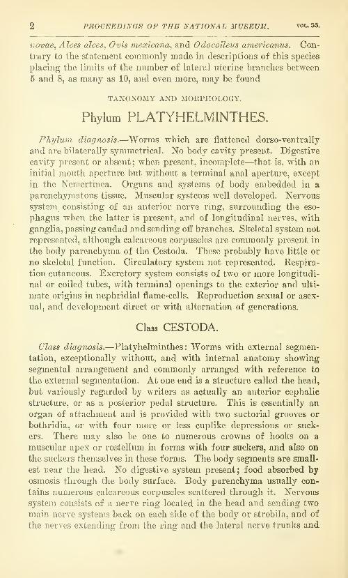
2 PROCEEDINGS OF THE NATIONAL MUSEUM. vol.55.
novae, Alces alces, Ovis mexicana, and Odocoileus americanus. Con-
trary to the statement commonly made in descriptions of this species
placing the limits of the number of lateral uterine branches between
6 and 8, as many as 10, and even more, may be found
TAXONOMY AND MORPHOLOGY.
Phylum PLATYHELMINTHES.
Phylmn diagnosis.—^Worms which are flattened dorso-ventrally
and are bilaterally symmetrical. No body cavity present. Digestive
cavity present or absent; when present, incomplete—that is, with an
initial mouth aperture but without a terminal anal aperture, except
in the Nemertinea. Organs and systems of body embedded in a
parenchyniatous tissue. Muscular systems well developed. Nervous
system consisting of an anterior nerve ring, surrounding the eso-
phagus when the latter is present, and of longitudinal nerves, with
ganglia, passing caudad and sending oif branches. Skeletal system not
represented, although calcareous corpuscles are commonly present in
the body parenchyma of the Cestoda. These probably have little or
no skeletal function. Circulatory system not represented. Respira-
tion cutaneous. Excretory system consists of two or more longitudi-
nal or coiled tubes, with terminal openings to the exterior and ulti-
mate origins in nephridial flame-cells. Reproduction sexual or asex-
ual, and development direct or with alternation of generations.
Class CESTODA.
Class diagnosis.—Platyhelminthes : Worms with external segmen-
tation, exceptionally without, and with internal anatomy showing
segmental arrangement and commonly arranged with reference to
the external segmentation. At one end is a structure called the head,
but variously regarded by writers as actually an anterior cephalic
structure, or as a posterior pedal structure. This is essentially an
organ of attachment and is provided with two suctorial grooves or
bothridia, or with four more or less cuplike depressions or suck-
ers. There may also be one to numerous crowns of hooks on a
muscular apex or rostellum in forms with four suckers, and also on
the suckers themselves in these forms. The body segments are small-
est near the head. No digestive system present; food absorbed by
osmosis through the body surface. Body parenchyma usually con-
tains numerous calcareous corpuscles scattered through it. Nervous
system consists of a nerve ring located in the head and sending two
main nerve systems back on each side of the body or strobila, and of
the nerves extending from the ring and the lateral nerve trunks and

NO. 2258 TAENIOID CEST0DE8 OF DOGS AND CATS—HALL. 3
ganglia. The excretory system consists, commonly, of a dorsal and
a ventral excretory vessel on each side of the strobila with a simple
or elaborate set of vessels connecting the vessels of the same side or of
opposite sides. The main longitudinal vessels terminate in open-
ings in the posterior extremity of the strobila and receive waste
matters poured in from canaliculi originating in flame-cells in the
parenchyma. With rare exceptions, the strobila is hermaphroditic,
the male and female sex organs being represented in each segment.
The eggs, or, more properly, embryophores, develop in the segments
to form an onchosphere armed with three pairs of hooks and sur-
rounded by one or several membranes, or they are similar to fluke
eggs, often with an operculum, do not contain an embryo when they
escape from the segments, and later develop an onchosphere with a
surrounding covering of cilia. Development always involves an al-
ternation of generations, the intermediate stage, in the form of a
bladderworm of some sort and with the developed head of the adult
as the essential structure and a surrounding membrane of some sort
as the incidental structure, developing in an intermediate host. This
larval stage develops to the adult worm on ingestion by a suitable
host.
Superfamily TAENIOIDEA Zwicke, 1841.
Synonym.—Cyclophyllidea van Beneden.
Superfamily diagnosis.—Cestoda: Head or scolex with four cup-
shaped suckers which may exceptionally (Tetrabothriidae) bear au-
ricular appendages, or exceptionally (Fimbriariidae) with a pseudo-
scolex in place of this scolex. Apical rostellum present or lacking.
Suckers and rostellum may be armed with hooks or unarmed. Neckpresent or lacking. Strobila with well-developed segmentation, or
exceptionally (Fimbriariidae) without division into segments. Asingle series of reproductive organs or a complete or incomplete dou-
ble series, both male and female organs present in the same segments
except in Dioicocestus where the strobilae are, respectively, male or
female. Genital pores usually present and marginal, exceptionally
ventral (Mesocestoididae), or lacking (Aporina). Testes usually
numerous, occasionally as few as two, and in medullary portijon of
segment. Ovary more or less bilobed. Yolk gland compact, usually
single and located near the median line. So-called shell-gland be-
tween ovary and yolk gland. Uterus v.ithout special opening for the
discharge of eggs to the exterior, except that rarely a secondarily
formed aperture may be present. Onchosphere with one or several
membranes and Avithout operculum. Larval stages in vertebrates or
invertebrates. Adults in the alimentary canal of vertebrates.
Type-family.—Taeniidae Ludwig, 1886a.
In limiting the scope of this paper to a consideration of the taenioid
cestodes, there have been excluded from consideration the carnivore

4 PROCEEDINGS OF THE NATIONAL MUSEUM. vol. 55.
tapeworms belonging to the Proteocephalidae and the Diphylloboth-
riidae. The only species of Proteocephalidae reported from car-
nivores is Ophiotaenia punica (Kholodkovski, 1908) La Rue, 1911.
This species was described from the dog by Kholodkovski (1908) as
Taenia punica^ but the present writer (Hall, 1910) pointed out that
this should be transferred to the genus Proteocephalus and that it
was presumably not a true parasite of the dog, but had been ingested
by the dog in eating the true hjost, some fish, reptile, or batrachian.
In his revision of the Proteocephalidae, La Rue (1911) created tho
new genus Ophiotaenia for the reptilian cestodes of that family, and
transferred this species to the new genus. This species is evidently
not to be considered as a parasite of carnivores and nothing would be
gained so far as concerns the purposes of the present paper by a de-
scription of its morphology. The presence of follicular yolk glands
in the lateral fields is one feature which distinguishes the Proteo-
cephalidae from the Taenioidea.
The omission of the Diphyllobothriidae from this paper is of more
importance than the omission of the Proteocephalidae. Species be-
longing in this group have been reported from carnivores in North
America and material of the sort is available to the writer for study,
but a casual examination of the material indicates that it would
require more time for adequate study than can be given at present.
This family is relatively much less important than the superfamily
Taenioidea covered in this paper. Members of the Diphyllobothrii-
dae are characterized by the presence of a rosette-shaped uterus which
has a special aperture in the midventral line for the discharge of eggs.
The keys given deal with each taxonomic group, from families to
the species of a given genus, separately. At the end of this paper is
a key covering the species of all genera involved.
KEY TO THE FAMILIES OF TAENIOIDEA.
1. Genital pores located on the ventral surface near the median line. Eggs In
gravid segments enclosed in a single thick-shelled egg capsule.
MESOCESTOIDIDAE, p. 59.
Genital pores lateral. Eggs in gravid segments contained in a uterus or
in numerous egg capsules 2
2. Usually large forms. Genital pores irregularly alternate. Rostellum usually
well developed and usually armed with a double crown of hooks, rarely
with a single (?) crown of hooks or unarmed. Suckers unarmed. Uterus
with a median stem and lateral branches. Eggs thick shelled—i. e.,
embryo sun-ounded by a thick embryophore TAENIIDAE, p. 5.
Usually small forms. Genital pores single or double; if single, regularly
or irregularly alternate. Rostellum present or absent; if present armed
with one to numerous rows of hooks. Suckers armed or unarmed. Uterus
saclike and persistent or with one or several parauterine organs to which
the eggs pass in the final stage of development. Eggs with thin transparent
shells (i. e., embryo surrounded by thin transparent embryophores).
HYMENOLEPIDIDAE, p. 61.
KO. 2258 TAENIOID CE8T0DE8 OF DOGS AND CATS—HALL. 5
Family TAENIIDAE Ludwig, 1886a.
/Synonyms.—Taeniaclao Baird, 1853(2; Taeniodea Diesing of Gold-
berg, 1855a; Taeniadea Cams, 1863; Teniadae Perrier, 1897a.
Family diagnosis.—Taenioidea : Rostelliim usually well developed,
rarely rudimentary, and usually armed with a double crown of hooks
composed of a circlet of large hooks and a circlet of small hooks, the
large and small hooks arranged alternately; rarely with a single (?)
circlet of hooks or unarmed. Suckers unarmed. Gravid segments
longer (that is, along the longitudinal axis of the strobila) than
broad (that is, along the transverse axis of the strobila) . A single set
of reproductive organs in each segment with the genital pores irregu-
larly alternate. Testes numerous. Ovary bilobed, or may be re-
garded as two ovaries. Uterus with a median stem and lateral
branches and without an opening to the exterior for the escape of the
eggs.
Type-genus.—Taenia Linnaeus, 1758.
Subfamily Taeniinae Stiles, 1896&.
Synonyms.—Taeniea Goldberg, 1855a; Cystotaeniae Glaus, 1876;
Taenianae Railliet, 1896.
Subfamily diagnosis.—Taeniidae: Usually large species. Gravid
segments usually considerably longer than broad. Scolex with ros-
tellum and usually armed with a double crown of hooks, rarely with
a single ( ? ) circlet of hooks or unarmed. Genital pores irregularly
alternate. Testes usually very numerous, mostly in the lateral por-
tions of the median field bordered by the longitudinal excretory
canals and to a less extent in the median portion of this field. Ovary,
shell-gland, and yolk-gland in the posterior portion of the median
field, distal from the head. Uterus with a median stem from which
develop lateral branches, the structure suppressing the genital glands,
wholly or partly, in gravid segments. Of the four longitudinal ex-
cretory canals, usually only the ventral are readily visible in gravid
segments. Egg shell thin, with or without filaments, usually disap-
pears after a time; embryophore thick and radially striate. Inter-
mediate larval stage a bladderworm of the cysticercus, coenurus, or
echinococcus type, occurring in herbivorous or omnivorous animals.
Adult stage a strobilate worm in carnivora or omnivora.
Type-genus.—Taenia Linnaeus, 1758a.
KEY TO THE GENEttA OF TABNIINAH.
1. Strobila less than 1 centimeter long and composed of a head and 3 segments,
only one of the segments being gravid at a time. Lateral uterine branches
often quite indistinct. Yolk-gland globular. Larval stage an echinococcus
with thick laminated wall, and developing brood capsules containing the
larval scolices Echinococcus, p. 56.

6 PROCEEDINGS OF THE NATIONAL MUSEUM. vot^ 55.
Strobila at least several centimeters long and composed of a head and numer-
ous segments, from 10 to hundreds, with a number of segments usually
gravid at one time. Lateral uterine branches usually distinct, at least in
early stages of formation. Yolk-gland posterior of ovaries and elongate or
triangular, with one side parallel to the posterior margin of the segment.
Larval stage a bladderworm with thin walls, and never containing brood
capsules 2.
2. Handle of large hook usually sinuous ; vagina usually shows a reflexed loop
in the vicinity of the longitudinal excretory canals. Larva a coenurus
—
i. e., a bladderworm with a thin wall containing numerous scolices, and oc-
casionally daughter bladders, but never brood capsules Miilticeps, p. 39.
Handle of large hook usually not sinuous; vagina straight or curved in the
vicinity of the longitudinal canals, but without a reflexed loop at this
point. Larva a cysticercus—i. e., a bladderworm containing one scolex.
Taenia, p. 6.
It appears difficult at the present time to write a key that will dif-
ferentiate between the strobilate forms of the genus Taenia and those
of the genus or subgenus MuUiceps. This is not surprising. Acoenurus for a larval stage may be regarded as a localized mechanical
device for reproductive purposes and need not be expected to exert
any noticeable effect on the strobilate morphology. It would natu-
rally be expected that species of this genus would show relationship,
but it would not necessarily follow that there would be a notable
departure in structure from that of the parent genus, Taenia.
Genus TAENIA Linnaeus, 1758a.
Synonyms.—Tenia Scopoli, 1777; Hydatigena Goeze, 1782a; Mego-
cephalos Goeze, 1782a; 'iPseudoechinorhynchus Goeze, 1782a; Finna
Werner, 1786a; Vesicaria Mueller, 1787a; Hydatula Abildgaard,
1790; %Baeruca Gmelin, 1790a; Hydath Blumenbach, 1797; Cysti-
cercus Zeder, 1800a; Alyselminthus Zeder, 1800a; Halysis Zeder,
1803a; Cisticercus Eudolphi, 1805a; Physchiosoma Brera, 1809a;
Finna Brera, 1809a; Goeziana Eudolphi, 1810a; Hydatigera La-
marck, 1816; Fischiosoma delle Chiaje, 1825a; Trachelocampylus
Fredault, 18475; Arhynchotaenia Diesing, 1850a; Halisis Goldberg,
1855a; Acanthotrias Weinland, 1858a; Cystotaenia Leuckart, 1863;
Neotenia Sodero, 1886a; Neotaenia Braun, 1894a; Gysticerhus of
authors; Cystizerkus of authors.
GeneHc diagnosis.—Taeniinae : Kostellum distinct and armed with
a double crown of hooks, or, exceptionally, with a single (?) crown
of hooks. Strobila composed of fi'om 10 to hundreds of segTnents.
Usually large forms. Larva a cysticercus in mammals ; adult strobila
in meat-eating mammals.Type-species.—Taenia solium Linnaeus. 1758a.

MO. 2258 TAENIOID CE8T0DES OF DOGS AND CATS—HALL. 7
KEY TO THE SPECIES OF TAENIA.
1. Rostellum with a single circlet of hooks of rose-thorn shape.
Taenia mono Stephanos, p. 38.
Rostellum with a douhle crown of hooks of conventional shape, i. e., with
a relatively long handle 2.
2. Large hooks 60 to 74 in number ; 320 to 355 fi long ; the large hooks arranged
alternately nearer to the center of the rostellum and farther from it,
forming, in effect, 2 circlets of large hooks Taenia macrocystis, p. 13,
Large hooks not over 60 in number 3.
3. No neck. Large hooks 380 to 420 m long; hooks 26 to 52 in number, well-
developed sphincter vaginae Taenia taeniaefonnis, p. 9.
Neck present, or if absent, large hooks 38 to 60 in number, no sphincter
vaginae 4.
4. No neck. Large hooks 38 to GO in number ; 380 to 420 fi long.
Taenia laticollis, p. 8.
Neck present. Large hooks not over 294 fi long , 5.
5. Large hooks 225 to 294 fi long. Small hooks deeply bifid. Testes extend
posterior of the vitellarium. Vas deferens originates in a vesicula semi-
nalis Taenia pisiformis, p. 22.
Large hooks not over 220 ix long 6.
6. Guard of small hook twisted so that its flat surface tends to lie in the plane
of the blade and handle Taenia brachysoma, p. 21,
Guard of small hook not so twisted 7.
7. Head acorn-shaped with hooks far anterior of the suckers. Mature seg-
ments approximately square Taenia Mlaniceps, p. 16.
Head not acorn-shaped and hooks not far anterior of the suckers. Maturesegments distinctly broader than long 8.
8. Large hooks 95 to 140 ii long Taenia brauni, p. 19.
Large hooks 148 n long or more (species of Multiccps will run down to here
with a range of 135 to 180 p.) , 9.
9. Gravid uterus with 20 to 25 lateral branches on each side of the medianstem. Vagina crosses the ovary on the pore side in some segments.
Taenia ovis, p. 32.
Gravid uterus with not over 10 lateral branches on each side. Vagina doesnot cross the ovary on the pore side of segments 10.
10. Large hooks 148 to 170 fi long. Genital papilla very large and prominent,
practically as long as the margin of the segments. Vagina does not forma crescent near segment margin. Gravid segments without a medianlongitudinal groove terminating in a posterior notch.
Taenia krahhei, p. 36.
Large hooks 170 to 220 m long. Genital papilla small and not prominent.
Vagina forms a sort of crescent by dilation and curvature near lateral
margin of segment. Gravid segments with a median longitudinal grooveterminating in a notch posteriorly Taenia hydatigena, p. 28.
In the above key the distinction between Taenia taeni-aeformis
and Taenia laticollis is not well drawn. As a matter of fact, it ap-
pears impossible to draw any adequate distinction. All the described
specim.ens of T. laticollis appear to have been based on immaturespecimens, and the descriptions of the genitalia with their topogra-phy, so essential in present-day specific concepts, have never beenpublished. It is an open question in the writer's mind whether there

8 PROCEEDINGS OF THE NATIONAL MUSEUM. vouOB.
is a distinct species, T, laticoUis, or whether 7'. laticollis is a synonymof T. taeniaeformis. So far as the descriptions are concerned, andin view of the host records from Felidae, including the lynx, there is,
if anything, more likelihood that it is a synonym.
TAENIA LATICOLLIS Radolphi, 1819a.
Specific diagnosis.—Taenia: Head spherical to club-shaped, about
1 to 1.22 mm. in diameter. Rostellum cylindrical to conical, and 600
to 700 (x in diameter, sharply circumscribed at its base. A crown of
38 to 60 hooks, the large hooks ( ? ) 380 to 420 p. long and with a blade
only slightly curved, the handle slightly wavy, and the guardrounded conical in outline with a slight bulge on the side toward the
blade. The small hooks (fig. 1) are 150 (?) to 183 [a
long with a blade even less curved than that of the
large hook, the handle thick and stumpy, roundedoblong in shape, and only slightly longer than the
thick, rounded conical guard. The suckers have a
diameter of 340 to 400 ^ and are quite prominent.
Fig. 1.—Taenia There is no neck, body segmentation beginning inmie-
smIll°ho"k. diately back of the head and with no diminution in
xiso. attee diameter. The first segments are short, later becom-
ing square and finally oblong and longer than broad
with a maximum diameter of 2 mm. The length of strobilae ob-
served is 50 to 95 mm. Genital pores irregularly alternate andprominent.
Male genitalia.—Not described.
Female genitalia.—Not described.
Hosts.—Lynx lynx {Felis lynx)^ Lynx canadensis.
Location.—Intestine.
Localities.—Europe, (?) United States.
Life history.—Unknown.The above description has been compiled from Eudolphi (1819a),
Diesing (1850^), Leuckart (1856a), and Liihe (1910, p. 697). Dies-
ing repeats Eudolphi's description, Leuckart had some new mate-
rial, while Liihe has reexamined Rudolphi's types, without, however,
adding more than a few details to our knowledge. Liihe thinks that
Taenia novella Neumann, 1896/, is probably identical with T. lati-
collis^ but the presence of a distinct neck in T. novella and the shape,
size, and number of hooks are all features that relate it to T. pisi-
formis, as Neumann noted in describing his new species. Leuckart
•states that the hook sizes are almost exactly those of T. taeniaeforniL'i,
and the figures in the specific diagnosis are made on the basis of this
statement. None of the described material has been fully developed,
so the important genital structures are unknown.

HO. 2258 TAENIOID CEST0DE8 OF DOGS AND CATS—HALL. 9
. -5!sr^
1^
Stiles and Hassall (18946?) record this parasite from Lynx cana-
densis and note the presence of the specimens in the Army Medical
Museum, but they give no further data as to where it was collected
or by whom determined, probably because there
was no record of these facts. It would require a
very careful examination before anyone would be
safe in saying that a tapeworm collected from
Lynx canadensis on the American continent was
identical with the imperfectly known Taenia lati-
collis from the European lynx, and the presence of
this tapeworm in North America must be regarded
as questionable.
TAENIA TAENIAEFORMIS (Batsch, 1786) Wolffhugel. 1911.
jSynonynis.—Vermis vesicularis muris Hart-
mann, 16956/ Fasclola muris hejMticae Roederer,
1762«/ Taenia hydafigena Pallas, 1766, part;
Vermis vesicularis teniaeformis Bloch, 1780(2/ Taenia collo hrevis-
simo Bloch, 1782«/ Taenia serrata Goeze, 1782a/ Hydatigena taeniae-
formis Batsch, 1786 ; Cysticercvs fasciolaris Rudolphi, 1808a/ Taenia
crassicoUis Rudolphi, 1810a/ Taenia teniaeformis (Bloch, 1780a)
Fig. 2.—Taenia tak-
ntaefoemis. headviewed feom theSIDE. X 15. AFTEENeumann in Rail-
LIET, 18930.
Fig. 3.—Taenia taeniaefoekis. Large and small hooks.
Stiles and Stevenson, 1905a. (For additional synonyms see Stiles
and Stevenson, 1905a.)
SpecifiG diagnosis.—Taenia: Head thick, cylindrical anteriorly (fig.
2), and 1.7 mm. thick. The rostellum is short and armed with a
double crown of 26 to 52 hooks. The large hooks (fig. 3) are 380

10 PROCEEDINGS OF TEE NATIONAL MUSEUM. vol.55.
to 420 [JL long. They have a blade of rather slight curvature; a
handle which maintains a generally straight direction except at its
distal extremity where it curves dorsad, and with a dorsal swelling
near its middle and another smaller dorsal SAvelling at its union with
the blade; and a guard with a tendency to bifid structure and pre-
senting in lateral view parallel borders proximally and a conical
termination distally. The small hooks (fig. o) are 250 to 270 \l long.
They have a blade of moderate curvature; the handle is straight
with a slight enlargement distally, the enlargement curved dorsad;
the guard enlarges just beyond its point of attachment, forming a
neck between the enlargement and the attachment, and then ter-
minates in a conical distal structure. The suckers are very promi-
nent, being set on the cylindrical head at an angle pointing forward
and outward. There is no neck, segmentation beginning directly
posterior of the suckers and the initial segments being as broad as
or broader than the head. The strobila attains a length of 15 to
60 cm. and a maximum width of 5 to 6 mm. The anterior segments
are very short, the following are cuneiform, and the terminal are
elongate, 8 to 10 mm. long by 5 to 6 mm. wide. Mature segments are
wider than long and only terminal
gravid segments are longer than wide.
Calcareous corpuscles are numerous andFIG. 4.-TAENIA TAENiAEFOKMis. DiA-
^^,^^|_ rj^YiQ parenchvma at times showsGRAM SHOWING THE TKANSVERSE EX- ^ "
CEETORY CANAL ENCIRCLING EACH numcrous rather large areas that fail toDORSAL CANAL. AFTER LovELAND,
gtalu wlth Carmine. The genital papilla
is in the middle of the lateral margin,
or anterior of the middle, is flat and inconspicuous, and is elongated
along the longitudinal axis of the strobila. The transverse excretory
canal is a single tube in the median portion of the strobila, but at its
union with the ventral canal it forms two branches which pass dor-
sally and ventrally and surround the dorsal excretory canal (fig.
4). The dorsal excretory canal i^ very sinuous and thick walled.
Male genitalia (fig. 5).—The testes are numerous, oval or spherical
in shape, and are set close together in the lateral portions of the
median field close to the excretory canals. For the most part they
leave a clear field in the vicinity of the median stem of the uterus,
but may extend across this anteriorly; they press close to the field
occupied by the genital canals, or even invade it, and lie in contact
with the lateral portions of the ovaries and extend posterior of the
ovaries but not quite to the vitellarium. The vas deferens is very
much looped in a dense mass of closely approximated coils along the
transverse and longitudinal axis of the strobila, and apparently
originates at some distance from the median stem of the uterus on
the pore side of the segment. The cirrus pouch is slender, frequently
curved in gravid segments, and is difficult to observe in toto mounts.

NO. 2258 TAENIOID CEST0DE8 OF DOGS AND CATS—HALL. 11
\

12 PROCEEDINGS OF THE NATIONAL MUSEUM. VOL. 55.
In mature segments it attains a length of 430 to 475 [x and a maxi-
mum diameter around 70 [x; in gravid segments the cirrus pouch
shortens and thickens, its length being 300 to 345 [jl and its maxinmmdiameter about 85 ^.
Female genitalia (fig. 5).—The ovaries are compact, circular in
outline, the one on the pore side being smaller than the one on the
aporal side. The vitellarium is elongated along the transverse axis
of the worm, stains very densely,
and is very conspicuous; it is in
contact vs^ith the posterior curva-
ture of the ovaries and extends
across the posterior portion of the
interovarian field; it does not ex-
tend as far laterally as do the ova-
ries. The shell-gland is inconspicu-
ous and appears to be commonly ob-
scured by either the ovaries or the
vitellarium. Near its union with
the genital sinus, the vagina com-monly presents a curve or even a
conspicuous loop toward the poste-
rior portion of the segment, and at
this point the vagina is encircled by
a well-developed sphincter. Fromhere the vagina parallels the course
of the cirrus pouch and the vas de-
ferens and then curves around the
nearest ovary to the interovarian
field. Even in the mature seg-
ments the median stem of the ute-
rus begins the formation of two
lateral branches, one on each side,
at the anterior end of the segment.
As these develop, other branches
form behind them, the newbranches being added posteriorly
until they invade the region of the
ovaries and obliterate them. The lateral branches are notably parallel
to one another along the transverse axis of the strobila and show com-
paratively little tendency to subdivide, but rather a tendency to be-
come sacculate at the distal extremities, so that the segment becomes
j&lled with eggs, not as the result of the formation of numerous
branches and the anastomosis of these branches, but as the result of
the sacculation of the main lateral branches and especially of their
distal extremities (fig. 6). The eggs are spherical and 31 to 37 n in
diameter.
'|/iomm.'
Fig. 6.—Taenia taeniaeformis. GravidSEGMENT.

NO. 2258 TAENIOID CESTODES OF DOGS AND CATS—HALL. 13
Hosts.—Primary: Felis catus {F. domestica) , F. maniculata, F.
macrom'a, F. concalor, F. melivora, F. onca, F. mitls^ F. tigrlna^ F.
eyr'a, F. sylvestris {Catus sylvestris).^Lynx uintd {Lynx tienta).,
Miistdla erminea {Putorius erminea). Secondary: Miis musculus^
Epimys rattus alexandrinus {M. rattus alexandrinus, M. tectorum)^
Epimys norvefficus, E. rattus rattus, Micro fus arvalls {Arvicola ar-
valis), A. amphibius {A. amphibia), ''''Lenh7mis tei'vestHs,^^ Ondatra
zibethica {Fiber zibethicus) , Talpa europaea, Plecotus auritus.
Location.—In small intestine of primary hosts. In liver of sec-
ondary hosts.
Localities.—Germany, Austria, Italy, France, Enghmd, Denmark,Iceland, Persia, Japan, United States.
Life history.—Eggs developed by the adult worm in the intestine
of the primary host pass out and are ingested by the secondary host
in contaminated food or water. In
the digestive tract of the second-
ary host, the embryo escapes fromthe shell and makes its way to the
liver, where it develops into the
larval stage or bladderworm, com-
monly known as Gysticercus fascio-
laris. This bladderworm is char-
acterized by the presence of a very
small caudal bladder or vesicle
filled with fluid and a very long
strobilate connection between this
caudal bladder and the head.
When the cysticercus is ingested by
the primary host, the caudal blad-
der digests off and new segments are formed back of the existing
strobilate portion, thereby developing into the strobilate tapeworm.
The record from Lynx uinta in the above list is new. It should
be noted that Liilie (1910) states that an examination of Diesing's
South American cestodes, on which some of Diesing's records of T.
taeniaeformis are based, did not disclose a single specimen of this
worm.TAENIA MACROCYSTIS (Diesing, 1850o) Liihe, 1910.
Synonym.—Cysticercus Tnacrocystis Diesing, 1850(Z.
Specific diagnosis.—Taenia: Head 1.25 to 1.6 mm. in diameter.
Rostellum from 515 to 690 [jl in diameter and armed with a double
crown of 60 to 74 hooks (fig. 7). The large hooks are alternated
with the small in the customary manner, but in addition every other
large hook is set a little closer to the center of the hook circlet than
is the case with the remaining large hooks, with the result that the
hook crown is arranged in one circlet of small hooks and two circlets
Fig. 7.—Taenia macrocystis. Hook circletVIEWED FEOM THE FRONT. X 60. AFTEBLt)HE, 1910.

14 PROCEEDINGS OF THE NATIONAL MUSEUM. TOU 6B.
of large hooks. This alternating arrangement of the large hooks
is also observed in a lateral view of the head (fig. 8). The large
hooks are 320 to 365 [j. long. When the head is viewed from the front,
showing the dorsal edge of the hooks, the large hooks are an elongate
spindle shape, with a slight constriction along the middle portion.
The blade and the handle attain their maximum thickness of 27 [>.
at their middle points, diminishing to 20 [i, near the guard. Fromthe lateral view (fig. 9) the large hooks present a blade wdth a
moderate curve; a handle which is very variable in outline; it maybe continuous dorsally with the straight line of the
blade, and be almost straight, with a very slight
undulation and a small enlargement at the end,
this end tending to bend slightly in the ventral
direction, or it may narrow to a rather acute tip
and have a smooth or Imobbed outline; and a
guard that is somewhat triangular in outline or in
some cases with the proximal portion presenting almost parallel sides
and terminating distally in a triangular portion. The small hooks
are 180 to 200 [x long. Froni the lateral view they present a blade of
moderate curve ; a thick, short handle curving dorsally ; and a thick
irregularly triangular guard. The suckers are not prominent andare 290 to 350 [>. in diameter. The neck is somewhat smaller than the
head and is 600 [i to 1.3 mm. long, measuring from the posterior
margin of the suckers to the first trace of segmentation. The strobila
Fig. 8.—Taenia macro-cYSTis. Lateral viewOFHEAD. X 42. AfterLOhe, 1910.
(fig. 10) attains a length of 12 cm., a
maximum width of 2 mm., and is com-
posed of 90 to 100 segments. The genital
pore lies near the middle of the segment
and is very prominent. The dorsal ex-
cretory canal lies lateral of the plane of
the ventral canal. The ventral canal is in
the ventral portion of the segment and not
merely ventral of the dorsal canal. Thetransverse canal connects the two ventral
canals in the usual way. Youngest seg-
ments about 750 \l wide. About the eight-
ieth segment, the maximum width occurs, segments being 2.2 mm.wide and 2.5 mm. long. The largest segments are 7 mm. long and
1.5 mm. wide. The genital primordia are visible about 1.5 mm.back of the head in the median portion of about the twentieth seg-
ment. The cirrus pouch and vagina lie between the dorsal and
ventral excretory vessel and the longitudinal nerve is ventral of
the cirrus pouch and vagina. The calcareous corpuscles are 19 by
13 \i. in diameter.
Male genitalia.—The testes are comparatively few in number and
are oval, the long axis paralleling the transverse axis of the strobila
;
Fig. 9.—Taenia macbocystisLarge and small hooks.

NO. 2258 TAENWID CE8T0DES OF DOGS AND CATS—HALL. 15
s©s
they lie in the lateral portion of the median field and cross the median
field anteriorly; they appear to cross the field of the
vas deferens and vagina at times, and extend close to
the ovaries laterally as a rule, occasionally occurring
posterior of the lateral portion of the ovaries ; they do
not lie so near the dorsal transverse musculature as in
I'aenia taeniaeformis. The vas deferens is very muchlooped, but pursues a fairly straight course from a
point near the median stem of the uterus, on the pore
side of the segment to the cirrus pouch. The cirrus
pouch is very long and narrow and extends from the
median border of the ventral excretory canal across
both canals; it is 300 to 345 [x long and 35 to 60 [/, wide.
Female genitalia.—The ovaries are elongated along
the transverse axis of the strobila and inclose, usually,
an oval to round interovarian field; the individual
branches of the ovary are only moder-
ately compact; the ovaries are of the
same size, or the one on the aporal side
may be slightly larger. The vitellarium
is elongated along the transverse axis of
the worm in contact with the posterior
curvature of the ovaries and extends
about as far laterally as do the ovaries.
The shell-gland is obscured or incon-
spicuous. The vagina comes in from the
genital pore in a long straight line and
makes a very slight curve around and
close to the nearest ovary. The uterine
stem begins its initial development in the
posterior portion of the segment, widening to fill the
interovarian field and sending branches posterior of
the ovaries. In the gravid seginents (fig. 11) there are
on each side 8 to 15 lateral branches, relatively short
and themselves branching. In the last segments the
main branches are amalgamated at the base. The egg
is oval, 34 to 38 [j. by 25 to 27 [jl in diameter, with a shell
4.5 [x thick.
Hosts.—Primary: Felis tigrina., F. yagouaroundi
{F. jaguarundi) , F. sp., Oalictis sp.. Lynx ruffu^ (L.
rufa)., L. haileyi. Secondary: Sylvilagus hrasiliensis
{Lepus hrasiliensis).
Location.—Intestine of primary host: Free in
body cavity or encapsuled in the liver, in the
region of the kidney or between the back muscles
in the secondarv host.
Fig. 11.—Taeniamacrocystis.Geavid seg-
ment. X 5.5.
After LChe,
1910.
imfnFio. 10.—Taeniamacrocystis.Entire stro-bila.

16 PROCEEDINGS OF THE NATIONAL MUSEUM. VOL. 50.
Localities.—Brazil, Paraguay, United States (North Carolina;
Boulder, Colorado).
Life history.—The eggs produced by the strobilate tapeworm in
the intestine of the primary host pass out and are ingested on food
or in water by the secondary host where they develop to form the
intermediate larval stage or bladderworm,
known as Oysticercus macrocystis andvery similar to Oysticercus fasciolaris.
On ingestion of this bladderworm by the
primary host in preying on the secondary
host, the terminal vesicle digests off while
the head and its strobilate connection with
the bladder develops attached segments
and so forms the strobilate worm.The material
recorded here
from theUnited States
is in the col-
lection of the
United States
Bureau ofAni-FiG.12.- -Taenia balaniceps.
Aftee Hall, 1910.
Head.
mal Industry.
That from Lynx rujfus was collected by
Doctors Hassall and Graybill from a
lynx sent from North Carolina to the
National Zoological Park at Washing-ton, District of Columbia, and that fromL. haileyi was collected at Boulder, Colo-
rado, by Dr. Max Ellis.
TAENIA BALANICEPS Hall, 1910.
Specific diagnosis.— Taenia: Headacorn-shaped (fig. 12), 735 tx long by534 to 753 \i. wide. Rostellum rounded
and prominent, 307 [x in diameter, andarmed with a double crown of 28 to 32
hooks, of which the larger are easily lost.
The hooks are set far forward of the
suckers. The large hooks (fig. 13) are
145 [JL long. They have a blade of moderate curvature; thfe handle
tapers toward its distal extremity; the distal extremity, which is
not enlarged, curves slightly dorsad, and in lateral view the ventral
outline is slightly convex, while the dorsal outline presents a slight
median swelling and another slight swelling at the union of the blade
Aomm.Fig. 13.—Taenia balaniceps. LabobAND SMALL HOOKS. AFTEE HAIX,1910.

NO. 2258 TAENIOID CE8T0DES OF DOGS AND CATS—HALL. 17
and handle; the guard is roughly conical with a protrusion toward
the blade at the point of union with the blade. The small hooks
m.m̂
/lS>^
. f.r.\^\.^
'Ml
y, . .
\ 1 'ioimn
Fig. 14.—Taenia balaniceps. Small hooks. Af-
ter Hall, 1910.
^'-*'i'^i^:^t'£a4fcjSiA^::d^**^^£*.^-
ImiTi.
Tig. 15.—Taenia balaniceps. MatueeSEGMENT. After Hall, 1910.
^fl-yVr^ct^
fSi
I
(figs. 13 and 14) are 93 to 98 tx long. They have a strongly curved
blade ; the handle is short and thick and may be straight or present a
slight curvature dorsad at its distal
extremity; the guard is rather
oval in lateral outline. The suckers
are round, the bulb of the sucker
being 215 to 265 [x in diameter.
The neck is distinct and rather
tong, the maximum length being
about 1.2 mm. from the posterior
margin of the suckers to the first
distinct segmentation. The strobila
may attain a length of over 24 cm.
Mature segments (fig. 15) are ap-
proximately square in outline and
are about 2 inm. long by 2 to 2.5
mm. wide. Gravid segments (fig.
16) are 5.5 to 10.5 mm. long and 2
to 4 mm. wide. The genital canals
pass out to the genital pore be-
tween the dorsal and ventral longi-
tudinal excretory vessels and either
dorsal or ventral of the main nerve
trunk. The transverse excretory
canal is very large and tends to lie between two adjacent segments
rather than along the posterior border of the segments. The pri-
62055—20—Proc.N.M.Vol.5E> 3
V
(mm.
Fig. 16.—Taenia balaniceps. Geavid seg-
ment. After Hall, 1910.

18 PROCEEDINGS OF THE NATIONAL MUSEUM. vol.55.
mordia of the genital organs appear a short distance back of the
head; the testes, genital canals, shell-gland, and the main trunk of
the uterus are clearly defined before the ovaries and yolk-gland can
be detected. The genital pores are irregularly alternate and are es-
pecially prominent in segments full of developing eggs, where they
may have an antero-posterior diameter of 480 jx, or about one-third
the segment length. Calcareous corpuscles abundant, of variable size
and shape, and with a maximum diameter of 20 pi.
3Iale genitalia.—The testes are commonly oval, with long axis
paralleling that of the strobila (fig. 15), and are principally confined
to two bands along the median side of the longitudinal excretory
canals. A narrow band of testes crosses the extreme anterior margin
of the segment, connecting the two lateral fields and leaving a large
space clear of testes, frequently approximately square in outline, be-
tween this band, the lateral testicular fields, and the ovaries. Thelateral testicular fields extend to the lateral margin of the ovaries,
frequently encroaching on the field of the vas deferens and vagina,
and are prolonged posterior of the ovaries to the yolk-gland. Thevas deferens arises near the plane of the median stem of the uterus,
either on the pore side or the aporal side. At the plane of the ventral
excretory canal, or just lateral of this, the vas deferens opens into
a tubular cirrus pouch 300 to 370 pi long, with an average length of
355 [X. The diameter of the cirrus pouch varies considerably, the
maximum diameter being about 110 [x . There is no vesicula seminalis
present. The length of the cirrus varies from 418 to 518 pi, with
a maximum diameter of about 33 \j. and a lumen diameter of
about 8 [X.
Female genitalia.—The ovaries are elliptical to crescentic in out-
line with their longitudinal axes paralleling that of the strobila, and
inclose an oval to oblong interovarian fields. The vitellarium is
elongated in the transverse axis of the strobila and extends a slight
distance between the ovaries but not lateral of them. The shell-gland
is very close to the vitellarium. The vagina swings in a wide curve
from the genital pore around the nearest ovary and opens into a small
receptaculum seminis in the neighborhood of the shell-gland. Theuterus originates as a median stem, and develops branches of un-
usual form. These branches are club-shaped and so closely approx-
imated and at times so united that the ultimate result resembles a
lobed sac (fig. 16). In many cases one uterine lobe extends over the
longitudinal excretory canals in the vicinity of the genital pore.
The eggs are ovoid in shape, are 29 to 37 pi by 27 to 33 pi in diameter,
the average being 35 to 31 ix. The shell is about 4 \j. thick.
Hosts.—Primary: Canis familiaris, Lynx rufiis mwnixmlatus
{Lynx rufus maculatus). Secondary: Unknown.
Na2258 TAEXIOID CB8T0DES OF DOGS AND CATS—HALL. 19
Location.—Intestine of primary host.
Localities.—Nevada (Fallon) ; Southern New Mexico.
Life history.—Unknown.The uterine structure is of the type found in such tapeworms as
T. taeniaeformis and indicates that the lynx is probably the normal
host, as the dog is certainly an
accidental or occasional host. Onthe other hand, the narrow neck
in T. halaniceps is quite different
from the thick neck or the absence
of a neck in many tapeworms
l^arasitic in the Felidae.
:iTAENIA BRAUNI Setti. 1897&.
Fig. 18.—TaeniaBRAUNI. LargeHOOKS. X 100.
After Setti,1897.
Flo. 17.—Taenia brauniAnterior extremityX 20. After Setti, 1897
Fig. 19.—TaeniaBRAUNI. SmallHOOKS. X 100.
After Setti,1S97.
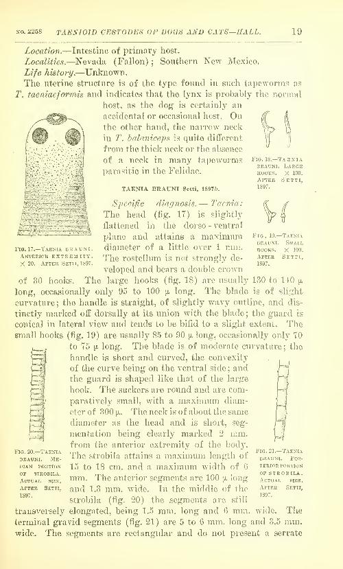
Specific diagnosis.— Taenia:
The head (fig. 17) is slightly
flattened in the dorso - ventral
plane and attains a maximmndiameter of a little over 1 mm.The rostellum is not strongly de-
veloped and bears a double crown
of 30 hooks. The large hooks (fig. 18) are usually 130 to 140 [x
long, occasionally only 95 to 100 y. long. The blade is of slight
curvature; the handle is straight, of slightly wavy outline, and dis-
tinctly marked off dorsally at its union with the blade ; the guard is
conical in lateral view and tends to be bifid to a slight extent. Thesmall hooks (fig. 19) are usually 85 to 90 [x long, occasionally only TO
to 75 [1. long. The blade is of moderate curvature; the
handle ig short and curved, the convexity^
of the curve being on the ventral side; andthe guard is shaped like that of the large
hook. The suckers are round and are com-
paratively small, with a maximum diam-
eter of 300 [x. The neck is of about the same
diameter as the head and is short, seg-
mentation being clearly marked 2 mm.from the anterior extremity of the body.
The strobila attains a maximum length of
15 to 18 cm. and a maximum width of 6
mm. The anterior segments are 100 \i. long
and 1.3 mm. wide. In the middle of the
strobila (fig. 20) the segments are still
transversely elongated, being 1.5 mm. long and G mm,terminal gravid segments (fig. 21) are 5 to G mm. long and 3.5 mm.wide. The segments are rectangular and do not present a serrate
Fig. 20.—TaeniaBRAUNI. Me-DLi.N PORTION
of strobila.
Actual size.
After Setti,
1897.
Fig. 21.—TaeniaBRAUNI. Pos-
terior portionOP strobila.Actual size.
After Setti,
1897.
wide. The
20 PROCEEDINGS OF THE NATIONAL MUSEUM. tol. 05.
edge on the margin of the strobila. The segments are dense owing
to their thickness and the abundance of calcareous corpuscles. Thegenital papillae are in the middle of the segments and are prominent.
The longitudinal excretory vessels are sinuous and are 500 to 700 \>.
from the margin of the strobila on each side.
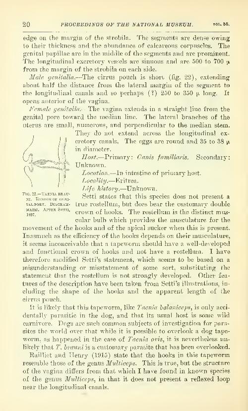
Male genitalia.—The cirrus pouch is short (fig. 22), extending
about half the distance from the lateral margin of the segment to
the longitudinal canals and so perhaps (?) 250 to 350 \i. long. It
opens anterior of the vagina.
Female genitalia.—The vagina extends m a straight line from the
genital pore toward the median line. The lateral branches of the
uterus are small, numerous, and perpendicular to the median stem.
They do not extend across the longitudinal ex-
cretory canals. The eggs are round and 35 to 38 ^.
in diameter.
Host.—Primary: Canis familiaris. Secondary:
Unknown.Location.—In intestine of primary host.
Locality.—Eritrea.
Life history.—Unknown.
^m^^REGi^oiroFTENi- Setti states that this species does not present a
TALPORE. DiAGKAM- true rostcllum, but does bear the customary doubleMATic. AFTEE setti.
^^^^^^ ^f j^^^j^^^rj.^^
rostcllum Is thc distinct mus-
cular bulb which provides the musculature for the
movement of the hooks and of the apical sucker when this is present.
Inasmuch as the efficiency of the hooks depends on their musculature,
it seems inconceivable that a tapeworm should have a well-developed
and functional crown of hooks and not have a rostellum. I have
therefore modified Setti's statement, which seems to be based on a
misunderstanding or misstatement of some sort, substituting the
statement that the rostellum is not strongly developed. Other fea-
tures of the description have been taken from Setti's illustrations, in-
cluding the shape of the hooks and the apparent length of the
cirrus pouch.
It is likely that this tapeworm, like Taenia halaniceps, is only acci-
dentally parasitic in the dog, and that its usual host is some wild
carnivore. Dogs are such common subjects of investigation for para-
sites the world over that while it is possible to overlook a dog tape-
worm, as happened in the case of T'aenia ovis, it is nevertheless mi-
likely that T. h7^auni is a customary parasite that has been overlooked.
Kailliet and Henry (1915) state that the hooks in this tapeworm
resemble those of the genus Multiceps. This is true, but the structure
of the vp.gina differs from that which I have found in known species
of the genus Multiceps, in that it does not present a reflexed loop
near the longitudinal canals.

NO. 2258 TAENIOID CEST0DE8 OP DOGS AND CATS—HALL. 21
TAENIA BRACHYSOMA Setti, 1899c.<
Fig. 24—Taeniabrachysom a.
Large hook.
X 200. AfterSetti, 1899.
Fig. 23.—Taeniabrachysoma.Anterior ex-
tremity. X20.After Setti,
1899.
Specific diagnosis.—Taenia: Head (fig, 23) about 700 \i. in diamete..-
and more or less elongate piriform in shape. Rostellmn prominentand bearing a double crown of 30 to 32
hooks. The large hooks (fig. 24) are 135
to 145 [I. long. The blade has a rather
slight curvature; the handle is slightl}^
curved, at times irregularly so, with the
convexity of the curve on the dorsal sur-
face and meeting the similar curve of the
blade in a distinct obtuse angle opposite
the middle of the guard; the guard is
thick, and in lateral view the sides of
the guard are approximately parallel, the
distal extremity being bluntly rounded.
The small hooks (fig. 25) are 95 to 105 ;j.
long. The blade makes a very sharp curve
toward the axis of the handle and sruard
and then straightens out, its distal portion
being roughly parallel to the axis of the
handle and guard; the handle is very short and blunt,
with a slight tendency to curve dorsally at the tip ; the
guard is rather broad, furrowed to show a trace of bifid-
ity, and tends to be twisted so that the lateral axis lies
in the plane of the blade and handle. The suckers are
round and have a maximum diameter of 250 to 270 \}.
with a circular or elliptical aperture of
about 150 [X. The neck is distinct, nar-
rower than the head, with an average
measurement of 300 to 400 \i and not ex-
ceeding 1 mm. long to the first distinct
segmentation. The strobila (fig. 26) at-
tains a maximum length of 10 cm. and a
maximum w^idth of 3 mm. The first seg-
ments are 40 to 70 [x long and 350 to 450 a
wide ; 5 mm. back of the head they are 200
to 230 [i. long and 600 to 900 ix wide ; 1 cm. back of the
head they are 240 to 260 [x long and 800 [x to 1 mm.wide; in the middle of the strobila they are 750 jx to
1.25 mm. long and 2.3 to 2.8 mm. wide; 2 cm. from the
posterior extremity they are 1.25 to 1.7 mm. long and2.5 to 3 mm. wide; the terminal segments are 2.5 to 3
mm. long and 2.o to 2.5 mm. wide. There are 140 to
180 segments. The first segments are trapezoidal with projecting
posterior angles forming a serrate strobila margin. The segments
Fig. 25.—Taeniabrachysoma.Small hooks.
X 200. AfterSetti, 1899.
Fig. 26.—Taeniabrachysoma.Entire stro-
bila. ActualSIZE. AfterSetti, 1899.

22 PROCEEDINGS OF THE NATIONAL MUSEUM. VOL. 55.
Fig. 27.—Taenia bra-CHYSOMA. GravidSEGMENT. En-larged. AfterSetti, 1899.
in the middle of the strobila are rectangular, almost as long as wide,
and with the posterior angles less prominent and the strobila margin
smoother than anteriorly. The posterior segments are almost quad-
rate, the last two or three longer than wide. Occasionally the middle
segments are campanulate and a little longer than wide. Calcareous
corpuscles are especially abundant in the anterior portion of the
gtrobila. The small genital papilla is near the middle of segment.
It is most distinct in segments in the middle of the strobila. Thelongitudinal excretory canals are about 500 y. from
the lateral margin of the segments.
Male genitalia.—The aperture of the cirrus pouch
is at the base of a genital sinus 100 to 170 ^ long.
The median extremity of the cirrus pouch is about
at the plane of the longitudinal excretory vessels.
Female genitalia.—The uterus (fig. 27) occupies
the median portion of the segments included be-
tween the longitudinal excretory canals. The me-
dian stem has 10 to 12 lateral branches on each side,
approximately perpendicular to the median stem,
and terminating distally in a variable number of
smaller branches of various sizes, shapes, and posi-
tions. Developed embryophores are only found in the last four or
five segments. The eggs are spherical and 32 p, in diameter.
Host.—Primary : Canis familiaris. Secondary : Unknown.
Location.—In intestine of primary host.
Locality.—Italy (Turin).
Life history.—Unknown.In a general way the circumstances indicate that this tapeworm,
like Taenia hrauni^ is also an accidental parasite of the dog.
Setti's statement that the guard of the small hook is twisted so that
the lateral axis tends to lie in the plane of the blade and handle, has
been noted by Ransom (1913) with the following comment: "Setti
does not make it clear whether this twisted condition is invariably
present. The small hooks of Taenia hydatigena commonly present a
similar appearance after subjection to the pressure of a cover glass."
This point is well taken. Tapeworm hooks are flexible structures,
capable of considerable distortion under pressure or torsion, up to
the limit of flexibility, at which point, of course, breaking occurs.
TAENIA PISIFORMIS (Bloch, 1780a) Gmelin, 1790a.
Synonyms.—Vermis vesicularis pisiformis Bloch, 1780a; Hydati-
gena pisiformis (Bloch, 1780<2) Goeze, 1782a; Hydatigena utricu-
lenta Goeze, 1782a; Hydatigena cordata Batsch, 1786a; Hydatigena
utricularis Batsch, 1786a; Vesicaria pisiformis (Bloch, 1780a)

NO. 2258 TA^NIOID CE8T0DES OF DOGS AND CATS—HALL. 28
Schrank, 1788a; Taenia serrata canis domestici et vulpis Rudolphi,
1793a; Cysticercus pisiformis (Bloch, 1780a) Zeder, 1803a; '''Taenia
serrata Goeze " of most authors ; Taenia novella Neumann, 1896/.
(For additional synonymy see Stiles and Stevenson, 1905a.)
Specific diagnosis.—Taenia: Head (fig. 28) 1.3 mm. in diameter.
Rostellum large and powerful, 515 to 640 [jl in diameter, and armed
MOmm
'^i^--:
^i^"-^)
rFig. 28.—Taenia pisiformis. Head, viewed from the front.
Fig. 29.—Taenia pisiformis.
Large and small hooks.'
^/"'^
with a double crown of 34 to 48 strong hooks. The large hooks (fig.
29) are 225 to 294 [jl long. They have a strongly curved blade; the
handle is very long and, observed from the side, usually has straight
sides, gradually diverging distally to a blunt rounded termination, a
truly club-shaped handle, which passes
dorsally rather directly into the blade save
for a short elevation dorsally, which is
usually present at the union of the blade
and handle; the guard is rather long,
thickest in its median portion, thinning:
slightly at its union with the blade andhandle, and terminating distally in a
bluntly rounded cone. The handle andguard form a very obtuse angle. Thesmall hooks (fig. 29) are 132 to 177 [x long.
They have a strongly curved blade; the
handle, viewed from the side, is thick and rather short, its sides dis-
tinctly or slightly curved and approximately parallel, the convexity
of the curve being ventral, and terminating as a rule, in a bluntly
rounded end distally; the guard is usually distinctly and often
strongly bifid (fig. 30), the depth of the cleft varying, rather oval in
!ommFig . 30.—Taenia pisiformis. Largeand small hooks a3 seen in alateral view of the head.

24 PROCEEDINGS OF THE NATIONAL MUSEUM. tol. 55.
outline when viewed from the side, and with a proximal protuberance
toward the side of the blade. The lines of the handle and guard donot meet but are separated by a rather long interval, slightly to
strongly convex in outline when viewed from the side. The distances
from the distal extremity of the guard to the distal extremities of the
blade and handle are very nearly equal. Viewed from the front the head(fig. 28) is approximately square with the suckers located at the corners
and separated by relatively wide intervals from one another. Thesuckers are round to elliptical with a maximum diameter of 310 to
330 \t.. The neck is but slightly narrower than the head and is 680 \).
to 1.7 mm. long from the posterior margin of the suckers to the first
distinct segmentation. The strobila attains a length of 60 cm. to 2
meters, average specimens being 90 to 100 cm. long and consisting of
about 400 segments. The maximum width is about 4.8 mm. The first
segments are very short and much wider than long. There are some-
thing less than 175 of these preceding the mature segments. Thesegments become mature and quadratic in shape about the hundred
and seventy-fifth. There are about 25 of these mature quadratic seg-
ments. They are about 4.9 mm. long and 4.2 mm. wide at the anterior
margin, 4.7 mm. wide at the posterior margin, and 4.8 mm. wide at
the genital pore. Complete maturity is attained in about the two-
hundredth segment, 25 cm. behind the head, and posterior of this the
segments transform into gravid segments. There are 30 to 40 gravid
terminal segments, making up almost half of the entire strobila, these
segments attaining a length of 1 cm. and a width of 4 mm. The pos-
terior angles of all segments are prominent, giving a characteristic
serrate appearance to the strobila. The calcareous corpuscles are
variable in shape and have a maximum diameter of 18 [x. The longi-
tudinal excretory canals are about 640 /x from the lateral margin of
the segment and 770 [jl from the genital pore. The transverse excre-
tory canal has the customary position in the posterior portion of the
segment and connects with the ventral canal. The genital pores are
irregularly alternate, commonly two in succession on one side and
rarely as many as four to six in succession. The genital papilla is
only moderately prominent and is located near the middle of the
segment except in gravid segments where it frequently is distinctly
posterior of the middle. The genital primordia are visible in toto
mounts in the fifth to the twelfth segments back of the head.
31ale genitalia.—The testes (fig. 31) are round or slightly elon-
gated in outline and are 132 by 96 jjl in diameter. There are about
400 to 500 in a segment, and they occupy nearly all the field included
between the longitudinal excretory canals not actually occupied by
other genital structures. In the posterior portion of the segment
they fill the lateral fields clear up to the median stem of the uterus,
leaving only little more than the width of the uterus free of testes

NO. 2258 TAENIOID CESTODES OF DOGS AND CATS—HALL. 25
in the median field, except in the region near the ovary where the
clear field is a little wider. On the pore side of the segment they
extend back to the vas deferens, the space between the vas and the
vagina being free of testes, and then extend from the vagina on this
side, and from the anterior margin of the segment on the aporal side,
back to the posterior margin of the segment. They press between
the loose lobes of the ovary and posterior and dorsal of the vitel-
larium. Aside from the space occupied by the median stem of the
uterus and the field of the vas deferens and vagina, the only space
free of testes is that between the ovaries. The testes are arranged
in two strata, a dorsal and a ventral, some overlying others in frontal
views. The vasa effer-
entia open into a dis-
tinct vesicula semi-
nalis, 210 by 350 \i in
diameter, located on
the pore side of the
median stem of the
uterus. From thevesicula seminalis the
vas deferens extends
posteriorly parallel to
the median stem of
the uterus for a short
distance and then
curves t o w a r d t h o
pore side of the seg-
ment. The vas def-
erens is very large,
much looped andirregular, with thethick loops lying very
close to one another.
The cirrus pouch extends in from the margin of the segment to the
plane of the ventral excretory canal, or very commonly to a point as
much as 107 n median of this plane, and is surrounded by a dis-
tinctive layer of cuboid cells. It is cylindrical or. rather, compressed
elliptical in outline, with its maximum diameter in the middle.
Maximum diameter, 130 to 140 [x ; length, 460 to 800 [>..
Feinale genitalia.—The ovaries (fig. 31) are somewhat reniform,
the concavities of the two inclosing an oval interovarian field, and
are of rather loose structure. They are very nearly equal in size. Thevitellarium is very large, extending laterally past the ovaries, and
forward to the posterior border of the ovaries and a short distance
into the interovarian field. The shell gland is large and in frontal
Fig. 31.—Taenia pisitoemis. Mature segment.
After Deffke, 1891.
Enlarged.
26 PROCEEDINGS OF THE NATIONAL MUSEUM. VOL. 55,
I
view of its posterior portion appears to be embedded in or overlaid
by the portion of the vitellarium. which projects into the interovarian
field. The vagina extends in from the genital pore almost straight
or somewhat inclined anteriorly and curves around the nearest ovary,
forming the receptaculum seminis in the interovarian field. In
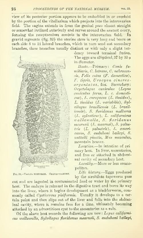
gravid segments (fig. 32) the uterine stem is very long and bears on
each side 8 to 14 lateral branches, which in turn send out secondary
branches, these branches usually distinct or with only a slight ten-
dency toward terminal fusion.
IThe eggs are elliptical, 37 by 32 \i.
Jr 1jin diameter.
^^ '
}Hosts.—Primary : Canis fa-
77iiUaris, C. latrans^ C. nehracen-
sis, Felis catus (F. domestica),
F. tigris, Urocyon cinereo-argentatus., fox. Secondary:
Oryctolagus cwniculus {Lepus
cuniculus ferus., L. c. domesti-
cus), L. europaeus {L. timidus)^
L. timidns {L. variabilis), Syl-
vilagus hrasiliensis {L. hrazil-
iensis), S. fyoridanus mallurue
(L. sylvaticus), L. califomicus
w allawalla., S. floridanufineanisii {L. mearnsi), S. palus-
tns {L. palustri-s)f
L. ameri-
canus, S. auduboni haileyi, S.
7iuttaUi pi7ietis, Mus irtusculus,
mountain beaver.
Location.—In intestine of Dri
marj^ host. In liver, mesenteries,
and free or attached in abdomi-
nal cavity of secondary host.
Locality.—More or less cosmo-
politan.
Life history.—Eggs produced
by the strobilate tapeworm pass
out and are ingested in contaminated food or water by the primary
host. The embryo is released in the digestive tract and bores its way
into the liver, where it begins development as a bladderworm, com-
monly called Cysticerous pisiformis. Usually it develops to a cer-
tain point and then slips out of the liver and falls into the abdom-
inal cavity, where it remains free for a time, ultimately becoming
attached by an adventitious cyst to the mesenteries as a rule.
Of the above host records the following are new : Lepus califomi-
cus wallaioalla, Sylvilagus fioridanus mearnsii, S. auduhom haileyi,
I mm.
Fig. 32.—Taenia pisifoemis. Geavid segment.

NO. 2258 TAENIOID CEST0DE8 OF DOGS AND CATS—HALL. 27
and mountain beaver. The records from Lepus sylvaticus by Stiles
and Hassall (1894^/) are covered in the above list by S. f. mcdlurus.
Other American records are given by Welch (1890a), Curtice
(1892^), Garrison (1911), Sommer (1896), Stiles and Hassall
(1898a), Young (1908a), Ward (1895a and 18975), Leidy (1855 and
1891a), and Hall (1913).
Taenia novella Neumann, 1896/, has been regarded here as a syno-
nym of Taenia pisiformis. It has already been noted in the discus-
sion of Taenia latirollis that Liihe (1910) has regarded T. novella
as a synonym of T. laticollis and the reasons given for disagreeing
•with this view. Neumann's specimens had a globular piriform head,
1.12 to 1.22 mm. in diameter, with four prominent suckers of slightly
oval contour, 400 by 340 [x in diameter, and with a slightly prominent
rostellum with a central depression and bearing 40 to 42 hooks.
The large hooks (fig. 33) are 250 to 260 jx long with a thin handle
of undulant contour and a little lonser than the
blade. The su^^all hooks are 150 to 155 \i. lone: with a
quite long handle and a broad cordiform guard. The
neck is about 3 mm. long. Substantially all of the
above is in agreement with the description of Taenia
pisifonnis, and the parts that are not in exact agree-
ment are well within the limit of variation already'
known. The large hooks of T. novella are described^^^ 33 _ taenia
as having a thin handle of undulant contour. An novella, larqe
examination of the figure given by Neumann shows x^ioo.^aTtkra handle which I would describe as thick, showing Neumann, 1893.
merely the inadequacy of such relative terms. The rs^xt^pian'ORm^
figure is very distinctly that of the large hook of
T. pisiformis. The undulation in outline is very slight, much less
than that found by Stevenson (1904) in his study of the variation
of the hooks of T. pisifoi^mis. Practically the same comments ap-
ply to Neumann's statement that Taenia novella has a long handle.
His figures show that it is relatively longer and narrower than the
usual handle in T. pisiformis., but it is well within the limits of
variation shown for these hooks by Stevenson. His statement that
the neck is 3 mm. long, is probably based on unmounted material,
while the measurements I have given are based on stained mounts,
and hence are due to the difference between a gross measurement anda microscopic measurement.
Neumann had 23 specimens of T. novella., the largest specimen
being 33 mm. long. This fact not only would influence the measure-
ment of the neck, in all probability, but it suggests that the wormsmight have been in a host animal in which they had not come to
maturity and in which they possibly never could mature. Taenia
pisiformis is certainl}'' not a normal parasite of the cat, but it might

28 PROCEEDINGS OF THE NATIONAL MUSEUM. tou 55.
be an occasional parasite, developing to a certain stage at least.
Dramard and Benoit-Bazille (1905) have recorded T. j^isiformis
from Felis tigris. [Since the above was v^^ritten, Ackert and Grant
(1917) have developed immature T. pisiformis^ up to 22 mm. long,
in kittens, by feeding Cyst. pisifor?nis.]
Some of the maximum measurements given in the specific diag-
nosis of this species are cited from Deflfke (1891«), and in the writer's
experience are much in excess of the usual maximum measurements.
Stevenson (1904) has noted that some of Defflie's measurements are
not substantiated by his illustrations. This is especially true of the
measurements of the cirrus pouch.
In counting the testes in toto mounts, a count of 300 is apt to be ob-
tained rather than 400 to 500, but this is probably due to the fact
that the testes are in two strata and that some overlie others.
It is commonly stated that this species has 8 to 10 lateral branches
on each side of the main uterine stem. In this case, as in the case of
other species of tapeworms examined by the present writer, the num-ber of lateral branches of the uterus may be larger. Stained andmounted specimens may show as many as 14 branches, exclusive of
the terminal anterior and posterior digitations of the main uterine
stem.TAENIA HYDATIGENA Pallas, 1766.
Synonyms.—Lurribricus hydropicus Tyson, 1691a, pre-Linnaean
;
Hydra hydatula Linnaeus, 1767a:; Vermis vesicularis eremita Bloch,
1780a; Hydatigena orbicularis Goeze, 1782a; Taenia margvnata
Batsch, 1786a; Cysticercus tenuicolli'i Rudolphi, 1810a. (For addi-
tional synonyms, see Stiles and Stevenson, 1905a.)
Specific diagnosis.—Taenia: Head variable in shape, reniform,
spherical, cylindrical or truncated pyramidal with the square to ob-
long base of the pyramid constituting the rostellar face of the head,
and with a head diameter of about 1 mm. Rostellum with a double
crown of 26 to 44 hooks. The large hooks (fig. 34) are 170 to 220 (i.
long. They have a blade of moderate curvature; the handle, viewed
from the side, has a rather sinuous contour, with its dorsal and ven-
tral margins approximately parallel, and meets the blade dorsally in
an obtuse angle ; the guard is actually and relatively long, about 40 [j.,
and rather narrow, somewhat cylindrical proximally and terminat-
ing conically distally, the cylindrical portion sometimes slightly
larger at its union with the conical portion, and the guard forming
almost a right angle with the ventral outline of the handle. Thesmall hooks are 110 to 160 [x long. They have a strongly curved
blade; the handle, viewed from the side, is long, narrow, and
curved, the convexity being on the ventral surface ; the guard is long,
narrow and cylindrical, viewed from the side, and is much expanded
and cordiform to Y-shaped when viewed along the longitudinal axis

NO. 2258 TAENIOID CESTODES OF DOGS AND CATS—HALL. 29
of the blade. The suckers are situated at the angles of the head.
They are relativel}^ large, about 810 ia in the longest diameter, and
are set rather close to one another. The neck is distinct or indistinct,
according to the state of contraction, and is approximately 500 ^
long from the posterior margin of the suckers to the first distinct evi-
dence of segmentation. The strobila is from 75 cm, to 5 meters long,
the average strobila being 2 meters long and consisting of 650 to 700
very thick segments. In such a strobila the short wide segments at
the anterior portion of the strobila gradually become larger, but the
mature segments are also wider than long. Mature segments begin
about 50 cm. behind the head about
the two hundred and seventy-fifth to
the three hundredth segment. These
segments are about half as long as
wide, being 3.78 mm. long and 7.5
mm. wide. These are followed bv
about 50 quadratic segments in which
the uterus branches are formine
and the genital glands undergoing
atrophy. Gravid segments begin
about the five hundred and eightieth
to the six hundred and tenth seer-
ment, and are longer than wide, be-
ing 10 to 15 mm. long and 4 to 5 mm.wide. The lateral margins of the
strobila are smooth and without ser-
ration, but the posterior margin of
each segment is continued posteriorly
over the anterior portion of the suc-
ceeding segment, forming an envel-
opiii-g cuff. In the gravid segments
there is a tendency, characteristic of
the species, to show a median longi-
tudinal furrow on the dorsal and
ventral surfaces, the furrow terminating posteriorly in a notch. The
genital papillae are near the middle of the lateral margins of the
segments and are not at all prominent. The calcareous corpuscles
are usually oval, with a maximum diameter of 20 [l. The longitudi-
nal excretory canals are about 700 p. from the lateral margin of the
segment.
Mole genitalia.—There are about 600 to 700 relatively small testes
(fig. 35). which are very thickly distributed in one plane and sepa-
rated bv a continuous sheet of parenchyma from the ovary and the
vitelline gland. The testes extend close to the median stem of the
uterus and the vas deferens and vagina, leaving little clear space
Fio. 34.—Taenia hydatigenaSMALL HOOKS.
Laege and

30 PROCEEDINGS OF THE NATIONAL MUSEUM. vou 55.
about these free fields, but they leave a fairly wide, distinct, clear
field about the ovaries and vitellaria and do not extend posterior of
these. The vas deferens is without a vesicula seminalis and arises at
a little distance from the median stem of the uterus on the pore side
of the segment. The vas deferens is narrow and is looped in com-
paratively open loops. It is quite commonlj^ pigmented. The cirrus
pouch is cylindrical, 450 \l long and 130 [i wide.
Female genitalia.—The ovaries are approximately circular in
dorso-ventral view, except for a flattening on the sides nearest one
another, by virtue of which they bound an interovarian space of
rectilinear outline. The ovary on the aporal side of the segment is
distinctly larger than that on the pore side. The vitellarium has a
very distinct and regular reticular structure. It is narrow and pro-
FiG. 35.—Taenia hydatigena. Mature segment. Enlarged. After Deffke, 1891.
longed along the transverse axis of the worm and does not extend be-
yond the ovaries laterally or into the interovarian field anteriorly.
The shell-gland is distinctly in the interovarian field and is not in
apparent contact with the vitellarium. The vagina curves posteriorly
from the genital cloaca and then anteriorly to the level of the ex-
cretory canals, forming a sort of crescent, which is widely dilated.
From the excretory canals it extends straight in toward the median
portion of the segment, paralleling practically the entire extent of
the vas deferens, and then curving around the nearest ovary to the
receptaculum seminis in the interovarian field. The vagina is not in-
frequently pigmented. In gravid segments (fig. 36) the uterine stem
bears on each side few, 5 to 10, thick lateral branches, which in turn
send out few thick secondary branches which remain fairly distinct

NO. 2258 TAEMOID CESTODES OF DOGS AND CATS—HALL. 31
backed jackal.
y-
Hi
V' ?
as a rule. The eggs are elliptical, 38 to 39 \). long and 34 to 35i».
wide. The shell is 4 [jl thick.
Hosts.—Primary : Canis familim^is, C. lupus, C. mesomelas, saddle-
Felis catus {F. domestica) . Secondary: Bos
taurus, Ovis aries^ Sus scrofa, S. scrofa
domestica, Capra hircus, Dniker,Springbok, Rooi reebok, ^^ Simia
faunus,''^ Preshytis entellus {Semno-
pithecus entellus), Lasiopyga cyno-
sura {Se^nnopithecus cynosurus),Lasiopyga mona {Cercopithecus
mona), Lasiopyga sahaeus (C. sah-
aeus) , Pithecus species {Macacus cyno-
m.olgus), Siviia sylvanus (31. inuus),
Papio maimon, Sciurus niger neg-
lectus {8. cinereus), S. vulgaHs, Ovis
argali, 0. musimon, Rupicapra rupi-
capra {R. tragus), Oryx heisa, 0. leu-
coryx. Saiga tartarica, Gazella dorcas,
Anfidorcas euchore [G. euchore),
Kohus ellipsvpryinus, Mazama imfa
{Gariacus rufus), Mazama nemori-
vaga (0. siTuplicicornus) , Capreolus
capreolus {G. caprea) ,Rangifer taran-
dus {Tarandus rangifer), Axis axis
{Gervus axis), G. elaphus, Rusa uni-
color (G. unicolor), PJiachochoerus
afrlcanus, P. aethiopicus, Potamo-
choerus koiropotamus (P. porcu^),
Bharrel, Odocoileus hemionus {Gari-
acus 7nacrotis), Rangifer terrae-novae
{R. novae-terrae) , Aloes alces [A.
macJilis), Ovis 7nexicana, Odocoileus
ainencanus {Gervus virginianus) , Co-
lumbia deer.
Location.—In small intestine of pri-
mary host. Imbedded in liver, or free
and especially to mesenteries, in abdominal
)
riG. 36.—Taenia HYDATiGENA. GravidSEGMENT. After Ransom, 1913.
or attached to viscera,
cavity of secondary host.
Locality.—Cosmopolitan.
Life history.—The eggs produced by the adult worm in the intes-
tine of the primary host pass out and are ingested in contaminated
food or water by the secondary host. In the digestive tract of the
secondary host the embryo is liberated from its shell and bores its

32 PROCEEDINGS OF THE NATIONAL MUSEUM. vol. 55.
way into the liver, where it wanders around for a time, apparently
following the course of the blood vessels and causing a condition re-
sembling angioma. The embryos then develop in the liver to the
bladderworm, called Cysticercus tenuicoUis, and are either found ad-
herent to the capsule of the liver, or, more commonly, they slip into
the abdominal cavity and are commonly found surrounded by an
adventitious cyst attached to the mesenteries or omentum. On in-
gestion of these bladderworms by the primary host in eating the sec-
ondary host, as would be the case under natural conditions, the head
on the bladderworm attaches in the intestine and develops segments,
forming the strobilate worm.
Of the hosts given above those not known to have been reported
previously are the Bharrel, Columbia deer, Odocolleus hemionus,
Rangifer novae-tem^ae., Alces alces^ Ovis mexicana, and Odocoileus
americanus.
Writers c(;mmonly state that this species has from five to eight
lateral uterine branches, but I have often found 10 and believe a fair
count in some cases would show more. Comment has already been
made on the count of uterine branches in connection with Taenia
pisifonnis.
Stiles and Hassall (1912) note that this tapeworm has been re-
ported for Felis catus {F. domestica). I have not found the refer-
ence in question and so am not in a position to comment on this. If it
were backed up by such evidence as there is to support the idea that
Taenia novella is really a dog tapeworm I would accept it. At pres-
ent, and for the purposes of this paper, the case will have to be re-
garded as not proven.
The very striking median groove and its posterior notch on the
gravid segments is figured by Stiles (1898), but is not mentioned by
him. Deffke (IBDla) apparently overlooked it and I have not found
any mention of it.
TAENIA OVIS (Cobbeld, 1869a) Ransom, 1913.
Synonyms.—Cysticercus ovis Cobbold, 1869a/ Cysticercus ovi-
pariens Maddox, 1873a.; Cysticercus cellulose of Kuchenmeister,
1878, in Kuchenmeister and Ziirn, 1878-1881a (misdetermination)
;
Cysticercus tenuicoUis of Chatin, in Kailliet, 1885fir (misdetermina-
tion) ; Cysticercus oviparus Leuckart, 1886</.
Specific diagnosis.—Taenia: The head is oblong, flattened in the
dorso-ventral direction and elongated along the transverse axis, and
is 800 {i, to 1.25 mm. wide. The rostellum is well developed, 375 to
430 p, in diameter, and bears a double crown of 24 to 36 hooks. The
large hooks (fig. 37) are 156 to 188 [/. long, the average being 173 p..
The blade is of slight to moderate curvature; the handle is narrow,
with its dorsal and ventral margins, viewed from the side, approxi-

NO. 2258 TAEXIOID CESTODES OF DOGS AND CATS—HALL. 33
mately parallel, usually quite distinctly wavy in outline but occa-
sionally almost straight, and usually with a more or less well-marked
convexity on the dorsal border near the union with the guard, the
union, as a result, forming a more or less obtuse angle, while the
other end of the convexity frequently terminates in a notch in the
dorsal margin of the handle; the guard is at right angles to the
blade and is roughly conical to elongate cordiform in lateral view,
the maximum thickness being a short distance from the point of
union and so forming a proximal neck. The small hooks are 96 to
128 \). long. The blade docs not present a prominent conca\'ity on its
ventral surface, but it makes a sharp curve, amounting at times al-
most to a bend, and then straightens out with the point past the ex-
tended longitudinal axis of the handle; the handle is long, narrow,
and tapering with a markedtendency, unusual in dog tape-
worms, to turn ventrally at
the distal extremity, and with
the dorsal and ventral mar-
gins rather straight or onlv
slightly wavy; the guard i:^
united to the handle at an
obtuse angle and is of approx-
imately the same shape as that
of the large hook, the guard
at times showing a slight
median ventral groove. Thesuckers are 270 to 320 a in
diainetcr and are set on at the
corners of the oblong head.
The neck is distinct, narrower
than the head, 650 to 900 :j,
wide, varying in length according to the state of contraction, a speci-
men in a state of moderate contraction having a neck about 600 [k long
from the posterior border of the suckers to the first distinct segmenta-
tion. The strobila i?< 45 to 110 cm. long, with a maximum width of 4
to 8.5 mm,, and has a tendency to twist in the form of a spiral. Thesegments have convex lateral borders. Immature and mature seg
ments are wider than long; gravid segments are longer than wide.
Mature segments are 1.9 to 3.5 mm. long and 5.5 to 6.5 mm. wide
Gravid segments attain a maximum length of 15 mm., the width
varying with the state of contraction, from 3 to 5.5 nun. The genital
papillae are very prominent, the entire lateral border on the pore
side being much more sharply convex than on the aporal side. Theyare situated in the middle of the segment and in gravid segments
may attain a diameter of over 1 mm. and an elevation of 750 [i
62055—20—Proc.N.M.Vol.55 4
Fig. 37.—Taenia ovis. Large and small hooks.

34 PROCEEDINGS OF THE NATIONAL MUSEUM. VOL. 55.
Calcareous corpuscles are numerous in the neck and less so in the
head.
Male genitalia.—There are about 300 testes visible in stained toto
mounts. Near the middle of the segment they are scattering and
are commonly elongated in the transverse axis of the worm (fig. 38) ;
in that part of the median field adjoining the longitudinal canals
they are crowded close together and appear to be of more irregular
outline and looser texture. The testes decrease in number toward
the median stem of the uterus, but only occasionally leave a narrow
clear field at this point, two or three testes usually occupying this
field. Along the lateral portion of the median field they form a
confused mass which presses in on the ovaries laterally, but does
I
not extend pos-
/ terior of the
ovaries or the
vitellarium.The t e s't e s
crowd close to
the vas defer-
ens and thevagina, leaving
only a narrowfree field in
that regionwith an occa-
sional testis occurring in this field. Anterior of the ovaries the testes
leave a free semicircular space, the ends of the semicircle following
approximately the antero-lateral borders of the ovaries. The vas def-
erens begins at a little distance from the median stem of the uterus
on the pore side and extends to the cirrus pouch in a series of some-
what irregular loops, an occasional loop in some segments crossing
the vagina. The cirrus pouch is 450 to 550 pL long, the inner end
being near the outer margin of the ventral excretory vessel.
Female genitalia.—The ovaries (fig. 38) are of notably loose and
open texture, the one on the aporal side of the segment being the
larger and frequently extending a projecting portion anteriorly and
medially. The ovaries are elongated in the transverse axis of the
segment and are concave on the median face, inclosing as a rule a
rather circular interovarian space. The vitellarium is a very open
reticular structure, elongated in the transverse axis of the segment,
not extending as far laterally as the ovaries and very little or not
at all between the ovaries. The reticulations of the vitellarimn
are also uniformly elongated along the transverse axis of the seg-
ment. The shell gland is small and distinct, and not in apparent
Imm.Fig. 38.—Taenia ovis. Mature segment. After Ransom, 1913.

NO. 2258 TAENIOID CESTODES OF DOGS AND CATS—HALL. 35
contact with the vitellarium. The vagina has usually a slightly
sinuous course in the lateral field of the segment, but after crossing
the longitudinal excretory canals it extends medially in a rather
straight or but slightly curved course and either barely clears the
anterior margin of the nearest ovary or crosses the anterior portion
of this ovar}^, a quite unusual and distinctive feature. In gravid
segments (fig. 39) the me-
dian uterine stem bears on
each side 20 to 25 lateral
branches, which in turn send
out numerous secondarybranches which frequently
fuse. The eggs are oval, 30
to 34 ;;. long by 24 to 28 ;j.
wide.
Hosts. — Primary : Canis
familiaris. Secondary : Ovharies, Capra hircus.
Location.—In small intes-
tine of primary host. Em-bedded in the heart, volun-
tary muscles, esophagus,lungs, walls of stomach ( ? )
,
and kidneys (?) of secondary
hosts.
Localities.— England,
France. Germany, Algeria,
German Southwest Africa,
Xew Zealand, and the United
States.
Life history.—Eggs devel-
oped by the adult worm in
the intestine of the primaryhost pass out and are in-
gested in contaminated food
or water by the secondary
host. In the digestive tract
of the secondary host the
embryos escape and boretheir way into the muscles and other suitable portions of the host's
anatomy and finally come to rest and develop to the bladderwormstage, called Cysticerciis ovis. On the ingestion of these larvae bythe primary host the head of the bladderworm develops segments
and forms the strobilate tapeworm.The evidence to the effect that the larvae of this parasite are widely
distributed over the world and that they have been overlooked in
^mmmi«=2S,\
U*//. iM i^iTv, vV) Y\\
imm.FlG.39.—TaENU OVIS. GEAVID SEGMENT. AFTER RAN-
SOM, 1913.

38 PROCEEDINGS OF THE NATIONAL MUSEUM. VOL. 55,
meat inspection in this and other countries indicates that this is
a fairly common tapeworm of dogs, and possibly of other carni-
vores, that has somehow been overlooked until Ransom (1913) called
attention to it. It is quite possible that it has been confused with T.
hydatigena^ though the macroscopic and microscopic differences be-
tween these forms are considerable and very evident when once
pointed out. The distinctive feature in this species is the crossing
of the ovary by the vagina on its way from the segment margin to
the interovarian field.
TAENIA KRABBEI Moniez, 1879c.
SpecJ-fic diagnosis.—Taenia: The head is very small, about 500 fx
in diameter, presenting a generally spherical appearance whenviewed from the side and a square outline when viewed en face.
The rostellum bears a double crown of 26 to 34 hooks. The large
/2omm.
Fig. 40.—Taenia krabbei. Large and small hooks.
hooks (fig. 40) are 148 to 170 [x long. The blade is of moderate
curvature; the handle is strikingly polymorphic, either straight,
curved in a simple curve, with the convexity of the curve dorsal or
ventral, or wavy, may have its sides approximately parallel or maytaper or thicken toward the distal extremity, may be slightly or
deeply notched on the dorsal border or not at all notched ; the guard
is generally cordiform to oval. The small hooks are 85 to 120 jx
long. They have a blade of strong curvature; the handle is tapering,
straight or somewhat curved, but in either case with a tendency to
curve dorsally at the tip; the guard is cordiform to oval in lateral
view and has usually a slight median groove. The suckers are in-
conspicuous. The neck is narrow and distinct and appears to be
about 1 mm. long in the cotype specimen. The strobila (fig. 41) at-
tains a length of over 26 cm. and a maximum width of over 1 cm.
All segments, except the few (7 or 8) terminal gravid segments are
very much wider than long, segments 8 mm. wide being about 1

NO. 2258 TAENIOID CESTODES OF DOGS AND CATS—HALL. 37
mm. long. Terminal gravid segments are 4 to 5.5 mm. long and
3.5 to 4.5 mm, wide. The genital papillae are extremely large, oecu-
pjdng practically the en-
tire lateral margin of a
segment, except in the ter-
minal gravid segments,and attaining a diameter
of about 1 mm.31ale genitalia.—-The
cirrus pouch is elongate
piriform and apparently
does not extend to the lat-
eral excretory canals.
Female genitalia.—Vagina often pigmented.
Median stem of the uterus
has about 10 lateral
branches on each side.
Eggs relatively small.
Hosts.—Primary : Canis
familiaris. Secondary:Rangifer tarandus {Taran-
dus rangifer)
.
Location.—In intestine
of primary host. In vol-
untary musculature of sec-
ondary host.
L o cality .—I c e 1 a n d
,
Alaska.
Life history.—Eggs pro-
duced by the adult worm in
the intestine of the pri-
mary host pass out and are
ingested by the secondary
host in contaminated food
or water. In the digestive
tract an embryo is released
and makes its way to the
nmsculature and heart of
the host animal. Here it
develops to the larvalstage, known as Cysticerftis
taramdi. On the ingestion of this bladderworm by the primary host,
the caudal vesicle digests and the head develops segments forming th(&
strobilate worm.
Fig. 41.—Taenia kkabbei. Entire strobila. ActualSIZE. After Moniez, 1880.
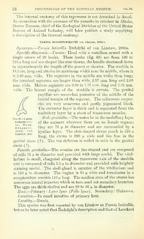
38 PROCEEDINGS OF THE NATIONAL MUSEUM. vol.55.
The internal anatomy of this tapeworm is not described in detail.
In connection with the presence of the parasite in reindeer in Alaska,
Doctor Eansom, chief of the Zoological Division of the United States
Bureau of Animal Industry, will later publish a study supplying
a description of the internal anatomy.
TAENIA MONOSTEPHANOS von Linstow. 1905cc,
Sy7\onym.—Taenia laticollis Rudolphi of von Linstow, 1903w.
SpedfiG diagnosis.—Taenia: Head with a rostellum armed with a
single crown of 29 hooks. These hooks (fig. 42) are from 190 to
210 [J, long and are shaped like rose-thorns, the handle shortened downto approximatelj'' the length of the guard or shorter. The strobila is
14.5 cm. long and attains its maximum width near its middle, where it
is 2.86 mm. wide. The segments in the middle are wider than long;
the terminal segments are longer than wide, 2.57 mm. long and 2.09
mm. wide. Mature segments are 0.99 to 1.18 mm. long and 2.25 mm.wide. The lateral margin of the strobila is serrate. The genital
papillae are somewhat posterior of the middle of the
lateral margin of the segment. The calcareous corpus-
cles are very numerous and partly pigmented black
The cuticular layer is thick and is separated from the
medullary layer by a sheet of transverse muscles.
Fig. 42.—Taenia Male genitalia.—The testes lie in the medullary layer
MONosTEPHAN- q^ \\^q scgmeut whercvcr there are no female organs;
enlaeged. they are 78 \}. in diameter and are surrounded by a
After von hyaline layer. The club-shaped cirrus pouch is 570 [x
long, the cirrus is 290 ]i. wide and lies free in the
genital sinus ( ? ) . The vas deferens is rolled in coils in the genital
sinus ( ? )
.
Female genitalia.—The ovaries are fan-shaped and are composed
of cells 13 [X in diameter and provided with large nuclei. The vitel-
larium is small, elongated along the transverse axis of the strobila
and is composed of cells 7.8 ^ in diameter and provided with brightly
staining nuclei. The shell-gland is anterior of the vitellarium and
is 160 [J, in diameter. The vagina is 34 p. wide and terminates in a
receptaculum seminis 110 [x long. The median stem of the uterus has
numerous lateral branches which in turn send out secondary branches.
The eggs are thick-shelled and are 29 to 36 p. in diameter.
Host.—Primary: Lynx lynx {Felis lynx). Secondary: Unknown.
Location.—In small intestine of primary host.
Locality.—Russia.
This species was first reported by von Linstow as Taenia laticollis^
but as he later noted that Rudolphi's description and that of Leuckart

NO. 225S TAENIOID CESTODES OF DOGS AND CATS—HALL. 39
called for the presence of the customary double crown of hooks, he
described this form as a new species. If von Linstow's material is to
be regarded as actually representative of the normal morphology of
the species to which it belongs, then it should be given generic or sub-
generic rank on the basis of the single crown of hooks, which is, if
anything, more striking than the entire absence of hooks in Taenia-
rhynchus saginatus, T. o,f7'icanus, etc., since the causes which might
operate to abolish one row of hooks in the genus Taenia s. s. might
reasonably be expected to abolish an intercalated row at the sametime. It has not seemed advisable to create a new genus for this
species for the reason that new data should be added in confirmation
of the idea that the conditions reported were normal. It is not anuncommon thing to find specimens of tapeworm in which all the
hooks are missing. It sometimes happens that a single row is miss-
ing, and the writer (Hall, 1910) reported the loss of the large hooks
as a common feature of Taenia halaniceps. In both cases, whether
one or both rows of hooks are missing, it appears to be usually the
result of trauma. What the nature of this traumatic injury may be
is uncertain, but even in mounting specimens hooks will occasionally
be detached. Eailliet (1893) has noted that Leidy has described a
specimen of Taenia pisiformis provided with a single circlet of hooks
(variety a monostephana Diesing), that Bremser has described one
without hooks (variety ^ astephana Diesing), and states in commentthat these facts are not important and that they deal with material
carelessly collected, or very old, or deteriorated material. There are,
therefore, grounds for suspecting that the presence of a single rowof hooks may be deemed accidental. It may be urged that there is
correlated with this an unusual hook shape, consisting of a consider-
able reduction of the handle. This condition differs only in degree
from that in other tapeworms where the handle is short and stubby.
Without regarding it as at all impossible that T. monostephanos mayprove to have a single row of hooks as a normal structure, the possi-
bility that this may not prove to be the case makes it advisable to
leave the matter as it stands and wait for further data.
The reason for including this species in a paper on the tapewormsof North America is that we have probably more information about
the tapeworms of the lynxes than of any other of our wild NorthAmerican carnivores, and a note of parasites found in other conti-
nents serves to aid in securing a really adequate study of the para-
sites of our native species.
Genus MULTICEPS Goeze, 1782a.
Rynonyms.—Taenia Linnaeus, 1758a, part; Cerehrina Acharius,
1782 ; Hydatigena Goeze, l782o, of Batsch, 1786a ; Vesicaria Schrank,

40 PROCEEDINGS OF THE NATIONAL MUSEUM. vol.55.
1788<j; Eydatula Abildgaard, 1790a, part; Hydatis Virey, l798a,
part; Polycefhalus Zeder, 1800a; Coenurus Rudolphi, 1808a; Poly-
cephops Rafinesque, 1815a; Multiplex Liautard in Hall, 1911 (re-
viewer's error). (For additional synonyms, see Hall, 1910.)
Generic diagnosis.—Taeniinae: Strobilate stage similar to that of
genus Taenia. Large hooks usually with a sinuous handle. Thevagina usually shows a reflexed loop in the vicinity of the lateral ex-
cretory canals. Larval stage a coenurus, a bladderworm with a par-
ent vesicle to which are attached numerous heads, internal or external
daughter bladders being present or absent.
Type-species.—Multiceps multiceps (Leske, 1780a) Hall, 1910.
KEY TO Sl'ECIES OF MULTICEPS.
1. Mature segments wider than long ; the hiteral margins of each segment at
times scalloped as a result of a number of constrictions or furrows pass-
ing around the segment transversely ; the posterior margin of each seg-
ment prolonged posteriorly to overlap the anterior margin of the suc-
ceeding segment like a cuff. Small hook with short, blunt curving handle.
Genital papilla very narrowly conical and near posterior margin of seg-
ment. Larva a coenurus with daughter bladders, found in the connective
tissue of rodents MtdUceps serialis, p. 51.
Mature segments longer than broad ; the lateral margins of each segmentsmootli and not scalloped ; the posterior margin of each segment prolonged
very little or not at all to form a projection over the following segment.
Small hooks with long slender handle. Genital papilla posterior of middle
of segment but not near posterior margin. Larva a coenurus without
daughter bladders, found in ungulates, especially ruminants 2.
2. Small hook with long curving handle terminating in a narrow distal ex-
tremity. Large hook with tapering handle with sinuous outline. Testes
do not extend posterior of the ovaries to the vicinity of the vitellarium
or between the vitellarium and the ovaries. Larva a coenurus in the cen-
tral nervous system of ungulates, especially ruminants.
Multiceps multiceps, p. 40.
Small hook with long straight handle terminating in a blunt distal ex-
tremity. Large hook with the handle not tapering, and either straight andblunt or bent dorsally just at the tip. Testes extend posterior of ovaries al-
most to the vitellarium and between the vitellarium and the ovaries.
Larva a coenurus in the central nervous system, lungs, parenchymatousorgans and connective tissue of ruminants Multiceps gaigeri, p. 45.
MULTICEPS MULTICEPS (Leske, 1780a) Hall, 1910.
Synonyms.—Taenia multiceps Leske, 1780ay Vermis vesicularis
socialis Bloch, l780a/ Taenia vesicularis cer^ehrina Goeze, 1782a/
Hydatigena cerebralis Batsch, 1786a/ Vesicaria socialis (Bloch,
1780a) Schrank, 1788a/ Taenia cerebralis (Batsch, 1786a) Gmelin,
1790a/ Polycephalus ovinus Zeder, 1803a/ Coenurus cerebralis

NO. 2258 TAENIOID CESTODES OF DOGS AND CATS—HALL. 41
(Batsch, 1786a) Rudolphi, 1808«y Polycephalus coenurus Tschudi,
1837(2/ Taenia multiplex Leuckart, 18526/ Taenia coenurus (Tschudi,
1837«) Kiichenmeister, 1853c/ Multiplex multiplex (Leuckart,
18526) Liautard in Hall, 1911 (reviewer's error). (For additional
synonyms, see Hall, 1910.)
Specific diagnosis.—Multiceps: The head is piriform in lateral
view and presents a square outline when viewed en face, and is about
800 [J, in diameter. The weakly developed rostellum is about 300 [x in
diameter and bears a double crown of 22 to 32 hooks. The large
hooks (fig. 43) are 150 to 170 pi, long. The blade has only a slight
curvature; the handle is straight in its general direction, its dorsal
and ventral borders sinuous and subparallel, commonly notched on
the dorsal border at a point between the middle of the handle and
the union with the blade, tapering more or less toward the distal
extremity and tending to turn dorsally at this extremity; the guard
is subcylindrical proximally and more or less
conical distally, usually with a slight thicken-
ing at the union of the proximal and distal
portions. The small hooks are 90 to 130 [j.
long. They have a blade of moderate to strong-
curvature; the handle is relatively long and
tapering, usually curved, with the convexity
on the dorsal side, along most of its lengthi/?nmm'
and with the distal extremity turning dorsally
;
the guard, in lateral view, is usually subcylin- fig. 43. - multiceps multi-
drical in the proximal portion and irregularlv ^'^'^^- large and small
• 1 T 11 1-1 • " HOOKS.rounded conical distally, the thickest point
being at the union of the proximal and distal portion, and is glightly
grooved in the median line in a way which gives the hook in some
views the appearance of being bifid. The suckers are located at the
corners of the head and have a diameter of 290 to 300 [jl. There is a
distinct neck which may be 2 to 3 mm. long from the posterior margin
of the suckers to the first distinct segmentation. The strobila
(fig. 44) attains a length of 40 to 100 cm., and consists of 200 to 250
thin and relatively translucent segments, with a maximum width of
about 5 mm. The genital primordia appear rather late and the first
things to stain clearly in toto mounts are the genital pore and the
genital canals from the genital pore inward. The pores are distinct
about 4.7 cm. back of the head, in about the eighteenth segment.
The segments are mature about 10 to 18 cm. back of the head, the
first mature segment being about the one hundred and twenty-fifth.
The mature segments (fig. 45) are either square or oblong and longer
than wide, but not wider than long. Mature segments have gently

42 PROCEEDINGS OF THE NATIONAL MUSEUM. VOD. 55.
convex lateral margins. There are from 12 to 20 gravid segments,
from 6 to 11 mm. long and 3 to 5 mm. wide, usually not over 4 mm.The calcareous corpuscles are very small, 16 to 16 jjl in diameter. Thelongitudinal excretory canals are small, the ventral lying about 420 [x
from the margin of the segment. The genital papilla is rather flat,
but is quite distinct as a result of the translucency of this worm.Afale genitalia.—There are about 200 testes in one horizontal
plane. They are principally confined to the lateral portions of the
median field near the longitudinal
canals, a few scattering testes oc-
cupying a more median position an-
teriorly, but leaving a fairly wide
and distinct field about the median
stem of the uterus (fig. 45). Thetestes do not press close to the field
of the vas deferens and vagina, and
leave a fairly wide free field on each
.side of these canals. They extend
alongside of the ovaries but do not
press close to them and do not pass
posterior of the ovaries in the direc-
tion of the vitellarium. The testes
are practically confined to the fields
lying lateral of lines drawn parallel
to the longitudinal axis of the stro-
bila through the lateral edges of the
ovaries. The vas deferens originates
close to the median stem of the uterus
on the pore side of the segment and
extends in a series of loops to the
cirrus pouch. The cirrus pouch usu-
ally originates in the field lateral of
the longitudinal excretory canals and
is either curved, with the convexity
of the curve toward the anterior por-
tion of the segment, in which case the vagina follows the curve on
the concave posterior side, or is straight and more or less piriform
or cylindrical, in which case the vagina comes in from the genital
eminence to about the middle of the cirrus pouch and then bends off
at a right angle or even turns back toward the lateral margin for
a short di^ance. The cirrus pouch is 315 to 350 \i. long and is 110
to 145 [JL wide.
Female genitalia.—The ovaries are somewhat elongated along the
longitudinal axis of the strobila and inclose an interovarian field
Fig. 44.— Multicep.s multiceps. Entire
STEOBiLA. Actual size. After Hall,
1910.

NO. 2258 TAENIOID CEST0DE8 OF DOGS AND CATS—HALL. 43
that is truncate conical to somewhat conical in shape (fig. 45) . They
are very nearly equal in size. The vitellarium is small, triangular,
and of reticular structure and rather widely set off from the other
male or female genital glands. The median point may or may not
project slightly between the ovaries but the vitellarium does not ex-
tend laterally as far as the ovaries. The shell-gland is small, dis-
tinct, and lies in the clear. The vagina follows the conformation of
the vas deferens, as noted above in the discussion of the male geni-
talia, and then curves in around the nearest ovary to the receptaculum
seminis in the interovarian field. In the transition from mature to
gravid segments, a striking feature is the almost constant forma-
ts"/-' s,.?'^' \
0^WM
Fig. 45.—Multiceps multiceps. Mature seg-
ment. Enlarged. After Beffke, 1891.
Fig. 46.—Multiceps multiceps.Gravid segment. After Hall,
1910.
tion of two lateral branches, one on each side, between the ovary
and the vitellarium. In gravid segments (fig. 46), the median stem
of the uterus has from 9 to 26 lateral branches, rather distinctly
parallel to one another for the most part, and most of these in turn
giving rise to a very few secondary branches, which usually remain
quite distinct and without tendency to fuse. The eggs are from 29
to 37 IX in diameter and have a shell 4 jx thick.
Hosts.—Primary: Ganis familiaris, O. nebracensis. Secondary:
Ovis aries, Bos tav/rus, Ca^pra hircus, Equus caballics, chamois, mouf-
lon, gazelle, antelope, African antelope, Bubalis sp., Ozanna equina
{Hippotragus eguinus), Homo sapiens.

44 PROCEEDINGS OF THE NATIONAL MUSEUM. vol. 55.
Location.—In small intestine of primary host. In central nervous
system, brain or spinal cord, of secondary host.
Localities.—Greece, Germany, Switzerland, England, France, Italy,
Ireland, Holland, Scotland, Austria, Denmark, Iceland, Argentine
Republic, Sardinia, South Australia, New Zealand, German South-
west Africa, India, Hungary, Cape Colony, Chile, Spain, Shetland,
Algeria, United States.
Life history.— Eggs developed by the adult worm in the intestine
of the primary host pass out and are ingested in contaminated food
or water by the secondary host. In the digestive tract an embryo is
released and bores its way through the tissues and into the blood
stream. An embryo which attains the central nervous system will
develop and form a coenurus which, unless removed by surgical inter-
ference, ultimately kills the host annual. The embryos which do not
attain the central nervous system begin development, but very soon
die and undergo degeneration. On the death of the host animal, as
a result of the pressure of the parasite or from other causes, if the
developed bladderworm is ingested by the primary host, part, or
possibly all, of the heads attached to the bladder may develo]> seg-
ments and form the strobilate worm.
The writer (Hall, 1910) has previously stated that the dog is the
only known host of the adult Multiceps multicej)s. Sinco thai time
two records from the coyote (Hall, 1911; Hall, 1912), as a res ilt of
experimental feedings, have been published. The writer also stated
that h(i had not found the record of the adult worm in Alopex laiopus
{Canis lagopvs) credited to Mobius by Railliet (1893a). Since then
the record by Mobius (1874) has been for.rid, but it does not app >ar to
be an acceptjible record of Midtice'ps multiceps. The woims in ques-
tion V ere 55 to 65 mm. long and had gravid segments, and unless weassume that these figures are an error for 55 to 65 cm., they can not
be considered as M. multiceps. In other respects the figures agree
fairly well with 31. multiceps—95 to 113 segments ; head 800 ]i. wide
;
a double crown of 26 hooks, the large 160 pi. long and the small 120 ^
long; the segments quadratic two-thirds of the distance from the
head and 2 mm. long and wide; the terminal segments 3 mm. long
and 2.6 mm. wide; uterus with 12 to 16 lateral branches; maximumQgg m.easurements 31.5 by 27.4 [jl. He states that the shape of the
hooks agrees witli those of M. multiceps. The size of the terminal
segments is not quite that of M. multiceps, but as there appears to be
some po'^sibility of error in the figures given for the strobila. there
may also be some in those for the segments. The writer further
stated, in the paper noted above, that there were no satisfactory
records of larval M. multiceps from man. Brumpt (1913) has since

NO. 2258 TAENIOID CESTODES OF DOGS AND CATS—HALL. 45
recorded a case in which he gives data and figures that must be
accepted as a case of this parasite from the brain of man.
MULTICEPS GAIGERI Hall, 1916.
Synonym.—Coenurus serialis Gervais, 1847a, of Gaiger, 1907, and
Dey, 1909.
Sjieeific diagnosis.—MuUiceps: The head is somewhat piriform in
lateral A'iew and almost square when viewed en face, and is about
950 iJi in diameter. The weak rostellum is about 360 [jl in diameter and
bears a double crown of 28 to 32 hooks. The large hooks (fig. 47)
are 160 to 180 ^ long. The blade is of slight curvature; the handle
is nearly straiglit or only slightly sinuous, with usually a notch,
slight or very pronounced, on the dorsal border between the middle
of the handle and the union with the blade, the dorsal and ventral
borders approximately parallel
and the handle not tapering but
ending bluntly and at times with
a slight curvature dorsally at the
distal extremity; the guard is ap-
proximately cordiform in lateral
view, the point of maximum thick-
ness being a short distance from
the union w4th the blade and
handle, thus forming a neck prox-
imally and a roundly conical distal
portion. The small hooks are 115
to 150 p. long. The blade is
strongly curved; the handle is
long, straight, slightly sinuous in
outline, and tapering to a rather
blunt tip ; the guard is rather oval in lateral view, has a slight medianventral depression without being bifid, and meets the handle at a
very obtuse angle, almost a straight angle. The suckers are compara-
tively large, set prominently at the angles of the quadrate head andAvith relatively small intervals between adjacent suckers, and have
a bulb diameter of 310 to 330 ii. The neck is quite distinct, of
pn-aller diameter than the head, and may measure around 690 [ji, in
length. The entire strobila may measure from 25 cm. to 1.82 meters,
according to conditions and individual development. In a well-de-
veloped strobila, about 40 cm. long, the segments become square about
the middle of the strobila, being 5 mm. long and 5 mm. wide at a
distance of 20 cm. from the head. As the segments become longer
than Avide they become mature. At 30 cm. from the head they are
7 mm. long and 5 mm. wide. At 40 cm. from the head the segments
are 14 mm. long and 2 to 3 mm. wide. The entire strobila is rather
-MlTLTICEPS GAJGERI. LARGE AND SMALLnooKS. From Hall, 1916o.

46 PROCEEDINGS OF THE NATIONAL MUSEUM. VOL. 55.
imm
Fig. 48.—MuLTiCEPS GAiGERi. Young STROBiLA. From Uat.l, lOlf.i.

NO. 225S TAENIOID CESTODES OF DOGS AND CATS—HALL. 47
thin, delicate, and comparatively translucent (fig. 48). The genital
primordia appear in about the thirtieth segment, between 2 and 3
mm. back of the head, first as deeper staining areas in the median
line. These areas become angular, the legs of the angles forming the
genital canals. The primordium of the median stem of the uterus is
the next thing visible. Calcareous corpuscles are especially large
and abundant in the medullary portion of the head and the anterior
portion of the neck, where they are elliptical and about 13 by 8 [x
in diameter, but are elsewhere small, oval, about 4 or 5 jjl in length,
and, though numerous, are relatively inconspicuous. The longitu-
dinal excretory canals are quite distinct, the ventral canal situated
about 225 \). from the margin of the segment and the dorsal canal
lying lateral of this. The genital
papilla is fiat and inconspicuous,
an actual papilla formation being
lacking as a rule, but is readily
observed, owing to the translu-
cency of the segments.
Male genitalia.—There are be-
tween 200 and 225 large, irregu-
larly spherical testes, confined
principally to the lateral portions
\
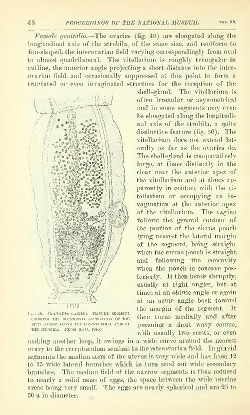
48 PROCEEDINGS OF THE NATIONAL MUSEUM. VOL,. 55.
Female genitalia.—The ovaries (fig. 49) are elongated along the
longitudinal axis of the strobila, of the same size, and reniform to
fan-shaped, the interovarian field varying correspondingly from oval
to almost quadrilateral. The vitellarium is roughly triangular in
outline, the anterior angle projecting a short distance into the inter-
ovarian field and occasionally suppressed at this point to form a
truncated or even invaginated structure for the reception of the
shell-gland. The vitellarium is
V^/
-R ®" '^^ ^ ^
,<^?.«^.
often irregular or asymmetrical
and in some segments may even
be elongated along the longitudi-
nal axig of the strobila, a quite
distinctive feature (fig. 50). Thevitellarium does not extend lat-
erally as far as the ovaries do.
The shell-gland is comparatively
large, at times distinctly in tlie
clear near the anterior apex of
the vitellarium and at times ap-
parently in contact with the vi-
tellarium or occupying an in-
vagination at the anterior apex
of the vitellarium. The vagina
follows the general contour of
the portion of the cirrus pouch
lying nearest the lateral margin
of the segment, being straight
when the cirrus pouch is straight
and following the concavity
when the pouch is concave pos-
teriorly. It then bends abruptly,
usually at right angles, but at
times at an obtuse angle or again
at an acute angle back toward
the margin of the segment. It
Ihen turns medially and after
pursuing a short wavy course,
with usually two crests, or even
niaking another loop, it swings in a wide curve around the nearest
ovary to the receptaculum seminis in the interovarian field. In gravid
segments the median stem of the uterus is very wide and has from 12
to 15 wide lateral branches which in turn send out wide secondary
branches. The median field of the narrow segments is thus reduced
to nearly a solid mass of egg3, the space between the wide uterine
areas being very small. The eggs are nearly spherical and are 25 to
30 |x in diameter.
1 1.,. 50.-
mm.-MULTICEPS GAIGERI. MATURE SEGMENT
SHOWING THE OCCASIONAL ELONGATION OF THE
VITELLARIUM ALONG THE LONGITUDINAL AXIS OF
THE STROBILA. FROM HALL, 191()rt.

NO. 225S TAENIOID CESTOUFJS OF DOGS AND CATS—HALL. 49
Hosts.—Primary : Canis familiaris. Secondary : Capra hircus.
Location.—In small intestine of primary host. In central nervous
system, liver, lungs, spleen, kidney, bladder, internuiscuhir connective
tissue, under peritonemn and subcutaneous in secondary host.
Locality.—India (Punjab, at Lahore, and Bengal) and Ceylon.
Type-material.—IJ. S. National Museum No. 16590. (Bureau of
Animal Industry Helminthological Collection.)
Life Justory.—Eggs produced by the adult worm in the primaryhost pass out and are ingested by the secondary host in contaminated
food and water. In the digestive tract the embryo escapes from its
shell and migrates into the tissues of the host, where it develops into
a coenuriform larva. On ingestion of this larva by the primary host,
some or all of the heads attached to it may give rise to strobilate
worms in the intestine.
Gaiger (1907) first recorded this parasite from the goat at Lahore,
India. He had two cases of the larval parasite occuring in the con-
nective tissue, and because of the site of the worm and the presence
of what ho regarded as daughter cysts, he concluded that the para-
site was Multiceps serialis^ the form commonly found in the connec-
tive tissues of the rabbit, rather than M. multiceps., the form com-monly found in the central nervous system of ungulates. He fed
some cyst material to a dog and to a rat. The rat died in two days
and the hooks were recovered from the stomach. The dog began pass-
ing segments of tapeworm on the fourteenth day and was killed on the
thirty-first day. The small intestines were found packed with tape-
worms from 1 to 40 cm. long.
Two years later, Dey (1909) reported (his parasite from the goat
in Bengal, India. In this case the parasites were found in the brain,
intermuscular connective tissue, in subcutaneous situations, and in
the mesenteries and att?.ched to the peritoneum of the abdominal wall
and the serous covering on the viscera. A dog was first treated with
taeniacides and purgatives and then fed some cyst material. In a
month and a half the dog began passing segments of tapeworm andand was killed two weeks later. Seventy-five tapeworms, the longest
1.82 meters long, were recovered from the small intestine, and the
scolices found to agree with those of the coenurus.
Southwell (1912) reports Coenurus serialis from the goat andTaenia serialis from the dog in Ceylon. The record from the goat
must be regarded as a record of M. gaigeri, and that from the dogmay be.
Gaiger (1915) lists it from the goat in India with a note to the
effect thnt it is common.In a previous paper (Hall, 19105), Gaiger's and Dey's records of
31. s-erialis from the goat were provisionally accepted as correct, with
6205.5—20—Proc.N.M.Vol .55 5

60 PROCEEDINGS OF THE NATIONAL MUSEUM. voi,. 55.
a note to the effect that the parasite would receive further attention
later. Through the courtesy of Doctor Gaiger, specimens of the
coenurus from the goat and of the tapeworm from the dog were
furnished to the United States Bureau of Animal Industry, and an
examination of these showed them to be a new species. That there
are no rabbits in India, a fact Gaiger (1909, 530) has himself noted,
would suggest something of the sort. The specimens of the adult
worm are much smaller than the largest specimens noted by Dey,
but as the size depends largely on whether fresh material is measured
in a stretched condition or preserved material is measured on a flat
surface this is a small matter. In the specimen which I have desig-
nated as type, the segments are mature about 5.5 to 6.5 cm. back of
the head. The general anatomy of the species is much more nearly
that of M. muUiceps than M. serialis. The strobila is delicate and the
vagina shows a peculiar bend which is strongly suggestive of the con-
dition in M. muUicejJS. The hooks are also suggestive of M. multi-
ceps. Gaiger states that the small hook has a bifid guard, but this
statement is often made of tapeworms that do not have a bifid guard.
In these cases, as in this species, there is an appearance of bifidity
found on focusing which is due to the fact that the lateral margins
of the guard are commonly thickened and the median ventral portion
thinned and often slightly grooved, but such conditions must be dif-
ferentiated from the condition of true bifidity which is found in such
hooks as those of Taenia pisiformis.
An examination of the larva shows it to be more closely related
to M. multiceps than to M. serialis morphologically. Placed in a
dish with typical specimens of these species it resembles the former
much more than the latter, and the same is true of scolices detached
from all three species. Gaiger (1907) states "there was a very dis-
tinct tendency in most cysts towards budding off of daughter cysts,
and although actual separation of a daughter cyst was not seen, in
one case there was a distinct neck dividing off a portion of the parent
cyst. Internal budding was complete and many of the cysts were
floating free. The daughter cysts were never more than 2 mm.broad and 4 long, and were always egg-shaped with one head at the
narrow end. The majority of them were attached by a minute
pedicle to the parent cyst, but could be easily detached, and often
they were present in bunches, both attached and free." I find on
examination that a striking feature of this species, so far as the
available material is concerned, is the extraordinary ease with which
the heads detach from the bladder wall and so come to lie free in
the internal fluid. The great majority of the heads in the available
specimen are free and lie in a mass inside the bladder, their former
position being marked by neat prominent apertures perforating the
bladder wall. This is the obvious explanation of Gaiger's statement

NO. 2258. TAENIOID CEST0DE8 OF DOGS AND CATS—HALL. 51
regarding daughter bladders. These are deciduous scolices, but they
are not daugliter bladders. It is interesting to note that we have
here a condition suggestive of what must have been the origin of
daughter bladders, namely, a deciduous scolex, but it is so recent
a development here that the cyst has not yet developed a reparative
process at the site where the scolex separates. This species is appar-
ently intermediate between M. muUiceps and M. serialis in this and
other respects. It presents an interesting study from a physiological
standpoint in that it is capable of development in the central nervous
systein and also in the connective tissues and on serous surfaces,
thus combining the sites of the other species, and causes the forma-
tion of an adventitious capsule, as M. serialis does and 31. niulticeps
X
V
Fig. 51.—MuLTicEi'.s o;.j;i,'i.i.s. IIe'-.d vievted from the front.
does not, even adhering tightly to the brain, from which M. muUi-
ceps slips very easily and with no trace of adhesions.
MULTICEPS SERIALIS (Gervais, 1847fl) Stiles and Stevenson, 1905a.
Synonytns.—Coenurus serialis Gervais, 1847«; Taenia serialis
(Gervais, 1847a) Baillet, 1863<2; Coenurus cuniculi (Diesing, 1863?))
Cobbold, 18645; Coenurus lowzoioi Lindemann, 18G7a; Multiplex
serialis (Gervais, 1847a) Liautard in Hall, 1911 (reviewer's error).
(For additional synonymy, see Hall, 1910.)
Specijic diagnosis.—MuUiceps: The head (fig. 51) is approxi-
mately spherical when viewed from the side, but quadrangular when
viewed en face, and is 850 \j. to 1.5 mm. in diameter. The rostellura is

52 PROCEEDINGS OF THE NATIONAL MUSEUM. VOL. 55.
about 390 \i. in diameter and bears a double crown of 26 to 32 hooks.
The large hooks (figs. 52 and 53) are 135 to 175 [x long. The blade is
of moderate curvature; the handle is slightly sinuous in outline, fre-
quently tapering slightly and with a tendency to turn dorsally at
the distal extremity; the guard is somewhat cordiform in lateral
view, the maximumdiameter being a
short distance fromthe union with the
blade and handle,
and with a projec-
tion toward the
blade along this line
of maximum thick-
ness. The smallhooks are 78 to 120 [k
long. The blade is
of strong to mod-erate curvature
;
the handle is short,
thick, and curved,
the convexity of the
curve being ventral
;
the guard is oval to cordiform and has a median ventral groove.
The suckers are large, with a muscular bulb about 300 [x in diameter,
but are set close together and are not conspicuous. The neck is dis-
tinct, narrow, almost 1 mm. long and may at times have a sharply
defined constriction encircling it. The developed strobila may be
from 20 to 72 cm. long, with a maximum width of
3.5 to 5 mm. and is very thick in the dorso-ventral
dimension. Mature segments are wider than long,
and are between 1.5 and 2 mm. long, and 2.5 and 3 mm.wide. Gravid segments are longer than wide and maybe 6 to 12 mm. long and 3 to 4 mm. wide. The out-
line of the strobila is neither serrate nor smooth, but
is uneven as a result of transverse furrows of the
segments, the normal convexity of the segments and
the prominence of the genital papillae. Calcareous
corpuscles are extremely abundant, oval in shape,
w^ith a maximum diameter of about 20 [x, and are a prominent factor
in making the strobila very dense and opaque. In young seg-
ments the posterior angles of each segment are considerably pro-
longed, but the prolongation extends posteriorly rather than lat-
FlG. 52.—MULTICEPS SERIALIS. LARGE AND SMALL HOOKS.
Fig. 53.—MulticepsSERIALIS. Largeand small hooks.
Enlarged. Af-
ter Neumann1S92.

NO. 2258. TAEXIOID CESTGDES OF DOGS AND CATS—HALL. 53
erally, and so lies close to the succeeding segment iixstead of pro-
jecting and forming a prominent angle and thus giving a serrate
appearance to the strobila. In older segments, the posterior seg-
ment margin is prolonged to form an infundibuliform portion which
surrounds the anterior portion of the succeeding segment (fig. 54).
In a naked eye inspection of the strobila this prolongation is seen
as a pale, fairly wide band separating the segments, whereas most
related tapeworms show a narrow depression between adjacent seg-
ments. The genital papilla is very prominent and is situated posterior
of the middle of the segment, sometimes slightly posterior and often
very much posterior, even practically at the posterior edge of the
lateral border; it is very rarely in the middle of the segment. Thegenital cone is frequently set very deep at the bottom of the genital
pore. The genital primcrdia appear almost immediately behind the
neck as densely staining areas in the median line. Farther back on
Fig. 54.—MULTiCEPS SERIALIS. MaTUJIE segment. K^^LAR,GED.
the strobila the genital canals and the median stem of the uterus
develop as outgrowths from this central area.
3Iale genitalia.—The testes (fig. 54) are numerous and set close
together throughout the testicular field. In the segments in which
they first appear they do not occur in the field of the median stem
of the uterus, but subsequently this field is invaded, as is also the
field of the vas deferens and vagina. They extend in intimate con-
tact with the lateral margins of the ovaries to the vitellarium and
posterior of the ovaries. Anterior of the ovaries there is an ap-
proximately square field free from testes, the lateral testicular fields
being connected by a rather straight band of testes crossing the an-
terior portion of the segment. The vas deferens apparently origi-
nates at some distance from the median stem of the uterus on the
pore side of the segment and is apparently very little looped. The

64 PROCEEDINGS OF THE NATIONAL MUSEUM. VOL. 55.
cirrus pouch is a very narrow, elongated, nozzle-shaped structure,
the distance from the median end of the cirrus pouch to the tip of
the very narrowlj'' elongate conical genital eminence being 200 to
300 [). and the maximum width, near the median end, 55 to 99 [J^;
the pouch is occasionally bent or curved; it is expanded to contain
the few loops of the cirrus only in the median portion. The external
^ ^
aperture is often oninirn. \ the side, rather than
the tip, of the gen-
ital cone.
F em ale genita-
lia.—The ovaries are
often of approxi-
mately equal size and
elongated along the
transverse axis ol
the strobila. Thevitellarium is like-
wise elongated in
the same direction;
it does not extend
laterally as far as
the ovaries; in toto
mounts it sometimes
seems to connect the
posterior portions of
the ovaries. Theshell gland is small
and inconspicuous.
The vagina comes in
from the lateralmargin of the seg-
ment, presents a re-
flexed loop, or sev-
eral loops, near the
longitudinal ex-
cretory canal andcurves sharply around the nearest ovary to the interovarian field. In
gravid segments (fig. 55) the median stem of the uterus has 20 to 25
lateral branches, or so many branches which in turn send out numerousanastomosing branches that a count is impossible. The entire me-
dian field is filled with uterine developments to the point where
almost no remaining parenchyma can be detected between the
growths. Even in gravid segments the posterior flap of the segments
overlaps the anterior portion of the succeeding segment so much as to
Fig. 55.—Multiceps skri.m.is. Gravid segment.

NO. 2258. TAENIOID CESTODES OF DOGS AND CATS—HALL. 55
distinctly overlap the anterior end of the uterus, whereas in related
tapeworms gravid segments show a tendency to become very dis-
tinctly separated and constricted at their point of contact with other
segments. The eggs are elliptical, 31 to 34 [x by 29 to 30 \i in
diameter.
Hosts.—Primary : Canis familiaris. Secondary : Lepus europaeus
{L. timidus), L. californicus, L. c. wallawalla, L. c. deserticola, L.
timidus {L. variabilis), L. californicus texiamis {L. texianus), L.
callotis, Oryctolagus cuniculus, L. cuniculus doinesticus {L. cunicu-
lus), Sciurus vulpinus (?), ^S". niger neglectus (S. vulgaris), jS. caro-
linensis (?), Myocastor coypus {Myopotamus coypus).
Location.—In the smaJl intestine of the primary host. In the con-
nective tissue of the secondary hosts, intermuscular, subcutaneous, etc.
Localities.—England, Scotland, France, Switzerland, Italy, Russia,
Siberia, Australia, New Zealand, Japan, India, Tunis, and the United
States.
Life history.—Eggs developed by the adult worm in the intestine
of the primary host pass out and are ingested by the secondary host
with contaminated food and water. In the digestive tract of the
secondary host the embryo escapes and bores into the tissues and
possibly into the circulation of the host. In the connective tissue
under the skin, between the muscles, and elsewhere, it comes to rest
and develops into a larval form, or coenurus, with numerous heads
attached and also with the production of internal and external
daughter bladders which in turn develop numerous heads. On in-
gestion of tliese bladders by the primary host the tapeworm heads
develop segments and form the strobilate worm.The thickness of this worm and the number of calcareous cor-
puscles make it an unfavorable subject for toto mounts. Unless
the calcareous corpuscles are eliminated by the use of some acid, the
resulting mounts will be very unsatisfactory, and even after they
are eliminated the thickness of the worm makes it a very much less
satisfactory mount than such translucent worms as M. muUiceps,
M. gaigeri, or even such worms as Taenia pisiformis. The descrip-
tion given above is based on specimens furnished to the UnitedStates Bureau of Animal Industry through the courtesj- of Professor
Eailliet. Material from North America available to the present
writer through the courtesy of Prof. F. D. Barker, of the University
of Nebraska, appears to conform in essential respects to the Europeanform.
In a previous paper (Hall, 1910) I have noted that the records of
this parasite from the squirrel were probably correct. Since then I
have recorded (Hall, 1911) the development of this species in Sciurus
carolvnensis.

56 PROCEEDINGS OF THE NATIONAL MUSEUM. ^l. 55.
In the same paper (Hall, 1910) I stated that the parasite appar-
ently did not occur in Germany. Later information is to the sameeffect. It would be interesting to know why a parasite which occurs
in France, Switzerland, Italy, Russia, and Siberia does not, appar-
ently, occur in Germany, Austria, and Hungary, in spite of the fact
that primary and secondary hosts are present in the countries in ques-
tion, and so far as any one knows, under circumstances presumablyas favorable as in the countries in which the parasite is found.
Genus ECHINOCOCCUS Rudolphi, 1801a.
Synonyms.—Acephalocystis Laennec, 1804; Liococcus Bremser,
1819« ; Splanchnococcus Bremser, 1819a ; Echinohokkus Buhl, 1856a
;
Echinococcifer Weinland, 1858*2. (For additional synonyms see
Stiles and Stevenson, 1905a.)
Generic diagnosis.—Taeniinae: Strobila composed of not over four
or five segments, of which only the posterior terminal segment is
gravid. Head armed with a double crown of
hooks. Genital pores marginal and irregularly
alternate. Larval stage an echinococcus, a blad-
FiG. 56.- Echinococcus derwomi with a thick laminated Avail and AvithGRANULOSUS. LARGE
qj^. v/itliout intcmal or external daughter cysts:AND SMALL HOOKS.
.
" .'JX300. After leuck- brood capsules develop in the mother or daugh-ART, 1880.
^^^ cj'sts and contain several scolices. Adults in
carnivorous animals; larval stage in herbivorous and omnivorous
animals.
Type-species.—Echinococcus granulosus (Batsch, 1786a) Rudolphi,
1805a.
ECHINOCOCCUS GRANULOSUS (Batsch, 1786fl) Rudolphi, 1805a.
Synx)nyins.—Taenia echinococcus Siebold, 1853 ; Echinococcifer
echinococcus (von Siebold, 1853) Weinland, 1861; Taenia echinococca
Koeberle, 1861a; Taenia {Echinococcifer) echinococcus of Leuckart,
1863; Taenia {Arhynchotaenia) echinococcus of Diesing, 1864a;
Taenia echinoccus von LinstoAv, 1878; Taenia {Echinococcus) echi-
nococcus of Railliet, 1885a. (For additional synonyms, see Stiles,
1906a.)
Specific diagnosis.—Echinococcus'. Head very small, subglobular,
scarcely 300 /* in diameter. Rostellum prominent, armed with a double
crown of 28 to 50 booklets. The large hooks (fig. 56) are 22 to 30 /*
long (40 to 45 yu,, according to Leuckart). They have a blade of very
slight curvature, which tends to lie in a prolongation of the longi-
tudinal axis of the handle, and which is very thick at the base; the
handle is rather straight, short, and thick, and with dorsal andventral swellings in the median portion; the guard is excessively
shortened and rounded, forming practically a hemispherical protu-

NO. :225s. TAENIOID CESTODES OF DOGS AND CATS—HALL. 57
Fig. 57.— Echino-coccus granu-LOSUS. Strori-
late tapeworm.Enlarged. Af-ter Leuckart,1S80.
berance. The small hooks (fig. 56) are 18 to 22 jjl long (30 to 38 ^,
according to Leuckart). The blade is very sliort and very little
curved, and tends to lie in a prolongation of the longi-
tudinal axis of the handle ; the handle is comparatively
long and narrow, with a prominent dorsal swelling
near the middle which causes a tapering to the distal
extremity ; the guard is relativel}' very much enlarged
and oval in outline. The suckers are 130 \i. in diameter
and are set well back from the rostelluni. The nock
attenuates posterior of the suckers and then gradually
increases in width to meet the anterior margin of the
first segment (fig. 57). This first segment is almost
square, about 21.0 to 260 [jl long and wide, and is rstcrile.
The second segment is about twice as broad in its pos-
terior portion as the first segment and is about foui-
times as long, and is mature. The third segment is
gravid and is much larger than the second, attaining
a maximum length of about 2 mm. and a width of
600 [/,. The longitudinal excretory canals are visible
and calcareous corpuscles are present.
Male genitalia.—Testes 70 ]x in diameter and about
40 to 60 to the segment, most of them in the anterior
and posterior ends and
on the aporal side of
the median field. Thevas deferens originates
near the median line of
the segment, anterior
of the ovary and on the
pore side of the medianstem of the uterus, and
has a few wide coils
before entering the cir-
rus pouch (fig. 58). In
its general course it
extends diagonallyforward to the cirrus
pouch, which is about
500 ]i. long and extends
almost to the medianline of the segment.
Female genitalia.—The ovary is horseshoe-
shaped (fig. 58), withthe median isthmus
and the concavity posterior is subme-
FlG. 58.—ECHINOCOCCUS GR.VNULOSUS. FeONTAL SECTIONS 01mature segments. 0, ovary; s, shell-gland; y, yolk-
After von Erlanger, 1890.GLAND. Enlarged.
narrower than the ends.

58 PROCEEDINGS OF THE NATIONAL MUSEUM. vol.55.
dian in position and about halfway between the plane of the
genital pore and the posterior border of the segment. Thevitellarium is situated near the posterior border of the segment; it
consists of two lobes, a dorsal and a ventral, lying one above the
other, the lobes in turn being formed of two portions, each with its
own duct. The shell-gland is spherical to oval. The ovaries, vitel-
larium, and uterus have the common openings of their tubes within
the shell-gland. The vagina ha3 a large, elongate, setose dilation
about 60 \x in diameter near the genital pore and bends at an obtuse
angle median of this to extend posteriorly and medially to a recep-
taculum seminis about 14 \i in diameter. The uterus develops com-
paratively late, forming a median stem and lateral enlargements, the
outlines of these later becoming quite indistinct. The eggs are 32 to
36 PL by 25 to 30 [i, in diameter.
Hosts.—Primary: Canis familiaris, C. lupus, C. aureus, C. dingo,
C. mesomelas, Felis catus {F. c. domestica), F. concolor. Second-
ary : Homo sapiens, Bos taurus, Ovis aries, Capra hircus, Sus scrofa
domestica, S. scrofa, Pithecus species (Simia cynomolgus) , Pithecus
silenus {Simia silenus), Simia sylvanus {Simia inuus), Ovis ammon,
Camelus hactrianus, C. dromedarius, Giraffa species {Camelopardalis
giraffa), Tetraceros quadricornis, Alces alces, Equus cdbaUus, E.
zehra, E. asinus, Tapirus indicus, Canis familiaris, Felis catus {F. c.
domestica), F. pardus, Macropus ma'pr, Sciurus vulgaris, Mungos
ichneumon {Herpestes ichneumon), Simia sylvanus {Inuus ecau-
datus), Macropus giganteus, Ovis argali, Tapirus ameHcanus, Oryc-
tolagus cuniculus {Lepus cuniculus).
Location.—In small intestine of primary host. In practically
every organ and tissue of the secondary hosts.
Localities.—Cosmopolitan.
Life history.—The eggs produced by the adult worm in the intes-
tine of the primary host pass out and are ingested by the secondary
host in contaminated food or water, as a rule. In the digestive tract
the embryo escapes from the shell and makes its way into the tissues.
Here it develops into the larval stage, a bladderworm which mayattain the size of a child's head, commonly called an echinococcus
or hydatid, and known under numerous scientific names, largely based
on morphological variations or hosts, and characterized by the forma-
tion of a laminated external layer and a delicate internal germinal
layer. As a defense reaction against unfavorable conditions, the
internal genninal membrane or scolices derived from it may give
rise to internal daughter bladders, while proliferative elements in
the cyst wall may form external daughter bladders. Daughter
bladders may form grand-daughter bladders and any bladder of the
series may form brood capsules containing scolices. When the larvae
are ingested by the primary host, the heads contained in these brood
capsules develop segments and form the strobilate worm.

NO. 2258. TAENIOID CESTODES OF DOGS AND VATS—HALL. 59
This tapeworm must be comparatively common in dogs in some
places in the United States, as the larval worms are not infre-
quently met with, especially in swine, in the meat inspection service
of the United States Bureau of Animal Industry. However, the
worm has only been reported by Curtice (1892^7), who collected it in
a dog from the pound in Washington, District of Columbia, and by
Welch (1890(7.), who developed the adult worm in the dog as the re-
sult of feeding experiments. It has been collected from dogs in
Alaska by Doctor Joss of the United States Bureau of Animal In-
dustry, its occurrence here being reported by Ransom (1915).
Family MESOCESTOIDIDAE Fuhrmann, 1907.
Synonym.—Mesocestoidae Ariola, 1899.
Family diagnosis.—Taenioidea : Scolex without rostellum or hooks.
Suckers unarmed. A single set of reproductive organs in each seg-
ment. Genital pores located on the ventral surface of the segment
near the median line. Vagina opens in front of or beside the cirrus
pouch. Eggs in gravid segments inclosed in a single thick-walled
Ggg capsule. Adults in mammals and birds.
Tyjye-genus.—Mesocestoides Vaillant, 1863.
Subfamily Mesocestoidinae Luhe, 1894.
Subfamily diagnosis.—Mesocestoididae : Characters of the family.
Type-genus.—Mesocestoides Vaillant, 1863.
Genus MESOCESTOIDES Vaillant, 1863.
Synonyms.—Monodoridium Walter, 1866; Ptychophysa Hamann,1885a.
Generic diagnosis.—Mesocestoidinae: Genital pores near the an-
terior fourth of the ventral surface. The uterus forms an elongated
median sac with wavy outline, irregularly constricted at certain
points, the single egg capsule forming at the posterior portion of the
uterus and of the segment.
Type-species.—Mesocestoides amhiguus Vaillant, 1863.
No kej^ has been written for the species of Mesocestoides^ for the
reasons given in the discussion of M. litteratus.
MESOCESTOIDES LINEATUS (Goeze, 1782a) Railliet. 1893a.
Synonyms.—Taenia lineata Goeze, 1782a; Holysis lineata (Goeze,
1782a) Zeder, 1803a; Taenia canis lagopodu Rudolphi, 1810a;
(?) Mesocestoides litteratus (Batsch, 1786a) Dolley, 1894a/ Taenia
pseudoelliptica Baillet, 1863a; Taenia pseudo-cupumerina Baillet,
1863a/ Ptychophysa lineata (Goeze, 1782a) Hamann, 1885a.

60 PROCEEDINGS OF THE NATIONAL MUSEUM. VOL. 55.
Fig. 59.— JIesoces-
toides lineatus.
Portion ofStro-
bila , showing ma-
ture segments.
Enlarged. Af-
ter Neumann,1896.
Specific diagnosis.—Mesocestoides: The head is 600 to 900 [i in
diameter, massive and somewhat flattened anteriorly. The rostellum
is rephiced by a slight central depression. The suckers are oval and
display a widely opened longitudinal aperture. The neck is com-
paratively short and thick. The strobila measures from 30 cm. to
2.5 meters, and shows a feebly reddish color along the median line.
The first segments are very short and indistinct. The
follow^ing segments become square, with slightly con-
vex lateral borders and with slightly prominent pos-
terior angles (fig. 59). Segments become mature in
the middle third of the strobila. The terminal seg-
ments measure 4 to 6 mm. long and 2 to 3 mm. wide,
are shaped like melon seeds and are swollen in the
median line by the ovoid uterine capsule filled with
eggs. The male genitalia develop first.
Male genitalia.—Testes large and numerous (about
50) and scattered through the segment both median
and lateral of the longitudinal excretory canals. Vas
deferens describes numerous loops near the mid-dorsal
line to the anterior extremity of the segment, where
it turns abruptly and enters the cirrus pouch. The
cirrus pouch is in the anterior portion of the segment, is well de-
veloped and prominent, piriform, and with its posterior aperture
opening alternately, usually regularly but at times irregularly, a
little to the right and to the left of the median line (fig. 59). Thecirrus is from 50 pi, to 1 mm. long, swollen at its proximal extremity
and commonly found protruded in mature segments.
Female genitalia.—The ovaries are located v^,^i_.__
in the posterior fourth of the segment, and / \
'"
are irregularly spherical to oval. There are ; i
,
two vitellaria which are partly posterior of / \\:'_
the ovaries and partly underneath the pos-
terior portion of the ovaries. The vagina ex-
tends anteriorly and then returns in a sinuous
curve posteriorly from the genital pore and
on the side of the median line opposite to the
cirrus pouch (fig. 60) ; it is without a re-
ceptaculum seminis. The uterus forms as an
elongate sac in the median line, and presents
anteriorly a curve to one side, the cirrus pouch always lying in
the concavity of this curve, and the curve being alternately, regularly
or irregularly, to the right or to the left (fig. 59). The posterior
dilation of the uterus transforms into an egg capsule, the remains
of the primitive uterus persisting as attached cord-like structures,
a short one posteriorly and a longer, sinuous one anteriorly. The
ilVV
Fig. 60.—Mesocestoides unea-
Tus. Mature segment. En-
larged. After Neumann,1896.

NO. 2258. TAENIOID CE8T0DES OF DOG8 AND CATS—HALL. 61
eggs are ovoid, 40 to 60 ja long by 35 to 43 [i, wide, and have two
very thin shells.
Hosts.—Primary: Canis familiaHs., C. lagopus., Yulpes vidpes
{V. vulgaris), Felis catus {F. domestica), Lynx rmhlcus {Lynckus
nuhicus), Lynx caracal, Fells sylvestns {Catus sylvestns, F. catus
ferus), f Mus musculus. Secondary: Unknown.
Location.—'^wvaW intestine.
Localities.—France, Iceland, Germany, Italy, Souidi Africa.
Life history.—Unlaiown.
MESOCESTOIDES LITTERATUS (Batsch, 1786u) Doiley, 1894a.
Synonyms.—Taenia litterata Batsch, 178G«; Alyselminthus lit-
teratus (Batsch, 1786a) Zeder, ISSOa; Halysis litterata (Batsch,
1786a) Zeder, 1803«; Taenia vtriculifera Walter, 1866; Ptychophysa
litterata (Batsch, 1786a) Loennberg, 1896a; Mcsoccstoides utt^icuU-
ferus (Walter, 1866) Muehling, 1898&.
Specific diagnosis.—Mesocestoides: See Mesocestoides lineatus.
Llosts.—Primary: Canis famiUaris, Vidpes vulpes {Canis vid-
pes, Vulpes vulgaris), Martes foina {Miistela foina)^ Canis aureus,
Felis sylvestris {Felis catus). Secondary: Unknown.Location.—Small intestine of primary host.
Locality.—Europe (Italy).
Life histo7vj.—Unknown.Setti (18976) notes the fact that a mnnber of prominent helmin-
thologists have regarded Mesocestoides litteratus as identical with
Mesocestoides lineatus, and then takes up the arguments adduced by
Zschokke (1888) and Condorclli (1891) and concludes that these two
writers are in turn in disagreement as to the specific differences said
to exist. He himself finds transitional and variable features in the
descriptions and his material, which lead him to conclude that the
two species are identical or of only varietal standing. The present
writer is in no position to pass on the validity of the two species, but
there appear to be no descriptions on wdiich a working key could be
constructed at present to differentiate the two species, if there are
two. Under the circumstances the two names have been retained,
but no attempt has been made to write a specific diagnosis, as any that
have been seen would vary in no tangible essential feature from that
of M. lineatus.
Family HYMENOLEPIDIDAE Railliet and Henry, 1909.
Synonijms.—Hymenolepidae Ariola, 1899; Echinocotylidae Ariohi,
1899; Dilepinidae Fuhrmann, 1907.
Family diagnosis.—Taenioidea : Scolex with an armed rostellum
or without rostellum. Hooks on rostellum not hammer-shaped.
Suckers usually unarmed. A single, or less frequently a double, set
62 PROCEEDINGS OF THE NATIONAL MUSEUM. vol. 55.
of reproductive organs in each segment. Genital pores marginal and
bilateral, unilateral, or regularly or irregularly alternate. Eggs with
thin transparent shells. Adults in mammals, birds, reptiles, and
amphibia.
Type-genus.—Hymenolepis Weinland, 1858a.
Subfamily Dipylidiinae Stiles, 1896.
Synonyms.—Ehynchotaenia Diesing, 1850«; Malacolepidota Wein-
land, 1858a; Cystoideae Leuckart, 1863; Cystoidei Leuckart, 1886;
Cystoidotaeniae Eailliet, 1885a; Microtaeniae Claus, 1891; Dipyli-
dinae Eailliet, 1896; Hymenolepinae Perrier, 1897a; Dilepininae
Fuhrmann, 1907 ; Dilepidinae Eailliet and Henry, 1909.
Subfamily diagnosis.—Hymenolepididae : Eostellum armed or, less
frequently, lacking. Suckers unarmed. A single set or, less fre-
quently, a double set of reproductive organs in each segment.
Uterus sac-like, simple or lobulated, or not persistent, breaking downinto numerous egg capsules, each containing one or several eggs.
Parauterine organs not developed. Adults in- mammals, birds, and
i-eptiles.
Type-genus.—Dipylidium Leuckart, 1863.
Genus DIPYLIDIUM Leuckart, 1863.
Synonyms.—Taenia Linnaeus, 1758a, part, of authors; Taenia
{Dipylidium) Leuckart, 1863; Alyselminthus Weinland, 1858a, not
Zeder, 1800a; Cryptocystis Villot, 1882; Microtaenia Claus, 1884;
Depylidium Sonsino, 1895; Dypilidium Castellani and Chalmers,
1910; BipyUduin Daniels, 1910.
Generic diagnosis.—Dipylidiinae: Eostellum armed with several
circlets of hooks which are of rose-thorn shape and usually provided
with a discoidal base. Suckers unarmed. A double set of reproduc-
tive organs in each segment. Genital pores double and opposite.
Testes very numerous, scattered throughout the entire medullary
parenchyma. Vas deferens coiled; vesicula seminalis lacking.
Gravid segments usually longer than broad. Uterus at first reticular,
later breaking up into egg capsules, each containing one or more eggs.
Adults in mammals and birds.
Type-species.—Dipylidium caninum (Linnaeus, 1758a) Eailliet,
1892v.KEY TO SPECIES OF DIPYLIDIDM.
1. Strobila not over 25 mm. long ; rostellum with 4 circlets of hooks, the hooks
45 II long and with a handle and guard ; the cirrus pouch opens ventral of
the vagina ; 1 egg to each capsule DipyUdium trinchesei, p. 69.
Strobila 5 cm. long or longer; hooks rose-thorn-shaijed 2.
2. Head with 16 circlets of hooks; strobila 20 to 30 cm. long; 1 egg to each
egg capsule Dipylidium pasqualei, p. 71.
Heads with 3 to 14 circlets of hooks 3.

NO. 2258. TAENIOID CESTODES OF DOOS AND CATS—HALL. 63
3. Head with 13 or 14 circlets of hooks; strobila 12 to 20 cm. long; 45 to 50
small testes; cirrus pouch 250 to 260 n long and crossing the longitudinal
excretory canals; vitellarium roughly splierical and smaller than the
ovarian lobes; 1 large egg, 52 to 53 ft in diameter, in each egg capsule.
Dipylidiuin chyzcH, p. G8.
Head with not over 6 rows of hooks; 90 or more testes; 2 or more eggs in
each capsule 4.
4. Heads with 6 circlets of hooks; strobila 10 to 23.5 cm. long; 130 to 140 testes;
cirrus pouch extends to the longitudinal excretory canals ; the median
ovarian lobes crescentic. the lateral spherical ; the vitellarium reniform
and as large as an ovarian lobe; egg capsules with 2 to 15 eggs and at
times extending lateral of the longitudinal excretory canals.
Dipylidium scxcoronatum, p. 65.
Heads with 3 to 5 circlets of hooks; cirrus pouch extends across the excre-
tory canals regularly or occasionally ; vitellarium smaller than the ovarian
lobes or smaller than the median lobes 5.
5. Head with 5 circlets of hooks; strobila 5 to 11 cm. long; 90 to 100 large testes;
cirrus pouch curved, convex posteriorly and extending distinctly across
the longitudinal excretory canals; vitellarium smaller than the ovarian
lobes ; spindle-shaped receptaculum seminis present ; vaguia opens posterior
of the cirrus pouch aperture ; eggs 25 ^ in diameter.
Dipylidium ocrleyi, p. 63,
Head with 3 to 4 circlets of hooks; strobila 15 to 40 cm. long; 100 to 200
testes; cirrus pouch piriform and extending to, or occasionally across, the
longitudinal excretory canals ; no receptaculum seminis present, the oviduct
showing a compensatory dilation ; vagina opens ventral of the cirrus
pouch aperture ; eggs 43 to 50 ii in diameter Dipylidium caninum, p. 73.
Parts of the above key are based on illustrations where the features
m question are not specifically covered in the text, and therefore
depend for their reliability on the accuracy of the illustrations and
the constancy of the features covered.
DIPYLIDIUM OERLEYI von Ratz. 1900o.
Specific diagnosis.—Dipylidium: The head (fig. 61) is broad andclub-shaped, and is 330 [jl long and 390 fx wide; it tapers anteriorly
to form a blunted cone ; it is provided with a rostellum 67 /* long and
71 [A wide at the base, conical in shape, with a rounded apex, con-
stantly retracted and lying in a deep depression. The rostellum bears
5 alternating circlets of hooks covering the lower portion of the cone,
the tip of the cone being free from hooks ; the hooks are of rose-thorn
shape, with a somewhat bent ventral root and with a bevel-edged
funnel-shaped base, so that the entire hook has the general form of
a bent funnel. The rostellum is prolonged posteriorly to form a thick
muscular mass, with a neck-like constriction anteriorly and termi-
nating posteriorly in a large thick conical mass. The 4 suckers are
oval and but slightly prominent. The neck is short and thick and
diminishes in diameter posteriorly. The entire strobila is 5 to 11 cm.
long and consists of 70 to 90 segments. The anterior segments are
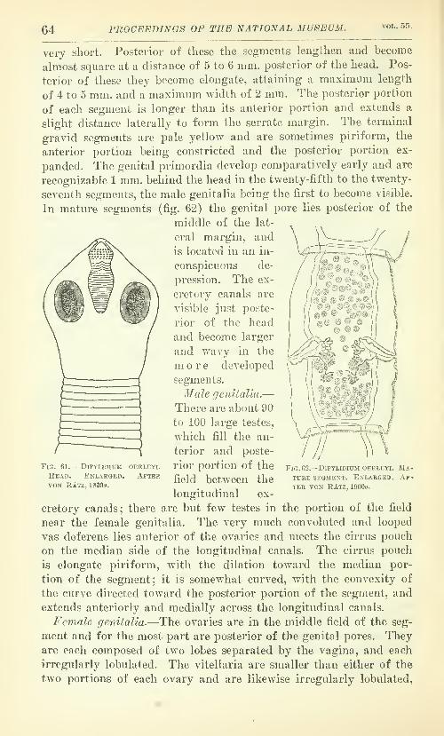
64 PROCEEDINGS OF THE NATIONAL MUSEUM. vol.. 55,
very short. Posterior of these the segments lengthen and become
almost square at a distance of 5 to 6 mm. posterior of the head. Pos-
terior of these they become elongate, attaining a maximum length
of 4 to 5 mm. and a maximum width of 2 mm. The posterior portion
of each segment is longer than its anterior portion and extends a
slight distance laterally to form the serrate margin. The terminal
gi-avid segments are pale yellow and are sometimes piriform, the
anterior portion being constricted and the posterior portion ex-
panded. The genital primordia develop comparatively early and are
recognizable 1 mm. behind the head in the twenty-fifth to the twenty-
seventh segments, the male genitalia being the first to become visible.
In mature segments (fig. 62) the genital pore lies posterior of the
middle of the lat-
eral margin, and
is located in an in-
conspicuous de-
pression. The ex-
cretory canals are
visible just poste-
rior of the head
and become larger
and wavy in the
m ore developed
segments.
Male genitalia.—There are about 90
to 100 large testes,
which fill the an-
terior and poste-
rior portion of the
field between the
longitudinal ex-
cretory canals; there are but few testes in the portion of the field
near the female genitalia. The very much convoluted and looped
vas deferens lies anterior of the ovaries and meets the cirrus pouch
on the median side of the longitudinal canals. The cirrus pouch
is elongate piriform, with the dilation toward the median por-
tion of the segment; it is somewhat curved, with the convexity of
the curve directed toward the posterior portion of the segment, and
extends anteriorly and medially across the longitudinal canals.
Female genitalia.—The ovaries are in the middle field of the seg-
ment and for the most part are posterior of the genital pores. Theyare each composed of two lobes separated by the vagina, and each
irregularly lobulated. The vitellaria are smaller than either of the
two portions of each ovary and are likewise irregularly lobulated,
Fig. 61. — Dipylidium
Head. EnlargedVON RAtz, 19009.
OERLE-iq.
AfterFig. 62.—Dipylidium OERLEYi. Ma-ture segment. Enlargeb. Af-ter VON RAtz, 1900o.

NO. 2258. TAENIOID CE8T0DE8 OF DOGS AND CATS—HALL. 65
and are situated just posterior of the ovaries. The small shell gland
is situated around the oviduct. The opening of tlie vagina is im-
mediately behind that of the cirrus pouch; from here the vagina
extends medially in a somewhat sinuous and curving course and
swings posteriorly between the two lobes of the nearest ovary to a
spindle-shaped receptaculum seminis in the comparatively wide clear
tield between these lobes. The uterus is only recognizable in the
most developed segments in the form of a reticulum lying between
y
\
\
Fig. 63.— DiPYUDiuM sexcorona-
TUM. Head. Enlarged. AfterVON RAtz, 1900.9.
/fo m m.
Fig. 61.—Diptlidium sexcoronatum.Head.
the testes. When full of eggs it bulges out in part and forms the
egg capsules. The eggs lie in groups in these capsules and are round
and 25 /x in diameter.
Host.—Primary: Fclis catus {F. domestica).
Location.—Small intestine.
Locality.—Hungary (Budapest).
Life history.—Unknown.I am indebted for the translation of von Ratz's Hungarian descrip-
tion of this and the next species to the courtesy of Dr. Adolph Eich-
horn, formerly of the United States Bureau of Animal Industry.
DIPTLIDIUM SEXCORONATUM yon Rdtz. 1900o.
Specific diagnosis.—Dipylidium: The head (figs. 63 and 64) is
330 ^ long and 370 {x wide, oval in shape, and terminating conically
62055—20—Proc.N.M.Vol.55 G

66 PROCEEDINGS OF THE NATIONAL MUSEUM. vol,. 56.
anteriorly. The conical rostellum (fig. 65) is usually retracted, andis 46 {x long and 54 [jl wide at the base. The middle and lower thirds
of the rostellum are covered with 6 circlets of hooks, which lie close
together and almost cover this portion of the rostellum. My Ameri-can specimens, as shown in figure 65, have 100 or more hooks. Thehooks are rose-thorn shaped, those of the most anterior row being the
largest and those of the posterior row being the smallest. The ros-
tellum is continued posteriorly as a muscular mass, of which the
anterior portion is thinner and the posterior portion is oval. Thesuckers are slightly prominent and are elongated longitudinally.
The neck is very short and is scalloped by an imperfect transverse
segmentation at the margin, these imperfect segmentations finally
becoming complete and forming the more anterior segments. Theentire strobila is about 10 to
23.5 cm. long. The anterior
segments are short and are
followed by longer ones which
become almost square about
1.5 cm. posterior of the head.
About the middle of the
strobila they are elongated
longitudinally, becoming sev-
eral times longer than wide.
The longest segments are
about at the union of the
middle and posterior thirds
of the strobila, and are 6 to 7
mm. long and 1 to 1.5 mm.wide. Posterior of this point
they become shorter, wider,
and thicker. The enlarged
posterior margin of each segment forms a sort of collar about the
smaller anterior margin of the succeeding segment. The primordium
of the genital pore forms on young segments as a small protuberance,
but in more mature segments this disappears and leaves instead a
small depression, which is posterior of the middle of the lateral
margin of the segment. The longitudinal excretory canals are very
prominent, even in young segments, and later show as wide wavycanals; the transverse canals are likewise very wide and are situated
at the union of adjacent segments.
Male genitalia.—The testes (fig. 66) are spherical, about 130 to
140 in number, and occupy nearly all the space between the longi-
tudinal canals not occupied by the other genitalia ; they are especially
numerous in the middle and posterior portions of this field and leave
a clear area in the anterior portion of the segment. The vas deferens
^omm.
Fig. 65.—Dipylidium sexcoronatum. Rostellum.

NO. 2258. TAENIOID CESTODES OF DOGS AND CATS—HALL. 67
is much looped; it lies just anterior of the ovary and pursues a fairly
straight course, aside from the loops, to the cirrus pouch. In youngsegments the cirrus pouch has the shape of an hourglass, widened
proximally and distally and constricted mediall}'; in mature seg-
ments it is piriform. It extends to the longitudinal excretory canal,
but does not cross it.
Female genitalia.—Each ovary (fig. 66) consists of two distinct
lobes, each of a loose, irregular structure, the lobe toward the medianside of the segment following the convexity of the curve of the vagina
in a roughly crescentic outline, and the one toward the lateral side of
the segment lying in the concavity of the curve of the vagina in a
roughly circular outline. The ovaries lie mostly posterior of the
genital pores. The vitellaria are relatively
compact reniform structures lying posterior
of the ovaries, and each vitellarium is about
as large as one of the two principal parts of
each ovary. The shell-gland is a relatively
large and compact reniform structure between
the ovary and the vitellarium and displaced
toward the median line. In young segments
the vagina passes almost straight medially;
in mature segments it is very slightly wavy in
the lateral field of the segment and then
curA'es posteriorly after crossing the excretory
canal to form a spindle-shaped receptaculum
seminis in the very narrow curved field be-
tween the tw^o principal portions of each
ovary. The uterus is only seen in later seg-
ments and is a fine tubular reticulum branch-
ing through the median field and especially
distinct between the ( ? female) genitalia
and posterior of these. After the constric-
tion of some parts it forms egg capsules containing 2-3-8-15 eggs
to a capsule. The eggs are spherical and are 21 [j, in diameter. Thedensest clumps of eggs are posterior of the oviducts, where the
capsules form almost a connected network and may lie lateral of the
longitudinal excretory canals.
Host.—Primary: Cants famUiaris.
Location.—Small intestine.
Localities.—Hungary (Budapest) ; United States (Bethesda,
Maryland; Detroit, Michigan).
Life history.—Unknown.This species, reported for the first time from the United States by
Hall (1917), the second record of its occurrence, was collected by the
writer from a dog shipped from Fallon, Nevada, in 1910, and exam-
FlG. Gil.—DiPYUDIUM SEXCORO-NATUM. Mature segment.Enlarged. After von RAtz,
18000.

68 PROCEEDINGS OF THE NATIONAL MUSEUM. VOL. 55.
ined post mortem for parasites in 1913. In view of what we knowof the length of life for dog cestodes, it is unlikely that the wormsfound y»ere present when the dog arrived at Bethesda, Maryland.
However, it is possible that the dog was infested with worms of this
species and that the worms found were some from infestation due to
eating intermediate hosts, presumably fleas or lice, as in D. caninum,
which had been infected from the original worms. It is also possible
that the infestation origi-
nated entirely at Bethesda.
A number of specimens
were collected at Detroit
from dogs Avhich undoubt-
edly became infested in
that locality.
Part of the specific de-
; cription is based on vonRatz's illustrations, and so
depends for its reliability on the accuracy of the illustrations and the
constancy of the features illustrated.
Fig. bT. — ] >iPYLi-
dium chyzeri.
Head. E n -
LARGE1'. AF-TF.K VON RAT2,
1S97&.
I'lG. 6S.—I'lPYUniUM CHYZEKI. RCS-TELLtTM. Enlarged. After vonRAtz, 18976.
DIPYLIDIUM CHYZERI von Ratz, 18976.
Specific diagnosis.—Dipylidium: The head (fig. 67) is small, 352
to 432 ^ in diameter, and conical. The rostellum (fig. 68) is conical,
112 [JL long, and terminates in a rounded tip.
There are 13 to 14 circlets of hooks, forming diag-
onal rows tovvurd the tip of the rostellum; these
hooks are of rose-thorn shape with a flat basal
portion attached at an angle. This basal part is
formed by a long flat plate, thinned at both ends
and bent at the anterior end, and the hook length
proper is not over two-thirds the length of this
basal portion. The largest hooks are near the tip
of the rostellum, where they are 14 pi. long and
5.5 ]}. thick; nearer the base they are 10.5 by 5 [j.;
and those at the base are still smaller. Thestrobila is 12 to 20 cm. long and attains its maxi-
mimi width, 1.4 to 1.6 mm., near the middle, at
which point the segments are almost quadrate.(
Fig. 69.—D I p y l I d I u mCHTZERi. Mature seg-
ment. Enlarged. Af-ter VON RAtz, 1897b.
The free gravid segments are 4.5 mm. long and
TOO to 750 [J. wide. The genital pores are anterior
of the middle of the lateral margin of the seg-
ment. The longitudinal and transverse excretory canals are of about
the same width.
Male genitalia.—The testes (fig. 69) are small, 45 to 60 in num-
ber, and are scattered at comparatively wide intervals throughout
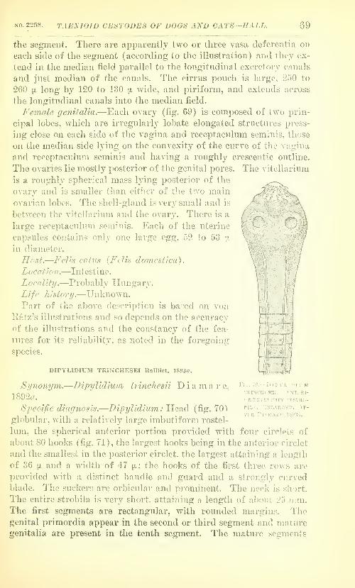
NO. 2258. TAEKIOID CESTODES OF U0G8 AND CATS—HALL. 69
the segment. There are apparently two or three vasa deferentia on
each side of the segment (according to the illustration) and they ex-
tend in the median field parallel to the longitudinal excretory canals
and just median of the canals. The cirrus pouch is large, 250 to
260 [). long by 120 to 130 jjl wide, and piriform, and extends across
the longitudinal canals into the median field.
Female genitalia.—Each ovary (fig. 69) is composed of two prin-
cipal lobes, which arc irregularly lobate elongated structures press-
ing close on each side of the vagina and receptacvilum seminis, those
on the median side lying on the convexity of the curve of the vagina
and receptaculum seminis and having a roughly crescentic outline.
The ovaries lie mostly posterior of the genital pores. The vitelhiriura
is a roughly spherical mass lying posterior of the
ovary and is smaller than either of the two main
ovarian lobes. The shell-gland is very small and is
between the vitellarium and the ovary. There is a
large receptaculum seminis. Each of the uterine
capsules contains only one large egg, 52 to 53 ;;.
in diameter.
nest.—Felis catus {Felis do7nesf/ca).
Location
.
—Intestine.
Locality.—Probably Hungary.
Life history/.—Unknown.Part of the above description is based on von
Ratz's illustrations and so depends on the accuracy
of the illustrations and the constancy of the fea-
tures for its reliability, as noted in the foregoing
si^ecie^.
DIPYLIDIUM TRINCHESEI Railliet, 1893rt.
-Dipylidium trinchesii Diamare, Fig. 70.—D ! p y l I d ut mTRiNCHisEi. .Anteri-
or EXTRrMITT OF STRO-
PILA. KnI-ARGKD. A^-TF.R Dl.\MARE, 18936.
Synonym.1892«.
Specific diagnosis.—Dipylidium: Head (fig. 70)
globular, with a relatively large imbutiform rostel-
lum, the spherical anterior portion provided with four circlets of
about 80 hooks (fig. 71) , the largest hooks being in the anterior circlet
and the smallest in the posterior circlet, the largest attaining a length
of 36 PL and a width of 47 pi.; the hooks of the first three rows are
provided with a distinct handle and guard and a strongly curved
blade. The suckers are orbicular and prominent. The neck is short.
The entire strobila is very short, attaining a length of about 25 mm.The first segments are rectangular, with rounded margins. Thegenital primordia appear in the second or third segment and maturegenitalia are present in the tenth segment. The mature segments

70 PROCEEDINGS OF THE NATIONAL MUSEUM. VOL. 55.
(fig. 72) are vase-shaped. Terminal gravid segments (fig. 73) be-
come four times as long as wide. The genital pores are considerably
anterior of the middle of the lateral margin of the segment, their
position marking the point of maximum width of the vase-shaped
segment.
31ale genitalia.—Testes, judging from the figure, are few in num-
ber and scattered through the median field. Vas deferens in loops
directed toward the anterior portion of the segment, and pigmented
black. Cirrus pouch cylindrical, somewhat twisted, and opening
ventral of the vagina; it apparently crosses the longitudinal canals
in a wide curve, the convexity of the curve directed posteriorly.
Female genitalia.—Ovaries compact and globular to reniform.
Vitellaria spherical. The vagina opens dorsal of the cirrus pouch
/
Fig. 71.—Dipylidium trinchesei. Hooks, a, Hook from first orMOST ANTERIOR ROW; 6, FROM SECOND ROW; C, FROM THIRD ROW;d, FROM FOURTH ROW. KnLARGED. AfTER DiAMARE, 18936.
A
Fig. 72.—Dipylidium trinche-
sEi. Mature segment. En-larged. After Dumare,18936.
and passes medially to a large receptaculum seminis between the
ovarian lobes. The egg capsules contain one egg each.
Hosts.—Primary : Felis catus {F. domestica) . Secondary : Zamenisviridiflavus.
Location.—In small intestine of primary host. In cysts in the
intestinal wall of the secondary host.
Localities.—Italy (Naples) ; Egypt (Alexandria).
Life history.—Diamare believes that Cysticercus acanthotetra Pa-
rona (1887) found in Sardinia in cysts in the intestinal wall of the
snake Zamenis viridiflavus, is the larva of Dipylidium trinchesai.
If this theory is correct, the worm is an accidental parasite of the
cat, as cats are not generally in the habit of eating snakes, as
Blanchard (1907) has noted. It might also be urged that there is
probably comparatively little opportunity for the ingestion of the
eggs of the adult worm by a secondary host like the snake except by

NO. 2258. TABNIOID CESTODES OF DOGS AND CATS—HALL. 71
^\
/
\
/[
WBSSAJl;fm^cm0-
ingesting the primary host, a thing which might readily occur if the
usual primary host is some small mammal. This would be a quite
unusual and interesting case, where the infestation with a larval
stage depended on the
ingestion of a primary
host by a secondary
host. But in view of
the fact that the sec-
ondary host must in
turn be eaten by the
primary host, and in
view of the fact that
the only known inter-
mediate hosts of a spe-
cies of Dvpylidium are
insects, Diamare's hy-
pothesis needs experi-
mental verification be-
fore it can be accepted.
Diamare (1892«) first
gave the number of
hooks in this species as
65, but later (Diamare,
1893) gave the numberas about 80.
DIPYLIDIUM PASQUALEIDiamare, 1893a.
Specific diagnosis.—Dipylidium: Head (fig
74) globular, 800 to 900
]}. in diameter. Theelongate,
claviform
rostel 1 umis acumi-
nate ante-
riorly and
i s armed
with 16 circlets of hooks which
have a discoid, almost circular base,
and are 7 ^ long and 8 [;, wide,
those of the last row being smaller.
The suckers are rounded. Thestrobila is from 20 to 30 cm. long
or longer. The genitalia develop
rather late and mature segments
(fig. 75) are still rather short and quadrilateral.
m^Fio. 74.—Dipylidium pas-
QUALEi. Anterior por-
tion OF strobila. En-larged. After Dia-mare, 18936.
Fig. 73.—Dipylidium trinchesei.
Gravid segment. Enlarged.
After Dumare, 18936.
Fig. 75.—Dipylidium pasqualei. Mature seg-
ment. Enlarged. After Diamare, 18936.
The genital pore

72 PROCEEDINGS OF THE NATIONAL MUSEUM. TOL. 5S.
is anterior of the middle of the lateral margin of the segment, and
the cirrus pouch and vagina open at the same level. Gravid segments
(fig. 76) are lanceolate in out-
line. The longitudinal ex-
cretory canals are very large,
three times as large as in
Dipylidium caninum. Calcare-
ous corpuscles are numerous.
Male genitalia.—Judgingfrom the illustration, the testes
are large, few in number(about 50?) and uniformly
distributed through the me-
dian field, not present lateral
of the longitudinal excretory
canals or of the ovaries. Thevas deferens appears to be
moderately looped distally,
and coiled in the ant6ro-laterai
corner of the median field
proximally. The cirrus pouch
is figured as extending to the
lateral canals or lying entirely
lateral of these canals, but not
crossing them.
Female genitalia.—Theovary is bilobed and branch-
ing. The vitellarium is bi-
lobed. Shell gland (?). Thevagina opens into a large fusi-
form receptaculum seminis.
The Qgg capsules contain one
egg each, and are distributed
throughout the fields lateral of
the longitudinal canal as well
as median of it.
Host.—Primary : Felis catus
{F. domestica)
.
Location.—Small intestine.
Locality.—Egypt (Alexan-
dria).
Life history.—Unknown.The figures for the size of the head are based on Diamare's state-
ment that the head is twice as large as that of D. echinorhynchoides.
The dimensions of the hooks are taken from Blanchard (1907).
Fig. 76.—DrPTLiDiuM pasqualei. Gravid segmentEnlarged. After Diamare, 18936.

NO. 2268. TAENJOID CEST0DE8 OF D008 AND CATS—HALL. 73
DIPYLIDIUM CANINUM (Lmnaeos. 1758rt) Rallliet, 1892 c.
Synonyms.—Taenia canina Linnaeus, 1758a; T. 7fioniliformis Pal-
las, 1781, not Batscli, 1786a.; T, cucumerina Bloch, 1782a; T. cateni-
formis Goeze, 1782a; T. elUptica Goeze, 1782a; T. ellyptica Batsch,
1786a; T. cateniformis canina Linnaeus of Gmelin, 1790a; T. c.
fells Gmelin, 1790a; Alyselminthus ellypticus (Batsch, 1786a) Zeder,
1800a; Taenia cunciceps Zeder, 1800a; Ilalysis ellyptica (Batsch,
1786a) Zeder, 1803a; Tenia canina (Linnaeus, 1758a) van Beneden,
I,-
M#
i/iomm
FlU. 77.—DlPYLIPIUM CANimJM. HEAD.
1861a; T. cucumerina (Bloch, 1782a) van Beneden, 1861a/ Taenia
{Dipylidium) cucumerina Bloch of Leuckart, 1863; Gryptocystis
trichodectis Villot, 1882; DypyUdium caninv/m (Linnaeus, 1758a)
von Katz, 1897.
Specific diagnosis.—Dipylidium: Head (fig. 77) small and rhom-
boidal, 350 to 460 ]}. in diameter in a state of contraction. K-ostellum
claviform, 185 [jl long in a state of complete evagination, 110 to 120 n
wide, and capable of retraction into a deep cephalic infundibulum.
It is armed with three or, usually, four circlets of about 60 hooks

74 PROCEEDINGS OF THE NATIONAL MUSEUM. VOL. 55,
(fig. 78) of rose-thorn shape; the hooks of the most anterior row are
the larger, 12 to 15 [i long, and those of the most posterior row the
smallest, 5 to 6 )u, long. The hooks are very easily lost and specimens
are frequently found without any hooks or with only a few still in
place. The suckers are very large and ellipsoidal. The neck is short
and thin. The strobila is 15 to 40 cm. long and is composed of 80 to
120 segments. The segments are at first very short, then trapezoidal,
and finally much longer than wide. Genitalia develop later. Matureand gravid segments (figs. 79
and 80) have distinctly convex
lateral margins, giving the seg-
ments the characteristic cucum-
ber-seed shape. Gravid seg-
ments are 8 to 11 mm. long and
1.5 to 3 mm. wide, and are often
reddish in color. Genital pores
C^
//7W- ?/' V
'
i/iomm
Fig. 78.—Dipyudium caninum. Hooks.
are in the middle of the lateral margin of the segment or at times
posterior of this. The lateral excretory canals are quite prominent.
Male genitalia.—T&stes numerous, 100 to 200 in number, and oc-
cupying most of the parenchyma not occupied by the female genitalia.
The vas deferens in mature segments extends anteriorly and laterally
from the vicinity of the median lobe of each uterus, toward the cirrus
pouch, only the final loop extending posteriorly to the cirrus pouch.
The cirrus pouch may extend to the longitudinal excretory canal, but
is usually entirely lateral of this ; its aperture
is dorsal to that of the vagina.
Female genitalia.—The two lobes of each
ovary are very distinct, separated into lo-
bules, and loosely formed; they are com-
monly said to be wing-shaped, but they are
very irregular and difficult to characterize.
The vitellarium is some distance posterior of
the ovary and is irregular and very loosely
lobulate; it is smaller than the median ova-
rian lobe, and equal to or smaller than the lateral lobe. The shell
gland is between the ovary and the vitellarium. The vagina extends
between the two lobes of each ovary and does not dilate to form a
receptaculum seminis. The oviduct is dilated to compensate for the
absence of a receptaculum seminis. Each egg capsule contains from
5 to 20 globular eggs, 43 to 50 fi in diameter, with thin shells and
with an onchosphere 25 to 36 n* in diameter, provided with hooks 11
to 14 fi long.
r
Fig. 79.—Dipyudium caninum.
Mature segment, x 12.5.
After Neumann in Rail-
LIET, 18936.

NO. 2258. TAENIOID CEST0DE8 OF DOGS AND CATS—HALL. 75
Hosts.—Primary : Canis familiaris, C. mesomelas, Felis srjlvestris
(F. catus), F. catus (F. catm domestica), F. inanicnlata, Homo
sapiens. Secondary : Tricho-
dectes cani s, Ctenocephalus
canis. Pulex irritans.
Location.—In small intestine
of primary host. In the visceral
cavity of secondary hosts.
Locality.—Cosmopolitan.
Life history—Eggs developed
by the adult worm in the in-
testine of the primary host were
once supposed to be ingested by
the secondary hosts either on
the skin of the primary host,
where it was soiled by feces, or
from the gravid segments di-
rect. Melnikov (18G9) has
shown that TricTiodectes eats
the eggs as it feeds on the skin
of the dog, but Joyenx (1916)
has demonstrated that fleas in-
gest eggs while the fleas are
larvae feeding on debris, the
adult flea being unable to ingest
a tapeworm ^^g. In the digest-
ive tract of the secondary host
the embryo escapes and makes
its way to the visceral cavity
where it develops into the tiny
larva, called Cryptocysti'^trichodectis. When lice and
fle«s infested with those larvao
are ingested by the primarj'
host, the larval head passes to
the intestine and gives rise to
the strobilate worm,
SYNOPTICAL KEY.
The tapeworms of the dog,
cat, and some related carnivores
may be separated into the fol-
lowing groups and genera by the use of the appended key from Hall
(19165), (corrected for stenographic errors involving Dipylidium
fttsqualei and D. chyseri), which is intended to designate the rela-
FlG. 80.—DiPYUDIUM CANINUM. ORAVID SEGMENT.
Enlarged. After Diamark, lSOn'».

76 PROCEEDINGS OF THE NATIONAL MUSEUM. vol. 55.
tions and importance of the tapeworms as well as to distinguish them
from one another. The key does not cover the species in the family
Diphyllobothriidae.
1. Head pi'ovided with two slit-like suckei-s, and segments with a rosette-shaped
utei'us having a special aperture in the midveutral line for the discharge
of eggs. Larvae in fish Diphyllobothriidae, p. 4.
Head with four cup-shaped suckers 2.
2. Follicular yolk glands in the lateral fields. Accidental in the dog.
Ophiotaenia punica, p. 4.
Yolk glands in median field, never in lateral fields 3.
3. Genital pore located on the ventral surface near the median line. Eggs In
gravid segments, contained in a single tliick-slielled egg capsule.
Mesocestoiiles lineatus, Mesocestoides litteratus
(probably identical species), pp. 59, 61.
Genital pore lateral. Eggs in gravid segments, contained in a uterus or in
numerous egg capsules 4.
4. Usually large forms. Genital pores irregularly alternate. Rostellum usually
well developed and usually armed with a double crown of hooks, rarely with
a single ( ?) crown of hooks or unarmed. Suckers unarmed. Uterus with a
median stem and lateral branches. Eggs thick shelled Taeniidae, 5, p. 5.
Usually small forms. Genital pores single or double; if single, regularly or
irregularly alternate. Rostellum present or absent ; if present, armed with
one to numerous rows of hook.s. Suckers armed or unarmed. Uterus sac-
like and persistent, or a single or double uterus with one or several para-
uterine organs, to which the eggs pass in the final stage of development.
Eggs vnth thin, transparent shells Hymenolepididae, 19, p. 61.
5. Strobila less tlian 1 cm. long and composed of a head and 3 segments, only
one of the segments being gravid at a time. Lateral uterine branches
often quite indistinct. Yolk-gland globular. Larval stage an echinococcus
with thick laminated wall, and developing brood capsules containing the
larval scolices Echinococcus granulosus, p. 56.
Strobila at least several centimeters long and coruposed of a head and nu-
merous segments, from 10 to hundreds, with a number of segments usually
gravid at one time. Lateral uterine branches usually distinct, at least in
early stages of formation. Yolk gland posterior of ovaries and elongate or
triangular, with one side parallel to the posterior margin of the segment.
Larval stage a bladderworm with thin walls and never containing brood
capsules 6.
6. Strobila without a neck and with a double circlet of many and very large
hooks, the large hook over 300 ^ long, or with only a single circlet of hooks
(?). B!add(Hv;orn!^:, so far as known, with a small caudal bladder and
with a long seguu']ited structure connecting the bladder and head. Para-
sitic in Felidae 7.
Strobila with a neck and with a double circlet of hooks, the large hook less
than 300 ji long. Bladderworms with a caudal bladder, a head and a mod-
erate-sized neck. Parasitic in Canidae 10.
7. Rostellum with a single circlet of hooks of rose-thorn-shape. Reported from
Lijvx lynx (Felis lijnx) Taenia monostephanos, p. 38.
Rostellum with a double circlet of hooks, the large hooks over 300 [jl long—8.
8. Large hooks 60 to 74 in number; 320 to 355 fi long; the large hooks ar-
ranged so as to form in effect 2 circlets of large hooks, being alternately
nearer to the center of the rostellum and farther from it.
Taenia macrocystis, p. IS.
Large hooks not over 60 in number 9.

NO. 2258. TAENIOID CESTODES OF DOGS AND CATS—HALL. 77
9. Hooks 26 to 52 in uuniber. Well developed sphincter vaginae.
Taenia iaeniaciorniin, p. 9.
Hooks 28 to CO in number. Probably identical with the preceding species.
Taenia laiicoUis, p. 8.
10. Vagina with a reflex curve near the lateral excretory caual^^. Large
hooks 135 to 180 fi long and usually with a sinuous handle.
Multiceps, 11, p. 39.
Vagina without a reflexed curve near the lateral canals Taenia, 13, p. 6.
11. Mature segments wider than long; the lateral margins of each segment
often scalloped as a result of a number of constrictions or furrows
passing around the segment ti'ansversely ; the posterior margin of each
segment prolonged posteriorly to overlap the anterior margin of tlie suc-
ceeding segment like a cuff. Small hook with short, blunt curving handle.
Genital papilla very narrowly coiiical and in posterior half of segment,
often near posterior margin. Larva a coenurus witli daughter bladders,
found in the connective tissue of rodents (including the Lagomorpha).
Multiceps serialis, p. 51.
Mature segments longer than broad ; the lateral margins of each segment
smooth and not scalloped ; the posterior margin of each segment pro-
longed very little or not at all to form a projection over the following
segment. Small hooks with long slender handle. Genital papilla pos-
terior of middle of segment but never near posterior margin. Larva a
coenurus without daughter bladders, found in ungulates, especially
ruminants 12.
12. Small hook witli long curving handle terminating in a blunt distal ex-
tremity. Large hook with tapering handle with sinuous outline. Testes
do not extend posterior of the ovaries to the vicinity of the vitellarium
or between the vitellarium and the ovaries. Larva a coenurus in the
central nervous system of ungulates, especially run:iinants.
Multiceps multiceps, p. 40.
Small hook with long straight handle terminating in a blunt distal ex-
tremity. Large hook with the handle not tapering, and either straight
and blunt or bent dorsally just at the tip. Testes extend posterior of
the ovaries, almost to the vitellarium and between the vitellarium andthe ovaries. Larva a coenurus in the central nervous system, lungs,
parenchymatous organs and connective tissue of ruminants.
Multiceps gaigeri, p. 45.
13. Forms in which the vagina crosses the ovary on the pore side in some seg-
ments, and forms in which the very large genital papilla is practically as
long as the margin of the narrow segments. Bladderworms in the
musculature of food animals 14.
Forms in which the vagina does not cross the ovary on the pore side, andin which the genital papilla is never nearly as long as the margin of
the segments 15.
14. Gravid uterus with 20 to 25 latei'al branches on each side of the medianstem. Vagina crosses the ovary on the pore side in some segments.
Genital papilla much shorter than the segment margin. Bladderwormin musculature of sheep Taenia ovis, p. 32.
Gravid uterus with not over 10 lateral branches on each side of the
median stem. Vagina does not cross the ovary on the pore side of the
segment. Genital papilla practically as long as the lateral margin of the
narrow segments. Bladderworm in the musculature of reindeer.
Taenia krahbei, p. 36.

78 FROCEEDINGS OF THE NATIONAL MUSEUM. vol. 55.
15. Forms with tlie books from 170 to 294 ii long. Bladderworms attached
to omenta, mesenteries, or peritoneal serosa 16.
Forms with the large hooks 185 to 145 m long. Probably accidental parasites
of the dog 17.
16. Large hooks 225 to 294 ^ long. Testes extend posterior of the vitellarium.
Mature segments approximately square. Gravid segments with 8 to 14
lateral branches on each side. Vagina only slightly dilated and curved
near margin of segnrent. Bladderworm in rabbits, rarely in mous© or
beaver Taenia pisiformis, p. 22.
Large hooks 170 to 224 ix long. Testes do not extend posterior of the
vitellarium. IMature segments distinctly wider than long. Gravid seg-
ments with 5 to 10 lateral branches on each side. Vagina forms a sort
of crescent by dilation and curvature near lateral margin of segment.
Gravid segments with a median longitudinal groove terminating in a
notch posteriorly. Bladderworm usually in ruminants, also reported from
monkeys and rodents Taenia hydati'jena, p. 28.
17. Guard of small hook twisted so that its flat surface tends to lie in the plane
of the blade and handle Taenia Iracfii/soma, p. 21.
Guard of small hook not so twisted 18.
18. Head acorn-shaped, with hooks far anterior of the suckers. Mature seg-
ments approximately square Taenia haJaniccps, p. 16.
Head not acorn-shaped, and hooks not far anterior of suckers. Mature seg-
ments distinctly broader than long Taenia Jjrauni, p. 19.
19. Strobila not over 25 mm. long ; rostellum with 4 circlets of hooks, the hooks
45 pi long and with a handle and guard ; the cirrus pouch opens ventral of
the vagina; 1 egg to each egg capsule Dipylidium trinchesei, p. 69.
Strobila 5 cm. long or longer ; hooks rose-thoru-shaped 20.
20. Head with IG circlets of hooks; strobila 20 to 30 cm. long; 1 egg to each
egg capsule Dipylidinm pasqualei, p. 71.
Head with 3 to 14 circlets of hooks 21.
21. Head M-ith 13 to 14 circlets of hooks, strobila 12 to 20 cm. long; 45 to 50
small testes : cirrus pouch 250 to 260 fi long and crossing the longitudinal
excretory canals; vitellarium rouglily spherical and smaller than the
ovarian lobes ; 1 large egg, 52 to 53 fi in diameter, in each egg capsule.
Dipylidium chyzeri, p. 68.
Head with not over 6 rows of hooks ; 90 or more testes ; 2 or more eggs in
each egg capsule 22.
22. Head with 6 circlets of hooks; strobila 10 to 23.5 cm. long; 130 to 140
testes, cirrus pouch extends to the longitudinal excretory canals ; the me-
dian ovarian lobes crescentic, the lateral spherical ; the vitellarium reni-
form and as large as an ovarian lobe ; capsules with 2 to 15 eggs and at
times extending lateral of the longitudinal excretory canals.
Dipylidium sexeoronatum, p. 65.
Head with 3 to 5 circlets of hooks; cirrus pouch extends across the ex-
cretory canals i*egularly or occasionally; vitellarium smaller than the
ovarian lobes or smaller than the median lobes 23.
23. Head with 5 circlets of hooks; strobila 5 to 11 cm. long; 90 to 100 large
testes; cirrus pouch curved, convex posteriorly, and extending distinctly
across the longitudinal excretory canals; vitellarium smaller than the
ovarian lobes ; spindle-shaped receptaculum seminis present ; vagina opens
posterior of the cirrus pouch aperture; eggs 25 n in diameter.
Dipylidium oerleyi, p. 63.

NO. 225S. TAENIOID CE8T0DES OF DOGS AND CATS—HALL. 79
Head with 3 to 4 circlets of hooks ; strobila 15 to 40 cm. long ; 100 to 200
testes ; cirrus pouch piriform and extending to and occasionally across
the longitudinal excretory canals ; no receptaculum seminis present, the
oviduct showing a compensatory dilation ; vagina opens ventral of the
cirrus pouch aperture ; eggs 43 to 50 /j- in diameter.
Dipylidium caninum, p. 73.
LIST OF HOSTS.
The writer is indebted to Mr. Gerrit S. Miller, of the United States
National Museum, for suppljnng the correct name in present usage
for the various hosts involved in this paper. The names used by
various authors which are no longer in good standing are covered
in cross references.BIAMMALS.
Maesxjpiala.
Macbopodidae.
Macropus giganteus:
Echinococcus granulosus.
Macropus major:
Echinococcus granulosus.
Insectivoba.
Talpidae.
Talpa europaca:
Taenia taeniaeforniis.
Cheibopteba.
Vespebtilionidae.
Flecotus auritus:
Taenia taeniaeformis.
Cabnivoba.
Canidae.
Cants aureus:
Echinococcus granulosus.
Mesocestoides litteratus.
Canis dlnffo:
Echinococcus granulosus.
Canis familiaris:
Dipylidium caninum.
Dipylidium sexcoronatum.
Echinococcus granulosus.
Mesocestoides lineatus,
Mesocestoides litteratus.
Multiceps gaigeri.
Multiceps multiceps.
Multiceps serialis.
Taenia halaniceps.
Taenia brachysoma.
Taenia brauni.
Taenia hydatigena.
Taenia krabbei.
Taenia ovis.
Taenia pisiformis.
Caenivoka—Continued.
Canidae—Continued.
Canis latrans:
Taenia, pisiformis.
Canis lupus:
Echinococcus granulosus.
Taenia hydatigena.
Canis mesomelas
:
Dipylidium eaninum.
Echinococcus granulosus.
Taenia hydatigena.
Canis nebraccnsis:
Multiceps multiceps.
Taenia pisiformis.
Canis lagopus. See Alopex
lagopus.
Canis vulpes. See Vulpea
vulpes.
Saddle-backed jackal:
Taenia hydatigena.
Alopex lagopus:
Mesocestoides lineatus.
Vulpes vulgaris. See Vulpes
vulpes.
Vulpes vulpes:
Mesocestoides littej'atus.
Mesocestoides lineatus.
Fox:Taenia pisiformis.
Vrocyon cinero-argentatus:
Taenia pisiformis.
Mustelidae.
Oalictis species:
Taenia macrocystis.
Maries foina:
Mesocestoides litteratus.

80 PROCEEDINGS OF THE NATIONAL MUSEUM. VOL. 55.
MAMMALS—Continued.
Cabnivoea—Coutiuued.
MusTELiDAE—Continued.
Mustela erminea:
Taenia taeniaeformis.
Mustela foina. See Maries
foina.
Putorius erminea. See Mu-stela erminea.
ViV'EBRlDAE.
Mungos ichneumon:
Echinococcus granulosus.
Ilcrpcstcs ichneumon. See
Mungos ichneumon.
Femdae.
Felis catus: (See also Felis
sylvestris.)
Dipylidium caninum.
Dipylidium chyseri.
Dipylidium oerleyi.
Dipylidium pasqualei.
Dipylidium ti'inchesei.
Mesocestoides lineatus.
Taenia hydatigena.
Taenia pisiformis.
Taenia taeniaeformis.
Echinococcus granulosus.
Felis catus domestica. See
Felis catus.
Felis catus ferus. See Felis
sylvestris.
Felis concolor:
Echinococcus granulosus.
Taenia taeniaeformis.
Felis domestica. See Felis
catus.
Felis eyra:
Taenia taeniaeformis.
Felis jaguarundi. See Felis
yagouaroundi.
Felis lynx. See Lynx lynx.
Felis macroura:
Taenia taeniaeformis.
Felis maniculata:
Taenia taeniaeformis.
Dipylidium canirmm.
Felis melivora:
Taenia taeniaeformis.
Felis mitis:
Taenia taeniaeformis.
Felis onca:
Taenia taeniaeformis.
Felis pardus:
Echinococcus granulosus.
Carnivora—Coutiuued.
Felidae—Continued.
Felis species
:
Taenia macrocystis.
Felis sylvestris:
Dipylidium caninum.
Mesocestoides lineatus.
Mesocestoides Utteratus.
Taenia taeniaeformis.
Felis species
:
Taenia macrocystis.
Taenia taeniaeformis.
Felis tigris:
Taenia pisiformis.
Felis yagouaroundi:
Taenia macrocystis.
Catus sylvestris. See Felis
sylvestris.
Lynx bailcyi:
Taenia macrocystis.
Lynx canadensis:
Taenia laticollis.
Lynx caracal:
Mesocestoides lineatus.
Lynx lynx:
Taenia laticollis.
Taenia monostephanos.
Lynx tiuhicus:
Mesocestoides lineatus.
Lynx rufa. See Lynx ruffus.
Lynx ruffus:
Taenia macrocystis.
Lynx ruffus maculatus:
Taenia balaniceps.
Lynx rufus maculatus. See
Lynx ruffus maculatus.
Lynx uenta. See Lynx uinta.
Lynx uinta:
Taenia taeniaeformis.
Lynchus nuhicus. See Lynxnuhicus.
RODENTIA.
MUKIDAE." Lemmus terrestris:"
Taenia taeniaeformis.
Ondatra sibethica:
Taenia taeniaeformis.
Fiber sibethicus. See Ondatra
zibethica.
Arvicola amphibia. See Arvi-
cola amphibius.
Arvicola amphibius:
Taenia taeniaeformis.

NO. 2258. TAENIOID CESTODES OF DOGS AND CATS—HALL. 81
MAMMALS—Con tinued.
EoDENTiA—Continued.
MuRiDAE—Continued.
Arvicola arvalis. See Micro-
tus arvalis.
Microtus arvalis:
Taenia taeniaeformis.
Epimys norvcgicus:
Taenia taeniaeformis.
Epimys rattiis alexandrinus:
Taenia taeniaeformis.
Epimys rattus ratlus:
Taenia tacniaeformis.
Mus musculus:
Mesocestoides lineatus.
Taenia pisiformis.
Taenia tacniaeformis.
Mus rattiis alexandrinus. See
Epimys rattus alexayidrinns.
Mus tectorum. See Epimysrattus alexandrinus.
Myocastoridae.
fMyocastor coypus:
Multiccps seriaUs.
Myopotamus coypus. See
Myocastor coypus.
SCIURIDAE.
Sciurus cariiVinensis
:
Multiccps se7ialis.
Sciurus cinercus. See Sciurus;
nigcr neglectus.
Sciurus niger neglectus:
Taenia hydatigcna.
Multiccps seria lis.
Sciurus vulgaris:
Ecliinococcus granulosiis.
Multiccps seridlis.
Taenia hydatigcna.
Sciurus vulpinus. See Sciurus
nigrr neglectus.
Castotjtdae.
Mountain benver
:
Taenia pisiformis.
LAGOMOltPHA.
Lepokidae.
Lcpiis aincricanus:
Taenia pisiformis.
Lepus hrasiliensis. See Sylvi-
lagus hrasiliensis.
Lepus cdlifornicus:
Multiccps serialis.
62055—20—Proc.N.]M.Yol.i"5 7
LagomorphA—Cent ii lued.
Leporiuae—Continued.
Lepus culifornicus deserti-
cola:
Multiccps serialis.
Lepus californicus texianus:
Multiceps serialis.
Lepus californicus ivalla-
walla:
Multiceps serialis.
Taenia pisiformis.
"Lepus callotis" (New Mex-ico) :
Multiccps serialis.
Lepus cuniculus. See Orycto-
lagus cuniculus.Lepus cuniculus domcsticus.
See Oryctolagus cuniculus.
Lepus cuniculus ferns. See
Oryctolagus cunicuhis.
Lepiis europaeus:
ifulticcps serialis.
Taenia pisiformis.
Lepus mcarnsi. See Sylvila-
gus floridanus mearnsii,
Lepus palustris. See Sylvila-
gus palustris.
Lepus sylvaticus. See Sylvila-
gus floridanus mallurus.
Lepus texianus. See Lepuscalifornicus texianus.
Lepus timidus: (See also
Lepus europaeus.)
Multiccps serialis.
Taenia pisiformis.
Lepus rariahilis. See JjCpus
timidus.
Oryctolagus cuniculus:
Echinococcus granulosus.
Multiceps serialis.
Taenia pisiformis.
Oryctolagus cuniculus domes-ticus. See Oryctolagus cu-
niculus.
Oryctolagus cuniculus ferus.
See Oryctolagus cuniculus.
Sylvilagus auduhoni baileyi:
Taenia ph i formis.
Sylvilagus brasiliensis
:
Taenia macrocystis.
Taenia pisiforjnis.

82 PROCEEDINGS OF THE NATIONAL MUSEUM. von. 55.
MAMMALS—Continued.
Lagomorpha—Continued.
LEa'ORiDAE—Continued.
Sylvilagus floridanus malliir
rus:
Taenia pisiformis.
Sylvilagus floridanus mearnsii:
Taenia pisiformis.
Sylvilagus mallurus. See Syl-
vilagus floridanus mallurus.
Sylvilagus nuttalU pinetis:
Taenia pisiformis.
Sylvilagus palustris:
Taenia pisiformis.
Artiodactyla.
Cekvidae.
Cervus axis. See Axis axis.
Cervus elaphus:
Taenia hydatigena.
Cervus unicolor. See Rusatinicolor.
Cervus virginianus. See Odo-
coileus americanus.
Odocoilcus americanus:
Taenia hydatigena.
Odocoileus hemionus:
Taenia hydatigena.
ilazama rufa:
Taenia hydatigena.
Mazama nemorivaga:
Taenia hydatigena.
Cariacus maerotis. See Odo-
coileus hemionus.
Cariacus rufus. See Mazamarufa.
Cariacus simplicicomus. See
Mazama nemorivaga.
Alces alces:
Echinococcus granulosus.
Taenia hydatigena.
Alces machlis. See Alces alces.
Rangifer tarandus:
Taenia hydatigena.
Taenia Icrabbei.
Rangifer tcrrae-novae:
'Taenia hydatigena.
Rangifer novae-terrae. See
Rangifer terrae-novae.
Tarandus rangifer. See Ran-gifer tarandus.
Capreolus caprea. See Capre-
olus capreolus.
Artiodactyla—Continued.
Cervidae—Continued.
Capreolus capreolus:
Taenia hydatigena.
Axis axis:
Taenia hydatigena.
Rusa unicolor:
Taenia hydatigena.
Duiker
:
Taenia hydatigena.
Springbok
:
Taenia hydatigena.
Rooi reebok
:
Taenia hydatigena.
Columbia deer
:
Taenia hydatigena.
Bharrel
:
Taenia hydatigena.
Chamois
:
Multiceps multiceps.
Mouflon
:
AIu Iticcps hi ulticeps.
Gazelle
:
Multiceps multiceps.
Antelope
:
Multiceps multiceps.
African antelope
:
Multiceps multiceps.
BOVIDAE.
Bos tatirus:
Echinococcus granulosus.
Multiceps tnulticeps.
Taenia hydatigena.
Bubalis species:
Miilticeps multiceps.
Capra hircus:
Echinococcus granulosus.
Multiceps gaigeri.
Multiceps multiceps.
Taenia hydatigena.
Taenia ovis.
Kobus ellipsiprymus:
Taenia hydatigena.
Ozanna equina:
Multiceps multiceps.
Eippotragus equinus. SeeOzanna equina.
Qazella dorcas:
Taenia hydatigena.
Gazella euchore. See Anti-
dorcas euchore.

NO. 2258. TAENIOID CESTODES OF DOGS AND CATS—HALL. 83
MAMMALS-
Artiodactyla—Continued.
BoviDAE—Continued.
Antidorcas euchore:
Taenia hydatUjena.
Oryx beisa:
Taenia liydatigcna.
Oryx leucoryx:
Taenia liydatigcna.
Ovis amnion:Echinococcus granulosus.
Ovis argali:
Echinococcus granulosus.
Taenia hydatigena.
Ovis aries:
Echinococcus granulosus.
Multiceps multiceps.
Taenia hydatigena.
Taenia oris.
Ovis m exicana
:
Taenia hydatigena.
Ovis musimon:Taenia hydatigena.
Rupicapra rupicapra:
Taenia hydatigena.
Rupicapra tragus. See Rupi-
capra rupicapra.
Saiga tartarica:
Taenia hydatigena.
Tetraceros guadricornis
:
Echinococcus granulos us.
SUIDAE.
Phacliochoerus aethiopicus:
Taenia hydatigena.
Phacliochoerus africanus:
Taenia hydatigena.
rotamochoerus koiropotamus.
Taenia hydatigena.
rotamochoerus porcus. S e e
Potamochoerus koiropota-
mus.
Bus scrofa:
Echinococcus granulosus.
Taenia hydatigena.
Sus scrofa domestica:
Echinococcus granulosus.
Taenia hydatigena.
GlKAI-FlDAE.
Oiraffa species
:
Echinococcus granulosus.
Camelopardalis girafja. See
Giraffa species.
-Continued.
Artiodactyla—Continued.
Camelidae.
Camelus hacirianus:
Echinococcus granulosus.
Cam el us droin edarius
:
Ecliinococcus granulosus.
Perissodactyla.
Equidae.
Equus asinus:
Echinococcus granulosus.
Equus caballus:
Echinococcus granulosus.
Multiceps multiceps.
Equus zebra:
Echinococcus granulosus.
Tapikidae.
Tapirus americanus:
Echinococcus granulosus.
Tapirus indicus:
Echinococcus granulosus.
Primates.
Hominidae.
Homo sapiens:
Dipylidium caninum.
Echinococcus granulosus.
Multiceps multiceps.
liASIOPYGIDAE.
Simia cynomolgus. See Pithe-
cus species.
Simla sylvanus:
Echinococcus granulosus.
Taenia hydatigena." Simia faunas:"
Taenia hydatigena.
Simia, inuus. See Simiasylvanus.
Simia silcnus. See Pithecus
silenus.
Papio mailnun:
Taenia hydatigena.
Pithecus species:
Echinococcus granulosus.
Taenia hydatigena.
Pithecus silenus:
Echinococcus granulosus.
Macacus cynomolgus. See
Pithecus species.
Macacus inuus. See Simia
sylvanus.
84 PROCEEDINGS OF TEE NATIONAL MUSEUM. VOL. 55,
MAMMALS—Continued.
Primates—Continued.
Lasiopygidae—Continued.
Presbytis entellus:
Taenia hydatigena.
Semnopithecus entellus.
Presbytis entellus.
Semnopithecus cynosurus.
Lasiopyga cynosura.
Lasiopyga cynosura:
Taenia hydatigena.
Lasiopyga mona:Taenia hydatigena.
See
See
Primates—Continued
.
Lasiopygidae—Continued
.
Lasiopyga sahaeus:
Taenia hydatigena.
Cercopithecus mona.Lasiopyga mona.
CercopitJi,ecus sahaeus.
Lasiopyga sabaeus.
Inuus ecaudatus. See Simiasylvanus.
See
See
REPTILES.
.Sekpentes.
colubridae.
Zamenis viridiflavus
:
Dipylidium trinchesei.
INSECTS.
SiPHONAPTEBA.
PULICIDAE.
Pulex irritans:
Dipylidium caninum.
Ctenocephalus canis:
Dipylidium caninum.
Mallophaga.Trichodectidae.
Trichodectes canis:
Dipylidium caninum.
BIBLIOGRAPHY.
[Small letters immediately after date usually refer to corresponding citations
by Stiles and Hassall (1902-1912).]
Abildgaard, Peter Christian.
1790. Alraindelige Betragtninger over Indvolde-Orme, Bemaerkninger ved
Hundsteilens Baendelorm, og Beskrivelse med Figurer af nogle nye
Baendelorme. Skr. af Naturli. Selsk., Ki0benli., vol. 1 (1), pp. 26-64, pi. 5,
figs. 1-6.
Acharius, Erik [Ericus].
1782. Animadversiones quaedam physico-medicae de Taenia. Diss. 37 pp.
Quarto. Lundae.
Ackert, J. E. ; and Grant, A. A.
1917. Another cestode from the young cat. Trans. Amer. Micros. Soc, vol.
36 (2), April, pp. 93-96.
Ariola, Vincenzo.
1899f?. II gen. Scyphocephalus Rigg. e proposta di una nuova classifica-
zione dei cestodi. Atti Soc. Li gust, di sc. nat. e geogr., Genova, vol. 10
(2), giugno, pp. 160-167, 1 table.

NO. 2258. TAENIOID CE8T0DES OF DOGS AND CATS—HALL. 85
Baillet, Casimib Celestin.
1863a. Recherclies sur un cystique polyc^phale du lapin et sur le ver qui
r^sulte (le sa transforinatioii dniis rintestia du chien. Mem. Acad. r. imp.
nat. d. sc. iiiscript. et b.-lett. de Toulouse, ser. G, vol. 1, pp. 452-482.
Baird, William.1853a. Catalogue of the species of entozoa oi" intestinal worms, contained
in the collection of the British Museum. 132 pp., 2 pis. Duodecimo.
London,
Batsch, Augustus Johannes Georg Carl.
ITSGo. Naturgeschichte der Bandwurmgattung iiberhaupt luid ihrer Artea
insbesondere, nach deu neueru Beobachtungen in einem systematischen
Auszuge. 298 pp., 1 1., 5 pis., 169 figs. Duodecimo. Halle.
VAN Beneuen, Pierre Joseph.
1861a. Memoire sur les vers intestinaux. Compt. rend. Acad. d. sc, Par.,
SuppL. vol. 2, pp. 1-376, pis. 1-27.
Blanchakd, Raphael.
1907. Parasitisme du Dipylidium caninum dans I'espece humaine. A propos
d'uu cas nouvean. Arch, de parasitol.. Par., vol. 11 (3), l^"" aodt, pp.
439-471, figs. 1-15.
Bloch, Marcus Elieser.
1780a. Beytrag zur Naturgeschichte der Blasenwiirmer. Schrift. d. Berl.
Gesellsch. naturf. Fr., vol. 1, pp. 335-347, pi. 10, figs. 1-9.
1782a. Abhandlung von der Erzeugung der Eingeweidewiirmer und den
Mitteln v^^ider dieselben. Eine von der Koniglich Dsinischeu Societiit
der Wissenschaften zu Copenhagen gekroute Preisschrift. 54 pp., 10 pis.
Octavo. Berlin.
Blumenbach, Johann Pbiedrich.
(1779.) Handbuch der Naturgeschichte. 448 pp., 2 pis. Octavo. Gottin-
gen. (Cited by Modeer, 1786, p. 23.)
Bbaun, Max.
1894a. Vermes. Bronn's Klass. u. Ordnung. d. Thier-Reichs, Leipz., vol.
4, Abt. I b, Lief. 31-32, pp. 927-1006; Lief. 33-35, pp. 1007-1118; Lief.
30-37, pp. 1119-1166, fig. 37, pis. 35-37.
Bremser, Joannes Godofredus.
1819a. Ueber lebende Wiirmer im lebenden Menschen. Ein Buch fiir
ausiibende Aerzte. Mit nach der Natur gezeichneten Abbildungen auf
vier Tafeln. Nebst einem Anhange fiber Pseudo-Helminthen. xii +284 pp.. 4 pis. Quarto. Wien.
Brera, ValerIxVno Luigi.
1809a. Memorio per S(>rvire di supplimento e di continuazione alle lezioni
medico-pratiche sopra i priucipali vermi del corpo uraano vivente e le
cosi dette malattie verminose. pt. 2, pp. 81-160, pi. 2. Quarto. Crema.
Brumpt, E.
1913. Precis de parasitologic. Preface de R. Blanchard. 4d. 2, entidi'eraent
remaniee. xxviii + 1011 pp., 698 figs., 4 pis. Duodecimo. Paris.
Buhl.1856a. Ueber die zusammengesetzte Echinokokkengeschwulst der Leber.
Verhandl. d. pliys.-med. Gesellsch. in Wiirzb., vol. 6 (3), pp. 428-429.
86 PROCEEDINGS OF THE NATIONAL MUSEUM. vol. 55.
Caeus, Julius Victor.
1S63. Radertliiere, Wiirmer, Echinoclermen, Coeleuteraten und Protozoen.
(In Peters; Cams; und Gerstaecker. Handbuch der Zoologie. Octavo.
Leipzig, vol. 2, pp. 422-600.)
Castellani, Aldo; and Chalmers, Albert J.
1910. Manual of tropical medicine. Univ. ser. xxv+1242 pp., 373 figs,,
14 pis. Octavo. London.
DELLE ChIAJE, StEFANO.
182.5«. Compendio di eluiintografia umana. viii+140 pp., 10 pis. Octavo.
Napoli.
Glaus, Carl Friedrich Wilhelm.
(1876.) Grundzuge der Zoologie. ed. 3. p. 39.
1884. Elementary textbook of zoology. General part and special part:
Protozoa to Insecta. Translated and edited by Adam Sedgwick, vpith
the assistance of F. G. Heathcote. 615 pp., 491 figs. Octavo. London,
(1891?). Lolubucli der Zoologie. ed. 5. [Not available.]
CoBBOLD, Thomas Spencer.
1864b. Entozoa: An introduction to the study of helminthology, with
reference, more particularly, to the internal parasites of man. xxvi+
480 pp., 82 figs., 21 pis. Octavo. Loudon.
1869a. Entozoa ; being a supplement to the introduction to the study of
helminthology. viii+124 pp., 3 figs. Quarto. London.
Condorelli-Francaviglia, Mario.
1891a. Contributo alio studio della Taenia litterata, Spallanzani, Roma,
ser. 1, vol. 29, ser. 2, vol. 20 (8-10), pp. 384-393, 1 pi., figs. 1-8 [missing].
Curtice, Cooper.
1892g. Parasites. Being a list of those infesting the domesticated animals
and man in the United States. Journ. Comp. Med. and Vet. Arch., N. Y.,
vol. 13 (4), Apr., pp. 223-236.
Daniels, Charles Wilberforce.
1910. Tropical medicine and hygiene. Pt. 2: Diseases due to the Metazoa.
With a chapter on snakes by A. Alcock. viii+283 pp., 98 figs., 1 pi., 7
figs. Octavo. London.
Deffke, O.
1891a. Die Entozoen des Hundes. Arch. f. wissensch. u. prakt. Thierh.,
Berl., vol. 17 (1-2), pp. 1-60; (4-5), pp. 253-289, pis. 1-2, figs. 1-11.
Dey, D.
1909. Coenurus serialis in a goat. Journ. Trop. Vet. Sc, Calcutta, vol. 4
(4), pp. 556-560, pi. 24, figs. Or-d.
DiAMARE, ViNCENZO.
1892a. Di un nuovo cestode del gen. Dipylidium, Lt. Boll. Soc. di nat.,
Napoli, ser. 1, vol. 6 (1), 5 settembre, pp. 46-48.
1893a. Note su' cestodi. Boll. Soc. di Nat., Napoli, ser. 1, vol. 7 (1-2), 28
agosto, pp. 9-13.
[1893&.] II geuere Dipylidium Lt. Atti r. Accad. d. sc. fis. e mat, Napoli,
ser. 2, vol. 6 (7), 31 pp., 3 pis., figs. 1-50.

110.2258. TAENIOID CE8T0DES OF DOGS AND CATS—HALL. 87
DiESiNG, Kabt- Mohitz.
1850a. Systetua belmiuthum. vol. 1, xiii pp., 1 1., 679 pp. Octavo. Vin-
dobonae.
1863&. Kevision der Cephalocotyleen. Abtheiluug: Cyclocotyleen. Sitz-
ungsb. d. k. Akad. d. Wissensch., Wien, inath.-naturw. CI., vol. 49,
1. Abt. (4), pp. 357-430.
18640. Idem. Reprint. 74 pp. (pp. 357-430.) Octavo. Wien.
DoiXEY, Charlks Sumner.
1894«. Synonymatic table of the animal parasites of man and of the ani-
mals commonly associated with him. Diet. Med. (Gould), Phila., pp.
978-1021, 60 figs.
Deamaed, J. ; and Benoit-Bazille, H.
1905. Deux nouveaux parasites du tigre royal. Naturaliste, Par., an. 27,
ser. 2 (428), vol. 19, l^r Jan., pp. 10-11.
TON Erlanger, Raphael Sijdeix.
1890rt. Der Geschleclitsapparat der Taenia echinococcus. Ztschr. f. wis-
sensch. Zool., Leipz., vol. 50 (4), 10. Oct., pp. 555-559, pi. 24.
Fr^dault.
1847«. Note sur un nouveau ver vesiculaire trouv6 dans le cerveau. Gaz.
med. de Par., vol. IS, 3. ser., vol. 2 (17), 24 avril, pp. 311-314.
1847&. Ueber einen neuen im Gehirne entdeckten Blasenwurm (Trachelo-
campylus). [Abstract of lS47a] Notiz. a. d. Geb. d. Nat.- u. Heilk.,
Weimar (42), 3 R., vol. 2 (20), Juni, pp. 309-312.
FuHEMANN, Otto.
1007. Die Systematik der Ordnung der Cyclopliyllidea. Zool. Anz., Leipz.,
vol. 32 (9-10), 29. Okt., pp. 289-297.
Gaiger, S. H.
1907. Coenurus serialis found in two goats in India. Journ. Trop. Vet. ScLCalcutta, vol. 2 (3), July, pp. 31G-321, figs. 5-8, pis. 15-17.
1909. Linguatula taenioides. Journ. Trop. Vet. Sci., Calcutta, vol. 4 (4),
pp. 528-531, pi. 21, 3 figs.
1915. A revised cheek list of the animal parasites of domesticated animals
in India. Journ. Comp. Path, and Therap., Edinb., Lond., vol. 28 (1), Mar.
31, pp. 67-76.
Garrison, P. E.
1911. Specimens added to the helminthological collection. United States
Naval Medical School, March-May, 1911. U. S. Naval Med. Bull., Wash.,
vol. 5 (3), July, p. 328.
Gebvais, Paul.
[1847a]. Sur quelques entozoaires taenioides et hydatides. Acad. d. sc, de
Montpel., m6m. sect. d. sc, vol. 1, pp. 85-103, pis. 1-2.
Omelin, Paul.
[1790a]. Caroli a Linne . . . Systema naturae per regna tria naturae,
secundum classes ordines, genera, species cum characteribus, dift'ereutiis,
synonymis, locis. v. 1. Editio decima tertia, aucta, reformata, cura Jo.
Fred. Gmelin. pt. 5 [insecta], pp. 2225-3020; pt. 6 [vermes], pp. 3021-
3910; pt. 7 [index], pp. 3911-4120. Octavo. [Lipslae.]

88 PROCEEDINGS OF THE NATIONAL MUSEUM. vol. 55.
GOEZE, JOHANN AUGUST EpHRAIM.
1782a. Versuch einer Naturgeschichte cler Eingeweidewiirmer thierischer
Korper. xi+471 pp., 44 [35] pis. Quarto. Blankenbiirg.
Goldberg, Oscarus Feodokus Paulus Feruinandus.
[1855«]. Helminthimi dispositio systematica. Diss. 130 pp., 2 1., 1 pi., 22
figs. Octavo. Berolini.
Hall, Maurice C.
1910a. A new species of cestode parasite (Taenia balaniceps) of the dog
and of the lynx, with a note on Proteocephalus punicus. pp. 139-151, 9
figs. Octavo. Washington. [Issued Oct. 2.5.] (Proc. U. S. Nat. Mus.
(1780), vol. 39, 1911.)
1910&. The gid parasite and allied species of the cestode genus Multiceps.
I. Historical review. U. S. Bur. Anim. ludust. Bull. 125, pt. 1, Wash.,
Oct. 10, 68 pp., 1 fig.
1911. The gid parasite and allied species of the cestode genus Multiplex
[sic]. [Editorial abstract] Amer. Vet. Rev., N. Y., vol. 38 (5), Feb.,
pp. 591-592.
1911. The coyote as a host of Multiceps multiceps. [Secretary's abstract
of paper read before 6 Meet. Helminthol. Soc, Wash., Apr. 11.] Science,
N. Y., n. s. (860), vol. 33, June 23, p. 975.
1912. A second case of Multiceps multiceps in the coyote. [Secretary's
abstract of paper read before 9. Meet. Helminthol. Soc, Wash., Feb. 16.]
Science, N. Y., n. s. (901), vol. 85, Apr. 5, p. 556.
1913. A new nematode, Rictularia splendida, from the coyote, with notes
on other coyote parasites, pp. 73-74. 6 figs. Octavo. Washington. [Issued
Aug. 23.] (Proc. U. S. Nat. Mus. (2012), vol. 46, 1914.)
1915. The dog as a carrier of parasites and disease. Bull. 260, U. S. Dept.
Agric, Wash., Nov. 23, pp. 1-27, figs. 1-14.
1916a. A ueAV and economically important tapeworm, Multiceps gaigeri,
from the dog. Journ. Amer. Vet. Med. Ass., vol. 50, n. ser., vol. 3 (2),
Nov., pp. 214-223, 4 figs.
1916&. A synoptical key to the adult taenioid cestodes of the dog, cat, and
some related carnivores. Journ. Amer. Vet. Med, Ass., vol. 50, n. ser.,
vol 3 (3), Dec, pp. 356-360.
1917, Parasites of the dog in Michigan. Journ. Amer. Vet. Med. Ass., vol,
51, n. ser., vol. 4 (3), June, pp. .383-396.
Hamann, Otto.
1885a. Taenia lineata Goeze. eine Tiinie mit flachenstandigen Geschlechts-
offnuugen. Ein Beitrag zur Kenntnis der Biindwiirmer. Zitschr. f. wis-
sensch. Zool., Leipz., vol. 42 (4), 24. Nov., pp. 718-744, pis. 29-30.
HaTvTmann, Philii' Jacob.
1695&. De vesicularibus vermibus in mure. Misc. Acad. nat. curios., Lips,
and Francof. (1694), Decur. 3, An. 2, Obs. 193, pp. 304-305.
JOYEUX, Ch.
1916. Sur le cycle gvolutif de quelques cestodes. Note preliminaire. Bull.
Soc path, exot.. Par., vol. 9 (8), Oct. 11, pp. 578-583.
Kholodkovski, N. a.
1908. Ueber eine neue Tiinie des Hundes. Zool. Anz., Leipz., vol. 33 (13),
15. Sept., pp. 418-420, figs. 1-4.

NO. 2258. TAENIOID CESTODES OF DOGS AND CATS—HALL. 89
KoEBERL^, Eugene.
1861«. Des C5'sticerque.s tie tenias chez I'homme. Gaz. hebd. de med., Par.,
vol. 8 (12), 22 luars, pp. 182-185; (14), 5 avril, pp. 21G-219; (17), 26
avril, pp. 263-265; (21), 24 mai, pp. 328-335.
KtJCHENMEISTER, GOTTLOB FrIEDKICH HEINRICH.
lS53e. Experimente iiber die Kntstehuug der Cestodeu 2ter Stufe zunachst
des Coeniiriis cei'cbralls. Unter Mitwirkung des Herrn Professor Haub-
ner aiif Befebl und Kosten des hohen k. sachsischen Staatsministerii des
Innern. Ztscbr. f. klin. Med., Bresl., v. 4, pp. 448-451.
KtJCHENMEISTER, GoTTLOB Friedbich Heinrich; and ZtJRN, Friedrich Anton.
[lS78-81tt.] Die Parasiteu des Meuschen. 2. Aufl., x+iv+5-5S2 pp., figs.,
15 pis. Octavo. Leipzig.
LaENNEC, RfiNfi-TH^OPHILE-HYACINTHE.
[1804a.] Memoire sur les vers v6sioulaires, et principalenieiit sur eeux qui
se trouvent dans le corps huuiain. [Read 26 pluviose An. 12 de la R^pub-
lique.] 176 pp., 3 pis. Octavo, [n.p.]
DE Lamarck, Jean-Baptiste-Pierre-Antoine de Monet.1816. Histoire naturelle des animaux sans vertebres. vol. 2, 568 pp. ; vol. 3,
586 pp. ; vol. 4, 603 pp. Octavo. Paris.
La Rue, George R.
1911. A revision of tbe cestode fainily Proteocephalidae. Zool. Anz., Leipz.,
vol. 38 (22-23), 21. Nov., pp. 473-482.
Leidy, Joseph.
lS55a. Notices of some tape worms. Proc. Acad. Nat. Sc. Phila. (1854-
55), vol. 7 (12), Nov.-Dec, pp. 443-444.
18910. Notices of Eutozoa. Proc. Acad. Nat. Sc. Phila. [vol. 42, ser. 3,
vol. 20] (3), Oct.-Dec, 1890, pp. 410-418. [Published Jan. 20.]
Leske, Nathanael Gottfried.
(17800.) Von dem Drehen der Schafe und dem Blasenbandwurme im Ge-
hlrne derselben, als die Ursache dieser Kraukheit. 52 pp., 1 pi. Octavo.
Leipzig.
Leuckart, Karl Georg Friedrich Rudolph.
1852?). Parasitismus und Parasiteu. Arch. f. physiol. Heilk., Stuttg., vol.
11 (2), pp. 199-259; (3), pp. 379-437, 1 pi., figs. 1-22.
1856a. Die Blasenbandwurmer und ihre Entwicklung. Zugleich ein Bei-
trag zur Kenntniss der Cysticercus-Leber. 162 pp., 3 pis. Quarto. Gies-
seu.
1863fl. Die menschlichen Parasiteu und die von ihnen herriihrenden Krank-heiten. Ein Hand- und Lehrbuch fiir Naturforscher und Aerzte. vol. 1,
viii+7G6 pp., 268 figs. Octavo. Leipzig and Heidelberg.
1886(7. Tlie parasites of man, and the diseases which proceed from them. Atextbook for students and practitioners. Natural history of parasites In
general. Systematic account of the parasites infesting man. Protozoa-
Cestoda. Transl. from the German, with the cooperation of the author,
by William E. Hoyle. xxvi pp., 771 pp., 404 figs. Octavo. Edinburgh.
Lindemann, Carl.
(1S67C). [Helminthologisches.] [Russian text.] Arch, sudebnoi med.,
St. Petersb. (4), Dec, pp. 118-154.

90 PROCEEDINGS OF THE NATIONAL MUSEUM. vol. 55.
Linnaeus, Cakolus.
175So. Systema naturae per regna tria naturae, secundum classes, ordines,
genera, species, cum characteribus, difEerentiis, synonymis, locis. Editio
decima, reforraata. vol. 1, 1 p. 1., 823 pp. Octavo. Holmiae. [Pub-
lished Jan.]
1767a. Systema naturae per regna tria naturae, secundum classes, ordines,
genera, species, cum characteribus, differentiis, synonymis, locis. Editio
duodecima, reformata. vol. 1, pt. 2, pp. 533-1327, 18 1. Octavo. Holmiae.
VON LiNSTOw, Otto Fkiedkich Bernhaed.
lS78a. Compendium der Helminthologie. Ein Verzeichniss der bekannten
Helmintheu, die frei oder in thierischen Korpern leben, geordnet nach
ihren Wohnthieren, unter Angabe der Organe, in denen sie gefunden sind,
und mit Beifiigung der Litteraturquellen. xxii + 382 pp. Octavo. Han-
nover. [Published 16. Mai.]
1903m. Eutozoa des zoologischen Museums der Kaiserlicheu Akademie der
Wissenschaften zu St. Petersburg. 2. Ann. Mus. zool. Acad. imp. d. sci. de
St. Petersb., vol. 8 (3-4), pp. 265-294, pis. 17-19, figs. 1-36.
1905CC. Neue Helmiuthen. Arch. f. Naturg., Berl., 71. J., vol. 1 (3), Dec, pp.
267-276, pi. 10, figs. 1-17.
LOENNBERG, EiNAB.
(lS96a.) Cestoden, Hamb. Magalhaenische Sammelreise (1), 9 pp., 1 pi.,
figs. 1-5. [MS. dated 21. Nov. 1894.]
LuDWiG, Hubert.
1886a. Dr. Johannes Leunis Synopsis der Thierkunde. Ein Handbuch fiir
hohere Lehranstalten und fur AUe, welche sich wissenschaftlich mit der
Naturgeschichte der Thiere beschtlftigen wollen. 3. giinzlich umgear-
beitete, vermehrte Auflage. vol. 2, xv + 1231 pp., IIGO figs. Octavo.
Hannover.
LuEHE, Maximilian Pbiedrich Ludwig.
1894a. Zur Morphologie des Taenienscolex. Diss. 133 pp., 2 1., 12 figs.
Octavo. Konigsberg i. Pr.
1910. Cystotanien siidamerikanischer Feliden. Zool. Jahrb., Jena, Suppl. 12,
Heft 3, pp. 687-710, figs, a-h, pis. 19-20, figs. 1-12.
Maddox, R. L.
1873o. On an entozoon with ova, found encysted in the muscles of a sheep.
[Read before Roy. Micr. Soc, May 7.] Month. Micr. Journ., Lond. (54),
vol. 9, June 1, pp. 245-253, pis. 18-19.
Melnikov, Nicolaus.
1869. Ueber die Jugendzustande der Taenia cucumerina. Arch. f. Naturg.,
Berl., 35. J., vol. 1 (1), pp. 62-70, pi. 3, figs. a-c.
MoBius, Karl.
1874. Mollusken, Wiirmer, Echinodermen und Coelenteraten 2. deutsche
Nordpolarfahrt . . . 1869 u. 1870 . . . Karl Koldewey, Leipz., vol. 2:
wissensch. Ergebn., 1. Abt., pp. 246-261, pi. 1, figs. 1-24.
Moniez, RoMAiN-Loxns.
1879c. Note sur le Taenia krabbei, esp&ce nouvelle de Taenia arrafi. Bull.
scient. dep. du nord [etc.], Lille, 2. ser., vol. 2 (5), mai, pp. 161-163.
1880a. Essai monographique sur les cysticerques. Thgse, 190 pp. 1 1., 3
pis. Quarto. Lille.

NO. 2258. TAENIOID CESTODES OF DOGS AND CATS—HALL. 91
MuEHLiNG, Paul.
189Sfj. Die Helminthen-Fauna der Wirbeltiere Ostpreussens. Arch. f.
Naturg., Berl., 64. J., vol. 1 (1), Mai, pp. 1-118, pis. 1-4, figs. 1-28. [MS.
dated Dec. 1897.]
MuETXEK, Otto Friedbich.
1787a. Verzeichniss der bisher entdeckten Eingeweidewiirmer, der Thiere,
in welclien sie gefunden worden, und besten Schriften, die derselben
erwiihneu. Nutuiforscher, Halle, vol. 22, pp. 33-86.
Neumann, Louis-Georges.
1896/. Notes sur des teiiiadfis du chien et du chat. M$m. Soc. zool. de
France, Par., vol. 9 (2-3), pp. 171-184, figs. 1-5.
Pallas, Peter Simon.
TiQGb. Miscellanea zoologica, quibiis novae imprimis atque obscurae ani-
malium species describuutur et observationibus iconibusqne illustrantur.
xii-f224 pp., 14 pis. Quarto. Hagae Comitum.
17Slo. Bemerkungen fiber die Bandwiirmer in Menscben und Tbieren.
N. nord. Beytr. z. pbys. u. geogr. Erd- u. Volkerbescbr. [etc.], St.
Petersb. und Leipz., vol. 1 (1), pp. 39-112, pis. 2-3, figs. 1-38.
Pabona, Corrado.
18S7&. Elmiutologia sarda. Contribuzione alio studio dei vermi parassiti in
animali di Sardegna. Ann. mus. civ. di storia nat. di Genova (1886-87),
vol. 24, ser. 2, vol. 4, 27 gennaio-18 feb., pp. 275-384, pis. 5-7, figs. 1-58.
Pebrieb, Edmond.
1897ff. Traite de zoologie. [Pt. 2], fasc. 4, pp. 1345-2136, figs. 980-1547.
Octavo. Paris.
Rafinesque [Rafinesque-Schmaltz], Constantine Schmaltz.
1815a. Analyse de la nature ou tableau de I'univers et des corps organisSs.
224 pp., port. Duodecimo. Palerme.
Railliet, Alcide.
1885a. ]5lements de zoologie mWicale et agricole. [Fasc. 1], 800 pp., 586
figs. Octavo. Paris. [Published oct.]
1886a. :fil6ments de zoologie mgdicale et agricole. [Fasc. 2], xv+801-1053
pp., figs. 587-705. Octavo. I'aris. [Published juin.]
1892v. Notices parasitologiqiies. Premi&re serie. [Read 10 mai.] Bull.
Soc. zool. de France, Par., vol. 17 (5), mai, pp. 110-117.
1893a. Traits de zoologie m6dicale et agricole. ed. 2. [Fasc. 1], 736 pp.,
494 figs. Octavo. Paris. [Published dSc]1896fL—Quelques rectifications a. la nomenclature des parasites. Rec. dem^d. v6t.. Par., vol. 73, 8. ser., vol. 3 (5), 15 mars., pp. 157-161.
Railliet, Alcide; and Henbt, A.
1909. Les cestodes des oiseaux. Rec. de m6d. v4t.. Par., vol. 86 (9),
15 mai, pp. 337-338.
1915. Sur un cenure de la gerbille k pieds veins. Bull. Soc. de path, exot,
Par., vol. 8 (4), 14 avril, pp. 173-177, figs. 1-3.
Ransom, B. H.
1913. Cysticercus ovis, the cause of tapeworm cysts in mutton. Journ. Agric.
Research, Dept. Agric, Wash., vol. 1 (1), Oct. 10, pp. 15-58, figs. 1-13,
pis. 2-A.
1915. The Zoological Division. In Rept. Chief Bu. Anim. Indust., Ann.Rept. U. S. Dept. Agric, Wash., pp. 58-60.

92 PROCEEDINGS OF THE NATIONAL MUSEUM. vol. 55.
VON Ratz, Stephan.
1897?). Dipylidium chyzeri n, sp. (A macska egy fij galandfSrge. ) Ter-
mgszet. fiizetek, Budapest, vol. 20 (1-2), 1 Feb., pp. 197-203, pi. 4, figs.
1-3; sumnmry iu German, pp. 259-266.
1900o. Parasitoligiai jegyzetek. Veterinarius, Budapest, vol, 23 (19),
okt. 1, pp. 525-534, figs. 1-4.
RoEDERER, Joannes Georgius.
1762a. [Zwo Gattuugen vou Fasdolis.] [Secretary's abstract.] Getting.
Anz. v. gelehrt. Sachen (1761-62), 61. St., vol. 2, 19 Junius, pp. 537-539.
RUDOLPHi, Carl Asmund.1793a. Observationes circa vermes intestinales. 46 pp. Octavo. Gryphis-
waldiae.
ISOla. Eeobaclitungen liber die Eingeweidevi^iirmer. Arcli. f. Zool. u. Zoot.,
Brnschwg., vol. 2 (1), pp. 1-65.
1805cf. Bemerkuugen aus dem Gebiet der Naturgeschichte, Medicin und
Tliierarzneykunde, auf einer Reise durch einen Theil von Deutschland^
Holland und Frankreich. 2. Theil, 222+xvi pp. Octavo. Berlin.
ISOSa. Entozoorum sive vermium intestinallum historia naturalis. vol. 1,
xxvi+527+xxvi pp., 6 pis. Octavo. Amstelaedami.
1810a. Idem. vol. 2 (2), xii+386 pp. Octavo. Amstelaedami.
1819a. Entozoorum synopsis cui accedunt mantissa duplex et indices
locupletissimi. x+Sll pp., 3 pis. Octavo. Beroliui,
Schrank, Franz von Paula.
1788a. Verzeichniss der bisher hinliinglich bekannten Kingeweidewurmer,
nebst einer Abhandlung liber ilire Anverwandtschaften. 5 p. 1., 116 pp.,
1 table. Duodecimo. Miinchen.
ScoPOLi, J. Ant.
(1777.) Introductio ad historiam naturalem, sistens genera lapidum,
plantarum et animalium, etc., in tribus divisa, subinde ad leges naturae.
Octavo. Pragae.
Setti, Ernesto.
ISQlb. Nuovi elminti dell' Eritrea. Atti Soc. Ligust. di sc. nat. e geogr.,
Genova, vol. 8 (2), giugno, pp. 198-247, pis. 8-9, figs. 1-41.
1899c. Una nuova tenia nel cane (Taenia brachysoma n. sp.). Atti Soc.
Ligust. di sc. nat. e geogr., Genova, vol. 10 (1), mar., pp. 11-20, pi. 1,
figs. 1-9.
VON SiEBOLD, Carl Theodor Ernst.
1853e. Ueber die Verwaudlung der Echinococcus-Brut in Taenien. Ztschr.
f. wisseusch. Zool., Leipz,, vol. 4 (4), 18. Apr. pp. 409^24, pi. 16A.
SODERO, GreGORIO.
1886a. Nuove ricerche sulla forma e struttura del cisticerco della cellulosa
Morgagni, Milano, vol. 28, pt. 1 (10), ottobre, pp. 650-662, figs. l-lO.
SoMMER, H. Otto.
1896c. Results of an examination of fifty dogs, at Washington, D. C, for
animal parasites. Vet. Mag., Phila., vol. 3 (8), Aug., pp. 483-487.
SONSINO, PbOSPERO.
1895c. Di alcuni entozoi raccolti in Egitto, finora non descritti. Monltore
zool. ital., Firenze, vol. 6 (6), giugno, pp. 121-125.

NO. 2258. TAENIOID CE8T0DES OF DOGS AND CATS—HALL. 93
Southwell, T.
1912. A description of ten new species of cestode parasites from marine
fislies of Ceylon, witli notes on otlier cestodes from tlie same region.
Ceylon Mar, Biol. Rep., Colombo, vol. 1 (6), Jan., pp. 259-278, pis. 1-3,
figs. 1-41.
Stevenson, Eable Clement.
1904a. Variation in the hooks of the dog tapeworms, Tnenia serrata and
Taenia seriali.s. Part 1. Univ. Stndies, Univ. Nebraska, Lincoln, vol.
4 (3), July 15, pp. 191-210 (20 pp.), pis. 1-6, figs. 1-50.
Stiles, Ch. Waedell.
1896/. Keport upon the present knowledge of the tapeworms of poultry.
Bull. 12, Bureau Animal Indust., U. S. Dept. Agric, Wash., July 11,
pp. 1-79, pis. 1-21. figs. 1-276; bibliography by Albert Hassall, pp. 81-88,
1898a, The flukes and tapeworms of cattle, sheep, and swine, with special
reference to the inspection of meats, {In The inspection of meats for
animal parasites). Bull 19, Bureau Animal Indust., U. S. Dept. Agric,
Wash., Feb. 8, pp. 11-136, figs. 1-124.
1906a. Illustrated key to the cestode parasites of man. Bull. 25, Hyg. Lab.,
U. S. Pub. Health & Mar.-Hosp. Serv., Wash., June, pp. 1-104, figs. 1-166.
Stiles. Ch. Wardell ; and Hassall, Albert.
1894e. A preliminary catalogue of the parasites contained in the collec-
tions of the United States Bureau of Animal Industry, United States
Army Medical Museum, Biological Department of the University of Penn-
sylvania (Coll. Leidy) and in Coll. Stiles and Coll. Hassall. Vet. Mag.,
Phila., vol. 1 (4), Apr., pp. 245-253; (5), May, pp. 331-3.54,
1898a. An inventory of the genera and subgenera of the trematode family
Fasciolidae. (Notes on parasites, 48.) Arch, de parasitol,. Par., vol.
1 (1), Jan., pp. 81-99.
1902-1912. Index-catalogue of medical and veterinary zoology. Bull. 39,
Bureau Animal Indust., U. S. Dept. Agric, Wash., Pts. 1-36, 2766 pp.
1912. Index-catalogue of medical and veterinnry zoology. Suhiects : Ces-
toda and Cestodaria. Bull. 85, Hyg. Lab., U. S. Pub. Health & Mar.-
Hosp. Serv., Wash., July, 467 pp.
Stiles, Ch. Wardell ; and Stevenson, Earle Clement.1905a. The synonymy of Taenia, T. crassicollis, T. marginata, T, serialis,
and Echinococcus. Bull. 80, Bureau Animal Indust,, U. S, Dept, Agric,
Wash., pp, 1-14,
TscHUDi, Ad,
lS37a. Die Blasenwiirmer. Ein monographischer Versuch. 75 pp., 2 1., 2
pis. Octavo. Freiburg im Breisgau.
Tyson, Edward,
1691a. Lurabricus hydropicus, or an essay to prove that hydatids often
met with in morbid animal bodies, are a species of worms, or imperfect
animals. Philos. Trans., Loud. (1693-94) (193), vol. 17, Mar.-Juiie, pp.
506-510, figs, 1-4.
Vaillant, LfioN.
(lS63c.) Sur deux helminthes cestoides de la genette. Institut, Par.
(1524), vol. 31, p. 87.
VlLLOT, FrAN(JOIS-ChARLES-ALFI!EI).
1882ft. Classification des cystiques des tenias fondle sur les divers modesde formation de la vesicule caudale. 9 pp. Quarto. Montpellier,

94 PROCEEDINGS OF THE NATIONAL MUSEUM. vol.55.
ViEEY, J. J.
[1798a.] M6moire sur la classe des vers, & principalement sur ceux qu'il
importe le plus de connoltre en ingdecine. Journ. de phys. [etc.], Par.
[vol. 47], vol. 4, frimaire, pp. 409-440. [An. 7 de la R§publique.]
Walteb, H.
1866a. Helminthologische Studien. 7. Ber. . . . d. Offenbacher Ver. £.
Naturk. (1865-66), pp. 51-79, pi. 1, figs. 1-6.
Ward, Henry Baldwin.
1895a. The parasitic worms of man and the domestic animals. Ann. Rep.
Nebraska Bd. Agric, Lincoln (1894), pp. 225-348, figs. 1-82, 2 pis.,
figs. 1-16.
1897b. Report of the zoologist. Animal parasites of Nebraska. Ann. Rep.
Nebraska Bd. Agric, Lincoln (1896), pp. 173-180, figs. 1-12.
Weinland, David Friedrich.
1858a. Human cestoides. An essay on the tapeworms of man, giving a
full account of their nature, organization, and embryonic development;
the pathological symptoms they produce, and the remedies which have
proved successful in modern practice. To which is added an appendix,
containing a catalogue of all species of helminthes hitherto found in man.
X + 93 pp., 12 figs. Octavo. Cambridge [Mass.].
1861&. Beschreibung zweier neuer Taenioiden aus dem Menschen; Notiz
fiber die Bandwiirmer der Indianer und Neger ; Beschreibung einer Mon-strositjit von Taenia solium L. uud Versuch einer Systematik der Taenien
uberhaupt. Nova acta Acad. nat. curios., Jenae, vol. 28, Abhandl., 24 pp.,
pis. 1-5, figs. 1-28.
Welch, William H.
1890a. Report of . . . remarks and exhibition of specimens of animal para-
sites at the meeting of the Johns Hopkins Medical Society, on March17th, 1890. [Abstract.] Johns Hopkins Hosp. Bull., Bait., vol. 1 (6),
July, pp. 72-73.
Werner, Paulus Chbistianus Fridericus.
1786a. Vermium intestinalium brevis expositionis continuatlo secunda
. . . post mortem auctoris. Edita et animadversionibus atque tabulis a
aeneis aucta a Joanne Leonardo Fischer, xvi + 96 pp., 4 pis. Duo-
decimo. Lipsiae.
Young, Robert Thompson.1908a. The histogenesis of Cysticercus pisiformis. Zool. Jahrb., Jena,
Abt. f. Anat., vol. 26 (2), pp. 183-254, pis. 8-11.
Zeder, Johann Georg Heineich.
1800a. Erster Nachtrag zur Naturgeschichte der Eingeweidewiirmer, mit
Zufjissen und Anmerkungen herausgegeben. xx -f 320 pp., 6 pis.
Quarto. Leipzig.
1803a.—Anleitung zur Naturgeschichte der Eingeweidewiirmer. xvi + 432
pp., 4 pis. Octavo. Bamberg.
Zschokke, Fritz.
lS88a. Ein Beitrag zur Kenntniss der VogeltJinien. Centralbl. f. Bakteriol.
[etc.], Jena, 2.J., vol. 3 (1), pp. 2-6, figs. 1-3; (2), pp. 41-46.
ZwicKE, Christophorus.
[1841a.] De entozois corporis humani. Diss. 32 pp. Octavo. Berolini