Embed Size (px)
Citation preview

Advanced Abstracting Issues for the Lung Cancer
Diagnosis
Judy Andrews, CTRMetropolitan Regional Coordinator
Georgia Center for Cancer StatisticsGATRA Fall Meeting 2004

ICD‑O-3 CODES
ICD‑0‑3 TERM
C34.0 Main bronchus
C34.1 Upper lobe, lung
C34.2 Middle lobe, lung
C34.3 Lower lobe, lung
C34.8 Overlapping lesion of lung
C34.9 Lung, NOS

Anatomy
Related adjectives: Lung= pneumo-, pulmono-, broncho-, bronchiolo-, alveolar, hilar
Breathing= -pnea (difficulty with breathing=dyspnea)
Paired organ
Subsites:
Right lobe: 3 lobes
Left Lobe: 2 lobes

Lung Anatomy
Carina
Upper Lobe
ObliqueFissure
Middle Lobe
Lower Lobe

Lung Anatomy: Key Words
Apex Hilum Carina Lingula Cardiac Notch Pleural Cavity Chest Wall Mediastinum Base

ICD-O-3 Morphology CodesSmall cell lung cancers include ICD‑O-3 morphology codes M‑80413, M‑80423, M‑80433, M‑80443, and M80453Common non‑small cell lung cancer histologies
Squamous or epidermoid (807_3) Adenocarcinoma (814_3) Bronchioloalveolar (82503) Large cell carcinoma (80123) Other subtypes of adenocarcinoma are acinar, papillary,and mucinous Adenosquamous carcinoma (85603) Mesothelioma (905_3) Bronchogenic carcinoma IS NOT a specific cell type
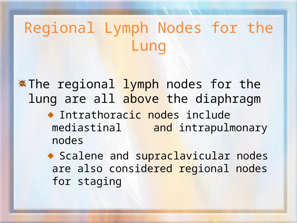
Regional Lymph Nodes for the Lung
The regional lymph nodes for the lung are all above the diaphragm
Intrathoracic nodes include mediastinal and intrapulmonary nodes
Scalene and supraclavicular nodes are also considered regional nodes for staging

Regional Lymph Nodes for the Lung
IntrathoracicPulmonary
12 Peribronchial
11 Intrapulmonary10 Hilar13 Segmental
Superior mediastinal
1 Superior Mediastinal
3Pretracheal,retrotracheal2 Paratracheal4 Lower paratracheal, azygos
IntrathoracicAortic
5 Subaortic (aortic window)6 Para-aortic (ascending
aorta or phrenic)
Inferior mediastinal
7 Carinal, subcarinal
8 Paraesophageal9 Pulmonary ligament

Regional Lymph Nodes for the Lung
Extrathoracic
Scalene
Supraclavicular or transverse cervical

Common Metastatic Sites for the Lung
Lymphatic Spread Cervical Lymph Nodes Contralateral Lung Contralateral Mediastinum
Hematogenic Spread Brain Bone Liver Adrenal glands Contralateral Lung
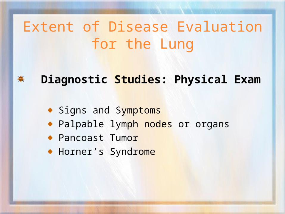
Extent of Disease Evaluation for the Lung
Diagnostic Studies: Physical Exam
Signs and Symptoms
Palpable lymph nodes or organs
Pancoast Tumor
Horner’s Syndrome
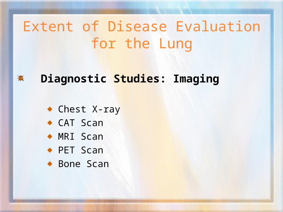
Extent of Disease Evaluation for the Lung
Diagnostic Studies: Imaging
Chest X-ray
CAT Scan
MRI Scan
PET Scan
Bone Scan

Extent of Disease Evaluation for the Lung
Diagnostic Studies: Endoscopies
Bronchoscopy
Mediastinoscopy
Thoracoscopy

Extent of Disease Evaluation for the Lung
Diagnostic Studies: Pathological
Closed Chest Needle Biopsy
Cytology (Thoracentesis)
Sputum Cytology
Bronchial Washings
Bone Marrow Biopsy
Extent of Disease Evaluation for the Lung
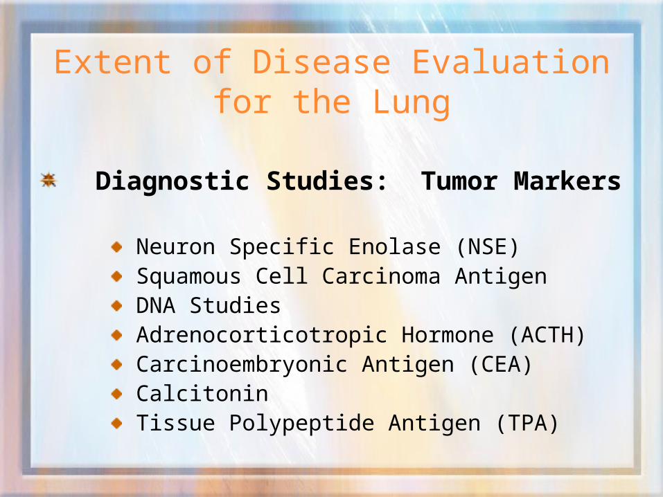
Diagnostic Studies: Tumor Markers
Neuron Specific Enolase (NSE) Squamous Cell Carcinoma Antigen DNA Studies Adrenocorticotropic Hormone (ACTH) Carcinoembryonic Antigen (CEA) Calcitonin Tissue Polypeptide Antigen (TPA)

Staging Systems for Lung
SEER Extent of Disease 3rd Edition (Diagnosis year 1988-2003)SEER Summary Staging 2000 (Diagnosis year 2001-2003)American Joint Committee on Cancer (AJCC) TNM 6th Edition(Diagnosis year 2003 forward) Collaborative Staging (Diagnosis year 2004 forward)

Staging Systems for Lung
SEER EOD
Size
Extent
Regional Lymph Nodes
Reg LN Positive
Reg LN Examined
SEER Summary Stage 2000In Situ
Localized
Reg by Direct Ext
Reg Ipsilateral LN only
Reg by Direct & Ipsilateral Reg LN
Reg, Nos
Distant Site/Node Involv
AJCC TNM 6th Edition
cT,pT: Extent of primary tumor
cN,pN: Absence or presence & Ext of regional lymph nodes
cM,pM: Absence/presence of metastasis
[Clinical (c) and pathologic (p)]
Collaborative StageCS Tumor Size
CS Tumor Size/Ext Eval
CS Lymph Nodes
CS Lymph Nodes Eval
CS Mets at Dx
CS Mets Eval
CS Site Specific Factors 1-6

Collaborative Stage
General Guidelines
Timing Rule
Site-specific guidelines
Highest applicable number for each field
Greatest extent of disease

Collaborative Stage for Bronchus and Lung
General Guidelines continued: CS Tumor Size/Ext CS Tumor Size/Ext Eval CS Regional LN CS Reg Nodes Eval CS Mets CS Mets Eval Regional LN Pos/Regional LN Exam Site Specific Factors

Collaborative Stage for Bronchus and Lung
CS Tumor Size000 No mass/tumor found001-998 001-998 millimeters (exact size)989 989 millimeters or larger990 Microscopic focus or foci only, no
size of focus given991 Described as less than 1 cm992-995 Described as less than (2,3,4,5 cm)996 Malignant cells in bronchopulmonary
secretions, but no tumor is seen radiographically or during bronchoscopy
997 Diffuse (entire lobe)998 Diffuse (entire lung)999 Unknown; size not stated
Not documented in patient record

Collaborative Stage for Bronchus and Lung
CS Extension
Primary Tumor extensionDO NOT CODE DISCONTINOUS METASTASES IN THIS FIELD
Priority order for extension
Notes 1-7: Read carefully

Collaborative Stage for Bronchus and Lung
CS TS/EXT EVAL
Evaluates source for “CS Tumor Size” and
“CS Extension”
Describes clinical or pathological staging
of the tumor

Collaborative Stage for Bronchus and Lung
CS TS/EXT EVAL
Code 0, 1, 9 No surgery
Code 2 Autopsy (diagnosis suspected)
Code 3 Surgery followed by other therapy
Code 5 Determined prior to neoadjuvant therapy
Code 6 Determined after neoadjuvant therapy
Code 8 Autopsy (diagnosis unsuspected)
Code 9 Unknown, Not assessed, Not stated

Collaborative Stage for Bronchus and Lung
CS Lymph Nodes
Regional Nodes and Nodes, NOS only
Highest Applicable Code
Exception (NOS)
New Rule for Inaccessible Site
Lung
Notes 1-4: Read carefully

Collaborative Stage for Bronchus and Lung
CS Reg Nodes Eval
Code 0, 1, 9 No LN(s) removal Code 2 Autopsy (diagnosis suspected) Code 3 LN(s) surgery followed by other
therapy Code 5 LN(s) status determined prior to
neoadjuvant therapy Code 6 LN(s) status determined after
neoadjuvant therapy Code 8 Autopsy (diagnosis unsuspected) Code 9 Unknown, Not assessed, Not stated

Collaborative Stage for Bronchus and Lung
Regional Nodes Positive00 All nodes examined
negative01-89 1-89 nodes positive90 90 or more nodes positive95 Positive aspiration of LN(s)
was performed97 Positive nodes
documented, number unspecified
98 No nodes examined99 Unknown if nodes
positive, not applicable, not stated in record
Regional Nodes Examined00 No nodes examined 01-89 1-89 nodes examined90 90 or more nodes examined95 No Reg LN removed,
Aspiration of Reg LN performed
96 Reg LN removal documented as sampling, number unknown/not stated
97 Reg LN removal documented as dissection, number unknown/not stated
98 Reg LN surgically removed, number of LNs unknown/not stated, not documented as sampling or dissection;
nodes examined, number unknown
99 Unknown, not applicable, not stated in record

Collaborative Stage for Bronchus and Lung
CS Mets at Dx 00 No; none
10 Distant Lymph Nodes (s), including cervical nodes
35 Separate tumor nodules (s) in different lobe, same lung
37 Extension to sternum, skeletal muscle, skin of chest
39 Extension to contralateral lung, contralateral main stem bronchus, separate tumor nodule(s) in contralateral lung
40 Abdominal organs, distant metastasis except to distant lymph nodes (code 10), distant mets, NOS, carcinomatosis
50 Distant mets + distant node(s)
(10) + any [35-40]
99 Unknown; distant metastasis cannot be assessed, not documented in patient record.

Collaborative Stage for Bronchus and Lung
CS Mets Eval
Code 0, 1, 9 No surgery Code 2 Autopsy (diagnosis suspected) Code 3 Surgery followed by other
therapy Code 5 Determined prior to
neoadjuvant therapy Code 6 Determined after
neoadjuvant therapy Code 8 Autopsy (diagnosis unsuspected) Code 9 Unknown, Not assessed, Not stated

Collaborative Stage for Bronchus and Lung
CS Site Specific Factors
SSF 1 Code 888
SSF 2 Code 888
SSF 3 Code 888
SSF 4 Code 888
SSF 5 Code 888
SSF 6 Code 888

Treatment for Bronchus and Lung
Surgery Non-small cell carcinoma: Stage I and II (Localized) Small cell carcinoma: Not recommended
Radiation Non-small cell carcinoma: Inoperable, Advanced Small cell carcinoma: Recommended
Systemic Therapy Non-small cell carcinoma: Chemotherapy Small cell carcinoma: Combination chemotherapy
Hormonal & Endocrine Not proven to be useful

Treatment for Bronchus and Lung
Surgery Codes
Primary Site
Scope of Regional Lymph Node
Surgical Procedure of Other Site

Treatment for Bronchus and Lung
Surgery Codes continued
Primary Site
Scope of Regional Lymph Node
Surgical Procedure of Other Site

Treatment for Bronchus and Lung
Surgery Codes continued
Primary Site
Scope of Regional Lymph Node
Surgical Procedure of Other Site

Treatment for Bronchus and Lung
Radiation Treatment Volume
Treatment Modality
Regional Dose
Boost Modality
Boost Dose
Prophylactic Cranial Radiation (PCI)

Treatment for Bronchus and Lung
Chemotherapy Non-Small Cell Carcinoma
Small Cell Carcinoma
Hormones
Immunotherapy
Other Endoscopic Photodynamic Therapy

Keys to Abstracting
Organization Dates
Procedure Documentation
Resources Diagnosis year
Text Pertinent
Acronyms