Embed Size (px)
Citation preview
8/7/2019 ADVANCED IMAGE ANALYSIS
http://slidepdf.com/reader/full/advanced-image-analysis 1/3
ADVANCED IMAGE ANALYSIS
Advanced Image Analysis Can Provide
Better Risk Assessment In Hardening Of The ArteriesScienceDaily (June 17, 2009) — Ultrasound examination of the carotid artery is a
patient-friendly and inexpensive method for assessing atherosclerosis and thereby predicting the risk of cardiovascular diseases. Peter Holdfeldt, who recently defendedhis doctoral thesis at Chalmers University of Technology in Sweden, has developed newanalytical methods for ultrasound images that can provide more reliable and more exactassessments of atherosclerosis.
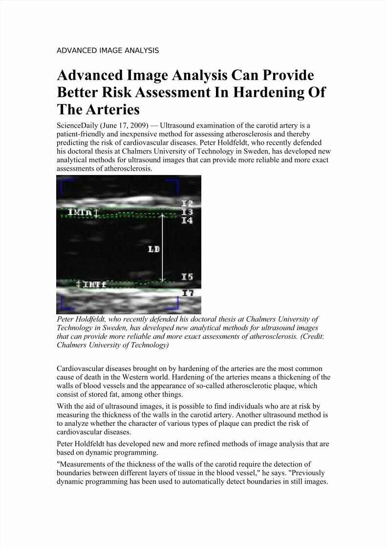
Peter Holdfeldt, who recently defended his doctoral thesis at Chalmers University of Technology in Sweden, has developed new analytical methods for ultrasound imagesthat can provide more reliable and more exact assessments of atherosclerosis. (Credit:Chalmers University of Technology)
Cardiovascular diseases brought on by hardening of the arteries are the most commoncause of death in the Western world. Hardening of the arteries means a thickening of the
walls of blood vessels and the appearance of so-called atherosclerotic plaque, whichconsist of stored fat, among other things.
With the aid of ultrasound images, it is possible to find individuals who are at risk bymeasuring the thickness of the walls in the carotid artery. Another ultrasound method isto analyze whether the character of various types of plaque can predict the risk of cardiovascular diseases.
Peter Holdfeldt has developed new and more refined methods of image analysis that are based on dynamic programming.
"Measurements of the thickness of the walls of the carotid require the detection of boundaries between different layers of tissue in the blood vessel," he says. "Previously
dynamic programming has been used to automatically detect boundaries in still images.

8/7/2019 ADVANCED IMAGE ANALYSIS
http://slidepdf.com/reader/full/advanced-image-analysis 2/3
But the new method uses dynamic programming for detection in image sequences of one and the same blood vessel instead."
Examining an entire image sequence instead of a single image provides a more correctresult, since it is possible to make use of the similarity between the images in thesequence - a boundary ought to be found in roughly the same place in two images in a
row. The method comprises two steps. First, several alternative locations of the boundary are determined in each image. Then one of the alternatives is selected fromeach image, and it is in this step that the program factors in the movement of boundaries
between images.
"This has proven to provide more correct detections of boundaries than what you canget from a program that detects boundaries on the basis of a single image," says Peter Holdfeldt.
He has also developed a method to automatically classify atherosclerotic plaque. This plaque can burst and form blood clots that cause heart attacks or strokes. In ultrasoundimages it is possible with the naked eye to see the type of plaque that often leads to
stroke, but such an assessment is subjective and is influenced by disturbances in theimage. The new automatic method entails a technological advancement of ultrasoundtechnology that can lead to more objective and quantifiable analysis.
Peter Holdfeldt's research has been part of a collaborative project between Chalmersand the Wallenberg Laboratory for Cardiovascular Research at Sahlgrenska UniversityHospital in Gothenburg. Björn Fagerberg, a physician and professor of cardiovascular research, is responsible for the clinical evaluation of the new methods together with thedoctoral candidate Ulrica Prahl.
"We're now busy testing the new automatic method for plaque classification in patientgroups," he says. "In its final form it should be an excellent aid in identifying high-risk
patients."
Measurement of the carotid artery is already in use today in cardiovascular research.There are other methods of measurement, but they are not as well validated as themethod that has been developed by the researchers at Chalmers and Sahlgrenska.
"Dynamic image analysis is an exciting new method that will no doubt offer great potential for elaboration," says Björn Fagerberg. "The advantage of using ultrasound isthat is practical, inexpensive, and patient-friendly."
The dissertation "Dynamic Programming for Ultrasound Image Analysis of Atherosclerosis" was defended on May 15.
Disclaimer : This article is not intended to provide medical advice, diagnosis or
treatment. Views expressed here do not necessarily reflect those of ScienceDaily or its staff.
Email or share this story:| More
Story Source:
The above story is reprinted (with editorial adaptations by Science Daily staff) frommaterials provided by The Swedish Research Council, via AlphaGalileo.
Parte superior do formulário
Need to cite this story in your essay, paper, or report? Use one of the following formats:

8/7/2019 ADVANCED IMAGE ANALYSIS
http://slidepdf.com/reader/full/advanced-image-analysis 3/3
APA
MLAThe Swedish Research Council (2009, June 17). Advanced Image Analysis Can Provide
Better Risk Assessment In Hardening Of The Arteries. ScienceDaily. RetrievedDecember 12, 2010, from http://www.sciencedaily.com/releases/2009/06/090605112331.htm
Parte inferior do formulário
Note: If no author is given, the source is cited instead.