Embed Size (px)
Citation preview
Advances in Biological Regulation 62 (2016) 1e10
Contents lists available at ScienceDirect
Advances in Biological Regulation
journal homepage: www.elsevier .com/locate/ jbior
Specific expression and function of inositol 1,4,5-trisphosphate 3-kinase C (ITPKC) in wild type and knock-outmice
Ariane Scoumanne a, 1, Patricia Molina-Ortiz a, Daniel Monteyne b, David Perez-Morga b, c, Christophe Erneux d, St�ephane Schurmans a, *
a Laboratoire de G�en�etique Fonctionnelle, GIGA-B34, Universit�e de Li�ege, avenue de l’Hopital 11, 4000 Li�ege, Belgiumb Laboratoire de Parasitologie Mol�eculaire, Institut de Biologie et de M�edecine Mol�eculaires (IBMM), Universit�e Libre de Bruxelles, rue desProfesseurs Jeener et Brachet 12, 6041 Gosselies, Belgiumc Center for Microscopy and Molecular Imaging (CMMI), Universit�e Libre de Bruxelles (ULB), 8 rue Adrienne Bolland, B-6041 Gosselies,Belgiumd Institut de Recherches Interdisciplinaires en Biologie Humaine et Mol�eculaire (IRIBHM), Campus Erasme, Universit�e Libre de Bruxelles,route de Lennik, 808, 1070 Bruxelles, Belgium
a r t i c l e i n f o
Article history:Received 17 February 2016Accepted 16 March 2016Available online 22 March 2016
Keywords:Inositol 1,3,4,5-tetrakisphosphateInositol 1,4,5-trisphosphate 3-kinaseItpkcTissue expressionMouse
Abbreviations: Bright field, BF; ciliary beat frequd�eshydrog�enase, GAPDH; in situ hybridization, ISH;disease, KD; mouse tracheal epithelial cells, MTEC;* Corresponding author.
E-mail address: [email protected] (S. Schurm1 Present address. Iteos Therapeutics, rue Cl�emen
http://dx.doi.org/10.1016/j.jbior.2016.03.0012212-4926/© 2016 Elsevier Ltd. All rights reserved.
a b s t r a c t
Inositol 1,4,5-trisphosphate 3-kinase C (ITPKC) is the last identified member of the inositol1,4,5-trisphosphate 3-kinases family which phosphorylates inositol 1,4,5-trisphosphateinto inositol 1,3,4,5-tetrakisphosphate. Although expression and function of the twoother family members ITPKA and ITPKB are rather well characterized, similar informationis lacking for ITPKC. Here, we first defined the expression of Itpkc mRNA and protein inmouse tissues and cells using in situ hybridization and new antibodies. Surprisingly, wefound that cells positive for ITPKC in the studied tissues express either a multicilium(tracheal and bronchial epithelia, brain ependymal cells), microvilli forming a brush border(small and large intestine, and kidney proximal tubule cells) or a flagellum (spermatozoa),suggesting a role for ITPKC either in the development or the function of these specializedcellular structures. Given this surprising expression, we then analyzed ITPKC function inmulticiliated tracheal epithelial cells and sperm cells using our Itpkc knock-out mousemodel. Unfortunately, no significant difference was observed between control and mutantmice for any of the parameters tested, leaving the exact in vivo function of this thirdIns(1,4,5)P3 3-kinase still open.
© 2016 Elsevier Ltd. All rights reserved.
ency, CBF; enhanced chemiluminescence, ECL; immunofluorescence, IF; glyc�erald�ehyde-3-phosphateinositol 1,4,5-trisphosphate, Ins(1,4,5)P3; inositol 1,3,4,5-tetrakisphosphate, Ins(1,3,4,5)P4; Kawasakiprotease inhibitors cocktail, PIC; Scanning electron microscopy, SEM.
ans).t Ader 16, 6041-Gosselies, Belgium.
A. Scoumanne et al. / Advances in Biological Regulation 62 (2016) 1e102
1. Introduction
Inositol 1,4,5-trisphosphate (Ins(1,4,5)P3), the well-known calcium mobilization messenger, can be phosphorylated intoinositol 1,3,4,5-tetrakisphosphate (Ins(1,3,4,5)P4) by Ins(1,4,5)P3 3-kinases (Itpk) isoforms a, b and c, or by Ipk2/Ipmk, theinositol phosphate multikinase (Choi et al., 1990; Takazawa et al., 1990, 1991; Dewaste et al., 2000; Saiardi et al., 2000;reviewed by York, 2006). The a, b and c isoforms of Ins(1,4,5)P3 3-kinases (or ITPKA, ITPKB and ITPKC) share a well conservedcarboxy-terminal catalytic domain, an ATP binding motif, an inositol binding motif, and a calmodulin binding domain(reviewed by Erneux et al., 2016). All three Ins(1,4,5)P3 3-kinase isoforms have been shown to be sensitive to Ca2þ to variousdegress; their activity is also regulated by direct protein phosphorylation (Communi et al., 1997). Tissue expression of these 3isoforms has been previously reported: using Northern blotting and RT-PCR, Itpkc mRNA has been shown to be ratherubiquitously expressed in human and mouse tissues, as Itpkb (Vanweyenberg et al., 1995; Dewaste et al., 2000). However, theprecise cellular localization of endogenous ITPKC in these tissues has never been reported in the literature, as no specificantibody has been reported to detect the endogenous protein. Production and analysis of mice genetically-deficient for Itpkaand Itpkb have strongly helped to characterize the in vivo functions of these enzymes and the corresponding mechanisms ofIns(1,3,4,5)P4 action (Pouillon et al., 2013; Schurmans et al., 2011, 2015). By contrast, the physiological function of ITPKC ispoorly characterized in vivo: Itpkc-deficient mice have been generated in our laboratory, but these mice appear healthy with anormal lifespan and growth (Pouillon et al., 2003). No gross morphological alterations have been detected in organs isolatedfrom these mice. T cell development and calcium response following stimulationwere found to be normal in these knock-outmice. Furthermore, inactivation of Itpkc on an Itpkb�/- genetic background did not demonstrate additional T cell alterationsbeyond those of Itpkb�/- mice, excluding a potential compensatory mechanism by the b isoform to explain the absence ofobvious phenotype in Itpkc�/- mice (Pouillon et al., 2003). Interestingly, an ITPKC functional genetic polymorphism wasdiscovered to be associated with Kawasaki disease susceptibility (Onouchi et al., 2008). Kawasaki disease (KD) is a severe andacute pediatric systemic vasculitis of unknown etiology. KD may lead to formation of coronary artery aneurysms and toischemic heart disease, myocardial infarction and sudden death (Hata and Onouchi, 2009). The link between this ITPKCpolymorphism and KD (or resistance to KD treatment) was confirmed in other studies, but others failed to reproduce theseresults and no association of the ITPKC locus with KD was detected (Burgner et al., 2009; Khor et al., 2011; Kuo et al., 2011; Linet al., 2011; Onouchi et al., 2013; Peng et al., 2012).
In order to better characterize the C isoform of the Ins(1,4,5)P3 3-kinases family, we defined the expression of ItpkcmRNAand protein in mouse tissues and cells using in situ hybridization and new antibodies. Then, based on these expressionstudies, we analyzed Itpkc function in specific cells using our Itpkc knock-out mouse model.
2. Materials and methods
2.1. Mice
Itpkc�/- mice were generated in our laboratory (Pouillon et al., 2003). Mice were maintained in a specific pathogen freefacility at the GIGA-research Center. Itpkc�/- and wild-type mice between 6 and 12 weeks of age were used in this study. Allprocedures involving mice were approved by the Animal Care and Use Review Committee of the Universit�e de Li�ege.
2.2. Antibodies
Affinity purified anti-mouse ITPKC polyclonal antibodies named GS2 and GS4 were made by immunizing rabbits withpeptide LPERDNKPRVDNLRC and QPGSDGFSS KDTESC, respectively (GenScript). Affinity purified anti-mouse ITPKC antibodynamed Aldwas produced by immunizing rabbits with three peptides: RGGRRRQPGLQRPGPGAG, KPRQNKELDGSNLQTHPRRN-C and SQTDDSLKGPSTQTAC (Aldevron). Those peptides were chosen in the sequence of mouse ITPKC (Pouillon et al., 2003).Specificity of detection was confirmed by Western blot and immunofluorescence analysis using anti-ITPKC antibodies pre-incubated or not with their corresponding immunizing peptides.
Anti-ezrin antibody was obtained from ThermoFisher Scientific. Anti-centrin was purchased from Millipore. Anti-acetylated tubulin, phalloidin-TRICT and anti-gamma tubulin were obtained from SigmaeAldrich. Rabbit IgGs and mouseIgGs were from Santa Cruz Biotechnologies. Anti-rabbit IgGs Atto 488 and Atto 594-conjugated secondary antibodies wereobtained from SigmaeAldrich. Horse-radish peroxidase (HRP)-conjugated secondary antibodies were obtained from SantaCruz Biotechnologies.
2.3. Immunodetection by Western blotting
To prepare mouse tissue lysates, fresh tissues were snap-frozen in liquid nitrogen, suspended in protein lysis buffer withProtease Inhibitors Cocktail (PIC) tablets (Roche) using an homogenizer (VDI 12, VWR) or a pestle and incubated at 95 �C for5 min. After centrifugation at 10,000� g for 5 min, protein of tissue lysates were quantified and 60 mg of lysates were used forWestern blot analysis. To prepareMTEC lysates, MTEC cultured onTranswell inserts werewashedwith PBS, suspended in lysisbuffer with PIC and incubated at 95 �C for 5 min. After quantification, 60 mg of cell lysates were used forWestern blot analysis.Briefly, SDS-PAGE was performed and transferred to nitrocellulose membrane using the Trans-blot Turbo transfer system
A. Scoumanne et al. / Advances in Biological Regulation 62 (2016) 1e10 3
(BioRad). Membranes were blocked with PBS containing 5% non-fat milk and 0.1% Tween-20 for 1 h and incubated withprimary antibodies in PBS containing 2% non-fat milk and 0.1% Tween-20 at 4 �C overnight. After 3 washes, the membraneswere incubated with HRP-conjugated secondary antibodies for 1 h, washed and revealed with enhanced chemiluminescence(ECL) onto Hyperfilm (Amersham). Images were quantitated with Image J software (National Institutes of Health).
2.4. HEK cell culture and transfection
HEK cells were cultured in DMEM medium supplemented with 10% FBS, 2% Penicillin-Streptomycin, 0.1 mM MEM-NonEssential Amino-Acids and 2 mM L-glutamine (Gibco) in an incubator with 5% CO2 at 37 �C. For transient transfection, cellswere seeded at 106 cells per well in a 6-well plate and transfected with FuGENE 6 transfection reagent (Promega), using 1 mgpCMV6-mItpkc cDNAvector (OriGene BC053450) in 40 ml serum-free Opti-MEM (ThermoFisher Scientific) combinedwith 6 mlFuGENE 6. After 24 h, transfected cells were washed with PBS and harvested in 50 ml lysis buffer with PIC for Western blot orimmunofluorescence analysis.
2.5. RTqPCR on mouse tissues
Mouse tissues were collected and snap-frozen in liquid nitrogen. Total RNAwas extracted using RNeasy Mini Kit (Qiagen)and cDNA was synthesized using iScript (BioRad). To determine Itpkc mRNA levels, qPCR was performed on an iQ5 real-timePCR detection system (BioRad) in a total volume of 20 ml containing 50 nM primers and 1� iQ SYBR Green Supermix (BioRad).For Itpkc amplification, primers mItpkc-F (50-CTG AAG TAC TCG CCC TTC GT-30) and mItpkc-R (50- TGC TCA CAC TGA CAG AAACG-30) were used. For 18S amplification, primers 18S-F (50- GCA ATT ATT CCC CAT GAA CG-30) and primers 18S-R (50-AGG GCCTCA CTA AAC CAT CC-30) were used.
2.6. Isolation and culture of mouse tracheal epithelial cells (MTEC)
MTEC culturewas based on the report of You et al. (2002). Briefly, micewere killed and tracheas were excised and trimmedof excess tissues. Tracheas were opened longitudinally to expose the lumen and incubated in DMEM/F12 medium (Ther-moFisher Scientific) containing 1.5 mg/ml pronase (Roche) at 4 �C for 18 h. Tracheal epithelial cells were dislodged by gentleagitation and collected in DMEM/F12 medium. The cells were treated with 0.5 mg/ml DNAse I (SigmaeAldrich) on ice for5 min and centrifuged at 400 � g at 4 �C for 5 min. The cell pellet was suspended in DMEM/F12 medium containing 10% FBS,plated in a T75 flask and placed in an incubator at 37 �C and 5% CO2 for 2 h to allow adherence of contaminating fibroblasts.Non-adherent cells were centrifuged, suspended in DMEM/F12 medium supplemented with 10 mg/ml insulin, 5 mg/mltransferrin, 25 ng/ml EGF, 30 mg/ml bovine pituitary extract, 5% FBS and freshly added 0.01 mM retinoic acid. The cells wereseeded at 7.5�104 cells per cm2 onto collagen-coated Transwell inserts (Corning) andmaintained in an incubator at 37 �C and5% CO2. Medium was changed every other day. Once reaching confluence (after ~ 10 days), the culture of undifferentiatedepithelial cells was changed to an air-liquid interface culture by adding DMEM/F12medium containing 2% NuSerum (Corning)to the bottom compartment only (You et al., 2002). MTEC were maintained in an incubator at 37 �C and 5% CO2 and mediumwas changed every other day until reaching full differentiation (after ~ 15 days). MTEC were used at air-liquid interface day15e19 in this study. All media were supplemented with 100 U/ml penicillin, 100 mg/ml streptomycin, 15 mM Hepes and0.25 mg/ml Fungizone.
2.7. Evaluation of ciliary beat frequency of MTEC
MTEC cultures at air-liquid interface day 17e19 were placed in a 37 �C chamber onto the stage of a Nikon A1R confocalmicroscope. Time-lapse images were captured at a 40�magnification objective and a frame-rate of 30 frames per sec, for 3 s.The ImageJ software was used to calculate the ciliary beat frequency (CBF): CBF (Hz) ¼ number of cyclical changes in pixelintensity in X frames * (frame-rate/X). Average ± S.D. was calculated from three counting.
2.8. Immunofluorescence and confocal microscopy
For MTEC, cells grown on Transwell inserts were rinsed with PBS, fixed in 10% formalin for 10 min and washed with PBS.Transwell filters were excised from their plastic supports and placed cell-side up on a slide. Filters were blocked in PBScontaining 5% goat serum, 3% BSA, 0.1% Triton X-100 and 1/100 dilution of unconjugated anti-mouse IgG antibody (JacksonImmunoResearch Laboratories) for 1 h. Filters were incubated with primary antibodies diluted in PBS containing 2% goatserum, 1% BSA, 0.1% Triton X-100 for 1 h or overnight at 4 �C. The filters were washed 3 times for 10 min and incubated withfluorochrome-labeled secondary antibodies (SigmaeAldrich) for 1 h. After 3 washes, the filters were incubated with 300 nMDAPI in PBS (ThermoFisher Scientific) for 5 min. After 3 washes, the filters were mounted using Prolong (ThermoFisherScientific). Images were acquired using the NIS 4.20.00 software on a Nikon A1R confocal microscope. Image stacks werecollected with a z-section of 0.2 mm and were processed using ImageJ (National Institutes of Health).
For mouse tissues, 5 mm cryosections were prepared and mounted on Superfrost Ultra Plus slide. The sections were fixedwith acetone for 10 min and blocked in PBS containing 5% goat serum, 0.1% Triton X-100 and 1/100 dilution of unconjugated
A. Scoumanne et al. / Advances in Biological Regulation 62 (2016) 1e104
anti-mouse IgG antibody (Jackson ImmunoResearch Laboratories) for 1 h or overnight at 4 �C. The sections were incubatedwith primary antibodies diluted in PBS containing 2% goat serum for 1 h, then washed 3 times for 5 min. The sections wereincubated with fluorochrome-conjugated secondary antibodies (SigmaeAldrich) for 1 h, washed and incubated with DAPI for5 min. After 3 washes, the slides were mounted in Prolong and images were acquired as described for MTEC.
2.9. In situ hybridization on mouse tissue sections
A mouse Itpkc oligo RNAscope probe was designed and provided by Advanced Cell Diagnostic. In situ hybridization wasperformed using the RNAscopeMultiplex Fluorescent Assay (ACD). Briefly, fresh tissues were quickly frozen in liquid nitrogenand embedded in OCT medium. Cryosections of 14 mmwere prepared and mounted on SuperFrost Plus slides. Sections werefixed and pre-treated according to the RNAscope guide for Fresh frozen Tissue (ACD). Following pre-treatment, the sectionswere hybridized with the Itpkc oligo probe using the HybEZ Hybridization System (ACD). After several amplification sets, thesections were counterstainedwith DAPI andmounted using Prolong. A Polar2A probe and a bacterial dapB probewere used aspositive and negative controls, respectively. Images were acquired using Nikon A1R confocal microscope and image stackswere processed using ImageJ.
2.10. Epididymal spermatozoa isolation and morphology
The epididymis was collected in TYH buffer containing 120 mM NaCl, 5 mM KCl, 1.2 mM MgSO4, 1.7 mM CaCl2,1.2 mM K2PO4, 25 mM NaHCO3, 1 mM sodium pyruvate, 5.6 mM glucose and 20 mM Hepes, pH 7.4 (Burnett et al., 2011). Theepididymis was cut and incubated for 15 min at 37 �C to allow the sperm to get out. For capacitation, an equal volume of TYHbuffer containing 5% BSAwas added and the sperm suspensionwas incubated at 37 �C for 1 h. For sperm flagellar analysis, thesperm suspension was mixed with equal volume of 2� stimulant solution (0e50 mM Lyral, SigmaeAldrich) (Fukuda et al.,2004). An aliquot of stimulated sperm suspension was mounted on slide and coverslip. Phase-contrast images were takenusing a Nikon Eclipse 90i microscope.
2.11. Scanning electron microscopy (SEM) on mouse trachea
Samples were washed with PBS, fixed overnight at 4 �C in glutaraldehyde 2.5%, 0.1M cacodylate buffer (pH 7.2) and post-fixed in OsO4 (2%) in the same buffer. After serial dehydration, samples were dried at critical point and coated with platinumby standard procedures. Observations were made in a Tecnai FEG ESEM QUANTA 200 (FEI) at 30 kV and images processed bySIS iTEM (Olympus) software.
2.12. Statistical analysis
All values are expressed as means ± standard error of the mean (SEM). Significance of differences between twomeans wascalculated using the GraphPad InStat software (GraphPad Software, San Diego, USA). Significance level of the differences ofthe means (p-value) is presented.
3. Results and discussion
3.1. Analysis of mouse Itpkc antibodies in HEK cells
Three antibodies, GS2, GS4 and Ald, were obtained after rabbit immunization with different peptides localized in the N-terminal region of the mouse ITPKC protein which is encoded by the first coding exon of the Itpkc gene (see SupplementaryMaterial). These antibodies were first tested by immunodetection on protein extracts isolated from HEK cells transfected ornot with an expression plasmid for the mouse Itpkc cDNA. Using all three antibodies, a strong signal was observed at ~90 kDain the mouse Itpkc cDNA-transfected HEK cell extract; no signal was detected in the non-transfected HEK cell extract (Fig. 1aand Supplementary Fig. 1). This signal was absent or markedly decreased when antibodies were incubated with the corre-sponding immunizing peptide(s) before addition onto the membrane (Fig. 1a and Supplementary Fig. 1). Second, GS2, GS4and Ald antibodies were tested for their capacity to detect the mouse ITPKC protein by immunofluorescence on coverslips inmouse Itpkc cDNA-transfected HEK cells (Fig. 1b). Only the GS2 antibody detected a homogeneous cytoplasmic staining in~30% of transfected HEK cells; the staining markedly decreased when the GS2 antibody was incubated with the corre-sponding immunizing peptide before addition on transfected HEK cells. No staining was detected with the GS2 antibody innon-transfected HEK cells, and no staining was observed in mouse Itpkc cDNA-transfected HEK cells using GS4 and Aldantibodies (data not shown).
3.2. Analysis of Itpkc mRNA and protein expression in normal mouse tissues and cells
Fourteen tissues were isolated from a normal adult male mouse; mRNA was extracted from each tissue and reverse-transcribed. cDNAs were then processed for qPCR in order to analyze Itpkc mRNA expression (Fig. 1c). Mouse Itpkc mRNA
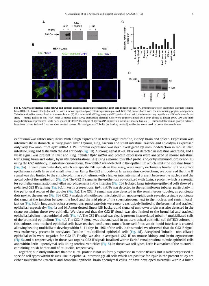
Fig. 1. Analysis of mouse Itpkc mRNA and protein expression in transfected HEK cells and mouse tissues: (A) Immunodetection on protein extracts isolatedfrom HEK cells transfected (þ) or not (�) with a mouse Itpkc (mItpkc) cDNA expression plasmid. GS2, GS2 preincubated with the immunizing peptide and gammaTubulin antibodies were added to the membrane. (B) IF studies with GS2 (green) and GS2 preincubated with the immunizing peptide on HEK cells transfected(HEK þ mouse Itpkc) or not (HEK) with a mouse Itpkc cDNA expression plasmid. Cells were counterstained with DAPI (blue) to detect DNA. Low and highmagnifications are presented. Scale bars: 25 mm. (C) RTqPCR analysis of ItpkcmRNA expression in various mouse tissues. (D) Immunodetection on protein extractsfrom four tissues isolated from an adult control mouse. Ald and gamma Tubulin (as loading control) antibodies were used to probe the membrane.
A. Scoumanne et al. / Advances in Biological Regulation 62 (2016) 1e10 5
expression was rather ubiquitous, with a high expression in testis, large intestine, kidney, brain and spleen. Expression wasintermediate in stomach, salivary gland, liver, thymus, lung, caecum and small intestine. Trachea and epididymis expressedonly very low amount of Itpkc mRNA. ITPKC protein expression was next investigated by immunodetection in mouse liver,intestine, lung and testis with the Ald antibody (Fig. 1d). A strong signal at ~90 kDa was detected in intestine and testis, and aweak signal was present in liver and lung. Cellular Itpkc mRNA and protein expression were analyzed in mouse intestine,testis, lung, brain and kidney by in situ hybridization (ISH) using amouse Itpkc RNA probe, and/or by immunofluorescence (IF)using the GS2 antibody. In intestine cryosections, ItpkcmRNAwas detected in the epitheliumwhich limits the intestine lumen(Fig. 2a). Indeed, punctuate dots, which are specific ISH signals in this assay, were nearly exclusively limited to the surfaceepithelium in both large and small intestines. Using the GS2 antibody on large intestine cryosections, we observed that the IFsignal was also limited to the simple columnar epithelium, with a higher intensity signal present between the nucleus and theapical pole of the epithelium (Fig. 2b). The GS2 IF signal in the epithelium co-localized with Ezrin, a proteinwhich is essentialfor epithelial organization and villus morphogenesis in the intestine (Fig. 2b). Isolated large intestine epithelial cells showed apolarized GS2 IF staining (Fig. 2c). In testis cryosections, ItpkcmRNAwas detected in the seminiferous tubules, particularly inthe peripheral region of the tubules (Fig. 3a). The GS2 IF signal was also detected in the seminiferous tubules, as punctuatedots next to the nucleus (Fig. 3b). GS2 IF analysis of motile sperm isolated frommouse epididymis revealed a single punctuatedot signal at the junction between the head and the mid piece of the spermatozoon, next to the nucleus and centrin local-ization (Fig. 3c). In lung and trachea cryosections, punctuate dots were nearly exclusively limited to the bronchial and trachealepithelia, respectively (Fig. 4a and b). A non-dotted, linear ISH background signal of unknown origin was also detected in thetissue sustaining these two epithelia. We observed that the GS2 IF signal was also limited to the bronchial and trachealepithelia, labeling most epithelial cells (Fig. 4c). The GS2 IF signal was clearly present in acetylated tubulinþ multiciliated cellsof the bronchial epithelium (Fig. 4c). The GS2 IF signal was also analyzed in mouse tracheal epithelial cell (MTEC) culture. Inthis culture, once tracheal epithelial cells have reached confluence onto a Transwell filter, an air-liquid interface is created,allowing beating multicilia to develop within 5e11 days in ~50% of the cells. In this model, we observed that the GS2 IF signalwas exclusively present in acetylated Tubulinþ multiciliated epithelial cells (Fig. 4d). Acetylated Tubulin� non-ciliatedepithelial cells were negative for GS2 IF. Finally, we also investigated GS2 IF on mouse kidney and brain cryosections(Fig. 5a and b, respectively). In these two organs, GS2 IF signals localized within Ezrinþ renal proximal tubule epithelial cellsand within Ezrinþ ependymal cells lining cerebral ventricles (Fig. 5). In these two cell types, Ezrin is a marker of the microvillicontaining brush border and of multicilia, respectively.
Together, our study indicates that the ITPKC protein is not uniformly expressed in mouse tissues, but is rather expressed inspecific cell types within tissues, like in epithelia. Interestingly, all cells which are positive for Itpkc in the present study areeither multiciliated (tracheal and bronchial epithelia, brain ependymal cells), or have developed microvilli within a brush
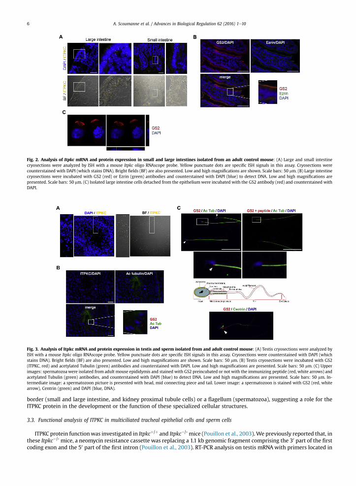
Fig. 2. Analysis of Itpkc mRNA and protein expression in small and large intestines isolated from an adult control mouse: (A) Large and small intestinecryosections were analyzed by ISH with a mouse Itpkc oligo RNAscope probe. Yellow punctuate dots are specific ISH signals in this assay. Cryosections werecounterstained with DAPI (which stains DNA). Bright fields (BF) are also presented. Low and high magnifications are shown. Scale bars: 50 mm. (B) Large intestinecryosections were incubated with GS2 (red) or Ezrin (green) antibodies and counterstained with DAPI (blue) to detect DNA. Low and high magnifications arepresented. Scale bars: 50 mm. (C) Isolated large intestine cells detached from the epitheliumwere incubated with the GS2 antibody (red) and counterstained withDAPI.
Fig. 3. Analysis of Itpkc mRNA and protein expression in testis and sperm isolated from and adult control mouse: (A) Testis cryosections were analyzed byISH with a mouse Itpkc oligo RNAscope probe. Yellow punctuate dots are specific ISH signals in this assay. Cryosections were counterstained with DAPI (whichstains DNA). Bright fields (BF) are also presented. Low and high magnifications are shown. Scale bars: 50 mm. (B) Testis cryosections were incubated with GS2(ITPKC, red) and acetylated Tubulin (green) antibodies and counterstained with DAPI. Low and high magnifications are presented. Scale bars: 50 mm. (C) Upperimages: spermatozoa were isolated from adult mouse epididymis and stained with GS2 preincubated or not with the immunizing peptide (red, white arrows) andacetylated Tubulin (green) antibodies, and counterstained with DAPI (blue) to detect DNA. Low and high magnifications are presented. Scale bars: 50 mm. In-termediate image: a spermatozoon picture is presented with head, mid connecting piece and tail. Lower image: a spermatozoon is stained with GS2 (red, whitearrow), Centrin (green) and DAPI (blue, DNA).
A. Scoumanne et al. / Advances in Biological Regulation 62 (2016) 1e106
border (small and large intestine, and kidney proximal tubule cells) or a flagellum (spermatozoa), suggesting a role for theITPKC protein in the development or the function of these specialized cellular structures.
3.3. Functional analysis of ITPKC in multiciliated tracheal epithelial cells and sperm cells
ITPKC protein functionwas investigated in Itpkcþ/þ and Itpkc�/- mice (Pouillon et al., 2003).We previously reported that, inthese Itpkc�/- mice, a neomycin resistance cassette was replacing a 1.1 kb genomic fragment comprising the 30 part of the firstcoding exon and the 50 part of the first intron (Pouillon et al., 2003). RT-PCR analysis on testis mRNA with primers located in
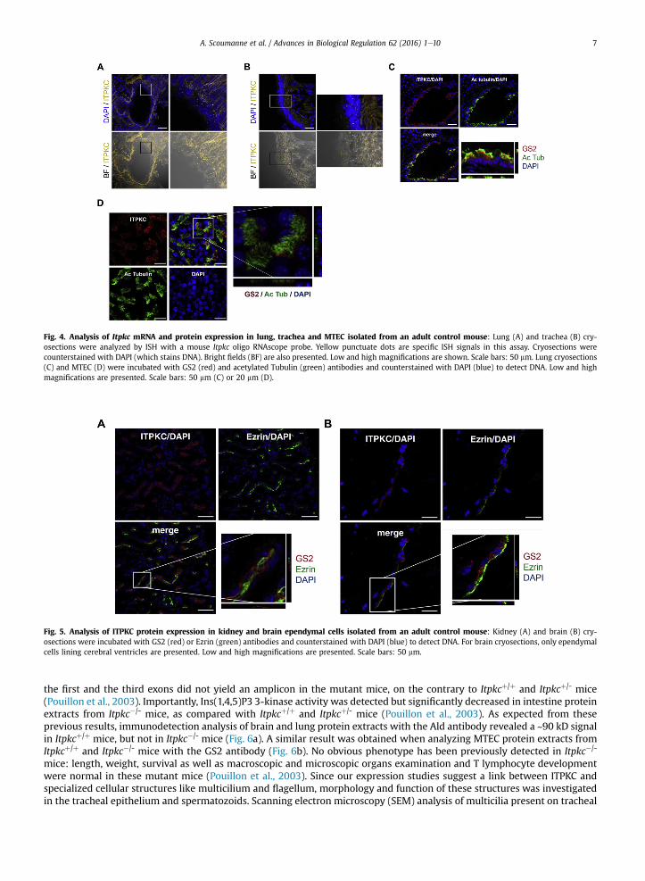
Fig. 4. Analysis of Itpkc mRNA and protein expression in lung, trachea and MTEC isolated from an adult control mouse: Lung (A) and trachea (B) cry-osections were analyzed by ISH with a mouse Itpkc oligo RNAscope probe. Yellow punctuate dots are specific ISH signals in this assay. Cryosections werecounterstained with DAPI (which stains DNA). Bright fields (BF) are also presented. Low and high magnifications are shown. Scale bars: 50 mm. Lung cryosections(C) and MTEC (D) were incubated with GS2 (red) and acetylated Tubulin (green) antibodies and counterstained with DAPI (blue) to detect DNA. Low and highmagnifications are presented. Scale bars: 50 mm (C) or 20 mm (D).
Fig. 5. Analysis of ITPKC protein expression in kidney and brain ependymal cells isolated from an adult control mouse: Kidney (A) and brain (B) cry-osections were incubated with GS2 (red) or Ezrin (green) antibodies and counterstained with DAPI (blue) to detect DNA. For brain cryosections, only ependymalcells lining cerebral ventricles are presented. Low and high magnifications are presented. Scale bars: 50 mm.
A. Scoumanne et al. / Advances in Biological Regulation 62 (2016) 1e10 7
the first and the third exons did not yield an amplicon in the mutant mice, on the contrary to Itpkcþ/þ and Itpkcþ/- mice(Pouillon et al., 2003). Importantly, Ins(1,4,5)P3 3-kinase activity was detected but significantly decreased in intestine proteinextracts from Itpkc�/- mice, as compared with Itpkcþ/þ and Itpkcþ/- mice (Pouillon et al., 2003). As expected from theseprevious results, immunodetection analysis of brain and lung protein extracts with the Ald antibody revealed a ~90 kD signalin Itpkcþ/þ mice, but not in Itpkc�/- mice (Fig. 6a). A similar result was obtained when analyzing MTEC protein extracts fromItpkcþ/þ and Itpkc�/- mice with the GS2 antibody (Fig. 6b). No obvious phenotype has been previously detected in Itpkc�/-
mice: length, weight, survival as well as macroscopic and microscopic organs examination and T lymphocyte developmentwere normal in these mutant mice (Pouillon et al., 2003). Since our expression studies suggest a link between ITPKC andspecialized cellular structures like multicilium and flagellum, morphology and function of these structures was investigatedin the tracheal epithelium and spermatozoids. Scanning electron microscopy (SEM) analysis of multicilia present on tracheal
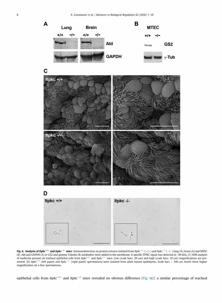
Fig. 6. Analysis of Itpkcþ/þ and Itpkc¡/¡ mice: Immunodetection on protein extracts isolated from Itpkcþ/þ (þ/þ) and Itpkc�/- (�/�) lung (A), brain (A) and MTEC(B). Ald and GAPDH (A) or GS2 and gamma Tubulin (B) antibodies were added to the membrane. A specific ITPKC signal was detected at ~90 kDa. (C) SEM analysisof multicilia present on tracheal epithelial cells from Itpkcþ/þ and Itpkc�/� mice. Low (scale bars: 20 mm) and high (scale bars: 10 mm) magnifications are pre-sented. (D) Itpkcþ/þ (left panel) and Itpkc�/� (right panel) spermatozoa were isolated from adult mouse epididymis. Scale bars ¼ 100 mm. Insets show highermagnification on a few spermatozoa.
A. Scoumanne et al. / Advances in Biological Regulation 62 (2016) 1e108
epithelial cells from Itpkcþ/þ and Itpkc�/- mice revealed no obvious difference (Fig. 6c): a similar percentage of tracheal
A. Scoumanne et al. / Advances in Biological Regulation 62 (2016) 1e10 9
epithelial cells presented with a multicilium (Itpkcþ/þ: 48 ± 5% of multiciliated cells, Itpkc�/�: 51 ± 7% of multiciliated cells;mean ± SEM, P ¼ 0.52), and multicilia were similar in term of cilia number, length and diameter (Fig. 6c). The trachealmulticiliary beating frequency was analyzed in the MTECmodel. Again, no significant difference was detected for multiciliarybeating frequency between Itpkcþ/þ (9.3 ± 2.2 Hz, mean ± SEM) and Itpkc�/- (10.0 ± 3.5 Hz, mean ± SEM; P ¼ 0.44) MTEC.Epididymal spermatozoa were isolated from 8-week old Itpkcþ/þ and Itpkc�/- male mice. No obvious morphological defectwas detected in Itpkc�/- spermatozoa, as compared to Itpkcþ/þ spermatozoa (Fig. 6d). In non-stimulated conditions, thepercentage of spermatozoa with a 'fishhook' like shape flagellum was similar in Itpkcþ/þ (16 ± 3%, mean ± SEM) and Itpkc�/-
(15 ± 3%, mean ± SEM; P ¼ 0.55) mice. Accordingly, reproductive capacity of male Itpkc�/- mice was normal: Itpkc�/- miceintercrosses gave litters of normal size, compared with Itpkcþ/þ intercrosses (Itpkcþ/þ: 5.8 ± 1.8 newborns per litter, Itpkc�/�:5.3 ± 1.9 newborns per letter; mean ± SEM, 20 litters, P ¼ 0.67).
4. Conclusions
Here, for the first time, we define the expression of Itpkc mRNA and protein in various mouse tissues using ISH and IFanalysis on tissue sections. We found that ITPKC is not uniformly expressed in mouse tissues, but it is rather expressed inspecific cell types within the studied tissues. Interestingly, cells positive for ITPKC in these tissues express either amulticilium,microvilli forming a brush border or a flagellum. This is specific for ITPKC and was not observed with ITPKA and ITPKB. Themouse endogenous ITPKC proteinwas never detected in the nucleus, in contrast to previously reported data of transfected ratand human ITPKC in NRK 52E renal cells (Nalaskowski et al., 2003, 2006). We also analyzed multiciliated tracheal epithelialcells and spermatozoa for ITPKC protein function in our Itpkcþ/þ and Itpkc�/- mice. Unfortunately, no significant differencewasobserved between control and mutant mice for any of the parameters tested, leaving the exact in vivo function of the lastreported isoenzyme of is third Ins(1,4,5)P3 3-kinase still open.
Acknowledgments
We thank Laoura Sacr�e (Laboratoire de G�en�etique Fonctionnelle, GIGA) and Colette Moreau (IRIBHM) for technical help aswell as the GIGA technology platforms (GIGA-Research Centre, Universit�e de Li�ege) for help with imaging and animal hus-bandry. The CMMI is supported by the European Regional Development Fund and Wallonia. This work was supported by anAction de Recherche Concert�ee (ARC project number 12/17-03 e ITPKC) to SS and by a grant from the Fonds de la RechercheScientifique (FRS-FNRS, project number T.004.13) to CE.
Appendix A. Supplementary data
Supplementary data related to this article can be found at http://dx.doi.org/10.1016/j.jbior.2016.03.001.
References
Burgner, D., Davila, S., Breunis, W.B., Ng, S.B., Li, Y., Bonnard, C., Ling, L., Wright, V.J., Thalamuthu, A., Odam, M., Shimizu, C., Burns, J.C., Levin, M., Kuijpers, T.W., Hibberd, M.L., International Kawasaki Disease Genetics Consortium, 2009. A genome-wide association study identifies novel and functionallyrelated susceptibility Loci for Kawasaki disease. PLoS Genet. 5, e1000319.
Burnett, L.A., Anderson, D.M., Rawls, A., Bieber, A.L., Chandler, D.E., 2011. Mouse sperm exhibit chemotaxis to allurin, a truncated member of the cysteine-rich secretory protein family. Dev. Biol. 360, 318e328.
Choi, K.Y., Kim, H.K., Lee, S.Y., Moon, K.H., Sim, S.S., Kim, J.W., Chung, H.K., Rhee, S.G., 1990. Molecular cloning and expression of a complementary DNA forinositol 1,4,5-trisphosphate 3-kinase. Science 248, 64e66.
Communi, D., Vanweyenberg, V., Erneux, C., 1997. D-myo-inositol 1,4,5-trisphosphate 3-kinase A is activated by receptor activation through a calcium:calmodulin-dependent protein kinase II phosphorylation mechanism. EMBO J. 16, 1943e1952.
Dewaste, V., Pouillon, V., Moreau, C., Shears, S., Takazawa, K., Erneux, C., 2000. Cloning and expression of a cDNA encoding human inositol 1,4,5-trisphosphate 3-kinase C. Biochem. J. 352, 343e351.
Erneux, C., Ghosh, S., Koenig, S., 2016. Inositol(1,4,5)P3 3-kinase isoenzymes: catalytic properties and importance of targeting to F-actin to understandfunction. Adv. Biol. Regul. 60, 135e143.
Fukuda, N., Yomogida, K., Okabe, M., Touhara, K., 2004. Functional characterization of a mouse testicular olfactory receptor and its role in chemosensing andin regulation of sperm motility. J. Cell. Sci. 117, 5835e5845.
Hata, A., Onouchi, Y., 2009. Susceptibility genes for Kawasaki disease: toward implementation of personalized medicine. J. Hum. Genet. 54, 67e73.Khor, C.C., Davila, S., Breunis, W.B., Lee, Y.C., Shimizu, C., Wright, V.J., Yeung, R.S., Tan, D.E., Sim, K.S., Wang, J.J., Wong, T.Y., Pang, J., Mitchell, P., Cimaz, R.,
Dahdah, N., Cheung, Y.F., Huang, G.Y., Yang, W., Park, I.S., Lee, J.K., Wu, J.Y., Levin, M., Burns, J.C., Burgner, D., Kuijpers, T.W., Hibberd, M.L., HongKongeShanghai Kawasaki Disease Genetics Consortium, Korean Kawasaki Disease Genetics Consortium, Taiwan Kawasaki Disease Genetics Consortium,International Kawasaki Disease Genetics Consortium, US Kawasaki Disease Genetics Consortium, Blue Mountains Eye Study, 2011. Genome-wide as-sociation study identifies FCGR2A as a susceptibility locus for Kawasaki disease. Nat. Genet. 43, 1241e1246.
Kuo, H.C., Yang, K.D., Juo, S.H., Liang, C.D., Chen, W.C., Wang, Y.S., Lee, C.H., His, E., Yu, H.R., Woon, P.Y., Lin, I.C., Huang, C.F., Hwang, D.Y., Lee, C.P., Lin, L.Y.,Chang, W.P., Chang, W.C., 2011. ITPKC single nucleotide polymorphism associated with the Kawasaki disease in a Taiwanese population. PLoS One 6,e17370.
Lin, M.T., Wang, J.K., Yeh, J.I., Sun, L.C., Chen, P.L., Wu, J.F., Chang, C.C., Lee, W.L., Shen, C.T., Wang, N.K., Wu, C.S., Yeh, S.Z., Chen, C.A., Chiu, S.N., Wu, M.H., 2011.Clinical implication of the C allele of the Itpkc gene SNP rs28493229 in Kawasaki disease. Pediatr. Infect. Dis. J. 30, 148e152.
Nalaskowski, M.M., Bertsch, U., Fanick, W., Stockebrand, M.C., Schmale, H., Mayr, G.W., 2003. Rat inositol 1,4,5-trisphosphate 3-kinase C is enzymaticallyspecialized for basal cellular inositol trisphosphate phosphorylation and shuttles actively between nucleus and cytoplasm. J. Biol. Chem. 278,19765e19776.
A. Scoumanne et al. / Advances in Biological Regulation 62 (2016) 1e1010
Nalaskowski, M.M., Windhorst, S., Stockebrand, M.C., Mayr, G.W., 2006. Subcellular localisation of human inositol 1,4,5-trisphosphate 3-kinase C: species-specific use of alternative export sites for nucleo-cytoplasmic shuttling indicates divergent roles of the catalytic and N-terminal domains. Biol. Chem.387, 583e593.
Onouchi, Y., Gunji, T., Burns, J.C., Shimizu, C., Newburger, J.W., Yashiro, M., Nakamura, Y., Yanagawa, H., Wakui, K., Fukushima, Y., Kishi, F., Hamamoto, K.,Terai, M., Sato, Y., Ouchi, K., Saji, T., Nariai, A., Kaburagi, Y., Yoshikawa, T., Suzuki, K., Tanaka, T., Nagai, T., Cho, H., Fujino, A., Sekine, A., Nakamichi, R.,Tsunoda, T., Kawasaki, T., Nakamura, Y., Hata, A., 2008. ITPKC functional polymorphism associated with Kawasaki disease susceptibility and formation ofcoronary artery aneurysms. Nat. Genet. 40, 35e42.
Onouchi, Y., Suzuki, Y., Suzuki, H., Terai, M., Yasukawa, K., Hamada, H., Suenaga, T., Honda, T., Honda, A., Kobayashi, H., Takeuchi, T., Yoshikawa, N., Sato, J.,Shibuta, S., Miyawaki, M., Oishi, K., Yamaga, H., Aoyagi, N., Iwahashi, S., Miyashita, R., Murata, Y., Ebata, R., Higashi, K., Ozaki, K., Sasago, K., Tanaka, T.,Hata, A., 2013. ITPKC and CASP3 polymorphisms and risks for IVIG unresponsiveness and coronary artery lesion formation in Kawasaki disease.Pharmacogenomics J. 13, 52e59.
Peng, Q., Chen, C., Zhang, Y., He, H., Wu, Q., Liao, J., Li, B., Luo, C., Hu, X., Zheng, Z., Yang, Y., 2012. Single-nucleotide polymorphism rs2290692 in the 30UTR ofITPKC associated with susceptibility to Kawasaki disease in a Han Chinese population. Pediatr. Cardiol. 33, 1046e1053.
Pouillon, V., Hascakova-Bartova, R., Pajak, B., Adam, E., Bex, F., Dewaste, V., Van Lint, C., Leo, O., Erneux, C., Schurmans, S., 2003. Inositol 1,3,4,5-tetrakisphosphate is essential for T lymphocyte development. Nat. Immunol. 4, 1136e1143.
Pouillon, V., Mar�echal, Y., Frippiat, C., Erneux, C., Schurmans, S., 2013. Inositol 1,4,5-trisphosphate 3-kinase B (Itpkb) controls survival, proliferation andcytokine production in mouse peripheral T cells. Adv. Biol. Regul. 53, 39e50.
Saiardi, A., Caffrey, J.J., Snyder, S.H., Shears, S.B., 2000. Inositol polyphosphate multikinase (ArgRIII) determines nuclear mRNA export in Saccharomycescerevisiae. FEBS Lett. 468, 28e32.
Schurmans, S., Pouillon, V., Mar�echal, Y., 2011. Regulation of B cell survival, development and function by inositol 1,4,5-trisphosphate 3-kinase B (Itpkb).Adv. Enzyme Regul. 51, 66e73.
Schurmans, S., Polizzi, S., Scoumanne, A., Sayyed, S., Molina-Ortiz, P., 2015. The Ras/Rap GTPase activating protein RASA3: from gene structure to in vivofunctions. Adv. Biol. Regul. 5, 153e161.
Takazawa, K., Lemos, M., Delvaux, A., Lejeune, C., Dumont, J.E., Erneux, C., 1990. Rat brain inositol 1,4,5-trisphosphate 3-kinase. Ca2þ-sensitivity, purificationand antibody production. Biochem. J. 268, 213e217.
Takazawa, K., Perret, J., Dumont, J.E., Erneux, C., 1991. Molecular cloning and expression of a new putative inositol 1,4,5-trisphosphate 3-kinase isoenzyme.Biochem. J. 278, 883e886.
Vanweyenberg, V., Communi, D., D'Santos, C.S., Erneux, C., 1995. Tissue- and cell-specific expression of Ins(1,4,5)P3 3-kinase isoenzymes. Biochem. 306,429e435.
York, J.D., 2006. Regulation of nuclear processes by inositol polyphosphates. Biochim. Biophys. Acta 1761, 552e559.You, Y., Richer, E.J., Huang, T., Brody, S.L., 2002. Growth and differentiation of mouse tracheal epithelial cells: selection of a proliferative population. Am. J.
Physiol. Lung Cell. Mol. Physiol. 283, L1315eL1321.