Embed Size (px)
Citation preview

1
Adverse effects of AMP-activated protein kinase 2-subunit
deletion and high-fat diet on heart function and ischemic
tolerance in aged female mice
Kristýna Slámová, František Papoušek, Petra Janovská, Jan Kopecký,
František Kolář*
Institute of Physiology of the Czech Academy of Sciences, 128 00 Prague, Czech Republic
* Corresponding author at: Institute of Physiology of the Czech Academy of Sciences,
Vídeňská 1083, 142 20 Prague, Czech Republic. Tel.: +420 24106 2559; fax: +420 24106
2125.
E-mail address: [email protected]
Short title: AMPK and heart aging

2
Summary
AMP-activated protein kinase (AMPK) plays a role in metabolic regulation under stress
conditions, and inadequate AMPK signaling may be also involved in aging process. The aim
was to find out whether AMPK 2-subunit deletion affects heart function and ischemic
tolerance of adult and aged mice. AMPK 2-/-
(KO) and wild type (WT) female mice were
compared at the age of 6 and 18 months. KO mice exhibited subtle myocardial AMPK 2-
subunit protein level, but no difference in AMPK 1-subunit was detected between the
strains. Both 1- and 2-subunits of AMPK and their phosphorylation decreased with
advanced age. Left ventricular fractional shortening was lower in KO than in WT mice of
both age groups and this difference was maintained after high-fat feeding. Infarct size induced
by global ischemia/reperfusion of isolated hearts was similar in both strains at 6 months of
age. Aged WT but not KO mice exhibited improved ischemic tolerance compared with the
younger group. High-fat feeding for 6 months during aging abolished the infarct size-
reduction in WT without affecting KO animals; nevertheless, the extent of injury remained
larger in KO mice. The results demonstrate that adverse effects of AMPK 2-subunit deletion
and high-fat feeding on heart function and myocardial ischemic tolerance in aged female mice
are not additive.
Key words:
AMP kinase, ischemia/reperfusion, myocardial infarction, aging, high-fat diet
3
1. Introduction
AMP-activated protein kinase (AMPK) is a heterotrimeric serine/threonine kinase
expressed in most mammalian tissues including myocardium. It acts as a cellular fuel gauge in
response to a depletion of ATP levels (Hardie 2003) and its activation is essential for the
control of whole body energy homeostasis during physiological and pathological stresses such
as exercise, pressure overload, nutritional deprivation, hypoxia or ischemia. Once activated,
AMPK phosphorylates a number of target proteins resulting in a stimulation of ATP-
producing processes and an inhibition of energy-consuming biosynthetic pathways. Increased
glucose uptake, glycogenolysis and glycolysis as well as increased fatty acid transport and
oxidation are the main acute metabolic actions of AMPK aiming at a restoration of cellular
energy balance (for review see Hardie and Carling 1997; Steinberg and Kemp 2009; Wang et
al. 2012; Zaha and Young 2012). In addition, AMPK inhibits protein synthesis, stimulates
protein degradation and promotes autophagy in line with its role in providing fuel during
energy deprivation (Zaha and Young 2012).
Rapid activation of AMPK during myocardial ischemia (Kudo et al. 1995; Folmes et
al. 2009) may help to preserve cardiac function and viability by stimulating glycolytic ATP
production. On the other hand, the AMPK-dependent stimulation of fatty acid oxidation at
reperfusion occurs at the expense of glucose oxidation with potentially harmful consequences
due to acidosis (Liu et al. 2002; Dyck and Lopaschuk 2006). Indeed, a number but not all
studies demonstrated beneficial effects of AMPK against various manifestations of acute
ischemia/reperfusion (I/R) injury (for review see Zaha and Young 2012) and this issue is still
a matter of debate.
Cardiovascular aging and senescence is associated with complex alterations at the
molecular level resulting in unfavorable myocardial biochemical and structural remodeling

4
and eventually in impaired cardiac contractility and pump function (Lakatta and Sollott 2002;
Ferrari et al. 2003). It has been repeatedly demonstrated that aged hearts are more susceptible
to I/R injury, and their endogenous protective mechanisms activated by various forms of pre-
and postconditioning are attenuated or lost (for review see Boengler et al. 2009). The cause is
obviously multifactorial and still poorly understood (Ashton et al. 2006).
AMPK controls various signaling pathways involved in the aging process (Salminen
and Kaarniranta 2012) and its chronic pharmacological activation has been proposed as a
strategy for delaying aging and extending the lifespan (McCarty 2004). Senescent mice
exhibited significant reduction in both AMPK 1 and 2 isoform activities in left ventricular
myocardium (Turdi et al. 2010) and the stimulation of AMPK 2 activity was blunted in
skeletal muscle of old rats (Reznick et al. 2007). It has been shown that AMPK deficiency
exacerbated cardiac contractile dysfunction in senescent mice (Turdi et al. 2010). In addition,
AMPK has been implicated in the mechanism of pronounced protective effect of caloric
restriction against myocardial I/R injury in aged mice (Edwards et al. 2010). On the other
hand, Gonzales at al. (2004) suggested that the age-associated decline in myocardial hypoxic
tolerance is caused by neither changes in AMPK activity nor blunted AMPK response to
hypoxia.
The purpose of the present study was to find out whether AMPK 2-subunit deletion
would affect heart function and ischemic tolerance of adult and aged mice. As high circulation
levels of fatty acids can contribute to myocardial I/R injury (Lopaschuk et al. 2007) and
AMPK 2-subunit plays an important role in fatty acid uptake (Abbott et al. 2012) and
prevention of metabolic disorders induced by high-fat (HF) feeding (Fujii et al. 2008), we also
assessed functional changes and the extent of I/R injury in hearts of mice fed HF diet for 6
months at advanced age. We hypothesized that deletion of AMPK 2-subunit, which is the
predominant AMPK -subunit expressed in mice hearts (Li et al. 2006), will impair heart

5
function and ischemic tolerance of aged mice and these effects will be further exacerbated by
HF diet.
2. Methods
2.1. Experimental animals
6-month-old (adult) and 18-month-old (aged) whole-body AMPK 2-subunit knock-
out (KO) female mice backcrossed to C57BL/6J mice for more than nine generations (Viollet
et al. 2003; Jeleník et al. 2010) and their wild-type (WT) littermate controls were employed.
Mice were housed in a controlled environment (21°C; 12-h light-dark cycle) with free access
to water and standard chow diet (extruded Ssniff R/M-H diet; Ssniff Spezialdieten GmbH,
Soest, Germany). Some mice were randomly assigned to corn oil-based HF diet from 12th
to
18th
month of age. Composition of the diets is given in Table 1 (for further details, see Kuda
et al. 2009). All mice were used in ad libitum fed state. The study was conducted in
accordance with the Guide for the Care and Use of Laboratory Animals published by the US
National Institutes of Health (NIH publication no. 85-23, revised 1996). The experimental
protocols were approved by the Animal Care and Use Committee of the Institute of
Physiology of the Czech Academy of Sciences.
2.2. Quantification of AMPK
Mice were killed by cervical dislocation, hearts were dissected and frozen in liquid
nitrogen. The heart lysates were prepared by homogenization in liquid nitrogen. The total
contents of catalytic α1- and α2-subunit of AMPK and the phosphorylated form of AMPK

6
were determined by Western blotting as described previously (Kůs et al. 2008; Matějková et
al. 2004).
2.3. Echocardiography
The echocardiographic evaluation of the geometrical and functional parameters of the
LV was performed using the GE Vivid 7 Dimension (GE Vingmed Ultrasound, Horten,
Norway) with a 12 MHz linear matrix probe M12L. The animals were anesthetized by the
inhalation of 2% isoflurane (Aerrane, Baxter SA) and their rectal temperature was maintained
within 36.5 and 37.5°C by a heated table throughout the measurements. For the baseline
evaluation, the following diastolic and systolic dimensions of the LV were measured: the
posterior wall thickness (PWTd and PWTs), anterior wall thickness (AWTd and AWTs), and
the cavity diameter (LVDd and LVDs). From these dimensions, the main functional
parameter, fractional shortening (FS) was derived by the following formula: FS [%] = 100 ×
(LVDd – LVDs) / LVDd.
2.4. Isolated perfused hearts
Animals were anesthetized with intraperitoneal injection of thiopental (VUAB
Pharma, Czech Republic). Hearts were rapidly excised and perfused according to Langendorff
under constant pressure of 80 mm Hg with non-recirculating modified Krebs-Henseleit
solution (mmol/l: 118.0 NaCl, 25.0 NaHCO3, 4.7 KCl, 1.2 MgSO4, 1.2 KH2PO4, 2.5 CaCl2,
0.5 EDTA, 11.0 glucose) gassed with 95% O2 and 5% CO2 (pH 7.4) and maintained at 37oC.
Coronary flow was measured by timed collection of coronary effluent and normalized to heart

7
weight. After 20 min of stabilization, the spontaneously beating hearts were subjected to 45
min of global no-flow normothermic ischemia and 60 min of reperfusion.
2.5. Infarct size determination
A 2 ml bolus of 1% 2,3,5 triphenyltetrazolium chloride (TTC) was injected through
the aorta followed by incubation of the heart in TTC for 20 min at 25°C and fixation
overnight in 10% neutral formaldehyde solution. After the right ventricle (RV) separation, the
left ventricle (LV including the septum) was cut perpendicularly to the long axis into 0.5 mm
thick slices. The infarct size (TTC-negative) and the size of the LV were determined from
photographs by a computerized planimetric method using the software Ellipse (ViDiTo,
Slovakia). The infarct size was normalized to the size of the LV.
2.6. Statistical analysis
Analyses were performed using GraphPad Prism software (version 2005; Graph Pad Inc., San
Diego, CA). A two-way ANOVA (with genotype and experimental conditions as categories)
was carried out to determine significant interactions, followed by a Tukey’s post-hoc
multiple-comparisons test to examine differences between groups. All values are expressed as
means ± SEM with p < 0.05 considered as statistically significant.
3. Results
3.1. Basic characteristics

8
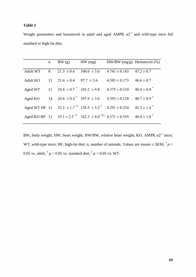
Body weight was significantly higher in aged mice than in adult ones and it was
further increased by HF diet-feeding without any effect of the genotype. Heart weight was
also higher in mice kept on HF diet and the increase was more pronounced in the KO group.
However, no difference among groups was observed in relative heart weight. Aging was
associated with a significant decrease in hematocrit level regardless the genotype or diet
(Table 2).
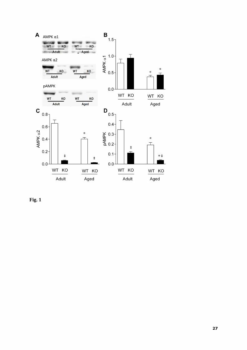
3.2. Protein expression and phosphorylation of AMPK
Western blot analysis of AMPK in the hearts from standard diet-fed mice (Fig. 1A)
revealed age-dependent decrease in the levels of both α1-subunit (Fig. 1B) and α2-subunit
(Fig. 1C). Whereas the level of AMPK α1-subunit was comparable to that of WT mice (Fig.
1B), a negligible amount of the α2-subunit was present in the KO mice, independent of age
(Fig. 1C). Phosphorylated AMPK levels were markedly reduced in KO compared with WT
mice and they were also decreased during aging in both genotypes (Fig. 1D).
3.3. Heart function
Echocardiography was used to assess effects of age, diet and AMPK deletion in
separate groups of mice. LVDs significantly increased in response to AMPK deletion in all
groups. Both LVDd and LVDs were significantly larger in the HF diet-fed as compared with
the standard diet-fed aged mice, but this effect reached statistical significance only in the KO
animals. Wall thickness measurements did not show any significant differences among
groups, except for a slight decrease in PWTs in aged KO compared to WT mice fed standard
diet (Table 3).
9
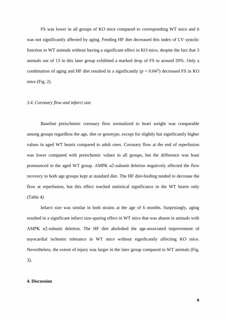
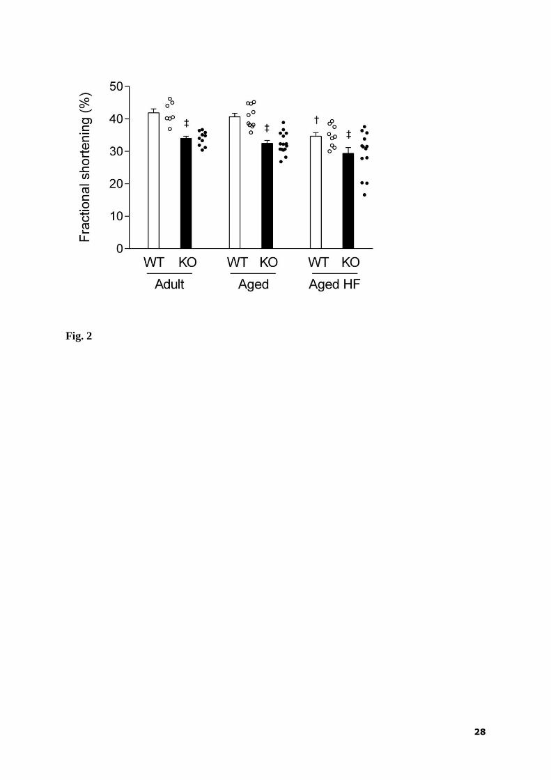
FS was lower in all groups of KO mice compared to corresponding WT mice and it
was not significantly affected by aging. Feeding HF diet decreased this index of LV systolic
function in WT animals without having a significant effect in KO mice, despite the fact that 3
animals out of 13 in this later group exhibited a marked drop of FS to around 20%. Only a
combination of aging and HF diet resulted in a significantly (p = 0.047) decreased FS in KO
mice (Fig. 2).
3.4. Coronary flow and infarct size
Baseline preischemic coronary flow normalized to heart weight was comparable
among groups regardless the age, diet or genotype, except for slightly but significantly higher
values in aged WT hearts compared to adult ones. Coronary flow at the end of reperfusion
was lower compared with preischemic values in all groups, but the difference was least
pronounced in the aged WT group. AMPK α2-subunit deletion negatively affected the flow
recovery in both age groups kept at standard diet. The HF diet-feeding tended to decrease the
flow at reperfusion, but this effect reached statistical significance in the WT hearts only
(Table 4).
Infarct size was similar in both strains at the age of 6 months. Surprisingly, aging
resulted in a significant infarct size-sparing effect in WT mice that was absent in animals with
AMPK α2-subunit deletion. The HF diet abolished the age-associated improvement of
myocardial ischemic tolerance in WT mice without significantly affecting KO mice.
Nevertheless, the extent of injury was larger in the later group compared to WT animals (Fig.
3).
4. Discussion

10
The results of the present study provide further evidence for the important role of
AMPK α2-subunit in the regulation of processes associated with heart aging. Aged female
mice exhibited decreased myocardial levels of both AMPK 1- and 2-subunits and AMPK
phosphorylation compared with adult littermates, the later effect being more pronounced in
KO animals. The major finding is that AMPK α2-subunit deletion and HF feeding
significantly impaired both cardiac contractile function and tolerance to acute I/R injury in
aged mice, but the negative effects of these two interventions were not additive to each other.
An increasing evidence suggests that AMPK activity can slow down aging process
and extend the lifespan. AMPK is involved in a complex network of signaling pathways that
control a number of cellular events helping to maintain energy balance under various stress
conditions and the loss of AMPK responsiveness may contribute to age-related metabolic
disturbances (Salminen and Kaarniranta 2012). However, reports concerning changes of
AMPK expression and activity during aging and senescence are rather controversial. For
example, Reznick et al. (2007) observed the loss of AMPK activation in skeletal muscle by
AICAR or exercise in aged rats without any change in the expression of AMPK 1- and 2-
subunits. Similarly, aging impaired phosphorylation of AMPK -subunit in rat skeletal
muscle but not the expression of either 1- or 2-subunits (Qiang et al. 2007). In contrast, an
increased AMPK activity with aging was observed in cultured human fibroblasts (Wang et al.
2003). Whereas the basal activity of AMPK 1-, but not 2-subunit, was higher in livers
from old mice compared to young animals, hypoxia-induced activation was blunted with
aging (Mulligan et al. 2005). Concerning the heart, neither basal activity of AMPK 1- and
2-subunit, nor its stimulation by AMP was afected by age in mice (Gonzales et al. 2004),
and no effect of aging on AMPK phosphorylation was found in human atrial tissue (Niemann
et al. 2013). On the other hand, recent reports showed that aging or senescence did not affect

11
murine myocardial AMPK expression, but it decreased its phosphorylation and activity as
well as the specific activities of both AMPK 1- and AMPK 2-subunits (Turdi et al. 2010;
Aurich et al. 2013). In the presents study, we found significant decreases in protein levels of
both -subunit isoforms and phosphorylated AMPK indicating its reduced basal activity in
the hearts of aged mice that was futher attenuated in animals with 2-subunit deletion.
Although the reason for these differences is unclear, our data are in line with the view that
AMPK function is likely compromised in aged hearts.
Despite the fact that AMPK is highly expressed in the myocardium, its role in the
pathogenesis of heart dysfunction associated with aging has not been fully understood. Our
echocardiographic data clearly show that the left ventricular systolic function in both adult
and aged KO mice was lower compared to WT animals as indicated by a decreased fractional
shortening. This observation is in agreement with the study of Turdi et al. (2010), who
demonstrated that the impairment of calcium handling and contractility of myocytes isolated
from aged murine hearts was more pronounced in transgenic animals overexpressing a
dominant negative AMPK 2 subunit (kinase dead). Moreover, aging-induced contractile
defects were attenuated by treatment with the AMPK activator metformin. These data suggest
that AMPK deficiency may contribute to age-induced cardiac dysfunction. It likely involves
oxidative stress, impaired intracellular calcium handling and disrupted mitochondrial function
(Turdi et al. 2010), but the complex mechanism remains to be elucidated.
The majority of studies that investigated an impact of aging on intrinsic cardiac
tolerance to I/R injury demonstrated its impairment with advanced age, possibly as a
consequence of enhanced oxidative stress (for review see Boengler et al. 2009). Our
observation of a reduced infarct size in female WT mice aged 18 months compared to their
younger littermates can be, therefore, considered rather surprising. However, available
evidence shows not only that aging is not always associated with exacerbated I/R injury

12
(Azhar et al. 1999; Peart et al. 2007) but also that myocardial ischemic tolerance can improve
with aging or senescence. For instance, several studies demonstrated infarct size reduction in
aged rats (Sniecinski and Liu 2004), mice (Gould et al. 2002; Boengler et al. 2007; Przyklenk
et al. 2008) or guinea-pigs (Rhodes et al. 2012). These discrepancies may be, in part, due to
marked differences in the age of animals used in various studies often regardless their sex.
Our preliminary observation that the infarct size reduction is absent in 18-month-old male
mice (Slámová at al. 2012) points to an important role of sex. Interestingly, Willems et al.
(2005) reported biphasic changes in the extent of myocardial injury caused by I/R in mice that
suggest a decreasing tolerance with aging and increasing tolerance with senescence; the
developmental profile of these changes differed between males and females. It seems,
therefore, that a certain sex-related window may exist during the aging process when intrinsic
protective mechanisms are more active allowing the heart to better survive acute I/R insult
than at younger stages. It has been proposed that aging-associated cardioprotection may be
linked to an attenuation of mitochondrial calcium overload (Rhodes et al. 2012) but the
underlying mechanism is unknown at present. Although in the present study the AMPK α2-
subunit deletion did not significantly worsen the extent of myocardial injury in young
animals, the absence of infarct-sparing effect of aging in KO mice suggests that the AMPK
pathway plays a role in this phenomenon.
Consistent with the view that high intake of fatty acids may cause cardiac lipotoxicity
(Lopaschuk et al. 2007), here we show that feeding WT mice with HF diet for 6 months
during aging decreased LV fractional shortening and impaired ischemic tolerance compared
to their age-matched littermates fed standard diet. These results support a number of the
previous reports indicating HF diet-induced cardiac contractile dysfunction (Ouwens et al.
2005; Relling et al. 2006; Turdi et al. 2011; Guo et al. 2013) and exacerbation of I/R injury by
increased levels of fatty acids (Lopaschuk et al. 2007; Thakker et al. 2008). However, it

13
should be mentioned that other reports failed to demonstrate cardiac lipotoxicity and
dysfunction following long term exposure to HF diet (Nascimento et al. 2011; Brainard et al.
2013) and this issue remains controversial. Besides the source of diet, the age of animals
likely plays a role due to the inability of old myocytes to adapt to high fatty acid load (Aurich
et al. 2013). In the present study, switch to HF diet took place at the age of 12 months when
reproductive function of female C57BL/6J starts to cease (Felicio et al. 1984) in association
with neuroendocrine and hormonal changes that may also influence effects of the diet.
It has been shown that AMPK α2-subunit activity is important in the regulation of
fatty acid uptake in HF diet-fed mice (Abbott et al. 2012). A limitation of our work is that we
could not measure AMPK subunits activity in aged mice fed HF diet. However, earlier studies
have demonstrated that HF diet decreases myocardial AMPK phosphorylation status and
activity (Guo et al. 2013; Lindholm et al. 2013), these effects being more pronounced with
advanced age (Aurich et al. 2013). In addition, AMPK α2-subunit deficiency exaggerates
insulin resistance (Fujii et al. 2008), cardiac contractile dysfunction and impaired intracellular
calcium handling (Turdi et al. 2011) induced by HF diet-feeding in middle age mice. In our
experiments, the LV systolic dysfunction and ischemic intolerance in mice on HF diet was
more pronounced in KO compared to WT group. However, these unfavourable effects of the
HF diet did not reach statistical significance on the background of AMPK α2-subunit deletion.
The reason for the absence of additive effects of HF diet and AMPK deficiency is unclear, but
it can be related to the fact that both heart function and ischemic tolerance of aged KO mice
fed standard diet were already compromised compared to WT animals, whereas any notable
effect of AMPK deficiency itself was not observed in the study of Turdi et al. (2011) on
younger mice. Alternatively, the defect in AMPK function may be apparent only under the
conditions promoting lipogenesis, i.e. in the animals fed standard diet when activation of
AMPK can increase fatty acid oxidation to preserve intracellular energy status, whereas

14
AMPK inactivation likely remains silent when lipogenesis is heavily suppressed in response
to HF diet-feeding. Our previous results on the functional significance of AMPK in the liver
support the later possibility (Jeleník et al. 2010).
5. Conclusions
Here we demonstrate that aging resulted in significant AMPK downregulation and
improved ischemic tolerance of female murine hearts. Global genetic ablation of AMPK α2-
subunit or long-term feeding HF diet similarly resulted in cardiac dysfunction and abolished
the anti-ischemic protection. However, the effects of AMPK α2-subunit deletion were not
further potentiated by HF diet. Our findings support the view that AMPK activity plays a role
in normal heart aging, suggesting this kinase as a potential target for cardioprotective
interventions.
Acknowledgements
This work was supported by the Czech Science Foundation (14-36804G) and the
institutional research project RVO:67985823. The authors thank Grahame Hardie for the
sheep AMPK α1- and α2-subunit antibodies and Benoit Viollet for the transgenic mice.

15
References
ABBOTT MJ, CONSTANTINESCU S, TURCOTTE LP: AMP-activated protein kinase α2 is
an essential signal in the regulation of insulin-stimulated fatty acid uptake in control-fed and
high-fat-fed mice. Exp Physiol 97: 603-617, 2012.
ASHTON KJ, WILLEMS L, HOLMGREN K, FERREIRA L, HEADRICK JP: Age
associated shifts in cardiac gene transcription and transcriptional responses to ischemic stress.
Exp Gerontol. 41: 189-204, 2006.
AURICH AC, NIEMANN B, PAN R, GRUENLER S, ISSA H, SILBER RE, ROHRBACH
S: Age-dependent effects of high fat-diet on murine left ventricles: role of palmitate. Basic
Res Cardiol 108: 369, 2013.
AZHAR G, GAO W, LIU L, WEI JY: Ischemia-reperfusion in the adult mouse heart
influence of age. Exp Gerontol 34: 699-714, 1999.
BOENGLER K, KONIETZKA I, BUECHERT A, HEINEN Y, GARCIE-DORADO D,
Heusch G, Schulz R: Loss of ischemic preconditioning's cardioprotection in aged mouse
hearts is associated with reduced gap junctional and mitochondrial levels of connexin 43. Am
J Physiol Heart Circ Physiol 292: H1764-H1769, 2007.
BOENNGLER K, SCHULZ R, HEUSCH G: Loss of cardioprotection with ageing.
Cardiovasc Res 83: 247-261, 2009.
BRAINARD RE, WATSON LJ, DEMARTINO AM, BRITTIAN KR, READNOWER RD,
BOAKYE AA, ZHANG D, HOETKER JD, BHATNAGAR A, BABA SP, JONES SP: High
fat feeding in mice is insufficient to induce cardiac dysfunction and does not exacerbate heart
failure. PloS One 8: e83174, 2013.
DYCK JR, LOPASCHUK GD: AMPK alterations in cardiac physiology and pathology:
enemy or ally? J Physiol 574(Pt 1): 95-112, 2006.

16
EDWARDS AG, DONATO AJ, LESNIEWSKI LA, GIOSCIA RA, SEALS DR, MOORE
RL: Life-long caloric restriction elicits pronounced protection of the aged myocardium: a role
for AMPK. Mech Ageing Dev 131: 739-742, 2010.
FELICIO LS, NELSON JF, FINCH CE: Longitudinal studies of estrous cyclicity in aging
C57BL/6J mice: II. Cessation of cyclicity and the duration of persistent vaginal cornification.
Biol Reprod 31: 446-453, 1984.
FERRARI AU, RADAELLI A, CENTOLA M: Aging and the cardiovascular system. J Appl
Physiol 95: 2591-2597, 2003.
FOLMES CD, WAGG CS, SHEN M, CLANACHAN AS, TIAN R, LOPASCHUK GD:
Suppression of 5'-AMP-activated protein kinase activity does not impair recovery of
contractile function during reperfusion of ischemic hearts. Am J Physiol Heart Circ Physiol
297: H313-H321, 2009.
FUJII N, HO RC, MANABE Y, JESSEN N, TOYODA T, HOLLAND WL, SUMMERS SA,
HIRSHMAN MF, GOODYEAR LJ: Ablation of AMP-activated protein kinase alpha2
activity exacerbates insulin resistance induced by high-fat feeding of mice. Diabetes 57:
2958-2966. 2008.
GONZALEZ AA, KUMAR R, MULLIGAN JD, DAVIS AJ, SAUPE KW: Effects of aging
on cardiac and skeletal muscle AMPK activity: basal activity, allosteric activation, and
response to in vivo hypoxemia in mice. Am J Physiol Regul Integr Comp Physiol 287:
R1270-R1275, 2004.
GOULD KE, TAFFAT GE, MICHAEL LH, CHRISTIE RM, KONKOL DL, POCIUS JS,
ZACHARIAH JP, CHAUPIN DF, DANIEL SL, SANDUSKY GE Jr, HARTLEY CJ,
ENTMAN ML: Heart failure and greater infarct expansion in middle-aged mice: a relevant
model for postinfarction failure. Am J Physiol Heart Circ Physiol 282: H615-H621, 2002.

17
GUO R, ZHANG Y, TURDI S, REN J: Adiponectin knockout accentuates high fat diet-
induced obesity and cardiac dysfunction: role of autophagy. Biochim Biophys Acta 1832:
1136-1148, 2013.
HARDIE DG, CARLING D: The AMP-activated protein kinase--fuel gauge of the
mammalian cell? Eur J Biochem 246: 259-273, 1997.
HARDIE DG: Minireview: the AMP-activated protein kinase cascade: the key sensor of
cellular energy status. Endocrinology 144: 5179-5183, 2003.
JELENÍK T, ROSSMEISL M, KUDA O, JÍLKOVÁ ZM, MEDŘÍKOVÁ D, KŮS V,
HENSLER M, JANOVSKÁ P, MIKŠÍK I, BARANOWSKI M, GORSKI J, HEBRARD S,
JENSEN TE, FLACHS P, HAWLEY S, VIOLLET B, KOPECKÝ J: AMP-activated protein
kinase α2 subunit is required for the preservation of hepatic insulin sensitivity by n-3
polyunsaturated fatty acids. Diabetes 59: 2737-2746, 2010.
KUDA O, JELENÍK T, JÍLKOVÁ Z, FLACHS P, ROSSMEISL M, HENSLER M,
KAZDOVÁ L, OGSTON N, BARANOWSKI M, GORSKI J, JANOVSKÁ P, KŮS V,
POLAK J, MOHAMED-ALI V, BURCELIN R, CINTI S, BRYHN M, KOPECKÝ J: n-3
Fatty acids and rosiglitazone improve insulin sensitivity through additive stimulatory effects
on muscle glycogen synthesis in mice fed high-fat diet. Diabetologia 52: 941-951, 2009.
KUDO N, BARR AJ, BARR RL, DESAI S, LOPASCHUK GD: High rates of fatty acid
oxidation during reperfusion of ischemic hearts are associated with a decrease in malonyl-
CoA levels due to an increase in 5'-AMP-activated protein kinase inhibition of acetyl-CoA
carboxylase. J Biol Chem 270: 17513-1720, 1997.
KŮS V, PRAŽÁK T, BRAUNER P, HENSLER M, KUDA O, FLACHS P, JANOVSKÁ P,
MEDŘÍKOVÁ D, ROSSMEISL M, JÍLKOVÁ Z, ŠTEFL B, PASTALKOVA E, DRAHOTA
Z, HOUŠTĚK J, KOPECKÝ J: Induction of muscle thermogenesis by high-fat diet in mice:
association with obesity-resistance. Am J Physiol Endocrinol Metab 295: E356-E367, 2008.

18
LAKKATTA EG, SOLLOTT SJ: The "heartbreak" of older age. Mol Interv 2: 431-446. 2002.
LI J, COVEN DL, MILLER EJ, HU X, YOUNG ME, CARLING D, SINUSAS AJ, YOUNG
LH: Activation of AMPK alpha- and gamma-isoform complexes in the intact ischemic rat
heart. Am J Physiol Heart Circ Physiol 291: H1927-H1934. 2006.
LINDHOLM CR, ERTEL RL, BAUWENS JD, SCHMUCK EG, MULLIGAN JD, SAUPE
KW: A high-fat diet decreases AMPK activity in multiple tissues in the absence of
hyperglycemia or systemic inflammation in rats. J Physiol Biochem 69: 165-175, 2013.
LIU Q, DOCHERTY JC, RENDELL JC, CLANACHAN AS, LOPASHUK GD: High levels
of fatty acids delay the recovery of intracellular pH and cardiac efficiency in post-ischemic
hearts by inhibiting glucose oxidation. J Am Coll Cardiol 39: 718-725, 2002.
LOPASCHUK GD, FOLMES CD, STANLEY WC: Cardiac energy metabolism in obesity.
Circ Res 101: 335-347, 2007.
MATĚJKOVÁ O, MUSTARD KJ, ŠPONAROVÁ J, FLACHS P, ROSSMEISL M, MIKŠÍK
I, THOMASON-HUGHES M, GRAHAME HARDIE D, KOPECKÝ J: Possible involvement
of AMP-activated protein kinase in obesity resistance induced by respiratory uncoupling in
white fat. FEBS Lett 569: 245-248, 2004.
McCARTY MF: Chronic activation of AMP-activated kinase as a strategy for slowing aging.
Med Hypotheses 63: 334-339, 2004.
MULLIGAN JD, GONZALEZ AA, KUMAR R, DAVIS AJ, SAUPE KW: Aging elevates
basal adenosine monophosphate-activated protein kinase (AMPK) activity and eliminates
hypoxic activation of AMPK in mouse liver. J Gerontol A Biol Sci Med Sci 60: 21-27, 2005.
NASCIMENTO AF, LUVIZOTTO RA, LEOPOLDO AS, LIMA-LEOPOLDO AP, SEIVA
FR, JUSTULIN LA Jr, SILVA MD, OKOSHI K, WANG XD, CICOGNA AC: Long-term
high-fat diet-induced obesity decreases the cardiac leptin receptor without apparent
lipotoxicity. Life Sci 88: 1031-1038, 2011.
19
NIEMANN B, PAN R, TESCHNER M, BOENING A, SILBER RE, ROHRBACH S: Age
and obesity-associated changes in the expression and activation of components of the AMPK
signaling pathway in human right atrial tissue. Exp Gerontol 48: 55-63, 2013.
OUWENS DM, BOER C, FODOR M, de GALAN P, HEINE RJ, MAASSEN JA,
DIAMANT M: Cardiac dysfunction induced by high-fat diet is associated with altered
myocardial insulin signalling in rats. Diabetologia 48: 1229-1237, 2005.
PEART JN, GROSS ER, HEADRICK JP, GROSS GJ: Impaired p38 MAPK/HSP27 signaling
underlies aging-related failure in opioid-mediated cardioprotection. J Mol Cell Cardiol 42:
972-980, 2007.
PRZYKLENK K, MAYNARD M, DARLING CE, WHITTAKER P, Aging mouse hearts are
refractory to infarct size reduction with post-conditioning. J Am Coll Cardiol 51: 1393-1398,
2008.
QIANG W, WEIQIANG K, QING Z, PENGJU Z, YI L: Aging impairs insulin-stimulated
glucose uptake in rat skeletal muscle via suppressing AMPK-alpha. Exp Mol Med 39: 535-
543, 2007.
RELLING DP, ESBERG LB, FANG CX, JOHNSON WT, MURPHY EJ, CARLSON EC,
SAARI JT, REN J: High-fat diet-induced juvenile obesity leads to cardiomyocyte dysfunction
and upregulation of Foxo3a transcription factor independent of lipotoxicity and apoptosis. J
Hypertens 24: 549-561, 2006.
REZNICK RM, ZONG H, LI J, MORINO K, MOORE IK, YU HJ, LIU ZX, DONG J,
MUSTARD KJ, HAWLEY SA, BEFROY D, PYPAERT M, HARDIE DG, YOUNG LH,
SHULMAN GI: Aging-associated reductions in AMP-activated protein kinase activity and
mitochondrial biogenesis. Cell Metab 5: 151-156, 2007.
RHODES SS, CAMARA AK, HEISNER JS, RIESS ML, ALDAKKAK M, STOWE DF:
Reduced mitochondrial Ca2+ loading and improved functional recovery after ischemia-

20
reperfusion injury in old vs. young guinea pig hearts. Eur J Clin Microbiol Infect Dis 31:
1551-1559, 2012.
SALMINEN A, KAARNIRANTA K: AMP-activated protein kinase (AMPK) controls the
aging process via an integrated signaling network. J Mol Med 89: 667-676, 2012.
STEINBERG GR, KEMP BE: AMPK in Health and Disease. Physiol Rev 89:1025-1078,
2009.
SLÁMOVÁ K, JANOVSKÁ P, PAPOUŠEK F, KOPECKÝ J, KOLÁŘ F: Influence of aging
and high-fat diet on myocardial ischemic tolerance in mice with AMP-activated protein
kinase α2-subunit deficiency. Act Nerv Super Rediviva 54: 100, 2012.
SNIECINSKI R, LIU H: Reduced efficacy of volatile anesthetic preconditioning with
advanced age in isolated rat myocardium. Anesthesiology 100: 589-597, 2004.
THAKKER GD, FRANGOGIANNIS NG, ZYMEK PT, SHARMA S, RAYA JL, BARGER
PM, TAEGTMEYER H, ENTMAN ML, BALLANTYNE CM: Increased myocardial
susceptibility to repetitive ischemia with high-fat diet-induced obesity. Obesity 16: 2593-
2600, 2008.
TURDI S, FAN X, LI J, ZHAO J, HUFF AF, DU M, REN J: AMP-activated protein kinase
deficiency exacerbates aging-induced myocardial contractile dysfunction. Aging Cell 9: 592-
606, 2010.
TURDI S, KANDADI MR, ZHAO J, HUFF AF, DU M, REN J: Deficiency in AMP-
activated protein kinase exaggerates high fat diet-induced cardiac hypertrophy and contractile
dysfunction. J Mol Cell Cardiol 50: 712-722, 2011.
VIOLLET B, ANDREELLI F, JØRGENSEN SB, PERRIN C, GELOEN A, FLAMEZ D, MU
J, LENZNER C, BAUD O, BENNOUN M, GOMAS E, NICOLAS G, WOJTASZEWSKI JF,
KAHN A, CARLING D, SCHUIT FC, BIRNBAUM MJ, RICHTER EA, BURCELIN R,

21
VAULONT S: The AMP-activated protein kinase alpha2 catalytic subunit controls whole-
body insulin sensitivity. J Clin Invest 111: 91-98, 2003.
WANG W, YANG X, LÓPEZ de SILANES I, CARLING D, GOROSPE M: Increased
AMP:ATP ratio and AMP-activated protein kinase activity during cellular senescence linked
to reduced HuR function. J Biol Chem 278: 27016-27023, 2003.
WANG S, SONG P, ZOU MH: AMP-activated protein kinase, stress responses and
cardiovascular diseases. Clin Sci 122: 555-573, 2012.
WILLEMS L, ZATTA A, HOLMGREN K, ASHTON KJ, HEADRICK JP: Age-related
changes in ischemic tolerance in male and female mouse hearts. J Mol Cell Cardiol 38: 245-
56. 2005.
ZAHA VG, YOUNG LH: AMP-activated protein kinase regulation and biological actions in
the heart. Circ Res 111: 800-814, 2012.

22
Table 1
Macronutrient composition and energy content of diets and fatty acid coposition of dietary
lipids.
Standard High-fat
diet diet
Macronutrient composition (% diet, wt/wt)
Lipid 3.4 35.2
Carbohydrate 55.3 35.4
Protein 19.3 20.5
Energy density (kJ/g) 16.3 22.8
Fatty acid composition (g/100g)
SFA 18.4 22.1
MUFA 17.8 28.5
n-6 PUFA 57.9 47.7
n-3 PUFA 6.0 1.8
SFA, saturated fatty acids; MUFA, monounsaturated fatty acids; PUFA, polyunsaturated fatty
acids.
23
Table 2
Weight parameters and hematocrit in adult and aged AMPK α2-/-
and wild-type mice fed
standard or high-fat diet.
n BW (g) HW (mg) HW/BW (mg/g) Hematocrit (%)
Adult WT 8 21.3 ± 0.6 100.6 ± 3.6 4.741 ± 0.143 47.2 ± 0.7
Adult KO 11 21.4 ± 0.4 97.7 ± 3.6 4.583 ± 0.173 46.6 ± 0.7
Aged WT 11 24.4 ± 0.7 * 103.2 ± 9.8 4.375 ± 0.310 40.4 ± 0.8
*
Aged KO 14 24.6 ± 0.3 * 107.9 ± 3.6 4.393 ± 0.128 40.7 ± 0.9
*
Aged WT HF 11 33.3 ± 1.7 *†
138.5 ± 5.2 * 4.291 ± 0.324 42.3 ± 1.4
*
Aged KO HF 11 35.1 ± 2.5 *†
162.3 ± 4.0 *†‡
4.371 ± 0.355 40.4 ± 1.8 *
BW, body weight; HW, heart weight; HW/BW, relative heart weight; KO, AMPK α2-/-
mice;
WT, wild-type mice; HF, high-fat diet; n, number of animals. Values are means ± SEM; *
p <
0.05 vs. adult; † p < 0.05 vs. standard diet;
‡ p < 0.05 vs. WT.

24
Table 3
Heart rate and echocardiographic parameters of adult and aged AMPK α2-/-
and wild-type mice fed standard or high-fat diet.
n HR (beat/min) LVDd (mm) LVDs (mm) AWTd (mm) PWTd (mm) AWTs (mm) PWTs (mm)
Adult WT 7 508 ± 12 3.40 ± 0.4 1.98 ± 0.06 0.75 ± 0.04 0.71 ± 0.03 1.15 ± 0.04 1.17 ± 0.04
Adult KO 10 516 ± 8 3.67 ± 0.06 2.42 ± 0.05 ‡ 0.73 ± 0.01 0.70 ± 0.02 1.12 ± 0.02 1.11 ± 0.03
Aged WT 10 539 ± 9 3.42 ± 0.08 2.04 ± 0.08 0.74 ± 0.02 0.75 ± 0.02 1.18 ± 0.02 1.19 ± 0.03
Aged KO 15 561 ± 8 3.66 ± 0.07 2.48 ± 0.07 ‡ 0.73 ± 0.02 0.73 ± 0.03 1.14 ± 0.02 1.12 ± 0.02
‡
Aged WT HF 10 525 ± 14 3.74 ± 0.04 2.45 ± 0.06 0.79 ± 0.03 0.80 ± 0.02 1.19 ± 0.02 1.18 ± 0.03
Aged KO HF 13 542 ± 10 4.08 ± 0.10 † ‡
2.90 ± 0.14 † ‡ 0.78 ± 0.02 0.81 ± 0.03 1.16 ± 0.02 1.15 ± 0.03
KO, AMPK α2-/-
mice; WT, wild-type mice; HF, high-fat diet; HR, heart rate; LVDd, diastolic cavity diameter; LVDs, systolic cavity diameter;
AWTd, diastolic anterior wall thickness; PWTd, diastolic posterior wall thickness; AWTs, systolic anterior wall thickness; PWTs, systolic
posterior wall thickness; n, number of animals. Values are means ± SEM; † p < 0.05 vs. standard diet;
‡ p < 0.05 vs. WT.

25
Table 4
Coronary flow before ischemia and at the end of reperfusion in isolated perfused hearts of
adult and aged AMPK α2-/-
and wild-type mice fed standard or high-fat diet.
Coronary flow (ml/g/min)
n Preischemic Reperfusion
Adult WT 8 13.3 ± 1.0 10.2 ± 0.6
Adult KO 11 13.2 ± 0.6 8.1 ± 0.5 ‡
Aged WT 11 17.8 ± 1.3 * 16.1 ± 1.3
*
Aged KO 14 15.2 ± 0.4 10.1 ± 0.9 * ‡
Aged WT HF 11 15.0 ± 1.6 8.8 ± 0.9 †
Aged KO HF 11 13.3 ± 0.9 8.5 ± 1.4
KO, AMPK α2-/-
mice; WT, wild-type mice; HF, high-fat diet; n, number of hearts. Values are
means ± SE; * p < 0.05 vs. adult;
† p < 0.05 vs. standard diet;
‡ p < 0.05 vs. WT.

26
Legend to Figures
Figure 1
Representative blots (A) and myocardial protein levels of AMPK 1-subunit (B), AMPK 2-
subunit (C) and phosphorylated AMPK (D) in adult and aged AMPK α2
-/- (KO) and wild-type
(WT) mice fed standard diet. Values are means ± SEM; n = 7 - 10 in each group; * p < 0.05
vs. adult; ‡ p < 0.05 vs. WT.
Figure 2
Left ventricular fractional shortening in adult and aged AMPK α2-/-
(KO) and wild-type (WT)
mice fed standard or high-fat (HF) diet. Circles indicate individual experiments. Values are
means ± SEM; n = 7 - 15 in each group; ‡ p < 0.05 vs. WT.
Figure 3
Myocardial infarct size expressed as percentage of the left ventricle (LV) in adult and aged
AMPK α2-/-
(KO) and wild-type (WT) mice fed standard or high-fat (HF) diet. Circles
indicate individual experiments. Values are means ± SEM; n = 11 - 16 in each group; * p <
0.05 vs. adult; † p < 0.05 vs. standard diet;
‡ p < 0.05 vs. WT.
27
Fig. 1
28
Fig. 2

29
Fig. 3


![Resveratrol suppresses glial activation and alleviates ...€¦ · sphate (AMP)-activated protein kinase (AMPK) [19]. Mul-tiple lines of evidence have indicated that AMPK is emerging](https://img.pdfslide.net/doc/110x75/60f875cb55012f48d821e488/resveratrol-suppresses-glial-activation-and-alleviates-sphate-amp-activated.jpg)


![Human Mitogen-activated Protein Kinase Kinase 4 as a ......(CANCERRESEARCH57. 4177—4182,October 1, 1997] Advances in Brief Human Mitogen-activated Protein Kinase Kinase 4 as](https://img.pdfslide.net/doc/110x75/6082557b7810d746a5071f39/human-mitogen-activated-protein-kinase-kinase-4-as-a-cancerresearch57.jpg)