Embed Size (px)
Citation preview

│ 1
Foreword
This Adverse Outcome Pathway (AOP) on binding of inhibition of the mitochondrial
complex I of nigro-striatal neurons leads to parkinsonian motor deficits, has been
developed under the auspices of the OECD AOP Development Programme, overseen by
the Extended Advisory Group on Molecular Screening and Toxicogenomics (EAGMST),
which is an advisory group under the Working Group of the National Coordinators for the
Test Guidelines Programme (WNT). The AOP has been reviewed internally by the
EAGMST, externally by experts nominated by the WNT, and has been endorsed by the
WNT and the Working Party on Hazard Assessment (WPHA) in xxxx.
Through endorsement of this AOP, the WNT and the WPHA express confidence in the
scientific review process that the AOP has undergone and accept the recommendation of
the EAGMST that the AOP be disseminated publicly. Endorsement does not necessarily
indicate that the AOP is now considered a tool for direct regulatory application.
The Joint Meeting of the Chemicals Committee and the Working Party on Chemicals,
Pesticides and Biotechnology agreed to declassification of this AOP on xxxx.
This document is being published under the responsibility of the Joint Meeting of the
Chemicals Committee and the Working Party on Chemicals, Pesticides and
Biotechnology.

2 │
Table of contents
Abstract .................................................................................................................................................. 4
Graphical Representation ..................................................................................................................... 5
Summary of the AOP ............................................................................................................................ 6
Molecular Initiating Events (MIE), Key Events (KE), Adverse Outcomes (AO) ............................... 6 Key Event Relationships ...................................................................................................................... 6 Stressors ............................................................................................................................................... 7
Overall Assessment of the AOP ............................................................................................................ 8
Domain of Applicability ...................................................................................................................... 8 Essentiality of the Key Events ............................................................................................................. 8 Weight of Evidence Summary ........................................................................................................... 11 Quantitative Consideration ................................................................................................................ 20
Considerations for Potential Applications of the AOP (optional) ................................................... 22
References ............................................................................................................................................ 23
Appendix 1: List of MIE(s), KE(s) and AO(s) in the AOP .............................................................. 36
List of MIE(s) in the AOP ................................................................................................................. 36 List of KE(s) in the AOP ................................................................................................................... 46 List of AO(s) in the AOP ................................................................................................................... 83
Appendix 2: List of Key Event Relationships in the AOP ............................................................... 94
List of Adjacent Key Event Relationships ......................................................................................... 94 List of Non Adjacent Key Event Relationships ............................................................................... 173

│ 3
Adverse Outcome Pathway on Inhibition of the mitochondrial
complex I of nigro-striatal neurons leads to parkinsonian
motor deficits
Short Title: Mitochondrial dysfunction and Neurotoxicity
Authors:
Anna Bal-Price, Chemical Safety and Alternative Methods Unit/EURL ECVAM of the European
Commission, Ispra, Italy
Marcel Leist, University of Konstanz, Konstanz, Germany
Stefan Schildknecht, University of Konstanz, Konstanz, Germany
Florianne Tschudi-Monnet, University of Lausanne and SCAHT, Lausanne, Switzerland
Alicia Paini, Chemical Safety and Alternative Methods Unit/EURL ECVAM of the European
Commission, Ispra, Italy
Andrea Terron, European Food Safety Authority, Parma Italy

4 │
Abstract
This Adverse outcome Pathway (AOP) describes the linkage between inhibition of
complex I (CI) of the mitochondrial respiratory chain and motor deficit as in parkinsonian
disorders. Binding of an inhibitor to complex I has been defined as the molecular
initiating event (MIE) that triggers mitochondrial dysfunction, impaired proteostasis,
which then cause degeneration of dopaminergic (DA) neurons of the nigro-striatal
pathway. Neuroinflammation is triggered early in the neurodegenerative process and
exacerbates it significantly. These causatively linked cellular key events result in motor
deficit symptoms, typical for parkinsonian disorders, including Parkinson's disease (PD),
described in this AOP as an Adverse Outcome (AO). Since the release of dopamine in the
striatum by DA neurons of the Substantia Nigra pars compacta (SNpc) is essential for
motor control, the key events refer to these two brain structures. The weight-of-evidence
supporting the relationship between the described key events is based mainly on effects
observed after an exposure to the chemicals rotenone and 1-methyl-4-phenyl-1,2,3,6-
tetrahydropyridine (MPTP), i.e. two well-known inhibitors of complex I. Data from
experiments with these two chemicals reveal a significant concordance in the dose-
response relationships between the MIE and AO and within key events (KEs). Also
essentiality of the described KEs for this AOP is strong since there is evidence from
knock out animal models, engineered cells or replacement therapies that blocking,
preventing or attenuating an upstream KE is mitigating the AO. Similarly, there is proved
experimental support for the key event relationships (KERs) as multiple studies
performed with modulating factors that attenuate (particularly with antioxidants) or
augment (e.g. overexpression of viral-mutated α-synuclein) a KE up show that such
interference leads to an increase of KE down or the AO. Information from in vitro and in
vivo experiments is complemented by human studies in brain tissues from individuals
with sporadic Parkinson's disease (Keeney et al., 2006) to support the pathways of
toxicity proposed in this AOP.

│ 5
Graphical Representation

6 │
Summary of the AOP
Molecular Initiating Event (MIE), Key Events (KEs), Adverse Outcomes (AOs)
Sequence Type Event
ID Title Short name
1 MIE 888 Binding of inhibitor, NADH-
ubiquinone oxidoreductase (complex I)
Binding of inhibitor, NADH-
ubiquinone oxidoreductase (complex I)
2 KE 887 Inhibition, NADH-ubiquinone
oxidoreductase (complex I)
Inhibition, NADH-ubiquinone
oxidoreductase (complex I)
3 KE 177 N/A, Mitochondrial dysfunction 1 N/A, Mitochondrial dysfunction 1
4 KE 889 Impaired, Proteostasis Impaired, Proteostasis
5 KE 890 Degeneration of dopaminergic neurons
of the nigrostriatal pathway
Degeneration of dopaminergic neurons
of the nigrostriatal pathway
6 KE 188 N/A, Neuroinflammation N/A, Neuroinflammation
7 AO 896 Parkinsonian motor deficits Parkinsonian motor deficits
Key Event Relationships (KERs)
Upstream Event Relationship
Type Downstream Event Evidence
Quantitative
Understanding
Binding of inhibitor, NADH-
ubiquinone oxidoreductase
(complex I)
adjacent Inhibition, NADH-
ubiquinone oxidoreductase
(complex I)
High Low
Inhibition, NADH-
ubiquinone oxidoreductase
(complex I)
adjacent N/A, Mitochondrial
dysfunction 1
High Moderate
N/A, Mitochondrial
dysfunction 1
adjacent Impaired, Proteostasis Moderate Low
N/A, Mitochondrial
dysfunction 1
non-adjacent Degeneration of
dopaminergic neurons of the
nigrostriatal pathway
Moderate Low
Impaired, Proteostasis adjacent Degeneration of
dopaminergic neurons of the
nigrostriatal pathway
High Moderate
Degeneration of
dopaminergic neurons of the
nigrostriatal pathway
adjacent N/A, Neuroinflammation Moderate Moderate
N/A, Neuroinflammation adjacent Degeneration of
dopaminergic neurons of the
nigrostriatal pathway
Moderate Moderate
Degeneration of
dopaminergic neurons of the
nigrostriatal pathway
adjacent Parkinsonian motor deficits High High

│ 7
Stressors
Name Evidence
MPP+ High
Rotenone High
MPP+
1-Methyl-4-phenyl-1,2,3,6-tetrahydropyridine (MPTP) is a meperidine analog intended
for illecit use as a ricreative drug resulted in life-long parkinsonism in a number of
abusers. MPTP is able to cross the blood-brain-barrier and is actually a pro-toxin, which
is converted by monoamine oxidase B in astrocytes to the neurotoxic active species 1-
methyl-4-phenyl-1,2,3,6-tetrahydropyridinium ion (MPP+). MPP+ has high affinity for
the DA transporter (DAT), thus preferentially enters dopaminergic neurons where is
accumulating in mitochondria to directely inhibit complex I (CI) and indirectly CII in the
respiratory transport chain. (Aguilar et al. 2015; pasquali et al.2 014).
Rotenone
The insecticide rotenone is a potent Inhibitor of CI in the respiratory transport chain of
mitochondroa with a relative selectivity for dopaminergig neurons in low
dosage.Additional non-specific treatment related effects include: vascular damage with
secondary ischemic neurodegeneration, tissue damage in heart and testicles and
interstitial haemorrhages in lungs and kidneys. Mitochondrial dysfunction is considered
to be the initial step in rotenone toxicity; though, multiple pathways are involved in the
process associated with rotenone-induced neurotoxicity. However, the impact of rotenone
on glial cells also impact on neuronal outcomes (Aguilar et al.2015). Rotenone provided
the first proof-of-concept that a systemic defect in mitochondrial function could lead to
selective nigrostriatal neurodegeneration. The rotenone animal model accurately
recapitulates many other features of Parkinson's disease, including: accumulation and
aggregation of endogenous wildtype alpha-synuclein; α-synuclein- and polyubiquitin-
positive Lewy bodies and Lewy neurites; apomorphine responsive behavioral deficits;
early and sustained activation of microglia; oxidative modification and translocation of
DJ-1 into mitochondria in vivo; impairment of the nigral ubiquitin-proteasome system;
accumulation of iron in the substantia nigra through a mechanism involving transferrin
and transferrin receptor 2; α-synuclein pathology in enteric neurons and functional
deficits in GI function, including gastroparesis (Greenamyre et al. 2010).

8 │
Overall Assessment of the AOP
Domain of Applicability
Life Stage Applicability
Life Stage Evidence
Adult High
Taxonomic Applicability
Term Scientific Term Evidence Links
human Homo sapiens High NCBI
rat Rattus norvegicus High NCBI
Sex Applicability
Sex Evidence
Mixed High
This proposed AOP is neither sex-dependent nor associated with certain life stage;
however, aged animals may be more sensitive. The relevance of this AOP during the
developmental period has not been investigated. In vivo testing has no species restriction.
The mouse was the species most commonly used in the experimental models conducted
with the chemical stressors; though experimental studies using alternative species have
been also performed (Johnson et al. 2015). However, animal models (rodents in
particular) would have limitations as they are poorly representative of the long human
life-time as well as of the human long-time exposure to the potential toxicants. Human
cell-based models would likely have better predictivity for humans than animal cell
models. In this case, toxicokinetics information from in-vivo studies would be essential to
test the respective concentrations in-vitro on human cells.
Essentiality of the Key Events
Essentiality of KEs for this AOP is strong. There is ample evidence from knock out
animal models, engineered cells or replacement therapies that blocking, preventing or
attenuating an upstream KE is mitigating the AO. In addition, there is experimental
support for the KERs as multiple studies performed with modulating factors that attenuate
(particularly with antioxidants) or augment (e.g. overexpression of viral-mutated α-
synuclein) a KE show that such interference leads to an increase of KE down or the AO.

│ 9
2 Support for
Essentiality of KEs
Defining
Question
Are downstream
KEs and/or the
AO prevented if
an upstream KE
is blocked?
High (Strong) Moderate Low(Weak)
Direct evidence from
specifically designed
experimental studies
illustrating essentiality
for at least one of the
important KEs (e.g.
stop/reversibility studies,
antagonism, knock out
models, etc.)
Indirect evidence
that sufficient
modification of an
expected
modulating factor
attenuates or
augments a KE
leading to increase
in KE down or AO
No or
contradictory
experimental
evidence of the
essentiality of
any of the KEs
KE1
Inhibition of
complex I
STRONG
Rationale: Inactivation of the NADH: Ubiquinone Oxidoreductase
Core Subunit S7 (Ndufs 4 gene knockout mice) that produces CI
deficiency causes encephalomyopathy, including ataxia and loss of
motor skills (Kruse et al., 2008). NDI1-transducted SK-N-MC cells
expressing the rotenone-insensitive single subunit NADH
dehydrogenase of yeast (NDI1) that acts as a replacement for the
entire CI in mammalian cells were completely resistant to 100 nM
rotenone-mediated cell death (at 48 hrs of exposure) indicating that
rotenone – induced toxicity requires rotenone biding of CI (Sherer et
al., 2003). In all rotenone models, mitochondria CI is inhibited at the
dose that cause neurodegeneration (Betarbet et al 2000 and 2006).
KE2
Mitochondrial
dysfunction
STRONG
Rationale: Many studies showing that antioxidants protect the cells
against rotenone or MPTP induced oxidative stress are published
(Chen et al. 2015; Lu et al., 2015; Saravanan et al., 2006; Chiu et al.,
2015, Sherer et al.2003, Nataraj et al.2015, Wu et al. 1994; Tseng et
al. 2014; Li et al. 2010; Kim-Han et al. 2011). This provides
(indirect) evidence for essentiality of KE2, if production of reactive
oxygen species (ROS) is assumed as direct consequence/sign of
mitochondrial dysfunction. Additional evidence comes from
experiments with overexpression or activation of antioxidative
enzymes (e.g.SOD or ALDH2) , which also prevent rotenone and
MPTP/MPP+ induced neurotoxicity (Mudo et al. 2012; Ciu CC et al.
2015). Furthermore, promotion of mitochondrial fusion or blocking
of mitochondrial fission prevents or attenuates rotenone and
MPTP/MPP+ induced neurotoxicity (Tieu K. et al. 2014).
KE3
Impaired
proteostasis
MODERATE
Rationale: Indirect evidence for the role of disturbed alpha-
synuclein proteostasis: Lacking of alpha-synuclein expression in
mice prevented induction of behavioural symptoms, neuronal
degeneration in the nigrostriatal pathway and loss of DA neurons
after chronic treatment with MPTP/MPP+ (Fornai et al. 2004; Dauer
et al. 2002) . Injection of adeno/lenti-associated virus that expresses
wild-type or mutant a-synuclyn into rat, mice or non-human primate
SN produced loss of dopaminergic neurons, but the effect is not
easily reproduced in transgenic mice overexpressing alpha-synuclein
(Kirk, 2002; Klein, 2002; Lo Bianco, 2002; Lauwers, 2003; Kirk,
2003).
Rationale for the role of autophagy: Early dendritic and axonal
dystrophy, reduction of striatal dopamine content, and the formation
of somatic and dendritic ubiquitinated inclusions in DA neurons
were prevented by ablation of Atg7 (an essential autophagy related
gene (Friedman et al. 2012)).
Rationale for the role of Ubiquitin Proteosomal System/Autophagic

10 │
Lysosomal Pathway (UPS/ALP): Protection from DA neuronal
death was also observed in multiple experiments through the
pharmacological modulation of the UPS, ALP system; however,
there are also contradicting data in the literature. (Inden et al. 2007;
Fornai et al. 2003; Dehay et al. 2010; Zhu et al. 2007, Fornai et al.
2005).
However, although many lines of evidence exist to support
essentiality of impaired proteostasis, a single molecular chain of
events cannot be established.
KE4 Degeneration of DA
neurons of
nigrostriatal pathway
MODERATE
Receptors for advanced glycated end product (AGEs) can activate
NF-kB (a transcription factor involved in the inflammatory
response) and they are found on microglia cells and astrocytes.
Ablation of receptor for advanced glycated end product (RAGE)
proved to be protective against MPTP-induced decreases of
TH+
neurons and mitigation of microglia and astrocytes reactivity
was observed (Teismann et al. 2012). Inhibition of RAGE, which is
upregulated in the striatum following rotenone exposure and in
response to neuroinflammation, decreases rotenone-induced
apoptosis by suppressing NF(Nuclear Factor)-kB activation, as well
as the downstream inflammatory markers TNF-alpha, iNOS and
myeloperoxidase (Abdelsalam and Safar, 2015). This showed
intermingled links between neuronal injury/death and
neuroinflammation. Rotenone-induced neurotoxicity was less
pronounced in neuron-enriched cultures than in neuron-glia co-
cultures (Gao et al., 2002), suggesting that neuron-glia interactions
are critical for rotenone-induced neurodegeneration. In addition, in
in vitro systems, a decrease in thyroxine hydrosilase (TH) mRNA
expression has been observed to be a sufficient signal to trigger
microglial reactivity (Sandström et al., 2017).
KE5 Neuroinflammation
MODERATE
Rationale: Following treatment with Rotenone or MPTP/ MPP+,
protection of DA neurons and terminals was observed in vivo and in
vitro by inhibiting different feature of neuroinflammation
(microglia/astrocyte); however, inhibition was different in different
models and considered as an indirect evidence of essentiality (Zhou
et al., 2007; Gao et al., 2002 and 2003 and 2015; ; Emmrich et al.,
2013; Salama et al., 2012; Chang et al., 2013; Wang et al., 2014; Liu
et al., 2012, 2015; Borrajo et al., 2013; Brzozowski et al., 2015;
Wang et al., 2006; Chung et al., 2011; Sriram et al., 2014; Feng et
al., 2002; Sathe et al., 2012; Khan et al., 2014; Ros-Bernal et al.,
2011; Ferger et al., 2004; Chao et al., 2009; Rojo et al., 2010; Qian
et al., 2011; Dehmer et al., 2000; Bodea et al., 2014). Mice lacking
the type-1 Interferons receptor showed an attenuated pro-
inflammatory response and reduced loss of dopaminergic neurons
induced by MPTP/MPP+. The neuro-protective potential was also
confirmed by treatment with a blocking monoclonal antibody
against type-1A IFN receptor (interferon receptor) that increased
survival of dopaminergic neurons of TH+ (Main et al., 2016).
KE4
Degeneration of DA
neurons of
nigrostriatal pathway
STRONG
Rationale: Clinical and experimental evidences show that the
pharmacological replacement of the dopamine (DA) neurofunction
by allografting fetal ventral mesencephalic tissues is successfully
replacing degenerated DA neurons resulting in the total reversibility
of motor deficit in animal model and partial effect is observed in
human patient for PD (Widner et al., 1992; Henderson et al., 1991;
Lopez-Lozano et al., 1991; Freed et al., 1990; Peschanski et al.,
1994; Spencer et al., 1992).
Also, administration of L-DOPA or DA agonists results in an
improvement of motor deficits (Calne et al 1970; Fornai et al. 2005).
The success of these therapies in man as well as in experimental
animal models clearly confirms the causal role of dopamine
depletion for PD motor symptoms ( Connolly et al., 2014; Lang et
al., 1998Silva et al., 1997; Cotzias et al., 1969; Uitti et al., 1996;
Ferrari-Tonielli et al., 2008; Kelly et al., 1987; Walter et al., 2004;
Narabayashi et al., 1984; Matsumoto et al., 1976; De Bie et al.,
1999; Uitti et al., 1997; Scott et al., 1998; Moldovan et al., 2015;
│ 11
Deuschl et al., 2006; Fasano et al., 2010; Castrito et al., 2011; Liu et
al., 2014; Widner et al., 1992; Henderson et al., 1991; Lopez-
Lozano et al., 1991; Freed et al., 1990; Peschanski et al.,
1994;Spencer et al., 1992).
Furthermore, experimental evidence from animal models of PD and
from in-vitro systems indicate that prevention of apoptosis through
ablation of BCL-2 family genes prevents or attenuates
neurodegeneration of DA neurons (Offen D et al., 1998; Dietz GPH
et al. 2002).
Weight of Evidence Summary
Concordance of dose-response relationship.
Data from experiments with the stressor compounds rotenone and MPTP (known
inhibitors of the CI) reveal a good concordance of the dose-response relationships
between the MIE and AO and within KEs. Although the different KEs have been
measured using different methodologies, comparison of data from multiple in-vitro/in-
vivo studies shows a general agreement in dose-relationship (see table 1 and 2). There is a
good consistency when comparing data on KE4 and the AO after exposure to rotenone
and MPTP. However, in vivo rodent studies proved that only exposure to low
concentrations of rotenone (rat brain concentration between 20-30 nM of rotenone;
Betrabet et al., 2000) or MPTP (mice striatum concentration of approximately 12-47 µM
MPP+; Fornai et al., 2005; Thomas et al. 2012) after chronic exposure (approximately 5
weeks) reproduced the anatomical, neurochemical behavioural and neuropathological
features similar to the ones observed in Parkinson’s disease (PD). Because of the
variability of experimental protocols used, a clear no-effect threshold could not be
established; nevertheless, these brain concentrations of rotenone (20-30 nM) and MPP+
(approximately 12-47µM) could serve as probabilistic thresholds for chronic exposure
that could reproduce features of PD as both concentrations trigger approximately a 50%
inhibition of CI (see table 3). Generally, a strong response-response relationship is
observed within studies. Some exceptions for this rule are observed between KE3/KE5
and KE4, likely because of the all biological complexity associated with these KEs. In
this AOP, neuroinflammation was considered to have a direct effect on degeneration of
DA neurons. However, it was not clear at which conditions it would become a
modulatory factor and for practical reasons was not included in table 1, 2 and 3 but
considered in the weight of evidence analysis.
Table 1. Dose-response and temporality table for rotenone.
Rotenone
Concentration
KE1aaa
Inhibition
of C I
KE2aaa
Mitochondrial
dysfunction
KE3aaa
Impaired
proteostasis
KE4
Degeneration of
DA neurons of
nigrostriatal
pathway
AO
Parkinsonian
motor symptoms
5-10 nM in-vitro
[1]
+
4-72 hours
[1]
+
4-72 hours [4]
+
24 hours [3] - -
20-30 nM ex-
vivo, rat brain
concentration
[4-5-2-6]
++
4-72 hours
(4-5)
++
4-72 hours
[4-5]
++
24 hours
[3-2-6]
++a
5 weeks [2-6]
+++aa
5 weeks [2-6]
100 nM in-vitro
[4]
+++
4-72 hours
+++
4-72 hours [4]
+++
24 hours [3]
Corresponding to a
concentration
Corresponding to a
concentration

12 │
[4] above the
maximum tolerated
dose in-vivo [2-6]
above the
maximum tolerated
dose in vivo [2-6]
References: Choi et al. 2008 [1]; Betarbet et al. 2006 [2]; Chou et al. 2010 [3]; Barrientos and Moraes 1999
[4]; Okun et al. 1999 [5]; Betarbet et al. 2000 [6]
-no data available
+: low severity score, ++ intermediate severity score, +++ high severity score
a: 50% of treated animals showed loss of DA neurons in SNpc
aa: All animals affected in KE4 showed impaired motor symptoms
aaa: KE 1, 2 and 3 showed a dose-related severity in the effect and the score ++ was normalized vs. the KE4
Table 2. Dose-Response and Temporality table for MPTP/MPP+
MPTP
Administered Dose
MPP+
Brain
Concentration
KE1bb
Inhibition
of C I
KE2bb
Mitochondrial
dysfunction
KE3b
Impaired
proteostasis
KE4
Degeneration of
DA neurons of
nigrostriatal
pathway
AO
Parkinsonian
motor
symptoms
1 mg/kg sc infusion
[1] - - -
+
4 weeks[1]
+aaa
4 weeks [1] No effect
5 mg/kg sc infusion
[1] - - -
++
4 weeks[1]
++aa
4 weeks [1]
+++
4 weeks [1]
20-30 mg/kg
sporadic ip.
injections (4 times
every 2 hours)
[2, 1]
47µM [2]^
12µM [1]
+++
4 hrs [2]
+++
4hrs [2]
+++
4 weeks [1]
+++a
1-4 weeks[2,1]
+++
4 weeks [1]
References: Fornai et al. 2005 [1]; Thomas et al. 2012 [2]
-no data available
a: approx 50% loss of DA neurons in SNpc
aa: approx 30% loss of DA neurons SN pc
aaa: no loss of DA neurons in SN pc. Reduced level of striata DA
b: for KE3, a dose response effect was observed.
bb: for KE 1 and 2 the severity of the effect was normalized vs. the KE4
^ After single dose MPTP administration, brain concentration was approx. 5.15 µM
Temporal concordance among the MIE, KEs and AO.
There is a strong agreement that loss of DA neurons of the SNpc that project into the
putamen is preceded by reduction in DA and degeneration of DA neuronal terminals in
the striatum (Bernheimer et al. 1973). The clinical symptoms of a motor deficit are
observed when 80% of striatal DA is depleted (Koller et al. 1992) and the sequence of
pathological events leading to the adverse outcome has been well-documented (Fujita, et
al.2014; O’Malley 2010, Dexter et al. 2013). Temporal concordance (see table 1 and 2)
among the KEs can be observed in the experimental models of PD using the chemical
stressors rotenone and MPTP (Betarbet 2000 and 2006; Sherer et al. 2003, Fornai et al.
2005). The acute administration of the chemical stressors can trigger a dose-related
change from the MIE to impaired proteostasis; however, to trigger KE4 (i.e. degeneration

│ 13
of DA neurons in SNpc with presence of intracytoplasmatic Lewy-like bodies) and motor
deficits (AO), proteostasis needs to be disturbed for a minimum period of time (Fornai et
al. 2005).
Strength, consistency, and specificity of association of AO and MIE.
Strength and consistency of the association of the AO with the MIE is strong. There is a
large body of evidence from in-vitro and in-vivo studies with chemical stressors, showing
association between the MIE that triggers an inhibition of CI and the AO (Sherer et al.
2003; Betarbet et al. 2000 and 2006, Fornai et al. 2005). Human data also suggest a link
between inhibition of CI and AO (Greenamyre et al. 2001; Schapira et al. 1989; Shults,
2004). Using the two different chemical stressors, rotenone and MPTP, data are
consistent and the pattern of activation of the MIE leading of the AO is similar. For
rotenone and MPTP, specificity is high; however, there are many inhibitors of the
mitochondrial CI without evidence of triggering the AO. When considering the limited
amount of chemical stressors for which empirical data are available for supporting the full
sequence of KEs, kinetic and metabolic considerations should be taken into account to
demonstrate specificity for these compounds; therefore, kinetic and metabolic
considerations should be taken into account to fully demonstrate specificity for these
compounds. The issue of specificity was also debated during the external review of this
AOP and the following information was added:
The vast majority of empirical support available in the literature is based on CI inhibitors,
such as rotenone and MPTP/MPP+, as well as on studies involving genetic impairment of
CI activity. A relatively wide spectrum of structurally different CI inhibitors have been
described over the course of recent decades. Prominent examples are acetogenins (Nat
Prod Rep 2005, 22, 269-303); alkaloids (J Neurochem 1996, 66, 1174-1181); antibiotics
(BBA 1998, 1364, 222-235; Eur J Biochem 1994, 219, 691-698; JBC 1970, 245, 1992-
1997; Bioorg Med Chem 2003, 11, 4569-4575); pesticides (Biochem Soc Trans 1994, 22,
230-233); quinones (JBC 1971, 246, 2346-2353); or vanilloids (ABB 1989, 270, 573-
577). Additional information can be also retreived from Fato et al 2009, Espositi et al.
1993, Lagoa et al. 2011 and Park et al. 2003.
All of these structurally different complex I inhibitors were characterised with isolated
mitochondria or with submitochondrial particles. Application of bovine heart
mitochondria revealed IC50 values in the range of 20-70 nM for piericidin A,
fenpyroximate, rotenone, and phenoxan (Eur. J. Biochem 1994, 219, 691-698). IC50
values in the range of 1-10 nM were detected by application of submitochondrial particles
with rotenone, molvizarin, rollinstatin-1 and -2, and piericidin A (Biochem J. 1994, 301,
161-167).
Studies involving neuronal cell cultures or in vivo models are in fact rather rare. A
systematic comparison of the IC50 values for complex I inhibition and EC50 values for
the reduction of ATP levels; cell death was performed with rat fetal striatal neurons (Exp
Neurol 2009, 220, 133-142). Due to the lipophilicity of most of the complex I inhibitors
tested, the detected EC50 values were in most cases lower than the IC50 values detected
for complex I inhibition. EC50 values detected were: annonacin (60 nM), fenazaquin (45
nM), piericidin A (1.6 nM), rollinstatin- 2 (1 nM), rotenone (8 nM), and squamocin (1
nM).
14 │
A systematic investigation involving mesencephalic cultures as well as rats was
performed for the complex I inhibitor annonacin, a major acetogenin of soursop, a plant
suspected to cause an atypical form of PD in Guadeloupe. Mesencephalic cultures treated
for 24 h with annonacin revealed EC50 values of 20 nM (annonacin), 34 nM (rotenone),
and 1900 nM (MPP+) (Neurosci 2003, 121(2), 287-296). Intravenous application by
minipumps over the course of 28 days indicated a passage of annonacin across the blood-
brain barrier, and an energy-dependent loss of ca. 30 % of DA neurons in the substantia
nigra (Champi et al.2004).
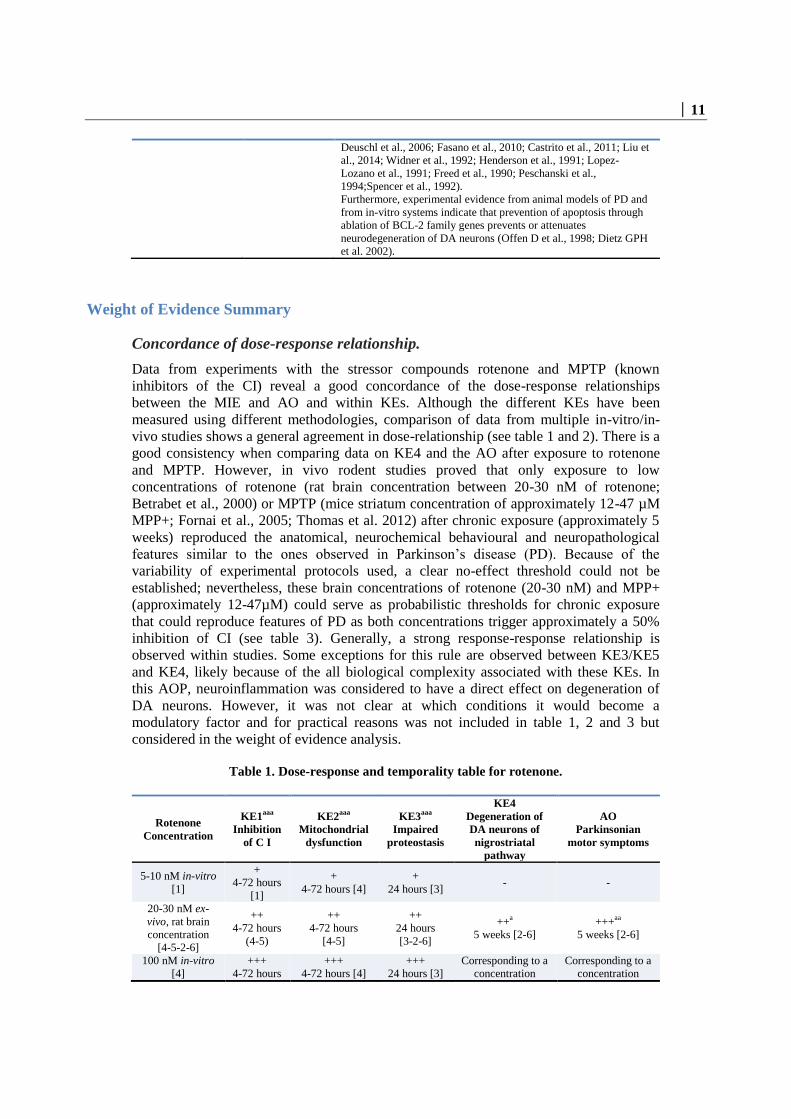
Weight of Evidence (WoE).
Biological plausibility, coherence, and consistency of the experimental evidence.
The biological plausibility of this AOP is overall considered strong. When using multiple
stressors in different studies and assays, the coherence and consistency of the
experimental data is well established. Furthermore, in-vivo and in-vitro studies are also in
line with the human evidence from PD patients. In addition, although the mechanistic
understanding of parkinsonian disorders (and PD in particular) are not fully clear, the
KEs and KERs described in this AOP are considered critical for the development of the
disease (Fujita et al. 2015, Shulman et al. 2011, Dexter et al. 2013, Dauer et al. 2003).
1 Support for
Biological
Plausibility of KERs
Defining
Question High (Strong) Moderate Low(Weak)
Is there a
mechanistic (i.e.
structural or
functional)
relationship
between KEup and
KE down
consistent with
established
biological
knowledge?
Extensive
understanding of
the KER based on
extensive previous
documentation and
broad acceptance
The KER is
plausible based on
analogy to accepted
biological
relationships, but
scientific
understanding is
not completely
established
There is empirical
support for a
statistical
association
between KEs but
the structural or
functional
relationship
between them is
not understood
MIE => KE1
Binding of inhibitor to
NADH-ubiquinone
oxidoreductase leads
of complex I
STRONG
Rationale: As describe in this KER there is an extensive
understanding of the functional relationship between binding of
an inhibitor to NADH-ubiquinone oxidoreductase (CI) and its
inhibition. Different complex I ligands, both naturally occurring,
like rotenone (from Derris scandens), piericidin A (from
Streptomyces mobaraensis), acetogenins (from various
Annonaceae species) and their derivatives, and synthetically
manufactured like pyridaben and various piperazin derivatives
inhibit the catalytic activity of complex I (Degli Esposti, 1994:
Ichimaru et al. 2008; Barrientos and Moraes, 1999; Betarbet et
al., 2000).
KE1 => KE2
Inhibition of complex
I leads to
mitochondrial
dysfunction
STRONG
Rationale: There is extensive understanding of the mechanisms
explaining how the inhibition of complex I lead to
mitochondrial dysfunction (i.e. failure to produce ATP, increase
in production of ROS etc). It is well documented that CI
inhibition is one of the main sites at which electron leakage to
oxygen occurs resulting in oxidative stress (Efremov and
Sazanow, 2011; lauren et al. 2010; Greenamyre et al. 2001).
These pathological mechanisms are well studied as they are
used as readouts for evaluation of mitochondrial dysfunction
(Graier et al., 2007; Braun, 2012; Martin, 2011; Correia et al.,
2012; Cozzolino et al., 2013
KE2 => KE3 MODERATE Rationale: The weight of evidence supporting the biological

│ 15
Mitochondrial
dysfunction results in
impaired proteostasis
plausibility behind the relationship between mitochondrial
dysfunction and impaired proteostasis, including the impaired
function of UPS and ALP that results in decreased protein
degradation and increase protein aggregation is well
documented but not fully understood. It is well established that
the two main mechanisms that normally remove abnormal
proteins (UPS and ALP) rely on physiological mitochondrial
function. The role of oxidative stress, due to mitochondrial
dysfunction, burdens the proteostasis with oxidised proteins and
impairs the chaperone and the degradation systems. This leads
to a vicious circle of oxidative stress inducing further
mitochondrial impairment (Powers et al., 2009; Zaltieri et al.,
2015; McNaught and Jenner, 2001). Therefore, the interaction
of mitochondrial dysfunction and UPS /ALP deregulation plays
a pivotal role in the pathogenesis of PD (Dagda et al., 2013; Pan
et al., 2008; Fornai et al., 2005; Sherer et al., 2002).
KE2 => KE4
Mitochondrial
dysfunction leads to
the degeneration of
dopaminergic neurons
of the nigrostriatal
pathway
STRONG
Rationale: Mitochondrial are essential for ATP production, ROS
management, calcium homeostasis and control of apoptosis.
Mitochondrial homeostasis by mitophagy is also an essential
process for cellular maintenance (Fujita et al. 2014). Because of
their anatomical and physiological characteristics, SNpc DA
neurons are considered more vulnerable than other neuronal
populations (Sulzer et al. 2013; Surmeier et al.2010).
Mechanistic evidence of mutated proteins relate the
mitochondrial dysfunction in familial PD with reduced calcium
capacity, increased ROS production, increase in mitochondrial
membrane permeabilisation and increase in cell vulnerability
(Koopman et al. 2012; Gandhi et al. 2009). Human studies
indicate mitochondrial dysfunction in human idiopathic PD
cases in the substantia nigra (Keeney et al., 2006; Parker et al.,
1989, 2008; Swerdlow et al., 1996). In addition, systemic
application of mitochondrial neurotoxicants such as rotenone or
MPTP leads to a preferential loss of nigrostriatal DA neurons
(Langston et al., 1983).
KE3 => KE4 Impaired proteostasis
leads to degeneration
of DA neurons of the
nigrostriatal pathway
MODERATE
Rationale: It is well known that impaired proteostasis refers to
misfolded and aggregated proteins including alfa-synuclein,
deregulated axonal transport of mitochondria and impaired
trafficking of cellular organelles. Evidences are linked to PD
and experimental PD models as well as from genetic studies
(McNaught et al. 2001, 2003; Tieu et al. 2014; Arnold 2011;
Rappold et al. 2014). Strong evidence for degeneration of the
nigrostriatal pathway comes from the experimental
manipulations that directly induce the same disturbances of
proteostasis as observed in PD patients (e.g. viral mutated alpha-
synuclein expression) or in chronic rotenone/MPTP models
trigger degeneration of the nigrostriatal pathway (Kirk et al.
2003; Betarbet et al. 2000 and 2006; Fornai et al. 2005).
However, a clear mechanistic proof for the understanding of the
exact event triggering cell death is lacking. There is only
moderate evidence showing that interventions that correct
disturbances of proteostasis after exposure to rotenone would
prevent neuronal degeneration and that the disturbances of
proteostasis correlate quantitatively under many conditions with
the extent of nigrostriatal neuronal degeneration.
KE4 => KE5 Degeneration of DA
neurons of the
nigrostriatal pathway
leads to
neuroinflammation
MODERATE
Rationale: The fact that neuronal injury/death can trigger
neuroinflammation is supported by evidence in human and
experimental models. NeThe evidence that neuroinflammation
triggered by neuronal damage can cause neuronal death (vicious
circle), is mostly indirect (blockade of any feature of
neuroinflammation) or by analogy (Hirsch and Hunot, 2009;
Tansey and Goldberg, 2009; Griffin et al., 1998; McGeer and
Mc Geer, 1998; Blasko et al., 2004; Cacquevel et al., 2004;
Rubio-Perez and Morillas-Ruiz, 2012; Thundyil and Lim, 2014;
Barbeito et al., 2010). Neuroinflammation is observed in

16 │
idiopathic and in genetic human PD as well as in complex I
inhibitor exposed humans, non-human primates, and
rodent.Components of damaged neurons lead to glial cells
activation via Toll-like receptors.Several chemokines and
chemokine receptors (fraktalkine, CD200) control the neuron-
microglia interactions. Neuroinflammation in response to
damaged neurons is not confined to PD, but is common to
several neurodegenerative diseases
KE5 => KE4 Neuroinflammation
leads to degeneration
of DA neurons of the
nigrostriatal pathway
MODERATE
Rationale: The fact that reactive glial cells (microglia and
astrocytes) may kill neurons is well accepted. The mechanisms
underlying this effect may include the release of cytotoxic
signals (e.g. cytokines) or production of ROS and RNS (Chao et
al., 1995; Brown and Bal-Price, 2003 ; Kraft and Harry, 2011 ;
Taetzsch and Block, 2013). However, the responsible mediators
differ from model to model. In humans or non-human primates,
an inflammatory activation of glial cells is observed years after
exposure to complex I inhibitors. Activated microglia and
astrocytes form pro-inflammatory cytokines and free radical
species, mostly responsible for neuronal damage. Glial
reactivity promotes an impairment of blood brain barrier
integrity, allowing an infiltration of peripheral leukocytes that
exacerbate the neuroinflammatory process and contribute to
neurodegeneration.The debris of degenerating neurons causes
neuroinflammation, which in turn can trigger
neurodegeneration, thus leading to a self-perpetuating vicious
cycle.
KE4 => AO
Degeneration of DA
neurons of the
nigrostriatal pathway
leads to parkinsonian
motor symptoms
STRONG
Rationale: The mechanistic understanding of the regulatory role
of striatal DA in the extrapyramidal motor control system is
well established. The loss of DA in the striatum is characteristic
of all aetiologies of PD and is not observed in other
neurodegenerative diseases (Bernheimer et al. 1973; Reynolds
et al. 1986). Characteristic motor symptoms such as
bradykinesia, tremor, or rigidity are manifested when more than
80 % of striatal DA is depleted as a consequence of SNpc DA
neuronal degeneration (Koller et al. 1992).
Empirical support.
Empirical support is strong. Many studies show evidence for the KERs by showing
temporal concordance and dose concordance when using different stressors.
3 Empirical
support for KERs
Defining Question
Does the empirical
evidence support
that a change in the
KEup leads to an
appropriate change
in the KE down?
Does KEup occur
at lower doses and
earlier time points
than KE down and
is the incidence of
KEup higher than
that for KE down?
Are inconsistencies
in empirical
support cross taxa,
species and
stressors that don’t
align with expected
pattern of
High (Strong) Moderate Low(Weak)

│ 17
hypothesized
AOP?
Multiple studies
showing
dependent
change in both
exposure to a
wide range of
specific stressors
(extensive
evidence for
temporal, dose-
response and
incidence
concordance) and
no or few critical
data gaps or
conflicting data.
Demonstrated
dependent change in
both events
following exposure
to a small number
of specific stressors
and some evidence
inconsistent with
expected pattern
that can be
explained by factors
such as
experimental
design, technical
considerations,
differences among
laboratories, etc.
Limited or no
studies reporting
dependent change in
both events
following exposure
to a specific stressor
(ie endpoints never
measured in the
same study or not at
all); and/or
significant
inconsistencies in
empirical support
across taxa and
species that don’t
align with expected
pattern for
hypothesized AOP
MIE => KE1 Binding of inhibitor
to NADH-
ubiquinone
oxidoreductase leads
to partial or total
inhibition of
complex I
STRONG
Rationale: The inhibition of complex I is well documented in a
variety of studies using isolated mitochondria or cells as well as
in in vivo experiments and in human post mortem PD brains. In
many experiments using different inhibitors ie rotenone and
MPTP, the observed relationship between the two events was
temporal, response and dose concordant (Betarbet et al., 2000
and 2006, Okun et al., 1999, Koopman et al., 2007, Choi et al.,
2008, Grivennikova et al., 1997, Barrientos and Moraes 1999).
KE1 => KE2 Inhibition of
complex I leads to
mitochondrial
dysfunction
STRONG
Rationale: There is a large amount of studies showing that the
inhibition of CI inhibition results in mitochondrial dysfunctions
in a response and dose dependent manner (Barriento and Moraes,
1999).
KE2 => KE3
Mitochondrial
dysfunction results
in impaired
proteostasis
STRONG
Rationale: Based on the existing in vitro and in vivo data it is
suggested that mitochondrial dysfunction impairs protein
homeostasis (impairment of the UPS and ALP system) through
oxidative and nitrosative stress resulting in accumulation of
misfolded proteins (including a-synuclein), disruption of
microtubule assembly and damaged intracellular transport of
proteins and cell organelles. A number of studies performed with
chemical stressors showed evidence of temporal, response and
dose concordance (Chou et al. 2010; Betarbet et al. 2000 and
2006; Fornai et al. 2005).
KE2 => KE4
Mitochondrial
dysfunction directly
leads to degeneration
of DA neurons of
nigrostriatal pathway
STRONG
Rationale: Multiple in vitro studies indicate dose and response-
response concordance. As most of the studies were conducted in
vitro, the temporal concordance is difficult to establish; however,
can be expected based on the well know temporal sequence of the
two KEs. (Park et al., 2014; Choi et al., 2014; Marella et al.,
2008; Du et al. 2010; Hajieva et al., 2009; Sherer et al., 2003;
Sherer et al., 2007; Wen et al. 2011; Swedlow et al., 1996; Jana
et al., 2011; Jha et al., 2000; Chinta et al., 2006)
KE3 => KE4a
Impaired proteostasis
leads to degeneration
of DA neurons of the
nigrostriatal pathway
STRONG
Rationale: The empirical support linking impaired proteostasis
with degeneration of DA neurons of the nigrostriatal pathway is
strong and comes from in-vivo and in-vitro studies performed
with different stressor (i.e. Rotenone, MPTP or proteasome
inhibitors) and post-mortem human evidences in PD patients
supporting a causative link between the two key events.
Temporal, effect and dose concordance was established in a
number of experiments (Fornai et al. 2005; Fornai et al. 2003;
Betabret et al. 2000 and 2006).
KE4a => KE5 Degeneration of DA
neurons of
nigrostriatal pathway
MODERATE
Rationale: multiple in vivo and in vitro experiments support the
link between degeneration of DA neurons in the nigrostriatal
pathway and neuroinflammation. The observation of concomitant
presence of reactive microglial and astrocytic cells and

18 │
leads to
neuroinflammation
degenerated/degenerating DA neurons is also reported in many
studies with a good temporal and response concordance. ATP
and other damage associated molecular patterns (DAMPs),
released from degenerating cells, stimulate P2Y receptors on
microglia, leading to their activation. Experimental injection of
DAMPs, fraktalkine, or neuromelanin, released by degenerating
DA neurons evokes neuroinflammation. Neutralization of
DAMPs (e.g. antibodies against HMGB1 or CX3CR1) decreases
MPTP-induced neuroinflammation .Toll-like receptor 4 deficient
mice display a reduced neuroinflammatory response upon MPTP
treatment. Inhibition of RAGE, which is upregulated in striatum
upon rotenone exposure, suppresses NF-kB activation and
downstream inflammatory markers.
KE5 => KE4b Neuroinflammation
leads to degeneration
of DA neurons of
nigrostriatal
pathway.
MODERATE
Rationale: multiple in vivo and in vitro experiments support the
link between neuroinflammation and degeneration of DA neurons
in the nigrostriatal pathway. The observation of concomitant
presence of reactive microglial and astrocytic cells and
degenerated/degenerating DA neurons is also reported in many
studies with a good temporal and response concordance.
Neuroinflammation has been implicated in dopaminergic
neuronal cell death in PD patients (Vivekanantham et al.,
2014). LPS injection into the CNS, or applied systemically,
evokes glial inflammation and a preferential degeneration of DA
neurons. In mouse models with a knockout of either IL-1b, IFN-
g, or TNF-a receptors 1 and 2, LPS no longer evokes
neuroinflammation and DA neurodegeneration. Experimental
interference with CD4+ T cell activation protects from DA
neurodegeneration.Transfer of immunosuppressive regulatory T
cells protect from DA neurodegeneration. Anti-inflammatory
TGF-b1 signaling protects from DA neurodegeneration . Clinical
trials indicate a protective influence on DA neuron survival by
the antibiotic minocycline blocking microglial reactivity, in
association with rasagiline (prevents DA degeneration), and
coenzyme Q10/creatine (restoration of cellular ATP).
KE4b => AO Degeneration of DA
neurons of
nigrostriatal pathway
leads to parkinsonian
motor symptoms
STRONG
Rationale: The experimental support linking the degeneration of
DA neurons of nigrostriatal pathways with the manifestation of
motor symptoms of PD comes from human in vivo observations
as well as from monkey, mice and rat in vivo models exposed to
an experimental toxin ie rotenone and MPTP. Observations in
human allow defining correlation between the levels of striatal
DA with the onset of motor dysfunction (Lloyd et al. 1975;
Hornykiewicz et al. 1986; Bernheimer et al. 1973). Temporal,
effect and dose concordance comes from studies performed in
multiple animal species following administration of rotenone and
MPTP ( Bezard et al. 2001; Cannon et al. 2009; Petroske et al.
2001; Alvarez-Fischer et al. 2008; Blesa et al. 2012; Lloyd et a.
1975).
Uncertainties and Inconsistencies.
The strength of this AOP is mainly based on MPP+ and rotenone and only limited
information on whether other mitochondrial complex I inhibitors also perturb the
KEs (specifically degeneration of DA neurons in the SNpc) or produce a similar
AO.
Conflicting data exists (Choi et al. 2008) showing that mitochondrial CI inhibition
is not required for DA neuron death induced by rotenone, MPTP/MPP+, or
paraquat, challenging the current AOP. The cited research article shows that
abolishment of CI’s activity by inactivation of a gene that codes for a subunit of
CI does not impact the survival of DA neurons in culture. The actions of rotenone

│ 19
and MPTP/MMP+ are independent of complex I. Since some complex I inhibitors
also target other complexes, it is possible that impairment of other respiratory
complexes may be involved. It was noted that this paper used the approach of
genetically deleting an essential chaperone in complex I assembly, and the authors
found that absence of complex I activity in this model did not affect the toxicity
of rotenone and MPP+. However, the findings have never been confirmed/
continued, neither in the originating laboratory, nor by others. Second, the work
did not consider the possibility that some functions of complex I were not affected
by the absence of the chaperone (e.g. reverse electron transfer from complex II
and III), and that rotenone and MPTP/MPP+ may well cause toxicity by
interfering with such residual function (e.g. by favoring channeling of electrons to
molecular oxygen). In light of this situation, the publication of Choi et al (2008)
should be considered weak in the overall weight of evidence and therefore
considered a minor inconsistency.
There is no strict linear relationship between inhibitor binding and reduced
mitochondrial function. Low doses of rotenone that inhibit CI activity partially do
not alter mitochondrial oxygen consumption. Therefore, bioenergetics defect
cannot account alone for rotenone-induced neurodegeneration. Instead, under
such conditions, rotenone neurotoxicity may result from oxidative stress (Betarbet
et al., 2000). Few studies used human brain cells/human brain mitochondria.
Therefore, full quantitative data for humans are not available.
It is molecularly unclear how rotenone binding alter CI function, switching it to
ROS production. It is still unclear whether the site of superoxide production in CI
inhibited mitochondria is complex I itself or not (Singer and Ramsay, 1994).
Some studies suggest that rotenone and MPTP/MPP+ may have effects other than
CI inhibition, e.g. MPTP and rotenone can induce microtubule disruption (Feng,
2006; Ren et al., 2005Cappelletti et al., 1999; Cappelletti et al., 2001, Brinkley et
al., 1974; Aguilar et al. 2015).
There are additional feedback loops possible between KEs, e.g. ROS production
from KE2 may damage CI, this leads to enhancement of KE1.
Some KEs e.g. KE 2, 3, 5 pool molecular processes that may need to be evaluated
individually at a later stage.
The exact molecular link from mitochondrial dysfunction to disturbed proteostasis
is still unclear (Malkus et al 2009; Zaltieri et al. 2015).
The role of ATP depletion vs. other features of mitochondrial dysfunction is not
clear.
The role of α-synuclein in neuronal degeneration is still unclear as well as the
mechanisms leading to its aggregation.
It is not clear under which conditions KE3 and KE5 become modulatory factors,
and when they are essential. MPTP/MPP+ can induce damage to nigrostriatal
neurons without formation of Lewy bodies (Dauer 2003; Forno 1986, 1993).
Similarly, discontinuous administration of rotenone, even at high doses, damages
the basal ganglia but produce no inclusions (Heikkila et al. 1985; Ferrante et al.
1997, Lapontine 2004). To reproduce the formation of neuronal inclusions,
continuous sc infusion of MPTP/MPP+ or rotenone is necessary. Acute
intoxication with rotenone seems to spare dopaminergic neurons (Dauer et al
2003, Ferrante 1997). In addition, in rats chronically infused with rotenone
showed a reduction in striatal DARPP-32-positive (dopamine- and cyclic-AMP-
regulated phosphoprotein of molecular weight 32,000), cholinergic and NADPH
diaphorase-positive neurons (Hoglinger 2003) or in other brain regions. These
20 │
results would suggest that Rotenone can induce a more widespread neurotoxicity
(Aguilar 2015) or the model is not reproducible in all laboratories.
Inconsistent effects of MPTP/MPP+ on autophagy (up or down regulation) are
reported (Drolet et al., 2004: Dauer et al., 2002). There is conflicting literature on
whether increased autophagy would be protective or enhances damage. Similarly,
a conflicting literature exists on extent of inhibition or activation of different
protein degradation system in PD and a clear threshold of onset is unknown
(Malkus et al. 2009; Fornai et al. 2005).
The selective vulnerability of the SN pc dopaminergic pathway does not have a
molecular explanation.
In some instances, the differential vulnerability of various brain regions towards a
generalised complex Iinhibition found non-dopaminergic lesions, particularly in
the striatum, in all animals with nigral lesion, as seen inatypical parkinsonism but
not in idiopathic parkinson's disease (Hoglinger et al.2003).
Priority of the pattern leading to cell death could depend on concentration, time of
exposure and species sensitivity; these factors have to be taken into consideration
for the interpretation of the study’s result and extrapolation of potential low-dose
chronic effect as this AOP refers to long-time exposure.
The model of striatal DA loss and its influence on motor output ganglia does not
allow to explain specific motor abnormalities observed in PD (e.g. resting tremor
vs bradykinesia) (Obeso et al. 2000). Other neurotransmitters (Ach) may play
additional roles. Transfer to animal models of PD symptoms is also representing
an uncertainty.
There are some reports indicating that in subacute rotenone or MPTP models
(non-human primates), a significant, sometimes complete, recovery of motor
deficits can be observed after termination of toxicant treatment. The role of
neuronal plasticity in intoxication recovery and resilience is unclear.
This AOP is a linear sequence of KEs. However, mitochondrial dysfunction (and
oxidative stress) and impaired proteostasis are influencing each other and this is
considered an uncertainty (Malkus et al. 2009).
Quantitative Consideration
The quantitative understanding of this AOP includes a clear response-response relationship and
the identification of a threshold effect. The WoE analysis clearly supports the qualitative AOP as
a means to identify and characterise the potential of a chemical to induce DA neuronal loss and
the AO. Importantly, both the AO and the KE4 are considered relevant regulatory endpoints for
this AOP. The empirical evidence supports existence of a response-response relationship. This
response-response is likely triggered by brain concentrations of approximately 20-30 nM and 17-
47 µM of rotenone and MPTP/MPP+, respectively and both concentrations trigger approx. a 50%
inhibition of mitochondrial complex I and this could be considered as a “threshold”. However, a
more detailed dose-response analysis for each KE is lacking as well as it is not clear which
temporal relationship exists for lower CI inhibitory effects. It is clear from the analysis of the
AOP that for the identification of these AOs, the design of the in-vivo studies should be tailored
as to a MIE which leads to a long-lasting perturbation of the KEs. This provides the most specific
and definite context to trigger neuronal death. To observe KEs relevant for this AOP, new
endpoints need to be introduced. Although a dose, response and temporal relationship is evident
for most KEs, the quantitative relationship between impaired proteostasis and degeneration of

│ 21
DA neurons has yet to be elucidated. Moving from a qualitative AOP to quantitative AOP would
need a clear understanding of effect thresholds and this is still representing a major hurdle for
several KEs of this AOP.
Table 3. Summary of quantitative effects at the concentration of rotenone and MPTP
triggering the AO.
Concentration KE1
Inhibition of C I
KE2
Mitochondrial
dysfunction
KE3
Impaired
proteostasis
KE4
Degeneration
of DA neurons
of nigrostriatal
pathway
AO
Parkinsonian
motor
symptoms
Rotenone 20-
30 nM rat brain
concentration
[1-2]
Approx. 53%[4-
5]
Approx. 20-53%
(decrease in
respiration
rate)[1-2]
Approx. 20-
60%
(decrease in
UPS (26S)
activity) [3]
Neuronal loss
(50% of animal
affected) [2]
Motor
impairment
(100% of
animals with
neuronal loss)
[2]
MPP+ 12-47
µM rat brain
concentration
[4-5]
Approx. 50-
75% [5]
Approx. 38%
(reduction in
phosphorylating
respiration) [5]
Approx. 60%
(decrease in
UPS activity)
[4]
Approx. 50%
of neuronal
loss [4-5]
Motor
impairment [4]
[1]; Okun et al. 1999 [2]; Barrientos and Moraes 1999; [3] Borland et al.2008 [4] Thomas et al 2012; [5]
Betarbet et al 2000 and 2006.
Summary of the proposed Key Events in this AOP:
Chronic, low level of exposure to environmental chemicals that inhibit complex I could result in
mitochondrial dysfunction and oxidative stress, triggering proteasomal dysfunction strongly implicated in
parkinsonian disorders, including aggregation/modifications in α-synuclein protein and organelles trafficking.
These cellular key events cause DA terminals degeneration in striatum and progressive cell death of DA
neurons in SNpc, accompanied by neuroinflammation that potentiates neuronal cell death, finally leading to
parkinsonian's motor symptoms. Important to notice that at each step, the effects become regionally restricted
such that systemic complex I inhibition eventually results in highly selective degeneration of the nigrostriatal
pathway.

22 │
Considerations for Potential Applications of the AOP (optional)
1. This AOP has been developed in order to evaluate the biological plausibility that
the adverse outcome i.e. parkinsonian motor deficits, is linked to a MIE that can
be triggered by chemical substances i.e. pesticides and chemicals in general. The
relevance of the AOP has been documented by tools compounds known to
trigger the described AOP. By means of using a human health outcome that has
been shown in epidemiological studies to be association with pesticide exposure,
the authors intend to draw attention on this AO in the process of hazard
identification. This AOP can be used to support the biological plausibility of this
association during the process of evaluation and integration of the
epidemiological studies into the risk assessment. It is biologically plausible that a
substance triggering the pathway, can be associated with the AO and ultimately
with the human health outcome, pending the MoA analysis.
2. In addition, this AOP can be used to support identification of data gaps that
should be explored when a chemical substance is affecting the pathway.
Moreover, the AOP provides a scaffold for recommendations on the most
adequate study design to investigate the apical endpoints. It is important to note
that, although the AO is defined in this AOP as parkinsonian motor deficits,
degeneration of DA neurons is already per se an adverse outcome even in
situations where it is not leading to parkinsonian motor deficits, and this should
be taken into consideration for the regulatory applications of this AOP.
3. The MIE and KEs identified in this AOP could serve as a basis for assays
development that could contribute to an AOP informed-IATA construction
which can be applied for different purposes such as: screening and prioritisation
of chemicals for further testing, hazard characterisation or even risk assessment
when combined with exposure and ADME information.
4. This AOP can be used for neurotoxicity assessment, since it is plausible that a
compound that binds to the mitochondrial complex I may eventually lead to
Parkinsonian motor deficits.
5. The regulatory applicability of this AOP would be to use experimental findings
in model systems representing the MIE and KEs as indicators/alerts for the AO.
Risk assessment may be possible if bioavailability at the target cells can be
estimated, the toxic concentrations in vitro can be extrapolated to in vivo and
exposure scenarios can be simulated.
6. This AOP can be applied for chemicals that have structural similarities to
rotenone or MPTP. However, this AOP may not at the moment be used for
chemicals that do not resemble rotenone or MPTP. It is however expected that
compounds acting on the same MIE, but belonging to different chemical classes
and those that are structurally different, will be tested in the near future in order
to substantiate a broader specificity for this AOP. However, it remains evident
that chemicals affecting the MIE are potential risk factors for this AO.

│ 23
References
Abdelsalam RM, Safar MM J Neurochem. Neuroprotective effects of vildagliptin in rat
rotenone Parkinson's disease model: role of RAGE-NFκB and Nrf2-antioxidant signaling
pathways. 2015 Jun;133(5):700-7. doi: 10.1111/jnc.13087. Epub 2015 Mar 26.
Aguilar JS, Kostrzewa RM. Neurotoxin mechanisms and processes relevant to
parkinson’s disease: un update. Neurotox Res. DOI 10.1007/s12640-015-9519-y.
Alvarez-Fischer D, Guerreiro S, Hunot S, Saurini F, Marien M, Sokoloff P, Hirsch EC,
Hartmann A, Michel PP. Modelling Parkinson-like neurodegeneration via osmotic
minipump delivery of MPTP and probenecid. J Neurochem. 2008 Nov;107(3):701-11.
doi: 10.1111/j.1471-4159.2008.05651.x. Epub 2008 Sep 16.
Arnold, B., et al. (2011). "Integrating Multiple Aspects of Mitochondrial Dynamics in
Neurons: Age-Related Differences and Dynamic Changes in a Chronic Rotenone Model."
Neurobiology of Disease 41(1): 189-200.
Barbeito AG, Mesci P, Boillee S. 2010. Motor neuron-immune interactions: the vicious
circle of ALS. J Neural Transm 117(8): 981-1000.
Barrientos A., and Moraes C.T. (1999) Titrating the Effects of Mitochondrial Complex I
Impairment in the Cell Physiology. Vol. 274, No. 23, pp. 16188–16197.
Bernheimer H, Birkmayer W, Hornykiewicz O, Jellinger K, Seitelberger F. Brain
dopamine and the syndromes of Parkinson and Huntington. Clinical, morphological and
neurochemical correlations. J Neurol Sci. 1973 Dec;20(4):415-55
Betarbet R, Sherer TB, MacKenzie G, Garcia-Osuna M, Panov AV, Greenamyre JT.
2000. Chronic systemic pesticide exposure reproduces features of Parkinson’s disease.
Nat Neurosci. 3:1301–6
Betarbet R, Canet-Aviles RM, Sherer TB, Mastroberardino PG,Mc Lendon C, Kim JH,
Lund S, Na HM, taylor G, Bence NF, kopito R, seo BB, Yagi T, Yagi A, Klinfelter G,
Cookson MR, Greenmyre JT. 2006. Intersecting pathways to neurodegeneration in
Parkinson’s disease: effects of the pesticide rotenone on DJ-1, α-synuclein, and the
ubiquitin-proteasome system. Neurobiology disease. (22) 404-20.
Bezard E, Dovero S, Prunier C, Ravenscroft P, Chalon S, Guilloteau D, Crossman AR,
Bioulac B, Brotchie JM, Gross CE (2001) Relationship between the appearance of
symptoms and the level of nigrostriatal degeneration in a progressive 1-methyl-4-phenyl-
1,2,3,6-tetrahydropyridine-lesioned macaque model of Parkinson's disease. J Neurosci.
21(17):6853-61.
Blasko I, Stampfer-Kountchev M, Robatscher P, Veerhuis R, Eikelenboom P, Grubeck-
Loebenstein B. 2004. How chronic inflammation can affect the brain and support the
development of Alzheimer's disease in old age: the role of microglia and astrocytes.
Aging cell 3(4): 169-176.

24 │
Blesa J, Pifl C, Sánchez-González MA, Juri C, García-Cabezas MA, Adánez R, Iglesias
E, Collantes M, Peñuelas I, Sánchez-Hernández JJ, Rodríguez-Oroz MC, Avendaño C,
Hornykiewicz O, Cavada C, Obeso JA (2012) The nigrostriatal system in the
presymptomatic and symptomatic stages in the MPTP monkey model: a PET, histological
and biochemical study. Neurobiol Dis. 48(1):79-91.
Bodea LG, Wang Y, Linnartz-Gerlach B, Kopatz J, Sinkkonen L, Musgrove R, et al.
2014. Neurodegeneration by activation of the microglial complement-phagosome
pathway. J Neurosci 34(25): 8546-8556.
Borrajo A, Rodriguez-Perez AI, Villar-Cheda B, Guerra MJ, Labandeira-Garcia JL. 2014.
Inhibition of the microglial response is essential for the neuroprotective effects of Rho-
kinase inhibitors on MPTP-induced dopaminergic cell death. Neuropharmacology 85: 1-8
Brinkley BR, Barham SS, Barranco SC, and Fuller GM. 1974. Rotenone inhibition of
spindle microtubule assembly in mammalian cells,” Experimental Cell Research.
85(1)41–46.
Brown GC, Bal-Price A (2003) Inflammatory neurodegeneration mediated by nitric
oxide, glutamate, and mitochondria. Mol Neurobiol 27: 325-355
Braun RJ. (2012). Mitochondrion-mediated cell death: dissecting yeast apoptosis for a
better understanding of neurodegeneration. Front Oncol 2:182.
Brzozowski MJ, Jenner P, Rose S. 2015. Inhibition of i-NOS but not n-NOS protects rat
primary cell cultures against MPP(+)-induced neuronal toxicity. J Neural Transm 122(6):
779-788.
Cacquevel M, Lebeurrier N, Cheenne S, Vivien D. 2004. Cytokines in neuroinflammation
and Alzheimer's disease. Curr Drug Targets 5(6): 529-534.
Calne DB, Sandler M (1970) L-Dopa and Parkinsonism. Nature. 226(5240):21-4.
Cannon JR, Tapias V, Na HM, Honick AS, Drolet RE, Greenamyre JT (2009) A highly
reproducible rotenone model of Parkinson's disease. Neurobiol Dis. 34(2):279-90.
Cappelletti G, Maggioni MG, Maci R. 1999. Influence of MPP+ on the state of tubulin
polymerisation in NGF-differentiated PC12 cells. J Neurosci Res. 56(1):28-35.
Cao S, Theodore S, Standaert DG.2010. Fc gamma receprors are required for NF-kB
signaling, microglial activation and dopaminergic neurodegeneration in an AAV-
synuclein mouse model of Parkinson's disease. molecular neurodegeneration.5-42.
Cappelletti G, Pedrotti B, Maggioni MG, Maci R. 2001. Microtubule assembly is directly
affected by MPP(+)in vitro. Cell Biol Int.25(10):981-4.
Castrioto A, Lozano AM, Poon YY, Lang AE, Fallis M, Moro E. 2011. Ten-year
outcome of subthalamic stimulation in Parkinson disease: a blinded evaluation. Arch
Neurol. 68(12):1550-6.
Chang CY, Choi DK, Lee DK, Hong YJ, Park EJ. 2013. Resveratrol confers protection
against rotenone-induced neurotoxicity by modulating myeloperoxidase levels in glial
cells. PLoS One 8(4): e60654.
Champy et al. (2004). Annonacin, a lipophilic inhibitor of mitochondrial complex I,
induces nigral and striatal neurodegeneration in rats: possible relevance for atypical
Parkinsonism in Guadeloupe. J Neurochem 88: 63-69.

│ 25
Chao YX, He BP, Tay SS. 2009. Mesenchymal stem cell transplantation attenuates blood
brain barrier damage and neuroinflammation and protects dopaminergic neurons against
MPTP toxicity in the substantia nigra in a model of Parkinson's disease. J Neuroimmunol
216(1-2): 39-50.
Chen Y, Zhang DQ, Liao Z, Wang B, Gong S, Wang C, Zhang MZ, Wang GH, Cai H,
Liao FF, Xu JP 2015. Anti-oxidant polydatin (piceid) protects against substantia nigral
motor degeneration in multiple rodent models of Parkinson's disease. Mol Neurodegener.
2;10(1):4.
Chinta SJ, Andersen JK (2006) Reversible inhibition of mitochondrial complex I activity
following chronic dopaminergic glutathione depletion in vitro: implications for
Parkinson's disease. Free Radic Biol Med. 41(9):1442-8.
Choi WS., Kruse S.E., Palmiter R, Xia Z., (2008) Mitochondrial complex I inhibition is
not required for dopaminergic neuron death induced by rotenone, MPP, or paraquat.
PNAS, 105, 39, 15136-15141
Choi BS, Kim H, Lee HJ, Sapkota K, Park SE, Kim S, Kim SJ (2014) Celastrol from
'Thunder God Vine' protects SH-SY5Y cells through the preservation of mitochondrial
function and inhibition of p38 MAPK in a rotenone model of Parkinson's disease.
Neurochem Res. 39(1):84-96.
Chiu CC, Yeh TH, Lai SC, Wu-Chou YH, Chen CH, Mochly-Rosen D, Huang YC, Chen
YJ, Chen CL, Chang YM, Wang HL, Lu CS. 2015. Neuroprotective effects of aldehyde
dehydrogenase 2 activation in rotenone-induced cellular and animal models of
parkinsonism. Exp Neurol. 263:244-53.
Chou AP, Li S, Fitzmaurice AG, Bronstein JM. 2010. Mechanisms of rotenone-induced
proteasome inhibition. NeuroToxicology. 31:367–372. Chung YC, Kim SR, Park JY,
Chung ES, Park KW, Won SY, et al. 2011. Fluoxetine prevents MPTP-induced loss of
dopaminergic neurons by inhibiting microglial activation. Neuropharmacology 60(6):
963-974.
Correia SC, Santos RX, Perry G, Zhu X, Moreira PI, Smith MA. (2012). Mitochondrial
importance in Alzheimer’s, Huntington’s and Parkinson’s diseases. Adv Exp Med Biol
724:205 – 221.
Cotzias GC, Papavasiliou PS, Gellene R. 1969. L-dopa in parkinson's syndrome. N Engl J
Med. 281(5):272.
Cozzolino M, Ferri A, Valle C, Carri MT. (2013). Mitochondria and ALS: implications
from novel genes and pathways. Mol Cell Neurosci 55:44 – 49.
Dagda RK, Banerjee TD and Janda E. 2013. How Parkinsonian Toxins Dysregulate the
Autophagy Machinery. Int. J. Mol. Sci. 14:22163-22189.
Dauer W, Kholodilov N, Vila M, Trillat AC, Goodchild R, Larsen KE, Staal R, Tieu K,
Schmitz Y, Yuan CA, Rocha M, Jackson-Lewis V, Hersch S, Sulzer D, Przedborski S,
Burke R, Hen R. 2002. Resistance of alpha -synuclein null mice to the parkinsonian
neurotoxin MPTP. Proc Natl Acad Sci U S A. 99(22):14524-9.

26 │
Dauer W, Przerdborski S. 2003. Parkinson’sdisease: Mechanisms and Models.Neuron.
39, 889-9.
De Bie RM, de Haan RJ, Nijssen PC, Rutgers AW, Beute GN, Bosch DA, Haaxma R,
Schmand B, Schuurman PR, Staal MJ, Speelman JD. 1999. Unilateral pallidotomy in
Parkinson's disease: a randomised, single-blind, multicentre trial. Lancet.
354(9191):1665-9.
Degli Esposti M, Ghelli A. 1994. The mechanism of proton and electron transport in
mitochondrial complex I. Biochim Biophys Acta.1187(2):116–120.
Dehay B, Bove J, Rodriguez-Muela N, Perier C, Recasens A, Boya P, Vila M. 2010.
Pathogenic lysosomal depletion in Parkinson’s disease. J. Neurosci. 30:12535–12544.
Dehmer T, Lindenau J, Haid S, Dichgans J, Schulz JB. 2000. Deficiency of inducible
nitric oxide synthase protects against MPTP toxicity in vivo. J Neurochem 74(5): 2213-
2216.
Deuschl G, Schade-Brittinger C, Krack P, Volkmann J, Schäfer H, Bötzel K, Daniels C,
Deutschländer A, Dillmann U, Eisner W, Gruber D, Hamel W, Herzog J, Hilker R, Klebe
S, Kloss M, Koy J, Krause M, Kupsch A, Lorenz D, Lorenzl S, Mehdorn HM,
Moringlane JR, Oertel W, Pinsker MO, Reichmann H, Reuss A, Schneider GH,
Schnitzler A, Steude U, Sturm V, Timmermann L, Tronnier V, Trottenberg T, Wojtecki
L, Wolf E, Poewe W, Voges J; German Parkinson Study Group, Neurostimulation
Section. 2006. A randomized trial of deep-brain stimulation for Parkinson's disease. N
Engl J Med. 355(9):896-908.
Dexter D. T., Jenner P.. Parkinson disease: from pathology to molecular disease
mechanisms. Free Radical Biology and Medicine 62 (2013) 132-144
Dietz GPH, Stockhausen KV, Dietz B et al. (2008) Membrane-permeable Bcl-xL
prevents MPTP-induced dopaminergic neuronal loss in the substantia nigra. J Neurochem
104:757-765. Doi:10.1111/j.1471-4159.2007.05028.
Drolet RE, Behrouz B, Lookingland KJ, Goudreau JL, 2004. Mice lacking α-synuclein
have an attenuated loss of striatal dopamine following prolonged chronic MPTP
administration. Neurotoxicology. 25(5):761-9.
Drouin-Ouellet J, St-Amour I, Saint-Pierre M, Lamontagne-Prolux J, Kriz J, Barker R,
Cicchetti F.2015. Toll-like receptor expression in the blood and brain of patients and a
mouse of Parkinson's disease. International Journal of Neuropsychopharmacology. 1-11.
Du T, Li L, Song N, Xie J, Jiang H (2010) Rosmarinic acid antagonized 1-methyl-4-
phenylpyridinium (MPP+)-induced neurotoxicity in MES23.5 dopaminergic cells. Int J
Toxicol. 29(6):625-33.
Efremov RG, Sazanov LA. Respiratory complex I: 'steam engine' of the cell? Curr Opin
Struct Biol. 2011 Aug;21(4):532-40. doi: 10.1016/j.sbi.2011.07.002. Epub 2011 Aug 8.
Review.
Efremov RG, Sazanov LA. Structure of the membrane domain of respiratory complex I.
Nature. 2011 Aug 7;476(7361):414-20. doi: 10.1038/nature10330.
Emmrich JV, Hornik TC, Neher JJ, Brown GC. 2013. Rotenone induces neuronal death
by microglial phagocytosis of neurons. The FEBS journal 280(20): 5030-5038.
Esposti et al. (1993) Complex I and Complex III of mitochondria have common inhibitors
acting as ubiquinone antagonists. Biochem Biophys Res Commun 190(3): 1090-6.

│ 27
Fasano A, Romito LM, Daniele A, Piano C, Zinno M, Bentivoglio AR, Albanese A.
2010. Motor and cognitive outcome in patients with Parkinson's disease 8 years after
subthalamic implants. Brain. 133(9):2664-76.
Fato et al. (2009) Differential effects of mitochondrial complex I inhibitors on production
of reactive oxygen species. Biochim Biophys Acta 1787(5): 384-392.
Feng ZH, Wang TG, Li DD, Fung P, Wilson BC, Liu B, et al. 2002. Cyclooxygenase-2-
deficient mice are resistant to 1-methyl-4-phenyl1, 2, 3, 6-tetrahydropyridine-induced
damage of dopaminergic neurons in the substantia nigra. Neurosci Lett 329(3): 354-358.
Feng J. Mictrotubule. A common target for parkin and Parkinson's disease toxins.
Neuroscientist 2006, 12.469-76.
Ferger B, Leng A, Mura A, Hengerer B, Feldon J. 2004. Genetic ablation of tumor
necrosis factor-alpha (TNF-alpha) and pharmacological inhibition of TNF-synthesis
attenuates MPTP toxicity in mouse striatum. J Neurochem 89(4): 822-833.
Ferrante RJ, Schulz JB, Kowall NW, Beal MF. 1997. Systematic administration of
rotenone produces selective damage in the striatum and globus pallidus, but not in the
substantia nigra. Brain Research. (753). 157-2.
Ferrari-Toninelli G, Bonini SA, Cenini G, Maccarinelli G, Grilli M, Uberti D, Memo M.
2008. Dopamine receptor agonists for protection and repair in Parkinson's disease. Curr
Top Med Chem. 8(12):1089-99.
Friedman LG, lachenmayer ML, Wang J, He L, Poulose SM, Komatsu M, Holstein GR,
Yue Z. 2012. Disrupted autophagy leads to dopaminergic axon and dendrite degeneration
and promotes presynaptic accumulation of α-synuclein and LRRK2 in the brain. The
Journal of Neuroscience. 32 (22) 7585-93.
Fornai F, Lenzi P, Gesi M, Ferrucci M, Lazzeri G, Busceti C, Ruffoli R, Soldani P,
Ruggieri S, Alessandri’ MG, Paparelli A. 2003. Fine structure and mechanisms
underlying nigrostriatal inclusions and cell death after proteasome inhibition. The journal
of neuroscience. 23 (26) 8955-6.
Fornai F., P. Lenzi, M. Gesi et al., “Methamphetamine produces neuronal inclusions in
the nigrostriatal system and in PC12 cells,” Journal of Neurochemistry, vol. 88, no. 1, pp.
114–123, 2004.
Fornai F, Schlüter OM, Lenzi P, Gesi M, Ruffoli R, Ferrucci M, Lazzeri G, Busceti CL,
Pontarelli F, Battaglia G, Pellegrini A, Nicoletti F, Ruggieri S, Paparelli A, Südhof TC.
2005. Parkinson-like syndrome induced by continuous MPTP infusion: Convergent roles
of the ubiquitinproteasome system and _α-synuclein. PNAS. 102: 3413–3418.
Freed CR, Breeze RE, Rosenberg NL, Schneck SA, Wells TH, Barrett JN, Grafton ST,
Huang SC, Eidelberg D, Rottenberg DA. 1990.
Transplantation of human fetal dopamine cells for Parkinson's disease. Results at 1 year.
Arch Neurol. 47(5):505-12.
Fujita KA, Ostaszewski M, Matsuoka Y, Ghosh S, Glaab E, Trefois C, Crespo I, Perumal
TM, Jurkowski W, Antony PM, Diederich N, Buttini M, Kodama A, Satagopam VP,
Eifes S, Del Sol A, Schneider R, Kitano H, Balling R. 2014. Integrating pathways of
Parkinson's disease in a molecular interaction map. Mol Neurobiol.49(1):88-102.

28 │
Gandhi S, Wood-Kaczmar A, Yao Z, et al. PINK1-associated Parkinson’s disease is
caused by neuronal vulnerability to calcium-induced cell death. Molecular Cell.
2009;33:627–638.
Gao HM, Hong JS, Zhang W, Liu B. 2002. Distinct role for microglia in rotenone-
induced degeneration of dopaminergic neurons. J Neurosci 22(3): 782-790.
Gao HM, Liu B, Hong JS. 2003. Critical role for microglial NADPH oxidase in rotenone-
induced degeneration of dopaminergic neurons. J Neurosci 23(15): 6181-6187.
Gao L, Brenner D, Llorens-Bobadilla E, Saiz-Castro G, Frank T, Wieghofer P, et al.
2015. Infiltration of circulating myeloid cells through CD95L contributes to
neurodegeneration in mice. J Exp Med 212(4): 469-480.
Graier WF, Frieden M, Malli R. (2007). Mitochondria and Ca2+ signaling: old guests,
new functions. Pflugers Arch 455:375–396.
Greenamyre, J T., Sherer, T.B., Betarbet, R., and Panov A.V. (2001) Critical Review
Complex I and Parkinson’s Disease Life, 52: 135–141.
Greenamayre et al. 2010. Lessons from the rotenone model of Parkinson's disease. Trends
pharmacol. Sci. 31(4):141-2
Griffin WS, Sheng JG, Royston MC, Gentleman SM, McKenzie JE, Graham DI, et al.
1998. Glial-neuronal interactions in Alzheimer's disease: the potential role of a 'cytokine
cycle' in disease progression. Brain Pathol 8(1): 65-72.
Grivennikova, V.G., Maklashina, E.O., E.V. Gavrikova, A.D. Vinogradov (1997)
Interaction of the mitochondrial NADH-ubiquinone reductase with rotenone as related to
the enzyme active/inactive transition Biochim. Biophys. Acta, 1319 (1997), pp. 223–232
Hajieva P, Mocko JB, Moosmann B, Behl C (2009) Novel imine antioxidants at low
nanomolar concentrations protect dopaminergic cells from oxidative neurotoxicity. J
Neurochem. 110(1):118-32.
Hoglinger G.U. et al.2003.Chronic systemic complex I inhibition induces a hypokynetic
multisystem degeneration in rats. J.neurochem 84:491-502.
Hornykiewicz O, Kish SJ. 1987. Biochemical pathophysiology of parkinson’s disease. In
Parkinson’s Disease. M Yahr and K.J. Bergmann, eds (New.York: Raven Press) 19-34.
Jana S, Sinha M, Chanda D, Roy T, Banerjee K, Munshi S, Patro BS, Chakrabarti S
(2011) Mitochondrial dysfunction mediated by quinone oxidation products of dopamine:
Implications in dopamine cytotoxicity and pathogenesis of Parkinson's disease. Biochim
Biophys Acta. 1812(6):663-73.
Jha N, Jurma O, Lalli G, Liu Y, Pettus EH, Greenamyre JT, Liu RM, Forman HJ,
Andersen JK (2000) Glutathione depletion in PC12 results in selective inhibition of
mitochondrial complex I activity. Implications for Parkinson's disease. J Biol Chem.
275(34):26096-101.
Johnson ME, Bobrovskaya L. 2015. An update on the rotenone models of parkinson’s
disease: Their ability to reproduce features of clinical disease and model gene-
environment interactions. 946). 101-16.
Heikkila RE, Nicklas WJ, Vyas I, Duvoisin RC. 1985. Dopaminergic toxicity of rotenone
and the 1-methyl-4-phenylpyridinium ion after their stereotaxic administration to rats:

│ 29
implication for the mechanism of 1-methyl-4-phenyl-1,2,3,6-tetrahydropyridine toxicity.
Neurosci Lett. 62(3):389-94.
Henderson BT, Clough CG, Hughes RC, Hitchcock ER, Kenny BG. 1991. Implantation
of human fetal ventral mesencephalon to the right caudate nucleus in advanced
Parkinson's disease. Arch Neurol. 48(8):822-7.
Hirsch EC, Hunot S. 2009. Neuroinflammation in Parkinson's disease: a target for
neuroprotection? Lancet Neurol 8(4): 382-397. Hoglinger GU, Feger J, Annick P, Michel
PP, Karine P, Champy P, Ruberg M, Wolfgang WO, Hirsch E. 2003. Chronic systemic
complexI inhibition induces a hypokinetic multisystem degeneration in rats. J.
Neurochem.. (84) 1-12.
Ichimaru N, Murai M, Kakutani N, Kako J, Ishihara A, Nakagawa Y, Miyoshi H. 2008..
Synthesis and Characterization of New Piperazine-Type Inhibitors for Mitochondrial
NADH-Ubiquinone Oxidoreductase (Complex I). Biochemistry. 47(40)10816–10826.
Inden M, Yoshihisa Kitamura, Hiroki Takeuchi, Takashi Yanagida, Kazuyuki Takata,
Yuka Kobayashi, Takashi Taniguchi, Kanji Yoshimoto, Masahiko Kaneko, Yasunobu
Okuma, Takahiro Taira, Hiroyoshi Ariga and Shun Shimohama. 2007.
Neurodegeneration of mouse nigrostriatal dopaminergic system induced by repeated oral
administration of rotenone is prevented by 4-phenylbutyrate, a chemical chaperone.
Journal of Neurochemistry. 101.(6).1491–4.
Keeney PM,Xie J,Capaldi RA,Bennett JP Jr. (2006) Parkinson's disease brain
mitochondrial complex I has oxidatively damaged subunits and is functionally impaired
and misassembled. J Neurosci. 10;26(19):5256-64.
Kelly PJ, Ahlskog JE, Goerss SJ, Daube JR, Duffy JR, Kall BA. 1987. Computer-assisted
stereotactic ventralis lateralis thalamotomy with microelectrode recording control in
patients with Parkinson's disease. Mayo Clin Proc. 62(8):655-64.
Khan MM, Kempuraj D, Zaheer S, Zaheer A. 2014. Glia maturation factor deficiency
suppresses 1-methyl-4-phenylpyridinium-induced oxidative stress in astrocytes. J Mol
Neurosci 53(4): 590-599.
Kim-Han JS, Dorsey JA, O’Malley KL. 2011. The parkinsonian mimetic MPP+,
specifically impairs mitochondrial transport in dopamine axons. The Journal of
Neuroscience. 31(19) 7212-1.
Kirk D, Rosenblad C, Burger C, Lundberg C, Johansen TE, Muzyczka N, Mandel R,
Bijorklund A. 2002. Parkinson-like neurodegeneration induced by targeted
overexpression of α-synuclein in the nigrostriatal system. 22(7) 2780-91.
Kirk D, Annett L, Burger C, Muzyczka N, Mandel R, Bijorklund A. 2003. Nigrostriatal
α-synucleinopathy induced by viral vector-mediated overexpression of human α-
synuclein: A new primate model of parkinson’s disease. PNAS (100) 2884-9.
Klein RL, King MA, Hamby ME, Meyer EM. 2002. Dopaminergic cell loss induced by
human A30P α-synuclein gene transfer to the rat substantia nigra. Hum.Gene.Ther. (13)
605-2.
Koller WC (1992) When does Parkinson's disease begin? Neurology. 42(4 Suppl 4):27-31
Koopman W, Hink M, Verkaart S, Visch H, Smeitink J, Willems P. 2007. Partial
complex I inhibition decreases mitochondrial motility and increases matrix protein

30 │
diffusion as revealed by fluorescence correlation spectroscopy. Biochimica et Biophysica
Acta 1767:940-947.
Koopman W, Willems P (2012) Monogenic mitochondrial disorders. New Engl J Med.
22;366(12):1132-41. doi: 10.1056/NEJMra1012478.
Kraft AD, Harry GJ. 2011. Features of microglia and neuroinflammation relevant to
environmental exposure and neurotoxicity. International journal of environmental
research and public health 8(7): 2980-3018.
Lagoa et al. (2011) Complex I and cytochrome c are molecular targets of flavonoids that
inhibit hydrogen peroxide production by mitochondria. Biochimica et Biophys Acta
1807: 1562-1572.
Lang AE, Lozano AM. 1998. Parkinson's disease. Second of two parts. N Engl J Med.
339(16):1130-43.
Langston JW, ballard P, Irwin I. 1983. Chronic parkinsonism in human due to a product
of meperidine-analog synthesis. Science. (219) 979-0.
Lapointe N, StHilaire M, martinoli MG, Blanchet J, gould P, Rouillard C, Cicchetti F.
2004. Rotenone induces non-specific central nervous system and systemic toxicity. The
FASEB Journal express article 10.1096/fj.03-0677fje
Lauwers E, Debyser Z, Van Drope J, DeStrooper B, Nuttin B. 2003. Neuropathology and
neurodegeneration in rodent brain induced by lentiviral vector-mediated overexpression
of α-synuclein. Brain pathol. (13) 364-72.
Liu Y, Hu J, Wu J, Zhu C, Hui Y, Han Y, et al. 2012. alpha7 nicotinic acetylcholine
receptor-mediated neuroprotection against dopaminergic neuron loss in an MPTP mouse
model via inhibition of astrocyte activation. J Neuroinflammation 9: 98.
Liu Y, Li W, Tan C, Liu X, Wang X, Gui Y, Qin L, Deng F, Hu C, Chen L. 2014. Meta-
analysis comparing deep brain stimulation of the globus pallidus and subthalamic nucleus
to treat advanced Parkinson disease. J Neurosurg. 121(3):709-18.
Liu Y, Zeng X, Hui Y, Zhu C, Wu J, Taylor DH, et al. 2015. Activation of alpha7
nicotinic acetylcholine receptors protects astrocytes against oxidative stress-induced
apoptosis: implications for Parkinson's disease. Neuropharmacology 91: 87-96.
Liu W, Kong S, Xie Q, Su J, Li W, Guo H, Li S, Feng X, Su Z, Xu Y, Lai X. Protective
effects of apigenin against 1-methyl-4-phenylpyridinium ion induced neurotoxicity in
PC12 cells. Int J Mol Med. 2015, 35(3):739-46.
Lloyd KG, Davidson L, Hornykiewicz O (1975) The neurochemistry of Parkinson's
disease: effect of L-dopa therapy. J Pharmacol Exp Ther. 195(3):453-64.
Lo Bianco C, Ridet JL, Deglon N, Aebischer P. 2002. Alpha-synucleopathy and selective
dopaminergic neuron loss in a rat lentiviral-based model of Parkinson’s disease.
Proc.natl.Sci.USA (99)10813-8.
López-Lozano JJ, Bravo G, Abascal J. 1991. Grafting of perfused adrenal medullary
tissue into the caudate nucleus of patients with Parkinson's disease. Clinica Puerta de
Hierro Neural Transplantation Group. J Neurosurg. 75(2):234-43.
Mangano EN, Litteljohn D, So R, Nelson E, Peters S, Bethune C, et al. 2012. Interferon-
gamma plays a role in paraquat-induced neurodegeneration involving oxidative and
proinflammatory pathways. Neurobiol Aging 33(7): 1411-1426.

│ 31
Marella M, Seo BB, Nakamaru-Ogiso E, Greenamyre JT, Matsuno-Yagi A, Yagi T
(2008) Protection by the NDI1 gene against neurodegeneration in a rotenone rat model of
Parkinson's disease. PLoS One. 3(1):e1433.
Martin LJ. (2011). Mitochondrial pathobiology in ALS. J Bioenerg Biomembr 43:569 –
579.
Matsumoto K, Asano T, Baba T, Miyamoto T, Ohmoto T. 1976. Long-term follow-up
results of bilateral thalamotomy for parkinsonism. Appl Neurophysiol. 39(3-4):257-60.
McGeer PL, McGeer EG. 1998. Glial cell reactions in neurodegenerative diseases:
Pathophysiology and therapeutic interventions. Alzheimer DisAssocDisord 12 Suppl. 2:
S1-S6.
McNaught KS, Jenner P. 2001. Proteasomal function is impaired in substantia nigra in
Parkinson’s disease. Neurosci. Lett. 297, 191– 194. McNaught KSC, Olanow W,
Halliwell B. 2001. Failure of the ubiquitine-proteasome system in parkinson’s disease.
Nature Rev. Neurosci. (2) 589-4.
McNaught KS, Belizaire R, Isacson O, Jenner P, Olanow CW. 2003. Altered proteasomal
function in sporadic Parkinson’s disease. Exp. Neurol. 179, 38– 46.
Moldovan AS, Groiss SJ, Elben S, Südmeyer M, Schnitzler A, Wojtecki L. 2015. The
treatment of Parkinson's disease with deep brain stimulation: current issues. Neural Regen
Res. 10(7):1018-22.
Mudò G, Mäkelä J, Di Liberto V, Tselykh TV, Olivieri M, Piepponen P, Eriksson O,
Mälkiä A, Bonomo A, Kairisalo M, Aguirre JA, Korhonen L, Belluardo N, Lindholm D.
(2012) Transgenic expression and activation of PGC-1α protect dopaminergic neurons in
the MPTP mouse model of Parkinson's disease. Cell Mol Life Sci. 69(7):1153-65.
Narabayashi H, Yokochi F, Nakajima Y. 1984. Levodopa-induced dyskinesia and
thalamotomy. J Neurol Neurosurg Psychiatry. 47(8):831-9. Nataraj J, Manivasagam T,
Justin Thenmozhi A, Essa MM 2015. Lutein protects dopaminergic neurons against
MPTP-induced apoptotic death and motor dysfunction by ameliorating mitochondrial
disruption and oxidative stress. Nutr Neurosci. 2015 Mar 2. [Epub ahead of print].
Obeso JA, Rodríguez-Oroz MC, Rodríguez M, Lanciego JL, Artieda J, Gonzalo N,
Olanow CW (2000) Pathophysiology of the basal ganglia in Parkinson's disease. Trends
Neurosci. 23(10 Suppl):S8-19.
Offen D, Beart PM, Cheung NS et al. (1998) Transegnic mice expressing human Bcl-2 in
their neurons are resistant to 6-hydroxydopamine and 1-methyl-4-phenyl-1,2,3,6-
tetrahydropyridine neurotoxicity. PNAS 95:5789-5794
O’Malley KL. 2010. The role of axonopathy in Parkinson’s disease. 2010. Experimental
Neurobiology. (19). 115-19.
Okun, J.G, Lümmen, P and Brandt U., (1999) Three Classes of Inhibitors Share a
Common Binding Domain in Mitochondrial Complex I (NADH:Ubiquinone
Oxidoreductase) J. Biol. Chem. 274: 2625-2630. doi:10.1074/jbc.274.5.2625
Pan T, Kondo S, Le W, Jankovic J. 2008. The role of autophagy-lysosome pathway in
neurodegeneration associated with Parkinson’s disease. Brain. 131, 1969-1978.
Park C.et al. (2003) Quercetin protects the hydrogen peroxide-induced apoptosis via
inhibition of mitochondrial dysfunction in H9c2 cardiomyoblast cells. Biochem
Pharmacol 66(7): 1287-1295.

32 │
Park SE, Sapkota K, Choi JH, Kim MK, Kim YH, Kim KM, Kim KJ, Oh HN, Kim SJ,
Kim S (2014) Rutin from Dendropanax morbifera Leveille protects human dopaminergic
cells against rotenone induced cell injury through inhibiting JNK and p38 MAPK
signaling. Neurochem Res. 39(4):707-18.
Parker WD Jr, Boyson SJ, Parks JK. 1989. Abnormalities of the electron transport chain
in idiopathic Parkinson's disease. Ann Neurol.26(6):719-23.
pasqualiL, Caldarazzo-Ienco Fornai . (2014). MPTP neurotoxicity:actions, mechanisms,
and animal modeling of Parkinson's disease. In: Kostrzewa RM (ed) Handbook of
neurotoxicity. Springer, Heidelberg, pp237-275.
Peschanski M, Defer G, N'Guyen JP, Ricolfi F, Monfort JC, Remy P, Geny C, Samson Y,
Hantraye P, Jeny R. 1994. Bilateral motor improvement and alteration of L-dopa effect in
two patients with Parkinson's disease following intrastriatal transplantation of foetal
ventral mesencephalon. Brain. 117 ( Pt 3):487-99.
Petroske E, Meredith GE, Callen S, Totterdell S, Lau YS (2001) Mouse model of
Parkinsonism: a comparison between subacute MPTP and chronic MPTP/probenecid
treatment. Neuroscience. 106(3):589-601.
Powers ET1, Morimoto RI, Dillin A, Kelly JW, Balch WE. 2009.. Biological and
Chemical Approaches to Diseases of Proteostasis Deficiency. Ann. Rev. Biochem 78:
959–91.
Purisai MG, McCormack AL, Cumine S, Li J, Isla MZ, Di Monte DA. 2007. Microglial
activation as a priming event leading to paraquat-induced dopaminergic cell degeneration.
Neurobiol Dis 25(2): 392-400. Parker WD Jr, Parks JK, Swerdlow RH (2008) Complex I
deficiency in Parkinson's disease frontal cortex. Brain Res. 1189:215-8.
Qian L, Wu HM, Chen SH, Zhang D, Ali SF, Peterson L, et al. 2011. beta2-adrenergic
receptor activation prevents rodent dopaminergic neurotoxicity by inhibiting microglia
via a novel signaling pathway. J Immunol 186(7): 4443-4454.
Rappold PM et al.2014. Drp1 inhibition attenuates neurotoxicity and dopamine release
deficits in vivo. Nature Communications. 5:5244 doi: 10.1038/ncomms6244.
Ren Y. et al., 2005. Selectivwe vulnerabity of dopaminergic neurons to microtubule
depolymerisation. J. Bio. Chem. 280:434105-12. Reynolds GP, Garrett NJ (1986) Striatal
dopamine and homovanillic acid in Huntington's disease. J Neural Transm. 65(2):151-5.
Rojo AI, Innamorato NG, Martin-Moreno AM, De Ceballos ML, Yamamoto M,
Cuadrado A. 2010. Nrf2 regulates microglial dynamics and neuroinflammation in
experimental Parkinson's disease. Glia 58(5): 588-598.
Ros-Bernal F, Hunot S, Herrero MT, Parnadeau S, Corvol JC, Lu L, et al. 2011.
Microglial glucocorticoid receptors play a pivotal role in regulating dopaminergic
neurodegeneration in parkinsonism. Proc Natl Acad Sci U S A 108(16): 6632-6637.
Rubio-Perez JM, Morillas-Ruiz JM. 2012. A review: inflammatory process in
Alzheimer's disease, role of cytokines. ScientificWorldJournal 2012: 756357.
Salama M, Helmy B, El-Gamal M, Reda A, Ellaithy A, Tantawy D, et al. 2013. Role of
L-thyroxin in counteracting rotenone induced neurotoxicity in rats. Environmental
toxicology and pharmacology 35(2): 270-277.
Sandström J, Broyer A, Zoia D, Schilt C, Greggio C1, Fournier M, Do KQ, Monnet-
Tschudi F. Potential mechanisms of development-dependent adverse effects of the

│ 33
herbicide paraquat in 3D rat brain cell cultures.Neurotoxicology. 2017 May;60:116-124.
doi: 10.1016/j.neuro.2017.04.010. Epub 2017 Apr 30.
Saravanan KS, Sindhu KM, Senthilkumar KS, Mohanakumar KP. 2006. L-deprenyl
protects against rotenone-induced, oxidative stress-mediated dopaminergic
neurodegeneration in rats. Neurochem Int.49(1):28-40.
Sathe K, Maetzler W, Lang JD, Mounsey RB, Fleckenstein C, Martin HL, et al. 2012.
S100B is increased in Parkinson's disease and ablation protects against MPTP-induced
toxicity through the RAGE and TNF-alpha pathway. Brain 135(Pt 11): 3336-3347.
Schapira AH, Cooper JM, Dexter D, Jenner P, Clark JB, and Marsden CD. 1989.
Mitochondrial complex I de. ciency in Parkinson’s disease. Lancet. 1,1269.
Scott R, Gregory R, Hines N, Carroll C, Hyman N, Papanasstasiou V, Leather C, Rowe J,
Silburn P, Aziz T. 1998. Neuropsychological, neurological and functional outcome
following pallidotomy for Parkinson's disease. A consecutive series of eight simultaneous
bilateral and twelve unilateral procedures. Brain. 121 ( Pt 4):659-75.
Sherer TB, Betarbet R, Stout AK, Lund S, Baptista M, Panov AV, Cookson MR,
Greenamyre JT. 2002. An in vitro model of Parkinson's disease: linking mitochondrial
impairment to altered alpha-synuclein metabolism and oxidative damage. J Neurosci.
22(16):7006-15.
Sherer TB, Betarbet R, Testa CM, Seo BB, Richardson JR, Kim JH, et al. 2003.
Mechanism of toxicity in rotenone models of Parkinson’s disease. J Neurosci. 23:10756–
64.
Sherer TB, Richardson JR, Testa CM, Seo BB, Panov AV, Yagi T, Matsuno-Yagi A,
Miller GW, Greenamyre JT (2007) Mechanism of toxicity of pesticides acting at complex
I: relevance to environmental etiologies of Parkinson's disease. J Neurochem.
100(6):1469-79.
Shulman JM, DeJager PL, Feany MB. 2011. Parkinson’s disease: Genetics and
Pathogenesis. Annu.Rev.Pathol.Mech.Dis. 6:193-2
Shults CW. 2004. Mitochondrial dysfunction and possible treatments in Parkinson’s
disease–a review. Mitochondrion 4:641– 648.
Singer TP, Ramsay RR.The reaction sites of rotenone and ubiquinone with mitochondrial
NADH dehydrogenase. Biochim Biophys Acta. 1994 Aug 30;1187(2):198-202.
Spencer DD, Robbins RJ, Naftolin F, Marek KL, Vollmer T, Leranth C, Roth RH, Price
LH, Gjedde A, Bunney BS. 1992. Unilateral transplantation of human fetal
mesencephalic tissue into the caudate nucleus of patients with Parkinson's disease. N
Engl J Med. 1992 Nov 26;327(22):1541-8
Sriram K, Matheson JM, Benkovic SA, Miller DB, Luster MI, O'Callaghan JP. 2002.
Mice deficient in TNF receptors are protected against dopaminergic neurotoxicity:
implications for Parkinson's disease. Faseb J 16(11): 1474-1476.
Sulzer D, Surmeier DJ. 2013. Neuronal vulnerability, pathogenesis, and Parkinson’s
disease. Movement Disorders. 28 (6) 715-24.
Surmeier DJ1, Guzman JN, Sanchez-Padilla J, Goldberg JA. 2010. What causes the death
of dopaminergic neurons in Parkinson's disease? Prog Brain Res. 2010;183:59-77. doi:
10.1016/S0079-6123(10)83004

34 │
Silva MA, Mattern C, Häcker R, Tomaz C, Huston JP, Schwarting RK. 1997. Increased
neostriatal dopamine activity after intraperitoneal or intranasal administration of L-
DOPA: on the role of benserazide pretreatment. Synapse. 27(4):294-302.
Swerdlow RH, Parks JK, Miller SW, Tuttle JB, Trimmer PA, Sheehan JP, Bennett JP Jr,
Davis RE, Parker WD Jr (1996) Origin and functional consequences of the complex I
defect in Parkinson's disease. Ann Neurol. 40(4):663-71.
Taetzsch T, Block ML. 2013. Pesticides, microglial NOX2, and Parkinson's disease. J
Biochem Mol Toxicol 27(2): 137-149.
Tansey MG, Goldberg MS. 2009. Neuroinflammation in Parkinson's disease: Its role in
neuronal death and implications for therapeutic intervention. Neurobiol Dis.
Thomas B., Banerjee R.,Starkova NN., Zhang S., Calingasan NY, Yang L., Wille E.,
Lorenzo B., Ho D., Beal M., Starkov A. 2012. Mitochondrial permeability transition pore
component cyclophilin D distinguishes nigrostriatal dopaminergic death paradigms in the
MPTP mouse model of Parkinson's disease. Antioxidants & redox signaling 16 (9) 855-
68
Thundyil J, Lim KL. 2014. DAMPs and Neurodegeneration. Ageing research reviews.
Tieu Kim, Imm Jennifer. 2014. Mitochondrial dynamics as potential therapeutic target for
Parkinson’s disease? ACNR 14 (1) 6-8.
Tseng YT, Chang FR, Lo YC2. 2014. The Chinese herbal formula Liuwei dihuang
protects dopaminergic neurons against Parkinson's toxin through enhancing antioxidative
defense and preventing apoptotic death. Phytomedicine. 21(5):724-33.
Uitti RJ, Ahlskog JE. 1996. Comparative Review of Dopamine Receptor Agonists in
Parkinson's Disease. C NS Drugs. 5(5):369-88.
Uitti RJ, Wharen RE Jr, Turk MF, Lucas JA, Finton MJ, Graff-Radford NR, Boylan KB,
Goerss SJ, Kall BA, Adler CH, Caviness JN, Atkinson EJ. 1997. Unilateral pallidotomy
for Parkinson's disease: comparison of outcome in younger versus elderly patients.
Neurology. 49(4):1072-7.
Vivekanantham S, Shah S, Dewji R,Dewji A, Khatri C & Ologunde R.2014.
Neuroinflammation in Parkinson's disease: role in neurodegeneration and tissue repair.
International Journal Of NeuroscienceAccepted. Author Version Posted Online.
Walter BL, Vitek JL. 2004. Surgical treatment for Parkinson's disease. Lancet Neurol.
3(12):719-28.
Widner H, Tetrud J, Rehncrona S, Snow B, Brundin P, Gustavii B, Björklund A, Lindvall
O, Langston JW. 1992. Bilateral fetal mesencephalic grafting in two patients with
parkinsonism induced by 1-methyl-4-phenyl-1,2,3,6-tetrahydropyridine (MPTP). N Engl
J Med. 26;327(22):1556-63.
walker DG, Lue LF, Serrano G, Adler CH, Caviness JN, Sue LI, Beach T.2016. altered
expression patterns of inflammation-associated and trophic molecules in substantia nigra
and striatum brain samples from Parkinson's disease, incidental Lewy Body disease and
normal control cases. Frontiers in Neuroscience. 9:507.
Wang Q, Chu CH, Oyarzabal E, Jiang L, Chen SH, Wilson B, et al. 2014. Subpicomolar
diphenyleneiodonium inhibits microglial NADPH oxidase with high specificity and
shows great potential as a therapeutic agent for neurodegenerative diseases. Glia 62(12):
2034-2043.
│ 35
Wang T, Zhang W, Pei Z, Block M, Wilson B, Reece JM, et al. 2006. Reactive
microgliosis participates in MPP+-induced dopaminergic neurodegeneration: role of 67
kDa laminin receptor. Faseb J 20(7): 906-915.
Wen Y, Li W, Poteet EC, Xie L, Tan C, Yan LJ, Ju X, Liu R, Qian H, Marvin MA,
Goldberg MS, She H, Mao Z, Simpkins JW, Yang SH (2011) Alternative mitochondrial
electron transfer as a novel strategy for neuroprotection. J Biol Chem. 286(18):16504-15.
Wu XF, Block ML, Zhang W, Qin L, Wilson B, Zhang WQ, et al. 2005. The role of
microglia in paraquat-induced dopaminergic neurotoxicity. Antioxidants & redox
signaling 7(5-6): 654-661.
Wu RM, Mohanakumar KP, Murphy DL, Chiueh CC. 1994. Antioxidant mechanism and
protection of nigral neurons against MPP+ toxicity by deprenyl (selegiline). Ann N Y
Acad Sci. 17;738:214-21.
Yadav S, Gupta SP, Srivastava G, Srivastava PK, Singh MP. 2012. Role of secondary
mediators in caffeine-mediated neuroprotection in maneb- and paraquat-induced
Parkinson's disease phenotype in the mouse. Neurochem Res 37(4): 875-884.
Zaltieri M, Longhena F, Pizzi M, Missale C, Spano P, Bellucci A. 2015. Mitochondrial
Dysfunction and α-Synuclein Synaptic Pathology in Parkinson's Disease: Who's on First?
Parkinsons Dis. 2015:108029.
Zhou F, Wu JY, Sun XL, Yao HH, Ding JH, Hu G. 2007. Iptakalim alleviates rotenone-
induced degeneration of dopaminergic neurons through inhibiting microglia-mediated
neuroinflammation. Neuropsychopharmacology 32(12): 2570-2580.
Zhu JH, Horbinski C, Guo F, Watkins S, Uchiyama Y, Chu CT. 2007.Regulation of
autophagy by extracellular signal-regulated protein kinases during 1-methyl-4-
phenylpyridinium-induced cell death. Am. J. Pathol. 170:75–86.

36 │
Appendix 1: List of MIE(s), KE(s) and AO(s) in the AOP
List of MIE(s) in the AOP
Event: 888: Binding of inhibitor, NADH-ubiquinone oxidoreductase (complex
I)
Short Name: Binding of inhibitor, NADH-ubiquinone oxidoreductase (complex I)
Key Event Component
Process Object Action
mitochondrial electron transport, NADH to
ubiquinone NADH-ubiquinone oxidoreductase chain
1 decreased
AOPs Including This Key Event
AOP ID and Name Event Type
Aop:3 - Inhibition of the mitochondrial complex I of nigro-striatal neurons leads to
parkinsonian motor deficits Molecular Initiating
Event
Stressors
Name
1',2'-dihydrorotenone
Biological Context
Level of Biological Organization
Molecular
Cell term
Cell term
eukaryotic cell

│ 37
Evidence for Perturbation by Stressor
Overview for Molecular Initiating Event
Numerous hydrophobic, amphipathic compounds are known to inhibit the proton
pumping NADH: ubiquinone oxidoreductase, also known as the ubiquinone reductase
reaction of respiratory chain complex I (Fendel et al., 2008). However, the most studied
examples of chemicals that inhibit CI are: rotenone and 1-methyl-4-phenyl-1,2,3,6-
tetrahydropyridine (MPTP) (Desplats et al., 2012; Lin et al., 2012; Sava et al., 2007).
Both, rotenone (pesticide) and MPP+ (the active metabolite of MPTP) are well known to
reproduce the anatomical, neurochemical, behavioural and neuropathological features of
PD-like syndrome (Betarbet et al., 2000; Greenamyre et al., 2001). Indeed, an
overwhelming evidence has accumulated in the existing literature suggesting such a link
and therefore these two inhibitors of CI will be discussed in the context of all KE
identified in this AOP.
1. Rotenone affinity to complex I binding sites.
Rotenone (a flavonoid, extracted from the several plants e.g. Derris scandens) is one of
the most powerful, an irreversible inhibitor of CI, binding with high affinity to CI and is
typically used to define the specific activity of this complex. Rotenone is extremely
lipophilic, it crosses biological membrane easily and it gets into brain very rapidly.
Rotenone inhibits 20 kDa subunit of complex I (PSST) labeling without effect on 36 kDa
subunit of complex I (ND1) (Schuler and Casida, 2001). The interaction of rotenone with
active ('pulsed') and thermally de-activated ('resting') membrane-bound Complex I as
revealed by inhibition of NADH-ubiquinone- and ubiquinol-NAD+ reductase activities
was studied. Ki = 1 x 10(-9) M, k(on) = 5 x 10(7) M-1 min-1 and k(off) = 0.02 min-1
(inhibitory effect of rotenone on NADH oxidation) and Ki = 2 x 10(-8) M (inhibition of
reverse electron transfer) were determined for pulsed enzyme. The equilibrium between
de-activated and active enzyme is reached (K approximately 100) after the slow strongly
temperature-dependent de-activation process has completed. Rotenone partially prevents
and reverses the enzyme de-activation. About two order of magnitude difference in
affinity of rotenone to the active and de-activated forms of the enzyme was demonstrated
(Grivennikova et al., 1997). Dose-dependent relative affinities of rotenone to the
inhibitory site of CI are shown in Fig. 3B (for more detail Grivennikova et al., 1997).
Most of the studies suggest that hydrophobic inhibitors like rotenone or Piericidin A most
likely disrupt the electron transfer between the terminal Fe-S cluster N2 and ubiquinone
(Fig. 3A).

38 │
Figure 3A. Rotenone structure and a schematic representation of its binding site (and other
Rotenone-like compounds) to CI.
IMS: inter-membrane space (based on Lummen, 1998)
Figure 3B. Fig. 2. Relative affinities of rotenone to the inhibitory site(s) of Complex I.
Panel (A): activated submitochondrial particles (SMP) (2.8 mg/ml, approx. 0.4 microM Complex I) were
incubated in the standard reaction mixture for 20 min at 25oC and residual initial rate of NADH oxidation
was measured. 100% correspond to the specific activity of 1.0 micromol/min per mg of protein. Panel (B):
curve 1 (o), SMP (48 microg/ml, approx. 8 nM Complex I) were activated in the assay cuvette and pre-
incubated with rotenone in the presence of gramicidin and 10 mM malonate for 20 min at 25oC and the
residual NADH oxidase activity was then measured; black circle: the same as (o), except that pre-incubation
with rotenone was made in the presence of 10 mM succinate (no gramicidin and malonate), 10 mM malonate
and gramicidin were added simultaneously with 100 microM NADH to measure the residual activity. Curve
2, presents the reverse electron transfer activity and curve 3, de-activated SMP were preincubated with
rotenone as described for curve 1(o) (for further details see Grivennikova et al., 1997). Panel (C): The same
as Panel B, curve 3, except for enzyme concentration was 0.5 mg/ml and rotenone concentration range which
was increased to show interaction of the inhibitor with de-activated enzyme. The activity was measured after

│ 39
200-fold dilution into the assay mixture. All the continues lines corresponds to the theoretical titration curves
for the reversible single site inhibition with Ki values of 1 nM, 20 nM and 80 nM for the curves 1, 2 and 3,
respectively (for further details see Grivennikova et al., 1997).
2. MPTP affinity to complex I binding sites. MPTP is not directly binding to CI and
it is therefore non-toxic to DA neurons. MPTP exerts its toxicity after it is metabolised by
mono-amino-oxidase, type B (MAO B), in astrocytes to 1-methyl-4-phenylpyridinium
(MPP+). This metabolite binds to CI, and is toxic. MPP+ is a good substrate for
dopamine transporters (DAT), expressed selectively by DA neurons (Greenamyre et al.
2001). Due to both a positive charge and an amphoteric character, MPP+ specifically
accumulates in mitochondria, where despite a lower affinity to the binding site of
complex I than rotenone, it reaches high enough intra-mitochondrial concentrations to
inhibit CI activity (Ramsay et al. 1991). The binding affinity of MPP+ is low (mM
range), and it can be totally reversed by washing out. Competitive binding experiments
with rotenone and MPP+ suggest that the two compounds bind to the same site of the CI
(Ramasay et al., 1991). Schuler and Casida (2001) reported that MPP+ inhibits PSST and
elevates ND1 labelling subunits of the mitochondrial complex I.
3. General characteristics of other complex I inhibitors: There is a variety of CI
inhibitors, both naturally occurring besides rotenone such as Piericidin A (from
Streptomyces mobaraensis), acetogenins (from various Annonaceae species) as well as
their derivatives, and synthetically manufactured compounds like pyridaben and various
piperazin derivatives (Ichimaru et al. 2008). They have been used to probe the catalytic
activity of complex I especially in order to clarify its ubiquinone binding site and indeed,
most of these compounds inhibit the electron transfer step from the Fe-S clusters to
ubiquinone (Friedrich et al. 1994). Therefore, classification of CI inhibitors is based on
their types of action. Type A inhibitors, like piericidin A, 2-decyl-4-quinazolinyl amine
(DQA), annonin VI and rolliniastatin-1 and -2, are considered to be antagonists of the
ubiquinone substrate. For piericidin A, it has been shown that it inhibits NADH:Q2
activity in a partially competitive manner. Contrary to type A, type B inhibitors, like the
commonly used rotenone, have hydrogen-bonding acceptors only in the cyclic head of the
molecule and are non-competitive towards UQ (ubiquinone), but are believed to displace
the semiquinone intermediate during the catalysis (Fig. 2). Finally, inhibitors classified as
type C, like stigmatellin and capsaicin, form a third group of hydrophobic CI inhibitors
that are believed to act as antagonists of reduced ubiquinone (Degli Esposti 1998,
Friedrich et al. 1994, Haefeli 2012) (Fig. 2). Competition studies with representatives of
all three different types of inhibitors revealed that type A and B and type B and C, but not
type A and C, compete with each other for binding. This led to a suggestion that all CI
inhibitors acting at the ubiquinone binding pocket share a common binding domain with
partially overlapping sites (Okun et al. 1999).
Some inhibitors bind to the outside of the ubiquinone reduction site and do not fit the
preceding classification. Examples of such compounds are ADP-ribose, which competes
for substrate binding at the NADH site (Zharova and Vinogradov, 1997), and
diphenyleneiodonium (DPI) that covalently binds to reduced flavin mononucleotide
(FMN) in the hydrophilic part of the enzyme blocking the electron transfer to the Fe-S
clusters (Majander et al., 1994). There are also new, commercially available
insecticides/acaricides with potential to inhibit mitochondrial respiration such as
benzimidazole, bullatacin, 6-chlorobenzothiadiazole, cyhalothrin, Fenazaquin
Fenpyroximate, Hoe 110779, Pyridaben, Pyrimidifen, Sandoz 547A, Tebufenpyrad and

40 │
Thiangazole (Greenamyre et al. 2001). It is clear that they are capable of inhibiting the
mammalian CI of mitochondrial respiratory chain, by binding to and blocking
ubiquinone-dependent NADH oxidation with high efficacy (Lummen, 1998).
Domain of Applicability
Taxonomic Applicability
Term Scientific Term Evidence Links
human Homo sapiens High NCBI
rat Rattus norvegicus High NCBI
mouse Mus musculus High NCBI
CI has a highly conserved subunit composition across species, from lower organisms to
mammals (Cardol, 2011). Fourteen subunits are considered to be the minimal structural
requirement for physiological functionality of the enzyme. These units are well conserved
among bacterial (E. coli), human (H. sapiens), and Bovine (B. taurus) (Vogel et al.
2007b; Ferguson, 1994). However, the complete structure of CI is reported to contain
between 40 to 46 subunits and the number of subunits differs, depending on the species
(Gabaldon 2005; Choi et al., 2008). In vertebrates, CI consists of at least 46 subunits
(Hassinen, 2007), particularly, in humans 45 subunits have been described (Vogel et al,
2007b). Moreover, enzymatic and immunochemical evidence indicate a high degree of
similarity between mammalian and fungal counterparts (Lummen, 1998). Mammalian CI
structure and activity have been characterised in detail (Vogel et al. 2007a; Vogel et al.
2007b), referring to different human organs including the brain. There is also a substantial
amount of studies describing CI in human muscles, brain, liver, as well as bovine heart
(Janssen et al., 2006; Mimaki et al. 2012) (Okun et al., 1999).
Key Event Description
Electron transport through the mitochondrial respiratory chain (oxidative
phosphorylation) is mediated by five multimeric complexes (I–V) that are embedded in
the mitochondrial inner membrane (Fig. 1). NADH-ubiquinone oxidoreductase is the CI
of electron transport chain (ETC). It is a large assembly of proteins that spans the inner
mitochondrial membrane. In mammals, it is composed of about 45-47 protein subunits
(human 45) of which 7 are encoded by the mitochondrial genome (ND1, ND2, ND3,
ND4, ND4L, ND5, and ND6) and the remainder by the nuclear genome (Greenamyre,
2001). CI oxidises NADH elevating the NAD+/NADH ratio by transferring electrons via
a flavin mononucleotide (FMN) cofactor and several iron-sulfur centers to ubiquinone
(Friedrich et al. 1994) (Fig. 1). Binding of an inhibitor to CI inhibits the NADH–
ubiquinone oxido-reductase activity, i.e. blocks the electron transfer. Recent studies
suggest that a wide variety of CI inhibitors share a common binding domain at or close to
the ubiquinone reduction site (Ino et al. 2003). Furthermore, the structural factors
required for inhibitory actions have been characterised on the basis of structure-activity
relationships (Miyoshi, 1998, Hideto, 1998). Based on molecular docking simulations, in
silico models mimicking the binding of chemicals to the pocket of NADH ubiquinone
oxidoreductase have been created according to the crystal structure of mitochondrial CI.
To investigate the ability of chemicals to bind to the active pocket, around 100 individual

│ 41
docking simulations have been performed. These confirmed the possible site of
interaction between the chemical and the pocket of CI. In particular, Miao YJ and
coworkers recently investigated the IC50 values of 24 chemicals (annonaceous
acetogenins) for inhibition of mitochondrial CI (Miao et al. 2014).
Based on their binding sites, CI inhibitors are classified as follows (Degli Esposti, 1998)
(Fig. 2): (i) type A inhibitors are antagonists of fully oxidised ubiquinone binding; (ii)
type B inhibitors displace the partially reduced ubisemiquinone intermediate; (iii) type C
inhibitors are antagonists of the fully reduced ubiquinol product. The affinity of the
different types of CI inhibitors to their diverse CI binding sites is described in the
paragraph Evidence for Chemical Initiation of this Molecular Initiating Event (see below)
in the context of a specific type of inhibitor.
Figure 1. The electron transport chain in the mitochondrion.
CI (NADH-coenzyme Q reductase or NADH dehydrogenase) accepts electrons from NADH and serves as the
link between glycolysis, the citric acid cycle, fatty acid oxidation and the electron transport chain. Complex II
also known as succinate-coenzyme Q reductase or succinate dehydrogenase, includes succinate
dehydrogenase and serves as a direct link between the citric acid cycle and the electron transport chain. The
coenzyme Q reductase or Complex III transfers the electrons from CoQH2 to reduce cytochrome c which is
the substrate for Complex IV (cytochrome c reductase). Complex IV transfers the electrons from cytochrome
c to reduce molecular oxygen into water. Finally, this gradient is used by the ATP synthase complex
(Complex V) to make ATP via oxidative phosphorylation. mtDNA: mitochondrial DNA; nDNA: nuclear
DNA.

42 │
Figure 2. Schematic representation of CI and proposed inhibition binding sites by inhibitors
of class A, B and C.
Nicotinamide adenine dinucleotide (NADH, reduced and NAD, oxidised), flavin mononucleotide (FMN) and
Ubiquinone (Q) (taken from Haefeli, 2012).
How it is Measured or Detected
Two different types of approaches have been used. The first is to measure binding as
such, and the corresponding assays are described below; the second is to infer binding
indirectly from assays that quantify e.g. CI activity and to assume that the activity can
only be altered upon binding. The second type of approach is dealt with in the chapter
entitled KE1: Inhibition of NADH ubiquinone oxidoreductase (complex I). However, it
has to be noted here that indirect assays can lead to wrong conclusions. For instance,
some compounds may trigger oxidative stress without actually binding to CI. Such
compounds, by triggering the generation of reactive oxygen species (ROS), may damage
CI protein components, thus causing a reduction of CI activity. Measurement of binding
by quantitative autoradiography To assess binding of an inhibitor at the rotenone binding
site of CI in tissues (e.g. in the substantia nigra or in the striatum), the standard approach
is to quantify the displacement of a radioactively labelled ligand of this binding site by
the toxicant under evaluation. Most commonly, binding of [3H]-labeled dihydrorotenone
(DHR) is measured and compared in control tissue and treated tissue. Binding of this
rotenone-derivative is detected by autoradiography. Unselective binding is determined by
measurement of [3H]-DHR binding in the presence of an excess of unlabeled rotenone.
Since a rotenone-derivative is used for the assay, only CI inhibitors that bind to the
rotenone-binding site in CI are detected. This was observed for e.g., meperdine,
amobarbital, or MPP+. This method allows a spatial resolution of CI expression and the
mapping of the binding of a competitive inhibitor on CI.
The method can be used for (a) in vitro measurements and for (b) ex vivo measurements:

│ 43
a) In vitro measurements. Tissues are embedded in a matrix for cutting by a cryostat. The
tissue slices are then mounted onto slides. For the binding experiment, they are incubated
with the test compound in the presence of labeled [3H]-DHR. Then the tissue slices are
washed and prepared for autoradiographic detection (Greenamyre et al. 1992; Higgins
and Greenamyre, 1996). b) Ex vivo measurements. As rotenone can pass the blood brain
barrier, the in vitro method was further extended for in vivo labeling of CI in the brains of
living animals, and detection of binding after preparation of the tissue from such animals.
Animals are exposed to test compounds and [3H]-DHR is applied intraventricularly for 2-
6 h before the brain is dissected and arranged for the preparation of tissue slices (Talpade
et al. 2000). In untreated animals, this method allows a precise spatial resolution of the
expression pattern of CI. In animals with impaired CI activity, either as a result of CI
deficiencies, or upon treatment with CI inhibitors, the assay allows an assessment of the
degree of CI inhibition.
Complex I Enzyme Activity (Colorimetric)
The analysis of mitochondrial OXPHOS CI enzyme activity can be performed using
human, rat, mouse and bovine cell and tissue extracts (abcam:
http://www.abcam.com/complex-i-enzyme-activity-microplate-assay-kit-colorimetric-
ab109721). Capture antibodies specific for CI subunits are pre-coated in the microplate
wells. Samples are added to the microplate wells which have been pre-coated with a
specific capture antibody. After the target has been immobilized in the well, CI activity is
determined by following the oxidation of NADH to NAD+ and the simultaneous
reduction of a dye which leads to increased absorbance at OD=450 nm. By analysing the
enzyme's activity in an isolated context, outside of the cell and free from any other
variables, an accurate measurement of the enzyme's functional state can be evaluated.
References
Betarbet R, Sherer TB, MacKenzie G, Garcia-Osuna M, Panov AV, Greenamyre JT.
(2000). Chronic systemic pesticide exposure reproduces features of Parkinson's disease.
Nat Neurosci 3:1301-1306.
Cardol, P., (2011) Mitochondrial NADH:ubiquinone oxidoreductase (complex I) in
eukaryotes: A highly conserved subunit composition highlighted by mining of protein
databases Biochimica et Biophysica Acta 1807, 1390–1397.
Choi WS., Kruse S.E., Palmiter R, Xia Z., (2008) Mitochondrial complex I inhibition is
not required for dopaminergic neuron death induced by rotenone, MPP, or paraquat.
PNAS, 105, 39, 15136-15141.
Degli Esposti (1998) Inhibitors of NADH-ubiquinone reductase: an overview Biochimica
et Biophysica Acta 1364-222-235.
Desplats P, Patel P, Kosberg K, Mante M, Patrick C, Rockenstein E, Fujita M, Hashimoto
M, Masliah E. (2012). Combined exposure to Maneb and Paraquat alters transcriptional
regulation of neurogenesis-related genes in mice models of Parkinson’s disease. Mol
Neurodegener 7:49.

44 │
Fendel U, Tocilescu MA, Kerscher S, Brandt U. (2008) Exploring the inhibitor binding
pocket of respiratory complex I.Biochim Biophys Acta. 2008, 1777(7-8):660-5.
Ferguson SJ. Similarities between mitochondrial and bacterial electron transport with
particular reference to the action of inhibitors. Biochem Soc Trans. 1994 Feb;22(1):181-
3.
Friedrich T, van Heek P, Leif H, Ohnishi T, Forche E, Kunze B, Jansen R,
TrowitzschKienast W, Hofle G & Reichenbach H (1994) Two binding sites of inhibitors
in NADH: ubiquinone oxidoreductase (complex I). Relationship of one site with the
ubiquinone-binding site of bacterial glucose:ubiquinone oxidoreductase. Eur J Biochem
219(1–2): 691–698.
Gabaldon, T., Rainey, D., Huynen, M.A. (2005) Tracing the evolution of a large protein
complex in the eukaryotes, NADH:ubiquinone oxidoreductase (Complex I), J. Mol. Biol.
348; 857–870.
Greenamyre, J T., Sherer, T.B., Betarbet, R., and Panov A.V. (2001) Critical Review
Complex I and Parkinson’s Disease Life, 52: 135–141.
Greenamyre JT, Higgins DS, Eller RV (1992) Quantitative autoradiography of
dihydrorotenone binding to complex I of the electron transport chain. J Neurochem.
59(2):746-9.
Grivennikova, V.G., Maklashina, E.O., E.V. Gavrikova, A.D. Vinogradov (1997)
Interaction of the mitochondrial NADH-ubiquinone reductase with rotenone as related to
the enzyme active/inactive transition Biochim. Biophys. Acta, 1319 (1997), pp. 223–232.
Haefeli, RH (2012) Molecular Effects of Idebenone. Doctoral thesis
http://edoc.unibas.ch/19016/1/Molecular_Effects_of_Idebenone_Roman_Haefeli.pdf.
Hassinen I (2007) Regulation of Mitochondrial Respiration in Heart Muscle. In
Mitochondria – The Dynamic Organelle Edited by Schaffer & Suleiman. Springer ISBN-
13: 978-0-387-69944-8.
Hideto M. Structure–activity relationships of some complex I inhibitors. Biochimica et
Biophysica Acta 1364 _1998. 236–244.
Higgins DS Jr1, Greenamyre JT. (1996). [3H]dihydrorotenone binding to NADH:
ubiquinone reductase (complex I) of the electron transport chain: an autoradiographic
study. J Neurosci. 1996 Jun 15;16(12):3807-16.
Ichimaru, N., Murai, M., Kakutani, N., Kako, J., Ishihara, A., Nakagawa, Y., … Miyoshi,
H. (2008). Synthesis and Characterization of New Piperazine-Type Inhibitors for
Mitochondrial NADH-Ubiquinone Oxidoreductase (Complex I). Biochemistry, 47(40),
10816–10826.
Ino T, Takaaki N, Hideto M. Characterization of inhibitor binding sites of mitochondrial
complex I using fluorescent inhibitor. Biochimica et Biophysica Acta 1605 (2003) 15–
20.
Janssen RJ, Nijtmans LG, van den Heuvel LP, Smeitink JA. Mitochondrial complex I:
structure, function and pathology. J Inherit Metab Dis. 2006 Aug;29(4):499-515.

│ 45
Keeney PM, Xie J,Capaldi RA,Bennett JP Jr. (2006) Parkinson's disease brain
mitochondrial complex I has oxidatively damaged subunits and is functionally impaired
and misassembled. J Neurosci. 10;26(19):5256-64.
Lin CJ, Lee CC, Shih YL, Lin CH, Wang SH, Chen TH, Shih CM. (2012). Inhibition of
Mitochondria- and Endoplasmic Reticulum Stress-Mediated Autophagy Augments
Temozolomide-Induced Apoptosis in Glioma Cells. PLoS ONE 7:e38706.
Lümmen, P., (1998) Complex I inhibitors as insecticides and acaricides1, Biochimica et
Biophysica Acta (BBA) - Bioenergetics, Volume 1364, Issue 2, Pages 287-296.
Majander A, Finel M, Wikstrom M. (1994) Diphenyleneiodonium inhibits reduction of
iron–sulfur clusters in the mitochondrial NADH–ubiquinone oxidoreductase (complex I)
J Biol Chem. 269:21037–21042.
Miao YJ, Xu XF, Xu F, Chen Y, Chen JW, Li X. (2014) The structure-activity
relationships of mono-THF ACGs on mitochondrial complex I with a molecular
modelling study. Nat Prod Res.28(21):1929-35.
Mimaki M, Wang X, McKenzie M, Thorburn DR, Ryan MT. Understanding
mitochondrial complex I assembly in health and disease. Biochim Biophys Acta. 2012
Jun;1817(6):851-62. doi: 10.1016/j.bbabio.2011.08.010.
Miyoshi H. Structure-activity relationships of some complex I inhibitors. Biochim
Biophys Acta. 1998, 6:236-244.
Okun, J.G, Lümmen, P and Brandt U., (1999) Three Classes of Inhibitors Share a
Common Binding Domain in Mitochondrial Complex I (NADH:Ubiquinone
Oxidoreductase) J. Biol. Chem. 274: 2625-2630. doi:10.1074/jbc.274.5.2625.
Ramsay R., Krueger MJ., Youngster SK., Gluck MR., Casida J.E. and Singer T.P.
Interaction of 1-Methyl-4-Phenylpyridinium Ion (MPP+) and Its Analogs with the
Rotenone/Piericidin Binding Site of NADH Dehydrogenase. Journal of Neurochemistry,
1991, 56: 4, 1184–1190.
Sava V, Velasquez A, Song S, Sanchez-Ramos J. (2007). Dieldrin elicits a widespread
DNA repair and antioxidative response in mouse brain. J Biochem Mol Toxicol 21:125-
135.
Schuler, F. and Casida, JE. (2001) Functional coupling of PSST and ND1 subunits in
NADH:ubiquinone oxidoreductase established by photoaffinity labeling, Biochimica et
Biophysica Acta (BBA) - Bioenergetics, Volume 1506, Issue 1, 2 July 2001, Pages 79-87,
ISSN 0005-2728, https://doi.org/10.1016/S0005-2728(01)00183-9.
Talpade DJ, Greene JG, Higgins DS Jr, Greenamyre JT (2000) In vivo labeling of
mitochondrial complex I (NADH:ubiquinone oxidoreductase) in rat brain using
[(3)H]dihydrorotenone. J Neurochem. 75(6):2611-21.
Vogel R.O., van den Brand M.A., Rodenburg R.J., van den Heuvel L.P., Tsuneoka M.,
Smeitink J.A., Nijtmans L.G. (2007a). Investigation of the complex I assembly
chaperones B17.2L and NDUFAF1 in a cohort of CI deficient patients. Mol. Genet.
Metab. 91:176–182.
Vogel, R.O. Smeitink, J.A. Nijtmans L.G. (2007b) Human mitochondrial complex I
assembly: a dynamic and versatile process Biochim. Biophys. Acta, 1767-. 1215–1227.

46 │
Zharova, TV, and Vinogradov, A.(1997) A competitive inhibition of the mitochondrial
NADH-ubiquinone oxidoreductase (Complex I) by ADP-ribose. Biochimica et
Biophysica Acta, 1320:256-64.
List of KE(s) in the AOP
Event: 887: Inhibition, NADH-ubiquinone oxidoreductase (complex I)
Short Name: Inhibition, NADH-ubiquinone oxidoreductase (complex I)
Key Event Component
Process Object Action
NADH dehydrogenase (ubiquinone) activity NADH-ubiquinone oxidoreductase chain 1 decreased
AOPs Including This Key Event
AOP ID and Name Event
Type
Aop:3 - Inhibition of the mitochondrial complex I of nigro-striatal neurons leads to parkinsonian
motor deficits KeyEvent
Stressors
Name
1',2'-dihydrorotenone
Biological Context
Level of Biological Organization
Cellular
Cell term
Cell term
eukaryotic cell
Domain of Applicability
Taxonomic Applicability
Term Scientific Term Evidence Links
human Homo sapiens High NCBI

│ 47
Rattus sp. Rattus sp. High NCBI
mouse Mus musculus High NCBI
The CI is well-conserved across species from lower organisms to mammals. The central
subunits of CI harboring the bioenergetic core functions are conserved from bacteria to
humans. CI from bacteria and from mitochondria of Yarrowia lipolytica, a yeast genetic
model for the study of eukaryotic CI (Kerscher et al., 2002) was analysed by x-ray
crystallography (Zickermann et al. 2015, Hofhaus et al. 1991; Baradaran et al. 2013). The
CI of the mitochondria of eukaryotes and in the plasma membranes of purple
photosynthetic bacteria are closely related to respiratory bacteria and the close homology
of sequences, function, and prosthetic groups shows a common ancestry (Friedrich et al.
1995).
Key Event Description
Under physiological conditions complex I (CI) couples the oxidation of NADH to NAD+
by reducing flavin mononucleotide (FMN) to FMNH2. FMNH2 is then oxidised through
a semiquinone intermediate. Each electron moves from the FMNH2 to Fe-S clusters, and
from the Fe-S clusters to ubiquinone (Q). Transfer of the first electron results in the
formation of the free-radical (semiquinone) form of Q, and transfer of the second electron
reduces the semiquinone form to the ubiquinol form (CoQH2). Altogether, four protons
are translocated from the mitochondrial matrix to the inter-membrane space for each
molecule of NADH oxidised at CI. This leads to the establishment of the electrochemical
potential difference (proton-motive force) that may be used to produce ATP (Garrett and
Grisham, 2010). Binding of an inhibitor attenuates or completely blocks the activity of
CI, i.e. the oxidation of NADH is impaired and protons are not moved. This causes two
major consequences: first, electrons are channelled toward oxygen instead Q. This
impairs normal oxygen reduction into water at complex IV and leads to the formation of
the ROS superoxide at other sites of the respiratory chain. Superoxide may cause damage
of proteins, lipid and DNA of the cell, or damage components of the mitochondria after
transformation into e.g. hydrogen peroxide. These processes result in mitochondrial
dysfunction (Voet and Voet, 2008). The second consequence is the increase of the
NADH/NAD+ ratio in mitochondria. This affects the function of key dehydrogenase
enzymes in the citric acid cycle and can lead to its block, resulting in an inhibition of
mitochondrial ATP production and mitochondrial respiration. Prolonged treatment with
an inhibitor results in a severe, progressive and irreversible inhibition of complex I, most
likely by indirect mechanisms involving oxidative damage (Cleeter et al, 1992). The
functional consequences of CI inhibition have been titrated in a time- and dose-dependent
manner (Barrientos and Moraes, 1999), with mitochondrial dysfunction measured by a
range of different assays (Barrientos and Moraes, 1999; Greenamyre et al., 2001). These
included quantification of ROS derived from mitochondria, and of cellular respiration
(see KE2: Mitochondrial dysfunction).
How it is Measured or Detected
As CI has an enzymatic function as such, but also contributes to the overall function of
oxidative phosphorylation, there are two fundamental approaches to assess CI inhibition.
The first approach measures the enzymatic activity of the complex itself; the second one

48 │
assesses the overall activity of oxidative phosphorylation of entire mitochondria, and
indirectly infers from this a potential dysfunction of CI.
I. Direct detection of complex I activity. This type of assay is always performed in
homogenates of cells or tissues, and requires at least a partial purification of mitochondria
or respiratory chain components. In order to focus on CI activity, the activities of
Complexes III (e.g. antimycin A) and complex IV (e.g. cyanide) need to be blocked by
pharmacological inhibitors in these setups.
1. Forward Electron Transfer. Submitochondrial particles or intact isolated mitochondria
are incubated with NADH as electron donor and with an electron acceptor to measure the
flow of electrons from NADH, through CI to the acceptor. As readout, either the
consumption of NADH, or the reduction of the electron acceptor is followed
photometrically or fluorometrically (Lenaz et al. 2004; Spinazzi et al. 2012; Long et al.
2009; Kirby et al. 2007). The physiological electron acceptor of CI is Coenzyme Q10
(CoQ10). Due to its hydrophobicity, it is not suitable for use in an experimental in vitro
setup. Short-chain analogs of CoQ10, such as CoQ1 or decylubiquinone (DB) with a 10
carbon-atom linear saturated side chain are hence applied as alternatives. With these non-
physiological electron acceptors, it is important to consider that the activity of CI can
easily be underestimated. As water-soluble electron acceptors, either ferricyanide or 2,6-
dichlorophenolidophenol (DCIP) are used. However the reduction of such compounds is
not strictly coupled to the transduction of energy. To identify the portion of rotenone-
inhibitable CI activity, all samples investigated are assayed in parallel following
treatment with rotenone. In contrast to the autoradiography assays, direct CI activity
detection allows the identification also of CI inhibitors that bind to sites of CI different
from the rotenone binding site.
2. Reverse Electron Transfer. An alternative setup for the direct measurement of CI
activity with minimal interference by the activities of complex III and complex IV make
use of the observation of a general reversibility of oxidative phosphorylation and electron
flow across the mitochondrial respiratory chain (Ernster et al. 1967). With this method,
electrons enter the respiratory chain via complex II. Based on the reverse flux, this
method allows the complete circumvention of complexes III and IV. As electron donor,
succinate is applied, together with NAD+ as electron acceptor. Formation of NADH from
NAD+ can be determined photometrically. The succinate-linked NAD+ reduction can be
performed either with intact isolated mitochondria or with submitochondrial particles. For
the direct assessment of CI activity, submitochondrial particles are used. For assays with
intact mitochondria, the succinate-linked reduction of NAD+ is performed in the presence
of ATP as energy source. Potassium cyanide (KCN) is added for inhibition of forward
electron transport towards complex IV.
3. Complex I activity dipstick assay. To assess CI activity and its inhibition in cell or
tissue homogenates without interference by other components of the respiratory chain,
CI-selective antibodies attached to a matrix (e.g. multiwell plates) are used (Willis et al.,
2009). Homogenised tissue can directly be added for capturing of CI, the unbound
supernatant is washed away and leaves a complex of the antibody and mitochondrial CI.

│ 49
For activity determination, NADH as electron donor and nitroblue tetrazolium (NBT) as
acceptor are added. Reduced NBT forms a colored precipitate, its signal intensity is
proportional to the amount of CI bound to the antibody. CI inhibitors can directly be
added for an assessment of their inhibitory potential. This method, when applied in e.g.
96-well or 384-well plates, allows screening of large sets of potential CI inhibitors
without any interference by other elements of the mitochondrial respiratory chain.
II. Indirect measurements of complex I activity. Such assays mostly require / allow the
use of live cells.
1. Oxygen consumption. Electrons, fed into the mitochondrial respiratory chain either by
CI or complex II, ultimately reduce molecular oxygen to water at complex IV. In a closed
system, this consumption of oxygen leads to a drop of the overall O2 concentration, and
this can serve as parameter for mitochondrial respiratory activity. Measurements are
traditionally done with a Clark electrode, or with more sophisticated optical methods. At
the cathode of a Clark electrode, oxygen is electrolytically reduced, which initiates a
current in the electrode, causing a potential difference that is ultimately recorded. Clark
electrodes however have the disadvantage that oxygen is consumed. Furthermore,
interferences with nitrogen oxides, ozone, or chlorine are observed (Stetter et al., 2008).
To circumvent these limitations, optical sensors have been developed that have the
advantage that no oxygen is consumed, combined with a high accuracy and reversibility.
Optical oxygen sensors work according to the principle of dynamic fluorescence
quenching. The response of the respective fluorescence dye is proportional to the amount
of oxygen in the sample investigated (Wang and Wolfbeis, 2014). In a model of isolated
mitochondria in the absence of complex II substrates, oxygen consumption can serve as
surrogate readout for the assessment of the degree of CI inhibition. It is however essential
to realize that also complex III and complex IV activities are involved and their inhibition
also results in a decline in O2 consumption. In addition to that, CI inhibitors can lead to a
one-electron reduction of molecular oxygen at the site of CI to yield superoxide. The
amount of superoxide formed hence contributes to the consumption of oxygen, but this
must not be interpreted as oxygen consumption as a result of controlled and coupled
electron flux through the complexes of the mitochondrial respiratory chain. A modern
convenient method to measure oxygen consumption is provided by the Seahorse
technology of extracellular flux (XF) analysis, in which cells are kept in a very small
volume, so that changes of oxygen levels can be detected very sensitively by an oxygen
sensor. To allow manipulation of the mitochondria in cells, the cell membrane can be
permeabilised with saponin (SAP), digitonin (DIG) or recombinant perfringolysin O
(rPFO) (XF-plasma membrane permeabiliser (PMP) reagent), to allow addition of
specific substrates to measure activity of different respiratory chain complexes, including
CI. (Salabei et al. 2014).
2. Intracellular ATP levels. Intracellular ATP levels originate both from mitochondria and
from glycolysis. If glycolytic ATP production is impaired or inhibited, the cellular
production of ATP is a measure of mitochondrial function. If it is assumed that the ATP
consumption remains constant, then the steady state ATP levels can serve as indirect
readout for mitochondrial activity, and the latter depends on the functioning of CI.
Inhibitors of CI reduce cellular ATP levels, but it has to be remembered that intracellular

50 │
ATP levels are also affected by inhibitors of other parts of the respiratory chain, of the
citric acid cycle or of the transport of energy substrates. For a proper interpretation of
assay results, it has to be ascertained in each particular test system, that ATP production
from other sources is excluded and that the cellular ATP consumption remains constant.
ATP levels can be easily measured from lysates of in vitro cell cultures or from tissues by
a luminometric luciferase/luciferin assay. The amount of light emitted is proportional to
the amount of ATP in the sample (Nguyen et al. 1988, Leist et al., 1997).
3. Other approaches As mitochondrial activity is coupled to many cellular functions, there
is a multitude of other indirect assays that are sensitive to inhibitors of CI. Some of these
tests may indeed be very sensitive, while they have a low specificity. Thus, their
application requires usually a good control of the experimental system and care with the
interpretation of the data. One exemplary approach is the measurement of NADH/NAD+
ratios in mitochondria by imaging methods. This provides resolution on the level of
individual mitochondria within a living cell (van Vliet et al., 2014).
References
Baradaran R., John M. Berrisford, Gurdeep S. Minhas , Leonid A. Sazanov. Crystal
structure of the entire respiratory complex I. Nature , 2013,| 494,443–448.
Barrientos A., and Moraes C.T. (1999) Titrating the Effects of Mitochondrial Complex I
Impairment in the Cell Physiology. Vol. 274, No. 23, pp. 16188–16197.
Cleeter MW, Cooper JM, Schapira AH. Irreversible inhibition of mitochondrial complex
I by 1-methyl-4-phenylpyridinium: evidence for free radical involvement. J Neurochem.
1992 Feb;58(2):786-9.
Degli Esposti (1998) Inhibitors of NADH-ubiquinone reductase: an overview Biochimica
et Biophysica Acta 1364-222-235.
Ernster L, Lee C (1967) Energy-linked reduction of NAD+ by succinate. Methods
Enzym. 10:729-738.
Friedrich, T., Steinmüller, K. & Weiss, H. (1995) The proton-pumping respiratory
complex I of bacteria and mitochondria and is homologue of chloroplasts. FEBS Lett.
(Minireview), 367, 107-111.
Garrett and Grisham, Biochemistry, Brooks/Cole, 2010, pp 598-611.
Greenamyre, J T., Sherer, T.B., Betarbet, R., and Panov A.V. (2001) Critical Review
Complex I and Parkinson’s Disease Life, 52: 135–141.
Hofhaus, G., Weiss, H. and Leonard, K. (1991): Electron microscopic analysis of the
peripheral and the membrane parts of mitochondrial NADH dehydrogenase (Complex I).
J. Mol. Biol. 221, 1027-1043.
Kerscher, S. Dröse, K. Zwicker, V. Zickermann, U. Brandt Yarrowia lipolytica, a yeast
genetic system to study mitochondrial complex I. Biochim. Biophys. Acta 1555, 83–91
(2002).
Kirby DM, Thorburn DR, Turnbull DM, Taylor RW (2007) Biochemical assays of
respiratory chain complex activity. Methods Cell Biol. 80:93-119.

│ 51
Leist M, Single B, Castoldi AF, Kühnle S, Nicotera P (1997) Intracellular adenosine
triphosphate (ATP) concentration: a switch in the decision between apoptosis and
necrosis. J Exp Med. 185:1481-6.
Leist M. Current approaches and future role of high content imaging in safety sciences
and drug discovery. ALTEX. 2014;31(4):479-93.
Lenaz G, Fato R, Baracca A, Genova ML (2004) Mitochondrial quinone reductases:
complex I. Methods Enzymol. 382:3-20.
Long J, Ma J, Luo C, Mo X, Sun L, Zang W, Liu J (2009) Comparison of two methods
for assaying complex I activity in mitochondria isolated from rat liver, brain and heart.
Life Sci. 85(7-8):276-80.
Nguyen VT, Morange M, Bensaude O. (1988) Firefly luciferase luminescence assays
using scintillation counters for quantitation in transfected mammalian cells. Anal
Biochem. 171(2):404-8.
van Vliet E, Daneshian M, Beilmann M, Davies A, Fava E, Fleck R, Julé Y, Kansy M,
Kustermann S, Macko P, Mundy WR, Roth A, Shah I, Uteng M, van de Water B,
Hartung T, Spinazzi M, Casarin A, Pertegato V, Salviati L, Angelini C (2012)
Assessment of mitochondrial respiratory chain enzymatic activities on tissues and
cultured cells. Nat Protoc. 7(6):1235-46.
Salabei J.K., Gibb A.A. and Hill BG. (2014) Comprehensive measurement of respiratory
activity in permeabilized cells using extracellular flux analysis. Nature Protocols, 9, 421–
438.
Stetter JR, Li J (2008) Amperometric gas sensors--a review. Chem Rev. 108(2):352-66.
Wang XD, Wolfbeis OS (2014) Optical methods for sensing and imaging oxygen:
materials, spectroscopies and applications. Chem Soc Rev. 43(10):3666-761.
Voet DJ and Voet JG; Pratt CW (2008). Chapter 18, Mitochondrial ATP synthesis.
Principles of Biochemistry, 3rd Edition. Wiley. p. 608. ISBN 978-0-470-23396-2.
Willis JH, Capaldi RA, Huigsloot M, Rodenburg RJ, Smeitink J, Marusich MF (2009)
Isolated deficiencies of OXPHOS complexes I and IV are identified accurately and
quickly by simple enzyme activity immunocapture assays. Biochim Biophys Acta.
1787(5):533-8.
Zickermann V., Christophe Wirth, Hamid Nasiri, Karin Siegmund, Harald Schwalbe,
Carola Hunte, Ulrich Brandt. Mechanistic insight from the crystal structure of
mitochondrial complex I. Science 2 January 2015: Vol. 347 no. 6217 pp. 44-49.

52 │
Event: 177: N/A, Mitochondrial dysfunction 1
Short Name: N/A, Mitochondrial dysfunction 1
Key Event Component
Process Object Action
mitochondrion functional change
AOPs Including This Key Event
AOP ID and Name Event
Type
Aop:48 - Binding of agonists to ionotropic glutamate receptors in adult brain causes excitotoxicity
that mediates neuronal cell death, contributing to learning and memory impairment. KeyEvent
Aop:77 - Nicotinic acetylcholine receptor activation contributes to abnormal foraging and leads to
colony death/failure 1 KeyEvent
Aop:78 - Nicotinic acetylcholine receptor activation contributes to abnormal role change within
the worker bee caste leading to colony death failure 1 KeyEvent
Aop:79 - Nicotinic acetylcholine receptor activation contributes to impaired hive thermoregulation
and leads to colony loss/failure KeyEvent
Aop:80 - Nicotinic acetylcholine receptor activation contributes to accumulation of damaged
mitochondrial DNA and leads to colony loss/failure KeyEvent
Aop:87 - Nicotinic acetylcholine receptor activation contributes to abnormal foraging and leads to
colony loss/failure KeyEvent
Aop:3 - Inhibition of the mitochondrial complex I of nigro-striatal neurons leads to parkinsonian
motor deficits KeyEvent
Aop:144 - Lysosomal damage leading to liver inflammation KeyEvent
Aop:178 - Nicotinic acetylcholine receptor activation contributes to mitochondrial dysfunction and
leads to colony loss/failure KeyEvent
Aop:200 - Estrogen receptor activation leading to breast cancer KeyEvent
Aop:17 - Binding of electrophilic chemicals to SH(thiol)-group of proteins and /or to seleno-
proteins during brain development leads to impairment of learning and memory KeyEvent
Biological Context
Level of Biological Organization
Cellular
Cell term
Cell term
eukaryotic cell

│ 53
Domain of Applicability
Taxonomic Applicability
Term Scientific Term Evidence Links
human Homo sapiens High NCBI
mouse Mus musculus High NCBI
rat Rattus norvegicus High NCBI
Mitochondrial dysfunction is a universal event occurring in cells of any species (Farooqui
and Farooqui, 2012). Many invertebrate species (drosophila, C, elegans) are considered as
potential models to study mitochondrial function. New data on marine invertebrates, such
as molluscs and crustaceans and non-Drosophila species, are emerging (Martinez-Cruz et
al., 2012). Mitochondrial dysfunction can be measured in animal models used for toxicity
testing (Winklhofer and Haass, 2010; Waerzeggers et al 2010) as well as in humans
(Winklhofer and Haass, 2010).
Key Event Description
Mitochondrial dysfunction is a consequence of inhibition of the respiratory chain leading
to oxidative stress.
Mitochondria can be found in all cells and are considered the most important cellular
consumers of oxygen. Furthermore, mitochondria possess numerous redox enzymes
capable of transferring single electrons to oxygen, generating the superoxide (O2-). Some
mitochondrial enzymes that are involved in reactive oxygen species (ROS) generation
include the electron-transport chain (ETC) complexes I, II and III; pyruvate
dehydrogenase (PDH) and glycerol-3-phosphate dehydrogenase (GPDH). The transfer of
electrons to oxygen, generating superoxide, happens mainly when these redox carriers are
charged enough with electrons and the potential energy for transfer is elevated, like in the
case of high mitochondrial membrane potential. In contrast, ROS generation is decreased
if there are not enough electrons and the potential energy for the transfer is not sufficient
(reviewed in Lin and Beal, 2006).
Cells are also able to detoxify the generated ROS due to an extensive antioxidant defence
system that includes superoxide dismutases, glutathione peroxidases, catalase,
thioredoxins, and peroxiredoxins in various cell organelles (reviewed in Lin and Beal,
2006). It is worth mentioning that, as in the case of ROS generation, antioxidant defences
are also closely related to the redox and energetic status of mitochondria. If mitochondria
are structurally and functionally healthy, an antioxidant defence mechanism balances
ROS generation, and there is not much available ROS production. However, in case of
mitochondrial damage, the antioxidant defence capacity drops and ROS generation takes
over. Once this happens, a vicious cycle starts and ROS can further damage
mitochondria, leading to more free-radical generation and further loss of antioxidant
capacity. During mitochondrial dysfunction the availability of ATP also decreases, which
is considered necessary for repair mechanisms after ROS generation.
A number of proteins bound to the mitochondria or endoplasmic reticulum (ER),
especially in the mitochondria-associated ER membrane (MAM) are playing an important
role of communicators between these two organelles (reviewed Mei et al., 2013). ER
stress induces mitochondrial dysfunction through regulation of Ca2+ signaling and ROS
production (reviewed Mei et al., 2013). Prolonged ER stress leads to release of Ca2+ at

54 │
the MAM and increased Ca2+ uptake into the mitochondrial matrix, which induces Ca2+-
dependent mitochondrial outer membrane permeabilisation and apoptosis. At the same,
ROS are produced by proteins in the ER oxidoreductin 1 (ERO1) family. ER stress
activates ERO1 and leads to excessive production of ROS, which, in turn, inactivates
SERCA and activates inositol-1,4,5- trisphosphate receptors (IP3R) via oxidation,
resulting in elevated levels of cytosolic Ca2+, increased mitochondrial uptake of Ca2+,
and ultimately mitochondrial dysfunction. Just as ER stress can lead to mitochondrial
dysfunction, mitochondrial dysfunction also induces ER Stress (reviewed Mei et al.,
2013). For example, nitric oxide disrupts the mitochondrial respiratory chain and causes
changes in mitochondrial Ca2+ flux which induce ER stress. Increased Ca2+ flux triggers
loss of mitochondrial membrane potential (MMP), opening of mitochondrial permeability
transition pore (MPTP), release of cytochrome c and apoptosis inducing factor (AIF),
decreasing ATP synthesis and rendering the cells more vulnerable to both apoptosis and
necrosis (Wang and Qin, 2010).
Summing up: Mitochondria play a pivotal role in cell survival and cell death because
they are regulators of both energy metabolism and apoptotic/necrotic pathways (Fiskum,
2000; Wieloch, 2001; Friberg and Wieloch, 2002). The production of ATP via oxidative
phosphorylation is a vital mitochondrial function (Kann and Kovács, 2007; Nunnari and
Suomalainen, 2012). The ATP is continuously required for signalling processes (e.g.
Ca2+ signalling), maintenance of ionic gradients across membranes, and biosynthetic
processes (e.g. protein synthesis, heme synthesis or lipid and phospholipid metabolism)
(Kang and Pervaiz, 2012), and (Green, 1998; McBride et al., 2006). Inhibition of
mitochondrial respiration contributes to various cellular stress responses, such as
deregulation of cellular Ca2+ homeostasis (Graier et al., 2007) and ROS production
(Nunnari and Suomalainen, 2012; reviewed Mei et al., 2013).). It is well established in
the existing literature that mitochondrial dysfunction may result in: (a) an increased ROS
production and a decreased ATP level, (b) the loss of mitochondrial protein import and
protein biosynthesis, (c) the reduced activities of enzymes of the mitochondrial
respiratory chain and the Krebs cycle, (d) the loss of the mitochondrial membrane
potential, (e) the loss of mitochondrial motility, causing a failure to re-localise to the sites
with increased energy demands (f) the destruction of the mitochondrial network, and (g)
increased mitochondrial Ca2+ uptake, causing Ca2+ overload (reviewed in Lin and Beal,
2006; Graier et al., 2007), (h) the rupture of the mitochondrial inner and outer
membranes, leading to (i) the release of mitochondrial pro-death factors, including
cytochrome c (Cyt. c), apoptosis-inducing factor, or endonuclease G (Braun, 2012;
Martin, 2011; Correia et al., 2012; Cozzolino et al., 2013), which eventually leads to
apoptotic, necrotic or autophagic cell death (Wang and Qin, 2010). Due to their structural
and functional complexity, mitochondria present multiple targets for various compounds.
How it is Measured or Detected
Mitochondrial dysfunction can be detected using isolated mitochondria, intact cells or
cells in culture as well as in vivo studies. Such assessment can be performed with a large
range of methods (revised by Brand and Nicholls, 2011) for which some important
examples are given. All approaches to assess mitochondrial dysfunction fall into two
main categories: the first assesses the consequences of a loss-of-function, i.e. impaired
functioning of the respiratory chain and processes linked to it. Some assays to assess this
have been described for KE1, with the limitation that they are not specific for complex I.
In the context of overall mitochondrial dysfunction, the same assays provide useful

│ 55
information, when performed under slightly different assay conditions (e.g. without
addition of complex III and IV inhibitors). The second approach assesses a ‘non-desirable
gain-of-function’, i.e. processes that are usually only present to a very small degree in
healthy cells, and that are triggered in a cell, in which mitochondria fail.
I. Mitochondrial dysfunction assays assessing a loss-of function.
1. Cellular oxygen consumption.
See KE1 for details of oxygen consumption assays. The oxygen consumption parameter
can be combined with other endpoints to derive more specific information on the efficacy
of mitochondrial function. One approach measures the ADP-to-O ratio (the number of
ADP molecules phosphorylated per oxygen atom reduced (Hinkle, 1995 and Hafner et al.,
1990). The related P/O ratio is calculated from the amount of ADP added, divided by the
amount of O2 consumed while phosphorylating the added ADP (Ciapaite et al., 2005;
Diepart et al., 2010; Hynes et al., 2006; James et al., 1995; von Heimburg et al., 2005).
2. Mitochondrial membrane potential (Δψm ).
The mitochondrial membrane potential (Δψm) is the electric potential difference across
the inner mitochondrial membrane. It requires a functioning respiratory chain in the
absence of mechanisms that dissipate the proton gradient without coupling it to ATP
production. The classical, and still most quantitative method uses a
tetraphenylphosphonium ion (TPP+)-sensitive electrode on suspensions of isolated
mitochondria. The Δψm can also be measured in live cells by fluorimetric methods.
These are based on dyes which accumulate in mitochochondria because of Δψm.
Frequently used are tetramethylrhodamineethylester (TMRE),
tetramethylrhodaminemethyl ester (TMRM) (Petronilli et al., 1999) or 5,5′,6,6′-
tetrachloro-1,1′,3,3′-tetraethylbenzimidazole carbocyanide iodide (JC-1). Mitochondria
with intact membrane potential concentrate JC-1, so that it forms red fluorescent
aggregates, whereas de-energised mitochondria cannot concentrate JC-1 and the dilute
dye fluoresces green (Barrientos et al., 1999). Assays using TMRE or TMRM measure
only at one wavelength (red fluorescence), and depending on the assay setup, de-
energised mitochondria become either less fluorescent (loss of the dye) or more
fluorescent (attenuated dye quenching).
3. Enzymatic activity of the electron transport system (ETS).
Determination of ETS activity can be done following Owens and King's assay (1975).
The technique is based on a cell-free homogenate that is incubated with NADH to
saturate the mitochondrial ETS and an artificial electron acceptor [l - (4 -iodophenyl) -3 -
(4 -nitrophenyl) -5-phenylte trazolium chloride (INT)] to register the electron
transmission rate. The oxygen consumption rate is calculated from the molar production
rate of INT-formazan which is determined spectrophotometrically (Cammen et al., 1990).
4. ATP content.
For the evaluation of ATP levels, various commercially-available ATP assay kits are
offered based on luciferin and luciferase activity. For isolated mitochondria various
methods are available to continuously measure ATP with electrodes (Laudet 2005), with
luminometric methods, or for obtaining more information on different nucleotide
phosphate pools (e.g. Ciapaite et al., (2005).
II. Mitochondrial dysfunction assays assessing a gain-of function.
56 │
1. Mitochondrial permeability transition pore opening (PTP).
The opening of the PTP is associated with a permeabilisation of mitochondrial
membranes, so that different compounds and cellular constituents can change intracellular
localisation. This can be measured by assessment of the translocation of cytochrome c,
adenylate kinase or AIF from mitochondria to the cytosol or nucleus. The translocation
can be assessed biochemically in cell fractions, by imaging approaches in fixed cells or
tissues or by life-cell imaging of GFP fusion proteins (Single 1998; Modjtahedi 2006).
An alternative approach is to measure the accessibility of cobalt to the mitochondrial
matrix in a calcein fluorescence quenching assay in live permeabilised cells (Petronilli et
al., 1999).
2. mtDNA damage as a biomarker of mitochondrial dysfunction.
Various quantitative polymerase chain reaction (QPCR)-based assays have been
developed to detect changes of DNA structure and sequence in the mitochondrial
genome. mtDNA damage can be detected in blood after low-level rotenone exposure, and
the damage persists even after CI activity has returned to normal. With a more sustained
rotenone exposure, mtDNA damage is also detected in skeletal muscle. These data
support the idea that mtDNA damage in peripheral tissues in the rotenone model may
provide a biomarker of past or ongoing mitochondrial toxin exposure (Sanders et al.,
2014a and 2014b).
3. Generation of ROS and resultant oxidative stress.
a. General approach. Electrons from the mitochondrial ETS may be transferred
‘erroneously’ to molecular oxygen to form superoxide anions. This type of side reaction
can be strongly enhanced upon mitochondrial damage. As superoxide may form hydrogen
peroxide, hydroxyl radicals or other reactive oxygen species, a large number of direct
ROS assays and assays assessing the effects of ROS (indirect ROS assays) are available
(Adam-Vizi, 2005; Fan and Li 2014). Direct assays are based on the chemical
modification of fluorescent or luminescent reporters by ROS species. Indirect assays
assess cellular metabolites, the concentration of which is changed in the presence of ROS
(e.g. glutathione, malonaldehyde, isoprostanes,etc.) At the animal level the effects of
oxidative stress are measured from biomarkers in the blood or urine.
b. Measurement of the cellular glutathione (GSH) status. GSH is regenerated from its
oxidised form (GSSH) by the action of an NADPH dependent reductase (GSSH +
NADPH + H+ à 2 GSH + NADP+). The ratio of GSH/GSSG is therefore a good indicator
for the cellular NADH+/NADPH ratio (i.e. the redox potential).. GSH and GSSH levels
can be determined by HPLC, capillary electrophoresis, or biochemically with DTNB
(Ellman’s reagent). As excess GSSG is rapidly exported from most cells to maintain a
constant GSH/GSSG ratio, a reduction of total glutathione (GSH/GSSG) is often a good
surrogate measure for oxidative stress.
c. Quantification of lipid peroxidation. Measurement of lipid peroxidation has historically
relied on the detection of thiobarbituric acid (TBA)-reactive compounds such as
malondialdehyde generated from the decomposition of cellular membrane lipid under
oxidative stress (Pryor et al., 1976). This method is quite sensitive, but not highly
specific.. A number of commercial assay kits are available for this assay using absorbance
or fluorescence detection technologies. The formation of F2-like prostanoid derivatives of
arachidonic acid, termed F2-isoprostanes (IsoP) has been shown to be more specific for
lipid peroxidation. A number of commercial ELISA kits have been developed for IsoPs,

│ 57
but interfering agents in samples requires partial purification before analysis.
Alternatively, GC/MS may be used, as robust (specific) and sensitive method.
d. Detection of superoxide production. Generation of superoxide by inhibition of complex
I and the methods for its detection are described by Grivennikova and Vinogradov
(2014). A range of different methods is also described by BioTek
(http://www.biotek.com/resources/articles/reactive-oxygen-species.html). The reduction
of ferricytochrome c to ferrocytochrome c may be used to assess the rate of superoxide
formation (McCord, 1968). Like in other superoxide assays, specificity can only be
obtained by measurements in the absence and presence of superoxide dismutase.
Chemiluminescent reactions have been used for their increased sensitivity. The most
widely used chemiluminescent substrate is lucigenin. Coelenterazine has also been used
as a chemiluminescent substrate. Hydrocyanine dyes are fluorogenic sensors for
superoxide and hydroxyl radical, and they become membrane impermeable after
oxidation (trapping at sit of formation). The best characterised of these probes are Hydro-
Cy3 and Hydro-Cy5. Generation of superoxide in mitochondria can be visualised using
fluorescence microscopy with MitoSOX™ Red reagent (Life Technologies). MitoSOX™
Red reagent is a cationic derivative of dihydroethidium that permeates live cells and
accumulates in mitochondria.
e. Detection of hydrogen peroxide (H2O2) production. There are a number of fluorogenic
substrates, which serve as hydrogen donors that have been used in conjunction with
horseradish peroxidase (HRP) enzyme to produce intensely fluorescent products in the
presence of hydrogen peroxide (Zhou et al., 1997: Ruch et al., 1983). The more
commonly used substrates include diacetyldichloro-fluorescein, homovanillic acid, and
Amplex® Red. In these examples, increasing amounts of H2O2 form increasing amounts
of fluorescent product (Tarpley et al., 2004).
Summing up, mitochondrial dysfunction can be measured by: • ROS production:
superoxide (O2-), and hydroxyl radicals (OH−) • Nitrosative radical formation such as
ONOO− or directly by: • Loss of mitochondrial membrane potential (MMP) • Opening of
mitochondrial permeability transition pores (MPTP) • ATP synthesis • Increase in
mitochondrial Ca2+ • Cytochrome c release • AIF (apoptosis inducing factor) release
from mitochondria • Mitochondrial Complexes enzyme activity • Measurements of
mitochondrial oxygen consumption • Ultrastructure of mitochondria using electron
microscope and mitochondrial fragmentation measured by labelling with DsRed-Mito
expression (Knott et al, 2008) Mitochondrial dysfunction-induced oxidative stress can be
measured by: • Reactive carbonyls formations (proteins oxidation) • Increased 8-oxo-dG
immunoreactivity (DNA oxidation) • Lipid peroxidation (formation of malondialdehyde
(MDA) and 4- hydroxynonenal (HNE) • 3-nitrotyrosine (3-NT) formation, marker of
protein nitration • Translocation of Bid and Bax to mitochondria • Measurement of
intracellular free calcium concentration ([Ca2+]i): Cells are loaded with 4 μM fura-
2/AM). • Ratio between reduced and oxidised form of glutathione (GSH
depletion)(Promega assay, TB369; Radkowsky et al., 1986 • Neuronal nitric oxide
synthase (nNOS) activation that is Ca2+-dependent All above measurements can be
performed as the assays for each readout are well established in the existing literature
(e.g. Bal-Price and Brown, 2000; Bal-Price et al., 2002; Fujikawa, 2015; Walker et al.,
1995). See also KE Oxidative Stress, Increase

58 │
References
Adam-Vizi V. Production of reactive oxygen species in brain mitochondria: contribution
by electron transport chain and non-electron transport chain sources. Antioxid Redox
Signal. 2005, 7(9-10):1140-1149.
Bal-Price A. and Guy C. Brown. Nitric-oxide-induced necrosis and apoptosis in PC12
cells mediated by mitochondria. J. Neurochemistry, 2000, 75: 1455-1464.
Bal-Price A, Matthias A, Brown GC., Stimulation of the NADPH oxidase in activated rat
microglia removes nitric oxide but induces peroxynitrite production. J. Neurochem. 2002,
80: 73-80.
Brand MD, Nicholls DG. Assessing mitochondrial dysfunction in cells. Biochem J. 2011
Apr 15;435(2):297-312.
Braun RJ. (2012). Mitochondrion-mediated cell death: dissecting yeast apoptosis for a
better understanding of neurodegeneration. Front Oncol 2:182.
Barrientos A., and Moraes C.T. (1999) Titrating the Effects of Mitochondrial Complex I
Impairment in the Cell Physiology. Vol. 274, No. 23, pp. 16188–16197.
Cammen M. Corwin, Susannah Christensen. John P. (1990) Electron transport system
(ETS) activity as a measure of benthic macrofaunal metabolism MARINE ECOLOGY
PROGRESS SERIES- (65) : 171-182.
Ciapaite, Lolita Van Eikenhorst, Gerco Bakker, Stephan J.L. Diamant, Michaela. Heine,
Robert J Wagner, Marijke J. V. Westerhoff, Hans and Klaas Krab (2005) Modular
Kinetic Analysis of the Adenine Nucleotide Translocator–Mediated Effects of Palmitoyl-
CoA on the Oxidative Phosphorylation in Isolated Rat Liver Mitochondria Diabetes 54:4
944-951.
Correia SC, Santos RX, Perry G, Zhu X, Moreira PI, Smith MA. (2012). Mitochondrial
importance in Alzheimer’s, Huntington’s and Parkinson’s diseases. Adv Exp Med Biol
724:205 – 221.
Cozzolino M, Ferri A, Valle C, Carri MT. (2013). Mitochondria and ALS: implications
from novel genes and pathways. Mol Cell Neurosci 55:44 – 49.
Diepart, C, Verrax, J Calderon, PU, Feron, O., Jordan, BF, Gallez, B (2010) Comparison
of methods for measuring oxygen consumption in tumor cells in vitroAnalytical
Biochemistry 396 (2010) 250–256.
Farooqui T. and . Farooqui, A. A (2012) Oxidative stress in Vertebrates and Invertebrate:
molecular aspects of cell signalling. Wiley-Blackwell,Chapter 27, pp:377- 385.
Fan LM, Li JM. Evaluation of methods of detecting cell reactive oxygen species
production for drug screening and cell cycle studies. J Pharmacol Toxicol Methods. 2014
Jul-Aug;70(1):40-7.
Fiskum G. Mitochondrial participation in ischemic and traumatic neural cell death. J
Neurotrauma. 2000 Oct;17(10):843-55.
Friberg H, Wieloch T. (2002). Mitochondrial permeability transition in acute
neurodegeneration. Biochimie 84:241–250.
Fujikawa DG, The Role of Excitotoxic Programmed Necrosis in Acute Brain Injury.
Computational and Structural Biotechnology Journal, 2015, 13: 212–221.

│ 59
Graier WF, Frieden M, Malli R. (2007). Mitochondria and Ca2+ signaling: old guests,
new functions. Pflugers Arch 455:375–396.
Green DR. (1998). Apoptotic pathways: the roads to ruin. Cell 94:695-698.
Grivennikova VG, Vinogradov AD. Generation of superoxide by the mitochondrial
Complex I. Biochim Biophys Acta. 2006, 1757(5-6):553-61.
Hafner RP, Brown GC, Brand MD: Analysis of the control of respiration rate,
phosphorylation rate, proton leak rate and protonmotive force in isolated mitochondria
using the ‘top-down’ approach of metabolic control theory. Eur J Biochem188 :313 –
319,1990.
Hinkle PC (1995) Measurement of ADP/O ratios. In Bioenergetics: A Practical
Approach. Brown GC, Cooper CE, Eds. Oxford, U.K., IRL Press, p.5 –6.
Hynes, J.. Marroquin, L.D Ogurtsov, V.I. Christiansen, K.N. Stevens, G.J. Papkovsky,
D.B. Will, Y. (2006)) Investigation of drug-induced mitochondrial toxicity using
fluorescence-based oxygen-sensitive probes, Toxicol. Sci. 92 186–200.
James, P.E. Jackson, S.K.. Grinberg, O.Y Swartz, H.M. (1995) The effects of endotoxin
on oxygen consumption of various cell types in vitro: an EPR oximetry study, Free Radic.
Biol. Med. 18 (1995) 641–647.
Kang J, Pervaiz S. (2012). Mitochondria: Redox Metabolism and Dysfunction. Biochem
Res Int 2012:896751.
Kann O, Kovács R. (2007). Mitochondria and neuronal activity. Am J Physiol Cell
Physiol 292:C641-576.
Knott Andrew B., Guy Perkins, Robert Schwarzenbacher & Ella Bossy-Wetzel.
Mitochondrial fragmentation in neurodegeneration. Nature Reviews Neuroscience, 2008,
229: 505-518.
Llaudet E, Hatz S, Droniou M, Dale N. Microelectrode biosensor for real-time
measurement of ATP in biological tissue. Anal Chem. 2005, 77(10):3267-73.
Lee HC, Wei YH. (2012). Mitochondria and aging. Adv Exp Med Biol 942:311-327.
Li N, Ragheb K, Lawler G, Sturgis J, Rajwa B, et al. Mitochondrial complex I inhibitor
rotenone induces apoptosis through enhancing mitochondrial reactive oxygen species
production. J Biol Chem.2003;278:8516–8525.
Lin MT, Beal MF. Mitochondrial dysfunction and oxidative stress in neurodegenerative
diseases. Nature 2006. 443:787-795.
Martin LJ. (2011). Mitochondrial pathobiology in ALS. J Bioenerg Biomembr 43:569 –
579.
Martinez-Cruz, Oliviert Sanchez-Paz, Arturo Garcia-Carreño, Fernando Jimenez-
Gutierrez, Laura Ma. de los Angeles Navarrete del Toro and Adriana Muhlia-Almazan.
Invertebrates Mitochondrial Function and Energetic Challenges (www.intechopen.com),
Bioenergetics, Edited by Dr Kevin Clark, ISBN 978-953-51-0090-4, Publisher InTech,
2012, 181-218.
McBride HM, Neuspiel M, Wasiak S. (2006). Mitochondria: more than just a
powerhouse. Curr Biol 16:R551–560.

60 │
McCord, J.M. and I. Fidovich (1968) The Reduction of Cytochrome C by Milk Xanthine
Oxidase. J. Biol. Chem. 243:5733-5760.
Mei Y, Thompson MD, Cohen RA, Tong X. (2013) Endoplasmic Reticulum Stress and
Related Pathological Processes. J Pharmacol Biomed Anal.. 1:100-107.
Modjtahedi N, Giordanetto F, Madeo F, Kroemer G. Apoptosis-inducing factor: vital and
lethal. Trends Cell Biol. 2006 May;16(5):264-72.
Nunnari J, Suomalainen A. (2012). Mitochondria: in sickness and in health. Cell
148:1145–1159. Hajnóczky G, Csordás G, Das S, Garcia-Perez C, Saotome M, Sinha
Roy S, Yi M. (2006). Mitochondrial calcium signalling and cell death: approaches for
assessing the role of mitochondrial Ca2+ uptake in apoptosis. Cell Calcium 40:553-560.
Oliviert Martinez-Cruz, Arturo Sanchez-Paz, Fernando Garcia-Carreño, Laura Jimenez-
Gutierrez, Ma. de los Angeles Navarrete del Toro and Adriana Muhlia-Almazan.
Invertebrates Mitochondrial Function and Energetic Challenges (www.intechopen.com),
Bioenergetics, Edited by Dr Kevin Clark, ISBN 978-953-51-0090-4, Publisher InTech,
2012, 181-218.
Owens R.G. and King F.D. The measurement of respiratory lectron-transport system
activity in marine zooplankton. Mar. Biol. 1975, 30:27-36.
Petronilli V, Miotto G, Canton M, Brini M, Colonna R, Bernardi P, Di Lisa F: Transient
and long-lasting openings of the mitochondrial permeability transition pore can be
monitored directly in intact cells by changes in mitochondrial calcein fluorescence.
Biophys J 1999, 76:725-734.
Promega GSH-Glo Glutathione Assay Technical Bulletin, TB369, Promega Corporation,
Madison, WI.
Pryor, W.A., J.P. Stanley, and E. Blair. (1976) Autoxidation of polyunsaturated fatty
acids: II. A Suggested mechanism for the Formation of TBA-reactive materials from
prostaglandin-like Endoperoxides. Lipids, 11:370-379.
Radkowsky, A.E. and E.M. Kosower (1986) Bimanes 17. (Haloalkyl)-1,5-
diazabicyclo[3.3.O]octadienediones (halo-9,10- dioxabimanes): reactivity toward the
tripeptide thiol, glutathione, J. Am. Chem. Soc 108:4527-4531.
Ruch, W., P.H. Cooper, and M. Baggiollini (1983) Assay of H2O2 production by
macrophages and neutrophils with Homovanillic acid and horseradish peroxidase. J.
Immunol Methods 63:347-357.
Sanders LH, McCoy J, Hu X, Mastroberardino PG, Dickinson BC, Chang CJ, Chu CT,
Van Houten B, Greenamyre JT. (2014a). Mitochondrial DNA damage: molecular marker
of vulnerable nigral neurons in Parkinson's disease. Neurobiol Dis. 70:214-23.
Sanders LH, Howlett EH2, McCoy J, Greenamyre JT. (2014b) Mitochondrial DNA
damage as a peripheral biomarker for mitochondrial toxin exposure in rats. Toxicol Sci.
Dec;142(2):395-402.
Single B, Leist M, Nicotera P. Simultaneous release of adenylate kinase and cytochrome
c in cell death. Cell Death Differ. 1998 Dec;5(12):1001-3.
Tahira Farooqui and Akhlaq A. Farooqui. (2012) Oxidative stress in Vertebrates and
Invertebrate: molecular aspects of cell signalling. Wiley-Blackwell,Chapter 27, pp:377-
385.

│ 61
Tarpley, M.M., D.A. Wink, and M.B. Grisham (2004) Methods for detection of reactive
Metabolites of Oxygen and Nitrogen: in vitro and in vivo considerations. Am . J. Physiol
Regul Integr Comp Physiol. 286:R431-R444.
von Heimburg, D. Hemmrich, K. Zachariah S.,. Staiger, H Pallua, N.(2005) Oxygen
consumption in undifferentiated versus differentiated adipogenic mesenchymal precursor
cells, Respir. Physiol. Neurobiol. 146 (2005) 107–116.
Waerzeggers, Yannic Monfared, Parisa Viel, Thomas Winkeler, Alexandra Jacobs,
Andreas H. (2010) Mouse models in neurological disorders: Applications of non-invasive
imaging, Biochimica et Biophysica Acta (BBA) - Molecular Basis of Disease, Volume
1802, Issue 10, Pages 819-839.
Walker JE, Skehel JM, Buchanan SK. (1995) Structural analysis of NADH: ubiquinone
oxidoreductase from bovine heart mitochondria. Methods Enzymol.;260:14–34.
Wang A, Costello S, Cockburn M, Zhang X, Bronstein J, Ritz B. (2011). Parkinson’s
disease risk from ambient exposure to pesticides. Eur J Epidemiol 26:547-555.
Wang Y., and Qin ZH., Molecular and cellular mechanisms of excitotoxic neuronal death,
Apoptosis, 2010, 15:1382-1402.
Wieloch T. (2001). Mitochondrial Involvement in Acute Neurodegeneration 52:247–254.
Winklhofer, K. Haass,C (2010) Mitochondrial dysfunction in Parkinson's disease,
Biochimica et Biophysica Acta (BBA) - Molecular Basis of Disease, 1802: 29-44.
Zhou, M., Z.Diwu, Panchuk-Voloshina, N. and R.P. Haughland (1997), A Stable
nonfluorescent derivative of resorufin for the fluorometric determination of trace
hydrogen peroxide: application in detecting the activity of phagocyte NADPH oxidase
and other oxidases. Anal. Biochem 253:162-168.

62 │
Event: 889: Impaired, Proteostasis
Short Name: Impaired, Proteostasis
Key Event Component
Process Object Action
Proteostasis deficiencies protein abnormal
AOPs Including This Key Event
AOP ID and Name Event
Type
Aop:3 - Inhibition of the mitochondrial complex I of nigro-striatal neurons leads to parkinsonian
motor deficits KeyEvent
Stressors
Name
1',2'-dihydrorotenone
Biological Context
Level of Biological Organization
Cellular
Cell term
Cell term
eukaryotic cell
Domain of Applicability
Taxonomic Applicability
Term Scientific Term Evidence Links
human Homo sapiens High NCBI
rat Rattus norvegicus High NCBI
mouse Mus musculus Moderate NCBI
The ubiquitin proteasome system is highly conserved in eukaryotes, from yeast to human.
Ubiquitin is a small (8.5 kDa) regulatory protein that has been found in almost all tissues
of eukaryotic organisms. For instance, drosophila has been used as PD model to study the
role of ubiquitin in α-synuclein induced-toxicity (Lee et al., 2009). Human and yeast
│ 63
ubiquitin share 96% sequence identity. Neither ubiquitin nor the ubiquitination machinery
are known to exist in prokaryotes. Autophagy is ubiquitous in eukaryotic cells and is the
major mechanism involved in the clearance of oxidatively or otherwise damaged/worn-
out macromolecules and organelles (Esteves et al., 2011). Due to the high degree of
conservation, most of the knowledge on autophagy proteins in vertebrates is derived from
studies in yeast (Klionsky et al., 2007). Autophagy is seen in all eukaryotic systems,
including fungi, plants, slime mold, nematodes, fruit flies and insects, rodents (i.e.,
laboratory mice and rats), and humans. It is a fundamental and phylogenetically
conserved self-degradation process that is characterised by the formation of double-
layered vesicles (autophagosomes) around intracellular cargo for delivery to lysosomes
and proteolytic degradation.
Key Event Description
The concept of proteostasis refers to the homeostasis of proteins in space and time, i.e.
the correct balance between protein synthesis, modification, transport and degradation.
Disturbance of proteostasis results in pathological changes either by loss of function
events (lack of a pivotal protein/protein function) or by a gain of undesired functions
(aggregation of a protein leading to the formation of inclusions and new structures in cells
and disturbing turnover of many unrelated proteins).
Proteostasis regulation is the main defence mechanism against toxic proteins, whose
accumulation could greatly compromise normal cellular function and viability. Therefore,
the chaperone and degradation systems assuring the removal of misfolded and aggregated
proteins, as well as damaged, dysfunctional cellular organelles (e.g., defective
mitochondria) play a key role in cellular homeostasis (Lee et al., 2012). The two major
degradation systems are the ubiquitin–proteasome system (UPS) and the autophagy–
lysosome pathway (ALP) (Korolchuk et al., 2010; Kroemer et al., 2010; Ravikumar et al.,
2010). The UPS works through the attachment of multiple ubiquitin molecules to a
protein substrate, followed by the subsequent degradation of the tagged polyubiquitinated
protein by the proteasome (Ciechanover, 1998; Ciechanover and Brundin, 2003). A
compromised function of the UPS leads to the accumulation of ubiquitylated proteins,
such as α-synuclein, (Ii et al. 1997; Spillantini et al. 1997; Sulzer and Zecca 2000). The
accumulation of polyubiquitinated proteins, as a consequence of a dysfunctional
proteasome activity, is observed in some pathologies, and experimental inhibition of the
proteasome has been shown to trigger parkinsonian neurodegeneration (McNaught and
Jenner 2001; Hardy et al., 2001).
ALP involves the engulfment of cytoplasmic materials into autophagosomes, which are
degraded by lysosomal enzymes after fusion of autophagosomes with lysosomes (Kuma
et al., 2004) or direct import of proteins into lysosomes (Cuervo, 2004; Mizushima et al.,
2008). Autophagy also plays an essential role for the removal of damaged organelles,
such as mitochondria. Both, excessive autophagy or reduced autophagic flux can
compromise cell survival (Rothermel and Hill, 2007), and several genetic forms of PD are
linked to the autophagy-related genes Pink1, Parkin or Uchl1. Autophagy enables cell
survival during mitochondrial stress by clearing the damaged organelles (Lee et al.,
2012).
One of the main aggregated proteins found to accumulate in nigrostriatal cells during
Parkinson's disease is α-synuclein. Aggregation of α-synuclein can obstruct normal
cellular transport, leading to impaired intracellular trafficking and/or trapping of cellular

64 │
organelles in inappropriate locations, this resulting in synaptic and cell dysfunctions
(Bartels et al., 2011; Bellucci A., et al., 2012; Cookson MR., 2005; Games D., et al.,
2013; Hunn BH., et al., 2015). Importantly, accumulation of α-synuclein affects
mitochondrial trafficking. The polarity and correct function of different types of cells
depend on an efficient transport of mitochondria to areas of high energy consumption
(Sheng, 2014). Therefore, the correct distribution of mitochondria to various parts of a
cell is essential to preserve cell function (Schwarz, 2013; Zhu et al., 2012).
How it is Measured or Detected
1. Evaluation of UPS function. General turnover assays Quantitative evaluation can be
based on the detection of increased ubiquitin or ubiquinated proteins, as well as
proteasomal subunits, either by immunocyto/histochemistry or by western blotting
(Rideout et al., 2001; Ortega and Lucas, 2014). UPS activity can be continuously
monitored by quantitating (by mean of flow cytometry or microscopy) the level of e.g.
EGFP-degron fusion proteins (green fluorescent protein) that are selectively degraded by
the proteasome (Bence et al., 2001).
Proteasome activity assay. Various fluorogenic substrates (e.g., Suc-Leu-Leu-Val-Tyr-
AMC for the chymotrypsin-like activity) can be used for the determination of
proteasomal activity in in vivo or in vitro applications. These substrates may be applied to
tissue or cell homogenates, but specific measurements require partial purification of the
proteasome (Kisselev and Goldberg, 2005).
Detection of α-synuclein (AS) aggregates. The most common methods to detect AS
aggregates use immunostaining for AS (in cells or in tissues). In cell culture, AS may also
be epitope-tagged or coupled to GFP to allow an indirect detection. The detection of
small, not microscopically-visible AS aggregates is indicative of protease-resistance.
Tissue slices may be exposed to proteases before immunostaining for AS. Alternatively,
small or large aggregates may be biochemically enriched by differential centrifugation
and proteolytic treatment, and then analysed, e.g., by western blot, mass spectrometry or
ELISA-like immunoquantification.
2. Evaluation of ALP function. Quantification of lysosomes or autophagosomes
Disturbances of ALP often result in counter-regulations that can be visualised by staining
of lysosomes or parts of the autophagy system. Several weakly basic dyes can be used to
stain acidic organelles (lysosomes) in live cells. For example, the dye LysoTracker Red
stains lysosomes and can be used to monitor autophagy (Klionsky et al., 2007; Klionsky
et al., 2008). The autofluorescent drug monodansylcadaverine (MDC) has also been used
as autophago-lysosome marker (Munafó and Colombo, 2002). A convenient way to stain
lysosomes in tissue or fixed cells is the use of antibodies against the Lysosomal-
Associated Membrane Protein 1 (LAMP-1) (Rajapakshe et al., 2015) or against
cathepsins (Foghsgaard et al., 2001). For qualitative or semiquantitative estimates of
lysosomes and related organelles, transmission electron microscopy has been frequently
used (Barth et al., 2010).
Monitoring of autophagy-related molecules. The amount and the localisation of
autophagy-related proteins can change during disturbance of the ALP. Especially in cell
culture, but also in transgenic mice, various techniques have been used to monitor
autophagy by mean of fluorescence-tags or other substrates, e.g., ATG, autophagy-related
protein or autophagy substrates, to monitor their fate in cells and thus provide information

│ 65
on disturbed ALP, or the over-expression of GFP–LC3, in which GFP (green fluorescent
protein) is expressed as a fusion protein at the amino terminus of LC3 (microtubule-
associated protein 1A/1B-light chain 3), which is the a mammalian homologue of S.
cerevisiae ATG8 (Kadowaki and Karim, 2009).
Monitoring autophagic flux. The lysosomal degradation of the autophagic cargo
constitutes the autophagic flux, which can be measured by assessing the rate of turnover
of long-lived proteins that are normally turned over by autophagy (Bauvy et al., 2009).
This is performed by labelling intracellular proteins with either [14C]-leucine or [14C]-
valine, followed by a long culture period in standard medium. The release of radioactive
leucin or valin into the culture medium corresponds to the protein degradation rate in
cells, and it may be measured by liquid scintillation counting.
Monitoring the conversion of LC3-I to LC3-II. The progression of autophagy (autophagic
flux) can be studied by the conversion of LC3-I into LC3-II (i.e. a post-translational
modification specific for autophagy) by mean of Western blot analysis. The amount of
LC3-II correlates with the number of autophagosomes. Conversion of LC3 can be used to
examine autophagic activity in the presence or absence of lysosomal activity (Klionsky et
al., 2007; Klionsky et al., 2008). The technology can also be used in vivo, e.g. by the use
of transgenic mice that overexpress GFP–LC3 (Kuma et al., 2004).
3. Evaluation of intracellular transport of mitochondria and other organelles. A range of
technologies has been used to visualise mitochondrial dynamics in live cells (Jakobs,
2006; Grafstein and Forman, 1980). They usually employ a combination of mitochondrial
labelling with fluorescent dyes (e.g. DiOC6 (3, 3′-Dihexyloxacarbocyanine iodide), JC-1
(5,5′,6,6′-Tetrachloro-1,1′,3,3′ tetraethylbenzimida-zolylcarbo-cyanine iodide),
MitoTracker, MitoFluor probes, etc.), followed by video- or confocal microscopy for live
cell imaging (Schwarz, 2013; Pool et al., 2006). Most frequently, mitochondrial mobility
is observed along neurites, and measurable endpoints may be mitochondrial speed and
direction with regard to the cell soma (Schildknecht et al. 2013). Additionally, also
mitochondrial fusion and fission have been monitored by such methods (Exner et al.,
2012). The transport of other organelles along neurites may be monitored using similar
methods, and the microtubule structures that serve as transport scaffold may be co-
stained.
References
Barth S., Danielle Glick, and Kay F Macleod, Autophagy: assays and artifacts. J Pathol.
2010 Jun; 221(2): 117–124.
Bartels T, Choi JG, Selkoe DJ (Sep 2011). "α-Synuclein occurs physiologically as a
helically folded tetramer that resists aggregation". Nature 477 (7362): 107–10.
Bauvy C, Meijer AJ, Codogno P. Assaying of autophagic protein degradation. Methods
Enzymol. 2009;452:47–61.
Bellucci A., M. Zaltieri, L. Navarria, J. Grigoletto, C. Missale, and P. Spano, “From α-
synuclein to synaptic dysfunctions: new insights into the pathophysiology of Parkinson’s
disease,” Brain Research, vol. 1476, pp. 183–202, 2012.
Bence NF, Sampat RM, Kopito RR. Impairment of the ubiquitin–proteasome system by
protein aggregation. Science 2001;292:1552–5.

66 │
Ciechanover A. (1998) The ubiquitin-proteasome pathway: on protein death and cell life.
EMBO J. 17, 7151±7160.
Ciechanover A., and Brundin P., 2003, The Ubiquitin Proteasome System in
Neurodegenerative Diseases: Sometimes the Chicken, Sometimes the Egg. Neuron, 427–
446
Cookson MR., “The biochemistry of Parkinson’s disease,” Annual Review of
Biochemistry, vol. 74, pp. 29–52, 2005.
Cuervo A.M., “Autophagy: many paths to the same end,” Molecular and Cellular
Biochemistry, vol. 263, no. 1, pp. 55–72, 2004.
Exner N, Lutz AK, Haass C, Winklhofer KF. Mitochondrial dysfunction in Parkinson's
disease: molecular mechanisms and pathophysiological consequences. EMBO J. 2012 Jun
26;31(14):3038-62.
Esteves AR, Arduíno DM, Silva DF, Oliveira CR, Cardoso SM. 2011. Mitochondrial
Dysfunction: The Road to Alpha-Synuclein Oligomerization in PD. Parkinsons Dis.
2011:693761.
Foghsgaard L, Wissing D, Mauch D, Lademann U, Bastholm L, Boes M, Elling F, Leist
M, Jäättelä M. Cathepsin B acts as a dominant execution protease in tumor cell apoptosis
induced by tumor necrosis factor. J Cell Biol. 2001 May 28;153(5):999-1010.
Games D., P. Seubert, E. Rockenstein et al., “Axonopathy in an α-synuclein transgenic
model of Lewy body disease is associated with extensive accumulation of c-terminal-
truncated α-synuclein,” American Journal of Pathology, vol. 182, no. 3, pp. 940–953,
2013.
Grafstein B., and Forman DS. Intracellular transport in neurons. Physiological Reviews
Published 1 October 1980 Vol. 60 no. 4.
Hardy J. Rideout, Kristin E. Larsen, David Sulzer and Leonidas Stefanis, Proteasomal
inhibition leads to formation of ubiquitin/a-synuclein-immunoreactive inclusions in PC12
cells. Journal of Neurochemistry, 2001, 78, 899±908
Hunn BH., S. J. Cragg, J. P. Bolam, M. G. Spillantini, and R. Wade-Martins, “Impaired
intracellular trafficking defines early Parkinson’s disease,” Trends in Neurosciences, vol.
38, no. 3, pp.178–188, 2015.
Ii K., Ito H., Tanaka K. and Hirano A. (1997) Immunocytochemical co-localization of the
proteasome in ubiquitinated structures in neurodegenerative diseases and the elderly. J.
Neuropathol. Exp. Neurol. 56, 125-131.
Jakobs S., High resolution imaging of live mitochondria, 2006, Biochimica et Biophysica
Acta (BBA) - Molecular Cell Research. 1763, Issues 5–6 Pages 561–575
Kadowaki M, Karim MR. Cytosolic LC3 ratio as a quantitative index of macroautophagy.
Methods Enzymol. 2009;452:199–213. [PubMed]
Kisselev AF, Goldberg AL. Monitoring activity and inhibition of 26S proteasomes with
fluorogenic peptide substrates. Methods Enzymol. 2005;398:364–378.
Klionsky DJ., Ana Maria Cuervo & Per O. Seglen. Methods for Monitoring Autophagy
from Yeast to Human. Autophagy 2007, 3:3, 181-206; Klionsky D.J., Abeliovich H.,
Agostinis P., Agrawal D.K., Aliev G., Askew D.S., Baba M., Baehrecke E.H., Bahr B.A.,

│ 67
Ballabio A., et al Guidelines for the use and interpretation of assays for monitoring
autophagy in higher eukaryotes. Autophagy 2008;4:151-175.
Korolchuk VI, Menzies FM, Rubinsztein DC (2010) Mechanisms of cross-talk between
the ubiquitin–proteasome and autophagy–lysosome systems. FEBS Lett 584:1393–1398
Kroemer G, Mariño G, Levine B (2010) Autophagy and the integrated stress response. J.
Molecular cell 40:280–293.
Kuma A, Hatano M, Matsui M, Yamamoto A, Nakaya H, Yoshimori T, et al. The role of
autophagy during the early neonatal starvation period. Nature. 2004;432:1032–1036.
Lee J, Giordano S, Zhang J; Giordano; Zhang (January 2012). "Autophagy, mitochondria
and oxidative stress: cross-talk and redox signalling". Biochem. J. 441 (2): 523–40.
Lee FK, Wong AK, Lee YW, Wan OW, Chan HY, Chung KK. The role of ubiquitin
linkages on alpha-synuclein induced-toxicity in a Drosophila model of Parkinson's
disease. J Neurochem. 2009 Jul;110(1):208-19
McNaught K. S. and Jenner P. (2001) Proteasomal function is impaired in substantia
nigra in Parkinson's disease. Neurosci. Lett. 297, 191-194.
Mizushima N. et al., 2008. Autophagy fights disease through cellular self-digestion.
Nature. 451(7182):1069-75. Review.
Munafó DB, Colombo MI. Induction of autophagy causes dramatic changes in the
subcellular distribution of GFP-Rab24. Traffic. 2002 Jul;3(7):472-82.
Ortega Z. and Lucas J.J. (2014) Ubiquitin–proteasome system involvement in
Huntington’s disease Front Mol Neurosci. 2014; 7: 77.
Pool M., Rippstein P., Mcbride H. Kothary R., 2006 Trafficking of Macromolecules and
Organelles in Cultured Dystonia musculorum Sensory Neurons Is Normal. J.
Comparative Neurology 494:549–558 (2006)
Rajapakshe AR, Podyma-Inoue KA, Terasawa K, Hasegawa K, Namba T, Kumei Y,
Yanagishita M, Hara-Yokoyama M. Lysosome-associated membrane proteins (LAMPs)
regulate intracellular positioning of mitochondria in MC3T3-E1 cells. Exp Cell Res. 2015
Feb 1;331(1):211-22. doi: 10.1016/j.yexcr.2014.09.014.
Ravikumar B, Sarkar S, Davies JE et al (2010) Regulation of mammalian autophagy in
physiology and pathophysiology. Physiol Rev 90:1383–1435.
doi:10.1152/physrev.00030.2009
Rothermel BA, Hill JA (2007) Myocyte autophagy in heart disease: friend or foe?
Autophagy 3:632–634.
Rideout HJ, Larsen KE, Sulzer D, Stefanis L. 2001.Proteasomal inhibition leads to
formation of ubiquitin/a-synuclein-immunoreactive inclusions in PC12 cells. Journal of
Neurochemistry. 78, 899-908.
Schildknecht S, Karreman C, Pöltl D, Efrémova L, Kullmann C, Gutbier S, Krug A,
Scholz D, Gerding HR, Leist M. Generation of genetically-modified human differentiated
cells for toxicological tests and the study of neurodegenerative diseases. ALTEX.
2013;30(4):427-44.
Schwarz TL.Mitochondrial trafficking in neurons. Cold Spring Harb Perspect Biol. 2013
Jun 1;5(6). pii: a011304.

68 │
Sheng ZH., Mitochondrial trafficking and anchoring in neurons: new insight and
implications. J of Cell Biology, vol. 204. No.7 pp. 1087-1098, 2014.
Spillantini M. G., Schmidt M. L., Lee V. M., Trojanowski J. Q., Jakes R. and Goedert M.
(1997) Alpha-synuclein in Lewy bodies.Nature 388, 839-840.
Sulzer D. and Zecca L. (2000) Intraneuronal dopamine-quinonem synthesis: a review.
Neurotoxicity Res. 1, 181-195.
Zhu XH, Qiao H, Du F, Xiong Q, Liu X, Zhang X, Ugurbil K, Chen W. Quantitative
imaging of energy expenditure in human brain. Neuroimage. 2012;60(4):2107-17)

│ 69
Event: 890: Degeneration of dopaminergic neurons of the nigrostriatal pathway
Short Name: Degeneration of dopaminergic neurons of the nigrostriatal pathway
Key Event Component
Process Object Action
loss of dopaminergic neurons increased
AOPs Including This Key Event
AOP ID and Name Event
Type
Aop:3 - Inhibition of the mitochondrial complex I of nigro-striatal neurons leads to parkinsonian
motor deficits Key Event
Biological Context
Level of Biological Organization
Organ
Domain of Applicability
Taxonomic Applicability
Term Scientific Term Evidence Links
human Homo sapiens High NCBI
rat Rattus norvegicus High NCBI
mouse Mus musculus High NCBI
Parkinson’s disease (PD), one of the best characterised parkinsonian disorder, is a
progressive age-related human neurodegenerative disease with a multi-factorial
pathogenesis implicating various genetic and environmental factors and is more prevalent
in males (Fujita et al. 2014). However, the anatomy and function of the nigrostriatal
pathway is conserved across mammalian species (Barron et al. 2010).
Pathological changes, similar to the one described in human PD, have been reproduced
with chemicals such as rotenone and MPTP. These chemicals have been tested
successfully in multiple mammalian species, including primates, rats and mice. The
mouse C57BL/6 strain is the most frequently used strain in the reported experiments. A
difference in vulnerability was observed, particularly for rats, depending on the strain and
route of administration, possibly indicating the relevance of genetic factors in the
development of this pathology. The Lewis strain gives more consistency in terms of
sensitivity when compared to the Sprague Dawley. In addition to rodents, the pesticide
rotenone has been also studied in Caenorhabditis elegans (C. elegans), Drosophila,
Zebrafish and Lymnaea Stagnalis (L. stagnalis) (Johnson et al., 2015).

70 │
Key Event Description
Degeneration of dopaminergic neurons (DA neurons) within the Substantia Nigra pars
compacta (SNpc) i.e. the nigrostriatal pathway, paralleled by the formation of
cytoplasmic fibrillar inclusions called Lewy bodies (LB), is regarded as a key event in
Parkinson’s disease (PD) and is, in a quantitative manner, directly linked to the
occurrence of clinical signs indicative of PD, i..e impaired motor behavior (Shulman et al.
2011; Jellinger et al. 2009, Dickinson 2012, Dauer et al. 2003). The severity of the
clinical signs correlates with the degree of nigral cell loss, and the reduced level of
dopamine in the striatum. It is estimated that at the onset of clinical signs, 60% of SNpc
neurons are lost, corresponding to an 80% depletion of striatal dopamine (Jellinger et al.
2009). PD is clinically and pathologically defined as a progressive disorder: There is a
temporally progress, according to a specific pattern, from the brain stem to the
nigrostriatal areas and to cortical locations (Braak et al. 2004 and 2009) and there is a
temporal increase in the occurrence of LB, of dopamine depletion in the striatum and of
loss of DA neurons in the SNpc (Shulman et al. 2012). Indeed, in patients with PD there
is a more evident loss of dopamine in striatum compared to SNpc, indicating that striatal
dopaminergic nerve terminals are the primary target of the degenerative process in the
nigrostriatal pathway and that neuronal loss in SNpc would result as a final outcome
(Hornykiewicz et al.1966; Dauer et al. 2003; Bernhaimer et al. 1973; Pavese. et al. 2009).
Studies in PD patients and experimental models are also suggesting that progression from
striatal terminal to loss of DA neurons occurs through a “dying back” axonopathy
pathology and that axonal dysfuction may be an important hallmark in PD (Orimo et al.
2005; Raff et. al. 2002; Kim-Han et al. 2011; O’Malley 2010).
In human brain, the classical Lewy body (LB) is characterised at light microscopy by
eosinophilic, spherical, intra-cytoplasmatic inclusion and it stains for α-synuclein and
ubiqutin proteins which form the ultrastructural fibrillar core of LB visible at
transmission electron microscopy. On autopsy, from individuals affected by PD,
accumulation of aggregates positive for α-synuclein protein are also observed within
neuronal processes, called Lewy neurites (LN), as well as by neurons showing a more
diffuse or granular peri-nuclear pattern (Dickson 2012). Because dopaminergic cells are
rich in melanin, their loss is detectable by depigmentation of the midbrain at gross
pathology examination (Dickson 2012; Shulman et al. 2010). However, it should be noted
that, although LB are recognised as characteristic of PD, they are not found in a minority
of clinically defined PD cases (Dauer 2003) and they can also be observed in other
diseases (Dickson 2012).
The biological function of the nigrostriatal pathway depends on the intactness of its
anatomical structure. Preservation of the striatum terminals and of neuronal cell bodies of
DA neurons in the SNpc is a prerequisite for the maintenance of the physiological
function (Fujita et al. 2014). The nigrostriatal system is anatomically located in the basal
ganglia loop which comprises the motor system structures caudate nucleus, putamen,
globus pallidum and subsatantia nigra. The caudate nucleus and the putamen are
collectively called striatum (David Robinson in: Neurobiology, Springer edition, 1997).
The system plays a unique integrative role in the control of movement as part of a system
called the “basal ganglia motor loop”. This anatomical loop includes structures in the
thalamus, motor and somatosensory cortex and wide regions of surrounding cortex.
Neurons of the SN produce dopamine (DA) and project to the striatum. They give
dopaminergic excitatory (D1 receptors) and inhibitory (D2 receptors) inputs to striatal

│ 71
interneurons (GABAergic). These control thalamic output to the motor cortex.
Degeneration within the SNpc leads to a decreased thalamic activation of the motor
cortex (Shulman et al, 2011).
The dopaminergic cells localised in the SNpc synthesise the transmitter substance
dopamine (DA) and make extensive contacts within the caudate and putamen (the
striatum). These DA neurons have a complex morphology and high energy demand. They
are provided with very long and dense arborisations projecting into the striatum where
DA is released. This unique morphological characteristics demand a high level of energy
to maintain the activity at the synaptic level, to compensate for the risk of depolarization
of the poorly myelinated fibres and to support a long distance axonal transport. This puts
a tremendous burden on mitochondrial functions (Pissadaki et al. 2013). SNpc neurons
are provided with specific calcium channels, the L-type Cav 1.3 which are intended to
regulate the autonomous firing as “pacemaker”. The high demand of calcium buffering
arising from this is handled by the endoplasmic reticulum (ER) and by the mitochondria.
This is a function specific for SNpc DA neurons, as the dopaminergic neurons belonging
to the ventral tegmental area (VTA) are using Na+ channels as a pacemaker. Additional
peculiarities of the neurons of the nigrostriatal pathway are the high number of synapses
and the higher probability of these neurons to accumulate misfolded proteins, including
α-synuclein. Furthermore, the nigrostriatal pathway metabolism of DA is known to
induce oxidative and nitrative stress (Fujita et al. 2014; Asanuma et al. 2003; Cantuti-
Castelvetri et al. 2003; Pissadaki et al. 2013) making DA neurons particularly sensitive to
oxidative stress (Lotharius and Brundin, 2002). DA neurons in SNpc also have a
relatively low mitochondria mass which may contribute to the vulnerability of these
neurons (Liang et al. 2007). In addition, increased levels of iron have been observed in
SN of PD patients (Gotz et al. 2004) and the high content of iron in dopamine neurons
has been reported to trigger oxidative/nitrosative stress and subsequent neurodegeneration
(Ayton and Lei 2014; Benshachar et al. 1991). As a consequence, these neurons are
particularly sensitive to various stressors that can contribute to their vulnerability and
preferential loss (Fujita et al. 2014).
How it is Measured or Detected
The presence of DA cells in the SNpc and DA terminals in the striatum can be visualised
using different phenotypic histological markers. Changes can be captured by
measurement of markers specific for dopaminergic neurons such as tyrosine hydroxylase
(TH), dopamine transporter (DAT) and vesicular monoamine transporter type 2
(VMAT2). Degenerating and/or degenerated neurons can be detected by the silver stains
and the Fluoro Jade stains. Depending on the chemical stressor, study design and animal
species/strain, the methodology required for the analysis of neuropathology and function
may vary as it is recognised that different endpoints can be quantitatively and/or
qualitatively affected differently. However, when testing potential neurotoxic chemicals,
it is important to assess the integrity and function of the nigra-striatal pathway. Some
well-developed and accepted techniques that are commonly used for these purposes are
described (Tieu, 2011): Quantification of dopaminergic neurons in the substantia nigra
par compacta, quantification of dopaminergic terminals in the striatum, quantification of
dopamine content in the striatum and detection of Lewy Body-like aggregation.
The silver degeneration stain is a method to trace degeneration of axons. By this
matter, products from disintegrated cells are visualised (Switzer R., 2000;

72 │
Betarbet et al. 2000). The mechanism by which the siver degeneration stain labels
degenerating neurons is unknown.Fluoro Jade stain is a fluorochrome derived
from fluorescein used in neuroscience disciplines to label degenerating neurons.
It is an alternative technique to traditional methods for labeling degenerating
neurons such as silver degeneration staining. Fluoro-Jade may be preferred to
other degeneration stains due to the simplicity of staining procedures, which are
common drawbacks of conventional stains. However, the mechanism by which
fluoro-jade labels degenerating neurons is unknown (Betarbet et al. 2000,
Schmued et al. 1997).
Detection of TH, the enzyme responsible for catalyzing the conversion of the
amino acid L-tyrosine to L-3,4-dihydroxyphenylalanine (L-DOPA), a precursor
for dopamine. Detection of TH can be done either by immunocytochemistry
followed by cell counting (quantitative evaluation) or by western blot followed
by densitometry analysis (Betarbet et al. 2000, Lee 1987, Fetissov 1999).
Counting of cells, immunostained for TH, or counting of nuclei by e.g. with
Nissel’s, DAPI (Kapuscinski, 1995) or Hoechst stain (Latt et al. 1976) should be
done following standard morphometric protocols. However, inclusion of
stereological cell counts to assess neurodegeneration is representing the most
sensitive method to confirm quantitatively specific morphological changes
(Dauer 2008, Brooks 1992, Thiruchelvam 2000a and 2000b).
Quantification of dopaminergic neurons in SNpc: the average number of DA
neurons in adult mouse SN is approximately 8.000 to 14.000, depending on strain
(Zaborszky and Vadasz 2001). Their distribution is not homogeneous with
difference in density between the caudal and rostral part of the SN. The gold
standard for counting neurons is then to use an unbiased stereological protocol
for cell counting with an optical dissector system (Tieu et al. 2011). This requires
a computerized stereology software. The count should include TH+ neurons as
well the total count of neurons using a non-specific cell stain (e.g. Nisell’s,
Fox3).
Quantification of dopaminergic terminals in the striatum: the density of
dopaminergic terminals is not homogeneous in the striatum, increasing from the
rostral to the caudal part and representative regions of the striatum should be
assessed. This can be done by digitalization of the fibres and quantification by
optical density or quantification of the fiber density identifyied by by TH+
immunoreactivity (Tieu et al. 2011; Fernagut et al. 2007). Alternatively, striatal
tissue can be isolated for immunobloting of TH or DAT.
DA transporters (DAT) and vesicular monoamine transporter type 2 (VMAT2)
can be visualised and quantified using immunocytochemistry (single cell levels)
or western blot followed by densitometry analysis, to quantify the changes in
their expression. (Hirata et al. 2007; Fornai et al. 2003; Tong et al. 2011; Ciliax et
al. 1995). • DA, DOPAC (DA metabolite) and homovanillic acid (HVA) content
in the striatum can be quantified through several methodologies such as capillary
electrophoresis, spectrofluorimetry and high performance liquid chromatography
(HPLC). The commonly used detectors for chromatography include MS, UV,
optical fiber detector, electrochemical detector and fluorescence detector (Zhao et
al. 2011, Fornai et al. 2005, Magnusson et al. 1980). • Indentification of LB in
standard histological sections stained with haematoxylin and eosin, they are
│ 73
characterised by the presence of pale eosinophilic vacuoles (Betarbet et al. 2000
and 2006; Pappolla et al. 1988; Dale et al. 1992).
Immuno staining for α-synuclein and ubiquitin to identify and quantify Lewy
bodies presence. In vivo, α-synuclein and ubiqutin can be evaluated in the fixed
tissue and quantified for fluorescence intensity (Betarbet et al. 2000 and 2006;
Forno et al. 1996, Tiller-Borcich 1988; Galloway et al. 199;, Kuzuhara et al.
1988; Kuusisto eyt al. 2003).
Imaging techniques: 18-fluoro-dopa positron emission tomography (PET)
quantification of various dopamine presynaptic markers (e.g. dopamine
transporter DAT, vescicular monoamine transporter type 2 VAT2) identified by
single photon emission tomography (SPECT). They permit to visualise the loss of
nigrostriatal DA neurons in patients (Shapira et al. 2013).
References
Ayton S and Lei P. 2014. Nigral Iron Elevation Is an Invariable Feature of Parkinson’s
Disease and Is a Sufficient Cause of Neurodegeneration. Biomed Research Inernational.
(2014) 1-9.
Asanuma M, Miyazaki I, Ogawa N. (3003). Dopamine or L-DOPA-induced
neurotoxicity: the role of dopamine quinone formation and tyrosinase in a model of
Parkinson’s disease. Neurotox Res (5) 165-76.
Barron AB, Søvik E, Cornish JL (2010). The roles of dopamine and related compounds in
reward-seeking behavior across animal phyla". Frontiers in Behavioral Neuroscience 4:
163.
Ben-Shachar D, Youdim MBH. 1991.Intranigral Iron Injection Induces Behavioral and
Biochemical “Parkinsonism” in Rat. Journal of Neurochemistry . 57(6), 2133–5.
Bernhaimer H, Birkmayer W, Hornykiewicz O, Jellinger K, Seitelberger F. 1973. Brain
dopamine and the syndrome of Parkinson and Huntington. Clinical, morphological and
neurochemical correlations. J Neurol.Sci.(20) 415-5.
Betarbet R, Sherer TB, MacKenzie G, Garcia-Osuna M, Panov AV, Greenamyre JT.
2000. Chronic systemic pesticide exposure reproduces features of Parkinson’s disease.
Nature neuroscience. 3 (12) 1301-6.
Betarbet R, Canet-Aviles RM, Sherer TB, Mastroberardino PG,Mc Lendon C, Kim JH,
Lund S, Na HM, taylor G, Bence NF, kopito R, seo BB, Yagi T, Yagi A, Klinfelter G,
Cookson MR, Greenmyre JT. 2006. Intersecting pathways to neurodegeneration in
Parkinson’s disease: effects of the pesticide rotenone on DJ-1, α-synuclein, and the
ubiquitin-proteasome system. Neurobiology disease. (22) 404-20.
Braak H, Ghebremedhin E, Rub U, Bratzke H, Del Tredici K. 2004.Stages in the
development of Parkinson’s disease-related pathology. CellTissue Res. (318) 121-4.
Braak H, Del Tredici K. 2009. Neuroanatomy and pathology of sporadic Parkinson’s
disease. Adv Anat Embryol Cell Biol (201) 1-119. Brooks AI, Chadwick CA, Gelbard
HA, Cory-Slechta DA, Federoff HJ. 1999. Paraquat elicited neurobehavioral syndrome
caused by dopaminergic neuron loss. Brain Research (823) 1-10.

74 │
Canuti-Silvestri I, Shukitt-Hale B, Joseph JA. (2003). Dopamine neurotoxicity: age
dependent behavioural and histological effects. Neurobiol aging (24) 697-6.
Dale GE, Probst A, Luthert P, Martin J, Anderton BH, Leigh PN. 1992.Relationships
between Lewy bodies and pale bodies in Parkinson's disease. Acta Neuropathol.
83(5):525-9.
Dauer W, Przerdborski S. 2003. Parkinson’sdisease: Mechanisms and Models.Neuron.
39, 889-9.
Dickinson D. 2012. Parkinson’s disease and parkinsonism: Neuropathology.Cold Spring
Harb Perspect Med. 2:a009258.
Efremova L, Scildknecht S, Adam M, pape R, Gutbier S, Hanf B, Burkle A, Leist M.
2015. Prevention of the degeneration of dopaminergic neurons in an astrocyte co-culture
system allowing endogenous drug metabolism. British Journal of Pharmacology; 172;
4119-32.
Fetissov SO, Marsais F. 1998. Combination of immunohistochemical and in situ
hybridization methods to reveal tyrosine hydroxylase and oxytocin and vasopressin
mRNA in magnocellular neurons of obese Zucker rats. Braian research Protocols. 4, 36-3.
Fornai F, Lenzi P, Gesi M, Ferrucci M, Lazzeri G, Busceti C, Ruffoli R, Soldani P,
Ruggieri S, Alessandri’ MG, Paparelli A. 2003. Fine structure and mechanisms
underlying nigrostriatal inclusions and cell death after proteasome inhibition. The journal
of neuroscience. 23 (26) 8955-6.
Forno LS, DeLanney LE, Irwin I, Langston JW. (1992). Electron microscopy of lewy
bodies in the amigdala-parahippocampal region. Comparison eith inclusion bodies in the
MPTP-treated squirrel monkey. Adv.neurol.(69) 217-8.
Forno LS. (1969). Concentric hyaline intraneuronal inclusion of Lewy type in brains of
elderly person (50 incident cases): Relationship to parkinsonism. J Am Geriatr Soc. (6)
557-5.
Fujita KA, Ostaszewski M, Matsuoka Y, Ghosh S, Glaab E, Trefois C, Crespo I, Perumal
TM, Jurkowski W, Antony PM, Diederich N, Buttini M, Kodama A, Satagopam VP,
Eifes S, Del Sol A, Schneider R, Kitano H, Balling R. Integrating pathways of
Parkinson's disease in a molecular interaction map. Mol Neurobiol. 2014 Feb;49(1):88-
102.
Galloway PG, Mulvihill P, perry G. 1992. Filaments of Lewy bodies contain insoluble
cytoskeletal elements. AmJ Pathol. (140) 809-2. Gotz ME, Double K, Gerlach M,
Youdim MBH, Riederer P. 2004. The relevance of iron in the pathogenesis of
Parkinson’s disease. Ann.N.Y. Acad. Sci. (1012) 193-8.
Hirata Y, suzuno S, tsuruta T, OH-hashi K, Kiuchi K. 2008. The role of dopamine
transporter in selective toxicity of manganese and rotenone. Toxicology. (244). 249-6.
Hornykiewicz O, Kish SJ. 1987. Biochemical pathophysiology of parkinson’s disease. In
Parkinson’s Disease. M Yahr and K.J. Bergmann, eds (New.York: Raven Press) 19-34.
Kapuscinski J. 1995. Biotechnic and Histochemistry, 70(5), 220-3.
Kim-Han JS, Dorsey JA, O’Malley KL. 2011. The parkinsonian mimetic MPP+,
specifically impairs mitochondrial transport in dopamine axons. The Journal of
Neuroscience. 31(19) 7212-1.

│ 75
Kuusisto E, Parkkinen L, Alafuzoff I. 2003. Morphogenesis of Lewy bodies: dissimilar
incorporation of α-synuclein, ubiquitin and 62. J Neuropathol Exp Neurol. (62)1241-3.
Kuzuhara S, mori H, Izumiyama N, Yoshimura M, Ihara Y. 1988. Lewy bodues are
ubiquinated. A light and electron microscopic immunocytochemical study. Acta
Neuropathol. (75) 345-3.
Jellinger KA. 2009. A critical evaluation of current staging of α-synuclein pathology in
Lewy body disorders. Biochemica and Biophysica Acta. 730-0.
Johnson ME, Bobrovskaya L. 2015. An update on the rotenone models of parkinson’s
disease: Their ability to reproduce features of clinical disease and model gene-
environment interactions. 946). 101-16.
Latt, SA; Stetten, G; Juergens, LA; Willard, HF; Scher, CD .1975. Recent developments
in the detection of deoxyribonucleic acid synthesis by 33258 Hoechst fluorescence. The
journal of histochemistry and cytochemistry : Official Journal of the Histochemistry
Society 23 (7): 493–5.
Liang CL; wang TT; Luby-Phelps K, German DC. 2007. Mitochondria mass is low in
mouse substantia nigra dopamine neurons: Implication for Parkinson’s disease.
Experimental Neurology (203) 370-80.
Lotharius, J., Brundin, P., 2002. Pathogenesis of Parkinson’s disease: dopamine, vesicles
and α-synuclein. Nat. Rev., Neurosci. 3, 932– 2.
Magnusson O., Nilsson LB, Westerlund D. 1980). Simultaneous determination of
dopamine, DOPAC, and homovanilic acid. Direct injection of supernatants from the brain
tissue homogenates in a liquid chromatography-electrochemical detection system.
J.Chromatogr, 221, 237-47.
Minnema DJ, Travis KZ, Breckenridge CB, Sturgess NC, Butt M, Wolf JC, Zadory D,
Beck MJ, Mathews JM, tisdel MO, Cook AR, Botham PA, Smith LL. 2014. Dietary
administration of paraquat for 13 weeks does not result in a loss of dopaminergic neurons
in the substantia nigra of C57BL/6J mice. (68). 250-8.
O’Malley KL. 2010. The role of axonopathy in Parkinson’s disease. 2010. Experimental
Neurobiology. (19). 115-19.
Orimo S, Amino T, Itoh Y, Takahashi A, Kojo T, Uchihara T, Tsuchiya K, Mori F,
Wakabayashi K, Takahashi h. 2005. Cardiac sympathetic denervation precedes neuronal
loss in the sympathetic ganglia in Lewy body disease. Acta Neuropathol (109) 583-8.
Ossowska K., Wardas J, Smialowska M, Kuter K, Lenda T, Wieronska JM, Zieba B,
Nowak P, Dabrowska J, Bortel A, Kwiecinski A, Wolfarth S. 2005. A slowly developing
dysfunction of dopaminergiv nigrostriatal neurons induced by long-term paraquat
administration in rats: an animal model of preclinical stages of Parkinso’s disease ?
European Journal of Neurosciences. (22) 1294-04.
Pappolla MA, Shank DL, Alzofon J, Dudley AW.1988. Colloid (hyaline) inclusion
bodies in the central nervous system: their presence in the substantia nigra is diagnostic of
Parkinson's disease. Hum. Pathol.19(1):27-1.
Pavese N, Brooks DJ. 2009. Imaging neurodegeneration in parkinson’s disease..
Biochimica and Biophysica Acta (1972) 722-9. Pissadaki EK, Bolam JP. 2013. The
energy cost of action potential propagation in dopamine neurons: clues to susceptibility in
Parkinson’s disease. (7) 1-17.

76 │
Raff MC, Whitemore AV, Finn JT. 2002. Axonal self-destruction and neurodegeneration.
Science (296) 868-1.
Robinson D.1997. Neurobiology. Published by Springer-Verlag. 245-247.
Schapira AHV. 2013. Recent developments in biomarkers in Parkinson disease.
Neurology. 26 (4) 395-0.
Shmued LC, Albertson C, Sikker W. 1997. Fluoro-jade: a novel fluorochrome for the
sensitive and reliable histochemical localization of neuronal degeneration. Brain research.
751, 37-6.
Shulman JM, DeJager PL, Feany MB. 2011. Parkinson’s disease: Genetics and
Pathogenesis. Annu.Rev.Pathol.Mech.Dis. 6:193-2.
Switzer RC. 2000. Application of silver degeneration stains for neurotoxicity testing.
Toxicologic pathology. 28(1) 70-83.
Thiruchelvam M, Brockel BJ, Richfield EK, Bags RB, Cory-Slechta DA. (2000a).
potential and preferential effects of combined paraquat and maneb on nigrostriatal
dopamine system: environmental risk factor for Parkinson’s disease? Brian research (873)
225-4.
Thiruchelvam M, Richfield EK, Bags RB, tank AW, Cory-Slechta DA. (2000b). The
nigrostriatal dopaminergic system as a potential target of repeated exposuresto combined
paraquat and maneb: implication for Parkinson’s disease. J.neurosci. (20). 9207-4.
Tieu K. 2011. A guide to neurotoxic animal models of Parkinson's disease.Cold Spring
Harb Perspect Med. 2011 Sep;1(1):a009316. doi: 10.1101/cshperspect.a009316.
Tong J, Boileau I, Furukawa Y, Chang LJ, Wilson AA, Houle S, Kish S. 2011.
Distribution of vescicular monoamine transporter 2 protein in human brain: implications
for brain imaging studies. Journal of cerebral blood flow & metabolism. (31) 20165-5.
Tiller-Borcich JK, Forno LS.(1988) Parkinson’s disease and dementia with neuronal
inclusions in the cerebral cortex: lewy bodies or Pick bodies. J Neuropathol Exp. Neurol.
(5) 526-5.
Zhao Hong-Xia, Mu Hui, Bai Yan-Hong, Hu Yu, Hu Ying-Mei.2011. A rapid method for
the determination in porcine muscle by pre-column derivatization and HPLC with
fluorescin detection. Journal of pharmaceutical analysis. 1(3); 208-12.

│ 77
Event: 188: N/A, Neuroinflammation
Short Name: N/A, Neuroinflammation
Key Event Component
Process Object Action
brain inflammation microglial cell pathological
brain inflammation astrocyte pathological
AOPs Including This Key Event
AOP ID and Name Event
Type
Aop:12 - Chronic binding of antagonist to N-methyl-D-aspartate receptors (NMDARs) during
brain development leads to neurodegeneration with impairment in learning and memory in aging KeyEvent
Aop:48 - Binding of agonists to ionotropic glutamate receptors in adult brain causes excitotoxicity
that mediates neuronal cell death, contributing to learning and memory impairment. KeyEvent
Aop:3 - Inhibition of the mitochondrial complex I of nigro-striatal neurons leads to parkinsonian
motor deficits KeyEvent
Aop:17 - Binding of electrophilic chemicals to SH(thiol)-group of proteins and /or to seleno-
proteins during brain development leads to impairment of learning and memory KeyEvent
Biological Context
Level of Biological Organization
Tissue
Organ term
Domain of Applicability
Taxonomic Applicability
Term Scientific Term Evidence Links
rat Rattus norvegicus High NCBI
mouse Mus musculus High NCBI
human Homo sapiens Moderate NCBI
Neuroinflammation is observed in human, monkey, rat, mouse, and zebrafish, in
association with neurodegeneration or following toxicant exposure. Some references
(non-exhaustive list) are given below for illustration:
In human: Vennetti et al., 2006
Organ term
brain

78 │
In monkey (Macaca fascicularis): Charleston et al., 1994, 1996
Iin rat: Little et al., 2012; Zurich et al., 2002; Eskes et al., 2002
In mouse: Liu et al., 2012
In zebrafish: Xu et al., 2014.
Key Event Description
Neuroinflammation or brain inflammation differs from peripheral inflammation in that
the vascular response and the role of peripheral bone marrow-derived cells are less
conspicuous. The most easily detectable feature of neuroinflammation is activation of
microglial cells and astrocytes. It is evidenced by changes in shape, increased expression
of certain antigens, and accumulation and proliferation of the glial cells in affected
regions (Aschner, 1998; Graeber & Streit, 1990; Monnet-Tschudi et al, 2007; Streit et al.,
1999; Kraft and Harry, 2011; Claycomb et al., 2013). Upon stimulation by cytokines or
inflammogens (e.g. from pathogens or from damaged neurons), both glial cell types
activate inflammatory signalling pathways, which result in increased expression and/or
release of inflammatory mediators such as cytokines, eicosanoids, and metalloproteinases
(Dong & Benveniste, 2001), as well as in the production of reactive oxygen (ROS) and
nitrogen species (RNS) (Brown & Bal-Price, 2003). Different types of activation states
are possible for microglia and astrocytes, resulting in pro-inflammatory or anti-
inflammatory signalling and other cellular functions (such as phagocytosis) (Streit et al.,
1999; Nakajima and Kohsaka, 2004).
Therefore, neuroinflammation can have both neuroprotective/neuroreparative and
neurodegenerative consequences (Carson et al., 2006 ; Monnet-Tschudi et al, 2007;
Aguzzi et al., 2013 ; Glass et al., 2010). Under normal physiological conditions,
microglial cells scan the nervous system for neuronal integrity (Nimmerjahn et al, 2005)
and for invading pathogens (Aloisi, 2001; Kreutzberg, 1995; Kreutzberg, 1996; Rivest,
2009). They are the first type of cell activated (first line of defence), and can subsequently
induce astrocyte activation (Falsig, 2008). Two distinct states of microglial activation
have been described (Gordon, 2003; Kigerl et al, 2009; Maresz et al, 2008; Mosser &
Edwards, 2008; Perego et al., 2011; Ponomarev et al, 2005): The M1 state is classically
triggered by interferon-gamma and/or other pro-inflammatory cytokines, and this state is
characterised by increased expression of integrin alpha M (Itgam) and CD86, as well as
the release of pro-inflammatory cytokines (TNF-alpha, IL-1β, IL-6), and it is mostly
associated with neurodegeneration. The M2 state is triggered by IL-4 and IL-13 (Maresz
et al, 2008; Perego et al, 2011; Ponomarev et al, 2007) and induces the expression of
mannose receptor 1 (MRC1), arginase1 (Arg 1) and Ym1/2; it is involved in repair
processes. The activation of astrocytes by microglia-derived cytokines or TLR agonists
resembles the microglial M1 state (Falsig 2006). Although classification of the M1/M2
polarization of microglial cells may be considered as a simplification of authentic
microglial reaction states (Ransohoff, 2016), a similar polarization of reactive astrocytes
has been descibed recently (Liddlelow et al., 2017): Interleukin-1 alpha (IL-1α), TNF and
subcomponent q (C1q) released by activated microglial cells induce A1-reactive
astrocytes, which lose the ability to promote neuronal survival, outgrowth, synaptogenesis
and phagocytosis and induce the death of neurons and oligodendrocytes.

│ 79
How it is Measured or Detected
Neuroinflammation, i.e. the activation of glial cells can be measured by quantification of
cellular markers (most commonly), or of released mediators (less common). As multiple
activation states exist for the two main cell types involved, it is necessary to measure
several markers of neuroinflammation:
1. Microglial activation can be detected based on the increased numbers of
labeled microglia per volume element of brain tissue (due to increase of binding
sites, proliferation, and immigration of cells) or on morphological changes. A
specific microglial marker, used across different species, is CD11b. Alternatively
various specific carbohydrate structures can be stained by lectins (e.g. IB4).
Beyond that, various well-established antibodies are available to detect microglia
in mouse tissue (F4/80), phagocytic microglia in rat tissue (ED1) or more
generally microglia across species (Iba1). Transgenic mice are available with
fluorescent proteins under the control of the CD11b promoter to easily quantify
microglia without the need for specific stains.
2. The most frequently used astrocyte marker is GFAP (99% of all studies)
(Eng et al., 2000). This protein is highly specific for astrocytes in the brain, and
antibodies are available for immunocytochemical detection. In neuroinflamatory
brain regions, the stain becomes more prominent, due to an upregulation of the
protein, a shape change/proliferation of the cells, and/or better accessibility of the
antibody. Various histological quantification approaches can be used.
Occasionally, alternative astrocytic markers, such as vimentin of the S100beta
protein, have been used for staining of astrocytes (Struzynska et al., 2007).
Antibodies for complement component 3 (C3), the most characteristic and highly
upregulated marker of A1 neurotoxic reactive astrocytes are commercially
available.
3. All immunocytochemical methods can also be applied to cell culture
models.
4. In patients, microglial accumulation can be monitored by PET imaging,
using [11C]-PK 11195 as a microglial marker (Banati et al., 2002).
5. Activation of glial cells can be assessed in tissue or cell culture models
also by quantification of sets of activation markers. This can for instance be done
by PCR quantification of inflammatory factors, by measurement of the respective
mediators, e.g. by ELISA-related immuno-quantification. Such markers include:
Pro- and anti-inflammatory cytokine expression (IL-1β; TNF-α, Il-6, IL-
4); or expression of immunostimmulatory proteins (e.g. MHC-II)
Itgam, CD86 expression as markers of M1 microglial phenotype
Arg1, MRC1, as markers of M2 microglial phenotype
(for descriptions of techniques, see also Falsig 2004; Lund 2006; Kuegler 2010; Monnet-
Tschudi et al., 2011; Sandström et al., 2014; von Tobel et al., 2014)

80 │
Regulatory example using the KE: Measurement of glial fibrillary acidic protein (GFAP)
in brain tissue, whose increase is a marker of astrocyte reactivity, is required by the US
EPA in rodent toxicity studies for fuel additives (40 CFR 79.67), but is optional for other
toxicant evaluations.
References
Aschner M (1998) Immune and inflammatory responses in the CNS: modulation by
astrocytes. ToxicolLett 103: 283-287
Banati, R. B. (2002). "Visualising microglial activation in vivo." Glia 40: 206-217.
Brown GC, Bal-Price A (2003) Inflammatory neurodegeneration mediated by nitric
oxide, glutamate, and mitochondria. Mol Neurobiol 27: 325-355
Charleston JS, Body RL, Bolender RP, Mottet NK, Vahter ME, Burbacher TM. 1996.
Changes in the number of astrocytes and microglia in the thalamus of the monkey
Macaca fascicularis following long-term subclinical methylmercury exposure.
NeuroToxicology 17: 127-138.
Charleston JS, Bolender RP, Mottet NK, Body RL, Vahter ME, Burbacher TM. 1994.
Increases in the number of reactive glia in the visual cortex of Macaca fascicularis
following subclinical long-term methyl mercury exposure. ToxicolApplPharmacol 129:
196-206.
Dong Y, Benveniste EN (2001) Immune Function of Astrocytes. Glia 36: 180-190
Eng LF, Ghirnikar RS, Lee YL (2000) Glial Fibrillary Acidic Protein: GFAP-Thirty-One
Years (1969-2000). NeurochemRes 25: 1439-1451
Eskes C, Honegger P, Juillerat-Jeanneret L, Monnet-Tschudi F. 2002. Microglial reaction
induced by noncytotoxic methylmercury treatment leads to neuroprotection via
interactions with astrocytes and IL-6 release. Glia 37(1): 43-52.
Falsig J, Latta M, Leist M. Defined inflammatory states in astrocyte cultures correlation
with susceptibility towards CD95-driven apoptosis. J Neurochem. 2004 Jan;88(1):181-
93.
Falsig J, Pörzgen P, Lund S, Schrattenholz A, Leist M. The inflammatory transcriptome
of reactive murine astrocytes and implications for their innate immune function. J
Neurochem. 2006 Feb;96(3):893-907.
Falsig J, van Beek J, Hermann C, Leist M. Molecular basis for detection of invading
pathogens in the brain. J Neurosci Res. 2008 May 15;86(7):1434-47.
Glass CK, Saijo K, Winner B, Marchetto MC, Gage FH (2010). Mechanisms underlying
inflammation in neurodegeneration. Cell. 2010 Mar 19;140(6):918-34.
Gordon S (2003) Alternative activation of macrophages. Nat Rev Immunol 3: 23-35
Graeber MB, Streit WJ (1990) Microglia: immune network in the CNS. Brain Pathol 1: 2-
5
Kigerl KA, Gensel JC, Ankeny DP, Alexander JK, Donnelly DJ, Popovich PG (2009)
Identification of two distinct macrophage subsets with divergent effects causing either
neurotoxicity or regeneration in the injured mouse spinal cord. J Neurosci 29: 13435-
13444

│ 81
Kuegler PB, Zimmer B, Waldmann T, Baudis B, Ilmjärv S, Hescheler J, Gaughwin P,
Brundin P, Mundy W, Bal-Price AK, Schrattenholz A, Krause KH, van Thriel C, Rao
MS, Kadereit S, Leist M. Markers of murine embryonic and neural stem cells, neurons
and astrocytes: reference points for developmental neurotoxicity testing. ALTEX.
2010;27(1):17-42
Kreutzberg GW (1995) Microglia, the first line of defence in brain pathologies.
Arzneimttelforsch 45: 357-360
Kreutzberg GW (1996) Microglia : a sensor for pathological events in the CNS. Trends
Neurosci 19: 312-318
Liddelow SA, Guttenplan KA, Clarke LE, Bennett FC, Bohlen CJ, Schirmer L, et al.
2017. Neurotoxic reactive astrocytes are induced by activated microglia. Nature
541(7638): 481-487.
Little AR, Miller DB, Li S, Kashon ML, O'Callaghan JP. 2012. Trimethyltin-induced
neurotoxicity: gene expression pathway analysis, q-RT-PCR and immunoblotting reveal
early effects associated with hippocampal damage and gliosis. Neurotoxicol Teratol
34(1): 72-82.
Liu Y, Hu J, Wu J, Zhu C, Hui Y, Han Y, et al. 2012. alpha7 nicotinic acetylcholine
receptor-mediated neuroprotection against dopaminergic neuron loss in an MPTP mouse
model via inhibition of astrocyte activation. J Neuroinflammation 9: 98.
Lund S, Christensen KV, Hedtjärn M, Mortensen AL, Hagberg H, Falsig J, Hasseldam H,
Schrattenholz A, Pörzgen P, Leist M. The dynamics of the LPS triggered inflammatory
response of murine microglia under different culture and in vivo conditions. J
Neuroimmunol. 2006 Nov;180(1-2):71-87.
Maresz K, Ponomarev ED, Barteneva N, Tan Y, Mann MK, Dittel BN (2008) IL-13
induces the expression of the alternative activation marker Ym1 in a subset of testicular
macrophages. J Reprod Immunol 78: 140-148
Monnet-Tschudi F, Zurich MG, Honegger P (2007) Neurotoxicant-induced inflammatory
response in three-dimensional brain cell cultures. Hum Exp Toxicol 26: 339-346
Monnet-Tschudi, F., A. Defaux, et al. (2011). "Methods to assess neuroinflammation."
Curr Protoc Toxicol Chapter 12: Unit12 19.
Mosser DM, Edwards JP (2008) Exploring the full spectrum of macrophage activation.
Nat Rev Immunol 8: 958-969
Nakajima K, Kohsaka S. 2004. Microglia: Neuroprotective and neurotrophic cells in the
central nervous system. Current Drug Targets-Cardiovasc & Haematol Disorders 4: 65-
84.
Perego C, Fumagalli S, De Simoni MG (2011) Temporal pattern of expression and
colocalization of microglia/macrophage phenotype markers following brain ischemic
injury in mice. J Neuroinflammation 8: 174
Ponomarev ED, Maresz K, Tan Y, Dittel BN (2007) CNS-derived interleukin-4 is
essential for the regulation of autoimmune inflammation and induces a state of alternative
activation in microglial cells. J Neurosci 27: 10714-10721
Ponomarev ED, Shriver LP, Maresz K, Dittel BN (2005) Microglial cell activation and
proliferation precedes the onset of CNS autoimmunity. J Neurosci Res 81: 374-389

82 │
Ransohoff RM. 2016. A polarizing question: do M1 and M2 microglia exist? Nat
Neurosci 19(8): 987-991.
Sandstrom von Tobel, J., D. Zoia, et al. (2014). "Immediate and delayed effects of
subchronic Paraquat exposure during an early differentiation stage in 3D-rat brain cell
cultures." Toxicol Lett. DOI : 10.1016/j.toxlet.2014.02.001
Struzynska L, Dabrowska-Bouta B, Koza K, Sulkowski G (2007) Inflammation-Like
Glial Response in Lead-Exposed Immature Rat Brain. Toxicol Sc 95:156-162
von Tobel, J. S., P. Antinori, et al. (2014). "Repeated exposure to Ochratoxin A generates
a neuroinflammatory response, characterized by neurodegenerative M1 microglial
phenotype." Neurotoxicology 44C: 61-70.
Venneti S, Lopresti BJ, Wiley CA. 2006. The peripheral benzodiazepine receptor
(Translocator protein 18kDa) in microglia: from pathology to imaging. Prog Neurobiol
80(6): 308-322.
Xu DP, Zhang K, Zhang ZJ, Sun YW, Guo BJ, Wang YQ, et al. 2014. A novel
tetramethylpyrazine bis-nitrone (TN-2) protects against 6-hydroxyldopamine-induced
neurotoxicity via modulation of the NF-kappaB and the PKCalpha/PI3-K/Akt pathways.
Neurochem Int 78: 76-85.
Zurich M-G, Eskes C, Honegger P, Bérode M, Monnet-Tschudi F. 2002. Maturation-
dependent neurotoxicity of lead aceate in vitro: Implication of glial reactions. J Neurosc
Res 70: 108-116.
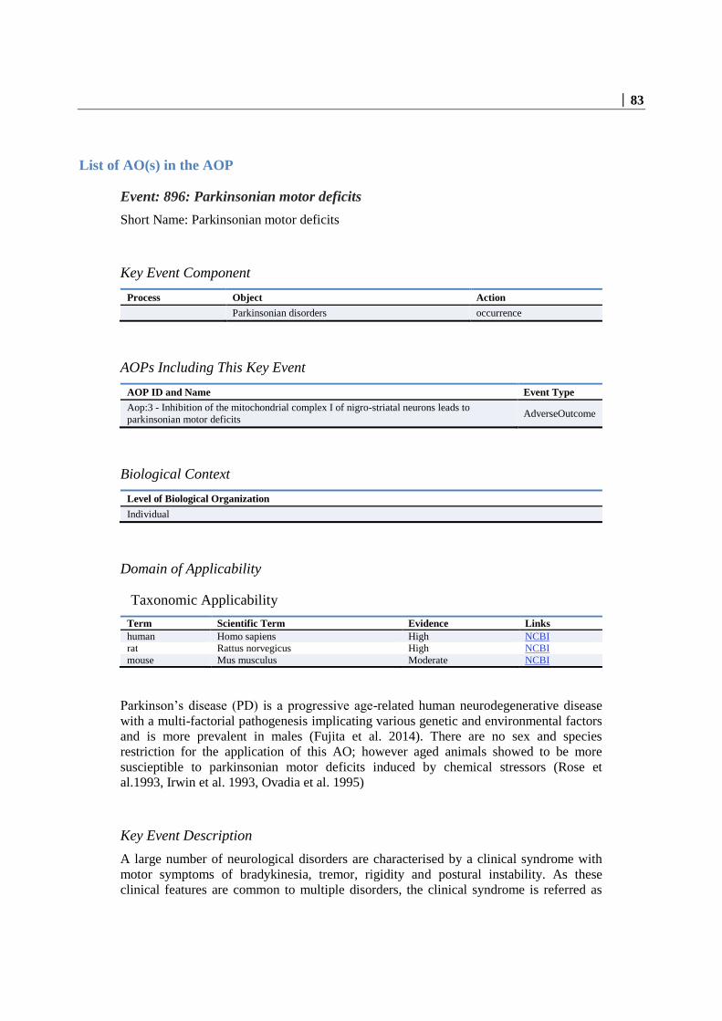
│ 83
List of AO(s) in the AOP
Event: 896: Parkinsonian motor deficits
Short Name: Parkinsonian motor deficits
Key Event Component
Process Object Action
Parkinsonian disorders occurrence
AOPs Including This Key Event
AOP ID and Name Event Type
Aop:3 - Inhibition of the mitochondrial complex I of nigro-striatal neurons leads to
parkinsonian motor deficits AdverseOutcome
Biological Context
Level of Biological Organization
Individual
Domain of Applicability
Taxonomic Applicability
Term Scientific Term Evidence Links
human Homo sapiens High NCBI
rat Rattus norvegicus High NCBI
mouse Mus musculus Moderate NCBI
Parkinson’s disease (PD) is a progressive age-related human neurodegenerative disease
with a multi-factorial pathogenesis implicating various genetic and environmental factors
and is more prevalent in males (Fujita et al. 2014). There are no sex and species
restriction for the application of this AO; however aged animals showed to be more
suscieptible to parkinsonian motor deficits induced by chemical stressors (Rose et
al.1993, Irwin et al. 1993, Ovadia et al. 1995)
Key Event Description
A large number of neurological disorders are characterised by a clinical syndrome with
motor symptoms of bradykinesia, tremor, rigidity and postural instability. As these
clinical features are common to multiple disorders, the clinical syndrome is referred as

84 │
“parkinsonism” and when parkinsonism is representing the prevalent part of the
syndrome, these are referred as “parkinsonian disorders”. Parkinson’s Disease (PD) is one
of parkinsonian disorders and can have an idiopathic, genetic or toxic (i.e. MPTP induced
parkinsonism) cause (Dickson 2012). All these disorders include a deregulation of the
extrapyramidal system.
The pyramidal motor system comprises bundles of neurons originating in the motor
centers of the cerebral cortex to terminate in the brainstem or in the spinal cord where
they are responsible for voluntary control of motor functions (Brooks 1971). The
extrapyramidal system, which is the anatomical organisation of the AO, is the part of the
motor system primarily involved in the control and regulation of involuntary motor
control, and in fine tuning (Barnes et al. 1983). Especially the initiation and maintenance
of complex movement patterns or of neuronal regulatory pathways involved in postural
control of the body are regulated by the nigrostriatal system that is affected in
parkinsonian states. The CNS input is modulated by extrapyramidal circuits before the
execution of complex motor movements. The modulated information from the basal
ganglia is looped back through the thalamus to the cortex, from where final motor signals
are sent via the pyramidal system; i.e. the basal ganglia system is not involved in the
control of motor neurons and striatal muscles, but it modulates the signals from the cortex
to these systems. Thus, an impaired input of dopamine into the striatum leads to an
impairment of this modulation loop, and a disturbance of basal ganglia feedback to the
thalamus and cortex. This ultimately manifests in key parkinsonian symptoms such as
tremor, rigidity, or bradykinesia (Bernheimer et al. 1973). These conditions can be
generated experimentally by dopamine depletion with reserpine, by inhibition of
dopamine receptors, by mechanical or chemical ablation of nigrostriatal dopamine
neurons (cut of the median forebrain bundle or injection of the toxicant 6-OH-dopamine)
or the application of toxicants that leading to a relatively selective death of dopaminergic
neurons in the substantia nigra (e.g. MPTP) and therefore a reduction of dopamine in the
striatum (Kolata et al. 1983).
The basal ganglia include the ventral striatum, the neostriatum composed of the putamen
and the caudate nucleus, the globus pallidus pars externa (GPe), the globus pallidus pars
interna (GPi), the subthalamic nucleus (STN), the substantia nigra pars reticulata (SNpr)
and the substantia nigra pars compacta (SNpc) (Obeso et al.2008). The main input sites
into basal ganglia are the striatum and the STN where cortical (glutamatergic)
innervations terminate in a topographically organised manner that largely reflects the
organisation in the cortex (Fallon et al. 1978; Takada et al. 1998). Both the GPi and the
SNpr represent the main output nuclei projecting into the thalamus (Parent et al. 1999;
Alexander et al. 1990). The connection between input and output nuclei is functionally
organised into a “direct” and an “indirect” pathway (Silverdale et al. 2003). These two
pathways in parallel regulate the activity of the basal ganglia output neurons of the GPi
and STN and are modulated by dopamine in the striatum. The dopaminergic terminals in
the striatum originate from dopaminergic projections from the SNpc. Striatal dopamine
modulates the activity of inhibitory GABAergic medium spiny neurons that make up 90%
of all neurons in the striatum (Smith et al. 1994). Medium spiny neurons that
preferentially express the D1 dopamine receptor are involved in the direct pathway and
directly project into the two main output nuclei (GPi and SNpr). Activation of the D1
medium spiny neuronal direct pathway results in a reduction of the inhibitory basal
ganglia output (GPi and SNpr) leading to a dis-inhibition of thalamic target neurons
(Bolam et al.2000). These events ultimately lead to an elevated activity in the respective
cortical neurons, i.e. D1 signalling in the striatum leads to an increase in motor activity.

│ 85
Medium spiny neurons predominantly expressing the D2 dopamine receptor mostly
project to the GPe (Gerfen et al. 1990). Activation of D2 expressing neurons leads to an
inhibition of their activity. D2 neurons of the indirect pathway connect the striatum with
GPi/SNpr via synaptic connections in the GPe and the STN. Activating neurons
originating in the STN project into the GPi/SNpr are glutamatergic. From the STN,
activating glutamatergic neuronal projections into the GPi/SNpr lead to a basal, low
activation. Activation of the indirect pathway by striatal dopamine from the substantia
nigra hence leads to a low basal inhibitory GABAergic output into thalamic structures,
and thus allows a strong motor cortex activation of the thalamus.
Functional anatomy of basal ganglia. A) Normal conditions. Striatal (STR) dopamine
mainly originates from projections originating in the substantia nigra pars compacta
(SNc). The STR is mainly composed of inhibitory GABAergic medium spiny neurons
(MSN). MSN involved in the direct pathway directly project to the globus pallidus pars
interna (GPi) and the substantia nigra pars reticulata (SNpr) leading to a basal inhibition
of these output nuclei. MSN involved in the indirect pathway send inhibitory projections
to the globus pallidus pars externa (GPe). Their activity is dampened by dopamine
binding to D2 receptor expressing MSN in the striatum. B) Lack of striatal dopamine.

86 │
Under conditions of a lack of striatal dopamine, inhibitory GABAergic neurons,
originating in the striatum, receive less activation, resulting in a declined inhibition of
GPi and SNpr inhibitory output. In the indirect pathway, the lack of dopamine causes a
lack of its inhibitory influence on inhibitory GABAergic projections into the GPe. This
accelerated inhibition of the GPe results in a decline in its inhibitory output into the STN.
The decline in STN inhibition allows its overactivation, resulting in an excessive
activation of stimulatory glutamatergic projections into the GPi and SNpr. (according to
Silverdale 2003).
Parkinson’s Disease is characterised by a decline in striatal dopamine input from the
substantia nigra pars compacta (Smith et al. 1994). Under normal conditions, ganglial
output via GPi/SNpr nuclei causes a moderate inhibitory influence on cortical and
brainstem motor neurons. A reduction in striatal dopamine leads to an underactivation of
D1 receptor-expressing medium spiny neurons of the direct pathway. This insufficient
activation of the inhibitory GABAergic medium spiny neurons results in a reduction of its
normal inhibitory influence on GPi and SNpr output nuclei. As a consequence, dopamine
depletion leads to the overactivation of the inhibitory GABAergic GPi/SNpr output via
the direct pathway (Mitchell et al.1989).
In the indirect pathway, the reduced activation of D2 receptors expressing neurons leads
to an overactivation of inhibitory output nuclei projecting into the GPe. The resulting
inhibitory output of the GPe is hence reduced, thus leading to a declined inhibition of the
STN. Overactivation of the stimulatory glutamatergic projections originating in the STN
leads to the hyperactivation of the output GPi/SNpr nuclei. As a consequence of striatal
dopamine depletion, the direct pathway becomes underactivated and the indirect pathway
becomes overactivated. This leads to an overactivation of the basal ganglia output nuclei.
Due to their inhibitory influence on thalamocortical motor centers, the resulting reduced
cortical activation leads to the prominent impairment of motor functions in parkinsonian
states (Silverdale et al. 2003).
The model of direct and indirect pathways linking striatal dopamine content with the
basal ganglia output nuclei has been criticised in recent years as it ignores the influence of
extrastriatal dopamine (Smith et al. 2000), or the fact that some medium spiny neurons
express dopamine receptors of both the D1 and of the D2 type (Surmeier et al. 1996).
Principal validity of the model and the central role of striatal dopamine was e.g.
demonstrated by L-DOPA-mediated supplementation of striatal dopamine content in
unprimed PD patients that causes a partial reduction in the overactivation of GPi/SNpr
output (Yuan et al. 2010; Heimer et al. 2006). As an alternative way for symptomatic
treatment of parkinsonian conditions, deep brain stimulation of either the STN or the GPi
was shown to relieve from parkinsonian motor features (Mazzone 2003; Odekerken
2013).
How it is Measured or Detected
For the analysis of striatal dopamine content and its correlation with motor control, both
biochemical analysis methods on the cellular and tissue level as well as behavioral tests
are required. Available test models are mice and rats on the one hand and non-human
primates and humans on the other. Motor impairment features associated with
parkinsonian states in man serve as reference standard. Monkey models have the
advantage to largely reflect complex motor impairment patterns observed in humans

│ 87
which are rather difficult to assess in rodents. Rodent models in contrast are cost-efficient
and allow both biochemical analysis that require major invasive methods as well as basic
behavioral tests. Due to the limitations in the assessment of moderate motor impairment
in rodents and the well-established correlation between striatal dopamine content and
impaired motor output, analysis of striatal dopamine is often applied as surrogate readout
for the assessment of motor deficits.
Detection of striatal dopamine (total or extracellular).
The standard method used in the majority of experimental work is the determination of
total contents of dopamine and its two degradation metabolites HVA and DOPAC. For
this purpose, the striatum is quickly removed from experimental animals, homogenised in
a suitable acidic buffer, and the dopamine (metabolites) determined by HPLC with
electrochemical detector or by HPLC-MS. For live in vivo detection of extracellular
dopamine levels, a microdialysis probe is inserted into the striatum. Microdialysis can be
performed in anesthetized animals or freely moving animals; basal dopamine levels or
stimulated levels (amphetamine, KCl) can be recorded. Dopamine and its metabolites are
detected in the dialysate either by HPLC or by HPLC-mass spectrometry analysis
(Saraswat 1981, Cui 2009, Gonzalez 2011).
Detection of dopamine neuron terminals in the striatum.
As alternative to the detection of striatal dopamine that is to a large extent limited to live
detection setups due to its instability in tissues, the number of remaining dopamine
neurons in the substantia nigra pars compacta was suggested as alternative readout (Burns
1983). It allows the analysis of ex vivo samples without the limitations associated with
the instability and reactivity of extravesicular dopamine. Although the number of
surviving dopamine neurons in the SNpc in PD or in complex-I inhibitor challenged test
animals is a valuable parameter on its own, it was discovered that the number of DA
neurons in the SNpc not necessarily correlates with the amount of dopamine released in
the striatum. Tyrosine hydroxylase (TH) was regularly stained as marker for DA neurons,
however it was observed that TH expression was very variable following MPTP
intoxication in the absence of cell death and therefore has only limited suitability for the
assessment of DA neuronal numbers (Aznavour 2012). Second, many DA neurites and
terminals displayed damage or degradation in the absence of death of the corresponding
neuronal cell (Ling 2015). Hence, even in the presence of viable DA neurons in the SNpc,
their corresponding terminals could no longer be able to release dopamine into the
striatum. Staining of DA neuronal terminals in the striatum is therefore used as a more
reliable indirect marker for striatal dopamine content. For the analysis of nigrostriatal
terminals, the dopamine transporter (DAT) is visualised either by antibody-mediated
staining in tissue slices or by the application of radioactively labeled DAT ligands that
allow their application both in vivo and in ex vivo samples (Morris 1996).
Behavioral tests: Rodent models.
Rotation: the rotation model of Ungerstedt and Arbuthnott (Ungerstedt and Arbuthnott
1970) is based on the unilateral lesion of the nigrostriatal dopamine neuron system either
in rodents or in non-human primates. The lesion can be produced either surgivally, or by
stereotaxic infusion of e.g. 6-OHDA into the nigrostriatal system of one hemisphere, or
by infusion of MPTP through one carotid (single sided). After the lesion, animals are left
to recover, then the dopamine system is stimulated by injection of amphetamine. The
asymmetry of remaining dopamine neurons (only on one side) triggers spontaneous
asymmetric motor behaviour, i.e. rotations of the animals. Each full turn of an animal is

88 │
recorded, the respective numbers of left- and right turns are plotted versus time,
respectively. In the standard rotation model, monkeys become hypokinetic in the limbs on
the contralateral side of the brain hemisphere treated. Rats preferentially rotate towards
the side of the lesion upon treatment with drugs that trigger activation of the remaining
dopamine neurons.
Rotarod: assessment of motor coordination. The animals are placed on a rotating rod that
is subjected to linear acceleration. The latency to fall from the rod is detected (Jones
1968). Hang test: Detection of neuromuscular strength. Mice are placed on a horizontal
grid. When the animals grabbed the grid with their fore- and hindpaws, the grid is
inverted with the animal hanging upside down. In a typical setup, mice are required to
remain on the grid for at least 30 s (Tillerson 2002).
Forepaw Stride length during walking. Ink is applied to the forepaws and the mice walk
across a blank sheet of paper. Training of the animals to walk across the white paper in a
straight line without stopping is performed before the respective treatment. The distance
between single steps on each side are measured (Klapdor 1997).
Grid test: Mice hang upside down for 30 s on the grid that is also used for the Hang test
and are recorded on video for closer analysis. With this method, the average forepaw
distance is measured by assessing the distance covered, divided by the number of
successful forepaw steps. In the course of the analysis, the number of unsuccessful
forepaw steps are detected and displayed as percentage of the total number of steps
performed (Crawley 1999).
Akinesia: the animal is placed on a flat surface and the latency until it has moved all of its
four limbs is assessed. Open field test: Infrared beams detect the animals activity for the
determination of parameters such as the time spent locomoting, the distance travelled, or
the number of rearings.
Pole test: the animal is placed on a gauze-taped pole with the head upwards below the top
of the pole. Two parameters are detected: 1) time until animals turn by 180°; 2) time until
the animals reach the floor.
Non-invasive imaging of DA neuron terminals
Positron emission tomography (PET): Based on its appropriate half life time of ca. 2 h for
clinical investigations, fluorine-18 labeled L-[18F]-fluorodopa is routinely used in trace
amounts for intravenous administration. Striatal uptake of L-[18F]-fluorodopa is followed
by applying positron emission tomography (PET) (Leenders 1986).
Single photon emission computed tomography (SPECT): monitoring of dopamine
transporter (DAT). Iodine-123-β-CIT is used as a sensitive ligand for dopamine and
serotonin transporters and was applied in monkeys and humans (Winogrodzka 2003).
Human neurological tests
A recent systematic review and evaluation of currently used rating scales for the
assessment of motor impairment and disability in PD patients identified the 1) Columbia
University rating scale, 2) the Northwestern University Disability Scale, and 3) the
Unified Parkinsons Disease rating scale as the most evaluated and reliable scales
available (Ramaker 2002). All scales evaluate several parameters, some of which are not
motor related. Thus, only subscales are useful for readout of motor symptoms (e.g. 13 of
the 42 UPDRS parameters). Of these, not all are equally dependent on nigrostriatal
dopamine. Examination needs to be done by a trained neurologist.

│ 89
Regulatory Significance of the AO
Neurotoxic effects shall be carefully addressed and reported in routine required regulatory
toxicological studies (acute toxicity studies, short-term toxicity studies, long term toxicity
and carcinogenicity studies and reproductive toxicity studies). Regarding neurotoxicity in
rodents, inclusion of neurotoxicity investigations in routine toxicology studies shall also
be considered. For pesticide active substances the circumstances in which neurotoxicity
studies should be performed are listed in Regulation (EU) No 283/2013:
Specific neurotoxicity studies in rodents shall be performed in case of one those
following conditions:
there is indication of neurotoxicity in routine toxicity studies carried out with the
active substance;
the active substance is a structurally related to known neurotoxic compound;
the active substance has a neurotoxic mode of pesticidal action.
As a result, specific neurotoxicity studies are not routinely required for all pesticide active
substances. Specific neurotoxicity testing becomes obligatory only if neurotoxicity has
been observed during histopathological evaluation or in case of structural analogy with a
known neurotoxic compound. Motor activity should be measured once in short-term
repeated dose toxicity studies (OECD 407, 408 and 422) and several times in specific
neurotoxicity studies (OECD 424, OECD 426 and cohort 2 of OECD 443). However, this
is not a requirement in chronic toxicity studies unless neurotoxic effects have been
reported in the shorter studies. The same test (measures horizontal and/or vertical
movements in a test chamber) is implemented in both routine studies and neurotoxicity
studies. Coordination and balance are evaluated by rotation or rotarod or pole tests, and
gait abnormalities by forepaw stride length test. Those tests are not required by any
repeated dose toxicity OECD guidelines and they can be optionally incorporated in the
design of neurotoxicity studies OECD 424 and OECD 426.
Although motor deficits is the AO in this AOP, degeneration of DA neurons, is also
considered an adverse effect in the regulatory framework, even in the absence of clear
clinical symptoms or motor deficits. Morphological assessment of brain structures is a
standard requirement in the regulatory toxicological studies supporting the risk
assessment of chemical substances and it is a regulatory expectation that the anatomical
structures belonging to the nigrostriatal pathway would be included and evaluated as part
of the standard evaluation of the brain. Treatment related neuronal degeneration, when
occurring as a consequence of the treatment, is generally dose-dependent in incidence and
severity. However, if not accompanied by clinical signs or behavioral changes indicative
of central nervous system pathology, minimal loss of DA neurons would likely remain
undetected in the standard histological evaluation, due to the presence of non DA neurons
or as a consequence of the subjectivity of non-quantifiable analysis, unless specific
markers are used. As multiple forms of perturbation can affect the neurons, some changes
are potentially still reversible (e.g. loss of TH or DA) and irreversibility should be
confirmed as part of the assessment. It is then important to apply a sensitive and
appropriate method (Switzer 2000) and evaluation of the phenotypic markers in the
striatum and in the SNpc should be always performed as a minimum standard (Minnema
et al 2014) when investigating perturbation of the nigrostriatal pathway. It should
additionally consider that rat is likely to be a poor model to capture this kind of hazard, as

90 │
demonstrated by the poor sensitivity of rat to MPTP or related compounds and this should
be taken into account for the design and interpretation of the studies.
Dissimilarities of chemical induced animal models to human disease are also important
and should be carefully weighted when considering the duration and schedule of the
study/treatment. Differently from the human disease, with the MPTP animal model, the
damage occurs rapidly, is hardly progressive, is little age-dependent and formation of
Lewy bodies is sometime not occurring (Efremova et al. 2015). Therefore, for different
animals models, the standard 90 days toxicity study could not match with the chronic and
progressive characteristics of the human disease and compensatory changes influencing
DA metabolism and turnover and protein catabolism can occur during the treatment
period with an impact on the time of onset of the lesion (Ossowska et al. 2005).
References
Alexander GE, Crutcher MD. Functional architecture of basal ganglia circuits: neural
substrates of parallel processing. Trends Neurosci. 1990 Jul;13(7):266-71.
Aznavour N, Cendres-Bozzi C, Lemoine L, Buda C, Sastre JP, Mincheva Z, Zimmer L,
Lin JS. MPTP animal model of Parkinsonism: dopamine cell death or only tyrosine
hydroxylase impairment? A study using PET imaging, autoradiography, and
immunohistochemistry in the cat. CNS Neurosci Ther. 2012 Nov;18(11):934-41.
Barnes CD. The basal ganglia in extrapyramidal dysfunction. Brain Res Bull. 1983
Aug;11(2):271-5.
Bernheimer H, Birkmayer W, Hornykiewicz O, Jellinger K, Seitelberger F. Brain
dopamine and the syndromes of Parkinson and Huntington. Clinical, morphological and
neurochemical correlations. J Neurol Sci. 1973 Dec;20(4):415-55.
Bolam JP, Hanley JJ, Booth PA, Bevan MD. Synaptic organisation of the basal ganglia. J
Anat. 2000 May;196 ( Pt 4):527-42. Brain Res. 1998 Apr 27;791(1-2):335-40.
Brooks VB, Stoney SD Jr. Motor mechanisms: the role of the pyramidal system in motor
control. Annu Rev Physiol. 1971;33:337-92.
Burns RS, Chiueh CC, Markey SP, Ebert MH, Jacobowitz DM, Kopin IJ. A primate
model of parkinsonism: selective destruction of dopaminergic neurons in the pars
compacta of the substantia nigra by N-methyl-4-phenyl-1,2,3,6-tetrahydropyridine. Proc
Natl Acad Sci U S A. 1983 Jul;80(14):4546-50.
Crawley JN. Behavioral phenotyping of transgenic and knockout mice: experimental
design and evaluation of general health, sensory functions, motor abilities, and specific
behavioral tests. Brain Res. 1999 Jul 17;835(1):18-26.
Cui M, Aras R, Christian WV, Rappold PM, Hatwar M, Panza J, Jackson-Lewis V,
Javitch JA, Ballatori N, Przedborski S, Tieu K. The organic cation transporter-3 is a
pivotal modulator of neurodegeneration in the nigrostriatal dopaminergic pathway. Proc
Natl Acad Sci U S A. 2009 May 12;106(19):8043-8.
Dickinson D. 2012. Parkinson’s disease and parkinsonism: Neuropathology.Cold Spring
Harb Perspect Med. 2:a009258. Fallon JH, Moore RY. Catecholamine innervation of the
basal forebrain. IV. Topography of the dopamine projection to the basal forebrain and
neostriatum. J Comp Neurol. 1978 Aug 1;180(3):545-80.

│ 91
Efremova L, Scildknecht S, Adam M, pape R, Gutbier S, Hanf B, Burkle A, Leist M.
2015. Prevention of the degeneration of dopaminergic neurons in an astrocyte co-culture
system allowing endogenous drug metabolism. British Journal of Pharmacology; 172;
4119-32
Fujita KA, Ostaszewski M, Matsuoka Y, Ghosh S, Glaab E, Trefois C, Crespo I, Perumal
TM, Jurkowski W, Antony PM, Diederich N, Buttini M, Kodama A, Satagopam VP,
Eifes S, Del Sol A, Schneider R, Kitano H, Balling R. Integrating pathways of
Parkinson's disease in a molecular interaction map. Mol Neurobiol. 2014 Feb;49(1):88-
102
Gerfen CR, Engber TM, Mahan LC, Susel Z, Chase TN, Monsma FJ Jr, Sibley DR. D1
and D2 dopamine receptor-regulated gene expression of striatonigral and striatopallidal
neurons. Science. 1990 Dec 7;250(4986):1429-32.
González RR, Fernández RF, Vidal JL, Frenich AG, Pérez ML. Development and
validation of an ultra-high performance liquid chromatography-tandem mass-
spectrometry (UHPLC-MS/MS) method for the simultaneous determination of
neurotransmitters in rat brain samples. J Neurosci Methods. 2011 Jun 15;198(2):187-94.
Heimer G, Rivlin-Etzion M, Bar-Gad I, Goldberg JA, Haber SN, Bergman H. Dopamine
replacement therapy does not restore the full spectrum of normal pallidal activity in the 1-
methyl-4-phenyl-1,2,3,6-tetra-hydropyridine primate model of Parkinsonism. J Neurosci.
2006 Aug 2;26(31):8101-14.
Irwin JK, 1993, Parkinson’s disease: Past, Present and Future. Neuropsycopharmacology.
9(1). 1-1.
Jones BJ, Roberts DJ. The quantiative measurement of motor inco-ordination in naive
mice using an acelerating rotarod. J Pharm Pharmacol. 1968 Apr;20(4):302-4.
Klapdor K, Dulfer BG, Hammann A, Van der Staay FJ. A low-cost method to analyse
footprint patterns. J Neurosci Methods. 1997 Jul 18;75(1):49-54.
Kolata G. Monkey model of Parkinson's disease. Science. 1983 May 13;220(4598):705.
Leenders KL, Palmer AJ, Quinn N, Clark JC, Firnau G, Garnett ES, Nahmias C, Jones T,
Marsden CD. Brain dopamine metabolism in patients with Parkinson's disease measured
with positron emission tomography. J Neurol Neurosurg Psychiatry. 1986 Aug;49(8):853-
60.
Ling H, Kearney S, Yip HL, Silveira-Moriyama L, Revesz T, Holton JL, Strand C, Davey
K, Mok KY, Polke JM, Lees AJ. Parkinson's disease without nigral degeneration: a
pathological correlate of scans without evidence of dopaminergic deficit (SWEDD)? J
Neurol Neurosurg Psychiatry. 2015 Jul 24. pii: jnnp-2015-310756. doi: 10.1136/jnnp-
2015-310756.
Mazzone P. Deep brain stimulation in Parkinson's disease: bilateral implantation of
globus pallidus and subthalamic nucleus. J Neurosurg Sci. 2003 Mar;47(1):47-51.
Minnema DJ, Travis KZ, Breckenridge CB, Sturgess NC, Butt M, Wolf JC, Zadory D,
Beck MJ, Mathews JM, tisdel MO, Cook AR, Botham PA, Smith LL. 2014. Dietary
administration of paraquat for 13 weeks does not result in a loss of dopaminergic neurons
in the substantia nigra of C57BL/6J mice. (68). 250-8.
Mitchell IJ, Clarke CE, Boyce S, Robertson RG, Peggs D, Sambrook MA, Crossman AR.
Neural mechanisms underlying parkinsonian symptoms based upon regional uptake of 2-

92 │
deoxyglucose in monkeys exposed to 1-methyl-4-phenyl-1,2,3,6-tetrahydropyridine.
Neuroscience. 1989;32(1):213-26.
Morris ED, Babich JW, Alpert NM, Bonab AA, Livni E, Weise S, Hsu H, Christian BT,
Madras BK, Fischman AJ. Quantification of dopamine transporter density in monkeys by
dynamic PET imaging of multiple injections of 11C-CFT. Synapse. 1996 Nov;24(3):262-
72.
Neural mechanisms underlying parkinsonian symptoms based upon regional uptake of 2-
deoxyglucose in monkeys exposed to 1-methyl-4-phenyl-1,2,3,6-tetrahydropyridine.
Obeso JA, Rodríguez-Oroz MC, Benitez-Temino B, Blesa FJ, Guridi J, Marin C,
Rodriguez M. Functional organization of the basal ganglia: therapeutic implications for
Parkinson's disease. Mov Disord. 2008;23 Suppl 3:S548-59.
Odekerken VJ, van Laar T, Staal MJ, Mosch A, Hoffmann CF, Nijssen PC, Beute GN,
van Vugt JP, Lenders MW, Contarino MF, Mink MS, Bour LJ, van den Munckhof P,
Schmand BA, de Haan RJ, Schuurman PR, de Bie RM. Subthalamic nucleus versus
globus pallidus bilateral deep brain stimulation for advanced Parkinson's disease
(NSTAPS study): a randomised controlled trial. Lancet Neurol. 2013 Jan;12(1):37-44.
Ossowska K., Wardas J, Smialowska M, Kuter K, Lenda T, Wieronska JM, Zieba B,
Nowak P, Dabrowska J, Bortel A, Kwiecinski A, Wolfarth S. 2005. A slowly developing
dysfunction of dopaminergiv nigrostriatal neurons induced by long-term paraquat
administration in rats: an animal model of preclinical stagesof Parkinso’s disease.
Ovadia A, Zhang Z, Gash DM. 1995. Increased susceptibility to MPTP in middle-aged
Rhesus Monkeys. Neurobiology of aging. 16 (6) 931-7
Parent A, Hazrati LN. Functional anatomy of the basal ganglia. I. The cortico-basal
ganglia-thalamo-cortical loop. Brain Res Brain Res Rev. 1995 Jan;20(1):91-127.
Ramaker C, Marinus J, Stiggelbout AM, Van Hilten BJ. Systematic evaluation of rating
scales for impairment and disability in Parkinson's disease. Mov Disord. 2002
Sep;17(5):867-76.
Rose S N M, Jackson EA, Gibb WR, Jaehnig P, Jenner P, Marsden CD. 1993. Age-
related effects of 1-methyl-4-phenyl-1,2,3,6-tetrahydropyridine treatment of common
marmosets. Eur J Pharmacol. 230(2).177-85.
Saraswat LD, Holdiness MR, Justice JB, Salamone JD, Neill DB. Determination of
dopamine, homovanillic acid and 3,4-dihydroxyphenylacetic acid in rat brain striatum by
high-performance liquid chromatography with electrochemical detection. J Chromatogr.
1981 Mar 13;222(3):353-62.
Silverdale MA, Fox SH, Crossman AR, Brotchie JM. Potential nondopaminergic drugs
for Parkinson's disease. Adv Neurol. 2003;91:273-91.
Smith Y, Bennett BD, Bolam JP, Parent A, Sadikot AF. Synaptic relationships between
dopaminergic afferents and cortical or thalamic input in the sensorimotor territory of the
striatum in monkey. J Comp Neurol. 1994 Jun 1;344(1):1-19.
Smith Y, Kieval JZ. Anatomy of the dopamine system in the basal ganglia. Trends
Neurosci. 2000 Oct;23(10 Suppl):S28-33.
Surmeier DJ, Song WJ, Yan Z. Coordinated expression of dopamine receptors in
neostriatal medium spiny neurons. J Neurosci. 1996 Oct 15;16(20):6579-91.

│ 93
Switzer RC. 2000. Application of silver degeneration stains for neurotoxicity testing.
Toxicologic pathology. 28(1) 70-83.
Takada M, Tokuno H, Nambu A, Inase M. Corticostriatal input zones from the
supplementary motor area overlap those from the contra- rather than ipsilateral primary
motor cortex.
Tillerson JL, Caudle WM, Reverón ME, Miller GW. Detection of behavioral impairments
correlated to neurochemical deficits in mice treated with moderate doses of 1-methyl-4-
phenyl-1,2,3,6-tetrahydropyridine. Exp Neurol. 2002 Nov;178(1):80-90.
Ungerstedt U, Arbuthnott GW. Quantitative recording of rotational behavior in rats after
6-hydroxy-dopamine lesions of the nigrostriatal dopamine system. Brain Res. 1970 Dec
18;24(3):485-93.
Winogrodzka A, Bergmans P, Booij J, van Royen EA, Stoof JC, Wolters EC.
[(123)I]beta-CIT SPECT is a useful method for monitoring dopaminergic degeneration in
early stage Parkinson's disease. J Neurol Neurosurg Psychiatry. 2003 Mar;74(3):294-8.
Yuan H, Zhang ZW, Liang LW, Shen Q, Wang XD, Ren SM, Ma HJ, Jiao SJ, Liu P.
Treatment strategies for Parkinson's disease. Neurosci Bull. 2010 Feb;26(1):66-76.

94 │
Appendix 2: List of Key Event Relationships (KERs) in the AOP
List of Adjacent Key Event Relationships
Relationship: 933: Binding of inhibitor, NADH-ubiquinone oxidoreductase
(complex I) leads to Inhibition, NADH-ubiquinone oxidoreductase (complex I)
AOPs Referencing Relationship
AOP Name Adjacency Weight of
Evidence
Quantitative
Understanding
Inhibition of the mitochondrial complex I of nigro-
striatal neurons leads to parkinsonian motor deficits adjacent High Low
Evidence Supporting Applicability of this Relationship
The CI is well-conserved across species from lower organism to mammals. The central
subunits of CI harboring the bioenergetic core functions are conserved from bacteria to
humans. CI from bacteria and from mitochondria of Yarrowia lipolytica, a yeast genetic
model for the study of eukaryotic CI (Kerscher et al., 2002) was analysed by x-ray
crystallography (Zickermann et al., 2015). However, the affinity of various chemicals to
cause partial or total inhibition of CI activity across species is not well studied (except
rotenone).
Key Event Relationship Description
It is well documented that binding of an inhibitor to CI inhibits its activity (see MIE).
Naturally occurring and synthetic CI inhibitors have been shown to inhibit the catalytic
activity of CI, leading to partial or total inhibition of its activity in a dose response
manner (Degli Esposti and Ghelli, 1994; Ichimaru et al. 2008; Barrientos and Moraes,
1999; Betarbet et al., 2000). Indeed, binding of inhibitors stops the electron flow from CI
to ubiquinone. Therefore, the Fe-S clusters of CI become highly reduced and no further
electrons can be transferred from NADH to CI. This leads to the inhibition of the NADH
oxido-reductase function, i.e. CI inhibition.
Evidence Supporting this KER
Biological Plausibility
The weight of evidence supporting the relationship between binding of an inhibitor to
NADH-ubiquinone oxidoreducatse and its inhibition is strong.

│ 95
Biological Plausibility
There is an extensive understanding of the functional relationship between binding of an
inhibitor to NADH-ubiquinone oxidoreductase (CI) and its inhibition. As the first entry
complex of mitochondrial respiratory chain, CI oxidises NADH and transfers electrons
via a flavin mononucleotide cofactor and several Fe-S complexes to ubiquinone. The
electron flow is coupled to the translocation of protons from the matrix to the
intermembrane space. This helps to establish the electrochemical gradient that is used to
fuel ATP synthesis (Greenamyre et al., 2001). If an inhibitor binds to CI, the electron
transfer is blocked. This compromises ATP synthesis and maintenance of Δψm, leading
to mitochondrial dysfunction. As CI exerts a higher control over oxidative
phosphorylation in synaptic mitochondria than in non-synaptic mitochondria in the brain
(Davey and Clark, 1996), specific functional defects observed in PD may be explained. It
is well documented that CI inhibition is one of the main sites at which electron leakage to
oxygen occurs. This results in a production of ROS, such as superoxide (Efremov and
Sazanow, 2011) and hydrogen peroxide, which are main contributors to oxidative stress
(Greenamyre et al., 2001).
Empirical Evidence
A variety of studies show a significant correlation between binding of an inhibitor to CI
and its inhibition, usually measured by the decreased mitochondrial respiration. Different
classes of CI inhibitors, such as rotenone, MPP+, piericidin A, acetogenins, pyridaben,
and various piperazin derivatives (Ichimaru et al. 2008) have been shown to bind to the
ubiquitin site of CI, leading to a partial or total inhibition of oxidoreductase activity in a
dose response manner (Grivennikova et al., 1997; Barrientos and Moraes, 1999; Betarbet
et al., 2000). The reduction of CI activity is well documented in a variety of studies using
isolated mitochondria or cells, as well as in in vivo experiments and in human post
mortem PD brains. Usually it is measured by assays described in 2nd Key Event
Relationship (KER): Inhibition of complex I leads to mitochondrial dysfunction. It has
been shown that binding of rotenone to CI (e.g. Betarbet et al., 2000, Greenamyre et al.,
2001) or MPP+ (e.g. Krug et al., 2014; Langston, 1996) can reproduce the anatomical,
neurochemical, behavioural and neuropathological features of PD. Therefore, the
empirical support for this KER will be mainly based on the experiments performed after
exposure to rotenone or MPP+.
The binding of rotenone to CI resulted in time- and dose-dependent inhibition of
CI activity measured in sub-mitochondrial particles. The kinetics of the active CI
inhibition was determined after exposure to rotenone at 20, 30 and 40 nM at
different times of exposure (30 sec, 1 min or 2 min) (Grivennikova et al., 1997).
This study suggests that two rotenone binding sites exist in CI: one affecting
NADH oxidation by ubiquinone and the other one operating in ubiquinol-NAD+
reductase action.
Partial inhibition of CI produces a mild, late-onset mitochondrial damage. The
threshold effect seen in brain mitochondria (25–50% decrease in activity) may not
directly impact ATP levels or Δψm but could have long-term deleterious effects
triggered by oxidative stress, as it has been shown that an electron leak upstream
of the rotenone binding site in CI leads to ROS production (Greenamyre et al.,
2001).
Exposure of rats to rotenone (2 days, 2 mg/kg) produced free brain rotenone
concentration of 20–30 nM and resulted in 73% inhibition of specific binding to

96 │
CI of [3H] dihydrorotenone (Betarbet et al., 2000). However, oximetry analysis
indicated that in brain mitochondria (but not liver mitochondria) this rotenone
concentration (30 nM maximum) was insufficient to inhibit glutamate (CI
substrate)-supported respiration (Betarbet et al., 2000) suggesting that this
rotenone concentration did not alter mitochondrial oxygen consumption in
isolated brain mitochondria.
Rotenone has been reported to be a specific and potent mitochondrial CI inhibitor
with IC50 values from 0.1 nM to 100 nM depending on the system and methods
used (Lambert and Brand, 2004; Ichimaru et al., 2008; Chinopoulos and Vizi,
2001; Beretta et al., 2006).
Mesencephalic cultures prepared from C57/BL6 mice and treated with 5, or 10
nM rotenone for 24 h inhibited CI activity by 11% or 33%, respectively (Choi et
al., 2008).
The inhibition of CI was studied in the human osteosarcoma-derived cell line
(143B) after the exposure to rotenone or using a genetic model (40% loss of CI
activity in human xenomitochondrial cybrids (HXC) lines). Different degrees of
CI inhibition were quantitatively correlated with levels of decreased cellular
respiration (Barrientos and Moraes, 1999). Only when CI was inhibited by 35-
40% (< 5 nM rotenone), cell respiration decreased linearly until 30% of the
normal rate. Increasing concentrations of rotenone produced further but slower
decrease in CI activity and cell respiration (Fig. 1). Cells with the complete
rotenone-induced CI inhibition still maintain a cell respiration rate of
approximately 20% because of an electron flow through complex II. At high
concentrations (5–6-fold higher than the concentration necessary for 100% CI
inhibition), rotenone showed a secondary, toxic effect at the level of microtubule
assembly (Barrientos and Moraes 1999).
Bovine sub-mitochondrial particles were used to test rotenone affinity binding at
20 nM. This concentration of rotenone reduced the NADH oxidation rate by
approximately 50% (Okun et al., 1999.
MPP+ (an active metabolite of MPTP) is an inhibitor of CI (Nicklas et al., 1987;
Mizuno et al, 1989; Sayre et al., 1986). Inhibition of the mitochondrial CI by
MPP+ supresses aerobic glycolysis and ATP production (Book chapter in
Cheville 1994).
MPP+ binds loosely to CI and causes reversible inhibition of its activity:
approximately 40% inhibition was observed at 10 mM concentration within 15
min of incubation. However, prolonged incubation (> 15min) produces up to 78%
of irreversible inhibition of CI (Cleeter et al., 1992).
MPP+ interrupts mitochondrial electron transfer at the NADH dehydrogenase-
ubiquinone junction, as do the respiratory chain inhibitors rotenone, piericidin A
and barbiturates. The 4'-alkyl derivatives of MPP+ inhibit NADH oxidation in
submitochondrial particles at much lower concentrations than does MPP+ itself.
The MPP+ analogues with short alkyl chains prevent the binding of 14C-labelled
piericidin A to the membrane and thus must act at the same site, but analogues
with alkyl chains longer than heptyl do not prevent binding of [14C]piericidin
(Ramsay et al., 1991).
13 complex I inhibitors were found to decrease labelling at the PSST site without
effect on ND1 labelling. The results suggest that the common action of MPP(+)
and stigmatellin on the functional coupling of the PSST and ND1 subunits is
initiated by binding at a semiquinone binding site in complex I (Schuler and
Casida, 1991).

│ 97
Human studies
There are many studies that show impaired catalytic activity of CI in multiple PD
post-mortem brain tissues. For example (Parker and Swerdlow, 1998), five PD
brains were used to measure activities of complexes I, III, IV, and of complexes
I/III together (NADH: cytochrome c reductase). These measurements were
performed in purified frontal cortex mitochondria and revealed a significant loss
of CI activity in these PD samples as compared to controls.
Human data indicate that impairment of CI activity may contribute to the
pathogenic processes of PD (for example, Greenamyre et al., 2001; Schapira et
al., 1989; Shults, 2004).
Uncertainties and Inconsistencies
It is not clear the number of subunits constituting CI in mammals, as according to the
existing literature different numbers are cited (between 41-46) (Vogel et al., 2007a;
Hassinen, 2007). The majority of data claims that mammalian CI is composed of 46
(Greenamyre et al., 2001; Hassinen, 2007) or 45 subunits (Vogel et al., 2007a). It is not
sure whether there may exist tissue-specific subunits of CI isoforms (Fearnley et al.,
2001). It is unclear, which subunit(s) bind rotenone or other inhibitors of CI.
Additionally, it is not clear whether CI has other uncharacterised functions, taking into
consideration its size and complexity (43-46 subunits vs. 11 subunits of complex III or 13
subunits of complex IV) (Greenamyre et al., 2001). There is no strict linear relationship
between inhibitor binding and reduced mitochondrial function. Low doses of rotenone
that inhibit CI activity partially do not alter mitochondrial oxygen consumption.
Therefore, bioenergetic defects can not account alone for rotenone-induced
neurodegeneration. Instead, under such conditions, rotenone neurotoxicity may result
from oxidative stress (Betarbet et al., 2000). Few studies used human brain cells/human
brain mitochondria. Therefore, full quantitative data for humans are not available.
References
Barrientos A, and Moraes CT. 1999. Titrating the Effects of Mitochondrial Complex I
Impairment in the Cell Physiology. 274(23)16188–16197.
Beretta S, et al. 2006. Partial mitochondrial complex I inhibition induces oxidative
damage and perturbs glutamate transport in primary retinal cultures. Relevance to Leber
Hereditary Optic Neuropathy (LHON). Neurobiol Dis. 24:308–317.
Betarbet R, Sherer TB, MacKenzie G, Garcia-Osuna M, Panov AV, Greenamyre JT.
2000. Chronic systemic pesticide exposure reproduces features of Parkinson's disease.
Nat Neurosci 3:1301-1306.
Cheville NF. 1994. Ultrastructural Pathology: The Comparative Cellular Basis of Disease
Wiley John Wiley & Sons, 09 dic 2009 - 1000 pagine Chinopoulos C, Adam-Vizi V.
2001. Mitochondria deficient in complex I activity are depolarized by hydrogen peroxide
in nerve terminals: Relevance to Parkinson’s disease. J Neurochem. 76:302–306.

98 │
Choi WS, Kruse SE, Palmiter R, Xia Z. 2008. Mitochondrial complex I inhibition is not
required for dopaminergic neuron death induced by rotenone, MPP, or paraquat. PNAS.
105(39):15136-15141.
Cleeter MW, Cooper JM, Schapira AH. 1992. Irreversible inhibition of mitochondrial
complex I by 1-methyl-4-phenylpyridinium: evidence for free radical involvement. J
Neurochem. 58(2):786-9.
Davey GP, Clark JB. 1996. Threshold effects and control of oxidative phosphorylation in
nonsynaptic rat brain mitochondria, J. Neurochem. 66:1617 24.
Degli Esposti M, Ghelli A. 1994. The mechanism of proton and electron transport in
mitochondrial complex I. Biochim Biophys Acta.1187(2):116–120.
Degli Esposti (1998) Inhibitors of NADH-ubiquinone reductase: an overview Biochimica
et Biophysica Acta 1364-222-235. Efremov RG, Sazanov LA. Structure of the membrane
domain of respiratory complex I. Nature. 2011 Aug 7;476(7361):414-20.
Fearnley IM, Carroll J, Shannon RJ, Runswick MJ, Walker JE, and Hirst J. 2001. GRIM-
19, a cell death regulatory gene product, is a subunit of bovine mitochondrial
NADH:ubiquinone oxidoreductas (complex I). J. Biol. Chem. 276(42):38345-8.
Greenamyre TJ, Sherer TB, Betarbet R, and Panov AV. 2001. Complex I and Parkinson’s
Disease. Critical Review.IUBMB Life, 52: 135–141.
Grivennikova VG, Maklashina EO, Gavrikova EV, Vinogradov AD. 1997. Interaction of
the mitochondrial NADH-ubiquinone reductase with rotenone as related to the enzyme
active/inactive transition. Biochim et Biophys. Acta. 1319:223–232.
Hassinen I. 2007. Regulation of Mitochondrial Respiration in Heart Muscle. In
Mitochondria – The Dynamic Organelle Edited by Schaffer & Suleiman. Springer ISBN-
13: 978-0-387-69944-8.
Ichimaru N, Murai M, Kakutani N, Kako J, Ishihara A, Nakagawa Y, Miyoshi H. 2008..
Synthesis and Characterization of New Piperazine-Type Inhibitors for Mitochondrial
NADH-Ubiquinone Oxidoreductase (Complex I). Biochemistry. 47(40)10816–10826.
Koopman W, Hink M, Verkaart S, Visch H, Smeitink J, Willems P. 2007. Partial
complex I inhibition decreases mitochondrial motility and increases matrix protein
diffusion as revealed by fluorescence correlation spectroscopy. Biochimica et Biophysica
Acta 1767:940-947. Krug AK, Gutbier S, Zhao L, Pöltl D, Kullmann C, Ivanova V,
Förster S, Jagtap S, Meiser J, Leparc G, Schildknecht S, Adam M, Hiller K, Farhan H,
Brunner T, Hartung T, Sachinidis A, Leist M (2014) Transcriptional and metabolic
adaptation of human neurons to the mitochondrial toxicant MPP(+). Cell Death Dis.
8(5):e1222. doi: 10.1038/cddis.2014.166. Lambert AJ, Brand MD. Inhibitors of the
quinone-binding site allow rapid superoxide production from mitochondrial
NADH:ubiquinone oxidoreductase (complex I). J Biol Chem. 2004 Sep
17;279(38):39414-20.
Langston JW. 1996. The etiology of Parkinson’s disease with emphasis on the MPTP
story. Neurology. 47, S153–160.
Mizuno Y, Ohta S, Tanaka M, Takamiya S, Suzuki K, Sato T, Oya H, Ozawa T, Kagawa
Y. 1989. Deficiencies in complex I subunits of the respiratory chain in Parkinson's
disease. Biochem Biophys Res Commun. 29;163(3):1450-5.

│ 99
Nicklas WJ, Yougster SK, Kindt MV, Heikkila RE. 1987. MPTP, MPP+ and
mitochondrial function. Life Sci. 40:721-729.
Okun JG, Lummen PL, Brandt U. 1999. Three Classes of Inhibitors Share a Common
Binding Domain in Mitochondrial Complex I (NADH:Ubiquinone Oxidoreductase)
274(5)2625–2630.
Parker Jr WD, Swerdlow RH. 1998. Mitochondrial dysfunction in idiopathic Parkinson
disease. Am J Hum Genet 62:758 –762.
Ramsay RR, Krueger MJ, Youngster SK, Singer TP. Evidence that the inhibition sites of
the neurotoxic amine 1-methyl-4-phenylpyridinium (MPP+) and of the respiratory chain
inhibitor piericidin A are the same. Biochem J. 1991 Jan 15;273(Pt 2):481-4.
Sayre LM, Arora PK, Feke SC, Urbach FL. 1986. Mechanism of induction of Parkinson’s
disease by I-methyl-4-phenyl- 1,2,3,6-tetrahydropyridine (MPTP). Chemical and
electrochemical characterization of a geminal-dimethyl-blocked analogue of a postulated
toxic metabolite. J Am Chem Sot. 108:2464-2466.
Schapira AH, Cooper JM, Dexter D, Jenner P, Clark JB, and Marsden CD. 1989.
Mitochondrial complex I deficiency in Parkinson’s disease. Lancet. 1,1269.
Schuler F, Casida JE.The insecticide target in the PSST subunit of complex I. Pest Manag
Sci. 2001 Oct;57(10):932-40.
Shults CW. 2004. Mitochondrial dysfunction and possible treatments in Parkinson’s
disease–a review. Mitochondrion 4:641– 648.
Vogel RO, van den Brand MA, Rodenburg RJ, van den Heuvel LP, Tsuneoka M,
Smeitink JA, Nijtmans LG. (2007a). Investigation of the complex I assembly chaperones
B17.2L and NDUFAF1 in a cohort of CI deficient patients. Mol. Genet. Metab. 91:176–
182.

100 │
Relationship: 934: Inhibition, NADH-ubiquinone oxidoreductase (complex I)
leads to N/A, Mitochondrial dysfunction 1
AOPs Referencing Relationship
AOP Name Adjacency Weight of
Evidence
Quantitative
Understanding
Inhibition of the mitochondrial complex I of nigro-
striatal neurons leads to parkinsonian motor deficits adjacent High Moderate
Evidence Supporting Applicability of this Relationship
Mitochondrial CI in eukaryotes has highly conserved subunit composition based on
protein databases (Cardol, 2011). The characterisation of induced mitochondrial
dysfunction phenotypes in zebrafish was studied in the presence of CI and CII inhibitors
(Pinho et al., 2013). Exposure of Caenorhabditis elegans (C. elegans) to rotenone,
reduced bioluminescence (an assay for mitochondrial dysfunction) after both relatively
short (2 hr) and longer exposures (24 hr) to a range of concentrations. A sharp decline in
bioluminescence (maximal inhibition) relative to controls occurred at the lowest rotenone
concentration of 2.5 μM. This decline in bioluminescence was consistent with reduced
cellular ATP (Lagido et al., 2015). The results obtained from C. elegans exposed to
rotenone suggested that chronic exposure to low concentration (2 or 4 μM) caused
mitochondrial damage through persistent suppression of mitochondrial biogenesis and
mitochondrial gene expression leading to mitochondrial dysfunction that contributed to
DA neuron degeneration (Zhou et al., 2013).
Drosophila melanogaster has been proven suitable to study signaling pathways implicated
in the regulation of mitochondrial function and integrity, such as the PINK1/parkin
pathway (controlling mitochondrial integrity and maintenance), DJ-1 and Omi/HtrA2
genes (associated with the regulation of mitochondrial functionality). Notably, PINK1,
PARKIN, and DJ-1 genes are associated with recessive forms of PD (Guo, 2012).
Drosophila flies lacking DJ-1 result to be viable, but show an increased sensitivity to
oxidative stress induced upon rotenone or Paraquat (an herbicide inducer of CI-dependent
ROS) feeding (Menzies et al. 2005; Meulener et al. 2005; Meulener et al. 2006).
Moreover, it has been reported in Drosophila that inhibition of CI by mean of sublethal
chronic exposure to rotenone (<750 μM) via the feeding medium caused a selective loss
of DA neurons in all of the brain regions and locomotor impairments, while L-dopa (3,4-
dihydroxy-L-phenylalanine) rescued the behavioral deficits (but not neuronal death)
(Coulom and Birman, 2004). MPTP causes Parkinsonism in primates including humans.
However, rodents (rats) are much less susceptible to MPTP+ but are fully susceptible to
MPP+ (due to the differences in toxicokineticks). In all species, CI inhibition leads to
mitochondrial dysfunction. Mitochondrial dysfunction is an universal event occurring in
cells of any species (Farooqui and Farooqui, 2012).
Key Event Relationship Description
Inhibited CI is unable to pass off its electron to ubiquinone and it cannot translocate
protons across the mitochondrial inner membrane. This creates a back-up of NADH

│ 101
within the mitochondrial matrix (Brown and Borutaite, 2004). This leads to an arrest of
the citric acid cycle and a failure to build a proton gradient (mitochondrial membrane
potential, Δψm) across the inner membrane. This results in impaired ATP production. In
addition, the direct transfer of electrons from CI to oxygen is increased. This leads to
oxidative stress as ROS (e.g. superoxide, hydrogen peroxide) are produced, which can
damage DNA, proteins, lipids and other cell components and function (Sanders et al.,
2014).
Evidence Supporting this KER
The weight of evidence supporting the relationship between inhibition of CI and
mitochondrial dysfunction is strong. The mechanisms behind this KER are partially
understood and well documented based on in vitro as well as in vivo experiments (e.g.,
Sanders et al., 2014), complemented by data from human post-mortem PD brain
evaluations (Parker et al., 1989; Greenamyre et al., 2001; Sherer et al., 2003; Schapira et
al., 1989).
Biological Plausibility
The biological plausibility that inhibition of CI activity triggers mitochondrial
dysfunction is strong. It is well understood, how the inhibition of CI can lead to
mitochondrial dysfunction as measured by: a) decreased oxygen consumption, b)
decrease or loss of ATP production, c) decrease of Δψm, d) the loss of mitochondrial
protein import and protein biosynthesis, e) reduced activities of enzymes of the
mitochondrial respiratory chain and the Krebs cycle, f) elevated levels of ROS, g) the loss
of mitochondrial motility, causing a failure of mitochondria to re-localise to sites of
increased energy demands (such as synapses), h) destruction of the mitochondrial
network, i) increased mitochondrial uptake of Ca2+ causing mitochondrial Ca2+ overload
(Graier et al., 2007) and opening of mitochondrial PTP, (j) rupture of the mitochondrial
inner and outer membranes, leading to release of mitochondrial pro-death factors,
including cytochrome c, AIF and endonuclease G (Braun, 2012; Martin, 2011; Correia et
al., 2012; Cozzolino et al., 2013). These pathological mechanisms are extremely well
studied.
Empirical Evidence
Many studies show that the pathophysiological consequences of a partial or total CI
inhibition are linked to mitochondrial dysfunction. In many of these experiments the
cellular damage caused by mitochondrial dysfunction is reduced (or entirely prevented)
by treatment with antioxidants. Different degrees of CI inhibition by rotenone have been
studied in the human osteosarcoma-derived cell line (143B). A quantitative correlation
between increasing inhibition of CI and mitochondrial dysfunction (as shown by
inhibition of mitochondrial respiration, reduced ATP production, increased ROS release
and lipid peroxidation, as well as decreased Δψm) was established (Fig. 1 and Table 1
based on Barrientos and Moraes, 1999). Based on the existing literature it is suggested
that rotenone exerts toxicity via oxidative stress, rather than via decrease of ATP
synthesis (bioenergetics effects).

102 │
A few examples illustrating mitochondrial damage and oxidative stress in animal
model of PD and human cells induced by:
Rotenone
Rotenone administered subcutaneously for 5 weeks (2.5 mg/kg/d) caused a
selective increase (by ~2 folds) in oxidative damage in the striatum, as compared
to the hippocampus and cortex, accompanied by massive degeneration of DA
neurons (~80% decrease) in the substantia nigra. Rotenone reduced intracellular
ATP levels in the striatum (by >40%), increases malondialdehyde (MDA,
indicative of lipid peroxidation, by ~60%), reduced GSH levels (by ~20%),
thioredoxin (by ~70%), and manganese superoxide dismutase (SOD, by ~15%)
(all parameters significantly changed in the striatum). Antioxidant polydatin
(Piceid) treatment significantly prevented the rotenone-induced changes by
restoring the above parameters to control levels, confirming that rotenone-
induced mitochondrial dysfunction resulted in oxidative stress (Chen et al., 2015).
Rotenone was administered 2.5 mg/kg body weight to male Wistar rats for 4
weeks in the presence or absence of ferulic acid (FA, at the dose of 50 mg/kg) that
has antioxidant and anti-inflammatory properties. Rotenone administration caused
DA neuronal cell death (~50%), significant reduction in endogenous antioxidants,
such as superoxide dismutase (~75%), catalase (~40%), and glutathione (~50%),
and induced lipid peroxidation evidenced by increased MDA formation (~2
folds). Treatment with FA rescued DA neurons in substantia nigra pars compacta
area and nerve terminals in the striatum, as well as restored antioxidant enzymes,
prevented depletion of glutathione, and inhibited lipid peroxidation induced by
rotenone (Ojha et al., 2015).
Many studies have shown that mitochondrial aldehyde dehydrogenase 2
(ALDH2) functions as a cellular protector against oxidative stress by
detoxification of cytotoxic aldehydes. Dopamine is metabolised by monoamine
oxidase to yield 3,4-dihydroxyphenylacetaldehyde (DOPAL) then converts to a
less toxic acid product by ALDH. The highly toxic and reactive DOPAL has been
hypothesized to contribute to the selective neurodegeneration of DA neurons. In
this study, rotenone (100 nM, 24 hr) in both SH-SY5Y cells and primary cultured
substantia nigra (SN) DA neurons, was shown to reduce DA cell viability (~40%),
reduce Δψm (~40%, as shown by TMRM), induce mitochondrial ROS production
(~30%, as shown by increase of MitoSox Red), and increased cytosolic protein
levels of proteins related to the mitochondrial apoptotic pathway (i.e. Bax,
cytochrome c, active caspase-9 and active caspase-3) (~ 2 folds for all proteins).
The neuroprotective mechanism of ALDH2 was observed as overexpression of wild-type
ALDH2 gene (but not the enzymatically deficient mutant ALDH2*2 (E504K)) reduced
rotenone-induced DA neuronal cell death, prevented rotenone-induced reduction in
TMRM signal (95.7±1.6% v.s. 67±3.5%), and prevented rotenone-induced increase in
MitoSox Red intensity (103.1±1% v.s. 133.4±0.8%). Additionally, pre-treatment of cells
with Alda-1 (activator of ALDH2) (1–10 μM, for 24 hr) prevented rotenone-induced loss
of Δψm and ROS production in a dose-dependent manner. These results were confirmed
by in vivo studies. Rotenone (50 mg/kg/day, oral administration for 14 days) or MPTP
(40 mg/kg/day, i.p. for 14 days) both administered to C57BL/6 mice caused significant
SN TH+ DA neuronal cell apoptosis (~50%). Alda-1 attenuated rotenone-induced
apoptosis by decreasing ROS accumulation, reversing Δψm depolarization, and inhibiting
the activation of proteins related to mitochondrial apoptotic pathway. The present study

│ 103
demonstrates that rotenone or MPP+ induces DA neurotoxicity through oxidative stress.
Moreover, Alda-1 is effective in ameliorating mitochondrial dysfunction by inhibiting
rotenone or MPP+ induced mitochondria-mediated oxidative stress that leads to apoptosis
(Chiu et al., 2015).
Rotenone-induced mitochondrial dysfunction was observed in human
neuroblastoma cells exposed to 5 nM rotenone for 1-4 weeks. After 3-4 weeks of
treatment, rotenone-treated cells showed evidence of oxidative stress, including
loss of GSH (by 5%) and increased oxidative DNA (qualitative, measured by
using antibodies to 8-oxo-dG) and protein damage (223 ± 29% of control, as
shown by the large increase in protein carbonyls in the insoluble fraction) (Sherer
et al. 2002). This chronic rotenone treatment markedly sensitized cells to further
oxidative challenge since in response to H2O2 cytochrome c release from
mitochondria and caspase-3 activation occurred earlier and to a greater extent in
rotenone-treated cells vs Ctr (1.44 ± 0.02% vs 0.38 ± 0.07% apoptosis/hr). This
study indicates that chronic, low-level CI inhibition by rotenone induces
progressive oxidative damage, and caspase-dependent neuronal cell death (Sherer
et al., 2002).
By using anti-oxidant, kaempferol (6 μM, 1 hr prior addition of rotenone) and
rotenone (50 nM, max up to 24 hr) on SH-SY5Y cells, kaempferol was found to
counteract rotenone-induced ROS production (especially superoxide: with
kaempferol, ethidium fluorescence decreased below the control (Ctr) levels),
rotenone-induced mitochondrial oxidative dysfunction (protein carbonyls values:
2.5 in Ctr, 6.2 with rotenone, 2.7 with kaempferol + rotenone), rotenone-induced
oxygen respiration (values of nmol of atomic oxygen/minute/mg protein: 5.89
Ctr, 0.45 with rotenone, 2.47 with kaempferol + rotenone), rotenone-induced
Δѱm decrease (~70% cells of with rotenone only vs ~30% with kaempferol +
rotenone) (Filomeni et al., 2012).
To model the systemic mitochondrial impairment, rats were exposed to rotenone.
A single rotenone dose (10 nM, for 24 hr) induced mtDNA damage in midbrain
neurons (>0.4 lesions/10kb vs 0 lesions/10kb in vehicle), but not in cortical
neurons; similar results were obtained in vitro in cultured neurons. Importantly,
these results indicate that mtDNA damage is detectable prior to any signs of
neuronal degeneration and is produced selectively in midbrain neurons. The
selective vulnerability of midbrain neurons to mtDNA damage was not due to
differential effects of rotenone on CI since rotenone suppressed respiration
equally (~60%) in midbrain and cortical neurons compared to vehicle. However,
in response to CI inhibition, midbrain neurons produced more mitochondrial
H2O2 (5 min of rotenone increased MitoPY1 fluorescence of ~10% in midbrain
mitochondria vs vehicle, and progressively for the duration of measurement), than
cortical neurons. The selective mtDNA damage in midbrain could serve as a
molecular marker of vulnerable nigral neurons in PD. Oxidative damage to cell
macromolecules in human PD and the rotenone model have been recently
reviewed (Sanders et al., 2014).
Adult male Sprague–Dawley rats were intranigrally infused with rotenone (6 μg
in 1 μl) alone or in the presence of L-deprenyl (0.1, 1, 5 and 10 mg/kg; i.p.) at 12
h intervals for 4 days. Rotenone alone (100 μM, 30 min) increased the levels of
hydroxyl radials in the mitochondrial P2 fraction 2,3-DHBA (122.90 ± 5.4
pmol/mg protein) and 2,5-DHBA (146.21 ± 6.3 pmol/mg protein). L-deprenyl
(100 nM–1 mM) dose-dependently attenuated rotenone-induced ·OH generation

104 │
in the mitochondrial P2 fraction. L-deprenyl-induced attenuation in the rotenone-
mediated 2,3-DHBA generation was from 17 ± 1.1% to 67 ± 4.3%, respectively,
for 100 nM–1 mM of the MAO-B inhibitor. Also, rotenone caused about 51 ±
3.3% reduction in GSH levels in the cell body region, SN and 34 ± 1.1% decrease
in the nerve terminal region, NCP (nucleus caudatus putamen). L-deprenyl alone
did not cause any significant difference in the GSH content in either region. L-
deprenyl treatment dose-dependently attenuated the rotenone-induced GSH
depletion in SN from 51 ± 3.1% to 44 ± 2.1%, 32 ± 1.7% and 9 ± 1.0%,
respectively, for doses of 1, 5 and 10 mg/kg. Additionally, SOD activity was
assayed in rotenone-lesioned animals, which were treated with l-deprenyl at
different doses (1–10 mg/kg). SN exhibited 2- and 3-fold activity of Cu/Zn-SOD
(i.e. cytosolic SOD fraction) and Mn-SOD (i.e. particulate SOD fraction),
respectively, compared to the nerve terminal region, NCP. L-deprenyl (5 and 10
mg/kg) in rotenone-lesioned animals caused a significant increase in the cytosolic
Cu/Zn SOD activity in SN of both the sides. Intranigral infusion of rotenone alone
caused a significant increase in the enzyme activity in SN of the side of infusion
as compared to the non-infused side (~20%). L-deprenyl (5 and 10 mg/kg) further
increased catalase activity in both ipsilateral SN and striatum, as compared to the
contralateral side of infusion. Finally, rotenone caused a 74% reduction in the
striatal TH staining intensity, which was partially recovered by L-deprenyl. These
results showed that oxidative stress is one of the major causative factors
underlying DA neurodegeneration induced by rotenone and they support the view
that L-deprenyl is a potent free radical scavenger and an antioxidant (Saravanan et
al., 2006). Similar results were obtained after exposure to MPP+ (Wu et al.,
1994).
Antioxidant (Piperaceae; PLL) with some anti-inflammatory activities
demonstrated in preclinical studies protective effects in PD animal models. Rats
treated with rotenone and PLL-derived alkaloids showed decreased ROS,
stabilized Δψm, and the opening of the mitochondrial PTP - which is triggered by
ROS production - was inhibited. In addition, rotenone-induced apoptosis was
abrogated in the presence of these alkaloids (Wang H. et al., 2015).
In SK-N-MC human neuroblastoma cells, rotenone (10 nM - 1µM, 48 hr) caused
dose-dependent ATP depletion (~35% reduction by 100 nM rotenone vs Ctr),
oxidative damage (100% increase of carbonyls levels upon 100 nM rotenone), and
death (100 nM rotenone after 48 hr caused 1.1 AU (arbitrary units) increase of
cell death vs untreated – 0.00 AU -). α-Tocopherol pre-treatment (62.5 or 125 μM
24 hr before rotenone (10 nm)) attenuated rotenone toxicity (Sherer et al., 2003).
MPTP (1-methyl-4-phenyl-1,2,3,6-tetrahydropyridine) or MPP+ (1-methyl-4-phenyl-
pyridinium ion)
MPTP converted into MPP+ inhibits mitochondrial CI activity, resulting in
excessive intracellular ROS production followed by further mitochondrial
dysfunction leading to mitochondrial-dependent apoptosis. Lutein, a carotenoid of
xanthophyll family (antioxidant) reversed MPTP-induced mitochondrial
dysfunction, oxidative stress, apoptotic cell death and motor abnormalities. These
results revealed that antioxidant protected DA neurons and diminished
mitochondrial dysfunction and apoptotic death (Nataraj et al., 2015).

│ 105
Antioxidant (salidroside; Sal) pre-treatment protected DA neurons against
MPTP/MPP+ induced toxicity in a dose-dependent manner by: (1) reducing the
production of ROS, (2) regulating the ratio of Bcl-2/Bax, (3) decreasing
cytochrome-c and Smac release, and inhibiting caspase-3, caspase-6, and caspase-
9 activation, which are known to trigger apoptosis following mitochondrial
dysfunction. Sal acted as an effective neuroprotective agent through modulation
of the ROS-induced mitochondrial dysfunction in vitro and in vivo (Wang S. et
al., 2015).
In an in vitro study, MPP+ (1 mM, 24 hr) was found to elicit production of ROS
(by 2 fold vs Ctr) and reduce by 50% SOD (by about 50%) and catalase (by about
65%) activity in SH-SY5Y human neuroblastoma cells. Pre-treatment with the
antioxidant astaxanthin (AST; 50 μM, 24 hr) inhibited MPP+- induced production
of ROS and attenuated both SOD and catalase activity decrease. Furthermore,
MPP+ (1 mM, 48 hr) increased caspase-3 activity to 243% of the Ctr and also
increased cleaved caspase-3 in the cells (qualitative). Addition of 50 μM AST
attenuated MPP+-induced caspase-3 activation (57% suppression). MPP
+ induced
also a 70% reduction of Δψm and cytochrome c release (qualitative), while AST
prevented both these effects. The protective effects of AST on MPP+ induced
mitochondrial dysfunction was due to its anti-oxidative properties and anti-
apoptotic activity via induction of expression of SOD and catalase (as shown
above) and regulating the expression of Bcl-2 and Bax (Bax/Bcl-2 ratio increased
to 1.6-fold vs Ctr upon treatment with MPP+, while AST prevented the MPP+-
induced increase of the Bax/Bcl-2 ratio). These results were confirmed by in vivo
studies (Lee et al., 2011).
DA neurons in primary mesencephalic cultures treated with MPP+ (100 μМ, for
48 hr) underwent reduction of cell viability (~55% MTT reduction), LDH release
(~90%), about 60% reduction of TH+ cells, disruption of Δψm (~45% decline)
and ROS production (~60% increase), upregulation of Nox2 (~45%) and Nox4
(~60%), while promoting a decrease of both SOD (~45%) and GSH activity
(~85%). Additionally, MPP induced apoptosis via mitochondrial dysfunction, as
shown by induction of cytochrome c (~55%), cleaved-caspase-3 (~75%),
upregulation of Bax expression (~55%), and downregulation of Bcl2 (~60%).
Liuwei dihuang (LWDH), a widely used traditional Chinese medicine (TCM), has
antioxidant characteristics. LWDH-WH, derivative of LWDH (0.01-10 μg/ml,
added 1 hr prior to MPP+
addition) reduced oxidative damage via increasing
antioxidant defence (SOD, GSH), decreasing ROS production, and down-
regulating NADPH oxidases (Nox2 and Nox4). LWDH-WH also inhibited
neuronal apoptosis by increasing anti-apoptotic protein Bcl-2 expression, and
down-regulating apoptotic signalling (Bax, cytochrome c, cleaved-caspase-3) in
MPP+-treated neurons. All these protective effects were induced in a dose-
dependent manner (Tseng et al., 2014).
PC12 cells treated with MPP+ (500 μM, for 24 hr) underwent reduction of
viability (~55% MTT reduction), oxidative stress (~160% increase in ROS
production) and downregulation of heme oxygenase-1 expression (~ 2 folds). Pre-
treatment with edaravone, a novel free radical scavenger, (25, 50, 75, 100 μM, for
1 h prior MPP+ treatment) protected PC12 cells against MPP+-cytoxicity via
inhibiting oxidative stress and up-regulating heme oxygenase-1 expression in a
dose-dependent manner (Cheng et al., 2014).
The protective effects of antioxidant, apigenin (AP), naturally occurring plant
flavonoids were observed on the MPP+ induced cytotoxicity in cultured rat

106 │
adrenal pheochromocytoma cells (PC12 cells). The PC12 cells were pre-treated
with various concentrations of the test compound for 4 h, followed by the
challenge with 1,000 µM MPP+ for 48 h. Pre-treatment with AP (3 - 6 - 12 µM)
before MPP+ significantly reduced the level of intracellular ROS and elevated
Δψm in the MPP+ treated PC12 cells. In addition, AP markedly suppressed the
increased rate of apoptosis and the reduced Bcl 2/Bax ratio induced by MPP+
in
the PC12 cells. The findings demonstrated that AP exerts neuroprotective effects
against MPP+
induced neurotoxicity in PC12 cells, at least in part, through the
inhibition of oxidative damage and the suppression of apoptosis through the
mitochondrial pathway (Liu et al., 2015).
Brain mitochondria isolated from ventral midbrain of mitochondrial matrix
protein cyclophilin D (CYPD) knockout mice were significantly less sensitive to
acute MPP+ (20 µM) -induced effects. CYPD ablation attenuated in vitro Ca2+-
induced mitochondrial dysfunction and ROS generation upon Ca2+ loading, both
in the absence and in the presence of MPP+, compared to wild-type mice. CYPD
ablation conferred a protection to mitochondrial functions upon in vivo treatment
with MPTP.
Ventral midbrain mitochondria (that constitutes < 5% of SNpc DA neurons) isolated from
brains of wild type (wt) mice acutely treated with MPTP (single MPTP 20 mg/kg
injection, analysis done after 4 hr), as compared with saline-treated mice, showed a
reduction of CI (by 53%), a reduced rate of phosphorylating respiration (by 38%), a
reduced respiratory control index (by 37%), and a decreased ADP/O ratio (by 18%).
Ventral midbrain mitochondria isolated from brains of CYPD knockout mice acutely
treated with MPTP, as compared with MPTP-treated wt mice, exhibited higher activity of
CI (~80%, vs 53% wt), higher rate of phosphorylating respiration (~82%, vs 62% wt), a
better respiratory control index (~79%, vs 63% wt), and a higher ADP/O ratio (~90% vs
82% wt) (Thomas et al., 2012). CYP plays as a regulatory component of a calcium-
dependent permeability transition pores (PTP), and the data suggest that PTP is involved
in MPP+-induced mitochondrial damage. Under oxidative stress, the prolonged opening
of the PTP results in calcium overload and with time mitochondrial dysfunction as they
get de-energised, depolarized, triggering apoptotic or necrotic cell death (Bernardi, 1999).
There are many other studies showing that MPP+ induces NADH-dependent SOD
formation and enhances NADH-dependent lipid peroxidation in submitochondrial
particles, confirming that oxidative stress is induced by MPP+ (e.g. Takeshige, 1994;
Ramsay and Singer, 1992).
Based on the human post mortem studies of PD brains it is well established that oxidative
stress and mitochondrial dysfunction accompany the pathophysiology of PD (e.g. Dias et
al., 2013; Zhu and Chu, 2010; Hartman et al., 2004; Fujita et al., 2014).
Examples of human data confirming the presence oxidative stress and mitochondrial
dysfunction in PD post mortem brains:
A significant decrease in CI activity has been identified in a large study of post-
mortem PD brains, specifically in substantia nigra compared with age matched
controls. In idiopathic PD all 10 patients studied had significant reductions of CI
activity (Parker et al., 1989). It is hypothesize that the CI dysfunction may have
an etiological role in the pathogenesis of PD (Greenamyre et al., 2001; Sherer et
al., 2003, Schapira et al., 1989).
The structure and function of mitochondrial respiratory-chain enzyme proteins
were studied post-mortem in the substantia nigra of nine patients with PD and

│ 107
nine matched controls. Total protein and mitochondrial mass were similar in the
two groups. CI and NADH cytochrome c reductase activities were significantly
reduced, whereas succinate cytochrome c reductase activity was normal. These
results indicated a specific defect of CI activity in the substantia nigra of patients
with PD (Schapira et al., 1990).
Post mortem human studies show that CI deficiency in PD is anatomically
specific for the substantia nigra, and they are not present in another
neurodegenerative disorder involving the substantia nigra. These results suggest
that CI deficiency may be the underlying cause of DA cell death in PD (Schapira
et al., 1990; Schapira, 1994).
The mitochondrial respiratory chain function was studied in various brain regions
as well as in skeletal muscle and in blood platelets from patients with idiopathic
PD and from matched controls. The evidence suggests that the CI deficiency in
PD is limited to the brain and that this defect is specific for the substantia nigra
(Mann et al., 1992).
Immunoblotting studies on mitochondria prepared from the striata of patients who
died of PD were performed using specific antisera against Complexes I, III and
IV. In 4 out of 5 patients with PD, the 30-, 25- and 24-kDa subunits of CI were
moderately to markedly decreased. No clear difference was noted in
immunoblotting studies on subunits of Complexes III and IV between the control
and PD. The authors claim that deficiencies in CI subunits seem to be one of the
most important clues to elucidate pathogenesis of PD (Mizuno et al., 1989).
Redox markers have been found unchanged in PD patient-derived vs Ctr-derived
fibroblasts at baseline. Basal mitochondrial respiration and glycolytic capacity
resulted similar at baseline between PD and Ctr fibroblasts, while rotenone-
sensitive respiration (analysed by using 0.5 μM rotenone) resulted lower in PD
fibroblasts vs Ctr (174.74 ± 48.71 vs 264.68 ± 114.84) (Ambrosi et al., 2014).
Augmented oxidative metabolism has been detected in PD brains by magnetic
resonance studies, in conjunction with energy unbalance. Decreased glucose
consumption (22% mean reduction), likely reflecting a decrease in neuronal
activity, has been reported in the nigrostriatal system of PD patients (Piert et al.,
1996). These symptoms were hypothesized to be indicative of mitochondrial
dysfunction as early markers, present in the brain of patients with PD even in the
absence of overt clinical manifestations (Rango et al., 2006). In particular, by
using high temporal and spatial resolution 31P magnetic resonance spectroscopy
(31P MRS) technique authors studied mitochondrial function by observing high-
energy phosphates (HEPs) and intracellular pH in the visual cortex of 20 PD
patients and 20 normal subjects at rest, during, and after visual activation. In
normal subjects, HEPs remained unchanged during activation, but rose
significantly (by 16%) during recovery, and pH increased during visual activation
with a slow return to rest values. In PD patients, HEPs were within the normal
range at rest and did not change during activation, but fell significantly (by 36%)
in the recovery period; pH did not reveal a homogeneous pattern with a wide
spread of values. Energy unbalance under increased oxidative metabolism
requirements, that is, the post-activation phase, discloses a mitochondrial
dysfunction that is present in the brain of patients with PD even in the absence of
overt clinical manifestations (Rango et al., 2006).

108 │
There are many other studies providing evidence that oxidative stress and mitochondrial
dysfunction play an important role in PD pathophysiology (see indirect KER
Mitochondrial dysfunction induced DA neuronal cell death of nigrastriatal pathway).
Uncertainties and Inconsistencies
Some studies suggest that rotenone may have effects other than CI inhibition, and it has
been claimed that rotenone induces microtubule disruption, rather than ETC CI inhibition
(Feng, 2006; Ren et al., 2005). Some studies suggested that there was no evidence for
significant change in mitochondrial CI function in PD patients' brains (Jenner et al.,
1992). It is still unclear whether the site of superoxide production in CI inhibited
mitochondria is CI itself or not (Singer and Ramsay, 1994).
References
Alam ZI, et al. Oxidative DNA damage in the parkinsonian brain: an apparent selective
increase in 8-hydroxyguanine levels in substantia nigra. J Neurochem. 1997; 69(3):1196–
203.
Ambrosi G, Ghezzi C, Sepe S, Milanese C, Payan-Gomez C, Bombardieri CR,
Armentero MT, Zangaglia R, Pacchetti C, Mastroberardino PG, Blandini F. Bioenergetic
and proteolytic defects in fibroblasts from patients with sporadic Parkinson's disease.
Biochim Biophys Acta. 2014 Sep;1842(9):1385-94.
Barrientos A, and Moraes CT. 1999. Titrating the Effects of Mitochondrial Complex I
Impairment in the Cell Physiology. 274(23):16188–16197.
Betarbet R, Sherer TB, MacKenzie G, Garcia-Osuna M, Panov AV, Greenamyre JT.
2000. Chronic systemic pesticide exposure reproduces features of Parkinson's disease.
Nat Neurosci. 3:1301-1306.
Bernardi P. Mitochondrial transport of cations: channels, exchangers, and permeability
transition. Physiol Rev. 1999 Oct; 79(4):1127-55.
Braun RJ. 2012. Mitochondrion-mediated cell death: dissecting yeast apoptosis for a
better understanding of neurodegeneration. Front Oncol 2:182.
Brown GC, and Borutaite V. 2004. Inhibition of mitochondrial respiratory complex I by
nitric oxide, peroxynitrite and S-nitrosothiols, Biochimica et Biophysica Acta (BBA) –
Bioenergetics 1658, 1–2.
Cardol P. 2011. Mitochondrial NADH:ubiquinone oxidoreductase (complex I) in
eukaryotes: a highly conserved subunit composition highlighted by mining of protein
databases.". Biochim Biophys Acta 1807 (11): 1390–7.
Chen Y, Zhang DQ, Liao Z, Wang B, Gong S, Wang C, Zhang MZ, Wang GH, Cai H,
Liao FF, Xu JP 2015. Anti-oxidant polydatin (piceid) protects against substantia nigral
motor degeneration in multiple rodent models of Parkinson's disease. Mol Neurodegener.
2;10(1):4.
Cheng B, Guo Y, Li C, Ji B, Pan Y, Chen J, Bai B. .Edaravone protected PC12 cells
against MPP(+)-cytoxicity via inhibiting oxidative stress and up-regulating heme
oxygenase-1 expression. J Neurol Sci. 2014 Aug 15;343(1-2):115-9.

│ 109
Chiu CC, Yeh TH, Lai SC, Wu-Chou YH, Chen CH, Mochly-Rosen D, Huang YC, Chen
YJ, Chen CL, Chang YM, Wang HL, Lu CS. 2015. Neuroprotective effects of aldehyde
dehydrogenase 2 activation in rotenone-induced cellular and animal models of
parkinsonism. Exp Neurol. 263:244-53.
Coulom H, Birman S. Chronic exposure to rotenone models sporadic Parkinson's disease
in Drosophila melanogaster. J Neurosci. 2004 Dec 1;24(48):10993-8.
Correia SC, Santos RX, Perry G, Zhu X, Moreira PI, Smith MA. 2012. Mitochondrial
importance in Alzheimer's, Huntington's and Parkinson's diseases. Adv Exp Med Biol
724:205221.
Cozzolino M, Ferri A, Valle C, Carri MT. 2013. Mitochondria and ALS: implications
from novel genes and pathways. Mol Cell Neurosci 55:44–49.
Dexter DT, et al. Increased levels of lipid hydroperoxides in the parkinsonian substantia
nigra: an HPLC and ESR study. Mov Disord. 1994; 9(1):92–7.
Dias V, Junn E. and Mouradian MM. 2013. “The role of oxidative stress in parkinson’s
disease,” Journal of Parkinson’s Disease, 3(4)461–491.
Farooqui T. and Farooqui, AA. 2012. Oxidative stress in Vertebrates and Invertebrate:
molecular aspects of cell signalling. Wiley-Blackwell, Chapter 27:377- 385
Feng J. Mictrotubule. A common target for parkin and Parkinson's disease toxins.
Neuroscientist 2006, 12.469-76.
Filomeni G, Graziani I, de Zio D, Dini L, Centonze D., Rotilio G, Ciriolo MR. 2012.
Neuroprotection of kaempferol by autophagy in models of rotenone-mediated acute
toxicity: Possible implications for Parkinson’s disease. Neurobiol. Aging. 33:767–785.
Fujita KA, Ostaszewski M, Matsuoka Y, Ghosh S, Glaab E, Trefois C, Crespo I, Perumal
TM, Jurkowski W, Antony PM, Diederich N, Buttini M, Kodama A, Satagopam VP,
Eifes S, Del Sol A, Schneider R, Kitano H, Balling R. 2014. Integrating pathways of
Parkinson's disease in a molecular interaction map. Mol Neurobiol.49(1):88-102.
Graier WF, Frieden M, Malli R. 2007. Mitochondria and Ca2+ signaling: old guests, new
functions. Pflugers Arch 455:375–396.
Greenamyre JT, Sherer TB, Betarbet R, and Panov AV. 2001. Critical Review Complex I
and Parkinson’s Disease. Life. 52:135–141.
Guo M. Drosophila as a model to study mitochondrial dysfunction in Parkinson's disease.
Cold Spring Harb Perspect Med. 2012 Nov 1;2(11). pii: a009944. Halliwell, BaG; JMC.
Free Radicals in Biology and Medicine. 4. Oxford University Press; 2007.
Hartman P, Ponder R, Lo HH, Ishii N. Mitochondrial oxidative stress can lead to nuclear
hypermutability. Mech Ageing Dev. 2004 Jun;125(6):417-20.
Hensley, K., Pye, Q. N., Maidt, M. L., Stewart, C. A., Robinson, K. A., Jaffrey, F., and
Floyd, R. A. (1998) Interaction of alpha-phenyl -N-tert-butyl nitrone and alternative
electron acceptor s with complex I indicates a substrate reduction site upstream from the
rotenone binding site. J. Neurochem. 71, 2549–2557.
Höglinger GU, Carrard G, Michel PP, Medja F, Lombès A, Ruberg M, Friguet B, Hirsch
EC. 2003. Dysfunction of mitochondrial complex I and the proteasome: interactions
between two biochemical deficits in a cellular model of Parkinson’s disease. J.
Neurochem. 86, 1297–1307.

110 │
Jenner P, Dexter DT, Sian J, Schapira AH, Marsden CD. Oxidative stress as a cause of
nigral cell death in Parkinson's disease and incidental Lewy body disease. The Royal
Kings and Queens Parkinson's Disease Research Group. Ann Neurol. 1992;32 Suppl:S82-
7.
Lagido C., McLaggan D., and Glover L.A.. A . Screenable In Vivo Assay for
Mitochondrial Modulators Using Transgenic Bioluminescent Caenorhabditis elegans. J
Vis Exp. 2015; (104): 53083.
Lee DH, Kim CS, Lee YJ. Astaxanthin protects against MPTP/MPP+-induced
mitochondrial dysfunction and ROS production in vivo and in vitro. Food Chem Toxicol.
2011 Jan;49(1):271-80.
Liu W, Kong S, Xie Q, Su J, Li W, Guo H, Li S, Feng X, Su Z, Xu Y, Lai X. Protective
effects of apigenin against 1-methyl-4-phenylpyridinium ion induced neurotoxicity in
PC12 cells. Int J Mol Med. 2015, 35(3):739-46.
Mann VM, Cooper JM, Krige D, Daniel SE, Schapira AH, Marsden CD. 1992. Brain,
skeletal muscle and platelet homogenate mitochondrial function in Parkinson's disease.
Brain. 115 ( Pt 2):333-42.
Martin LJ. 2011. Mitochondrial pathobiology in ALS. J Bioenerg Biomembr 43:569–579.
Mecocci P, et al. Oxidative damage to mitochondrial DNA shows marked age-dependent
increases in human brain. Ann Neurol. 1993; 34(4):609–16.
Menzies FM, Yenisetti SC, Min KT. 2005. Roles of Drosophila DJ-1 in survival of
dopaminergic neurons and oxidative stress. Curr Biol 15: 1578–1582.
Meulener M, Whitworth AJ, Armstrong-Gold CE, Rizzu P, Heutink P, Wes PD, Pallanck
LJ, Bonini NM. 2005. Drosophila DJ-1 mutants are selectively sensitive to environmental
toxins associated with Parkinson's disease. Curr Biol 15: 1572–1577.
Meulener MC, Xu K, Thomson L, Ischiropoulos H, Bonini NM. 2006. Mutational
analysis of DJ-1 in Drosophila implicates functional inactivation by oxidative damage
and aging. Proc Natl Acad Sci 103: 12517–12522.
Mizuno Y, Ohta S, Tanaka M, Takamiya S, Suzuki K, Sato T, Oya H, Ozawa T, Kagawa
Y. 1989. Deficiencies in complex I subunits of the respiratory chain in Parkinson's
disease. Biochem Biophys Res Commun. 163(3):1450-5
Nataraj J, Manivasagam T, Justin Thenmozhi A, Essa MM 2015. Lutein protects
dopaminergic neurons against MPTP-induced apoptotic death and motor dysfunction by
ameliorating mitochondrial disruption and oxidative stress. Nutr Neurosci. 2015 Mar 2.
[Epub ahead of print].
Navarro A, et al. Human brain cortex: mitochondrial oxidative damage and adaptive
response in Parkinson disease and in dementia with Lewy bodies. Free Radic Biol Med.
2009; 46(12):1574–80.
Ojha S, Javed H, Azimullah S, Abul Khair SB, Haque ME Neuroprotective potential of
ferulic acid in the rotenone model of Parkinson's disease. Drug Des Devel Ther. 2015 Oct
7;9:5499-510.
Parker WD Jr, Boyson SJ, Parks JK. 1989. Abnormalities of the electron transport chain
in idiopathic Parkinson's disease. Ann Neurol.26(6):719-23.

│ 111
Piert M, Koeppe RA, Giordani B, Minoshima S, Kuhl DE. Determination of regional rate
constants from dynamic FDG-PET studies in Parkinson's disease. J Nucl Med. 1996
Jul;37(7):1115-22.
Pinho BR, Santos MM, Fonseca-Silva A, Valentão P, Andrade PB, Oliveira JM. How
mitochondrial dysfunction affects zebrafish development and cardiovascular function: an
in vivo model for testing mitochondria-targeted drugs. Br J Pharmacol. 2013
Jul;169(5):1072-90.
Rango M, Bonifati C, and Bresolin N. 2006. Parkinson’s disease and brain mitochondrial
dysfunction: a functional phosphorus magnetic resonance spectroscopy study.Journal of
Cerebral Blood Flow & Metabolism. 26(2)283–290.
Ren Y. et al., 2005. Selectivwe vulnerabity of dopaminergic neurons to microtubule
depolymerisation. J. Bio. Chem. 280:434105-12.
Ramsay RR, Singer TP. 1992. Relation of superoxide generation and lipid peroxidation to
the inhibition of NADH-Q oxidoreductase by rotenone, piericidin A, and MPP+.
Biochem Biophys Res Commun. 189(1):47-52.
Sanders LH, and Greenamyre JT. 2013. Oxidative damage to macromolecules in human
Parkinson disease and the rotenone model. Free Radic Biol Med.62:111-20.
Sanders LH, McCoy J, Hu X, Mastroberardino PG, Dickinson BC, Chang CJ, Chu CT,
Van Houten B, Greenamyre JT. 2014. Mitochondrial DNA damage: molecular marker of
vulnerable nigral neurons in Parkinson's disease. Neurobiol Dis. 70:214-23.
Saravanan KS, Sindhu KM, Senthilkumar KS, Mohanakumar KP. 2006. L-deprenyl
protects against rotenone-induced, oxidative stress-mediated dopaminergic
neurodegeneration in rats. Neurochem Int.49(1):28-40.
Schapira AH, Cooper JM, Dexter D, Jenner P, Clark JB, and Marsden CD. 1989.
Mitochondrial complex I deficiency in Parkinson’s disease. Lancet. 1,1269.
Schapira AH, Cooper JM, Dexter D, Clark JB, Jenner P, Marsden CD. 1990a.
Mitochondrial complex I deficiency in Parkinson's disease. J Neurochem. 54(3):823-7.
Schapira AH, Mann VM, Cooper JM, Dexter D, Daniel SE, Jenner P, Clark JB, Marsden
CD. 1990b. Anatomic and disease specificity of NADH CoQ1 reductase (complex I)
deficiency in Parkinson's disease. J Neurochem. 55(6):2142-5.
Schapira AH. 1994. Evidence for mitochondrial dysfunction in Parkinson's disease--a
critical appraisal. Mov Disord. 9(2):125-38.
Shamoto-Nagai M, Maruyama W, Kato Y, Isobe K, Tanaka M, Naoi M, Osawa T. 2003.
An inhibitor of mitochondrial complex I, rotenone, inactivates proteasome by oxidative
modification and induces aggregation of oxidized proteins in SH-SY5Y cells. J Neurosci
Res. 74:589–97.
Sherer TB, Betarbet R, Stout AK, Lund S, Baptista M, Panov AV, Cookson MR,
Greenamyre JT. 2002. An in vitro model of Parkinson's disease: linking mitochondrial
impairment to altered alpha-synuclein metabolism and oxidative damage. J Neurosci.
22(16):7006-15.
Sherer TB, Betarbet R, Testa CM, Seo BB, Richardson JR, Kim JH, et al. 2003.
Mechanism of toxicity in rotenone models of Parkinson’s disease. J Neurosci. 23:10756–
64.

112 │
Singer TP, Ramsay RR.The reaction sites of rotenone and ubiquinone with mitochondrial
NADH dehydrogenase. Biochim Biophys Acta. 1994 Aug 30;1187(2):198-202.
Takeshige K 1994. Superoxide formation and lipid peroxidation by the mitochondrial
electron-transfer chain. Rinsho Shinkeigaku. 34(12):1269-71.
Testa CM, Sherer TB, Greenamyre JT. Rotenone induces oxidative stress and
dopaminergic neuron damage in organotypic substantia nigra cultures. Brain Res Mol
Brain Res. 2005; 134(1):109–18.
Thomas B, Banerjee R, Starkova NN, Zhang SF, Calingasan NY, Yang L, Wille E,
Lorenzo BJ, Ho DJ, Beal MF, Starkov A. Mitochondrial permeability transition pore
component cyclophilin D distinguishes nigrostriatal dopaminergic death paradigms in the
MPTP mouse model of Parkinson's disease. Antioxid Redox Signal. 2012 May
1;16(9):855-68.
Tseng YT, Chang FR, Lo YC2. 2014. The Chinese herbal formula Liuwei dihuang
protects dopaminergic neurons against Parkinson's toxin through enhancing antioxidative
defense and preventing apoptotic death. Phytomedicine. 21(5):724-33.
Wang S, He H, Chen L, Zhang W, Zhang X, Chen J. Protective effects of salidroside in
the MPTP/MPP(+)-induced model of Parkinson's disease through ROS-NO-related
mitochondrion pathway. Mol Neurobiol. 2015, 51(2):718-28.
Wang H, Liu J, Gao G, Wu X, Wang X, Yang H. Protection effect of piperine and
piperlonguminine from Piper longum L. alkaloids against rotenone-induced neuronal
injury. Brain Res. 2015 Jul 29. pii: S0006-8993(15)00558-2. doi:
10.1016/j.brainres.2015.07.029. [Epub ahead of print].
Wu RM, Mohanakumar KP, Murphy DL, Chiueh CC. 1994. Antioxidant mechanism and
protection of nigral neurons against MPP+ toxicity by deprenyl (selegiline). Ann N Y
Acad Sci. 17;738:214-21.
Zhou S, Wang Z, Klaunig JE. Caenorhabditis elegans neuron degeneration and
mitochondrial suppression caused by selected environmental chemicals. Int J Biochem
Mol Biol. 2013 Dec 15;4(4):191-200. eCollection 2013.
Zhu J, and Chu CTT. 2010. Mitochondrial dysfunction in Parkinson’s disease. Journal of
Alzheimer’s Disease, 20(2):S325–S334.

│ 113
Relationship: 904: N/A, Mitochondrial dysfunction 1 leads to Impaired,
Proteostasis
AOPs Referencing Relationship
AOP Name Adjacency Weight of
Evidence
Quantitative
Understanding
Inhibition of the mitochondrial complex I of nigro-
striatal neurons leads to parkinsonian motor deficits adjacent Moderate Low
Evidence Supporting Applicability of this Relationship
The ubiquitin proteasome system is highly conserved in eukaryotes, from yeast to human.
Ubiquitin is a small (8.5 kDa) regulatory protein that has been found in almost all tissues
of eukaryotic organisms. For instance, drosophila has been used as PD model to study the
role of ubiquitin in α-synuclein induced-toxicity (Lee et al., 2009). Human and yeast
ubiquitin share 96% sequence identity. Neither ubiquitin nor the ubiquitination machinery
are known to exist in prokaryotes. Autophagy is ubiquitous in eukaryotic cells and is the
major mechanism involved in the clearance of oxidatively or otherwise damaged/worn-
out macromolecules and organelles (Esteves et al., 2011). Due to the high degree of
conservation, most of the knowledge on autophagy proteins in vertebrates is derived from
studies in yeast (Klionsky et al., 2007). Autophagy is seen in all eukaryotic systems,
including fungi, plants, slime mold, nematodes, fruit flies and insects, rodents (i.e.,
laboratory mice and rats), and humans. It is a fundamental and phylogenetically
conserved self-degradation process that is characterised by the formation of double-
layered vesicles (autophagosomes) around intracellular cargo for delivery to lysosomes
and proteolytic degradation.
Key Event Relationship Description
In any cell type, including neurons, the protein homeostasis (proteostasis) plays a key role
in cellular functions. There are two major systems involved in the removal of damaged
cellular structures (e.g. defective mitochondria) and misfolded or damaged proteins, the
ubiquitin-proteasome system (UPS) and the autophagy–lysosome pathway (ALP). These
processes are highly energy demanding and highly susceptible to oxidative stress. Upon
mitochondrial dysfunction UPS and ALP functions are compromised resulting in
increased protein aggregation and impaired intracellular protein/organelles transport (e.g.
Zaltieri et al., 2015; Song and Cortopassi, 2015; Fujita et al., 2014; Esteves et al., 2011;
Sherer et al., 2002).
Evidence Supporting this KER
The weight of evidence supporting the relationship between mitochondrial dysfunction
and impaired proteostasis, including the impaired function of UPS and ALP that results in
decreased protein degradation and increase protein aggregation is strong.
Biological Plausibility

114 │
The biological relationship between Mitochondrial dysfunction and Impaired proteostasis
(unbalanced protein homeostasis) that involves dysregulation of proteins degradation
(misfolded or damaged) as well as removal of cell organelles is partly understood. Under
physiological conditions, mechanisms by which proteostasis is ensured include regulated
protein translation, chaperone assisted protein folding and functional protein degradation
pathways. Under oxidative stress, the proteostasis function becomes burdened with
proteins modified by ROS (Powers et al., 2009; Zaltieri et al., 2015). These changed
proteins can lead to further misfolding and aggregation of proteins (especially in non-
dividing cells, like neurons). Particularly in DA cells, oxidative stress from dopamine
metabolism and dopamine auto-oxidation may selectively increase their vulnerability to
CI inhibitors (such as rotenone) and cause additional deregulation of protein degradation
(Lotharius and Brundin, 2002; Esteves et al., 2011). As most oxidised proteins get
degraded by UPS and ALP (McNaught and Jenner, 2001), mitochondrial dysfunction and
subsequent deregulation of proteostasis play a pivotal role in the pathogenesis of PD
(Dagda et al., 2013; Pan et al., 2008; Fornai et al., 2005; Sherer et al., 2002). It is also
well documented that increased oxidative stress changes the protein degradation
machinery and leads to a reduction of proteasome activity (Lin and Beal, 2006; Schapira,
2006).
Empirical Evidence
Based on the existing in vitro and in vivo data it is suggested that mitochondrial
dysfunction impairs protein homeostasis through oxidative and nitrosative stress resulting
in protein aggregation, disruption of microtubule assembly and damaged intracellular
transport of proteins and cell organelles.
Mitochondrial dysfunction by rotenone or MPP+ reduces UPS activity:
Mitochondrial dysfunction induced by systemic and chronic CI inhibition by
rotenone, results in a selective inhibition of proteasomal function in the midbrain
(not in cortical or striatal homogenates) of rats that had lost the TH-positive
terminals in the striatum. Initially, proteasomal activity showed an acute increase
prior to a decrease by 16-31 %, during chronic rotenone exposure (3.0 mg/kg/day,
through subcutaneous osmotic pump during 5 weeks). In the same animals a
significant and selective increase in ubiquitinated proteins was observed (~ 25%)
in the ventral midbrain of lesioned rats, indicating an increase in the proteins
levels that have been marked for degradation by UPS. These results were
confirmed immunocyto-chemically, pointing out that ubiquitin levels were
elevated selectively in DA neurons present in SNpc (Betarbet et al., 2006).
Nigral neurons in chronically rotenone-treated rats (up to 5 weeks, continuous
intrajugular infusion of rotenone at 2.5 mg/kg/day) accumulate fibrillar
cytoplasmic inclusions that contain ubiquitin and α-synuclein (the main protein of
Lewy bodies observed in PD) (qualitative data, obtained by immuno-electron
microscopy) (Betarbet et al., 2000).
Inhibition of proteasomal function was also observed in in vitro systems using
SK-N-MC human neuroblastoma. Exposure to 5 nM rotenone, for up to 4 weeks
caused 60% increase in the levels of ubiquinated proteins, suggesting that chronic
exposure to rotenone increased the level of misfolded or oxidised proteins
targeted for degradation by UPS (Betarbet et al., 2006).

│ 115
To determine whether rotenone-induced proteasomal inhibition was due to CI
inhibition or direct effects of rotenone on the UPS, proteasomal activity was
determined in SKN-MC cells expressing the rotenone-insensitive single-subunit
NADH dehydrogenase of Saccharomyces cerevisiae (NDI1), which acts as a
"replacement" for the entire CI in mammalian cells (Bai et al., 2001; Seo et al.,
2000, 2002). The obtained results confirmed that rotenone-induced proteasomal
dysfunction is due to CI inhibition and not to direct effects of rotenone on
proteasomal function (Betarbet et al., 2006). In the same study the decreased
proteasomal activity and an accumulation of ubiquitinated proteins was
completely prevented by continuous treatment with α-tocopherol (62.5 μM added
1 week prior to and continuously thereafter along with 5 nM rotenone)
(qualitative data), confirming that oxidative damage played a major role in
rotenone-induced proteasomal dysfunction rather than bioenergetic defects.
Indeed, chronic, low levels of rotenone exposure did not changed significantly
ATP levels (111.5 ± 1.5% of control), but produced ROS (not shown in this
study). Similar results were published by Shamoto-Nagai's group (Shamoto-Nagai
et al. 2003).
Rotenone significantly lowered UPS activity in a concentration dependent manner
in HEK (human embryonic kidney cells) and SK-N-MC human neuroblatoma
cells even after 24 h exposure to doses as low as 10 nM. It caused a reduction in
the 20S proteasome activity (by 5-25%) and of the 20S proteasome subunit (by
20-60%) (as shown by increase of GFP-U fluorescence) (Chou et al., 2010).
Similar results were obtained using other pesticides that inhibit CI, including
pyridaben and fenazaquin (Wang et al., 2006). This effect was mediated by
oxidative stress as anti-oxidants, such as butylated-hydroxy toluene (BHT), and
catalase attenuated rotenone-induced UPS inhibition. Additionally, nitric oxide
(NO) and peroxinitrite contributed to this effect as well, since neuronal nitric
oxide synthase (nNOS) inhibitor (LNMMA) attenuated rotenone-induced
proteasome inhibition by 20% (Chou et al., 2010) indicating that both oxidative
and nitrative stress can directly inhibit the proteasome activity through increased
degradation of proteasome subunits. The same mechanisms of proteasome
inhibition were suggested by many other studies (e.g. Szweda et al, 2002; Osna et
al., 2004; Shamoto-Nagai et al., 2003).
CI inhibition-induced proteasomal dysfunction has been reported in ventral
mesencephalic cultures following acute rotenone or MPP+
exposure (Hoglinger et
al., 2003). In DA neurones derived from rat (embryonic day 15.5) ventral
mesencephalon, it has been showen that proteasome inhibition (by 100 nm
epoxomicin) exacerbated the neurotoxicity of CI inhibitors (by mean of rotenone
30 nM, or MPP+ 3 µM, for 24 hr). All three proteasomal peptidase activities (i.e.,
chymotrypsin (CT)-like, trypsin (T)-like, and peptidylglutamyl-peptide hydrolase
(PGPH) activity) significantly decreased in cultures upon 6 hr treatment with 30
nM rotenone (by 50+-60%) or 30 µM MPP+ (by 25-30%) (Hoglinger et al., 2003).
CI inhibition-induced proteasomal dysfunction has been reported in human SH-
SY5Y neuroblastoma cells following acute rotenone exposure (Shamoto-Nagai et
al., 2003). After 96 hr of incubation with 25 or 50 nM rotenone, the activity was
reduced respectively to 28.7% and 21.9% of control, and adding ATP did not
increase the activity. After 120 hr, the activity was virtually undetectable (with or
without added ATP). On the contrary, the levels of the proteins composing
proteasome did not change with rotenone treatment (Shamoto-Nagai et al., 2003).

116 │
The ability of rotenone to cause proteasome inhibition via disruption of
microtubules (MT) assembly has been also documented. In human embryonic
kidney (HEK) and neuroblastoma SK-N-MC cells rotenone (10-100-100 nM, 24
hr) was found to inhibit 26S UPS activity (by 25%, at 10 nM) (Chou et al., 2010).
Rotenone was found to interfere with MT assembly at concentrations as low as 10
nM, providing evidence that there could be additional mechanisms implicated in
the rotenone induced UPS inhibition, possibly mediated by nitric oxide (NO). In
the same study, nocodazole, a MT disrupter (positive control), strongly inhibited
the UPS activity (e.g., 10 µM nocodazole caused ~80% decrease of 26S UPS
activity) (Chou et al., 2010).
• Oxidative stress triggered by the MPP+ inhibited CI (1 mM, for 2-6-24 hr) led to
a decrease in proteolytic activity, as shown in NT2 human teratocarcinoma cells
containing mitochondrial DNA (ρ+) and NT2 cells depleted of mtDNA (ρ0)
(Domingues et al., 2008). In particular, MPP+ (1 mM, 2 hr) elevated
ubiquitinylated protein content (by ~3 fold compared to untreated Ctr), and after
24 hr induced a significant decrease of chymotrypsin-like activity (by ~30%) and
peptidyl-glutamyl peptide hydrolytic-like activity (by ~75%) compared to
untreated cells (Domingues et al., 2008).
Mice following continuous MPTP infusion (1-5-30 mg/kg daily) exhibited
inhibition of the UPS (respectively by 40-50-60%) and increased inclusions of
ubiquitin and α-synuclein in the neurons in the substantia nigra (Fornai et al.,
2005).
A mouse model of mitochondrial CI deficiency (Ndufs4-/- mice) showed an
impaired 20S proteasomal activity (by ~50%), leading to increased ubiquitin
protein levels (by ~40%) in the substantia nigra (not in cortex and hippocampus),
increased of ubiquitin+/TH+ neurons (by ~2 fold, compared to WT mice), and
increased ubiquitinated neurofilaments in the midbrain (values of 1.2 - 2.8 vs 1.0
in WT) (Song and Cortopassi, 2015).
Human studies.
PD patients appear to have an impaired UPS. The presence of aggregated, poly-
ubiquitinated proteins in Lewy Bodies indicates that proteolytic dysfunction and
proteo-toxicity are critical steps in the pathogenic cascade of PD (Betarbet et al.,
2005). In this regard, impairment of proteasomal activity and reduced expression
of proteasomal subunits have been reported selectively in subtantia nigra of
sporadic PD post-mortem brains (McNaught et al., 2003; McNaught and Jenner,
2001). In particular, in PD, there was a 40.2% reduction in the amount of α-
subunits in the SNc. On the opposite α-subunits levels were increased by 9.2% in
the cerebral cortex and by 29.1% in the striatum in PD compared to Ctr
(McNaught et al., 2003). Chymotrypsin-like, trypsin-like, and peptidyl glutamyl-
peptide hydrolytic (PGPH) 20/26S proteasomal activities were significantly
decreased in the substantia nigra (by 43.9%, 45.9%, and 44.6% respectively) (not
in the cortex or striatum) in PD patients. At the same time, in PD there was a
marked increase in the levels of PA700 subunits (the 19S regulatory complex of
the 26S proteasome) in the frontal cortex and/or the striatum compared to
controls, while in the SNpc PA700 subunits resulted decreased up 33%, whereas
levels of nigral PA28 were almost undetectable in both normal and PD subjects
(McNaught et al., 2003).
Steady-state levels of soluble AF-6 (modulates parkin ubiquitin-ligase activity)
have been found significantly lower in the caudate/putamen (~66% lower) as well
as in the SN of PD patients (~66% lower). AF-6 was also detected in ∼25% of

│ 117
mature Lewy bodies and in occasional Lewy neurites in the substantia nigra of the
four PD brains analysed, and may contribute to the disruption of mitochondrial
homeostasis (Haskin et al. 2013).
HDAC6 has recently been identified by immunocytochemistry as a constituent in
Lewy bodies of PD and dementia with LBs (DLB), as well as in glial cytoplasmic
inclusions in multiple system atrophy (MSA) (Kawaguchi et al. 2003; Miki et al.
2011; Chiba et al. 2012). HDAC6 is considered a sensor of proteasomal inhibition
and a cellular stress surveillance factor. Upon proteasomal inhibition, HDAC6 is
relocated and recruited to polyubiquitin-positive aggresomes. HDAC6 inhibition
elicits tubulin acetylation and restores microtubule (MT)-dependent transport
mechanisms in neurons (Richter-Landsberg and Leyk, 2013).
Basal activity of 20S proteasome was significantly reduced (by ~33%) in PD as
compared to control fibroblasts. Higher accumulation of ubiquitinated proteins
(by ~2 fold), representative of impaired 26S proteasome function, were found in
PD as compared to Ctr cells at baseline. In the presence of rotenone (20 and 500
μM, 6 hr) PD-derived fibroblasts showed a higher induction of 20S proteasome
activity (~15% higher) as compared to Ctr fibroblasts, with no significant changes
in autophagy (except from increased LC3-II accumulation in both groups after
exposure to 500 μM rotenone) (Ambrosi et al., 2014).
Mitochondrial dysfunction by rotenone or MPP+ deregulates ALP activity
Exposure to rotenone (10 μM, 24 hr) induced neurotoxicity in human neuronal
SH-SY5Y cells (number of dead cells was 8 folds higher than Ctr group) and pre-
treatment with rapamycin (3 μM, 48 hrs) (strong inducers of autophagy) robustly
protected against rotenone-mediated toxicity (number of dead cells was 3 folds
higher than Ctr group) and this was due to the induction of autophagy. Indeed,
suppression of autophagy (by silencing of Atg5) blocked the neuroprotection of
rapamycin (Pan et al., 2009).
Similar results were produced using kaempferol (6 μM, 1 hr prior addition of
rotenone) and rotenone (50 nM, max up to 24 hr) on SH-SY5Y cells. Kaempferol
was found to counteract rotenone-induced effects (see KER2) and these protective
effects were related to induction of autophagy (6 hr kaempferol induced LC3-II
formation, as shown by Western blot) (Filomeni et al., 2012).
Treatment of SH-SY5Y cells with high doses of rotenone (500 nM, 48 hr)
induced Atg5–Atg12 dependent autophagy, which leads to lysosomal
dysfunction, increased p62 levels, and an aberrant accumulation of α-synuclein
(Pan et al., 2009; Dadakhujaev et al., 2010). In particular, in α-synuclein
expressing SH-SY5Y cells Atg5–Atg12 were increased by addition of rotenone
and rapamycin (100 nM, 48 hr). Co-treatment with rotenone and autophagy
inhibitors (e.g., 3-MA, bafilomycin or wortmannin) similarly diminished the level
of Atg5–Atg12 in α-synuclein expressing cells (western blot analyses)
(Dadakhujaev et al., 2010).
A few studies have suggested that rotenone can act as an inducer of autophagic
flux. For instance, treating human embryonic kidney cells (HEK 293) and U87
glioma cells with rotenone (50 μM, for 0-72 hr) caused cell death (in HEK 293
cells, rotenone induced 30% cell death, after 72 hr; in U87 cells, 40%) by
upregulating autophagy and mitophagy (as shown by increase of cells with AVOs
(indicative of autophagosomes and autolysosomes, analysed by flow cytometry):
118 │
by ~14% in HEK 293 cells, and by ~20% in U87 cells, as compared to untreated
cells, 0%), a process that is supposed to be triggered by mitochondrial superoxide
(Chen et al., 2007).
Increased autophagic flux has been observed in SH-SY5Y cells and primary
cortical neurons treated respectively with 1 μM and 250 nM of rotenone.
Rotenone elicited increases in autophagy (~ 2 folds vs Ctr) and mitophagy (i.e., as
shown by the percentage of GFP-LC3 puncta colocalising with mitochondria (~ 4
folds vs Ctr), indicating a preferential increase in “mitophagosomes” relative to
total autophagosomes. Additionally, rotenone induced a decrease in p62
(SQSMT1), levels (~40% decrease with 250 nM), consistent with increased
autophagic flux. This effect was reversed by co-treating cells with bafilomycin
A2, a specific inhibitor of vacuolar-type H(+)-ATPase, or by RNAi (knockdown
of ATG7 and ATG8/LC3). The mechanism by which LC3 recognises damaged
mitochondria in rotenone-treated neurons involves, among others, the
externalization of cardiolipin and recruitment of LC3 at the mitochondria
initiating rotenone induced-mitophagy and lysosomal-mediated degradation of
mitochondria (Chu et al., 2013).
In the study by Wu et al., (2015) chronically rotenone-treated rats (male Lewis
rats received rotenone 1mg/kg subcutaneously twice a day for 8 weeks) had a
robust loss of TH+ neurons in striatum (~50%) and in SNpc (~30%). However, in
the remaining DA neurons of SNpc, cytoplasmic inclusions containing α-
synuclein were observed (~7% of α-synuclein+/TH+ cells vs ~2% in Ctr),
probably due to rotenone-induced decreased degradation of the autophagosomes
(upregulation of LC3-II by ~30%, Beclin 1 by ~10%, and p62 by ~150%, after 24
hr rotenone) indicating decreased ALP function. Compared with the control
group, the nigral DA neurons of the rotenone-treated group exhibited an increased
diffuse distribution of LAMP2 (~15% vs ~25% Ctr) and cathepsin D (~22% vs
~60% Ctr) instead of punctuate pattern, indicating impaired lysosome integrity
and a redistribution of cathepsin D from lysosomes to the cytosol. In parallel in
vitro studies by the same group showed that PC12 cells exposed to rotenone (500
nM for 24 hr) underwent increased protein levels (but not mRNA levels) of α-
synuclein (~4.5 folds vs Ctr), indicating an impairment of protein degradation. In
TEM pictures, the majority of neurons displayed mitochondrial swelling, crista
fragmentation, and accumulation of double membrane structures containing
damaged mitochondria, which were stalled autophagosomes (Wu et al., 2015).
Similar results, showing impaired autophagic flux resulting in α-synuclein
accumulation and the rupture of lysosomes in neuronal cell lines exposed to
rotenone have been described in many other studies (e.g. Mader et al., 2012;
Sarkar et al., 2014).
Rotenone produced bidirectional effects on macroautophagy (decrease or
increase). This may be attributed to differences in the dosage, the duration, and
cell type which can produce variable levels of ROS and mitochondrial damage
(Pan et al., 2009; Dadakhujaev et al., 2010; Chen et al., 2007; Filomeni et al.,
2012; Mader et al., 2012).
MPP+ (2.5 mM, 24 - 48 hr) increased autophagy (~14 folds increase vs Ctr, of
LC3-II) and mitochondrial loss in SH-SY5Y cells (a DA neuronal cell line widely
used as a cell culture model of PD) by increased MAP kinase signalling (MEK
inhibition by UO126 reversed by both autophagy and mitochondrial loss elicited
by MPP+) (Zhu et al., 2007).

│ 119
Another study from the same group showed that longer MPP+ treatment (250 μM,
2 weeks) induced formation of enlarged, coarse GFP-LC3 puncta, in a time- and
dose-dependent manner (~1.8% of cells presenting coarse GFP-LC3 puncta, vs
~0.2% in Ctr, at 14 days with 250 μM rotenone) (Zhu et al., 2012).
An in vitro study on MN9D cells (a fusion of embryonic ventral mesencephalic
and neuroblastoma cells, used as a model of DA neurons) showed that MPP+ (50
μM, for 24 hr) blocked autophagic flux, as evidenced by increased steady-state
levels of p62 (qualitative data, Western blot), increased of authophagic vacuoles
numbers (~3 folds vs Ctr) along with lysosomal depletion and dysfunction
presumably due to leakage of lysosomes, impaired lysosomal biogenesis, and
increased proteasomal-mediated degradation of proteins (as shown by time-
dependent increase of ubiquitinated proteins, by IC) (Lim et al., 2011).
In another study human neuroblastoma BE-M17 cells were treated with
MPP+ (0.25-2.5 mM, 24 hr); Lamp1 protein levels were decreased in a dose-
dependent manner in MPP+-treated cells (by ~40% at 2.5 mM), without
concomitant decreases in mRNA expression levels. Also, LC3-II increased in a
dose-dependent manner with MPP+treatment (~3000% increase at 2.5 mM vs
Ctr), indicating lysosome depletion and autophagosome accumulation upon
MPP+ treatment. These data were confirmed in vivo: lysosomal depletion and
accumulation of autophagosomes (as shown by ~600% increase of LC3-II, and
~40% decrease of Lamp1, after 1 day of MPTP injection compared to saline)
occurred also in MPTP-intoxicated mice (30 mg/kg/day, for 5 consecutive days)
(Dehay et al., 2010).
Other in vivo data support a negative role of MPTP on autophagic flux. Mice
were i.p. injected with 2 mg/ml MPTP (30 mg/kg) for 7 days. Suppression of
autophagic flux induced by MPTP (~20% reduction vs Ctr) was detrimental to
neuronal survival (as shown by ~60% decrease of TH+ neurons). Treating mice
with the autophagy inducer rapamycin after seven days of MPTP treatment (daily
i.p. injections of 2 mg/ml MPTP (30 mg/kg) for 7 days, followed by 0.1 ml of 20
µg/ml rapamycin by i.v. for an additional 7 days), significantly increased the
number of surviving dopamine neurons (~60% TH+ neurons vs ~30% with MPTP
alone, as compared to Ctr 100%) and the levels of TH protein (~75% vs ~60%
with MPTP alone, as compared to Ctr 100%) and decreased the levels of α-
synuclein aggregates (~210% of α-synuclein protein level, vs ~300% with MPTP
alone, as compared to Ctr 100%) (Liu et al., 2013).
Treating mice with the autophagy inducer rapamycin after seven days of MPTP
treatment (daily i.p. injections of 2 mg/ml MPTP (30 mg/kg) for 7 days, followed
by 0.1 ml of 20 µg/ml rapamycin by i.v. for an additional 7 days), significantly
increased the number of surviving dopamine neurons (~75% of TH protein level
vs ~60% with MPTP alone) and decreases the levels of α-synuclein aggregates
(~210% of α-synuclein protein level, vs ~300% with MPTP alone) (Liu et al.,
2013).
MPP+ induced dysregulation of macroautophagy in neurons is discussed in
recently published reviews (e.g. Cherra et al., 2010; Jiang et al., 2010). The
potential other mechanisms by which rotenone or MPTP induce mitochondrial
dysfunction are further discussed in recent publications (e.g. Dagda et al., 2013;
Esteves et al., 2011).
Impaired UPS and ALP function leads to α-synuclein aggregation:

120 │
α-synuclein is one of the most abundant neuronal proteins (Vekrellis et al., 2011). Several
PD-related mutations and environmental toxicants cause autophagy dysfunction and lead
to the accumulation of misfolded proteins in DA neurons, including α-synuclein. Both
monomeric and aggregated forms of α-synuclein can be degraded by macroautophagy,
whereas only wild-type α-synuclein (not Ala30Pro, Ala53Thr and Glu46Lys mutant
forms) is degraded by the process of chaperone-mediated autophagy (CMA) (Vekrellis et
al., 2011).
Rotenone-induced α-synuclein aggregation has the ability to inhibit proteasome
activity due to its propensity to assemble into filaments (as reviewed in Zaltieri et
al., 2015). In particular, expression of α-synuclein was found to inhibit
proteasome activity in SH-SY5Y cells. Increased levels of GFP-CL1 band were
observed in cells coexpressing GFP-CL1 and α-synuclein (~9000 arbitrary units
(au) vs ~500 au in DMSO-Ctr), indicating that proteasome activity is inhibited
effectively by expression of α-synuclein (Nonaka and Hasegawa, 2009).
By using stable PC12 cell lines expressing wild-type (WT) or A53T mutant
human α-synuclein it has been shown that cells expressing mutant α-synuclein
showed: (1) disruption of the ubiquitin-dependent proteolytic system, manifested
by small cytoplasmic ubiquitinated aggregates and by an increase in
polyubiquitinated proteins (qualitative data); (2) marked accumulation of
autophagic-vesicular structures (qualitative data); (3) reduction of lysosomal
hydrolysis and chymotrypsin-like proteasomal function (by ~ 30%, compared to
WT) (Stefanis et al., 2001).
Rotenone- (or MPP+)-induced inhibition of CI results in calcium (Ca2+) release
from mitochondria. Calcium rise and oxidative stress cooperatively can promote
α-synuclein aggregation (Follett et al., 2013; Goodwin et al., 2013; Nath et al.,
2011).
For instance, to investigate the influence of raised Ca2+ in response to plasma
membrane depolarization on the aggregation of a-synuclein, HEK293T and SH-
SY5Y neuroblastoma cells have been used and depolarized by addition of KCl to
the cell culture medium. After KCl treatment (50 mM) increase of cellular Ca2+
was observed (~90% increase 20 min after KCl treatment), leading to the
formation of frequent perinuclear α-synuclein focal aggregates at 26–74 hr post-
treatment (qualitative IC images). By adding TMO (a selective T-type Ca2+
channel blocker) no a-synuclein aggregates were detected (Follett et al., 2013).
Similarly, increased intracellular free Ca2+ (obtained by treating cells with either
calcium ionophore or thapsigargin) induced the formation of α-synuclein
aggregates in α-synuclein-GFP-transfected 1321N1 glioma cells (~65% increase
compared to Ctr-untreated cells) (Nath et al., 2011).
On the other hand, α-synuclein can control mitochondrial calcium homeostasis by
enhancing endoplasmic reticulum-mitochondria interactions. Silencing of
endogenous α-synuclein (siRNA-α-syn) in HeLa cells was found to impair
mitochondrial Ca2+ transients (~35% decrease compared to Ctr-scrambled
siRNA) and morphology (Calì et al., 2012). Also, α-synuclein oligomerization
exacerbates calcium dysregulation by increasing mitochondria permeability
transition (Danzer et al., 2007). Therefore, it is possible that mitochondrial
dysfunction-induced calcium rise precede the onset of α-synuclein accumulation
leading to UPS inhibition (Chou et al., 2010).
It has been demonstrated that rotenone increased the intracellular calcium levels,
triggering aggregation and phosphorylation of α-synuclein in a calcium-dependent

│ 121
manner. The aggregation of α-synuclein in PC12 cells following rotenone
exposure was observed in a dose and time-dependent manner (1, 10 and 100 nM
for 48 hrs, 3 days, 1 and 3 weeks) (~4 fold increase of α-syn with 100 nM
rotenone for 48 hr, vs Ctr; and also, ~2.5 fold increase of α-syn with 1 nM
rotenone for 1 week, vs Ctr) as evaluated via a variety of methods, including
western blotting, immunofluorescence and electron microscopy. The observed
attenuation of autophagy and α-synuclein aggregation was reversed by scavenging
calcium (by using the calcium chelator BAPTA at 10 μM). Aggregated α-
synuclein is typically degraded by autophagy, but rotenone impaired this process
(Yuan et al., 2015).
Under physiological conditions, α-synuclein is degraded by both the proteasome
and autophagy. Mutant α-synuclein inhibits ALP functioning by tightly binding to
the receptor on the lysosomal membrane for autophagy pathway control (e.g. Pan
et al., 2009; Betarbet et al., 2000).
The strongest evidence supporting that mitochondrial dysfunction precedes the
onset of α-synuclein pathology derives from studies on rotenone and MPTP in
which repetitive exposure of rodents and monkeys to these chemicals via oral,
intraperitoneal, intragastric, or nasal administration resulted in the pathological
accumulation of α-synuclein in central as well as peripheral neurons (Cannon et
al., 2009; Drolet et al., 2009; Mandel et al., 2004; Pan-Montojo et al., 2012 and
2010; Tristão et al., 2014). For example, male Lewis rats were injected with
rotenone (2.0 mg/kg, i.p.) and sacrificed at 0, 4, 8, 16, or 32 h after injection and
showed α-synuclein and poly-ubiquitin accumulation and aggregation (as shown
by IHC data) (Cannon et al., 2009).
Drolet and colleagues injected rats with rotenone (2.0 mg/kg, 1.0 ml/kg, i.p. 5
injections/week for 6 weeks) and found formic acid-resistant α-synuclein
aggregates in the small intestine myenteric plexus, particularly 6-months after the
last rotenone injection (3.5 median, vs 2.0 in Ctr) (Drolet et al., 2009). Mandel et
al. injected male C57-BL mice with MPTP (24 mg/kg/day, ip for 5 days) and
found α-synuclein aggregates (IHC data), which were decreased by using the
radical scavengers apomorphine (injected s.c. at 10 mg/kg/day) or
epigallocatechin-3-gallate (EGCG, given alone orally, 2 mg/kg/d) for 10 days) or
a combination of both (Mandel et al., 2004).
Inhibition of the mitochondria respiratory chain induces oxidative stress that in
turn leads to lipid peroxidation of cellular and vesicular membranes at synaptic
sites, resulting in dysfunction of neurotransmitter release. These effects facilitate
α-synuclein conformational changes, such as accumulation, and aggregation. It
has been demonstrated that synaptic dysfunction (caused by mitochondrial
dysfunction) triggered the accumulation of α-synuclein (Nakata et al., 2012).
Also, alterations of mitochondrial fission or dynamics can reduce synaptic
mitochondrial load and impair neuronal function by hindering the proper energy
demand to ensure synaptic function. Mitochondrial behaviours, especially those
regulated by neuronal activity and synapse location, determine their distribution
in the axon (Obashi and Okabe, 2013). These observations support the idea that
mitochondrial dysfunction can affect synaptic environment and consequently
result in α-synuclein accumulation at synapses (Zaltieri et al., 2015).
It was found that continuous administration of MPTP produced formation of
nigral inclusions immunoreactive for ubiquitin and α-synuclein (Fornai et al.,
2005). Mice were implanted with osmotic pump to deliver MPTP-HCl. Delayed
and prolonged inhibition of striatal proteasome activity (i.e., 40-50-60%

122 │
inhibition of UPS) occurred after continuous MPTP administration (respectively,
1-5-30 mg/kg MPTP daily) for the indicated time periods (Fig. 1) (Fornai et al,
2005). Continuous MPTP infusions caused also a long-lasting activation of
glucose uptake. Additionally, in mice lacking α-synuclein, the MPTP-induced
inhibition of the UPS system and the production of inclusion bodies were reduced
(e.g., Ctr mice showed ~40% inhibition of postglutamyl peptidase (PGPH)
activity, vs ~13% inhibition observed in α-synuclein KO mice) (Fig. 2),
suggesting that α-synuclein could play an important role in UPS inhibition
induced by MPP+ (Fornai et al., 2005). These data suggest that continuous, low-
level exposure of mice to MPTP causes a Parkinson-like syndrome in a α-
synuclein-dependent manner (Fornai et al., 2005).
These results are supported by other studies showing that α-synuclein−/− mice are
resistant to MPTP toxicity (Dauer et al., 2002; Drolet et al., 2004). MPTP
exposure (0.5, 5, 50 µM, 48 hr) increases in a dose-dependent manner the α-
synuclein protein level in mesencephalic neurons in culture (e.g., ~70% increase
at 5 µM vs Ctr) (Duka et al., 2006). Increased expression of α-synuclein
predisposes DA neuronal cells to proteasomal dysfunction (~50% decrease
compared to Ctr-vector cells) (Sun et al., 2005).
Accumulation/overexpression of α-synuclein, both wild type and mutant,
potentiates inhibition of proteasomal activity. Cells expressing mutant α-synuclein
showed a reduction of lysosomal hydrolysis and chymotrypsin-like UPS function
(by ~30%, compared to WT) (Stefanis et al., 2001).
Proteasomal inhibition (by mean of lactacystin, a proteasome inhibitor, used at
different concentrations for 24 hr) contributes to the accumulation of α-synuclein
as it has been described by immunostaining in PC12 cells (Rideout et al., 2001)
and in primary mesencephalic neurons (McNaught et al., 2002).
α-Synuclein levels were selectively increased in the ventral midbrain (VMB)
region of rotenone-infused rats with or without lesion (~ 110% increase vs Ctr)
(Fig. 3) (Betarbet et al., 2006). Rotenone was administered up to 5 weeks, at 2.5
mg/kg/day. Additionally, 4 weeks of in vitro rotenone exposure (5 nM, on SK-N-
MC human neuroblastoma cells) increased α-Synuclein levels by 24%, while
lactacystin (9 μM, overnight) did not induce any detectable changes in α-
synuclein levels. α-Tocopherol attenuated the rotenone-induced increase in α-
synuclein (comparable to Ctr) (Fig. 4). Furthermore, levels of ubiquitinated
proteins detected in solubilized protein fractions from SK-N-MC cells resulted
increased (by 60%) with rotenone treatment (5 nM), and even more (by 484%)
with rotenone combined with lactacystin (Fig. 5) (Betarbet et al., 2006).
CI inhibition-induced proteasomal dysfunction has been reported in human SH-
SY5Y neuroblastoma cells following acute rotenone exposure (Shamoto-Nagai et
al., 2003). The proteasome activity decreased in the cells treated with rotenone
(25 or 50 nM) in a time- and dose-dependent way. ATP addition restored the
reduction of proteasome activity in the cells treated with 25 nM rotenone for 72
hr. However, after 96 hr of incubation with 25 or 50 nM rotenone, the activity
was reduced respectively to 28.7% and 21.9% of control, and adding ATP did not
increase the activity. After 120 hr, the activity was virtually undetectable (with or
without added ATP) (Fig. 6). On the contrary, the levels of the proteins
composing proteasome did not change with rotenone treatment (Shamoto-Nagai
et al., 2003).

│ 123
Cytoskeletal damage further enhances disturbed proteostasis:
α-synuclein can trigger hyperphosphorylation of Tau. Treatment of primary
mesencephalic neurons acutely (48 h) or subchronic treatment of wild-type (WT)
mice with MPP+/MPTP results in selective dose-dependent hyperphosphorylation
of Tau at Ser396/404 (p-Tau). The presence of α-synuclein was absolutely
mandatory to observe MPP+/MPTP-induced increases in p-Tau levels, since no
alterations in p-Tau were seen in transfected cells not expressing α-synuclein or in
α-synuclein-/- mice. MPP+/MPTP also induced a significant accumulation of α-
synuclein in both mesencephalic neurons and in WT mice striatum. Sub-chronic
MPTP exposure increased phosphorylated-Tau in striatum of WT (but not α-Syn-
/- mice) causing microtubule (MT) cytoskeleton instability that affects cellular
microtubule transport (including axonal transport) (Qureshi et al., 2009; Duka et
al., 2006). For instance, MPTP was found to elicit an increase of phosphorylated
Tau at Ser262 by 2.8-, 4.5-, 4.6-, and 4.0-fold higher in 1, 5, 25, and 50 μM
MPTP-treated cells than the basal level observed in Ctr/vehicle-treated cells,
respectively. Additionally, MPTP caused a dose-dependent increase in the
intracellular α-synuclein level in M17 human neuroblastoma cells (~3.5 fold
increase in cells treated with 25 μM MPTP vs Ctr) (Qureshi and Paudel, 2009).
These results were confirmed by other studies (e.g. Dauer et al., 2002; Drolet et
al., 2004 etc.).
α-synuclein accumulation followed by MT depolymerisation induces disruption in
axonal transport, which leads to an accumulation of damaged organelles,
aggregated/misfolded proteins and impaired vesicular release. Dopamine is
leaking from the vesicles to the cytosol promoting an increase in oxidative stress,
potentiated by dopamine oxidation (Feng, 2006; Kim et al., 2007). When
microtubule network is disrupted, the amount of free tubulin increases, triggering
α-synuclein fibrillization (Payton et al., 2001).
Axonal transport might be impaired by misfolded α-synuclein through
perturbation of microtubule assembly (Esposito et al., 2007; Lee et al., 2002;
Chen et al., 2007 ), especially together with MAPT protein (Qureshi and Paudel,
2011; Giasson et al., 2003). It induces not only microtubule disruption but also
impairs microtubule-dependent trafficking (Lee at al., 2006). MT-dependent
transport is important for maintaining the Golgi structure, and thus,
depolymerization of the MT leads to a specific pattern of Golgi fragmentation
(Cole et al., 1996). When the MT network was disrupted by nocodazole treatment
(5 µg/mL) or α-synuclein was overexpressed, this normally compact organelle
was fragmented and dispersed (IC images) as shown in COS-7 cells (Lee at al.,
2006). Similarly, overexpression of α-synuclein in differentiated SH-SY5Y cells
caused Golgi fragmentation (e.g., ~190% increased fragmented Golgi at 12 m.o.i.
(multiplicity of infection) of α-synuclein vs Ctr) (Lee at al., 2006).
It was found that α-synuclein mutants associated with PD exhibit reduced
transport in neurons, as shown in rat primary neuronal cortical cultures transfected
with wild-type (WT), A53T or A30P α-synuclein. For instance, the rate of
transport (expressed in µm/hr) was reduced of ~55% and ~60% after 3-4 hr for
A30P and A53T respectively (vs Ctr-WT) (Saha et al., 2004).
Damaged cytoskeletal proteins disrupt also mitochondrial trafficking.
Mitochondria use cytoskeletal proteins as tracks for their directional movement
(Nogales, 2000). The cytoskeletal system regulates not only mitochondrial
movement but also their morphology and function. Therefore, damage to

124 │
microtubules perturbs transport of mitochondria through axons, increasing their
retrograde movement. These changes in mitochondria dynamics lead to a decrease
of mitochondria numbers in axons and mitochondria accumulation in cell bodies
(De vos et al., 2007; Miller and Sheetz, 2004). Depletion of mitochondria quantity
and function in axons occurs in neurodegenerative disorders (Brownlees et al.,
2002; Stamer et al., 2002). Since mitochondria are ATP suppliers and
microtubules need ATP to accomplish their function, mitochondrial dysfunction
has a profound effect on axonal transport and function (De Vos et al., 2008).
Mitochondrial dysfunction may damage mitochondrial trafficking through
calcium dysregulation. Cytosolic Ca2+ is one of the best-studied regulators of
mitochondrial movement. Elevation of cytosolic Ca2+ stops both the anterograde
and retrograde trafficking of mitochondria in neurons and in many cell lines.
(Chang et al. 2006; Szabadkai et al. 2006). In H9c2 cells simultaneous
measurements of free Ca2+ levels and mitochondrial dynamics showed that 50%
reductions in mitochondrial movement occurred at concentrations of
approximately 400 nM Ca2+, and a complete arrest in the low micromolar range
(Yi et al. 2004; Saotome et al., 2008). These are indirect proofs suggesting that
inhibition of CI, followed by mitochondrial dysfunction, could damage
mitochondrial trafficking. Also, chronic exposure to rotenone (50 nM at different
times of exposure) was reported to reduce mitochondrial movement in
differentiated SH-SY5Y cells (e.g., ~30% reduction of mitochondrial movement
(µm/sec) after 8 days of rotenone treatment vs Ctr) (Borland et al., 2008).
Human studies
In PD patient postmortem cortical tissues, levels of oligomeric α-synuclein in
SNpc (~1000% vs Ctr samples) and expression of LC3-II levels (~130% vs Ctr
samples) were up-regulated (Yu et al., 2009) (for further info, see the review from
Vekrellis et al., 2011).
The pathological observations in PD autopsy brains showed that LC3-II levels
were elevated in the SNpc and amygdala of PD brain samples, suggesting an
increase in macroautophagy (but they did not reach statistical significance). LC3
colocalised with α-synuclein in most LBs and Lewy neurites in PD SNpc as well
as in small punctate α-synuclein immunoreactive inclusions (IC images) (Alvarez-
Erviti et al., 2010).
Analogously, another study reported that brain homogenates derived from the
temporal cortex of dementia with LB (DLB) patients vs non-demented controls
were characterised by higher levels of both mTor (~130% vs Ctr) and p-mTor (~
10 folds higher than Ctr), and levels of Atg7 (molecular initiator of autophagy)
were moderately reduced in DLB cases compared to Ctr (~ 40% lower than Ctr).
Consistent with the studies in human brains, levels of both mTor and p-mTor
were increased in the membrane fractions from brains of α-synuclein tg mice
compared to non tg controls (respectively, by ~250% and ~200% vs Ctr), and
levels of Atg7 were reduced in α-synuclein tg brains compared to non tg controls
(~75% less than Ctr) (Crews et al., 2010).
Another study showed that post-mortem brain samples derived from PD patients,
compared to age-matched controls, presented significant reductions of LAMP1
(2069.10 ± 329.52), CatD (1809.35 ± 533.47), HSP73 (2604.92 ± 494.56), and
20S proteasome (1660.84 ± 229.87) calculated by optic density (OD) measures
│ 125
(Chu et al., 2009). These data globally indicate that the functions of both the UPS
and ALP systems is compromised in PD patients.
Uncertainties and Inconsistencies
The exact molecular link from mitochondrial dysfunction to disturbed proteostasis
is not known. It is not clear which is the oxidative modification that drives the
process.
The sequence of events taking place after inhibition of CI is not entirely clear
(Zaltieri et al., 2015). Some studies suggest that induced oxidative stress leads to
α-synuclein aggregation that triggers proteosomal dysfunction (Betarbet et al.,
2006). Such order of events is suggested to take place in vivo (McNaught and
Jenner, 2001). However, in other studies opposite sequence of events is proposed
suggesting that first proteosomal dysfunction take place that leads to α-synuclein
aggregation.
A vicious circle is observed here as α-synuclein aggregation potentiates
proteosomal dysfunction and v/v. In this vicious cycle it is difficult to establish
exact quantitative relationship of these two events.
Whether α-synuclein is a substrate for proteasome remains controversial since
both positive and negative data have been reported (Paxinou et al., 2001).
Furthermore, polyubiquitination of α-synuclein, a prerequisite for 26S
proteasomal degradation has yet to be reported (Stefanis et al., 2001). It is also not
clear whether polyubiquitination of α-synuclein is necessary for its degradation.
However, α-synuclein gets targeted by the UPS in the SHSY5Y neuroblastoma
cell line. Phosphorylated α-synuclein gets targeted to mono- or di-ubiquitination
in synucleinopathy brains (Hasegawa et al., 2002), but it is not clear if this
modification can play any role in proteasomal degradation since
monoubiquitination of proteins serves mainly as a signal for endocytosis or
membrane trafficking.
On the contrary to the increased α-synuclein levels observed in the midbrain,
decreased α-synuclein levels were found in the cerebellums of PD patients when
compared to controls, suggesting an imbalance of α-synuclein levels in different
parts of the brain (Westerlund et al., 2008).
Although mitochondrial alterations have been reported in PD patients (Ikawa et
al., 2011) and disease models, it is not clear whether they represent a primary
pathogenic mechanism. In particular, the critical interplay between mitochondrial
dysfunction and oxidative stress, which has been widely reported in PD (Dias et
al., 2013) and could constitute either a cause or a consequence of mitochondrial
damage, hampers an effective comprehension of the above mentioned studies.
Oxidative stress can constitute a bridge connecting mitochondrial dysfunction to
the induction of α-synuclein misfolding, aggregation, and accumulation, but
otherwise it may be also triggered by these latter events that in turn could induce
mitochondrial alterations (Zhu and Chu, 2010; Dias et al., 2013).
It is still unclear whether the involvement of α-synuclein in chronic MPTP
toxicity reflects a physiological function for α-synuclein that has been activated in
the wrong context, or whether α-synuclein produces an accidental pathogenicity
that contributes to MPTP toxicity but is unrelated to the normal function of α-
synuclein (Fornai et al., 2005).
The inconsistent effects of MPP+ on autophagy (up or down regulation) are
reported. It may be attributed to differences observed between immortalized cell
lines and primary neurons, different timing or dose. While dysregulation of

126 │
autophagy is always described, the direction is not clear. Further studies are
required to clarify this issue.
MPTP administration does not induce Lewy body formation (in contrast to
rotenone) characteristic of PD, even after repeated injections (Drolet et al., 2004;
Dauer et al., 2002).
There is also controversy over whether the increase in autophagic markers is
protective or, on the contrary, causative of neuronal death.
MPP+ may have effects apart from CI inhibition, e.g., on microtubules but it is
still unclear whether this is a primary effect. Indeed, MPP+ binds to microtubules
in PC12 cells and inhibits their polymerization and stability (Cappelletti et al.,
1999; Cappelletti et al., 2001).
It is not clear whether microtubules disruption may be associated with α-synuclein
aggregation since tubulin was shown to co-localise with α-synuclein in Lewy
bodies. Furthermore, tubulin folding is dependent on ATP and GTP hydrolysis,
and mitochondrial dysfunction with subsequent energy failure could trigger
microtubules disruption. Cytoskeletal microtubule (MT) injury is likely to be
responsible for altered rearrangement and movement of cell organelles, being a
common feature of several neurodegenerative diseases including PD (Wade,
2009; Mattson et al., 1999).
It is not clear whether rotenone could cause microtubules depolymerization in
vivo and in vitro (Brinkley et al., 1974) by binding to the colchicine site on
tubulin heterodimers (Marshall et al., 1978). Ren and Feng (2007) found that
microtubule depolymerization induced by rotenone caused vesicle accumulation
in the soma and kills neurons.
References
Alvarez-Erviti L, Rodriguez-Oroz MC, Cooper JM et al. 2010. Chaperone-mediated
autophagy markers in Parkinson disease brains. Arch Neurol. 67:1464–1472.
Ambrosi G, Ghezzi C, Sepe S, Milanese C, Payan-Gomez C, Bombardieri CR,
Armentero MT, Zangaglia R, Pacchetti C, Mastroberardino PG, Blandini F. Bioenergetic
and proteolytic defects in fibroblasts from patients with sporadic Parkinson's disease.
Biochim Biophys Acta. 2014 Sep;1842(9):1385-94.
Bai Y, Hajek P, Chomyn A, Chan E, Seo BB, Matsuno-Yagi A,Yagi T, Attardi G. 2001.
Lack of complex I activity in human cells carrying a mutation in MtDNA-encoded ND4
subunit is corrected by the Saccharomyces cerevisiae NADH-quinone oxidoreductase
(NDI1) gene. J. Biol. Chem. 276: 38808– 38813.
Betarbet R, Canet-Aviles RM, Sherer TB, Mastroberardino PG, McLendon C, Kim JH, et
al. 2006. Intersecting pathways to neurodegeneration in Parkinson’s disease: effects of the
pesticide rotenone on DJ-1, α-synuclein, and the ubiquitin–proteasome system. Neurobiol
Dis. 22:404–20.
Betarbet R., Sherer T.B., Greenamyre J.T. Ubiquitin-proteasome system and Parkinson's
diseases. Exp. Neurol., 191 (Suppl.) (2005), pp. S17–S27
Betarbet R, Sherer TB, MacKenzie G, Garcia-Osuna M, Panov AV, Greenamyre JT.
2000. Chronic systemic pesticide exposure reproduces features of Parkinson’s disease.
Nat Neurosci. 3:1301–6.

│ 127
Borland MK, Trimmer PA, Rubinstein JD, Keeney PM, Mohanakumar KP, Liu L and
Bennett JP. 2008. Chronic, low-dose rotenone reproduces lewy neurites found in early
stages of Parkinson’s disease, reduces mitochondrial movement and slowly kills
differentiated SH-SY5Y neural cells. Molecular Neurodegeneration. 3,(1) 21.
Bove J, Martinez-Vicente M, and Vila M. 2011. Fighting neurodegeneration with
rapamycin: Mechanistic insights. Nat. Rev. Neurosci. 12:437–452.
Brinkley BR, Barham SS, Barranco SC, and Fuller GM. 1974. Rotenone inhibition of
spindle microtubule assembly in mammalian cells,” Experimental Cell Research.
85(1)41–46.
Brownlees J, Ackerley S, Grierson AJ. Jacobsen NJ, Shea K, Anderton BH, Leigh PN,
Shaw CE, Miller CC. 2002. Charcot- Marie-Tooth disease neurofilament mutations
disrupt neurofilament assembly and axonal transport, Human Molecular Genetics, vol.
11, no. 23, pp. 2837–2844.
Calì T, Ottolini D, Negro A, Brini M. α-Synuclein controls mitochondrial calcium
homeostasis by enhancing endoplasmic reticulum-mitochondria interactions. J Biol
Chem. 2012 May 25;287(22):17914-29. doi: 10.1074/jbc.M111.302794. Epub 2012 Mar
27.
Cannon JR, Tapias V, Na HM, Honick AS, Drolet RE, and Greenamyre JT. 2009. “A
highly reproducible rotenone model of Parkinson’s disease”. Neurobiology of Disease.
34(2) 279–290.
Cappelletti G, Maggioni MG, Maci R. 1999. Influence of MPP+ on the state of tubulin
polymerisation in NGF-differentiated PC12 cells. J Neurosci Res. 56(1):28-35.
Cappelletti G, Pedrotti B, Maggioni MG, Maci R. 2001. Microtubule assembly is directly
affected by MPP(+)in vitro. Cell Biol Int.25(10):981-4.
Chang DT, Honick AS, Reynolds IJ. 2006. Mitochondrial trafficking to synapses in
cultured primary cortical neurons. J Neurosci. 26: 7035–7045.
Chen L, Jin J, Davis J, Zhou Y, Wang Y, Liu J, Lockhart PJ, Zhang J. 2007. Oligomeric
α-synuclein inhibits tubulin polymerization. Biochem Biophys Res Commun. 356: 548–
553.
Chen Y, McMillan-Ward E, Kong J, Israels SJ, Gibson SB. 2007. Mitochondrial electron-
transport-chain inhibitors of complexes I and II induce autophagic cell death mediated by
reactive oxygen species. J. Cell Sci. 120:4155–4166.
Cherra SJ,Kulich SM, Uechi G, Balasubramani M, Mountzouris J, Day BW, Chu CT.
2010. Regulation of the autophagy protein LC3 by phosphorylation. J. Cell Biol.
190:533–539.
Chiba Y., S. Takei, N. Kawamura, Y. Kawaguchi, K. Sasaki, S. Hasegawa-Ishii, A.
Furukawa, M. Hosokawa, A. Shimada. Immunohistochemical localization of aggresomal
proteins in glial cytoplasmic inclusions in multiple system atrophy. Neuropathol. Appl.
Neurobiol., 38 (2012), pp. 559–571.
Chou AP, Li S, Fitzmaurice AG, Bronstein JM. 2010. Mechanisms of rotenone-induced
proteasome inhibition. NeuroToxicology. 31:367–372.
Chu CT, Ji J, Dagda RK, Jiang JF, Tyurina YY, Kapralov AA, Tyurin VA, Yanamala N,
Shrivastava IH, Mohammadyani D, Qiang Wang KZ, Zhu J, Klein-Seetharaman J,
Balasubramanian K, Amoscato AA, Borisenko G, Huang Z, Gusdon AM, Cheikhi A,

128 │
Steer EK, Wang R, Baty C, Watkins S, Bahar I, Bayır H, Kagan VE. 2013. Cardiolipin
externalization to the outer mitochondrial membrane acts as an elimination signal for
mitophagy in neuronal cells. Nat. Cell Biol. 15(10):1197-205.
Chu Y, Dodiya H, Aebischer P, Olanow CW, Kordower JH. 2009. Alterations in
lysosomal and proteasomal markers in Parkinson’s disease: relationship to α-synuclein
inclusions. Neurobiol Dis. 35(3):385-98.
Cole, N.B., Sciaky, N., Marotta, A., Song, J. & Lippincott-Schwartz, J. (1996) Golgi
dispersal during microtubule disruption: regeneration of Golgi stacks at peripheral
endoplasmic reticulum exit sites. Mol. Biol. Cell, 7, 631–650.
Crews L, Spencer B, Desplats P, Patrick C, Paulino A, Rockenstein E, Hansen L, Adame
A, Galasko D, Masliah E. 2010. Selective molecular alterations in the autophagy pathway
in patients with Lewy body disease and in models of α-synucleinopathy. PLoS One
19;5(2):e9313.
Dadakhujaev S, Noh HS, Jung EJ, Cha JY, Baek SM, Ha JH, Kim DR. 2010. Autophagy
protects the rotenone-induced cell death in α-synuclein overexpressing SH-SY5Y cells.
Neurosci. Lett. 472:47–52.
Dagda RK, Banerjee TD and Janda E. 2013. How Parkinsonian Toxins Dysregulate the
Autophagy Machinery. Int. J. Mol. Sci. 14:22163-22189.
Danzer KM, Haasen D, Karow AR, Moussaud S, Habeck M, Giese A, Kretzschmar H,
Hengerer B, Kostka M. 2007. Different species of α-synuclein oligomers induce calcium
influx and seeding. The Journal of Neuroscience, 27(34):9220–9232.
Dauer W, Kholodilov N, Vila M, Trillat AC, Goodchild R, Larsen KE, Staal R, Tieu K,
Schmitz Y, Yuan CA, Rocha M, Jackson-Lewis V, Hersch S, Sulzer D, Przedborski S,
Burke R, Hen R. 2002. Resistance of alpha -synuclein null mice to the parkinsonian
neurotoxin MPTP. Proc Natl Acad Sci U S A. 99(22):14524-9.
Domingues AF, Arduíno DM, Esteves AR, Swerdlow RH, Oliveira CR, Cardoso SM.
Mitochondria and ubiquitin-proteasomal system interplay: relevance to Parkinson's
disease. Free Radic Biol Med. 2008 Sep 15;45(6):820-5.
De Vos KJ, Grierson AJ, Ackerley S, and Miller CCJ. 2008. Role of axonal transport in
neurodegenerative diseases, Annual Review of Neuroscience. 31:151–173.
De Vos KJ, Chapman AL, Tennant ME, Manser C, Tudor EL, Lau KF, Brownlees J,
Ackerley S, Shaw PJ, McLoughlin DM, Shaw CE, Leigh PN, Miller CC, Grierson AJ.
2007. Familial amyotrophic lateral sclerosis-linked SOD1 mutants perturb fast axonal
transport to reduce axonal mitochondria content”. Human Molecular Genetics.
16(22):2720–2728.
Dehay B, Bove J, Rodriguez-Muela N, Perier C, Recasens A, Boya P, Vila M. 2010.
Pathogenic lysosomal depletion in Parkinson’s disease. J. Neurosci. 30:12535–12544.
Dias V, Junn E, and Mouradian MM. 2013. The role of oxidative stress in parkinson’s
disease. Journal of Parkinson’s Disease. 3(4):461–491.
Drolet RE, Cannon JR, Montero L, and Greenamyre JT. 2009. Chronic rotenone exposure
reproduces Parkinson’s disease gastrointestinal neuropathology. Neurobiology of
Disease. 36(1):96–102.

│ 129
Drolet RE, Behrouz B, Lookingland KJ, Goudreau JL, 2004. Mice lacking α-synuclein
have an attenuated loss of striatal dopamine following prolonged chronic MPTP
administration. Neurotoxicology. 25(5):761-9.
Duka T, Rusnak M, Drolet RE, Duka V, Wersinger C, Goudreau JL, and Sidhu A. 2006.
α-synuclein induces hyperphosphorylation of Tau in the MPTP model of parkinsonism.
FASEB J. 20, 2302–2312.
Esposito A, Dohm CP, Kermer P, Bähr M, Wouters FS. 2007. α-synuclein and its
disease-related mutants interact differentially with the microtubule protein tau and
associate with the actin cytoskeleton. Neurobiol Dis 26:521–531.
Esteves AR, Arduíno DM, Silva DF, Oliveira CR, Cardoso SM. 2011. Mitochondrial
Dysfunction: The Road to Alpha-Synuclein Oligomerization in PD. Parkinsons Dis.
2011:693761.
Feng J, 2006. Microtubule: a common target for Parkin and Parkinson's disease toxins,
Neuroscientist. 12(6)469–476.
Filomeni G, Graziani I, de Zio D, Dini L, Centonze D., Rotilio G, Ciriolo MR. 2012.
Neuroprotection of kaempferol by autophagy in models of rotenone-mediated acute
toxicity: Possible implications for Parkinson’s disease. Neurobiol. Aging. 33:767–785.
Follett J, Darlow B, Wong MB, Goodwin J, and Pountney DL. 2013. Potassium
depolarization and raised calcium induces α-synuclein aggregates.. Neurotoxicity
Research. 23(4)378–392.
Fornai F, Schlüter OM, Lenzi P, Gesi M, Ruffoli R, Ferrucci M, Lazzeri G, Busceti CL,
Pontarelli F, Battaglia G, Pellegrini A, Nicoletti F, Ruggieri S, Paparelli A, Südhof TC.
2005. Parkinson-like syndrome induced by continuous MPTP infusion: Convergent roles
of the ubiquitinproteasome system and _α-synuclein. PNAS. 102: 3413–3418.
Fujita KA, Ostaszewski M, Matsuoka Y, Ghosh S, Glaab E, Trefois C, Crespo I, Perumal
TM, Jurkowski W, Antony PM, Diederich N, Buttini M, Kodama A, Satagopam VP,
Eifes S, Del Sol A, Schneider R, Kitano H, Balling R. 2014. Integrating Pathways of
Parkinson's Disease in a Molecular Interaction Map. Mol Neurobiol. 49:88–102. Giasson
BI, Forman MS, Higuchi M, Golbe LI, Graves CL, Kotzbauer PT, Trojanowski JQ, Lee
VM. 2003. Initiation and synergistic fibrillization of tau and α-synuclein. Science.
300:636–640.
Goodwin J, Nath S, Engelborghs Y, Pountney DL. 2013. Raised calcium and oxidative
stress cooperatively promote α-synuclein aggregate formation. Neurochemistry
International. 62(5)703–711.
Hasegawa M, Fujiwara H, Nonaka T, Wakabayashi K, Takahashi H. Lee VMY,
Trojanowski JQ, Mann D, and Iwatsubo T. 2002. Phosphorylated R-synuclein is
ubiquitinated in R-synucleinopathy lesions, J. Biol. Chem. 277:49071-49076.
Haskin J, Szargel R, Shani V, Mekies LN, Rott R, Lim GG, Lim KL, Bandopadhyay R,
Wolosker H, Engelender S. AF-6 is a positive modulator of the PINK1/parkin pathway
and is deficient in Parkinson's disease. Hum Mol Genet. 2013 May 15;22(10):2083-96.
Höglinger GU, Carrard G, Michel PP, Medja F, Lombès A, Ruberg M, Friguet B, Hirsch
EC. 2003. Dysfunction of mitochondrial complex I and the proteasome: interactions
between two biochemical deficits in a cellular model of Parkinson’s disease. J.
Neurochem. 86, 1297–1307.

130 │
Ikawa M, Okazawa H, Kudo T, Kuriyama M, Fujibayashi Y, Yoneda M. 2011.
“Evaluation of striatal oxidative stress in patients with Parkinson’s disease using
[62Cu]ATSM PET” Nuclear Medicine and Biology. 38(7)945–951.
Jiang H, Cheng D, Liu W, Peng J, Feng J. 2010. Protein kinase C inhibits autophagy and
phosphorylates LC3. Biochem. Biophys. Res. Commun.395:471–476.
Kawaguchi Y., J.J. Kovacs, A. McLaurin, J.M. Vance, A. Ito, T.P. Yao. The deacetylase
HDAC6 regulates aggresome formation and cell viability in response to misfolded protein
stress. Cell, 115 (2003), pp. 727–738.
Kim I, Rodriguez-Enriquez S, and Lemasters JJ. 2007. Selective degradation of
mitochondria by mitophagy. Archives of Biochemistry and Biophysics. 462(2)245–253.
Lee HJ, Khoshaghideh F, Lee F, and Lee SJ. 2006. Impairment of microtubule-dependent
trafficking by overexpression of α-synuclein. European Journal of Neuroscience.
24(11)3153–3162.
Lee HJ, Shin SY, Choi C, Lee YH, Lee SJ. 2002. Formation and removal of α-synuclein
aggregates in cells exposed to mitochondrial inhibitors. J Biol Chem. 277:5411–5417.
Lim J, Kim HW, Youdim MB, Rhyu IJ, Choe KM, Oh YJ. 2011. Binding preference of
p62 towards LC3-ll during dopaminergic neurotoxin-induced impairment of autophagic
flux. Autophagy. 7:51–60.
Lin MT and Beal MF 2006.Mitochondrial dysfunction and oxidative stress in
neurodegenerative diseases. Nature. 443:787-95.
Liu K, Shi N, Sun Y, Zhang T, Sun X. 2013. Therapeutic effects of rapamycin on MPTP-
induced Parkinsonism in mice. Neurochem. Res. 38:201–207.
Lotharius J, and Brundin P. 2002. Pathogenesis of Parkinson’s disease: dopamine,
vesicles and α-synuclein. Nat. Rev., Neurosci. 3, 932– 942.
Mader BJ, Pivtoraiko VN, Flippo HM, Klocke BJ, Roth KA, Mangieri LR, Shacka JJ.
2012. Rotenone inhibits autophagic flux prior to inducing cell death. ACS Chem
Neurosci. 3:1063–1072.
Mandel S, Maor G, and Youdim MBH. 2004. Iron and α-synuclein in the substantia nigra
ofMPTP-treated mice: effect of neuroprotective drugs R-apomorphine and green tea
polyphenol (−)-epigallocatechin-3-gallate. Journal ofMolecular Neuroscience.24(3)401–
416.
Marshall LE, and Himes RH. 1978. “Rotenone inhibition of tubulin self-assembly,”
Biochimica et Biophysica Acta. 543(4)590–594.
Mattson MP, Pedersen WA, Duan W, Culmsee C, Camandola S. 1999. Cellular and
molecular mechanisms underlying perturbed energymetabolismand neuronal
degeneration in Alzheimer’s and Parkinson’s diseases. Annals of the New York Academy
of Sciences. 893:154–175.
McNaught KS, Belizaire R, Isacson O, Jenner P, Olanow CW. 2003. Altered proteasomal
function in sporadic Parkinson’s disease. Exp. Neurol. 179, 38– 46.
McNaught KS, Belizaire R, Jenner P, Olanow CW, Isacson O. 2002. Selective loss of
20S proteasome alpha-subunits in the substantia nigra pars compacta in Parkinson’s
disease. Neurosci. Lett. 326, 155–158.

│ 131
McNaught KS, Jenner P. 2001. Proteasomal function is impaired in substantia nigra in
Parkinson’s disease. Neurosci. Lett. 297, 191– 194.
Miki Y., F. Mori, K. Tanji, A. Kakita, H. Takahashi, K. Wakabayashi. Accumulation of
histone deacetylase 6, an aggresome-related protein, is specific to Lewy bodies and glial
cytoplasmic inclusions. Neuropathology, 31 (2011), pp. 561–568.
Miller KE, and Sheetz MP. 2004. “Axonal mitochondrial transport and potential are
correlated,” Journal of Cell Science, vol. 117, no. 13, pp. 2791–2804.
Müftüoglu M, Elibol B, Dalmizrak O, Ercan A, Kulaksiz G, Ogüs H, Dalkara T, Ozer N.
Mitochondrial complex I and IV activities in leukocytes from patients with parkin
mutations. Mov Disord. 2004 May; 19(5):544-8.
Nakata Y, Yasuda T, Fukaya M, Yamamori S, Itakura M, Nihira T, Hayakawa H,
Kawanami A, Kataoka M, Nagai M, Sakagami H, Takahashi M, Mizuno Y, Mochizuki
H. 2012. Accumulation of α-synuclein triggered by presynaptic dysfunction.The Journal
of Neuroscience. 32(48):17186–17196.
Nath S, Goodwin J, Engelborghs Y, and Pountney DL. 2011. Raised calcium promotes α-
synuclein aggregate formation. Molecular and Cellular Neuroscience. 46 (2):516–526.
Nogales E. 2000. Structural insights into microtubule function, Annual Review of
Biochemistry, vol. 69, pp. 277–302.
Nonaka T, and Hasegawa M. 2009. A cellular model to monitor proteasome dysfunction
by α-synuclein. Biochemistry. 48(33) 8014–8022.
Obashi K, and Okabe S. 2013 Regulation of mitochondrial dynamics and distribution by
synapse position and neuronal activity in the axon. European Journal of Neuroscience.
38(3) 2350–2363, 2013.
Osna NA, Haorah J, Krutik VM, Donohue TM. Jr 2004. Peroxynitrite alters the catalytic
activity of rodent liver proteasome in vitro and in vivo. Hepatology. 40:574–82.
Pal R, Monroe TO, Palmieri M, Sardiello M, Rodney GG. Rotenone induces
neurotoxicity through Rac1-dependent activation of NADPH oxidase in SHSY-5Y cells.
FEBS Lett. 2014 Jan 31;588(3):472-81.
Pan T, Rawal P, Wu Y, Xie W, Jankovic J, Le W. 2009. Rapamycin protects against
rotenone-induced apoptosis through autophagy induction. Neuroscience. 164:541–551.
Pan T, Kondo S, Le W, Jankovic J. 2008. The role of autophagy-lysosome pathway in
neurodegeneration associated with Parkinson’s disease. Brain. 131, 1969-1978.
Pan-Montojo F, Schwarz M, Winkler C, Arnhold M, O'Sullivan GA, Pal A, Said J,
Marsico G, Verbavatz JM, Rodrigo-Angulo M, Gille G, Funk RH, Reichmann H. 2012.
Environmental toxins trigger PD-like progression via increased alphasynuclein release
from enteric neurons in mice. Scientific Reports. 2, 898.
Pan-Montojo FJ, and Funk RHW. 2010. Oral administration of rotenone using a gavage
and image analysis of α-synuclein inclusions in the enteric nervous system. Journal of
Visualized Experiments, no. 44, article 2123.
Paxinou E, Chen Q, Weisse M, Giasson BI, Norris EH, Rueter SM, Trojanowski JQ, Lee
VM. 2001. Ischiropoulos H. Induction of alpha-synuclein aggregation by intracellular
nitrative insult. J Neurosci.21(20):8053-61.

132 │
Payton JE, Perrin RJ, Clayton DF, and George JM. 2001. Protein-protein interactions of
α-synuclein in brain homogenates and transfected cells. Molecular Brain Research. 95(1-
2):138–145.
Powers ET1, Morimoto RI, Dillin A, Kelly JW, Balch WE. 2009.. Biological and
Chemical Approaches to Diseases of Proteostasis Deficiency. Ann. Rev. Biochem 78:
959–91.
Qureshi HY and Paudel HK. 2011. Parkinsonian neurotoxin 1- methyl-4-phenyl-1,2,3,6-
tetrahydropyridine (MPTP) and alphasynuclein mutations promote Tau protein
phosphorylation at Ser262 and destabilize microtubule cytoskeleton in vitro. J Biol Chem
286:5055–5068.
Ren Y, and Feng J. 2007. Rotenone selectively kills serotonergic neurons through a
microtubule-dependent mechanism. Journal of Neurochemistry. 103(1)303–311.
Richter-Landsberg C, Leyk J. Inclusion body formation, macroautophagy, and the role of
HDAC6 in neurodegeneration. Acta Neuropathol. 2013 Dec;126(6):793-807.
Rideout HJ, Larsen KE, Sulzer D, Stefanis L. 2001.Proteasomal inhibition leads to
formation of ubiquitin/a-synuclein-immunoreactive inclusions in PC12 cells. Journal of
Neurochemistry. 78, 899±908.
Saha AR, Hill J, Utton MA, Asuni AA, Ackerley S, Grierson AJ, Miller CC, Davies AM,
Buchman VL, Anderton BH, Hanger DP. 2004.Parkinson’s disease α-synuclein mutations
exhibit defective axonal transport in cultured neurons. Journal of Cell Science.
117(7):1017–1024.
Saotome M, Safiulina D, Szabadkai G, Das S, Fransson A, Aspenstrom P, Rizzuto R,
Hajnoczky G. 2008. Bidirectional Ca2þ-dependent control of mitochondrial dynamics by
the Miro GTPase. Proc Natl Acad Sci 105: 20728–20733.
Sarkar S, Chigurupati S, Raymick J, Mann D, Bowyer JF, Schmitt T, Beger RD, Hanig
JP, Schmued LC, Paule MG. 2014. Neuroprotective effect of the chemical chaperone,
trehalose in a chronic MPTP-induced Parkinson’s disease mouse model. Neurotoxicology
44C:250–262.
Scarffe LA, Stevens DA, Dawson VL, Dawson TM. Parkin and PINK1: much more than
mitophagy. Trends Neurosci. 2014 Jun;37(6):315-24.
Schapira AH. 2006. Etiology of Parkinson's disease. Neurology. 66:S10-23.
Seo BB, Nakamaru-Ogiso E, Flotte TR, Yagi T, Matsuno-Yagi A. 2002. A single-subunit
NADH-quinone oxidoreductase renders resistance to mammalian nerve cells against
complex I inhibition. Mol. Ther. 6, 336–341.
Seo BB, Wang J, Flotte TR, Yagi T, Matsuno-Yagi A. 2000. Use of the NADH-quinone
oxidoreductase (NDI1) gene of Saccharomyces cerevisiae as a possible cure for complex
I defects in human cells. J. Biol. Chem. 275, 37774–37778.
Shamoto-Nagai M, Maruyama W, Kato Y, Isobe K, Tanaka M, Naoi M, Osawa T. 2003.
An inhibitor of mitochondrial complex I, rotenone, inactivates proteasome by oxidative
modification and induces aggregation of oxidized proteins in SH-SY5Y cells. J Neurosci
Res. 74:589–97.
Sherer TB, Betarbet R, Stout AK, Lund S, Baptista M, Panov AV, Cookson MR,
Greenamyre JT. 2002. An in vitro model of Parkinson's disease: linking mitochondrial

│ 133
impairment to altered alpha-synuclein metabolism and oxidative damage. J Neurosci.
22(16):7006-15.
Sherer TB, Betarbet R, Testa CM, Seo BB, Richardson JR, Kim JH, Miller GW, Yagi T,
Matsuno-Yagi A, Greenamyre JT. 2003. Mechanism of toxicity in rotenone models of
Parkinson’s disease. J Neurosci. 23: 10756–64.
Song L and Cortopassi G. 2015. Mitochondrial complex I defects increase ubiquitin in
substantia nigra. Brain Res. 12;1594:82-91.
Stamer K, Vogel R, Thies E, Mandelkow E, and.Mandelkow EM. 2002 Tau blocks traffic
of organelles, neurofilaments, and APP vesicles in neurons and enhances oxidative
stress,” Journal of Cell Biology. 156(6):1051–1063.
Stefanis L, Larsen KE, Rideout HJ, Sulzer D, and Greene LA. 2001. Expression of A53T
mutant but not wild-type α-synuclein in PC12 cells induces alterations of the
ubiquitindependent degradation system, loss of dopamine release, and autophagic cell
death. Journal of Neuroscience, vol. 21, no. 24, pp. 9549–9560, 2001.
Sun F, Anantharam V, Latchoumycandane C, Kanthasamy A, Kanthasamy AG. 2005.
Dieldrin induces ubiquitin-proteasome dysfunction in α-synuclein overexpressing
dopaminergic neuronal cells and enhances susceptibility to apoptotic cell death. J
Pharmacol Exp Ther. 315(1):69-79.
Szabadkai G, Simoni AM, Bianchi K, De Stefani D, Leo S, Wieckowski MR, Rizzuto R.
2006. Mitochondrial dynamics and Ca2þ signaling. Biochim Biophys Acta 1763: 442–
449.
Szweda PA, Friguet B, Szweda LI. 2002. Proteolysis, free radicals, and aging. Free Radic
Biol Med. 33:29–36.
Thomas B, Banerjee R, Starkova NN, Zhang SF, Calingasan NY, Yang L, Wille E,
Lorenzo BJ, Ho DJ, Beal MF, Starkov A. Mitochondrial permeability transition pore
component cyclophilin D distinguishes nigrostriatal dopaminergic death paradigms in the
MPTP mouse model of Parkinson's disease. Antioxid Redox Signal. 2012, 16(9):855-68.
Tristão FS, Amar M, Latrous I, Del-Bel EA, Prediger RD, Raisman-Vozari R. 2014.
Evaluation of nigrostriatal neurodegeneration and neuroinflammation following repeated
intranasal 1-methyl-4-phenyl-1,2,3,6-tetrahydropyridine (MPTP) administration in mice,
an experimental model of Parkinson’s disease. Neurotoxicity Research. 25(1) 24–32.
Vekrellis K, Xilouri M, Emmanouilidou E, Rideout HJ, Stefanis L. 2011. Pathological
roles of α-synuclein in neurological disorders. Lancet Neurol. 10:1015–1025.
Wade RH. 2009. On and around microtubules: an overview. Molecular Biotechnology.
43(2) 177–191.
Wang XF, Li S, Chou AP, Bronstein JM. 2006. Inhibitory effects of pesticides on
proteasome activity: implication in Parkinson’s disease. Neurobiol Dis. 23:198–205.
Westerlund M, Belin AC, Anvret A, Håkansson A, Nissbrandt H, Lind C, Sydow O,
Olson L, Galter D. 2008.Cerebellar alpha-synuclein levels are decreased in Parkinson's
disease and do not correlate with SNCA polymorphisms associated with disease in a
Swedish material. FASEB J. 22(10):3509-14.
Wu F., Xu HD,_ Guan JJ, Hou YS, Gu JH, Zhen XC and Qin ZH. 2015 Rotenone impairs
autophagic flux and lysosomal functions in Parkinsons's disease. Neuroscience. 284: 900–
911.

134 │
Yi M, Weaver D, Hajnoczky G. 2004. Control of mitochondrial motility and distribution
by the calcium signal: A homeostatic circuit. J Cell Biol 167: 661–672.
Yu WH, Dorado B, Figueroa HY, Wang L, Planel E, Cookson MR, Clark LN, Clark LN,
Duff KE. 2009. Metabolic activity determines efficacy of macroautophagic clearance of
pathological oligomeric α-synuclein. Am J Pathol. 175:736–747.
Yuan YH, Yan WF, Sun JD, Huang JY, Mu Z, Chen NH. 2015.The molecular
mechanism of rotenone-induced α-synuclein aggregation: emphasizing the role of the
calcium/GSK3β pathway. Toxicol Lett. 233(2):163-71.
Zaltieri M, Longhena F, Pizzi M, Missale C, Spano P, Bellucci A. 2015. Mitochondrial
Dysfunction and α-Synuclein Synaptic Pathology in Parkinson's Disease: Who's on First?
Parkinsons Dis. 2015:108029.
Zhang H, Duan C, Yang H. Defective autophagy in Parkinson's disease: lessons from
genetics. Mol Neurobiol. 2015 Feb;51(1):89-104. doi: 10.1007/s12035-014-8787-5.
Zhu J and Chu CT. 2010. “Mitochondrial dysfunction in Parkinson’s disease,” Journal of
Alzheimer’s Disease. 20(2):S325–S334.
Zhu JH, Gusdon AM, Cimen H, van Houten B, Koc E, Chu CT. 2012. Impaired
mitochondrial biogenesis contributes to depletion of functional mitochondria in chronic
MPP+ toxicity: Dual roles for ERK1/2. Cell Death Dis. 2012;3:e312.
Zhu JH, Horbinski C, Guo F, Watkins S, Uchiyama Y, Chu CT. 2007.Regulation of
autophagy by extracellular signal-regulated protein kinases during 1-methyl-4-
phenylpyridinium-induced cell death. Am. J. Pathol. 170:75–86.

│ 135
Relationship: 905: Impaired, Proteostasis leads to Degeneration of
dopaminergic neurons of the nigrostriatal pathway
AOPs Referencing Relationship
AOP Name Adjacency Weight of
Evidence
Quantitative
Understanding
Inhibition of the mitochondrial complex I of nigro-
striatal neurons leads to parkinsonian motor deficits adjacent High Moderate
Evidence Supporting Applicability of this Relationship
Multiple animal modeles have been used to mimic PD (Johnson et al. 2015). There are no
sex restriction; however, susceptibility to MPTP increases with age in both non-human
primates and mice (Rose et al.1993, Irwin et al. 1993, Ovadia et al. 1995).
Key Event Relationship Description
One of the critical functions in the long-lived cells such as neurons is the clearing system
for the removal of the unfolded proteins. This function is provided by two major systems,
the Ubiquitin Proteosome System (UPS) and the Autophagy-Lysosome Pathway (ALP)
(Tai HC et al. 2008; Korolchuck VI et al. 2010 and Ravikumar B et al. 2010). Impaired
proteostasis with formation of misfolded α-synuclein aggregates deregulates microtubule
assembly and stability with reduction in axonal transport and impairment of
mithocondrial trafficking and energy supply (Esposito et al. 2007; Chen et al. 2007;
Borland et al. 2008; O’Malley 2010; Fujita et al. 2014; Weihofen et al. 2009).
Pathological consequences of these deregulated process include interference with the
function of synapses, formation of toxic aggregates of proteins, impaired energy
metabolism and turnover of mitochondria and chronic endoplasmic reticulum stress; all
eventually leading to degeneration of DA neurons in the nigrostriatal pathway (Fujita et
al. 2010, Shulman et al. 2011, Dauer et al. 2003, Orimo et al.2008, Raff et al. 2005;
Schwarz 2015).
Evidence Supporting this KER
The weight of evidence for the relationship between impaired proteostasis and
degeneration of dopaminergic neurons of the nigrostriatal pathway is strong. The
biological plausibility is based on the knowledge of the physiological cellular process
governing the cleaning processes of degradated proteins and organells and on the
observations done in genetic and idiopathic forms of Parkinson's disease. Dose and time
concordance support a strong response-respose relationships which is also supported by
the very well known chronic and progressive behviour of the Parkinson's disease.
Although essentiality has been demonstrated in multiple models and lines of evidence,
including knockout animals, a single molecular chain of events cannot be established;
therefore essentiality for this KEs relationship was considered moderate.
Biological Plausibility

136 │
The fact that impaired proteostasis can induce degeneration of DA neurons of the
nigrostriatal pathway is well known and based on the understanding of the physiological
cellular processes involved in removing degraded/misfolded proteins as they are critical
for normal mitochondria and axonal transport. Accumulation of misfolded and/or
aggregated α-synuclein and the presence of abnormal mitochondria is a consequence of
deregulation of this clearing process, and the Lewy bodies, a pathological hallmark of
sporadic PD, stain specifically for proteins associated with UPS (Fornai et al., 2003; Gai
et al., 2000; McNaught et al., 2002).
Impaired proteostasis has been described in humans affected by sporadic PD (McNaught
et al.; 2001, 2003), and changes induced by excess cellular levels of degraded proteins in
nigral dopaminergic neurons cause a progressive decline in lysosome function, i.e. ALP
system, contributing to neurodegeneration (Decressac et al. 2013). In this context, the
ALP system is likely working in a complementary way, with the UPS being the major
cleaning system in the soma and the ALP playing a role at pre-synaptic sites (Friedman et
al., 2012). Pathological observations from patients affected by PD and from animal
models show an increased number of autophagic vacuoles or autophagic markers
(Alvarez-Erviti et al., 2010; Crews et al. 2010). Additional observations support the role
of impaired proteostasis in nigrostriatal toxicity such as : several genetic variants of
sporadic PD are due to susceptible genes able to participate in or modify proteostasis
(Shulman et al. 201, Fornai et al. 2003, Shimura et al. 2000, Leroy et al.1998) and striatal
microinfusion of proteasome inhibitors induce selective nigrostriatal toxicity with loss of
DA and DA metabolites (DA, DOPAC and HVA) in the striatum, retrograde loss of
nigral DA cell and intracytoplasmatic inclusions positive for protein of the UPS (Fornai et
al. 2003).
Transgenic overexpression of mutant or wild-type forms of α-synuclein in mice causes
neuropathological changes including dystrophic neurites and α-synuclein positive LB-
inclusion (Dauer et al. 2003; Masiliah et al. 2000). However, they fail to reproduce
specific cell death in the nigrostriatal pathway. In contrast, injection of human α-
synuclein expressing viral vectors into the SN of adult rats causes a selective death of
dopaminergic neurons and formation of LB inclusions (Dauer et al. 2003; Kirik et al.
2002; Lo Bianco et al. 2002). These effects were observed with adeno-associated virus –
mediated expression of A30P α-synuclein and with lentiviral-mediated expression of α-
synuclein in rats, mice and non-human primates (Shulman 2010; Kirk et al.2003; Klein et
al. 2002; Lo Bianco et al. 2002 and 2004; Lauwerset al. 2003).
Impaired proteostasis and formation of proteins aggregates also affect the axonal
transport and mitochondrial trafficking. α-synuclein mutants accumulate in the neuronal
soma when overexpressed, reducing the axonal transport (Kim-Han et al. 2011; Saha et
al.2004); in addition, overexpressed vesicle-associated α-synuclein binds to the
microtubules with a detrimental role on axonal transport (Kim-Han et al. 2011; Yang et
al. 2010). Postmortem studies on PD patients are indicative of axonal damage. It appears
that axonal changes precede neuronal loss, supporting the idea that axonal impairments
are early events in neurodegenerative disorders (Orimo et al. 2005 and 2008, Raff 2002,
Braak et al. 2004). These changes, and observation from animals models using the
chemical stressor MPTP (Meissner et al. 2003, Serra et al. 2002, Hasbani et al. 2006) are
supporting the notion that DA neurons of the nigrostriatal pathway degenerate through a
“dying back” axonopathy (Raff et al. 2002). It was demonstrated that axonal degeneration
follows an active process distinct from cell body loss in a Wallerian degeneration slow
(WldS) mutant mouse transgenic model. In this model, axonal degeneration in a variety
of disorders is inhibited. In WldS mice, acute treatment with MPTP (20 mg/kg ip for 7

│ 137
days) resulted in attenuated nigrostriatal axon degeneration, and attenuated DA loss, but
cell bodies were not rescued (Hasbani et al. 2006). Indeed, multiple evidences from
genetic and experimental models (particularly using MPTP as a chemical stressor)
support an early and critical role of axonal impairment with early occurrence of Lewy
neurites preceding Lewy bodies formation and cell death (O’Malley 2010).
In addition, a strong link between mitochondrial dysfunction and PD came from the
discovery that mutations in PINK1, α-synuclein, LRRK2, parkin and DJ-1, all linked with
genetic causes of PD, can affect mitochondrial function (Rappold et al.2014, O’Malley
2010). Deregulation of mitochondrial dynamics (fission, fusion and movement of
mitochondria) can affect neuronal activity and viability and imbalance of mitochondrial
dynamics have been reported in experimental models of PD with mutated α-synuclein
(Tieu, 2014) or chronic model of primary neuronal cells treated with low concentrations
(0.1-1 nM) of rotenone (Arnold et al. 2011). Progression of neuronal changes with
formation of Lewy neurites and reduction of mitochondrial movement leading to cell
death has been also observed in-vitro in a chronic cell-based model (SH-SY5Y
neuroblastoma cell line) treated with Rotenone (50nM for 21 days). In this assay,
reduction in mitochondrial movement was associated with a progressive damage, first
including formation of Lewy neurites, followed by cell death (Borland et al.2008).
Empirical Evidence
Degeneration of DA neurons of the nigrostriatal pathway, similar to the one observed in
PD, have been reproduced in human and experimental animal models following exposure
to MPTP (Dauer 2003; Kitamura et al. 2000; Meissner et al. 2003; Serra et al. 2002;
Langston et al. 1983; Rose et al. 1993; Irwin et al. 1993; Forno et al. 1993; Ovadia et al.
1995; Porras et al. 2012 ) and in animals following administration of rotenone through
multiple routes of exposure (Betarbet et al. 2000 and 2006, Fleming et al.2004, Schmidt
et al. 2002, Inden et al.2007, Saravanan et al. 2005, Sherer et al. 2003; Pan-Montojo et al.
2010 and Johnson et al. 2015). This indicates that both chemicals can be used as a tool
compound for experimental investigations on PD and exploring the key event relationship
between impaired proteostasis and degeneration of DA neurons of nigrostriatal pathway.
Also, similar to PD, susceptibility to MPTP increases with age in both non-human
primates and mice (Rose et al.1993, Irwin et al. 1993, Ovadia et al. 1995). It should be
noted that the upstream key event includes multiple pathological events, eventually
leading to the downstream key event. As it is difficult to assess real time changes for a
series of complex and dynamic events in a single experiment, most of the empirical
supporting evidences are performed by exploring single factors (e.g. impairment of ALP
or UPS or axonal transports) and their role in the degeneration of DA neurons. A
selection of studies supporting the empirical evidence is reported below.
Empirical support using MPTP/MPP+
Inhibition of the UPS was observed following continuous infusion of MPTP at 1
mg/kg/day for 28 days in mice. A dose related decrease in the enzyme activity of
the UPS was observed and this effect was associated with a dose-related decrease
of TH positive terminals (densitometry analysis) in the dorsal and ventral
striatum. This effect was accompanied by a dose-related cell loss in the SN
(counting of TH positive cells) at 5 and 30 mg/kg/day. At 30 mg/kg/day the
authors reported cytoplasmic inclusions positively staining for ubiquitin and α-
synuclein in neurons of the SN (and locus coeruleus). In the same experiment,
acute administration of MPTP (single injection of 30 mg/kg/ or 4 separate
injections of 20 mg/kg) induced a transient inhibition of the UPS activity,

138 │
neuronal loss but no intracytoplasmatic inclusions, indicating that a continuous
infusion is necessary to induce permanent inhibition and pathological changes
similar to the one observed in PD (Fornai et al. 2005).
In mice lacking α-synuclein, continuous infusion of up to 30 mg/kg/day for 28
days of MPTP neuronal cell death and behavioral symptoms were almost
alleviated (Fornai et al. 2005, Dauer et al. 2002).
Administration of MPTP to mice (30 mg/kg/day ip for 5 days) produced
autophagosome (AP) accumulation (increase in LC3II) and dopaminergic cell
death which was preceded by a decrease in the amount of lysosomes in DA
neurons. MPTP also induced mitochondrial- derived ROS and permeabilisation of
the lysosomal membrane. This resulted in a decrease in Lamp 1 lysosome
structural protein and accumulation of undegraded AP and release of lysosomal
enzymes into the cytosol. The effect observed in-vivo was quantitatively
confirmed in-vitro (human neuroblastoma cell line BEM17(M17EV)). MPP+ was
tested in-vitro at the concentrations of 0.25 to 2.5 µM and induced a
concentration- related decrease in Lamp1, increase in LC3II, increase in cell death
and decrease in lysotracker. In the same in-vitro system, MPP+ also induced
lysosome membrane permeabilisation. In the same experiment, induction of
lysosome biogenesis by the autophagy-enhancer compound rapamycin attenuated
the dopaminergic neurodegeneration, both in vitro and in vivo, by restoring
lysosomal levels (Dehay et al. 2010).
In an in-vitro microchamber that allowed specific exposure of neuritis of murine
mesencephalic neurons, treatment with 1 to 5 µM of MPP+ induced impairment
of mitochondrial transport, neurite degeneration (degeneration of proximal
dendrites) and autophagy, before cell death (Kim-Han et al. 2011). The number of
TH positive cell bodies and neurites was reduced at 1 µM, and axonal
fragmentation and LC3 dots increased while tubulin density decreased (Kim-Han
et al. 2011).
Mice treated with MPTP at 20mg/kg/day ip for 5 days showed loss of DA
neurons in SN which was attenuated by the pharmacological block of
mitochondrial fission protein Drp1. Drp 1 blockade also promoted mitochondrial
fusion and enhanced the release of DA from the striatal terminals in a PINK1
knockout model showing a defective DA release (Rappold et al. 2014; Tieu et al.
2014).
In differentiated (d6) LUHMENS cell system stably expressing eGFP/mito-tRFP,
treatement with MPP+ (5µM) for 24 hours revealed a reuction in the total number
of mitochondria in neuritis and a significant reduction in velocity. Partial
protection from MPP+ dependent mitochondrial immobilization in neuritis as well
as from drop in mitochondria numbers in neuritis was detects following co-
treatment with the anti-oxidant Vitamin C (Schildknecht et al. 2013).
Proteasome inhibitors
Intracerebral microinfusion of proteasome inhibitors (lactacystein or epoxomycin
at , 100 and 1000 µM) induced loss of TH and DAT immunostaining and decrease
in DA and DOPAC in DA terminals in the striatum and loss of nigral cells in SN
(counting of TH positive cells). Formation of cell inclusions (positively
immunostained for α-synuclein and ubiquitin) and apoptosis were observed after
treatment with proteasome inhibitors (0.1 to 50 µM) in an in-vitro system (PC 12
cells). The concentration response curve for apoptosis was shifted to the right
compared to the concentration response curve for cellular inclusions indicating

│ 139
that inclusions occurred earlier and independently of cell death. A maximum
effect was reached between 1 and 10 µM (Fornai et al.2003).
Empirical support using Rotenone
Administration of rotenone, via osmotic mini pumps implanted to rats (3
mg/kg/day for 7 days) induced decrease of TH in substantia nigra and striatum
and decrease in α-synuclein, in its native form, in substantia nigra and striatum,
while monoubiquitinated alpha-synuclein increased in the same regions. Valproic
acid (VPA) treatment (effective inhibitor of histone deacetylases) significantly
counteracted the death of nigral neurons and the 50% drop of striatal dopamine
levels caused by rotenone administration.VPA treatment also counteracted both
type of α-synuclein alterations. Furthermore, monoubiquitinated alpha-synuclein
increased its localisation in nuclei isolated from substantia nigra of rotenone-
treated rats, an effect also prevented by VPA treatment. Nuclear localisation of
alpha-synuclein has been recently described in some models of PD and its
neurodegenerative effect has been ascribed to histone acetylation inhibition
(Monti et al. 2010).
Chronic oral administration of rotenone at 30mg/kg/day in mice produced
neuronal loss and degeneration of TH positive terminals in the striatum
accompanied by an increase in α-synuclein, ubiquinated proteins and decrease in
proteaosomal activity. Concomitant treatment with 4-PBA (a chemical chaperone
able to reverse the mislocalisation and/or aggregation of proteins) inhibited
rotenone-induced neuronal death and decreased protein level of α-synuclein
(Inden et al. 2007).
Treatment of Lewis rat with 2 mg/kg/day of rotenone, administered sc for 8 weeks
impaired autophagic flux, induced lysosomal dysfunction and degeneration of DA
neurons (decrease in number of TH positive cells and decrease in density of TH
positive fibers) in SNpc . The effect of rotenone was paralleled by an increase in
LC3 immunopositive dots and upregulation of the LC3II in DA neurons. A
concomitant effect was observed and characterised by a decrease in LAMP2 and
catepsin immunodots with a diffuse morphological pattern, possibly indicative of
decreased lysosomal membrane integrity and leaking to cytosol. In-vitro (PC12
cells) at 500 nM, rotenone also induced increases in α-synuclein, microtubule
associated protein 1, light chain 3-II, Beclin 1, p62, increased lysosome
permeability and induced cell death. In PC12 cell, the concomitant treatment with
trehalose (autophagic inducer) attenuated the rotenone-induced cell death while
in-vivo trehalose treatment decreased the rotenone-induced dopaminergic neurons
loss (Wu et al. 2015).
Rotenone LD50 of 10 nM in differentiated SH-SY5Y cells decreased autophagic
flux at both 2 and 24h. Up-regulation of autophagy by rapamycin protected
against cell death while inhibition of autophagy by 3-methyladenine exacerbated
cell death (Giordano et al. 2014)
Treatment of embryonic midbrain neuronal cells with 0.1 to 10 µM rotenone for
30 minutes induces a decrease in polymerized tubulin and increased the number
of apoptotic TH+ cells. Similar effects were observed with colchicine treatment, a
well-known microtubule-depolyrizing agent and prevented by taxol, a well-
known microtubule –stabilizing agent. The effect was considered specific to DA
neurons as the effect on apoptosis and cell death was much less evident in
GABAergic and glutamatergic neurons (Ren et al. 2005).
140 │
Human studies
Inclusion bodies in DA neurons (ie Lewy bodies), a pathological hallmark for
sporadic PD, stains specifically for proteins associated with the UPS (Fornai et al.
2003, Gai et al. 2000, Mcnaught et al. 2002), including α-synuclein, parkin and
ubiquitin; possibly indicating that failure of the UP system represents a common
step in the pathogenesis of PD and impairment of the proteasome system was
found in humans affected by sporadic PD (McNaught et al. 2001, 2003).
Lysosomal breakdown and autophagosome (AP) accumulation with co-
localisation of lysosomal markers in Lewy Bodies is reported to occur in PD brain
samples where Lewy bodies were strongly immunoreactive for the
autophagosome markers (LC3II), (Dehay et al. 2010).
Postmortem studies on PD patients show axonal pathology that is likely to
precede the loss of neuronal bodies In this investigation, TH immunoreactive
fibers had almost entirely disappeared with preservation of neuronal bodies
(Orimo et al. 2005 and 2008).
Uncertainties and Inconsistencies
MPTP can induce damage to nigrostriatal neurons without formation of Lewy
bodies (hall mark of PD). Acutely intoxicated humans and primates with MPTP
lack LB-like formation (Dauer et al. 2003; Forno et al. 1986, 1993). Similarly,
discontinuous administration of rotenone, even at high doses, damages the basal
ganglia but produce no inclusions (Heikkila et al. 1985; Ferrante et al. 1997,
Lapontine 2004). To reproduce the formation of neuronal inclusions, continuous
infusion of MPTP or rotenone is necessary.
Acute intoxication with rotenone seems to spare dopaminergic neurons (Dauer et
al 2003, Ferrante 1997). In addition, in rats chronically infused with rotenone
showed a reduction in striatal DARPP-32-positive, cholinergic and NADPH
diaphorase-positive neurons (Hoglinger et al. 2003) or in other brain regions.
These results would suggest that Rotenone can induce a more widespread
neurotoxicity (Aguilar et al. 2015).
The vulnerability of the dopaminergic pathway still remains circumstantial. The
selectivity of MPP+ for dopaminergic neurons is due to its selective uptake via
dopamine transporter (DAT), which terminates the synaptic actions of dopamine
(Javitch et al. 1985, Pifl et al. 1993, Gainetdinov et al.1997, Hirata et al. 2008).
Selectivity of rotenone for dopaminergic neurons is not fully understood (Hirata
2008).
Transgenic overexpression of α-synuclein induces neurotoxicity (ie neuronal
atrophy, distrophic neuritis, astrocytosis and LB-like formation). However they
fail to cause death of dopaminergic neurons. Nevertheless, injection of the human
protein or mutated form expressing viral vectors into the SN, are able to induce all
the pathological changes characteristic of PD. This discrepancy could be due to
the higher expression of α-synuclein in the viral vector model or because in these
models, α-synuclein overexpression would occur suddenly in adult animals
(Dauer et al. 2003). In addition, transgenic expression of C-terminal truncated α-
synuclein also leads to motor symptoms but neuronal degeneration is not reported
(Halls et al. 2015).
There is conflicting literature on whether increased autophagy would be
protective or enhances damage. Similarly, a conflicting literature exists on extent

│ 141
of inhibition or activation of different protein degradation system in PD and a
clear threshold of onset is unknown (Fornai et al. 2005).
Several mechanisms may affect the axonal transport in neurons showing swelling
of neurites positive for α-synuclein. These include e.g. ROS production, lysosome
and mitochondria membranes depolarization, increased permeability and
microtubule depolymerization (Kim-Ham et al.2011, Borland et al.2008, Choi et
al.2008). As both MPTP and rotenone could directly trigger these effects, a clear
mechanistic understanding leading to cell death is difficult to identify (Aguilar et
al. 2015).
Different features of imbalanced proteostasis can trigger one another (e.g.
disturbed protein degradation, pathological protein aggregation, microtubule
dysfunction); and each of them can lead to cell death. Therefore, the “single”
triggering event triggering axonal degeneration or neuronal death is not known.
For instance, for α-synuclein aggregation, it is not clear whether this causes death
because some vital function of neurons is lost, or whether some protein increases
e.g. because of inhibited chaperone-mediate autophagy (Kaushik et al. 2008,
Cuervo et al. 2014).
Real-time changes in DA axons are difficult to assess, accounting for the
limitation of testing models of structural or trafficking impairment in-vivo.
References
Agiular JS, Kostrzewa RM. Neurotoxin mechanisms and processes relevant to
parkinson’s disease: un update. Neurotox Res. DOI 10.1007/s12640-015-9519-y.
Alvarez-Erviti L, Rodriguez-Oroz MC, Cooper JM, Caballero JD, Ferrer I, Obeso JI,
Schapira AHV. 2010. Chaperone-Mediated Autophagy Markers in Parkinson Disease
Brains. Arch Neurol. 67(12). 1464-2.
Arnold, B., et al. (2011). "Integrating Multiple Aspects of Mitochondrial Dynamics in
Neurons: Age-Related Differences and Dynamic Changes in a Chronic Rotenone Model."
Neurobiology of Disease 41(1): 189-200.
Betarbet R, Sherer TB, MacKenzie G, Garcia-Osuna M, Panov AV, Greenamyre JT.
2000. Chronic systemic pesticide exposure reproduces features of Parkinson’s disease.
Nature neuroscience. 3 (12) 1301-6.
Betarbet R, Canet-Aviles RM, Sherer TB, Mastroberardino PG,Mc Lendon C, Kim JH,
Lund S, Na HM, taylor G, Bence NF, kopito R, seo BB, Yagi T, Yagi A, Klinfelter G,
Cookson MR, Greenmyre JT. 2006. Intersecting pathways to neurodegeneration in
Parkinson’s disease: effects of the pesticide rotenone on DJ-1, α-synuclein, and the
ubiquitin-proteasome system.2006. Neurobiology disease. (22) 404-20.
Borland MK, Trimmer PA, Rubinstein JD, et al (2008). Chronic, low dose rotenone
reproduces Lewy neuritis found in early stages of Parkinson’s disease, reduces
nitochondrial movement and slowly kill differentiated SH-SY5Y neural cells. Mol
Neurodegener 3-21.
Chen L, Jin J, Davis J (2007). Oligomeric α-synuclein inhibits tubulin polymerization.
Biochem Biophys Res Commun (356) 548-3.
Choi WS, Kruse SE, Palmiter RD, Xia Z. 2008. Mitochondrial complexI inhibition is not
required for dopaminergic neuron death induced by rotenone, MPP+, or paraquat. PNAS.
105 (39) 15136-41.

142 │
Crews L, Spencer B, Desplats P, Patrick C, Paulino A, Rockenstein E, Hansen L, Adame
A, Galasko D, Malsiah E. 2010. Selective molecular alterations in the autophagy pathway
in patients with lewy body disease and in models of α-synucleinopathy. 5(2)1-16.
Cuervo AM, Wong E. 2014. Chaperone-mediated autophagy: roles in disease and aging.
Cell Research. (24); 92-104.
Decressac M, Björklund A. 2013. Pathogenic role and therapeutic target in Parkinson
disease. Autophagy. 9, (8). 1–3.
Dehay B, bove J, Rodriguez-manuela N, perier C, Recasens A, boya P, vila M. 2010.
Pathogenic lysosomal depletion in parkinson’s disease. The journal of neuroscience.
30(37) 12535-12544.
Dauer W, Kholodilov N, Vila M, Trillat AC, Goodchild R, Larsen KE, Staal R, Tieu K,
Schmitz Y, Yuan CA, Rocha M, Lewis VJ, Hersch S, Sulzer D, Przedborski S, burke R,
Hen R. 2002. Resistance of α-synuclein null mice to the parkinsonian neurotoxicity
MPTP. PNAS. (99) 14524-9.
Dauer W, Przerdborski S. 2003. Parkinson’sdisease: Mechanisms and Models.Neuron.
39, 889-9. Esposito A, Dohm CP, Kermer P. (2007). α-synuclein and its disease-related
mutants interact differentially with the microtubule protein tau and associate with actin
cytoskeleton. Neurobiol Dis. (26) 521-1.
Fleming SM, Zhu C, Fernagut PO, Mehta A, DiCarlo CD, Seaman R, Chesselet MF.
2004. Behavioral and immunohistochemical effects of chronic intravenous and
subcutaneous infusion of varying doses of rotenone. Experimental neurology. (187). 418-
9.
Ferrante RJ, Schulz JB, Kowall NW, Beal MF. 1997. Systematic administration of
rotenone produces selective damage in the striatum and globus pallidus, but not in the
substantia nigra. Brain Research. (753). 157-2.
Fornai F, Lenzi P, Gesi M, Ferrucci M, Lazzeri G, Busceti C, Ruffoli R, Soldani P,
Ruggieri S, Alessandri’ MG, Paparelli A. 2003. Fine structure and mechanisms
underlying nigrostriatal inclusions and cell death after proteasome inhibition. The journal
of neuroscience. 23 (26) 8955-6.
Fornai F, Schluter OM, lenzi P, Gesi M, Ruffoli R, Ferrucci M, Lazzeri G, Busceti CL,
pontarelli F, battaglia G, pellegrini A, Nicoletti F, Ruggeri S, paparelli A, Sudhof TC.
2005. Parkinson-like syndrome induced by continuous MPTP infusion: Convergent roles
of the ubiquitin-proteasome system and α-synuclein. PNAS.102(9) 3413-18.
Forno LS, DeLanney LE, Irwin I, Langston JW.1993. Similarities and differences
between MPTP-induced parkinsonsim and Parkinson's disease. Neuropathologic
considerations. Adv Neurol. (60). 600-8.
Forno LS, Langston JW, DeLanney LE, Irwin I, Ricaurte GA. 1986. Locus ceruleus
lesions and eosinophilic inclusions in MPTP-treated monkeys. 20,( 4) 449–5.
Friedman LG, lachenmayer ML, Wang J, He L, Poulose SM, Komatsu M, Holstein GR,
Yue Z. 2012. Disrupted autophagy leads to dopaminergic axon and dendrite degeneration
and promotes presynaptic accumulation of α-synuclein and LRRK2 in the brain. The
Journal of Neuroscience. 32 (22) 7585-93.
Fujita KA, Ostaszewski M, Matsuoka Y, Ghosh S, Glaab E, Trefois C, Crespo I, Perumal
TM, Jurkowski W, Antony PM, Diederich N, Buttini M, Kodama A, Satagopam VP,

│ 143
Eifes S, Del Sol A, Schneider R, Kitano H, Balling R. Integrating pathways of
Parkinson's disease in a molecular interaction map. Mol Neurobiol. 2014 Feb;49(1):88-
102.
Gai WP, Yuan HX, Li XQ, Power JT, Blumbergs PC, Jensen PH. 2000. In situ and in
vitro study of colocalization and segregation of alpha-synuclein, ubiquitin, and lipids in
Lewy bodies. Exp Neurol. 166(2):324-33.
Gainetdinov RR, Fumagalli F, Jones SR, Caron MG.1997. Dopamine transporter is
required for in vivo MPTP neurotoxicity: evidence from mice lacking the transporter.
J.Neurochem.(69). 1322-5.
Giordano, S., et al. (2014). "Bioenergetic Adaptation in Response to Autophagy
Regulators During Rotenone Exposure." Journal of Neurochemistry 131: 625-633.
Hall K, Yang S, Sauchanka O, Spillantini MG, Anichtchik O. 2015. Behavioural deficits
in transgenic mice expressing human truncated (1-120 amino acid) alpha-synuclein. Exp
Neurol. 264:8-13.
Hasbani DM, O’malley KL. 2006. Wlds mice are protected against Parkinsonisn mimetic
MPTP. Experimental Neurology. (202) 93-9. Heikkila RE, Nicklas WJ, Vyas I, Duvoisin
RC. 1985. Dopaminergic toxicity of rotenone and the 1-methyl-4-phenylpyridinium ion
after their stereotaxic administration to rats: implication for the mechanism of 1-methyl-
4-phenyl-1,2,3,6-tetrahydropyridine toxicity. Neurosci Lett. 62(3):389-94.
Hirata Y, Suzuno H, Tsuruta T, Oh-hashi K, Kiuchi K. 2008. The role of dopaminergic
transporter in selective toxicity of manganese and rotenone.Toxicology.(244). 249-6.
Hoglinger GU, Feger J, Annick P, Michel PP, Karine P, Champy P, Ruberg M, Wolfgang
WO, Hirsch E. 2003. Chronic systemic complexI inhibition induces a hypokinetic
multisystem degeneration in rats. J. Neurochem. (84) 1-12.
Javitch JA, D’Amato RJ, Strittmatter SM, Snyder SH. 1985. Parkinson inducing
neurotoxin, MPTP,: uptake of the metabolite MPP+ by dopamine neurons explains
selective toxicity. Proc. Natl.Acad.Sci.(82). 2173-77
Johnson ME, Bobrovskaya L. 2015. An update on the rotenone models of parkinson’s
disease: Their ability to reproduce features of clinical disease and model gene-
environment interactions. 946). 101-16.
Kaushik S. and A. M. Cuervo. 2008. Chaperone Mediated Autophagy. Methods Mol Biol.
2008 ; 445: 227–244.
Kim-Han JS, Dorsey JA, O’Malley KL. 2011. The parkinsonian mimetic MPP+,
specifically impairs mitochondrial transport in dopamine axons. The Journal of
Neuroscience. 31(19) 7212-1.
Kirk D, Rosenblad C, Burger C, Lundberg C, Johansen TE, Muzyczka N, Mandel R,
Bijorklund A. 2002. Parkinson-like neurodegeneration induced by targeted
overexpression of α-synuclein in the nigrostriatal system. 22(7) 2780-91.
Kirk D, Annett L, Burger C, Muzyczka N, Mandel R, Bijorklund A. 2003. Nigrostriatal
α-synucleinopathy induced by viral vector-mediated overexpression of human α-
synuclein: A new primate model of parkinson’s disease. PNAS (100) 2884-9.
Kitamura Y, Shimohama S, Akaike A, Taniguchi T. The Parkinsonian Models:
Invertebrates to Mammals. 2000 The Japanese Journal of Pharmacology. 84 (3) 237-3.

144 │
Klein RL, King MA, Hamby ME, Meyer EM. 2002. Dopaminergic cell loss induced by
human A30P α-synuclein gene transfer to the rat substantia nigra. Hum.Gene.Ther. (13)
605-2.
Korolochuk VI, Menzies FM, Rubinsztein DC. 2010. Mechanism of cross-talk between
the ubiquitin-proteasome and autophagy-lysosome systems. FEBS Lett. (584) 1393-8.
Inden M, Yoshihisa Kitamura, Hiroki Takeuchi, Takashi Yanagida, Kazuyuki Takata,
Yuka Kobayashi, Takashi Taniguchi, Kanji Yoshimoto, Masahiko Kaneko, Yasunobu
Okuma, Takahiro Taira, Hiroyoshi Ariga and Shun Shimohama. 2007.
Neurodegeneration of mouse nigrostriatal dopaminergic system induced by repeated oral
administration of rotenone is prevented by 4-phenylbutyrate, a chemical chaperone.
Journal of Neurochemistry. 101.(6).1491–4.
Irwin JK, 1993, Parkinson’s disease: Past, Present and Future. Neuropsycopharmacology.
9(1). 1-1. Langston JW, ballard P, Irwin I. 1983. Chronic parkinsonism in human due to a
product of meperidine-analog synthesis. Science. (219) 979-0.
Lapointe N, StHilaire M, martinoli MG, Blanchet J, gould P, Rouillard C, Cicchetti F.
2004. Rotenone induces non-specific central nervous system and systemic toxicity. The
FASEB Journal express article 10.1096/fj.03-0677fje
Lauwers E, Debyser Z, Van Drope J, DeStrooper B, Nuttin B. 2013. Neuropathology and
neurodegeneration in rodent brain induced by lentiviral vector-mediated overexpression
of α-synuclein. Brain pathol. (13) 364-72.
Leroy E, Boyer R, Auburger G, Leube B, Ulm G, Mezey E, Harta G, Brownstein MJ,
Jonnalagada S, Chernova T, Dehejia A, Lavedan C, gasser T, Steinbach PI, Wilkinson
KD, Polymeopoulos MH. 1998. The ubiquitin pathway in parkinson’s disease. Nature.
(395) 451-2.
Lo Bianco C, Ridet JL, Deglon N, Aebischer P. 2002. Alpha-synucleopathy and selective
dopaminergic neuron loss in a rat lentiviral-based model of Parkinson’s disease.
Proc.natl.Sci.USA (99)10813-8.
Lo Bianco C, Schneider BL, bauer M, Sajadi A, Brice A. 2004. Lentiviral vector delivery
of parkin prevents dopaminergic degeneration in a α-synuclein rat model of parkinson’s
disease. Proc.Natl.Acad.Sci. (101). 17510-15.
Masiliah E, Rockenstein E, Veibergs I, malloty M, Hashimoto M, takeda A, Sagara Y,
Sisk A, Mucke L. 2000. Dopaminergic loss and inclusion body formation in α-synuclein
mice: inmplications for neurodegenerative disorders. Science (287) 1265-9.
Meissner W, prunier C, Guilloteau D, Chalon S, Gross CE, bezard E. 2003. Time-course
of nigrostriatal degeneration in a progressive MPTP-lesioned macaque model of
parkinson’s disease. Molecular Neurobiology. (3) 209-8.
McNaught KSC, Olanow W, Halliwell B. 2001. Failure of the ubiquitine-proteasome
system in parkinson’s disease. Nature Rev. Neurosci. (2) 589-4.
McNaught KSP, Belizaire R, Isacson O, Jenner P, Olanow CW. 2002. Altered
proteasomal function in sporadic Parkinson’s disease. Experimental Neurology (179) 38-
46.
McNaught KSP, Olanow CW. 2003). Proteolytic Stress: A Unifying Concept for the
Etiopathogenesis of Parkinson’s Disease. Ann Neurol ;53 (3):73–6.

│ 145
Monti, B. Gatta V, Piretti F, Raffaelli S,Virgili M, Contestabile A. (2010). "Valproic
Acid Is Neuroprotective in the Rotenone Rat Model of Parkinson's Disease: Involvement
of Alpha-Synuclein." Neurotoxicity Research 17: 130-41.
O’Malley KL. 2010. The role of axonopathy in Parkinson’s disease. 2010. Experimental
Neurobiology. (19). 115-19.
Orimo S, Amino T, Itoh Y, Takahashi A, Kojo T, Uchihara T, Tsuchiya K, Mori F,
Wakabayashi K, Takahashi h. 2005. Cardiac sympathetic denervation precedes neuronal
loss in the sympathetic ganglia in Lewy body disease. Acta Neuropathol (109) 583-8.
Ovadia A, Zhang Z, Gash DM. 1995. Increased susceptibility to MPTP in middle-aged
Rhesus Monkeys. Neurobiology of aging. 16 (6) 931-7 Pan-Montojo F, Anichtchik O,
Dening Y, knels L, pursche S, Jung R, Jackson S, gille G, Spillantini MG, Reichmann H,
Funk RHW. 2010. Progression of Parkinson’s disease pathology is reproduced by
intragastric administration of rotenone in mice. PloS ONE. 5(1) 1-10.
Pifl C, Giros B, Caron MG. 2004. Dopamine transporter expression confers cytotoxicity
to low doses of the parkinsonism-inducing neurotoxin MPTP. J.Neurosci. (13) 4246-3.
Porras G, Bezard E, 2012. Modelling Parkinson’s disease in primates: The MPTP model.
Cold Spring Harb Perspect Med. 2. 1-0. Raff MC, Whitemore AV, Finn JT. 2002. Axonal
self-destruction and neurodegeneration. Science (296) 868-1.
Rappold PM et al.2014. Drp1 inhibition attenuates neurotoxicity and dopamine release
deficits in vivo. Nature Communications. 5:5244 doi: 10.1038/ncomms6244.
Ravikumar Bb, Sarkar S, Davies JE et al . 2010. Regulation of mammalian autophagy in
physiology and pathophysiology. Physiol rev. (90) 1383-435.
Ren Y, liu W, Jiang H, jiang Q, Feng J. 2005. Selective vulnerability of dopaminergic
neurons to microtubule depolymerisation. 280(40). 34105-12. Rose S N M, Jackson EA,
Gibb WR, Jaehnig P, Jenner P, Marsden CD. 1993. Age-related effects of 1-methyl-4-
phenyl-1,2,3,6-tetrahydropyridine treatment of common marmosets. Eur J Pharmacol.
230(2).177-85.
Saha AR, Utton MA, Asuni AA, Ackerley S, Grierson AJ, Miller CC, Davies AM,
Bucham VI, Anderton BH, Hanger DP. 2004. Parkinson’s disease alpha-synuclein
mutation exhibit defective axonal transport in cultured neurons J.Cell Sci (117) 1017-24.
Saravan KS, Sindhu K, Mohanakumar P. 2005. Acute intranigral infusion of rotenone in
rats causes progressive biochemical lesions in the striatum similar to parkinson’s disease.
Brian research. (1049). 147-5.
Serra PA, Sciola L, Delogu MR, Spano A, Monaco G, Miele E, Rocchitta G, Miele M,
Migheli R, Desole MS. 2002. The neurotoxin MPTP induces apoptosis in mouse
nigrostriatal glia.. The Journal of Biological Chemestry. 277(37) 34451-61
Sherer TB, kim JH, betarbet R, Greenmayre JT. 2002. Subcutaneous rotenone exposure
causes highly selective dopaminergic degeneration and α synuclein aggregation.
Experimental neurology. (179). 9-16.
Schildknecht S, Karreman C, Pöltl D, Efrémova L, Kullmann C, Gutbier S, KrugA,
Scholz D, Gerding HR, Leist M. 2013.Generation of genetically-modified human
differentiated cells for toxicological tests and the study of neurodegenerative diseases.
ALTEX. ;30(4):427-44. Shimura H, hattori N, Kubo S, Mizuno Y, Asakawa S,

146 │
Minoshima S, Shimizu N, Chiba IK, Tanaka K, Suzuki T. 2000. Familial Parkinson’s
disease gene product, parkin, is an ubiquitin-protein ligase. Nat Genett.(25). 302-5.
Schmidt MA. 2002. Rotenone destroys dopaminergic neurons and induces parkinsonian
symptoms in rats. Behavioural brain research. (136) 317-4.
Schwarz TL. 2015. Mitochondrial trafficking in neurons. Cold Spring Harb Perspect Biol.
2013-5.
Shulman JM, DeJager PL, Feany MB. 2011. Parkinson’s disease: Genetics and
Pathogenesis. Annu.Rev.Pathol.Mech.Dis. 6:193-2. Tai HC, Schuman EM. 2008.
Ubiquitin, the proteasome and the protein degradation in neuronl function and
dysfunction. Nat.Rev. Neurosci (9) 826-38.
Tieu Kim, Imm Jennifer. 2014. Mitochondrial dynamics as potential therapeutic target for
Parkinson’s disease? ACNR 14 (1) 6-8. Yang MI, Hasasdri L, Woods WS, George JM.
2010. Dynamic transport and localization of alpha-synuclein in primary hippocampal
neurons. Molneurodegener 5-9.
Weihofen A, Thomas KJ, Ostaszewski BL. (2009). Pink1 forms a multiprotein complex
with Miro and Milton, linking Pink1 function to mitochondrial trafficking. Biochemistry
(48). 2045-2. Wu F, Xu HD, Guan JJ, Hou YS, Gu JH, Zhen XC, Qin ZH. 2015.
Rotenone impairs autophagic flux and lysosomal functions in parkinson’s disease. (284)
900-11.

│ 147
Relationship: 907: Degeneration of dopaminergic neurons of the nigrostriatal
pathway leads to N/A, Neuroinflammation
AOPs Referencing Relationship
AOP Name Adjacency Weight of
Evidence
Quantitative
Understanding
Inhibition of the mitochondrial complex I of nigro-
striatal neurons leads to parkinsonian motor deficits adjacent Moderate Moderate
Evidence Supporting Applicability of this Relationship
Beside the rodent models, the concept of vicious circle with neuronal injury leading to
neuroinflammation and neuroinflammation triggering or enhancing neurodegeneration is
described in several neurodegenerative diseases in human, without any sex restriction
(Hirsch and Hunot, 2009; Tansey and Goldberg, 2009; Griffin et al., 1998; McGeer and
Mc Geer, 1998; Blasko et al., 2004; Cacquevel et al., 2004; Rubio-Perez and Morillas-
Ruiz, 2012; Thundyil and Lim, 2014; Barbeito et al., 2010). Aging is an aggravating
factor and increases the risk for developing a neurodegenerative disease (Kawas et al.,
2000; Blasko et al., 2004).
Key Event Relationship Description
Several chemokines and chemokines receptors (fraktalkine, CD200) control the neuron-
microglia interactions and a loss of this control on the side of neurons can trigger
microglial reactivity without any further positive signal required (Blank and Prinz, 2013;
Chapman et al., 2000; Streit et al., 2001). Upon neuronal injury, signals termed “Damage-
Associated Molecular Patterns (DAMPs)” are released by damaged neurons to promote
microglial reactivity (Marin-Teva et al., 2011; Katsumoto et al., 2014). These are for
instance detected by Toll-like receptors (TLRs) (for review, see Hayward and Lee, 2014).
TLR-2 functions as a master sensing receptor to detect neuronal death and tissue damage
in many different neurological conditions including nerve transection injury, traumatic
brain injury and hippocampal excitotoxicity (Hayward and Lee, 2014). Astrocytes, the
other cellular actor of neuroinflammation besides microglia (Ranshoff and Brown, 2012)
are also able to sense tissue injury via e.g. TLR-3 (Farina et al., 2007; Rossi, 2015), and
neuronal injury can result in astrocytic activation (Efremova, 2015).
The SNpc can be particularly vulnarable to the inflammatory process; its contains more
microglia than astrocytes when compared with other areas of the brain and this can
promote stronger neuroinfammation (Mena et al. 2008, Kim et al. 2000).
Evidence Supporting this KER
Biological Plausibility
Kreutzberg and coworkers (1995, 1996) showed that neuronal injury generally leads to
activation of microglia and astrocytes. This is a general phenomenon: for instance it is
always observed in ischemic damage (stroke; often in the form of glial activation

148 │
following neuronal injury (Villa 2007)) as well as in stab or freeze injuries (Allahyari and
Garcia, 2015). It is also observed regularly when neurons are killed by highly specific
neurotoxicants that do not affect glia directly, such as injection of quinolinic acid or of 6-
hydroxydopamine into the striatum (Hernandez-Baltazar et al., 2013; Arlicot et al., 2014).
The vicious circle of neuronal injury triggering glial activation and glial activation
triggering/enhancing neurodegeneration is often assumed to be a key element in the
pathogenesis of neurodegenerative diseases, not just PD, but also Alzheimer's disease,
prion disease and many others(Hirsch and Hunot, 2009; Tansey and Goldberg, 2009;
Griffin et al., 1998; McGeer and Mc Geer, 1998; Blasko et al., 2004; Cacquevel et al.,
2004; Rubio-Perez and Morillas-Ruiz, 2012; Thundyil and Lim, 2014; Barbeito et al.,
2010).
Innate immune system, mainly microglia and astrocytes is primary involved in
Parkinson's disease the(Lucin et al. 2009, Glass et al. 20101, Rocha et al. 2012), and
neurons are knowns to actively regulate the microglia response to stress (Mott et al. 2004,
Cardona et al. 2006). Presence of reactive microglia has been observed in post-mortem
brain tissue from PD patients or in people following intoxication with MPTP as well as in
animal models of PD (McGeer et al. 1988, Langston et al. 1999, McGeer et al. 2003,
Czlonkowska et al. 1996, Walsh et al. 2011). In co-cultures of neurons and microglia
neuronal damage/cell death triggers microglia activation that potentiates MPTP-induced
neuronal injury (Gao et al. 2003).
Empirical Evidence
MPP+: The chemokine fractalkine (regulating neuron-glia interactions) was found to be
released by neurons after unilateral injection of MPP+ in substantia nigra. It induced
microglial activation by binding on the microglial receptor CXCR1 (Shan et al., 2011).
Similarly, in chronically MPTP- injected macaques, metalloproteinases-9 (MMP-9)
released by injured neurons favor glial activation (Annese et al., 2015). Advanced
glycation endproducts (AGEs), which are endproducts of reactions involving ROS,
colocalised with DAergic neurons 2 days post last MPTP injection, suggesting neuronal
injury (Teismann et al., 2012). In contrast, the receptors for AGEs (RAGEs) were found
on microglial cells and astrocytes (Teismann et al., 2012). RAGE can activate NF-
kappaB, the transcription factor involved in the inflammatory response (Abdelsalam and
Safar, 2015). Ablation of RAGE proved to be protective against MPTP-induced decreases
of TH+ neurons, by decreasing NF-kappaB p65 nuclear translocation and by mitigating
microglia and astrocyte reactivities (Teismann et al., 2012).
Rotenone: Rotenone-induced neurotoxicity was less pronounced in neuron-enriched
cultures, than in neuron-glia co-cultures (Gao et al., 2002), sugggesting that neuron-glia
interactions are critical for rotenone-induced neurodegeneration. Indeed, CD200-CD200R
signaling regulates neuron-glia interactions and holds microglia in a quiescent state
(Biber et al., 2007). Therefore, inhibition of CD200R by blocking antibodies increased
rotenone-induced DA neurotoxicity in neuron-glia mesencephalic co-cultures (Wang et
al., 2011). Aging is associated with a decrease of CD200 expression (Wang et al., 2011)
and deficits in neuronal CD200 production is also observed in several animal models of
Parkinson’s disease (Sung et al., 2012 ; Wang et al., 2011 ; Zhang et al., 2011). Inhibition
of RAGE, which is upregulated in the striatum following rotenone exposure and in
response to neuroinflammation, decreases rotenone-induced apoptosis by decreasing
mitochondrial cytochrome c release and caspase-3 activation and suppresses NF-kappaB
activation, as well as the downstream inflammatory markers TNF-alpha, i-NOS and

│ 149
myeloperoxidase (Abdelsalam and Safar, 2015), showing again intermingled links
between neuronal injury/death and neuroinflammation.
Uncertainties and Inconsistencies
Triggering of glia by injured neurons may not necessarily be due to the damage of
neurons, but it may also be due to released synuclein (Sanchez-Guajardo, 2010)
In a AAV alpha-synucleinoptahy model, it was shown that cytoskeletal
perturbation and accumulation of alpha-synuclein were sufficient to induce
microglial reactivity, suggesting that neuroinflammation appears early in the
disease process and is not a result triggered by cell death (Chung et al., 2009)
Direct effects of toxicants on glia cannot be completely excluded. They have been
reported for most toxicants in one or the other publication (rotenone, paraquat,
MPP+) (Zhang et al., 2014; Rappold et al., 2011; Brooks et al., 1989). The
overwhelming evidence speaks against such effects for rotenone and MPP+
(Klintworth et al., 2009), but for paraquat there is evidence of direct interaction
with microglial membrane NADPH oxidase (Rappold et al., 2011).
References
Abdelsalam RM, Safar MM. 2015. Neuroprotective effects of vildagliptin in rat rotenone
Parkinson's disease model: role of RAGE-NFkappaB and Nrf2-antioxidant signaling
pathways. J Neurochem 133(5): 700-707.
Allahyari RV, Garcia AD. 2015. Triggering Reactive Gliosis In Vivo by a Forebrain Stab
Injury. Journal of visualized experiments: JoVE(100): e52825.
Annese V, Herrero MT, Di Pentima M, Gomez A, Lombardi L, Ros CM, et al. 2015.
Metalloproteinase-9 contributes to inflammatory glia activation and nigro-striatal
pathway degeneration in both mouse and monkey models of 1-methyl-4-phenyl-1,2,3,6-
tetrahydropyridine (MPTP)-induced Parkinsonism. Brain structure & function 220(2):
703-727.
Arlicot N, Tronel C, Bodard S, Garreau L, de la Crompe B, Vandevelde I, et al. 2014.
Translocator protein (18 kDa) mapping with [125I]-CLINDE in the quinolinic acid rat
model of excitotoxicity: a longitudinal comparison with microglial activation,
astrogliosis, and neuronal death. Molecular imaging 13: 4-11.
Barbeito AG, Mesci P, Boillee S. 2010. Motor neuron-immune interactions: the vicious
circle of ALS. J Neural Transm 117(8): 981-1000.
Biber K, Neumann H, Inoue K, Boddeke HW. 2007. Neuronal 'On' and 'Off' signals
control microglia. Trends Neurosci 30(11): 596-602.
Blank T, Prinz M. 2013. Microglia as modulators of cognition and neuropsychiatric
disorders. Glia 61(1): 62-70.
Blasko I, Stampfer-Kountchev M, Robatscher P, Veerhuis R, Eikelenboom P, Grubeck-
Loebenstein B. 2004. How chronic inflammation can affect the brain and support the
development of Alzheimer's disease in old age: the role of microglia and astrocytes.
Aging cell 3(4): 169-176.
150 │
Brooks WJ, Jarvis MF, Wagner GC. 1989. Astrocytes as a primary locus for the
conversion MPTP into MPP+. J Neural Transm 76(1): 1-12.
Cacquevel M, Lebeurrier N, Cheenne S, Vivien D. 2004. Cytokines in neuroinflammation
and Alzheimer's disease. Curr Drug Targets 5(6): 529-534.
Cardona AE, Pioro EP, Sasse ME et al (2006) Control of microglial neurotoxicity by the
fractalkine receptor. Nat Neurosci 9:917–924.
Chung CY, Koprich JB, Siddiqi H, Isacson O. 2009. Dynamic changes in presynaptic and
axonal transport proteins combined with striatal neuroinflammation precede
dopaminergic neuronal loss in a rat model of AAV alpha-synucleinopathy. J Neurosci
29(11): 3365-3373.
Cicchetti F, Lapointe N, Roberge-Tremblay A, Saint-Pierre M, Jimenez L, Ficke BW, et
al. 2005. Systemic exposure to paraquat and maneb models early Parkinson's disease in
young adult rats. Neurobiol Dis 20(2): 360-371.
Chapman GA, Moores K, Harrison D, Campbell CA, Stewart BR, Strijbos PJLM. 2000.
Fractalkine Cleavage from Neuronal Membrans Represents an Acute Event in
Inflammatory Response to Excitotoxic Brain Damage. J Neurosc 20 RC87: 1-5.
Czerniczyniec A, Lanza EM, Karadayian AG, Bustamante J, Lores-Arnaiz S. 2015.
Impairment of striatal mitochondrial function by acute paraquat poisoning. Journal of
bioenergetics and biomembranes 47(5): 395-408.
Efremova L, Schildknecht S, Adam M, Pape R, Gutbier S, Hanf B, et al. 2015. Prevention
of the degeneration of human dopaminergic neurons in an astrocyte co-culture system
allowing endogenous drug metabolism. Br J Pharmacol 172(16): 4119-4132.
Farina C, Aloisi F, Meinl E. 2007. Astrocytes are active players in cerebral innate
immunity. Trends Immunol 28(3): 138-145.
Gao HM, Hong JS, Zhang W, Liu B. 2002. Distinct role for microglia in rotenone-
induced degeneration of dopaminergic neurons. J Neurosci 22(3): 782-790.
Gao H, Liu B, Zhang W, Hong J (2003) Critical role of microglial NADPH oxidase-
derived free radicals in the in vitro MPTP model of Parkinson’s disease. FASEB J
17:1954–1956. doi:10.1096/fj.03-0109fje.
Glass CK, Saijo K, Winner B et al (2010) Mechanisms underlying inflammation in
neurodegeneration. Cell 140:918–934. doi:10.1016/j.cell.2010.02.016.
Czlonkowska A, Kohutnicka M, Kurkowska-Jastrzebska I, Czlonkowski A (1996)
Microglial reaction in MPTP (1-methyl-4-phenyl-1,2,3,6-tetrahydropyridine) induced
Parkinson’s disease mice model. Neurodegeneration 5:137–143.
Griffin WS, Sheng JG, Royston MC, Gentleman SM, McKenzie JE, Graham DI, et al.
1998. Glial-neuronal interactions in Alzheimer's disease: the potential role of a 'cytokine
cycle' in disease progression. Brain Pathol 8(1): 65-72.
Hayward JH, Lee SJ. 2014. A Decade of Research on TLR2 Discovering Its Pivotal Role
in Glial Activation and Neuroinflammation in Neurodegenerative Diseases. Experimental
neurobiology 23(2): 138-147.
Hernandez-Baltazar D, Mendoza-Garrido ME, Martinez-Fong D. 2013. Activation of
GSK-3beta and caspase-3 occurs in Nigral dopamine neurons during the development of
apoptosis activated by a striatal injection of 6-hydroxydopamine. PLoS One 8(8): e70951.

│ 151
Hirsch EC, Hunot S. 2009. Neuroinflammation in Parkinson's disease: a target for
neuroprotection? Lancet Neurol 8(4): 382-397.
Katsumoto A, Lu H, Miranda AS, Ransohoff RM. 2014. Ontogeny and functions of
central nervous system macrophages. J Immunol 193(6): 2615-2621.
Kawas C, Gray S, Brookmeyer R, Fozard J, Zonderman A. 2000. Age-specific incidence
rates of Alzheimer's disease: the Baltimore Longitudinal Study of Aging. Neurology
54(11): 2072-2077.
Kim WG, Mohney RP, Wilson B et al (2000) Regional difference in susceptibility to
lipopolysaccharide-induced neurotoxicity in the rat brain: role of microglia. J Neurosci:
Off J Soc Neurosci 20:6309–6316.
Klintworth H, Garden G, Xia Z. 2009. Rotenone and paraquat do not directly activate
microglia or induce inflammatory cytokine release. Neurosci Lett 462(1): 1-5.
Kreutzberg GW. 1995. Microglia, the first line of defence in brain pathologies.
Arzneimttelforsch 45: 357-360.
Kreutzberg GW. 1996. Microglia : a sensor for pathological events in the CNS. Trends
Neurosci 19: 312-318.
Langston JW, Forno LS, Tetrud J et al (1999) Evidence of active nerve cell degeneration
in the substantia nigra of humans years after 1-methyl-4-phenyl-1,2,3,6-
tetrahydropyridine exposure. Ann Neurol 46:598–605.
Lucin KM, Wyss-Coray T (2009) Immune activation in brain aging and
neurodegeneration: too much or too little? Neuron 64:110–122
Marin-Teva JL, Cuadros MA, Martin-Oliva D, Navascues J. 2011. Microglia and
neuronal cell death. Neuron glia biology 7(1): 25-40.
McGeer PL, McGeer EG. 1998. Glial cell reactions in neurodegenerative diseases:
Pathophysiology and therapeutic interventions. Alzheimer DisAssocDisord 12 Suppl. 2:
S1-S6.
McGeer PL, Itagaki S, Boyes BE, McGeer EG (1988) Reactive microglia are positive for
HLA-DR in the substantia nigra of Parkinson’s and Alzheimer's disease brains.
Neurology 38:1285–1285.
McGeer PL, Schwab C, Parent A, Doudet D (2003) Presence of reactive microglia in
monkey substantia nigra years after 1-methyl-4-phenyl-1,2,3,6-tetrahydropyridine
administration. Ann Neurol 54:599–604. doi:10.1002/ana.10728.
Mena M a, Yébenes G d J (2008) Immune activation in brain aging and
neurodegeneration: too much or too little? Neuroscientist 14:544–560.
doi:10.1177/1073858408322839.
Mitra S, Chakrabarti N, Bhattacharyya A. 2011. Differential regional expression patterns
of alpha-synuclein, TNF-alpha, and IL-1beta; and variable status of dopaminergic
neurotoxicity in mouse brain after Paraquat treatment. J Neuroinflammation 8: 163.
Mott RT, Ait-Ghezala G, Town T et al (2004) Neuronal expression of CD22: novel
mechanism for inhibiting microglial proinflammatory cytokine production. Glia 46:369–
379.

152 │
Purisai MG, McCormack AL, Cumine S, Li J, Isla MZ, Di Monte DA. 2007. Microglial
activation as a priming event leading to paraquat-induced dopaminergic cell degeneration.
Neurobiol Dis 25(2): 392-400.
Ransohoff RM, Brown MA. 2012. Innate immunity in the central nervous system. J Clin
Invest 122(4): 1164-1171.
Rappold PM, Cui M, Chesser AS, Tibbett J, Grima JC, Duan L, et al. 2011. Paraquat
neurotoxicity is mediated by the dopamine transporter and organic cation transporter-3.
Proc Natl Acad Sci U S A 108(51): 20766-20771.
Rocha SM, Cristovão AC, Campos FL et al (2012) Astrocyte-derived GDNF is a potent
inhibitor of microglial activation. Neurobiol Dis 47:407–415.
doi:10.1016/j.nbd.2012.04.014
Rossi D. 2015. Astrocyte physiopathology: At the crossroads of intercellular networking,
inflammation and cell death. Prog Neurobiol 130: 86-120.
Rubio-Perez JM, Morillas-Ruiz JM. 2012. A review: inflammatory process in
Alzheimer's disease, role of cytokines. ScientificWorldJournal 2012: 756357.
Sanchez-Guajardo V, Febbraro F, Kirik D, Romero-Ramos M. Microglia acquire distinct
activation profiles depending on the degree of alpha-synuclein neuropathology in a rAAV
based model of Parkinson's disease. PLoS One. 2010 Jan 20;5(1):e8784.
Sandstrom von Tobel J, Zoia D, Althaus J, Antinori P, Mermoud J, Pak HS, et al. 2014.
Immediate and delayed effects of subchronic Paraquat exposure during an early
differentiation stage in 3D-rat brain cell cultures. Toxicol Lett.
10.1016/j.toxlet.2014.02.001
Shan S, Hong-Min T, Yi F, Jun-Peng G, Yue F, Yan-Hong T, et al. 2011. New evidences
for fractalkine/CX3CL1 involved in substantia nigral microglial activation and behavioral
changes in a rat model of Parkinson's disease. Neurobiol Aging 32(3): 443-458.
Streit WJ, Conde J, Harrison JK. 2001. Chemokines and Alzheimer's disease. Neurobiol
Aging 22: 909-913.
Sung YH, Kim SC, Hong HP, Park CY, Shin MS, Kim CJ, et al. 2012. Treadmill exercise
ameliorates dopaminergic neuronal loss through suppressing microglial activation in
Parkinson's disease mice. Life sciences 91(25-26): 1309-1316.
Tansey MG, Goldberg MS. 2009. Neuroinflammation in Parkinson's disease: Its role in
neuronal death and implications for therapeutic intervention. Neurobiol Dis.
Teismann P, Sathe K, Bierhaus A, Leng L, Martin HL, Bucala R, et al. 2012. Receptor
for advanced glycation endproducts (RAGE) deficiency protects against MPTP toxicity.
Neurobiol Aging 33(10): 2478-2490.
Thundyil J, Lim KL. 2014. DAMPs and Neurodegeneration. Ageing research reviews.
Villa P, van Beek J, Larsen AK, Gerwien J, Christensen S, Cerami A, Brines M, Leist M,
Ghezzi P, Torup L. Reduced functional deficits, neuroinflammation, and secondary tissue
damage after treatment of stroke by nonerythropoietic erythropoietin derivatives. J Cereb
Blood Flow Metab. 2007 Mar;27(3):552-63
Walsh S, Finn DP, Dowd E (2011) Time-course of nigrostriatal neurodegeneration and
neuroinflammation in the 6-hydroxydopamine-induced axonal and terminal lesion models
of Parkinson’s disease in the rat. Neuroscience 175:251–261.

│ 153
Wang XJ, Zhang S, Yan ZQ, Zhao YX, Zhou HY, Wang Y, et al. 2011. Impaired CD200-
CD200R-mediated microglia silencing enhances midbrain dopaminergic
neurodegeneration: roles of aging, superoxide, NADPH oxidase, and p38 MAPK. Free
Radic Biol Med 50(9): 1094-1106.
Zhang S, Wang XJ, Tian LP, Pan J, Lu GQ, Zhang YJ, et al. 2011. CD200-CD200R
dysfunction exacerbates microglial activation and dopaminergic neurodegeneration in a
rat model of Parkinson's disease. J Neuroinflammation 8: 154.
Zhang XY, Chen L, Yang Y, Xu DM, Zhang SR, Li CT, et al. 2014. Regulation of
rotenone-induced microglial activation by 5-lipoxygenase and cysteinyl leukotriene
receptor 1. Brain Res 1572: 59-71.

154 │
Relationship: 906: N/A, Neuroinflammation leads to Degeneration of
dopaminergic neurons of the nigrostriatal pathway
AOPs Referencing Relationship
AOP Name Adjacency Weight of
Evidence
Quantitative
Understanding
Inhibition of the mitochondrial complex I of nigro-
striatal neurons leads to parkinsonian motor deficits adjacent Moderate Moderate
Evidence Supporting Applicability of this Relationship
Rodent models have been mainly used to study the impact of neuroinflammation on
DAergic nigrostriatal pathway degeneration, without any sex restriction.
Neuroinflammation preceding neuronal death was detected in monkeys exposed to MPTP
(Barcia et al., 2011); and in human, neuroinflammation is considered as an early event in
the disease process (Innaccone et al., 2012).
Key Event Relationship Description
Cells of the innate (microglia and astrocytes) and adaptive (infiltrating monocytes and
lymphocytes) immune system of the brain have, like other immune cells (in peripheral
tissues), various ways to kill neighboring cells. This is in part due to evolutionary-
conserved mechanisms evolved to kill virus-infected cells or tumor cells; in part it is a
bystander phenomenon due to the release of mediators that should activate other cells and
contribute to the killing of invading microorganisms. An exaggerated or unbalanced
activation of immune cells can thus lead to parenchymal (neuronal) cell death (Gehrmann
et al., 1995). Mediators known to have such effects, and that are also known to be
produced during inflammation in the brain comprise components of the complement
system and cytokines/death receptor ligands triggering programmed cell death (Dong and
Benveniste, 2001). Besides these specific signals, various secreted proteases (e.g. matrix
metalloproteases), lipid mediators (e.g. ceramide or gangliosides) or reactive oxygen
species can contribute to bystander death of neurons (Chao et al., 1995; Nakajima et al.,
2002; Brown and Bal-Price, 2003; Kraft and Harry, 2011; Taetzsch and Block, 2013).
Especially the equimolar production of superoxide and NO from glial cells can lead to
high steady state levels of peroxynitrite, which is a very potent cytotoxicant (Yuste et al.,
2015). Already damaged neurons, with an impaired anti-oxidant defence system, are
more sensitive to such mediators. An important role of microglia in the brain is the
removal of cell debris (Xu et al., 2015). Healthy cells continuously display anti-“eat me”
signals, while damaged and stressed neurons/neurites display “eat-me” signals that may
be recognbized by microglia as signal to start phagocytosis (Neher et al., 2012), thus
accelerating the loss of DA neurites in the striatum. Activated microglia surrounding
DAergic neurons in PD express the M1 neurodegenerative phenotype (Hunot et al.,
1999), which promote proliferation and function of CD4+ T cells (for review Appel et al.,
2010), which in turn induce DA neuron toxicity, as assessed by experiments with
immunodeficient mice (Brochard et al., 2009). Possible infiltration of other myeloid cells,
such as monocytes or macrophages through a compromised blood-brain barrier, may also

│ 155
be involved in phagocytosis and neurodegeneration (Depboylu et al., 2012 ; Pey et al.,
2014).
Evidence Supporting this KER
Biological Plausibility
Histopathological studies have shown that glial activation is a hallmark of every
neurodegenerative disease, including Parkinson’s disease (Whitton, 2007 ; Tansey and
Goldberg, 2009 ; Niranjan, 2014 ; Verkhratiky et al., 2014). PET studies in PD patients
have revealed that microglial activation in the substantia nigra is an early event in the
disease process (Iannaccone et al., 2012), and that it is extremely persistent. The role of
astrocytes is less clear than the one of microglia, but reactive astrocytes are able to release
neurotoxic molecules (Mena and Garcia de Ybenes, 2008 ; Niranjan, 2014). However,
astrocytes may also be protective due to their capacity to quench free radicals and secrete
neurotrophic factors. The activation of astrocytes reduces neurotrophic support to
neurons, and the proportion of astrocytes surrounding dopaminergic neurons in the
substantia nigra is the lowest for any brain area suggesting that dopaminergic neurons are
more vulnerable in terms of glial support (for review, Mena and Garcia de Ybenes, 2008).
In vitro co-culture experiments have demonstrated that reactive glial cells (microglia and
astrocytes) can kill neurons (Chao et al., 1995 ; Brown and Bal-Price, 2003 ; Kraft and
Harry, 2011 ; Taetzsch and Block, 2013), and that interventions with e.g. i-NOS
inhibition can rescue the neurons (Yadav et al., 2012; Brzozowski et al., 2015). Direct
activation of glial cells with the inflammogen LPS has also resulted in vivo in the death
of DA neurons (Sharma and Nehru, 2015; Zhou et al., 2012; Li et al., 2009).
Circulating monocytes and lymphocytes: Neuroinflammation can disrupt blood-brain
barrier integrity (Zhao et al., 2007), facilitating infiltration of circulating monocytes and
lymphocytes (Machado et al., 2011; Quian et al., 2010). T cell infiltration has been found
in CNS tissue of PD patients (Miklossy et al., 2006; Qian et al., 2010), and in animal
models, in which depletion or inactivation of lymphocytes has been found to protect
striatal DA terminals (for review, Appel et al., 2010).
Empirical Evidence
LPS injections: Lipopolysaccharide (LPS, a known activator of microglia) injected into
the substantia nigra successfully replicated the pathogenic features of Parkinson’s disease
in rats. An increase in the mRNA expression of pro-inflammatory cytokines (TNF-alpha,
IL-1 beta) was observed 7 days post-injection; alterations in oxidative stress markers
(ROS, lipid peroxidation, NO formation, NADPH oxidase activity, GSH system, SOD
and catalase) became significant 14 days post-injection, and this was followed by a
significant decline in tyrosine hydroxylase (TH), as marker of dopaminergic neurons
(Sharma and Nehru, 2015). LPS-induced downregulation of TH expression seemed to
depend on the pro-inflammatory cytokine IL-1 beta, since it was not observed in LPS-
injected IL-1 knockout mice (Tanaka et al., 2013). Progressive hypokinesia, selective loss
of dopaminergic neurons in substantia nigra and reduction of striatal dopamine content, as
well as alpha-synuclein aggregation in substantia nigra was also achieved by unilateral
intranasal instilation of LPS every other day for 5 months, mimicking a progressive
inflammation-mediated chronic pathogenesis of Parkinson’s disease (He et al., 2013). It is
important to note that LPS administrated either directly in the brain, intraperitoneally or

156 │
in utero results in a delayed and progressive loss of nigral DA neurons that persists well
after the initial inflammatory stimulis (for review, Taetzsch and Block, 2013).
Rotenone: Chronic systemic rotenone exposure reproduces features of Parkinsons’
disease with loss of DA neurons and putative Lewis bodies in substantia nigra,
accompanied by neuroinflammation and oxidative stress, and reduction of TH
immunoreactivity in striatum together with an increase in reactive astrocytes (Betarbet et
al., 2000; Ferris et al., 2013). In this chronic rotenone model (2-3 mg/kg per day up to 4
weeks), microglia activation precedes neuronal death (Sherer et al., 2003). Several
interventions aiming at blocking several features of microglial activation (NADPH
oxidase, myeloperoxidase, phagocytosis, opening of K ATP channels,…) protected DA
neurons from death (Wang et al., 2014 ; Emmrich et al., 2013 ; Chang et al., 2013 ;
Salama et al., 2013 ; Zhou et al., 2007 ; Gao et al., 2003). An enhanced sensitivity of
dopaminergic neurons to rotenone-induced toxicity was observed with aging, in parallel
with the increase of glial cell activation in older rats (Phiney et al., 2006). In vitro, little
neurotoxicity was detected in primary DA neuron cultures (low glia-content) exposed to
rotenone, whereas significant and selective dopaminergic neurodegeneration was
observed in neuron/glia cultures (Gao et al., 2002).
MPTP/MPP+: Following MPTP treatment, microglial cells are activated by a mechanism
secondary to dopaminergic neuron injury (Zhou et al., 2005). However, elevation of
interferon-gamma and TNFalpha in substantia nigra was detected before the death of
DAergic neurons (Barcia et al., 2011); and serum levels of IFN-gamma and TNFalpha
remain elevated for years in monkeys exposed to MPTP (Barcia et al., 2011). The role of
microglia in the progression of DA neurodegeneration is suggested by in vivo and in vitro
experiments in which feature of microglial reactivity (TNF-alpha, i-NOS, NADPH-
oxydase, ROS generation) were blocked (Brzozowski et al., 2015; Wang et al., 2006 ; Liu
et al., 2015 ; Wang et al., 2014 ; Chung et al., 2011 ; Borrajo et al., 2014 ; Bodea et al.,
2014 ; Sriram et al., 2002 ; Feng et al., 2002 ; Dehmer et al., 2000 ; Ferger et al., 2004).
Some evidence from above studies also extends to astrocytes (Sathe et al., 2012; Khan et
al., 2014). For instance, systemic adminstration of nicotine (stimulating the anti-
inflammatory role of alpha 7 nicotinic acetylcholine receptors on astrocytes and
microglia) reduced MPTP-induced motor symptoms, and protected against
neurodegeneration in the substantia nigra by (Liu et al., 2012; 2015). Entrance into the
brain of bone marrow-derived cells expressing i-NOS may also play a deleterious role in
neurodegeneration (Kokovay and Cunningham, 2005). Indeed, pharmacological
inhibition or deletion of CD95 in peripheral myeloid cells hampered brain infiltration and
was protective for MPTP-induced DA loss in striatum (Gao et al., 2015 ; Chung et al.,
2015). Similarly, therapies aiming at suppressing immune reactivity, such as
administration of Treg cells (CD4+CD25+ regulatory T cells) lead in MPTP treated mice,
to a robust nigrostriatal protection associated to an inhibition of microglial reactivity
(Reynolds et al., 2010).
A deleterious role of type-1 interferons (key modulators of early neuroinflammation), was
demonstrated in mice treated with MPTP. Mice lacking the type-1 IFN receptor showed
an attenuated pro-inflammatory response and reduced loss of dopaminergic neurons and
the neuroprotective potential was confirmed by treatment with a blocking monoclonal
IFNAR1 antibody (Main et al. 2016).

│ 157
Uncertainties and Inconsistencies
Mice deficient in microglia (depletion by a ganciclovir-thymidine kinase system
under the CD11b promoter) were still susceptible to MPTP toxicity, while mixed
cell cultures prepared from these deficient mice showed partial protection
(Kinugawa et al., 2013).
Although some publications show strong protection by COX-2
inhibition/deletion, others showed that mice deficient for COX-2 were partly
protected against MPTP-induced decrease of DAergic neurons in substantia nigra,
but not against DA terminal loss in striatum (Feng et al., 2000).
Mice deficient in IL6 (IL6-/-) showed an increased vulnerability of the
nigrostriatal pathway following MPTP treatment associated to a normal
astrogliosis but a transient microgliosis, suggesting that transient microgliosis and
IL6 may have also protective effects (Cardenas and Bolin, 2003).
MMTV integration site 1 (Wnt 1) is a key transcript involved in DAergic
neurodevelopment, and is dynamically regulated during MPTP-induced DA
degeneration and glial activation. MPTP-activated astrocytes of the ventral
midbrain were identified as candidate source of Wnt 1 by in situ hybridization and
RT-PCR in vitro, suggesting that reactive astrocytes may be rather involved in
neuroprotective/neurorescue pathways, as further demonstrated by deletion of
Wnt 1 or pharmacological activation of Wnt/-catenin signaling pathway
(L’Episcopo et al, 2011).
The role of microglia, NADPH-oxidase and oxidative stress in paraquat-induced
neurodegeneration is well established. Nevertheless, the mechanism connecting
these three elements remain poorly understood since direct evidence for
extracellular and/or intracellular formation of radical paraquat and superoxide is
controversial.
Rotenone (1-3 nM) applied directly on BV2 microglial cells increased their
phagocytosis and the release of pro-inflammatory cytokines (TNF-alpha, IL-1
beta), suggesting that microglial cell can also be a primary target of rotenone
(Zhang et al., 2014). However, these results in a transformed microglial cell line
contrast with the experiments performed on isolated primary microglial cells,
where rotenone (10-50 nM) was not able to trigger a direct activation (Klintworth
et al., 2009).
The regulation of inducible nitric oxide synthase (for production of peroxynitrite)
differs strongly between rodents and human, and thus, the role of NO in human
remains unclear (Ganster et al., 2001).
While in human long-term use of anti-inflammatory drugs (NSAIDs, aspirin,
iboprufen) for preventing PD onset or for slowing the progression is still
controversial, a new strategy is emerging aiming at targeting microglial cells by
modulating their activity, rather than simply trying to counteract their
inflammatory neurotoxicity. The advantage of this therapeutic approach could be
to reduce neuroinflammation and neurotoxicity, while at the same time
strengthening intrinsic neuroprotective properties (Pena-Altamira et al., 2015)
References
Appel SH, Beers DR, Henkel JS. 2010. T cell-microglial dialogue in Parkinson's disease
and amyotrophic lateral sclerosis: are we listening? Trends Immunol 31(1): 7-17.

158 │
Barcia C, Ros CM, Annese V, Gomez A, Ros-Bernal F, Aguado-Yera D, et al. 2011.
IFN-gamma signaling, with the synergistic contribution of TNF-alpha, mediates cell
specific microglial and astroglial activation in experimental models of Parkinson's
disease. Cell death & disease 2: e142.
Betarbet R, Sherer TB, MacKenzie G, Garcia-Osuna M, Panov AV, Greenamyre JT.
2000. Chronic systemic pesticide exposure reproduces features of Parkinson's disease.
Nat Neurosci 3(12): 1301-1306.
Bobyn J, Mangano EN, Gandhi A, Nelson E, Moloney K, Clarke M, et al. 2012. Viral-
toxin interactions and Parkinson's disease: poly I:C priming enhanced the
neurodegenerative effects of paraquat. J Neuroinflammation 9: 86.
Bodea LG, Wang Y, Linnartz-Gerlach B, Kopatz J, Sinkkonen L, Musgrove R, et al.
2014. Neurodegeneration by activation of the microglial complement-phagosome
pathway. J Neurosci 34(25): 8546-8556.
Bonneh-Barkay D, Reaney SH, Langston WJ, Di Monte DA. 2005. Redox cycling of the
herbicide paraquat in microglial cultures. Brain Res Mol Brain Res 134(1): 52-56.
Borrajo A, Rodriguez-Perez AI, Villar-Cheda B, Guerra MJ, Labandeira-Garcia JL. 2014.
Inhibition of the microglial response is essential for the neuroprotective effects of Rho-
kinase inhibitors on MPTP-induced dopaminergic cell death. Neuropharmacology 85: 1-
8.
Brochard V, Combadiere B, Prigent A, Laouar Y, Perrin A, Beray-Berthat V, et al. 2009.
Infiltration of CD4+ lymphocytes into the brain contributes to neurodegeneration in a
mouse model of Parkinson disease. J Clin Invest 119(1): 182-192.
Brown GC, Bal-Price A. 2003. Inflammatory neurodegeneration mediated by nitric oxide,
glutamate, and mitochondria. Mol Neurobiol 27(3): 325-355.
Brzozowski MJ, Jenner P, Rose S. 2015. Inhibition of i-NOS but not n-NOS protects rat
primary cell cultures against MPP(+)-induced neuronal toxicity. J Neural Transm 122(6):
779-788.
Cardenas H, Bolin LM. 2003. Compromised reactive microgliosis in MPTP-lesioned IL-6
KO mice. Brain Res 985(1): 89-97.
Chang CY, Choi DK, Lee DK, Hong YJ, Park EJ. 2013. Resveratrol confers protection
against rotenone-induced neurotoxicity by modulating myeloperoxidase levels in glial
cells. PLoS One 8(4): e60654.
Chao CC, Hu S, Peterson PK. 1995. Glia, cytokines, and neurotoxicity. CritRevNeurobiol
9: 189-205.
Chao YX, He BP, Tay SS. 2009. Mesenchymal stem cell transplantation attenuates blood
brain barrier damage and neuroinflammation and protects dopaminergic neurons against
MPTP toxicity in the substantia nigra in a model of Parkinson's disease. J Neuroimmunol
216(1-2): 39-50.
Chung YC, Kim SR, Park JY, Chung ES, Park KW, Won SY, et al. 2011. Fluoxetine
prevents MPTP-induced loss of dopaminergic neurons by inhibiting microglial activation.
Neuropharmacology 60(6): 963-974.
Chung ES, Lee G, Lee C, Ye M, Chung HS, Kim H, et al. 2015. Bee Venom
Phospholipase A2, a Novel Foxp3+ Regulatory T Cell Inducer, Protects Dopaminergic

│ 159
Neurons by Modulating Neuroinflammatory Responses in a Mouse Model of Parkinson's
Disease. J Immunol.
Cicchetti F, Lapointe N, Roberge-Tremblay A, Saint-Pierre M, Jimenez L, Ficke BW, et
al. 2005. Systemic exposure to paraquat and maneb models early Parkinson's disease in
young adult rats. Neurobiol Dis 20(2): 360-371.
Dehmer T, Lindenau J, Haid S, Dichgans J, Schulz JB. 2000. Deficiency of inducible
nitric oxide synthase protects against MPTP toxicity in vivo. J Neurochem 74(5): 2213-
2216.
Depboylu C, Stricker S, Ghobril JP, Oertel WH, Priller J, Hoglinger GU. 2012. Brain-
resident microglia predominate over infiltrating myeloid cells in activation, phagocytosis
and interaction with T-lymphocytes in the MPTP mouse model of Parkinson disease. Exp
Neurol 238(2): 183-191.
Dexter DT, Jenner P. 2013. Parkinson disease: from pathology to molecular disease
mechanisms. Free Radic Biol Med 62: 132-144.
Dong Y, Benveniste EN. 2001. Immune Function of Astrocytes. Glia 36: 180-190.
Emmrich JV, Hornik TC, Neher JJ, Brown GC. 2013. Rotenone induces neuronal death
by microglial phagocytosis of neurons. The FEBS journal 280(20): 5030-5038.
Feng ZH, Wang TG, Li DD, Fung P, Wilson BC, Liu B, et al. 2002. Cyclooxygenase-2-
deficient mice are resistant to 1-methyl-4-phenyl1, 2, 3, 6-tetrahydropyridine-induced
damage of dopaminergic neurons in the substantia nigra. Neurosci Lett 329(3): 354-358.
Ferger B, Leng A, Mura A, Hengerer B, Feldon J. 2004. Genetic ablation of tumor
necrosis factor-alpha (TNF-alpha) and pharmacological inhibition of TNF-synthesis
attenuates MPTP toxicity in mouse striatum. J Neurochem 89(4): 822-833.
Ferris CF, Marella M, Smerkers B, Barchet TM, Gershman B, Matsuno-Yagi A, et al.
2013. A phenotypic model recapitulating the neuropathology of Parkinson's disease.
Brain and behavior 3(4): 351-366.
Gao HM, Hong JS, Zhang W, Liu B. 2002. Distinct role for microglia in rotenone-
induced degeneration of dopaminergic neurons. J Neurosci 22(3): 782-790.
Gao HM, Liu B, Hong JS. 2003. Critical role for microglial NADPH oxidase in rotenone-
induced degeneration of dopaminergic neurons. J Neurosci 23(15): 6181-6187.
Gao L, Brenner D, Llorens-Bobadilla E, Saiz-Castro G, Frank T, Wieghofer P, et al.
2015. Infiltration of circulating myeloid cells through CD95L contributes to
neurodegeneration in mice. J Exp Med 212(4): 469-480.
Gehrmann J, Banati RB, Wiessnert C, Hossmann KA, Kreutzberg GW. 1995. Reactive
microglia in cerebral ischaemia: An early mediator of tissue damage?
NeuropatholApplNeurobiol 21: 277-289.
He Q, Yu W, Wu J, Chen C, Lou Z, Zhang Q, et al. 2013. Intranasal LPS-mediated
Parkinson's model challenges the pathogenesis of nasal cavity and environmental toxins.
PLoS One 8(11): e78418.
Hunot S, Dugas N, Faucheux B, Hartmann A, Tardieu M, Debre P, et al. 1999.
FcepsilonRII/CD23 is expressed in Parkinson's disease and induces, in vitro, production
of nitric oxide and tumor necrosis factor-alpha in glial cells. J Neurosci 19(9): 3440-3447.

160 │
Iannaccone S, Cerami C, Alessio M, Garibotto V, Panzacchi A, Olivieri S, et al. 2013. In
vivo microglia activation in very early dementia with Lewy bodies, comparison with
Parkinson's disease. Parkinsonism & related disorders 19(1): 47-52.
Khan MM, Zaheer S, Nehman J, Zaheer A. 2014. Suppression of glia maturation factor
expression prevents 1-methyl-4-phenylpyridinium (MPP(+))-induced loss of
mesencephalic dopaminergic neurons. Neuroscience 277: 196-205.
Kinugawa K, Monnet Y, Bechade C, Alvarez-Fischer D, Hirsch EC, Bessis A, et al.
2013. DAP12 and CD11b contribute to the microglial-induced death of dopaminergic
neurons in vitro but not in vivo in the MPTP mouse model of Parkinson's disease. J
Neuroinflammation 10: 82.
Klintworth H, Garden G, Xia Z. 2009. Rotenone and paraquat do not directly activate
microglia or induce inflammatory cytokine release. Neurosci Lett 462(1): 1-5.
Kokovay E, Cunningham LA. 2005. Bone marrow-derived microglia contribute to the
neuroinflammatory response and express iNOS in the MPTP mouse model of Parkinson's
disease. Neurobiol Dis 19(3): 471-478.
Kraft AD, Harry GJ. 2011. Features of microglia and neuroinflammation relevant to
environmental exposure and neurotoxicity. International journal of environmental
research and public health 8(7): 2980-3018.
L'Episcopo F, Tirolo C, Testa N, Caniglia S, Morale MC, Cossetti C, et al. 2011.
Reactive astrocytes and Wnt/beta-catenin signaling link nigrostriatal injury to repair in 1-
methyl-4-phenyl-1,2,3,6-tetrahydropyridine model of Parkinson's disease. Neurobiol Dis
41(2): 508-527.
Li XZ, Bai LM, Yang YP, Luo WF, Hu WD, Chen JP, et al. 2009. Effects of IL-6
secreted from astrocytes on the survival of dopaminergic neurons in lipopolysaccharide-
induced inflammation. Neuroscience research 65(3): 252-258.
Liu Y, Hu J, Wu J, Zhu C, Hui Y, Han Y, et al. 2012. alpha7 nicotinic acetylcholine
receptor-mediated neuroprotection against dopaminergic neuron loss in an MPTP mouse
model via inhibition of astrocyte activation. J Neuroinflammation 9: 98.
Liu Z, Chen HQ, Huang Y, Qiu YH, Peng YP. 2015. Transforming growth factor-beta1
acts via TbetaR-I on microglia to protect against MPP-induced dopaminergic neuronal
loss. Brain, behavior, and immunity.
Liu Y, Zeng X, Hui Y, Zhu C, Wu J, Taylor DH, et al. 2015. Activation of alpha7
nicotinic acetylcholine receptors protects astrocytes against oxidative stress-induced
apoptosis: implications for Parkinson's disease. Neuropharmacology 91: 87-96.
Machado A, Herrera AJ, Venero JL, Santiago M, De Pablos RM, Villaran RF, et al. 2011.
Peripheral inflammation increases the damage in animal models of nigrostriatal
dopaminergic neurodegeneration: possible implication in Parkinson's disease incidence.
Parkinson's disease 2011: 393769.
Main BS, Zhang M, Brody KM, Ayton S, Frugier T, Steer D, Finklestein D, Crack PJ,
Taylor JM. (2016) Type-1 Interferons contribute to the neuroinflammatory response and
disease progression of the MPTP mouse model of Parkinson's disease. Glia: 64; 1590-
1604.
Mangano EN, Peters S, Litteljohn D, So R, Bethune C, Bobyn J, et al. 2011. Granulocyte
macrophage-colony stimulating factor protects against substantia nigra dopaminergic cell

│ 161
loss in an environmental toxin model of Parkinson's disease. Neurobiol Dis 43(1): 99-
112.
Mangano EN, Litteljohn D, So R, Nelson E, Peters S, Bethune C, et al. 2012. Interferon-
gamma plays a role in paraquat-induced neurodegeneration involving oxidative and
proinflammatory pathways. Neurobiol Aging 33(7): 1411-1426.
Mena MA, Garcia de Yebenes J. 2008. Glial cells as players in parkinsonism: the "good,"
the "bad," and the "mysterious" glia. Neuroscientist 14(6): 544-560.
Miklossy J, Doudet DD, Schwab C, Yu S, McGeer EG, McGeer PL. 2006. Role of
ICAM-1 in persisting inflammation in Parkinson disease and MPTP monkeys. Exp
Neurol 197(2): 275-283.
Miller RL, Sun GY, Sun AY. 2007. Cytotoxicity of paraquat in microglial cells:
Involvement of PKCdelta- and ERK1/2-dependent NADPH oxidase. Brain Res 1167:
129-139.
Mitra S, Chakrabarti N, Bhattacharyya A. 2011. Differential regional expression patterns
of alpha-synuclein, TNF-alpha, and IL-1beta; and variable status of dopaminergic
neurotoxicity in mouse brain after Paraquat treatment. J Neuroinflammation 8: 163.
Nakajima K, Tohyama Y, Kohsaka S, Kurihara T. 2002. Ceramide activates microglia to
enhance the production/secretion of brain-derived neurotrophic factor (BDNF) without
induction of deleterious factors in vitro. J Neurochem 80: 697-705.
Neher JJ, Neniskyte U, Brown GC. 2012. Primary phagocytosis of neurons by inflamed
microglia: potential roles in neurodegeneration. Frontiers in pharmacology 3: 27.
Niranjan R. 2014. The role of inflammatory and oxidative stress mechanisms in the
pathogenesis of Parkinson's disease: focus on astrocytes. Mol Neurobiol 49(1): 28-38.
Pena-Altamira E, Prati F, Massenzio F, Virgili M, Contestabile A, Bolognesi ML, et al.
2015. Changing paradigm to target microglia in neurodegenerative diseases: from anti-
inflammatory strategy to active immunomodulation. Expert opinion on therapeutic
targets: 1-14.
Pey P, Pearce RK, Kalaitzakis ME, Griffin WS, Gentleman SM. 2014. Phenotypic profile
of alternative activation marker CD163 is different in Alzheimer's and Parkinson's
disease. Acta neuropathologica communications 2: 21.
Phinney AL, Andringa G, Bol JG, Wolters E, van Muiswinkel FL, van Dam AM, et al.
2006. Enhanced sensitivity of dopaminergic neurons to rotenone-induced toxicity with
aging. Parkinsonism & related disorders 12(4): 228-238.
Purisai MG, McCormack AL, Cumine S, Li J, Isla MZ, Di Monte DA. 2007. Microglial
activation as a priming event leading to paraquat-induced dopaminergic cell degeneration.
Neurobiol Dis 25(2): 392-400.
Qian L, Flood PM, Hong JS. 2010. Neuroinflammation is a key player in Parkinson's
disease and a prime target for therapy. J Neural Transm 117(8): 971-979.
Qian L, Wu HM, Chen SH, Zhang D, Ali SF, Peterson L, et al. 2011. beta2-adrenergic
receptor activation prevents rodent dopaminergic neurotoxicity by inhibiting microglia
via a novel signaling pathway. J Immunol 186(7): 4443-4454.

162 │
Rappold PM, Cui M, Chesser AS, Tibbett J, Grima JC, Duan L, et al. 2011. Paraquat
neurotoxicity is mediated by the dopamine transporter and organic cation transporter-3.
Proc Natl Acad Sci U S A 108(51): 20766-20771.
Reynolds AD, Stone DK, Hutter JA, Benner EJ, Mosley RL, Gendelman HE. 2010.
Regulatory T cells attenuate Th17 cell-mediated nigrostriatal dopaminergic
neurodegeneration in a model of Parkinson's disease. J Immunol 184(5): 2261-2271.
Rojo AI, Innamorato NG, Martin-Moreno AM, De Ceballos ML, Yamamoto M,
Cuadrado A. 2010. Nrf2 regulates microglial dynamics and neuroinflammation in
experimental Parkinson's disease. Glia 58(5): 588-598.
Ros-Bernal F, Hunot S, Herrero MT, Parnadeau S, Corvol JC, Lu L, et al. 2011.
Microglial glucocorticoid receptors play a pivotal role in regulating dopaminergic
neurodegeneration in parkinsonism. Proc Natl Acad Sci U S A 108(16): 6632-6637.
Saint-Pierre M, Tremblay ME, Sik A, Gross RE, Cicchetti F. 2006. Temporal effects of
paraquat/maneb on microglial activation and dopamine neuronal loss in older rats. J
Neurochem 98(3): 760-772.
Salama M, Helmy B, El-Gamal M, Reda A, Ellaithy A, Tantawy D, et al. 2013. Role of
L-thyroxin in counteracting rotenone induced neurotoxicity in rats. Environmental
toxicology and pharmacology 35(2): 270-277.
Sathe K, Maetzler W, Lang JD, Mounsey RB, Fleckenstein C, Martin HL, et al. 2012.
S100B is increased in Parkinson's disease and ablation protects against MPTP-induced
toxicity through the RAGE and TNF-alpha pathway. Brain 135(Pt 11): 3336-3347.
Sharma N, Nehru B. 2015. Characterization of the lipopolysaccharide induced model of
Parkinson's disease: Role of oxidative stress and neuroinflammation. Neurochem Int 87:
92-105.
Sherer TB, Betarbet R, Kim JH, Greenamyre JT. 2003. Selective microglial activation in
the rat rotenone model of Parkinson's disease. Neurosci Lett 341(2): 87-90.
Sriram K, Matheson JM, Benkovic SA, Miller DB, Luster MI, O'Callaghan JP. 2002.
Mice deficient in TNF receptors are protected against dopaminergic neurotoxicity:
implications for Parkinson's disease. Faseb J 16(11): 1474-1476.
Taetzsch T, Block ML. 2013. Pesticides, microglial NOX2, and Parkinson's disease. J
Biochem Mol Toxicol 27(2): 137-149.
Tanaka S, Ishii A, Ohtaki H, Shioda S, Yoshida T, Numazawa S. 2013. Activation of
microglia induces symptoms of Parkinson's disease in wild-type, but not in IL-1 knockout
mice. J Neuroinflammation 10: 143.
Tansey MG, Goldberg MS. 2009. Neuroinflammation in Parkinson's disease: Its role in
neuronal death and implications for therapeutic intervention. Neurobiol Dis.
Verkhratsky A, Parpura V, Pekna M, Pekny M, Sofroniew M. 2014. Glia in the
pathogenesis of neurodegenerative diseases. Biochemical Society transactions 42(5):
1291-1301.
Wang T, Zhang W, Pei Z, Block M, Wilson B, Reece JM, et al. 2006. Reactive
microgliosis participates in MPP+-induced dopaminergic neurodegeneration: role of 67
kDa laminin receptor. Faseb J 20(7): 906-915.

│ 163
Wang Q, Chu CH, Qian L, Chen SH, Wilson B, Oyarzabal E, et al. 2014. Substance P
exacerbates dopaminergic neurodegeneration through neurokinin-1 receptor-independent
activation of microglial NADPH oxidase. J Neurosci 34(37): 12490-12503.
Wang Q, Chu CH, Oyarzabal E, Jiang L, Chen SH, Wilson B, et al. 2014. Subpicomolar
diphenyleneiodonium inhibits microglial NADPH oxidase with high specificity and
shows great potential as a therapeutic agent for neurodegenerative diseases. Glia 62(12):
2034-2043.
Whitton PS. 2007. Inflammation as a causative factor in the aetiology of Parkinson's
disease. Br J Pharmacol 150(8): 963-976.
Wu XF, Block ML, Zhang W, Qin L, Wilson B, Zhang WQ, et al. 2005. The role of
microglia in paraquat-induced dopaminergic neurotoxicity. Antioxidants & redox
signaling 7(5-6): 654-661. Xu L, He D, Bai Y. 2015. Microglia-Mediated Inflammation
and Neurodegenerative Disease. Mol Neurobiol. Yadav S, Gupta SP, Srivastava G,
Srivastava PK, Singh MP. 2012. Role of secondary mediators in caffeine-mediated
neuroprotection in maneb- and paraquat-induced Parkinson's disease phenotype in the
mouse. Neurochem Res 37(4): 875-884. Yuste JE, Tarragon E, Campuzano CM, Ros-
Bernal F. 2015. Implications of glial nitric oxide in neurodegenerative diseases. Frontiers
in cellular neuroscience 9: 322.
Zhao C, Ling Z, Newman MB, Bhatia A, Carvey PM. 2007. TNF-alpha knockout and
minocycline treatment attenuates blood-brain barrier leakage in MPTP-treated mice.
Neurobiol Dis 26(1): 36-46.
Zhang XY, Chen L, Yang Y, Xu DM, Zhang SR, Li CT, et al. 2014. Regulation of
rotenone-induced microglial activation by 5-lipoxygenase and cysteinyl leukotriene
receptor 1. Brain Res 1572: 59-71. Zhou Y, Wang Y, Kovacs M, Jin J, Zhang J. 2005.
Microglial activation induced by neurodegeneration: a proteomic analysis. Molecular &
cellular proteomics : MCP 4(10): 1471-1479.
Zhou F, Wu JY, Sun XL, Yao HH, Ding JH, Hu G. 2007. Iptakalim alleviates rotenone-
induced degeneration of dopaminergic neurons through inhibiting microglia-mediated
neuroinflammation. Neuropsychopharmacology 32(12): 2570-2580.
Zhou Y, Zhang Y, Li J, Lv F, Zhao Y, Duan D, et al. 2012. A comprehensive study on
long-term injury to nigral dopaminergic neurons following intracerebroventricular
injection of lipopolysaccharide in rats. J Neurochem 123(5): 771-780.
Relationship: 910: Degeneration of dopaminergic neurons of the nigrostriatal
pathway leads to Parkinsonian motor deficits
AOPs Referencing Relationship
AOP Name Adjacency Weight of
Evidence
Quantitative
Understanding
Inhibition of the mitochondrial complex I of nigro-
striatal neurons leads to parkinsonian motor deficits adjacent High High

164 │
Evidence Supporting Applicability of this Relationship
Parkinonian disorders are generally recognised as progressive age-related human
neurodegenerative diseases more prevalent in males. However, the anatomy and function
of the nigrostriatal pathway is conserved across mammalian species (Barron et al. 2010)
and no sex and species restrictions were evidenciated using the chemical stressors
rotenone and MPTP. It should be noted that animal behaviour models can only be
considered as surrogates of human motor disorders as occuring in Parkinson's disease.
Key Event Relationship Description
Degeneration of dopaminergic (DA) neuron terminals in the striatum and the
degeneration of DA neurons in the substantia nigra pars compacts (SNpc) are the defining
histopathological events observed in idiopathic, familial, and toxicant-evoked cases of
Parkinson’s Disease (PD) (Tolwani et al. 1999; Bove et al. 2012). The loss of nigrostriatal
DA neurons leads to a decline in the levels of DA in the striatum (Koller et al. 1992).
Striatal DA is involved in the modulation of extrapyramidal motor control circuits. A
decline in striatal DA leads to an overactivation of the two principal basal ganglia output
nuclei (GPi/STN). Therefore, the inhibitory GABAergic neurons that project to thalamo-
cortical structures are overactivated and inhibit cortical pyramidal motor output
performance. This inhibited output activity is responsible for key clinical symptoms of
PD such as bradykinesia and rigor.
Evidence Supporting this KER
Biological Plausibility
The mechanistic understanding of striatal DA and its regulatory role in the
extrapyramidal motor control system is well established (Alexander et al. 1986; Penney et
al. 1986; Albin et al. 1989; DeLong et al. 1990; Obeso et al. 2008; Blandini et al. 2000).
The selective degeneration of DA neurons in the SNpc (and the subsequent decline in
striatal DA levels) has been known to be linked to PD symptoms for more than 50 years
(Ehringer et al. 1960). The reduction of DA in the striatum is characteristic for all
etiologies of PD (idiopathic, familial, chronic manganese exposure) and related
parkinsonian disorders (Bernheimer et al. 1973), and it is not observed in other
neurodegenerative diseases, such as Alzheimer’s or Huntington’s Diseases (Reynolds et
al. 1986). In more progressive stages of PD, not only a loss of DA neuronal terminals in
the striatum, but also a degeneration of the entire DA neuron cell bodies in the substantia
nigra pars compacta (SNpc) was detected (Leenders et al. 1986; Bernheimer et al. 1973).
The different forms of PD exhibit variations in the degradation pattern of the SNpc DA
neurons. In idiopathic PD, for example, the putamen is more severely affected than the
caudate nucleus (Moratalla et al. 1992; Snow et al. 2000). All different PD forms
however are characterised by the loss in striatal DA that is paralleled by impaired motor
output (Bernheimer et al. 1973). Characteristic clinical symptoms of motor deficit
(bradykinesia, tremor, or rigidity) of PD are observed when more than 80 % of striatal
DA is depleted (Koller et al. 1992). These findings on the correlation of a decline in
striatal DA levels as a consequence of SNpc DA neuronal degeneration with the onset of
clinical PD symptoms in man provide the rationale for the current standard therapies that
aim to supplement striatal DA, either by the application of L-DOPA, or by a

│ 165
pharmacological inhibition of the endogenous DA degradation-enzyme monoaminde
oxidase B (MAO-B). These treatments result in an elevation of striatal DA that is
correlated with an improvement of motor performance (Calne et al 1970). The success of
these therapies in man as well as in experimental animal models clearly confirms the
causal role of dopamine depletion for PD motor symptoms.
Empirical Evidence
The experimental support linking the degeneration of DA neurons of nigrostriatal
pathways with the manifestation of motor symptoms characteristics of parkinsonian
disorders comes from human clinical observations as well as from primates, mice and rat
in vivo models using DA neuron ablation by toxicants. The levels of striatal DA corrected
with the onset of PD symptoms, and dopaminergic degeneration preceed the onset of
motor symptoms. The exemplary animal studies selected here are based on the use of
MPTP or rotenone. The efficacy of MPTP or rotenone treatment depends on the regimen
applied (acute, subacute, chronic administration), the age of the animals, and the strains
used. For the interpretation of the studies, it is important that in some animal models the
initial depletion of DA is only partially explained by neurite degeneration. The other
contributing factors are downregulation of TH, and depletion of DA from synaptic
terminals. These effects recover after 1-2 weeks. This makes the time point of
measurement important for the correlation of effects. Moreover, the mouse brain has a
very high plasticity after damage, so that motor deficits can recover after several weeks
although there is pronounced dopaminergic neuro degeneration.
Rat in vivo models
Rat/rotenone: Correlation between striatal DA, SNpc DA neurons, and motor
deficits. Lewis rats exposed to systemic rotenone (3 mg/kg/ day i.p.) exhibited a
loss of TH positive neurons in the SNpc by 45 %. Motor deficits were assessed by
the postural instability test and by the rearing test. While 3 month old animals
developed motor symptoms after 12 days of rotenone exposure, 7 month and 12
month old animals developed motor symptoms already after 6 days of exposure.
Rotenone treatment elicited a progressive development of motor deficits that was
reversible when treated with a DA agonist. Similar to that, the loss of rearing
performance evoked by rotenone was reversed by the DA agonist apomorphine.
Rotenone elicited terminal loss in the dorsolateral structures. While in the
dorsolateral striatum, a significant loss of TH-positive neurites was detected,
striatal cell bodies were spared from degeneration. Initial striatal DA levels (75
ng/ mg protein) dropped to 45 % following rotenone treatment (Cannon et al.
2009).
Rat/6-OHDA: Destruction of nigrostriatal DA neurons. Unilateral injection of 6-
OHDA into the dopaminergic nigrostriatal pathway leads to a preferential loss of
DA neurons that is correlated with the onset of rotational motor deficits (Luthman
et al. 1989; Perese et al. 1989; Przedborski et al. 1995).
Rats/rotenone: Correlation between striatal dopamine and motor symptoms;
partial reversibility by L-DOPA. Rats were exposed to 2.5 mg/kg rotenone, daily,
for 48 days. Dopamine detected in the anterior striatum and posterior striatum was
reduced by ca. 50 % after rotenone treatment. Rotenone treatment resulted in a
significantly prolonged descent latency compared to control in the bar test and
166 │
grid test. In the catalepsy test, descent latency dropped from 35 s of the controls to
5 s. In the grid test, a reduction from 30 s (control) down to 4 s (rotenone) was
observed. The average distance travelled within 10 min by the animals was
reduced from 37 m to 17 m in the rotenone group. Average number of rearings
declined from 65 to 30; the time of inactive sitting of 270 s in controls was
increased to 400 s in the rotenone group (Alam et al. 2004).
Rat/rotenone: Correlation between striatal dopamine and motor symptoms. Rats
were treated with rotenone either at doses of 1.5 mg/kg or 2.5 mg/kg over two
months with daily i.p. injections. In the 2.5 mg/kg group, striatal DA levels
dropped from 6400 pg/mg in the controls to 3500 pg/mg in the rotenone group.
Rotenone treated animals showed an extended descent latency (5 to 50). In a
vertical grid test, latency time increased from 9 s to 72 s (Alam et al. 2002).
Rats/rotenone: Correlation between nigrostriatal TH intensity and motor
symptoms. Rats were treated with different doses of rotenone for 21 days with
daily i.v. or s.c. injections. In the 2.5 mg/kg group, TH intensity in the striatum
dropped from 0.2 to 0.12. The average time to initiate a step increased from 5 s in
the controls to 11 s in the rotenone group. Spontaneous rearing scores dropped
from 80 % of the vehicle treated controls to 20 % in the rotenone group (Fleming
et al. 2004).
Rat/rotenone: In middle-aged rats exposed to rotenone (3 mg/kg/day for 6 days), a
reduction of striatal DA levels and TH positive neurons by ca. 50 % correlated
with impairments rearing performance and postural instability tests (Cannon et al.
2009).
Rat/rotenone: In rats, exposed to rotenone (2.5 mg/kg/day), spontaneous
locomotor activity was reduced by ca. 50 % after 1 week of rotenone treatment.
This impaired motor performance was correlated with a loss of striatal DA fibers
by 54 % and a loss of nigral DA neurons by 28.5 % (Höglinger et al. 2003).
Mouse in vivo models
Mouse/MPTP: In mice exposed to MPTP in combination with probenecid, both a
chronic treatment scheme (MPTP 25 mg/kg, in 3.5 day intervals for 5 weeks) as
well as a subacute treatment scheme (25 mg/kg, 1x per day for 5 days) resulted in
a deletion of striatal DA that was directly correlated with impairments in motor
symptoms (Petroske et al. 2001).
Mouse/MPTP: In a mouse model exposed to MPTP at 15 day intervals (36
mg/kg), lower rotarod performance was observed after the fourth injection. The
decline in motor performance was correlated with the decline in TH-
immunoreactivity in the striatum (r2 = 0.87) (Rozas et al. 1998).
Mouse/D2 receptor knockout. Mice deficient in D2 receptors displayed akinesia,
bradykinesia and a reduction in spontaneous movement (Baik et al. 1995).
Monkey in vivo models
Monkey/MPTP: Correlation between striatal DA, SNpc DA neuron number and
PD symptoms. Macaca exposed to MPTP (i.v) (0.2 mg/kg, daily) display signs of
PD at day 15, including motor abnormalities. The transition between the
presymptomatic and symptomatic period occurred between day 12 and day 15 of
MPTP exposure. At day 15, TH neurons in the SNpc were reduced by 50%, DAT

│ 167
binding autoradiography studies revealed a decline in binding also by 50% at day
15. Compared with control values of 150 pg/µg protein, the DA content of the
caudate nucleus dropped to values < 10 pg/µg protein at day 15. In the putamen,
DA levels dropped from 175 pg/µg protein to 20 pg/µg protein at day 15 (Bezard
et al. 2001).
Monkey/MPTP: Correlation between striatal DA, SNpc DA neurons, and PD
symptoms. Monkeys display a motor symptom pattern similar to that observed in
humans. In order to optimise a MPTP intoxication protocol that allows a gradual
development of nigral lesion, different states of PD symptom severity were
defined and correlated with the amount of striatal DA and the number of TH-
positive neurons in the SNpc. Asymptomatic monkeys displayed a reduction in
striatal DA by 30 %, a neuronal loss in the SNpc by 40 %, and a decline in striatal
expression of TH, DAT and VMAT2 by 50-60 %. Monkeys that recovered from
early PD symptoms displayed a reduction of striatal DA of 50 %, a loss of TH
neurons in the SNpc and a loss of DAT and VMAT2 expression up to 60 %. In
animals with moderate PD symptoms, striatal DA levels as well as TH positive
neurons and DAT and VMAT 2 expression were reduced by 70-80 %. Animals
with severe PD symptoms displayed remaining levels of striatal DA and SNpc
expression of TH, DAT and VMAT2 of around 20 % compared to untreated
controls (Blesa et al. 2012).
Monkey/MPTP: The established model of basal ganglia wiring received ample
experimental support in recent years. For instance, an increase in the inhibitory
output by GPi/STN has been observed in MPTP treated monkeys, similar to the
situation in idiopathic PD patients. These findings were corroborated by
observations indicating an elevated mitochondrial activity and an elevated firing
rate of the inhibitory output nuclei detected on the level of individual neurons
(Mitchell et al. 1989; Filion et al. 1991). Lesions in the output ganglia of monkeys
lead to a reduction in the output and to an improvement in motor control
(Bergman et al. 1990; Aziz et al. 1991). In analogy to these lesion experiments,
deep brain stimulation of these regions results in a profound improvement of
motor performance in PD patients (Limousin et al. 1999; Ceballos-Baumann et al.
1994).
Human PD
Human PD: Association of PD phenotype with impaired striatal DA. In the brains
of human PD patients, a significant decrease of striatal DA was observed (Lloyd
et al. 1975). In the caudate nucleus, levels of DA dropped from control values of 4
µg/g tissue to levels of 0.2 µg/g. In the putamen, control values were in the range
of 5 µg/g and 0.14 µg/g in the PD patient group. The levels of DA in the striata of
DA patients that received L-DOPA treatment was 9-15 times higher compared
with non-treated PD cases.
Human PD: Correlation between striatal DA loss and degeneration of DA neurons
in the SNpc. Examinations of the brains of PD patients revealed morphological
damage in the SNpc, accompanied by the degeneration of DA neurons (Earle et
al. 1968).
Human PD: Association of striatal DA levels and motor performance. In order to
substitute degenerated DA neurons in the SNpc, human fetal tissue from the
ventral mesencephalon was transplanted to the caudate and putamen in idiopathic

168 │
cases PD as well as in patients that developed PD-related motor deficits as a
consequence to MPTP intoxication. Transplanted cells led to a reinnervation of
the striatum with DA projections (Widner et al. 1992; Kordower et al. 1995,
1998). In these case studies, patients demonstrated a sustained improvement in
motor function (decline in rigidity score by more than 80 %).
Human PD: correlation between nigrostriatal DA neuron content and motor
symptoms. Imaging of DAT was performed by the use of 123I-FP-CIT SPECT
(single photon emission computed tomography). Clinical PD severity was
determined by using the Unified Parkinsons Disease Rating Score (UPDRS). In
PD patients, DAT binding in the striatum, caudate, and putamen correlated with
disease severity and duration of disease (Benamer et al. 2000).
Human PD: correlation between 18F-dopa uptake measured by PET and the onset
of motor symptoms detected according the UPDRS. 18F-dopa influx rate
constants (Ki/min) were reduced in the midbrain from 0.008 to 0.006, in the right
putamen from 0.017 to 0.0036, and in the left putamen from 0.017 to 0.005
(Rakshi et al. 1999).
Human PD: correlation between putamen influx rate (Ki/min). Ki (control):
0.0123; asymptomatic PD (no observable motor deficits): 0.0099; symptomatic
PD (clinically evident motor deficits): 0.007. Mean UPDRS value was "15.1
7.5". A correlation coefficient of -0.41 was detected between motor UPDRS and
putamen influx (Ki) (Morrish et al. 1995).
Human PD: Correlation of the degree of monoaminergic degeneration in early PD
with motor symptoms assed by the UPDRS and the Hoehn and Yahr Stage scale.
For PET imaging, 18F-9-fluoropropyl-dihydrotetrabenzazine that targets VMAT2
was used. Uptake of the tracer was reduced by 20-36 % in the caudate, by 45-
80 % in the putamen, and by 31 % in the substantia nigra. This correlated with a
total UPDRS value of "12.1 7.1" in the PD group, respectively with a HY value
of "1.0 0.1" in the PD group compared to controls (Lin et al. 2014).
Human PD: Correlation between the decline in 18F-dopa rate constant (Ki) and
the onset of motor deficits. The 18F-dopa rate constant Ki was reduced in the
caudate nucleus (0.011 down to 0.0043) and inversely correlated with an increase
in the UPDRS from "11.9 5.2" to "50 11.6" (Broussolle et al. 1999).
Human PD: Correlation between striatal DAT binding measured by the use of
123I-CIT SPECT and motor deficits. A correlation coefficient between 123I-CIT
binding and UPDRS motor scale of -0.56 was detected. A correlation coefficient
of -0.64 between 123I-CIT binding and Hoehn and Yahr stage scale was detected.
Motor symptoms in the clinically less affected body side show a closer correlation
with striatal DAT binding (Pirker et al. 2003).
Human PD: Correlation between the reduction in the putamen uptake of 18F-CFT
and the severity of PD motor symptoms. 18F-CFT uptale was reduced to 18 % in
the putamen, to 28% in the anterior putamen, and to 51 % in the caudate nucleus
(Rinne et al. 1999).
Human PD: Reduction in 123I-CIT binding in the putamen by 65 % correlated
with a mean UPDRS score of 27.1 (Tissingh et al. 1998).
Association between striatal DA and motor performance. Application of L-DOPA
leads to a substitution of DA in the striatum and improves motor performance.
(Boraud et al. 1998; Gilmour et al. 2011; Heimer et al. 2002; Papa et al. 1999;
Hutchonson et al. 1997; Levy et al. 2001).

│ 169
Uncertainties and Inconsistencies
Motor abnormalities observed in PD display large interindividual variations.
The model of striatal DA loss and its influence on motor output ganglia does not
allow to explain specific motor abnormalities observed in PD (e.g. resting tremor
vs bradykinesia) (Obeso et al. 2000). Other neurotransmitters (Ach) may play
additional roles.
There are some reports indicating that in subacute rotenone or MPTP models
(non-human primates), a significant, sometimes complete, recovery of motor
deficits can be observed after termination of toxicant treatment. While the
transient loss of striatal DA can be explained by an excessive release of DA under
acute toxicant treatment, the reported losses of TH-positive neurons in the SNpc
and their corresponding nerve terminals in the striatum are currently not explained
(Petroske et al. 2001).
In MPTP treated baboons, the ventral region of the pars compacta was observed
to be more severely degenerated that the dorsal region. This pattern is similar to
the degeneration pattern in idiopathic PD in humans. These observations indicate
that two subpopulations of nigrostriatal DA neurons with different vulnerabilities
might exist (Varastet et al. 1994).
According to the classical model of basal ganglia organisation, DA is assumed to
have a dichotomous effect on neurons belonging either to the direct or indirect
pathway. More recent evidence however rather indicates that D1 and D2 receptors
are expressed on most striatal neurons in parallel (Aizman et al. 2000).
Large variability exists regarding the onset of the downstream AO. This is
dependent upon the the stressor used and the route of exposure and variability in
the experimentl outcome consequent to differences in the route of exposure is a
frequent inconsistencies.
References
Aizman O, Brismar H, Uhlén P, Zettergren E, Levey AI, Forssberg H, Greengard P,
Aperia A (2000) Anatomical and physiological evidence for D1 and D2 dopamine
receptor colocalization in neostriatal neurons. Nat Neurosci. 3(3):226-30.
Alam M, Schmidt WJ (2002) Rotenone destroys dopaminergic neurons and induces
parkinsonian symptoms in rats. Behav Brain Res. 136(1):317-24.
Alam M, Schmidt WJ (2004) L-DOPA reverses the hypokinetic behaviour and rigidity in
rotenone-treated rats. Behav Brain Res. 153(2):439-46. Albin RL, Young AB, Penney JB
(1989) The functional anatomy of basal ganglia disorders. Trends Neurosci. 12(10):366-
75.
Alexander GE, DeLong MR, Strick PL (1986) Parallel organization of functionally
segregated circuits linking basal ganglia and cortex. Annu Rev Neurosci. 9:357-81.
Asenbaum S, Brücke T, Pirker W, Podreka I, Angelberger P, Wenger S, Wöber C, Müller
C, Deecke L (1997) Imaging of dopamine transporters with iodine-123-beta-CIT and
SPECT in Parkinson's disease. J Nucl Med. 38(1):1-6.
Aziz TZ, Peggs D, Sambrook MA, Crossman AR (1991) Lesion of the subthalamic
nucleus for the alleviation of 1-methyl-4-phenyl-1,2,3,6-tetrahydropyridine (MPTP)-
induced parkinsonism in the primate. Mov Disord. 6(4):288-92.

170 │
Baik JH, Picetti R, Saiardi A, Thiriet G, Dierich A, Depaulis A, Le Meur M, Borrelli E
(1995) Parkinsonian-like locomotor impairment in mice lacking dopamine D2 receptors.
Nature. 377(6548):424-8.
Benamer HT, Patterson J, Wyper DJ, Hadley DM, Macphee GJ, Grosset DG (2000)
Correlation of Parkinson's disease severity and duration with 123I-FP-CIT SPECT striatal
uptake. Mov Disord. 15(4):692-8.
Bergman H, Wichmann T, DeLong MR (1990) Reversal of experimental parkinsonism by
lesions of the subthalamic nucleus. Science. 249(4975):1436-8.
Bernheimer H, Birkmayer W, Hornykiewicz O, Jellinger K, Seitelberger F (1973) Brain
dopamine and the syndromes of Parkinson and Huntington. Clinical, morphological and
neurochemical correlations. J Neurol Sci. 20(4):415-55.
Bezard E, Dovero S, Prunier C, Ravenscroft P, Chalon S, Guilloteau D, Crossman AR,
Bioulac B, Brotchie JM, Gross CE (2001) Relationship between the appearance of
symptoms and the level of nigrostriatal degeneration in a progressive 1-methyl-4-phenyl-
1,2,3,6-tetrahydropyridine-lesioned macaque model of Parkinson's disease. J Neurosci.
21(17):6853-61.
Blandini F, Nappi G, Tassorelli C, Martignoni E (2000) Functional changes of the basal
ganglia circuitry in Parkinson's disease. Prog Neurobiol. 62(1):63-88.
Blesa J, Pifl C, Sánchez-González MA, Juri C, García-Cabezas MA, Adánez R, Iglesias
E, Collantes M, Peñuelas I, Sánchez-Hernández JJ, Rodríguez-Oroz MC, Avendaño C,
Hornykiewicz O, Cavada C, Obeso JA (2012) The nigrostriatal system in the
presymptomatic and symptomatic stages in the MPTP monkey model: a PET, histological
and biochemical study. Neurobiol Dis. 48(1):79-91.
Boraud T, Bezard E, Guehl D, Bioulac B, Gross C (1998) Effects of L-DOPA on
neuronal activity of the globus pallidus externalis (GPe) and globus pallidus internalis
(GPi) in the MPTP-treated monkey. Brain Res. 787(1):157-60.
Bové J, Perier C (2012) Neurotoxin-based models of Parkinson's disease. Neuroscience.
211:51-76.
Broussolle E, Dentresangle C, Landais P, Garcia-Larrea L, Pollak P, Croisile B, Hibert O,
Bonnefoi F, Galy G, Froment JC, Comar D (1999) The relation of putamen and caudate
nucleus 18F-Dopa uptake to motor and cognitive performances in Parkinson's disease. J
Neurol Sci. 166(2):141-51.
Calne DB, Sandler M (1970) L-Dopa and Parkinsonism. Nature. 226(5240):21-4.
Cannon JR, Tapias V, Na HM, Honick AS, Drolet RE, Greenamyre JT (2009) A highly
reproducible rotenone model of Parkinson's disease. Neurobiol Dis. 34(2):279-90.
Ceballos-Baumann AO, Obeso JA, Vitek JL, Delong MR, Bakay R, Linazasoro G,
Brooks DJ (1994) Restoration of thalamocortical activity after posteroventral pallidotomy
in Parkinson's disease. Lancet. 344(8925):814.
DeLong MR (1990) Primate models of movement disorders of basal ganglia origin.
Trends Neurosci. 13(7):281-5.
Earle KM (1968) Studies on Parkinson's disease including x-ray fluorescent spectroscopy
of formalin fixed brain tissue. J Neuropathol Exp Neurol. 27(1):1-14.

│ 171
Ehringer H, Hornykiewicz O (1960) Distribution of noradrenaline and dopamine (3-
hydroxytyramine) in the human brain and their behavior in diseases of the extrapyramidal
system. Klin Wochenschr. 38:1236-9.
Filion M, Tremblay L (1991) Abnormal spontaneous activity of globus pallidus neurons
in monkeys with MPTP-induced parkinsonism. Brain Res. 547(1):142-51.
Fleming SM, Zhu C, Fernagut PO, Mehta A, DiCarlo CD, Seaman RL, Chesselet MF
(2004) Behavioral and immunohistochemical effects of chronic intravenous and
subcutaneous infusions of varying doses of rotenone. Exp Neurol. 187(2):418-29.
Gilmour TP, Lieu CA, Nolt MJ, Piallat B, Deogaonkar M, Subramanian T (2011) The
effects of chronic levodopa treatments on the neuronal firing properties of the
subthalamic nucleus and substantia nigra reticulata in hemiparkinsonian rhesus monkeys.
Exp Neurol. 228(1):53-8.
Heimer G, Bar-Gad I, Goldberg JA, Bergman H (2002) Dopamine replacement therapy
reverses abnormal synchronization of pallidal neurons in the 1-methyl-4-phenyl-1,2,3,6-
tetrahydropyridine primate model of parkinsonism. J Neurosci. 2002 Sep 15;22(18):7850-
5.
Höglinger GU, Féger J, Prigent A, Michel PP, Parain K, Champy P, Ruberg M, Oertel
WH, Hirsch EC (2003) Chronic systemic complex I inhibition induces a hypokinetic
multisystem degeneration in rats. J Neurochem. 84(3):491-502.
Hutchinson WD, Levy R, Dostrovsky JO, Lozano AM, Lang AE (1997) Effects of
apomorphine on globus pallidus neurons in parkinsonian patients. Ann Neurol.
42(5):767-75.
Koller WC (1992) When does Parkinson's disease begin? Neurology. 42(4 Suppl 4):27-31
Kordower JH, Freeman TB, Chen EY, Mufson EJ, Sanberg PR, Hauser RA, Snow B,
Olanow CW (1998) Fetal nigral grafts survive and mediate clinical benefit in a patient
with Parkinson's disease. Mov Disord. 13(3):383-93.
Kordower JH, Freeman TB, Snow BJ, Vingerhoets FJ, Mufson EJ, Sanberg PR, Hauser
RA, Smith DA, Nauert GM, Perl DP (1995) Neuropathological evidence of graft survival
and striatal reinnervation after the transplantation of fetal mesencephalic tissue in a
patient with Parkinson's disease. N Engl J Med. 332(17):1118-24.
Leenders KL, Palmer AJ, Quinn N, Clark JC, Firnau G, Garnett ES, Nahmias C, Jones T,
Marsden CD (1986) Brain dopamine metabolism in patients with Parkinson's disease
measured with positron emission tomography. J Neurol Neurosurg Psychiatry. 49(8):853-
60.
Levy R, Dostrovsky JO, Lang AE, Sime E, Hutchison WD, Lozano AM (2001) Effects of
apomorphine on subthalamic nucleus and globus pallidus internus neurons in patients
with Parkinson's disease. J Neurophysiol. 86(1):249-60.
Limousin P, Brown RG, Jahanshahi M, Asselman P, Quinn NP, Thomas D, Obeso JA,
Rothwell JC (1999) The effects of posteroventral pallidotomy on the preparation and
execution of voluntary hand and arm movements in Parkinson's disease. Brain. 122 ( Pt
2):315-27.
Lin SC, Lin KJ, Hsiao IT, Hsieh CJ, Lin WY, Lu CS, Wey SP, Yen TC, Kung MP, Weng
YH (2014) In vivo detection of monoaminergic degeneration in early Parkinson disease
by (18)F-9-fluoropropyl-(+)-dihydrotetrabenzazine PET. J Nucl Med. 55(1):73-9.

172 │
Lloyd KG, Davidson L, Hornykiewicz O (1975) The neurochemistry of Parkinson's
disease: effect of L-dopa therapy. J Pharmacol Exp Ther. 195(3):453-64.
Luthman J, Fredriksson A, Sundström E, Jonsson G, Archer T (1989) Selective lesion of
central dopamine or noradrenaline neuron systems in the neonatal rat: motor behavior and
monoamine alterations at adult stage. Behav Brain Res. 33(3):267-77.
Mitchell IJ, Clarke CE, Boyce S, Robertson RG, Peggs D, Sambrook MA, Crossman AR
(1989) Neural mechanisms underlying parkinsonian symptoms based upon regional
uptake of 2-deoxyglucose in monkeys exposed to 1-methyl-4-phenyl-1,2,3,6-
tetrahydropyridine. Neuroscience. 32(1):213-26.
Moratalla R, Quinn B, DeLanney LE, Irwin I, Langston JW, Graybiel AM (1992)
Differential vulnerability of primate caudate-putamen and striosome-matrix dopamine
systems to the neurotoxic effects of 1-methyl-4-phenyl-1,2,3,6-tetrahydropyridine. Proc
Natl Acad Sci U S A. 89(9):3859-63
Morrish PK, Sawle GV, Brooks DJ (1995) Clinical and [18F] dopa PET findings in early
Parkinson's disease. J Neurol Neurosurg Psychiatry. 59(6):597-600.
Obeso JA, Rodríguez-Oroz MC, Benitez-Temino B, Blesa FJ, Guridi J, Marin C,
Rodriguez M (2008) Functional organization of the basal ganglia: therapeutic
implications for Parkinson's disease. Mov Disord. Suppl 3:S548-59.
Obeso JA, Rodríguez-Oroz MC, Rodríguez M, Lanciego JL, Artieda J, Gonzalo N,
Olanow CW (2000) Pathophysiology of the basal ganglia in Parkinson's disease. Trends
Neurosci. 23(10 Suppl):S8-19.
Papa SM, Desimone R, Fiorani M, Oldfield EH. (1999) Internal globus pallidus discharge
is nearly suppressed during levodopa-induced dyskinesias. Ann Neurol. 46(5):732-8.
Penney JB Jr, Young AB (1986) Striatal inhomogeneities and basal ganglia function.
Mov Disord. 1(1):3-15.
Perese DA, Ulman J, Viola J, Ewing SE, Bankiewicz KS (1989) A 6-hydroxydopamine-
induced selective parkinsonian rat model. Brain Res. 494(2):285-93.
Petroske E, Meredith GE, Callen S, Totterdell S, Lau YS (2001) Mouse model of
Parkinsonism: a comparison between subacute MPTP and chronic MPTP/probenecid
treatment. Neuroscience. 106(3):589-601.
Pirker W (2003) Correlation of dopamine transporter imaging with parkinsonian motor
handicap: how close is it? Mov Disord. 18 Suppl 7:S43-51.
Przedborski S, Levivier M, Jiang H, Ferreira M, Jackson-Lewis V, Donaldson D,
Togasaki DM (1995) Dose-dependent lesions of the dopaminergic nigrostriatal pathway
induced by intrastriatal injection of 6-hydroxydopamine. Neuroscience. 67(3):631-47.
Rakshi JS, Uema T, Ito K, Bailey DL, Morrish PK, Ashburner J, Dagher A, Jenkins IH,
Friston KJ, Brooks DJ (1999) Frontal, midbrain and striatal dopaminergic function in
early and advanced Parkinson's disease A 3D [(18)F]dopa-PET study. Brain. 122 ( Pt
9):1637-50.
Reynolds GP, Garrett NJ (1986) Striatal dopamine and homovanillic acid in Huntington's
disease. J Neural Transm. 65(2):151-5.

│ 173
Rinne JO, Kuikka JT, Bergström KA, Rinne UK (1995) Striatal dopamine transporter in
different disability stages of Parkinson's disease studied with [(123)I]beta-CIT SPECT.
Parkinsonism Relat Disord. 1(1):47-51.
Rinne JO, Ruottinen H, Bergman J, Haaparanta M, Sonninen P, Solin O (1999)
Usefulness of a dopamine transporter PET ligand [(18)F]beta-CFT in assessing disability
in Parkinson's disease. J Neurol Neurosurg Psychiatry. 67(6):737-41.
Rozas G, López-Martín E, Guerra MJ, Labandeira-García JL (1998) The overall rod
performance test in the MPTP-treated-mouse model of Parkinsonism. J Neurosci
Methods. 83(2):165-75.
Snow BJ, Vingerhoets FJ, Langston JW, Tetrud JW, Sossi V, Calne DB (2000) Pattern of
dopaminergic loss in the striatum of humans with MPTP induced parkinsonism. J Neurol
Neurosurg Psychiatry. 68(3):313-6.
Tissingh G, Bergmans P, Booij J, Winogrodzka A, van Royen EA, Stoof JC, Wolters EC
(1998) Drug-naive patients with Parkinson's disease in Hoehn and Yahr stages I and II
show a bilateral decrease in striatal dopamine transporters as revealed by [123I]beta-CIT
SPECT. J Neurol. 245(1):14-20.
Tolwani RJ, Jakowec MW, Petzinger GM, Green S, Waggie K (1999) Experimental
models of Parkinson's disease: insights from many models. Lab Anim Sci. 49(4):363-71.
Varastet M, Riche D, Maziere M, Hantraye P (1994) Chronic MPTP treatment reproduces
in baboons the differential vulnerability of mesencephalic dopaminergic neurons
observed in Parkinson's disease. Neuroscience. 63(1):47-56.
Widner H, Tetrud J, Rehncrona S, Snow B, Brundin P, Gustavii B, Björklund A, Lindvall
O, Langston JW. (1992) Bilateral fetal mesencephalic grafting in two patients with
parkinsonism induced by 1-methyl-4-phenyl-1,2,3,6-tetrahydropyridine (MPTP). N Engl
J Med. 327(22):1556-63.
List of Non Adjacent Key Event Relationships
Relationship: 908: N/A, Mitochondrial dysfunction 1 leads to Degeneration of
dopaminergic neurons of the nigrostriatal pathway
AOPs Referencing Relationship
AOP Name Adjacency Weight of
Evidence
Quantitative
Understanding
Inhibition of the mitochondrial complex I of nigro-
striatal neurons leads to parkinsonian motor deficits
non-
adjacent Moderate Low
Evidence Supporting Applicability of this Relationship
There are no sex or age restiction for the applicability of this KEr and mitochondrial are
essential for most of eukariotyc cells. Rotenone and MPTp have been tested successfully
in primates and mice. The mouse C57BL/6 strain is the most frequently used strain in the
reported experiments. A difference in vulnerability was observed, particularly for rats,
depending on the strain and route of administration. The Lewis strain gives more

174 │
consistency in terms of sensitivity when compared to the Sprague Dawley. In addition to
rodents, the pesticide rotenone has been also studied in Caenorhabditis elegans
(C.elegans), Drosophila, zebrafish and Lymnaea Stagnalis (L.stagnalis) (Johnson et al.,
2015), indicating that the system is preseved across species.
Key Event Relationship Description
Neurons are characterised by the presence of neurites, the formation of action potentials,
and the release and re-uptake of neurotransmitters into the synaptic cleft. The presence of
long extensions implies a significant enlargement of total cell surface. In combination
with the transmission of action potentials that require a continuous maintenance of active
transport processes across the membrane, the steady state energy demand of these
neurons is significantly higher compared with non-neuronal cells. Dopaminergic (DA)
neurons located in the substantia nigra pars compacta (SNpc) that project into the striatum
are unique with respect of the total length of their neurites and the number of synapses
that are significantly higher compared with other neuronal cell types (Bolam et al., 2012).
Besides this complex morphology DA neurons have a distinctive physiological phenotype
that could contribute to their vulnerability (Surmeier et al., 2010). Other features such as
high energy demand, high calcium flux, dopamine autoxidation process as well as high
content of iron and high content of microglia makes these DA neurons at vulnerable
population of cells to oxidative stress produced by mitochondrial dysfunction. These
architectural features of SNpc DA neurons render this cell type as particularly vulnerable
to impairments in energy supply. Mitochondrial dysfunction, either evoked by
environmental toxins such as the complex I inhibitor rotenone or MPTP, by oxidative
modifications of components of the mitochondrial respiratory chain, or by genetic
impairments of mitochondrial ATP generation hence have direct influence on the function
and integrity of SNpc DA neurons.
Evidence Supporting this KER
Biological Plausibility
Mitochondria are organelles essentials for multiple cellular processes, including
production of ATP, maintenance of calcium homeostasis, management of ROS
production and apoptosis. Mitochondrial dynamics are also critical for the maintenance of
cellular homeostasis, which involve multiple factors controlling mitophagy (Youle et al.
2012). Deregulation of mitochondrial functions may impact any neuronal population;
however, SNpc DA neurons are indeed the most vulnerable population in PD. Multiple
factors are related to their vulnerability: These include autonomous activity, broad action
potentials, low intrinsic calcium buffering capacity, poorly myelinated long highly
branched axons and terminal fields, and use of a catecholamine neurotransmitter, often
with the catecholamine-derived neuromelanin pigment (Sulzer et al. 2013; Surmeier et
al.2010).
The above mentioned factors imply a significantly higher total cell surface and a high
energy requirement in order to maintain the re-distribution of ions across the membrane
following an action potential. In addition, SNpc DA neurons are characterised by
significantly higher numbers of synapses compared with other neuronal types or with DA
neurons of different anatomical localisations (Anden et al., 1966; Kawaguchi et al., 1990;

│ 175
Kita et al., 1994; Bevan et al., 1998; Wu et al., 2000; Tepper et al., 2004). In humans, ca.
10 times higher numbers of synapses compared with rats are expected, making human
DA neurons particularly vulnerable (Bolam et al., 2012; Matsuda et al., 2009). These
extreme bioenergetics demands pose SNpc DA neurons energetically “on the edge”. Any
stressor that might perturb energy production would hence lead to conditions under which
the energy demand would exceed energy supply, resulting in cell damage and ultimately
to cell death.
The mechanistic link between mitochondrial dysfunction and loss of SNpc DA neurons
also comes from evidence of mutated proteins related to mitochondrial function in
familial PD, resulting in reduced calcium capacity, increased ROS production, increase in
mitochondrial membrane permeabilisation and increase in cell vulnerability (Koopman et
al. 2012; Gandhi et al. 2009). In addition, excessive ROS production can damage
mitochondrial DNA and activate the intrinsic pathway of apoptosis (Tait et al. 2010).
Additional sources of oxidative stress come from the autoxidation of dopamine and the
active generation of ROS by activated glia cells; furthermore, the mitochondrial
respiratory chain itself represents a source of constant superoxide formation, even under
normal conditions (Moosmann et al., 2002).
Imbalance of mitochondrial dynamics have been also reported in a wide range of
experimental models of PD and inhibition of the mitochondrial fission proteins (i.e. Drp1)
promote mitochondrial fusion and fission and enhanced the release of dopamine from the
nigrostriatal terminals (Tieu et al. 2014).
Additional link between mitochondrial dysfunction and the degeneration of DA neurons
of the nigrostriatal pathway comes from studies indicating a reduced activity of
mitochondrial complex I in human idiopathic PD cases in the substantia nigra (Keeney et
al., 2006; Parker et al., 1989, 2008; Swerdlow et al., 1996). The impairment in complex I
activity was directly correlated with an elevated sensitivity of SNpc DA neurons and their
demise. Transfer of mitochondria from human platelets collected from idiopathic PD
subjects into fibroblasts or neuronal cells resulted in elevated levels of basal oxidative
stress, a declined supply with ATP, and an elevated vulnerability towards exogenous
stressors such as the complex I inhibitors rotenone or the redox cycler paraquat
(Swerdlow et al., 1996; Gu et al., 1998). Systemic application of complex I inhibitors
such as rotenone or MPTP lead to a preferential loss of nigrostriatal DA neurons, while
other brain areas or peripheral cells are not affected to the same degree (Langston et al.,
1983).
Empirical Evidence
The experimental support linking mitochondrial dysfunction with the degeneration of
DA neurons of the nigrostriatal system is based on the analysis of mitochondria from PD
patients, from genetic mouse models, from in vitro knockdown and overexpression
systems, and from in vitro and in vivo toxin models.
- In vitro/rotenone: Prevention of ROS formation protects from cell death. The concept
of mitochondrial dysfunction as a consequence of defects in complex I has been fueled by
observations of impaired complex I activity in the SNpc, muscle, and in platelets of PD
patients. Human neuroblastoma SK-N-MC cells, exposed to rotenone, displayed a time-
and concentration-dependent decline in viability. Transfection of rotenone-insensitive
single subunit NADH dehydrogenase (NDI 1) allowed a replacement of endogenous
complex I activity. NDI 1 transfected cells showed no oxidative damage, no declined

176 │
mitochondrial activity, or cell death. A significant amount of endogenously formed ROS
at complex I was identified in SK-N-MC cells and in a chronic midbrain slice culture
exposed to rotenone. Antioxidants such as -tocopherol prevented cell death evoked by
rotenone, but not the rotenone-induced drop in ATP (Sherer et al. 2003).
- In vitro/rotenone/MPP+: Antioxidants protect from rotenone/MPP+ cell death.
Analysis of post mortem nigrostriatal material from PD patients regularly revealed the
presence of elevated levels of oxidative modified proteins, lipids, and DNA. These
observations indicate an elevated formation of ROS in the cells affected by the disease
and triggerd the concept of antioxidants as a potential intervention strategy to slow down
the progression of PD. In MES23.5 cells, a reduction in viability, DA content, NADH
levels, as well as an increase in ROS formation and elevated nuclear condensation was
observed upon treatment with MPP+. Rosmarinic acid is well known for its radical
scavenging activities and displayed a complete protection from MPP+-mediated cell
death and rescued NADH levels. In addition, it lead to a partial protection from the loss
of DA and resulted in a rate of nuclear condensation that was about half of that observed
with MPP+ alone (Du et al. 2010). The flavonoid rutin has been demonstrated to protect
from oxidative stress in 6-OHDA induced motor deficits in rats as well as to inhibit the
formation of nitric oxide and proinflammatory cytokines (Khan et al., 2012). In a model
of SH-SY5Y cells, exposure to rotenone lead to a reduction in viability by ca. 50% that
was almost completely protected in the presence of rutin. Rotenone-dependent increase of
ROS formation and an elevation of intracellular Ca2+ was significantly dampened by the
presence of rutin, similar to its rescue from rotenone-dependent decrease in mitochondrial
membrane potential (Park et al., 2014). Comparable observations were made with the
quinone triterpene celastrol that protected SH-SY5Y cells exposed to rotenone almost
completely from cell death, from a rotenone-dependent elevation in ROS levels, and from
a rotenone-dependent loss of the mitochondrial membrane potential (Choi et al., 2014).
- In vitro/different complex I inhibitors: Inhibition of complex I triggers oxidant
formation and cell death. The majority of experimental PD studies were either conducted
using rotenone or MPP+. In order to demonstrate that the concept of complex I inhibition
and its ROS-mediated triggering of mitochondrial dysfunction and cell demise can be
regarded as a general principle, alternative complex I inhibitors were applied to
substantiate previous observations made with rotenone. In human SK-N-MC
neuroblastoma cells, rotenone as well as the pesticides fenazaquin, fenpyroximate,
pyridaben, tebufenpyrad, pyridaben were tested. In all cases, a time- and concentration-
dependent decline in intracellular ATP and cell viability was observed. Expression of the
rotenone-insensitive NADH dehydrogenase from Saccharomyces cerevisiae (NDI 1)
prevented from the toxicity of the different complex I inhibitors completely. Rotenone-
and pyridaben-dependent cell death was prevented by ca. 75 % by the presence of the
antioxidant α-tocopherol.(Sherer et al., 2007).
- In vitro/rotenone: Mitochondrial dysfunction-dependent cell death is prevented by
antioxidants. In a human neuroblastoma SH-SY5Y model, exposed either to the complex
I inhibitors MPP+ or rotenone, the imine antioxidants iminostilbene, phenothiazine,
phenoxazine in the low nanomolar concentration range partially protected from MPP+ or
rotenone toxicity. A reduction in the membrane potential evoked by MPP+ and rotenone
was completely prevented by these antioxidants (Hajteva et al., 2009).
- In vitro/rotenone: Circumvention of dysfunctional mitochondria protects from cell
death. Assuming a direct causal relationship between complex I inhibition, mitochondrial
dysfunction, and the demise of DA neurons, the circumvention of endogenous complex I

│ 177
by expression of the NADH dehydrogenase of Saccharomyces cerevisiae (NDI 1)
provided initial evidence for the essential role of complex I inhibition in this sequence of
events. As an alternative electron carrier, capable of transferring electrons from NADH to
cytochrome c, methylene blue was identified. In hippocampal HT-22 cells, a rotenone-
mediated reduction in the oxygen consumption rate was completely reversed by the
addition of methylene blue. A rotenone-mediated decline in cell viability by 70 % was
almost completely prevented by 0.1 µg/ml methylene blue. In rats, rotenone-mediated
decline in striatal DA was entirely prevented by methylene blue, the observed elevation
of ROS formation evoked by rotenone was reduced to control levels, and rotarod
performance impairments evoked by rotenone were completely avoided by administration
of methylene blue. These observations illustrate a causal relationship between
dysfunctional mitochondria, the degeneration of nigrostriatal DA neurons, and impaired
motor performance (Wen et al. 2011).
- In vivo/rotenone: Circumvention of dysfunctional mitochondria prevents from
nigrostriatal cell degeneration. Circumvention of a dysfunctional complex I by the
rotenone-insensitive NADH dehydrogenase NDI 1 in vivo and its influence on
nigrostriatal DA neuron integrity was demonstrated in a rat model with an unilateral
injection of a recombinant adeno-associated virus, carrying the NDI 1 gene into close
special vicinity to the SNpc. The animals were treated with rotenone after the unilateral
expression of NDI 1. NDI 1 almost completely prevented from the rotenone-mediated
loss of TH staining in the SNpc and the striatum. Striatal DA levels that were reduced by
ca. 50 % by rotenone, in the presence of NDI 1, DA levels were also almost identical to
the values of untreated controls. These observations highlight a causal relationship
between the inhibition of complex I and the degeneration of nigrostriatal DA neurons
(Marella et al. 2008).
- In vitro/DA: Exogenously added oxidants lead to mitochondrial dysfunction and cell
death. Next to an elevated formation of reactive oxygen species evoked by endogenous
defects in complex I or in response to pharmacological inhibitors of complex I,
nigrostriatal DA neurons are characterised by the neurotransmitter dopamine and its
tendency to undergo autoxidation when exposed to physiological pH and oxygen tension
conditions. To assess the role of DA-mediated oxidative stress as a cause of
mitochondrial dysfunction and its influence on cell viability, PC12 cells were exposed to
DA. The observed increase in intracellular ROS was completely reversed by the presence
of the antioxidant N-acetly-cysteine (NAC). The amount of oxidative modified protein
increased by DA treatment, its rise was completely prevented by the presence of NAC,
and partially prevented by the presence of exogenously added GSH. DA-dependent PC12
cell death, decline in the transmembrane potential and in intracellular ATP, and decline in
complex II/III activities were observed and were all completely prevented by the presence
of NAC (Jana et al., 2011).
- In vitro/ GSH depletion: Oxidative stress causes mitochondrial dysfunction and
neurodegeneration. Several reports indicated a declined activity of complex I in the brain,
but also in muscle and platelets of PD patients. In order to investigate the mutual
interaction between pro-oxidative conditions and complex I activity, a PC12 subclone
was generated, allowing the inducible downregulation of -glutamyl-cystein synthetase
involved in the synthesis of glutathione (GSH). This system allows a controlled decrease
of intracellular GSH by ca. 50 % and a decrease in mitochondrial GSH by ca. 40 %.
Under these conditions, intracellular and intramitochondrial ROS increased by ca. one
third, mitochondrial complex I activity and ATP levels were reduced by ca. two thirds.
The observed inhibition of complex I was completely reversed by DTT. These

178 │
observations indicate that an impairment of complex I activity as a key event in the
initiation of mitochondrial dysfunction and ultimately cell death, can be evoked by
elevated levels of oxidants, respectively by a declined cellular antioxidant capacity (Jha et
al., 2000).
- In vitro/ GSH depletion: Oxidative stress causes mitochondrial dysfunction and
neurodegeneration. PD is characterised by the depletion of glutathione (GSH) in the
SNpc. Declined cellular levels of GSH were reported to be associated with morphological
changes of mitochondria (Perry et al., 1982; Jain et al., 1991). To investigate the
influence of declined GSH levels, N27 cells were exposed to buthionine-S-sulfoximine
(BSO), an inhibitor of glutamate cysteine ligase and hence of de novo GSH synthesis.
The BSO concentration chosen allowed a reduction in intracellular GSH levels by 50 %
in the absence of cell death. Chronic GSH depletion resulted in the S-nitrosation of
complex I and its inhibition. Both effects were completely reversed by the addition of
DTT (Chinta et al., 2006).
- Isolated mitochondria: Exogenous oxidants cause mitochondrial dysfunction. In order
to further address the aspect on how DA autoxidation contributes to mitochondrial
dysfunction and DA neurodegeneration, isolated rat brain mitochondria were exposed to
DA, resulting in an inhibition of complex I by ca. 30 % and in an inhibition of complex
IV by ca. 50 %. Both activities of complex I and complex IV were completely protected
from DA-dependent inactivation by the presence of GSH. These observations point to a
direct inhibitory action of endogenous DA and its autoxidation derivatives on the activity
of the mitochondrial respiratory chain. (Khan et al., 2005)
- In vitro/cybrid cells: Sensitization of neuronal cells for degeneration by transfer of
dysfunctional mitochondria. In a subclone of human neuroblastoma cells (SH-SY5Y),
devoid of mitochondrial DNA, mitochondria from platelets of PD patients were
transplanted. Analysis after 5-6 weeks in culture after transplantation of mitochondria
indicated a 20 % reduction in complex I activity, a 2-fold increase in the basal formation
of reactive oxygen species, and a ca. 2-fold higher sensitivity towards the mitochondrial
PD toxin MPP+ (Swerdlow et al., 1996).
- In vitro/cybrid cells: Sensitization of neuronal cells for degeneration by transfer of
dysfunctional mitochondria. In a subclone of the human A549 cell line, devoid of
mitochondrial DNA, mitochondria of platelets from PD patients were transplanted.
Complex I activity in platelets of PD patients displayed a reduction of 25 % compared
with age-matched controls. After transplantation into the A549 cells, complex I activity
was reduced by 25% in its activity (Gu et al., 1998).
- In vivo: Induction of mitochondrial dysfunction by Drp1 deletion leads to neuronal cell
loss. Maintenance of functional mitochondria in a cell is regulated by fission/fusion
processes that allow the elimination of damaged mitochondria and the spreading of intact
mitochondria. Deletion of the central fission protein dynamin related protein 1 (Drp1)
leads to an elimination in DA neuron terminals in the caudate putamen and to a loss of
DA neuron cell bodies in the midbrain. In Drp1 deficient mice, mitochondrial mass
decreases, particularly in axons (Berthet et al., 2014)
- In vivo: Induction of mitochondrial dysfunction by Tfam knockdown leads to neuronal
cell loss. Mitochondrial transcription factor A (Tfam) is a key regulator of mitochondrial
biogenesis. Conditional knockout mice with a selective disruption of the gene for
mitochondrial Tfam in DA neurons indicated a reduction in mtDNA levels and
deficiencies in the respiratory chain in midbrain DA neurons that progressed to DA cell

│ 179
death. The demise of DA neurons in the SNpc was associated with the onset of PD
symptoms such as a reduction in locomotor activity of these mice by ca. 30 %. The
decrease in locomotor activity was reversed by L-DOPA treatment (Ekstrand et al.,
2007).
- In vivo: MPTP dependent mitochondrial dysfunction and cell death is protected by
PGC-1 overexpression. Peroxisome proliferator-activated receptor gamma coactivator
1 (PGC-1) is a key regulator of mitochondrial biogenesis and metabolism. Transgenic
mice overexpressing PGC-1 show protection against MPTP intoxication (50 %). The
SNpc in these mice is characterised by elevated levels of SOD2, Trx2. Resveratrol is a
known activator of SIRT1, leading to enhanced PGC-1 gene transcription. In MPTP
mice, resveratrol protected TH-positve neurons by 80% from cell loss (Mudo et al.,
2012).
- In vivo: Prevention of mitochondrial dysfunction protects from nigrostriatal cell loss. In
order to demonstrate the causative connection between complex I-dependent
mitochondrial dysfunction and the degeneration of DA neurons, a series of in vivo
experiments were conducted that indicated partial restoration by antioxidants or by
compounds supporting a dysfunctional mitochondrial ATP generation. In MPTP
challenged mice that additionally received Q10 treatment, a 37 % higher striatal DA level
compared with the MPTP group was detected. TH positive staining in the striatum
dropped by ca. 65 % after MPTP. In the MPTP + Q10 group, the loss in striatal TH
staining was reduced to ca. 40 % compared with the untreated controls. (Beal et al.,
1998). In MPTP challenged marmosets, TH positive cell body numbers were reduced by
ca. 60 %, co-administration with ebselen resulted in a reduction of TH staining of only ca.
25 % (Moussaoui et al., 2000). In MPTP challenged mice, a reduction of striatal DA by
ca. 70 % was detected. Co-treatment with creatine resulted in a reduction of DA levels of
only 42 %. In the same setup, TH positive neuron number in the SNpc was reduced by
70 % in response to MPTP, in the presence of creatine, a drop of only 4 % was observed
(Matthews et al., 1999).
- In vivo/rotenone: Antioxidants prevent from rotenone-dependent nigrostriatal cell
death. Rotenone administered subcutaneously for 5 weeks (2.5 mg/kg/d) caused a
selective increase in oxidative damage in the striatum as compared to the hippocampus
and cortex, accompanied by massive degeneration of dopaminergic neurons in the
substantia nigra. Antioxidant polydatin (Piceid) treatment significantly prevented the
rotenone-induced changes in the levels of glutathione, thioredoxin, ATP,
malondialdehyde and the manganese superoxide dismutase (SOD) in the striatum,
confirming that rotenone- induced mitochondrial dysfunction resulted in oxidative stress
(Chen et al., 2015).
- In vivo/rotenone: Degeneration of DA neurons depends on oxidative stress evoked by
mitochondrial dysfunction. Many studies have shown that mitochondrial aldehyde
dehydrogenase 2 (ALDH2) functions as a cellular protector against oxidative stress by
detoxification of cytotoxic aldehydes. Dopamine is metabolised by monoamine oxidase to
yield 3,4-dihydroxyphenylacetaldehyde (DOPAL) then converts to a less toxic acid
product by ALDH. The highly toxic and reactive DOPAL has been hypothesized to
contribute to the selective neurodegeneration of dopamine (DA) neurons. In this study,
the neuroprotective mechanism of ALDH2 was observed as overexpression of wild-type
ALDH2 gene, but not the enzymatically deficient mutant ALDH2*2 (E504K), reduced
rotenone-induced DA neuronal cell death. Application of a potent activator of ALDH2,
Alda-1, was effective in protecting against rotenone-induced (100 nM, 24 hr exposure)

180 │
apoptotic cell death in both SH-SY5Y cells and primary cultured substantia nigra (SN)
DA neurons. These results were confirmed by in vivo studies. Intraperitoneal
administration of Alda-1 to C57BL/6 mice treated with rotenone (50 mg/kg/day, oral
administration for 14days) or MPTP (40 mg/kg/day, i.p. for 14 days) significantly
reduced death of SN tyrosine hydroxylase-positive dopaminergic neurons. The
attenuation of rotenone-induced apoptosis by Alda-1 resulted from decreasing ROS
accumulation, reversal of mitochondrial membrane potential depolarization, and
inhibition of activation of proteins related to mitochondrial apoptotic pathway. The
present study demonstrates that rotenone or MPP+ induces DA neurotoxicity through
oxidative stress. Moreover, Alda-1 is effective in ameliorating mitochondrial dysfunction
by inhibiting rotenone or MPP+ induced mitochondria-mediated oxidative stress that leads
to apoptosis (Chiu et al., 2015).
Human studies: PD patients were found to show striatal oxidative stress directly relating
to the progression of disease severity (Ikawa et al., 2011), suggesting that oxidative stress
may cause synaptic dysfunction. Indeed, interruption of the activity-driven local ATP
synthesis by synaptic mitochondria (an auto-regulated mechanism) can impair synaptic
function (Rangaraju et al., 2014).
Additional empirical support added after the external review:
Humans + MPTP. Neuropathological examination reveals severe depletion of
neurons of the SNpc, no Lewy bodies, clustering of glia; indications for self-
perpetuating neurodegeneration following initiation by MPTP. Patients display
Parkinsonian motor deficits; responsive to L-Dopa (Ann Neurol 1999, 46(4), 588-
605). Nigrostriatal DA neurodegeneration confirmed in live patients by PET
analysis (Ann Neurol 1994, 36(5), 765-770).
Mice/rats + MPTP. Intrastriatal infusion of MPP+. EC50 for DA neurotoxicity was
0.4 mM (mice) and 4.3 mM (rats). Depletion of striatal DA reflected this ca. 10-
fold difference between mice and rats (JPET 1994, 270(3), 1008-1014).
Rats/intranasal infusion of MPTP (0.1 mg/nostril). Reduction in DA neurons in
the olfactory bulb and the substantia nigra by ca. 50 %. Reduction in DA by ca.
50 %. Correlation of nigra-striatal DA neuron loss with the onset of impaired
motor functions (Exp Neurol 2006, 202(2), 391-403).
Rats + intranasal MPTP. Decrease in TH staining in the olfactory bulb and SNpc
by ca. 30 %. Reduction of DA by ca. 50% in olfactory bulb and 25 % in striatum.
Correlated with olfactory, cognitive, and motor impairments (Ann NY Acad Sci
2009, 1170, 629-636).
Mice + MPTP. Strain differences in the response towards MPTP. Comparison of
C57/bl vs. CD 1 mice. C57/bl mice displayed 90 % reduction in striatal DA, while
CD1 mice exhibited a reduction of only 30 %. TH expression levels reduced by
45 % in C57/bl mice vs. 27 % reduction in CD1 mice (Exp Neurol 1994, 126(2),
195-204).
Mice + MPTP. Mice lacking DAT are resistant towards MPTP. Loss of TH-
positive neurons in the nigrostriatal system by 34 % in wt (+/+) mice; loss of
22.5% in DAT (+/-) mice; loss of 0 % in DAT (-/-) mice (Exp Neurol 1999, 155,
268-279).
Rats + rotenone. Loss of striatal fibers (54 %), loss of nigral neurons (28.5 %).
TH staining intensity reduced by 50 % in the striatum, reduced by 70 % in the
substantia nigra (J. Neurochem 2003, 84, 491-502).

│ 181
Rats + rotenone (low 1.5 mg/kg/day vs. high 2.5 mg/kg/day) Dose-dependent loss
of striatal DA, loss of TH immunoreactivity in the striatum; dose-dependent
catalepsy (Behav Brain Res 2002, 136, 317-324).
C57/bl 6 mice + rotenone. Oral administration of rotenone for 28 days leads to
nigra-striatal DA neurodegeneration, correlation with the onset of motor deficits
(J. Neurochem 2007, 101, 1491-1504).
Uncertainties and Inconsistencies
- Several in vitro studies applying rotenone to evoke mitochondrial dysfunction came
to the conclusion that rotenone-dependent ROS formation, and not the rotenone-
evoked drop in ATP is the primary cause for cell degeneration. These observations
are largely based on experimental systems employing the rotenone insensitive NADH
dehydrogenase NDI 1. Expression of NDI 1 protected rotenone exposed cells from
degeneration. The presence of NDI 1 however results in a substitution of ATP.
Endogenously expressed complex I is still present in these models and it can be
assumed that rotenone exposure would still lead to a complex I-dependent formation
of ROS that precludes the modeling of a precise cause-consequence relationship
between either ATP depletion or elevated ROS levels with the demise of DA neurons.
- Several studies indicate a dominant role of ROS in the degeneration of DA neurons,
based on models in which rotenone/MPP+ mediated mitochondrial dysfunction and
cell degeneration was protected by the presence of exogenously added antioxidants.
Maintenance of the endogenous redox potential however is a highly ATP-dependent
process. Clear-cut separations between the respective contribution of ROS or the role
of an inhibited mitochondrial ATP synthesis on the degeneration of DA neurons is
hence difficult to postulate.
- Studies with chronic partial GSH depletions indicated that an experimental reduction
of GSH/GSSG by ca. 50 % has no influence on cell viability. Reports involving
rotenone and MPP+ however regularly observe degeneration of DA neurons under
conditions of GSH depletion around 50 %. These observations indicate a more
prominent role of the intracellular drop of ATP evoked by the complex I inhibitors in
the process of cell degeneration.
- Studies in which oxidative stress is generated e.g. by the application of DA or 6-
OHDA not only observed a challenge of the cellular redox potential, but also
reversible and irreversible inhibitory mechanisms of mitochondrial respiratory chain
complexes (nitration, S-nitrosation) that are accompanied by an inhibition of the
respiratory chain in the absence of pharmacological complex I inhibitors. These
observations illustrate the close mutual interaction between oxidative stress and the
inhibition of mitochondrial respiration and point to a profound role of direct
mitochondrial inhibition also under oxidative stress conditions.
- Mitochondrial dysfunction is generally associated with conditions of oxidative stress.
Dysfunctional mitochondria can act as potent source of superoxide. Oxidative stress
associated with PD however not only originates from mitochondrial ROS, but also
from DA autoxidation and the Fenton reaction, as well as from inflammatory
activated adjacent glia. Interpretations on the role of oxidative stress in DA neurons
and its role in DA neurodegeneration is hence hampered by the fact that the
respective origin of the reactive oxygen species formed (mitochondria, DA

182 │
autoxidation, inflammation of glia cells) is rather difficult to identify and often shows
overlappings (Murphy et al., 2009; Starkov et al., 2008, Cebrian et al., 2015).
- In PD patients, a reduction in complex I activity in the SNpc, but also in peripheral
tissue and cells such as platelets, was reported. Studies with isolated mitochondria
indicated that for efficient inhibition of mitochondrial ATP formation, an inhibition
of complex I by ca. 70 % is necessary (Davey et al., 1996). Reports on the reduction
of complex I activity in PD patients however repeatedly indicated an inhibition of
only 25-30 % (Schapira et al., 1989; Schapira et al., 1990; Janetzky et al., 1994).
- Data available on the respective inhibition of the components of the respiratory chain
are highly dependent on the experimental setup used. Analysis of mitochondrial
respiratory chain complex activities in mitochondrial homogenates provide results
different from data obtained with intact, isolated mitochondria. These aspects need to
be considered in the interpretation of such data (Mann et al., 1992; Parker et al.,
2008; Mizuno et al., 1989; Schapira et al., 1990; Cardellach et al., 1993)
References
Alam M, Schmidt WJ.Behav Brain Res. Rotenone destroys dopaminergic neurons and
induces parkinsonian symptoms in rats. 2002 Oct 17;136(1):317-24.
Andén NE, Hfuxe K, Hamberger B, Hökfelt T (1966) A quantitative study on the nigro-
neostriatal dopamine neuron system in the rat. Acta Physiol Scand. 67(3):306-12.
Antunes F, Han D, Rettori D, Cadenas E. (2002) Mitochondrial damage by nitric oxide is
potentiated by dopamine in PC12 cells. Biochim Biophys Acta. 1556(2-3):233-8.
Beal MF, Matthews RT, Tieleman A, Shults CW (1998) Coenzyme Q10 attenuates the 1-
methyl-4-phenyl-1,2,3,tetrahydropyridine (MPTP) induced loss of striatal dopamine and
dopaminergic axons in aged mice. Brain Res. 783(1):109-14.
Berthet A, Margolis EB, Zhang J, Hsieh I, Zhang J, Hnasko TS, Ahmad J, Edwards RH,
Sesaki H, Huang EJ, Nakamura K. (2014) Loss of mitochondrial fission depletes axonal
mitochondria in midbrain dopamine neurons. J Neurosci. 34(43):14304-17.
Betarbet R, Sherer TB, MacKenzie G, Garcia-Osuna M, Panov AV, Greenamyre JT
(2000) Chronic systemic pesticide exposure reproduces features of Parkinson's disease.
Nat Neurosci. 3(12):1301-6.
Bevan MD, Booth PA, Eaton SA, Bolam JP (1998) Selective innervation of neostriatal
interneurons by a subclass of neuron in the globus pallidus of the rat. J Neurosci.
18(22):9438-52.
Bezard E, Gross CE, Fournier MC, Dovero S, Bloch B, Jaber M Exp Neurol. Absence of
MPTP-induced neuronal death in mice lacking the dopamine transporter.1999
Feb;155(2):268-73.
Bolam JP, Pissadaki EK (2012) Living on the edge with too many mouths to feed: why
dopamine neurons die. Mov Disord. 27(12):1478-83. Cardellach F, Martí MJ, Fernández-
Solá J, Marín C, Hoek JB, Tolosa E, Urbano-Márquez A. (1993) Mitochondrial
respiratory chain activity in skeletal muscle from patients with Parkinson's disease.
Neurology. 43(11):2258-62.

│ 183
Cebrián C, Loike JD, Sulzer D. (2015) Neuroinflammation in Parkinson's disease animal
models: a cell stress response or a step in neurodegeneration? Curr Top Behav Neurosci.
22:237-70.
Chen Y, Zhang DQ, Liao Z, Wang B, Gong S, Wang C, Zhang MZ, Wang GH, Cai H,
Liao FF, Xu JP (2015) Anti-oxidant polydatin (piceid) protects against substantia nigral
motor degeneration in multiple rodent models of Parkinson's disease. Mol Neurodegener.
10:4. doi: 10.1186/1750-1326-10-4.
Chinta SJ, Andersen JK (2006) Reversible inhibition of mitochondrial complex I activity
following chronic dopaminergic glutathione depletion in vitro: implications for
Parkinson's disease. Free Radic Biol Med. 41(9):1442-8.
Chiu CC, Yeh TH, Lai SC, Wu-Chou YH, Chen CH, Mochly-Rosen D, Huang YC, Chen
YJ, Chen CL, Chang YM, Wang HL, Lu CS (2015) Neuroprotective effects of aldehyde
dehydrogenase 2 activation in rotenone-induced cellular and animal models of
parkinsonism. Exp Neurol. 263:244-53.
Choi BS, Kim H, Lee HJ, Sapkota K, Park SE, Kim S, Kim SJ (2014) Celastrol from
'Thunder God Vine' protects SH-SY5Y cells through the preservation of mitochondrial
function and inhibition of p38 MAPK in a rotenone model of Parkinson's disease.
Neurochem Res. 39(1):84-96.
Davey GP, Clark JB. (1996) Threshold effects and control of oxidative phosphorylation
in nonsynaptic rat brain mitochondria. J Neurochem. 66(4):1617-24.
Du T, Li L, Song N, Xie J, Jiang H (2010) Rosmarinic acid antagonized 1-methyl-4-
phenylpyridinium (MPP+)-induced neurotoxicity in MES23.5 dopaminergic cells. Int J
Toxicol. 29(6):625-33.
Ekstrand M, Terzioglu M, Galter D, Zhu S, Hofstetter C, Lindqvist E, Thams S,
Bergstrand A, Hansson FS, Trifunovic A, Hoffer B, Cullheim S, Mohammed AH, Olson
L, Larsson NG. (2007) Progressive parkinsonism in mice with respiratory-chain-deficient
dopamine neurons. Proc Natl Acad Sci U S A. 104(4):1325-30.
Gandhi S, Wood-Kaczmar A, Yao Z, et al. PINK1-associated Parkinson’s disease is
caused by neuronal vulnerability to calcium-induced cell death. Molecular Cell.
2009;33:627–638.
Giovanni A, P K Sonsalla and R E Heikkila. Studies on species sensitivity to the
dopaminergic neurotoxin 1-methyl-4-phenyl-1,2,3,6-tetrahydropyridine. Part 2: Central
administration of 1-methyl-4-phenylpyridinium.Journal of Pharmacology and
Experimental Therapeutics September 1994, 270 (3) 1008-1014;
Gu M, Cooper JM, Taanman JW, Schapira AH (1998) Mitochondrial DNA transmission
of the mitochondrial defect in Parkinson's disease. Ann Neurol. 44(2):177-86.
Hajieva P, Mocko JB, Moosmann B, Behl C (2009) Novel imine antioxidants at low
nanomolar concentrations protect dopaminergic cells from oxidative neurotoxicity. J
Neurochem. 110(1):118-32.
Höglinger GU, Féger J, Prigent A, Michel PP, Parain K, Champy P, Ruberg M, Oertel
WH, Hirsch EC (2003) Chronic systemic complex I inhibition induces a hypokinetic
multisystem degeneration in rats. J Neurochem. 84(3):491-502.
Inden M, Kitamura Y, Takeuchi H, Yanagida T, Takata K, Kobayashi Y, Taniguchi
T, Yoshimoto K, Kaneko M, Okuma Y, Taira T, Ariga H, Shimohama S.

184 │
Neurodegeneration of mouse nigrostriatal dopaminergic system induced by repeated oral
administration of rotenone is prevented by 4-phenylbutyrate, a chemical chaperone. J
Neurochem. 2007 Jun;101(6):1491-1504.
Jain A, Mårtensson J, Stole E, Auld PA, Meister A (1991) Glutathione deficiency leads to
mitochondrial damage in brain. Proc Natl Acad Sci U S A. 88(5):1913-7.
Jana S, Sinha M, Chanda D, Roy T, Banerjee K, Munshi S, Patro BS, Chakrabarti S
(2011) Mitochondrial dysfunction mediated by quinone oxidation products of dopamine:
Implications in dopamine cytotoxicity and pathogenesis of Parkinson's disease. Biochim
Biophys Acta. 1812(6):663-73.
Janetzky B, Hauck S, Youdim MB, Riederer P, Jellinger K, Pantucek F, Zöchling R,
Boissl KW, Reichmann H. (1994) Unaltered aconitase activity, but decreased complex I
activity in substantia nigra pars compacta of patients with Parkinson's disease. Neurosci
Lett. 169(1-2):126-8.
Jha N, Jurma O, Lalli G, Liu Y, Pettus EH, Greenamyre JT, Liu RM, Forman HJ,
Andersen JK (2000) Glutathione depletion in PC12 results in selective inhibition of
mitochondrial complex I activity. Implications for Parkinson's disease. J Biol Chem.
275(34):26096-101. Kawaguchi Y, Wilson CJ, Emson PC (1990) Projection subtypes of
rat neostriatal matrix cells revealed by intracellular injection of biocytin. J Neurosci.
10(10):3421-38.
Johnson ME, Bobrovskaya L. 2015. An update on the rotenone models of parkinson’s
disease: Their ability to reproduce features of clinical disease and model gene-
environment interactions. 946). 101-16.
Keeney PM, Xie J, Capaldi RA, Bennett JP Jr (2006) Parkinson's disease brain
mitochondrial complex I has oxidatively damaged subunits and is functionally impaired
and misassembled. J Neurosci. 26(19):5256-64.
Khan FH, Sen T, Maiti AK, Jana S, Chatterjee U, Chakrabarti S (2005) Inhibition of rat
brain mitochondrial electron transport chain activity by dopamine oxidation products
during extended in vitro incubation: implications for Parkinson's disease. Biochim
Biophys Acta. 1741(1-2):65-74.
Khan MM, Raza SS, Javed H, Ahmad A, Khan A, Islam F, Safhi MM, Islam F (2012)
Rutin protects dopaminergic neurons from oxidative stress in an animal model of
Parkinson's disease. Neurotox Res. 22(1):1-15.
Kita H, Kitai ST (1994) The morphology of globus pallidus projection neurons in the rat:
an intracellular staining study. Brain Res. 636(2):308-19.
Koopman W, Willems P (2012) Monogenic mitochondrial disorders. New Engl J Med.
22;366(12):1132-41. doi: 10.1056/NEJMra1012478. Langston JW, Ballard PA Jr (1983)
Parkinson's disease in a chemist working with 1-methyl-4-phenyl-1,2,5,6-
tetrahydropyridine. N Engl J Med. 309(5):310.
Mann VM, Cooper JM, Krige D, Daniel SE, Schapira AH, Marsden CD (1992) Brain,
skeletal muscle and platelet homogenate mitochondrial function in Parkinson's disease.
115 ( Pt 2):333-42.
Marella M, Seo BB, Nakamaru-Ogiso E, Greenamyre JT, Matsuno-Yagi A, Yagi T
(2008) Protection by the NDI1 gene against neurodegeneration in a rotenone rat model of
Parkinson's disease. PLoS One. 3(1):e1433.

│ 185
Matsuda W, Furuta T, Nakamura KC, Hioki H, Fujiyama F, Arai R, Kaneko T (2009)
Single nigrostriatal dopaminergic neurons form widely spread and highly dense axonal
arborizations in the neostriatum. J Neurosci. 29(2):444-53.
Matthews RT, Ferrante RJ, Klivenyi P, Yang L, Klein AM, Mueller G, Kaddurah-Daouk
R, Beal MF (1999) Creatine and cyclocreatine attenuate MPTP neurotoxicity. Exp
Neurol. 157(1):142-9.
Mizuno Y, Ohta S, Tanaka M, Takamiya S, Suzuki K, Sato T, Oya H, Ozawa T, Kagawa
Y. (1989) Deficiencies in complex I subunits of the respiratory chain in Parkinson's
disease. Biochem Biophys Res Commun. 163(3):1450-5.
Moosmann B, Behl C (2002) Antioxidants as treatment for neurodegenerative disorders.
Expert Opin Investig Drugs. 11(10):1407-35. Moussaoui S, Obinu MC, Daniel N,
Reibaud M, Blanchard V, Imperato A (2000) The antioxidant ebselen prevents
neurotoxicity and clinical symptoms in a primate model of Parkinson's disease. Exp
Neurol. 166(2):235-45.
Mudò G, Mäkelä J, Di Liberto V, Tselykh TV, Olivieri M, Piepponen P, Eriksson O,
Mälkiä A, Bonomo A, Kairisalo M, Aguirre JA, Korhonen L, Belluardo N, Lindholm D.
(2012) Transgenic expression and activation of PGC-1α protect dopaminergic neurons in
the MPTP mouse model of Parkinson's disease. Cell Mol Life Sci. 69(7):1153-65.
Murphy MP. (2009) How mitochondria produce reactive oxygen species. Biochem J.
417(1):1-13.
Muthane U, Ramsay KA, Jiang H, Jackson-Lewis V, Donaldson D, Fernando S, Ferreira
M, Przedborski S.Differences in nigral neuron number and sensitivity to 1-methyl-4-
phenyl-1,2,3,6-tetrahydropyridine in C57/bl and CD-1 mice. Exp Neurol. 1994
Apr;126(2):195-204.
Orimo S, Uchihara T, Kanazawa T, Itoh Y, Wakabayashi K, Kakita A, Takahashi H
(2011) Unmyelinated axons are more vulnerable to degeneration than myelinated axons
of the cardiac nerve in Parkinson's disease. Neuropathol Appl Neurobiol. 37(7):791-802.
Park SE, Sapkota K, Choi JH, Kim MK, Kim YH, Kim KM, Kim KJ, Oh HN, Kim SJ,
Kim S (2014) Rutin from Dendropanax morbifera Leveille protects human dopaminergic
cells against rotenone induced cell injury through inhibiting JNK and p38 MAPK
signaling. Neurochem Res. 39(4):707-18.
Parker WD Jr, Boyson SJ, Parks JK (1989) Abnormalities of the electron transport chain
in idiopathic Parkinson's disease. Ann Neurol. 26(6):719-23.
Parker WD Jr, Parks JK, Swerdlow RH (2008) Complex I deficiency in Parkinson's
disease frontal cortex. Brain Res. 1189:215-8. Perry TL, Godin DV, Hansen S (1982)
Parkinson's disease: a disorder due to nigral glutathione deficiency? Neurosci Lett. 1982
Dec 13;33(3):305-10.
Prediger RD, Rial D, Medeiros R, Figueiredo CP, Doty RL, Takahashi RN. Risk is in the
air: an intranasal MPTP (1-methyl-4-phenyl-1,2,3,6-tetrahydropyridine) rat model of
Parkinson's disease. Ann N Y Acad Sci. 2009 Jul;1170:629-36. doi: 10.1111/j.1749-
6632.2009.03885.x.
Prediger RD, Aguiar AS Jr, Matheus FC, Walz R, Antoury L, Raisman-Vozari R, Doty
RL.Intranasal administration of neurotoxicants in animals: support for the olfactory

186 │
vector hypothesis of Parkinson's disease. Neurotox Res. 2012 Jan;21(1):90-116. doi:
10.1007/s12640-011-9281-8. Epub 2011 Oct 15. Review
Rangaraju V, Calloway N, and Ryan TA. 2014. Activity-driven local ATP synthesis is
required for synaptic function. Cell. 156(4)825–835.
Schapira AH, Cooper JM, Dexter D, Clark JB, Jenner P, Marsden CD (1990)
Mitochondrial complex I deficiency in Parkinson's disease. J Neurochem. 54(3):823-7.
Schapira AH, Cooper JM, Dexter D, Jenner P, Clark JB, Marsden CD (1989)
Mitochondrial complex I deficiency in Parkinson's disease. Lancet. 1(8649):1269.
Sherer TB, Betarbet R, Testa CM, Seo BB, Richardson JR, Kim JH, Miller GW, Yagi T,
Matsuno-Yagi A, Greenamyre JT (2003) Mechanism of toxicity in rotenone models of
Parkinson's disease. J Neurosci. 23(34):10756-64.
Sherer TB, Richardson JR, Testa CM, Seo BB, Panov AV, Yagi T, Matsuno-Yagi A,
Miller GW, Greenamyre JT (2007) Mechanism of toxicity of pesticides acting at complex
I: relevance to environmental etiologies of Parkinson's disease. J Neurochem.
100(6):1469-79.
Snow BJ, Vingerhoets FJ, Langston JW, Tetrud JW, Sossi V, Calne DB. Pattern of
dopaminergic loss in the striatum of humans with MPTP induced parkinsonism. J Neurol
Neurosurg Psychiatry. 2000 Mar;68(3):313-6.
Starkov AA (2008) The role of mitochondria in reactive oxygen species metabolism and
signaling. Ann N Y Acad Sci. 1147:37-52.
Swerdlow RH, Parks JK, Miller SW, Tuttle JB, Trimmer PA, Sheehan JP, Bennett JP Jr,
Davis RE, Parker WD Jr (1996) Origin and functional consequences of the complex I
defect in Parkinson's disease. Ann Neurol. 40(4):663-71.
Surmeier DJ1, Guzman JN, Sanchez-Padilla J, Goldberg JA. 2010. What causes the death
of dopaminergic neurons in Parkinson's disease? Prog Brain Res. 2010;183:59-77. doi:
10.1016/S0079-6123(10)83004-3.
Sulzer D, Surmeier DJ. 2013. Neuronal vulnerability, pathogenesis, and Parkinson’s
disease. Movement Disorders. 28 (6) 715-24.
Tait SWG, Green DR. Mitochondria and cell death: outer membrane permeabilization
and beyond. Nat Rev Mol Cell Biol. 2010;11:621–632.
Tepper JM, Bolam JP (2004) Functional diversity and specificity of neostriatal
interneurons. Curr Opin Neurobiol. 14(6):685-92.
Wen Y, Li W, Poteet EC, Xie L, Tan C, Yan LJ, Ju X, Liu R, Qian H, Marvin MA,
Goldberg MS, She H, Mao Z, Simpkins JW, Yang SH (2011) Alternative mitochondrial
electron transfer as a novel strategy for neuroprotection. J Biol Chem. 286(18):16504-15.
Wu Y, Richard S, Parent A (2000) The organization of the striatal output system: a
single-cell juxtacellular labeling study in the rat. Neurosci Res. 38(1):49-62.
Youle RJ, van der Bliek AM. 2012. Mitochondrial fission, fusion and stress. Science.
337:1062–1065.
Zhu C, Vourc'h P, Fernagut PO, Fleming SM, Lacan S, Dicarlo CD, Seaman RL,
Chesselet MF (2004) Variable effects of chronic subcutaneous administration of rotenone
on striatal histology. J Comp Neurol. 478(4):418-26.