Embed Size (px)
Citation preview
Copyright 2005 Psychonomic Society, Inc. 362
Cognitive, Affective, & Behavioral Neuroscience2005, 5 (3), 362-372
Electrophysiological studies of performance monitor-ing have described a sequence of response-locked event-related potential (ERP) components, the ERN/Ne and Pe(error-related negativity/positivity), related to error de-tection. The ERN/Ne and Pe components, occurring within400 msec after an executed response, show maximumamplitude over frontal-central cortical areas and havebeen observed after errors committed in numerous cogni-tive tasks (Falkenstein, Hoormann, Christ, & Hohnsbein,2000). The ERN/Ne and Pe signals are thought to initiatecompensatory effort toward minimizing subsequent errors.In support of this view, ERN/Ne and Pe features havebeen shown to correlate with corrective behaviors in re-sponse to stimuli presented subsequent to an error trial(Hajcak, McDonald, & Simons, 2003; Pailing, Segal-owitz, Dywan, & Davies, 2002; Rabbitt & Vyas, 1970).
There has been controversy over whether the ERN/Neand Pe signal the detection of discrepancy between theneural representation of a correct and that of an incorrectresponse, or an evaluative index of the degree of compe-tition or conflict between response options. To resolvethis issue, Gehring and Fencsik (2001) showed that theERN/Ne was not sensitive to perceptual disparity be-tween reafferent feedback about an incorrect responseand the representation of the correct response. Rather,the ERN/Ne increased with similarity between correctand incorrect response options, supporting the idea thatthe ERN/Ne depicts relative conflict between competingresponse options (Gehring & Fencsik, 2001).
Studies have also described a response-locked ERPcomparable to the ERN/Ne that increases after correctresponses during task conditions that feature increasedresponse conflict. This ERP has a latency range, mor-phology, and scalp distribution similar to those of theERN/Ne and has been termed the conflict-related nega-tivity (CRN; Luu, Flaisch, & Tucker, 2000; Vidal, Has-broucq, Grapperon, & Bonnet, 2000). The ERN/Ne, Pe,and CRN may represent components of a neural responsesystem to a more general class of conditions in whichrepresentations of goals, relevant stimuli, response op-tions, or actions are poised to compete for ensuing atten-tion and processing resources.
Several other ERPs, the stimulus-locked N2, and thefeedback-related medial frontal negativity (MFN), alsohave scalp distributions comparable to the ERN/Ne, CRNand Pe and are similarly modulated by factors relevant toperformance monitoring. The N2 occurs approximately200 msec after presentation of task stimuli and shows in-creased amplitude for stimuli that convey increased conflict(Mathalon, Whitfield, & Ford, 2003; Van Veen & Carter,2002). The MFN occurs approximately 200–300 msecafter performance feedback and shows increased ampli-tude when the feedback indicates the loss of a reward orpoor task performance (Gehring & Willoughby, 2002;Ruchsow, Grothe, Spitzer, & Kiefer, 2002). The sensi-tivity of these components to information that is salientfor assessing performance demands and requirements foradjustment of processing resources suggests that theyrepresent processes similar to the ERN/Ne, Pe, and CRN.The analogous scalp distributions and overlapping func-tional sensitivity of the ERN/Ne, CRN, Pe, N2, and MFNsuggest that these ERPs represent components of a neuralresponse system in medial and lateral frontal brain regions
Correspondence should be addressed to: E. R. Simon-Thomas, HelenWills Neuroscience Institute, 132 Barker Hall, MC 3190, University of Cal-ifornia, Berkeley, CA 94720-3190 (e-mail: [email protected]).
Affective and cognitive modulationof performance monitoring:
Behavioral and ERP evidence
EMILIANA R. SIMON-THOMAS and ROBERT T. KNIGHT University of California, Berkeley, California
This study investigates the effects of negative affect on performance monitoring. EEG was acquiredduring a lateralized, numeric Stroop working memory task that featured task-irrelevant aversive andneutral pictures between stimuli. Performance accuracy showed a right-hemisphere advantage for stim-uli that followed aversive pictures. Response-locked event-related potentials (ERPs) from accurate tri-als showed an early negative component (CRN; correct response/conflict-related negativity) followedby a positive wave comparable to the Pe (error positivity). The CRN was bi-peaked with an earlier peakthat was sensitive to aversive pictures during early portions of the experiment and a later peak that in-creased with error likelihood later in the experiment. Pe amplitude was increased with aversive pic-tures early in the experiment and was sensitive to picture type, Stroop interference, and hemisphereof stimulus delivery during later trials. This suggests that ERP indices of performance monitoring, theCRN and Pe, are dynamically modulated by both affective and cognitive demands.
AFFECT, COGNITION, AND PERFORMANCE MONITORING 363
that are sensitive to circumstances warranting realloca-tion of processing resources in cognitive and affectivedomains. This system may be theorized to promote real-location of neural processing resources to produce be-havior that maximizes coherence between cognitive andaffective goals.
The sensitivity of the ERN/Ne, CRN, Pe, N2, and MFNto affective changes such as the unease that arises withcommitting an error or the stress associated with height-ened cognitive conflict or increased demand on process-ing resources or with the disappointment associated withthe detection of an outcome that is worse than expectedhas not been determined. One study has concluded thatawareness of errors was not critical for emergence of theERN/Ne, relegating an aspect of primary error detectionto subconscious cognitive and affective domains (Nieu-wenhuis, Ridderinkhof, Blom, Band, & Kok, 2001). An-other study has suggested that ERN/Ne amplitude corre-lates with perceived inaccuracy during error commission,and a third has concluded that motivation to perform wellheightens the sensitivity of the ERN/Ne, suggesting thatboth cognitive and affective processes mediate error de-tection and compensatory responses (Scheffers & Coles,2000; Stemmer, Witzke, & Schönle, 2001). In a more re-cent study, Nieuwenhuis, Yeung, Holroyd, Schurger, andCohen (2004) have shown that the classically affect-relatedMFN and cognition-related ERN were dynamically sensi-tive to utilitarian (loss) versus performance (error) feed-back information, depending on which aspect of feedbackwas more salient, concluding further that they emergefrom a common monitoring system (Nieuwenhuis et al.,2004). Affective personality traits have also been relatedto ERN magnitude in response to errors, providing furtherevidence that emotional processes are involved in perfor-mance monitoring (Pailing & Segalowitz, 2004). However,the specific influence or mechanism whereby emotionalprocesses influence the ERPs implicated in performancemonitoring has not been well characterized.
Several behavioral studies have described emotionalinfluences on response selection and performance-monitoring functions, providing a useful exploratoryframework for investigating the influence of affectivechanges on underlying ERPs like the ERN/Ne, Pe, andCRN. For example, in several studies using a lexical de-cision task, subjects showed accuracy improvementsafter errors or external feedback for stimuli presented tothe LVF (right hemisphere) but no difference after errorsor improvement after external feedback for stimuli pre-sented to the RVF (left hemisphere), which was interpretedto reflect a generalized right-hemisphere error-processingadvantage (Iacoboni, Rayman, & Zaidel, 1997; Kaplan,& Zaidel, 2001). There is also behavioral, patient, electro-physiological and neuroimaging evidence that the righthemisphere plays a dominant role in processing negativeor withdrawal-related emotion (Derryberry, 1990; Har-tikainen, Ogawa, & Knight, 2000; Müller, Keil, Gruber,& Elbert, 1999; Pizzagalli, Regard, & Lehmann, 1999;Schutter, van Honk, D’Alfonso, Postma, & de Haan, 2001;Schwartz, Davidson, & Maer, 1975; Simon-Thomas,
Role, & Knight, 2005; Tucker, Hartry-Speiser, McDou-gal, Luu, & deGrandpre, 1999). The primary affectiveshift associated with error commission, increased diffi-culty, and loss is in the negative direction. Therefore,right-hemisphere negative-emotion-related processingmay drive the right-hemisphere error-processing advan-tage described above. Habituation, defined as the uni-versal tendency to show diminished responses to initiallysalient or emotionally evocative stimuli that are not as-sociated with significant consequences is also likely tobe a factor in any study of emotion–cognition inter-action. As experimentally induced emotional reactionsdiminish (Phan, Liberzon, Welsh, Britton, & Taylor,2003), the relative influence of emotion on cognitiveprocessing is also likely to change. Evaluation of the in-fluence of emotion on performance-monitoring functionrequires consideration of hemispheric specialization forcognitive and emotional processes, as well as dynamicchanges in the strength of emotion elicitation over time.
The ERN/Ne, CRN, Pe, N2, and MFN all show dipolesource localizations that are consistent with a neuralgenerator in various subregions of the anterior cingulatecortex (ACC) (Luu, Tucker, Derryberry, Reed, & Poulsen,2003; Van Veen & Carter, 2002). Intracerebral recordingstudies have reported ERN/Pe responses from neuralpopulations in ACC tissue (Brazdil et al., 2002), andneuroimaging studies have shown increased ACC activ-ity during error trials (Carter, Braver, Barch, Botvinick,Noll, & Cohen, 1998; Kiehl, Liddle, & Hopfinger, 2000)and during increased conflict conditions (Carter, Mac-donald, et al., 2000).
Correlations between ACC activity levels and responselatencies during increased conflict conditions have alsobeen reported, implicating the ACC in the slowing of re-sponses associated with performing a more difficult task(Botvinick, Nystrom, Fissell, Carter, & Cohen, 1999).Furthermore, Mathalon et al. (2003) showed that fMRI-determined ACC activity correlated with ERN, CRN,and N2 amplitudes from the same individual performing aresponse-conflict task. Sanfey, Rilling, Aronson, Nystrom,and Cohen’s (2003) fMRI study suggested that affectiveprocessing of “unfairness” during decision making pro-duced increased ACC activity. The ACC is also broadlyimplicated in pain-related functioning, emotion process-ing, motivation, and personality (Devinsky, Morrell, &Vogt, 1995; Drevets, 1998; Drevets, Ongür, & Price, 1998;Heller, & Nitschke, 1997; Phan, Wager, Taylor, & Liber-zon, 2002; Taylor, Phan, Decker, & Liberzon, 2003). Forexample, Sewards and Sewards (2002) proposed that theACC represents the motivational aspect of pain based onintegrating input from central and peripheral structures.Potentially expressed from the same ACC cortical net-work, the ERN/Ne, CRN, Pe, N2, and MFN may repre-sent the initiation of dynamic, information-driven ad-justments in performance strategy to meet both cognitiveand affective goals. The MFN, in particular, may signala primarily affect-driven impetus for readjustment.
This study was carried out to determine whether neg-ative emotion elicitation contemporaneous with but un-
364 SIMON-THOMAS AND KNIGHT
related to task-response requirements modulates response-locked ERPs associated with performance monitoring.ERP components from correct-response trials on a work-ing memory delay/Stroop task were measured in twoemotion (negative vs. neutral) and cognitive conflict(congruent vs. incongruent) conditions. Highly aversiveor neutral pictures were presented at the center of thetask monitor immediately before each brief, laterallypresented numeric Stroop stimulus. Subjects were ex-plicitly instructed to maintain central fixation through-out the task and told that they would see a variety of pic-tures between task-relevant stimuli. They were asked tonotice the intervening pictures as naturalistically as pos-sible and not to ignore or avoid looking at them whiledoing the task. Numeric Stroop task stimuli were pre-sented very briefly immediately after the pictures, mean-ing that subjects had to be looking at the pictures in orderto detect and respond correctly to the task stimuli. Ourhypothesis was that the pictures would induce transient(to each picture) and sustained (cumulative effect of re-peated aversive pictures) negative emotional processes,which would dynamically modulate response-lockedERP components over the course of the experiment. Wepredicted that performance-monitoring response-lockedERPs would show increased amplitude during condi-
tions featuring maximal competition between cognitiveand negative emotional processing demands in a lateral-ized and temporally sensitive manner.
METHOD
SubjectsFourteen healthy volunteers (8 female, mean age: 21) were re-
cruited from the University of California community to participatein this study. The subjects were prescreened for right-handedness,lack of past or present psychiatric or neurological disorder or in-jury, and absence of current prescription or nonprescription druguse. The use of human subjects was approved by the Committee forthe Protection of Human Subjects at UC Berkeley, and informedconsent was obtained prior to participation. The subjects earnedcourse credit and/or an hourly honorarium for their time and effort.
EEGEEGs (0.1–100-Hz bandwidth; 256 samples/sec) were recorded
from 60 scalp electrodes located in standard 10/20 electrode posi-tions embedded in an elastic cap recording device (ElectroCap,Inc.).
Electrooculogram (EOG)Electrodes were placed at the outer canthi of each eye to measure
horizontal eye movements. Vertical eye movements were measuredfrom one electrode placed on the right suborbital ridge and fromFP2, an electrode located just above the right eyebrow. All elec-
Stroop stimulus frompreceding trial(in working memory)
Trial start 0
600
or
1,550
1,600
1,750
4,000
Tim
e(m
sec)
(600 msec)+
(950 msec)
(50 msec)+
+
(150 msec)
(2,250 msec)Match NonmatchTrial end
Left visual field Center Right visual field
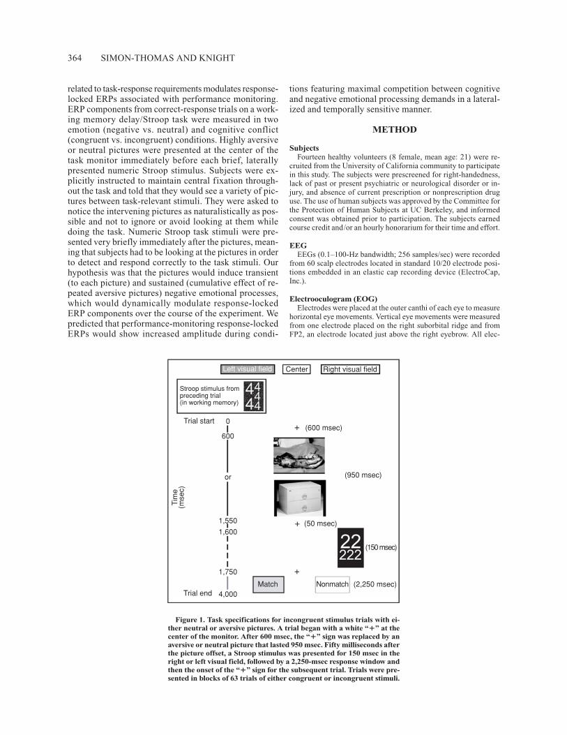
Figure 1. Task specifications for incongruent stimulus trials with ei-ther neutral or aversive pictures. A trial began with a white “�” at thecenter of the monitor. After 600 msec, the “�” sign was replaced by anaversive or neutral picture that lasted 950 msec. Fifty milliseconds afterthe picture offset, a Stroop stimulus was presented for 150 msec in theright or left visual field, followed by a 2,250-msec response window andthen the onset of the “�” sign for the subsequent trial. Trials were pre-sented in blocks of 63 trials of either congruent or incongruent stimuli.
AFFECT, COGNITION, AND PERFORMANCE MONITORING 365
trodes were referenced to linked mastoids. Skin impedances for ref-erence and ground electrodes were brought to below 5 kΩ. Otherelectrode impedances were brought to below 20 kΩ.
TaskA 1-back working memory task that used numeric Stroop stim-
uli was designed for this study. The Stroop stimuli consisted ofgroups of numbers, each with one to five repetitions of an integerfrom 1 to 5. There were 10 “congruent” Stroop stimuli, where theinteger used was the same as the number of instances—for exam-ple, 333—and 10 “incongruent” stimuli—for example, 444. Thesubjects were instructed to detect the number of integers in eachstimulus, and to indicate whether this value matched the quantity ofintegers from the immediately preceding stimulus (see Figure 1)The subjects responded by pressing a “match” or “nonmatch” but-ton on a button control panel with the index and middle fingers oftheir right hand. Between presentations of the number stimuli, onehighly aversive or neutral picture from the International AffectivePicture System (IAPS; Lang, 1999) was presented. The picturesused in this experiment were selected according to normative pleas-antness and arousal ratings that accompanied the IAPS set. Highlyaversive images had the lowest pleasantness and highest arousal rat-ings and included gruesome scenes. Neutral images had low arousalratings and included ordinary people and objects. Twenty-one im-ages of each type were used, with repetition, throughout the exper-iment. Pictures appeared in pseudorandom order (with repetitiononly after exhausting the complete set of 42 within a block and noimmediate sequential repetition of the same picture) between theStroop stimuli. Each picture was repeated no fewer than three andno more than six times during the course of a subject’s experiment.
Each task trial began with the appearance of a white “�” sign ona black background, presented at the center of a computer monitorfor 600 msec. The subjects were instructed to fixate their gaze onthis “�” sign continuously during performance of the task. A neu-tral or aversive picture replaced the fixation “�” sign at the end of600 msec and remained on the monitor for 950 msec. IAPS picturesoccupied up to 5.3º of visual angle on both sides of the visual mid-line. After the picture disappeared, the monitor showed the blackbackground for 50 msec, and then a white congruent or incongru-ent numeric Stroop stimulus was presented to either the left or theright visual field for 150 msec. Stroop stimuli occupied between5.5º and 7.5º of visual angle on either side of the visual midline.Stroop stimuli were followed by a uniform 2,250-msec responsewindow, during which the white “�” sign was visible at the centerof the monitor. At the end of each 4-sec trial, a new trial began withthe reappearance of the white “�” sign uninterrupted from the re-sponse window fixation “�” sign. To increase cognitive load, aftereach Stroop stimulus, the subjects indicated whether the number ofintegers in the current trial matched the number of integers in theimmediately preceding trial, in a continuously updating fashion.Congruent and incongruent stimuli were presented in separateblocks of 63 trials.
ProcedureUpon arrival, the subjects signed consent forms and completed a
screening form confirming their eligibility to participate in EEGrecording. If all criteria were met, they were led into a sound-attenuatedexperimental booth, seated approximately 52 in. in front of a mon-itor, and given a button control panel. The subjects were instructedto perform four blocks (two congruent and two incongruent) of atraining version of the task where the pictures were omitted fromeach trial. Training and experimental task block order were coun-terbalanced across subjects and alternated between starting withcongruent or incongruent stimulus types. The subjects were en-couraged to respond as quickly and as accurately as possible, andthe experimenter gave general performance feedback between com-pleted training blocks.
After completing the training, the subjects were prepared forEEG recording. After the recording cap was in place, the subjects’ongoing EEG signals were displayed on a monitor for them to ob-serve, while the experimenters described the basic principles ofEEG and encouraged them not to blink excessively, have tense fa-cial muscles, clench their jaw, or chew during task performance.The experimenters demonstrated how each of these activities pro-duced artifacts in each subject’s ongoing EEG signal, providing fur-ther incentive for the subjects to minimize these activities in orderto generate useful data.
Prior to starting the first experimental task block, the subjectswere given a Positive and Negative Affect Scale (PANAS; Watson,Clark, & Tellegen, 1988) with instructions to rate how accuratelyeach of the terms on the scale described the way that they were feel-ing at that moment (e.g., distressed: 1 � not at all, 5 � very much).
The subjects were also told that they would now see pictures be-tween presentations of the number stimuli while doing the task, andthat the pictures were irrelevant to the task. The subjects then per-formed four alternating congruent and incongruent blocks of thetask and completed an additional PANAS after every two blocks.
Data Analysis MethodSubjective ratings. Changes in positive and negative affect were
assessed from the subjects’ PANAS scores. Ratings for each posi-tive and negative term were summed within affect category to yieldan overall positive and negative score from each successive admin-istration of the PANAS. Scores were submitted to one-factor (time:before task, after two task blocks, after four task blocks, after sixtask blocks, after eight task blocks) within-subjects repeated mea-sures analyses of variance (ANOVAs). Greenhouse–Geisser cor-rected p values are reported for these and all subsequent analyses tocorrect for violation of the sphericity assumption.
Performance. Response times (RTs) from each subject’s correctresponses were divided into eight groups according to experimen-tal conditions: Stroop (congruent vs. incongruent), picture valence(aversive vs. neutral), and field of stimulus presentation (LVF vs.RVF), then averaged within conditions across experimental blocks.RTs were submitted to a three-factor (Stroop � valence � field)within-subjects repeated measures ANOVA. Planned follow-uppaired t tests were carried out to determine how individual factorscontributed to main effects. Performance data were also analyzedwith respect to time. RTs from early (Blocks 1 and 2) and late(Blocks 3 and 4) experimental blocks were divided by experimen-tal condition, then submitted to a four-factor (Stroop � valence �field � time) within-subjects repeated measures ANOVA.
Because the task relied on “match” and “nonmatch” responses,accuracy was assessed using d′ values to minimize the influence ofindividual response bias on group means. d′ scores were computedfrom correct hit and false positive rates within each block, then av-eraged into the same experimental condition groups as were the RTdata and submitted to the same statistical analyses.
Electrophysiology. ERPs were computed from artifact-free dataepochs extending from 600 msec prior to 400 msec after the button-press responses from trials in which the subjects responded correctly.The mean signal over a 200-msec window from 600 to 400 msec priorto the response was subtracted from each EEG epoch to eliminatenoise related to between-trial baseline amplitude shifts. A band-passfilter (1–20 Hz) was applied to grand-average ERPs prior to visualinspection and extraction of amplitude and latency features to re-duce spurious contributions from signals below and above the fre-quency band that encompasses response-locked ERP componentsrelated to conflict detection. Components with latency and scalpdistribution features similar to those of the ERN/Ne and Pe gener-ated from error trials obtained during this experiment were identi-fied as the CRN and Pe on correct trials, and features of interestwere extracted from each subject’s data for statistical analysis. Am-plitudes were represented as the average amplitude across a time win-
366 SIMON-THOMAS AND KNIGHT
dow that spanned the maximal condition-related differences in thegrand-average waveforms. Amplitudes were measured on a clusterof frontal-central electrodes over the medial prefrontal cortex (F1,Fz, F2, FCz, and Cz), and then averaged together prior to statisticalanalysis.
Response-locked EEG epochs were divided into eight groups ac-cording to experimental condition (Stroop, valence, and field) andwere then averaged within group for ERP analysis. Amplitudes ofCRN and Pe features were submitted to a three-factor (Stroop �valence � field) within-subjects repeated measures ANOVA. Plannedfollow-up paired t tests were carried out to explore how differencesbetween pairs of conditions contributed to main effects. EEG epochswere further grouped according to experimental time (early blocks,Blocks 1 and 2 and late blocks, Blocks 3 and 4) and were then av-eraged into early and late CRN and Pe components. Amplitudeswere submitted to the same three-factor analysis as above to explorehow condition effects may have changed between the early and laterparts of the experiment.
RESULTS
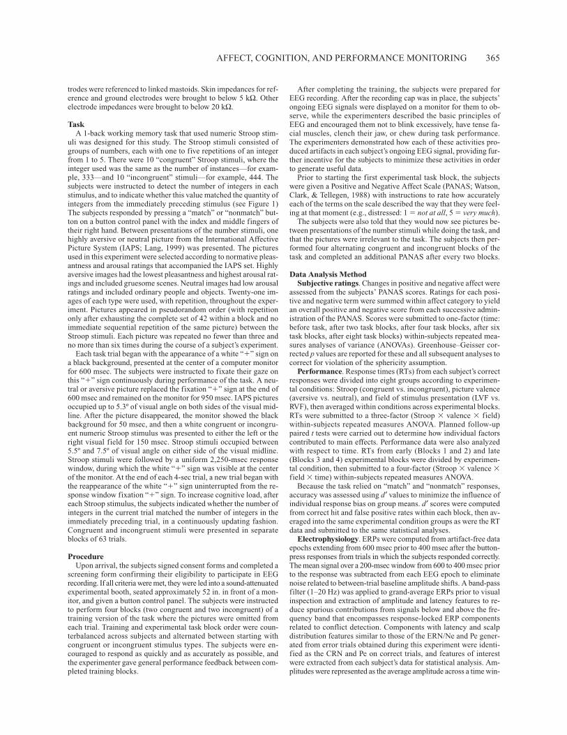
PerformanceSubjective ratings. There was a main effect of time on
positive PANAS scores [F(4,52) � 9.7, p � .001]: Sub-jective ratings of positive affect decreased throughoutthe experiment. There was also a main effect of time fornegative PANAS scores [F(4,52) � 9.4, p � .001]: Rat-ings of negative affect increased between the baseline(before starting the task) and the second block of the task( p � .005), then gradually decreased throughout the re-mainder of the experiment (see Figure 2).
RTs. RTs showed a predicted main effect of Stroop[F(1,13) � 17.6, p � .001]: Subjects’ responses werefaster to congruent stimuli (CS) than to incongruentstimuli (IS; 591 � 36 and 645 � 46, respectively). Therewere no main effects or interactions involving valence orfield on RT data. There was a main effect of time [F(1,13)� 57.5, p � .001] due to slower RTs during early taskblocks (674 � 44 and 563 � 40, respectively). There wasalso a time � field interaction [F(1,13) � 6.2, p � .05]:RTs tended to be faster for stimuli presented to the LVFthan for those presented to the RVF during early taskblocks (662 � 44 vs. 685 � 45, p � .06), whereas therewere no differences related to field in later blocks (568 �42 vs. 556 � 37, p � .3).
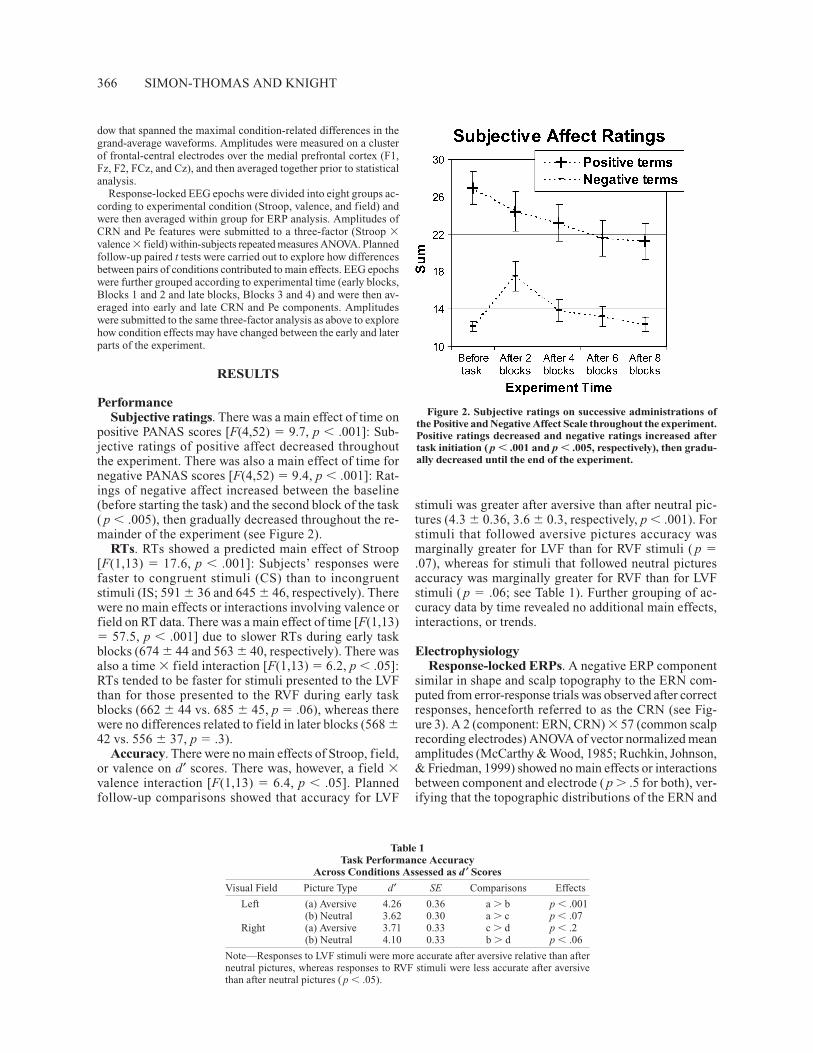
Accuracy. There were no main effects of Stroop, field,or valence on d′ scores. There was, however, a field �valence interaction [F(1,13) � 6.4, p � .05]. Plannedfollow-up comparisons showed that accuracy for LVF
stimuli was greater after aversive than after neutral pic-tures (4.3 � 0.36, 3.6 � 0.3, respectively, p � .001). Forstimuli that followed aversive pictures accuracy wasmarginally greater for LVF than for RVF stimuli ( p �.07), whereas for stimuli that followed neutral picturesaccuracy was marginally greater for RVF than for LVFstimuli ( p � .06; see Table 1). Further grouping of ac-curacy data by time revealed no additional main effects,interactions, or trends.
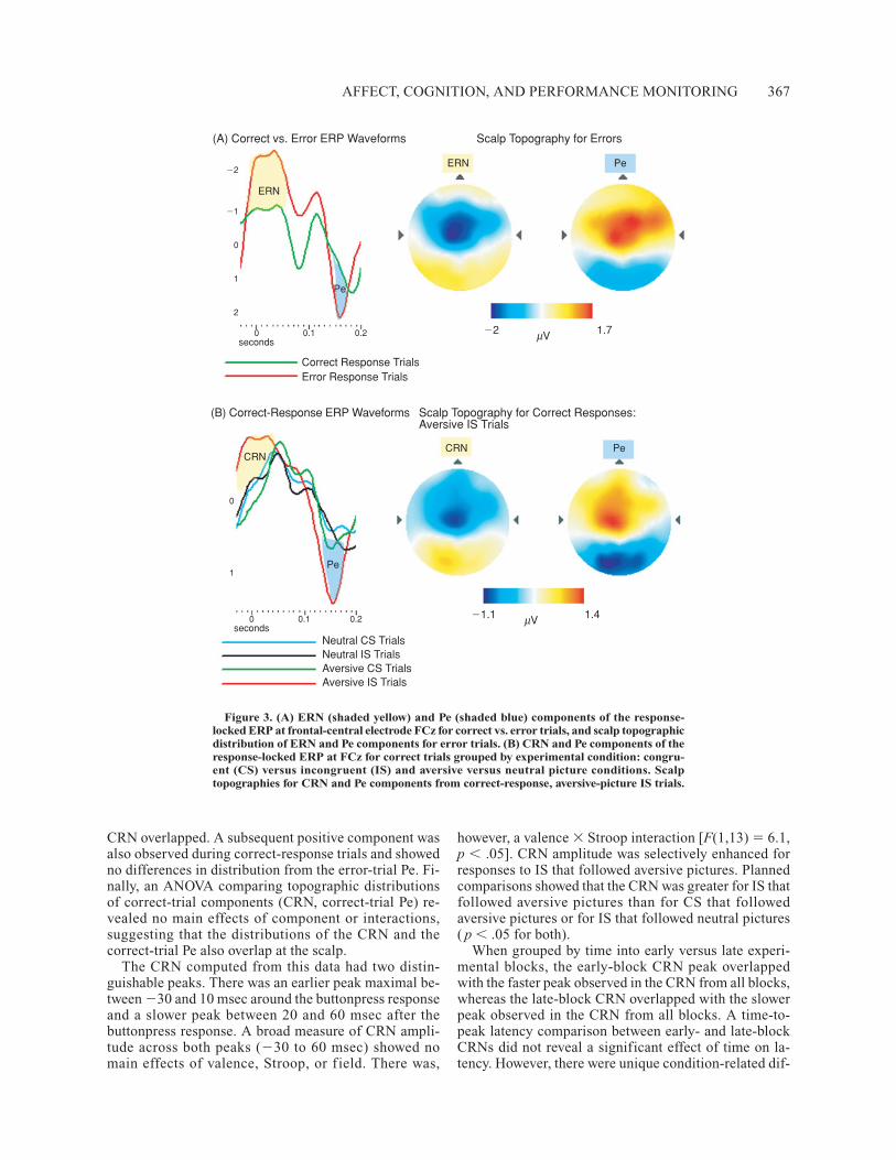
ElectrophysiologyResponse-locked ERPs. A negative ERP component
similar in shape and scalp topography to the ERN com-puted from error-response trials was observed after correctresponses, henceforth referred to as the CRN (see Fig-ure 3). A 2 (component: ERN, CRN) � 57 (common scalprecording electrodes) ANOVA of vector normalized meanamplitudes (McCarthy & Wood, 1985; Ruchkin, Johnson,& Friedman, 1999) showed no main effects or interactionsbetween component and electrode ( p � .5 for both), ver-ifying that the topographic distributions of the ERN and
Figure 2. Subjective ratings on successive administrations ofthe Positive and Negative Affect Scale throughout the experiment.Positive ratings decreased and negative ratings increased aftertask initiation ( p � .001 and p � .005, respectively), then gradu-ally decreased until the end of the experiment.
Table 1Task Performance Accuracy
Across Conditions Assessed as d ′ Scores
Visual Field Picture Type d′ SE Comparisons Effects
Left (a) Aversive 4.26 0.36 a � b p � .001(b) Neutral 3.62 0.30 a � c p � .07
Right (a) Aversive 3.71 0.33 c � d p � .2(b) Neutral 4.10 0.33 b � d p � .06
Note—Responses to LVF stimuli were more accurate after aversive relative than afterneutral pictures, whereas responses to RVF stimuli were less accurate after aversivethan after neutral pictures ( p � .05).
AFFECT, COGNITION, AND PERFORMANCE MONITORING 367
CRN overlapped. A subsequent positive component wasalso observed during correct-response trials and showedno differences in distribution from the error-trial Pe. Fi-nally, an ANOVA comparing topographic distributionsof correct-trial components (CRN, correct-trial Pe) re-vealed no main effects of component or interactions,suggesting that the distributions of the CRN and thecorrect-trial Pe also overlap at the scalp.
The CRN computed from this data had two distin-guishable peaks. There was an earlier peak maximal be-tween �30 and 10 msec around the buttonpress responseand a slower peak between 20 and 60 msec after thebuttonpress response. A broad measure of CRN ampli-tude across both peaks (�30 to 60 msec) showed nomain effects of valence, Stroop, or f ield. There was,
however, a valence � Stroop interaction [F(1,13) � 6.1,p � .05]. CRN amplitude was selectively enhanced forresponses to IS that followed aversive pictures. Plannedcomparisons showed that the CRN was greater for IS thatfollowed aversive pictures than for CS that followedaversive pictures or for IS that followed neutral pictures( p � .05 for both).
When grouped by time into early versus late experi-mental blocks, the early-block CRN peak overlappedwith the faster peak observed in the CRN from all blocks,whereas the late-block CRN overlapped with the slowerpeak observed in the CRN from all blocks. A time-to-peak latency comparison between early- and late-blockCRNs did not reveal a significant effect of time on la-tency. However, there were unique condition-related dif-
(A) Correct vs. Error ERP Waveforms Scalp Topography for Errors
(B) Correct-Response ERP Waveforms Scalp Topography for Correct Responses:Aversive IS Trials
Correct Response TrialsError Response Trials
�2
�1
0
1
2
0 0.1 0.2seconds
ERN
ERN Pe
�2 1.7μV
CRN
Pe
0
1
0 0.1 0.2seconds
CRN Pe
�1.1 1.4μV
Neutral CS TrialsNeutral IS TrialsAversive CS TrialsAversive IS Trials
Pe
Figure 3. (A) ERN (shaded yellow) and Pe (shaded blue) components of the response-locked ERP at frontal-central electrode FCz for correct vs. error trials, and scalp topographicdistribution of ERN and Pe components for error trials. (B) CRN and Pe components of theresponse-locked ERP at FCz for correct trials grouped by experimental condition: congru-ent (CS) versus incongruent (IS) and aversive versus neutral picture conditions. Scalptopographies for CRN and Pe components from correct-response, aversive-picture IS trials.
368 SIMON-THOMAS AND KNIGHT
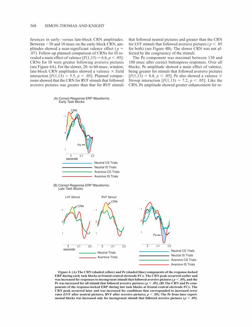
ferences in early- versus late-block CRN amplitudes. Between �30 and 10 msec on the early-block CRN, am-plitudes showed a near-significant valence effect ( p �.07). Follow-up planned comparison of CRNs for IS re-vealed a main effect of valence [F(1,13) � 6.6, p � .05]:CRNs for IS were greater following aversive pictures(see Figure 4A). For the slower, 20- to 60-msec, window,late-block CRN amplitudes showed a valence � fieldinteraction [F(1,13) � 5.5, p � .05]. Planned compar-isons showed that the CRN for RVF stimuli that followedaversive pictures was greater than that for RVF stimuli
that followed neutral pictures and greater than the CRNfor LVF stimuli that followed aversive pictures ( p � .05for both) (see Figure 4B). The slower CRN was not af-fected by the congruency of the stimuli.
The Pe component was maximal between 130 and 180 msec after correct buttonpress responses. Over allblocks, Pe amplitude showed a main effect of valence,being greater for stimuli that followed aversive pictures[F(1,13) � 8.8, p � .05]. Pe also showed a valence �Stroop interaction [F(1,13) � 7.2, p � .05]. Like theCRN, Pe amplitude showed greater enhancement for re-
(A) Correct-Response ERP Waveforms:Early Task Blocks
(B) Correct-Response ERP Waveforms:Late Task Blocks
Neutral CS Trials
Neutral IS Trials
Aversive CS Trials
Aversive IS Trials
Neutral CS Trials
Neutral IS Trials
Aversive CS Trials
Aversive IS Trials
Neutral Trials
Aversive Trials
CRN
Pe
0 0.1 0.2seconds
�1
0
1
0 0.1 0.2seconds
0 0.1 0.2 0 0.1 0.2
�1
0
1
�1
0
1
�1
0
1
CRN
CRN
Pe
LVF Stimuli RVF Stimuli
Figure 4. (A) The CRN (shaded yellow) and Pe (shaded blue) components of the response-lockedERP during early task blocks at frontal-central electrode FCz. The CRN peak occurred earlier andwas increased for responses to incongruent stimuli that followed aversive pictures ( p � .05), and thePe was increased for all stimuli that followed aversive pictures ( p � .05). (B) The CRN and Pe com-ponents of the response-locked ERP during late task blocks at frontal central electrode FCz. TheCRN peak occurred later and was increased for conditions that corresponded to increased errorrates (LVF after neutral pictures, RVF after aversive pictures; p � .05). The Pe from later experi-mental blocks was increased only for incongruent stimuli that followed aversive pictures ( p � .05).
AFFECT, COGNITION, AND PERFORMANCE MONITORING 369
sponses to IS that followed aversive pictures (see Fig-ure 4A). Planned comparisons showed that error positiv-ity values were greater for IS following aversive picturesthan for CS following aversive, or than for IS followingneutral pictures ( p � .05 for both).
Measured from early block ERPs, the Pe showed a maineffect of valence [F(1,13) � 6.7, p � .05]; its amplitudewas greater for responses to stimuli that followed aver-sive pictures. Error positivity values from later blocks,however, showed more complex main effects of valence[F(1,13) � 5.7, p � .05] and Stroop [F(1,13) � 5.6, p �.05] and a valence � Stroop interaction [F(1,13) � 7.7,p � .05]. During later experimental blocks, Pe amplitudewas increased for all stimuli that followed aversive pic-tures, all IS, and most prominently for responses to ISthat followed aversive pictures (see Figure 4B). Plannedcomparisons revealed that this effect was weighted bysignificantly increased Pe amplitude for responses to ISafter aversive pictures versus after neutral pictures andversus CS after aversive pictures ( p � .01 for both).
DISCUSSION
This study explored the influence of negative emotionon behavior and response-locked ERPs associated withperformance monitoring. EEG was acquired from sub-jects performing a working memory delay task that usednumeric Stroop stimuli interspersed with neutral andaversive pictures from the IAPS (Lang, 1999). Subjec-tive measures revealed that subjects felt more negative,and task behavior showed that they performed the taskdifferently as a result of viewing the aversive pictures.Early in the experiment, when self-ratings of negativefeeling were the highest, RTs were faster for stimuli pre-sented to the right hemisphere (LVF) than to the lefthemisphere (RVF). Throughout the experiment, responseswere more accurate for stimuli presented to the righthemisphere (LVF) and less accurate for stimuli presentedto the left hemisphere (RVF) following aversive pictures.Correct-trial response-locked ERPs revealed CRN andPe components that were dynamically sensitive to stim-ulus visual field (i.e., initial processing hemisphere), Stroopinterference, and picture valence.
Self-ratings on the PANAS showed that subjects feltemotionally affected as a consequence of viewing theaversive IAPS pictures (Codispoti, Bradley, & Lang,2001; Sutton, Davidson, Donzella, Irwin, & Dottl, 1997).After performing two blocks of the task (one congruentand one incongruent), subjects associated their currentfeeling significantly more with negative terms and lesswith positive terms. With further repetition, successivenegative affect ratings gradually decreased, suggestingthat subjects’ feelings and underlying emotional responseswere habituating to the repeated presentation of aversivepictures (Carretié, Hinojosa, & Mercado, 2003; Fein-stein, Goldin, Stein, Brown, & Paulus, 2002; Fischer et al.,2003). Ratings of positive affect continued to decrease,albeit less precipitously, throughout the experiment.
The PANAS ratings provided a useful measure of thesustained, tonic effects that viewing aversive pictureshad on subjects’ emotional states. Phasic, individual picture-related effects on subjective emotion were notexplicitly measured in this study. Several studies have re-ported similar affective responses to IAPS pictures pre-sented for brief trial intervals and for runs of longer series,suggesting that brief and sustained affective responseswere elicited with repeated picture presentation in thisstudy (Bradley, Cuthbert, & Lang, 1996; Codispoti et al.,2001).
Subjects’ RTs during incongruent stimulus blockswere slower than their RTs during congruent stimulusblocks, confirming that the incongruent condition wasmore demanding than the congruent condition. However,since congruent and incongruent stimuli were presentedseparately in blocks, the difficulty of the incongruentcondition cannot be directly attributed to traditionalStroop interference—that is, increased conflict betweenprepotent and task-instructed processing of and respond-ing to the number of stimuli (Stroop, 1935). Subjectsmay have developed a rule-based cognitive strategy forresponding during the incongruent block condition, re-sponding without necessarily choosing or switching be-tween the prepotent and task-relevant options, since thetask-relevant option was always the appropriate one withineach block. RTs were slower during earlier experimentalblocks, suggesting that subjects’ performance improvedwith task repetition. There was also an interaction betweenexperiment time and field of stimulus presentation: Dur-ing earlier task blocks, RTs to LVF (right-hemisphere)stimuli were faster than those to RVF (left-hemisphere)stimuli, whereas RTs were not significantly differentacross fields later in the experiment. Faster RTs to LVFstimuli suggests that there was a right-hemisphere ad-vantage during the early blocks of the task. This lateral-ity difference was not observed during the four practicetrials that excluded picture stimuli ( p � .58), indicatingthat the cognitive demands of the task did not afford anintrinsic right-hemisphere advantage. Therefore, the RTdifferences suggest that the early right-hemisphere RTadvantage was driven by the tonic or sustained aversiveemotional state invoked by the repeated negative pic-tures. In further support of this claim, the right-hemisphereRT advantage diminished as negative-emotion subjectiveratings decreased later in the experiment. This early right-hemisphere advantage may reflect a tonic facilitation ofright-hemisphere processing induced by subjects’ shifttoward a more negative emotional state. In agreementwith the valence hypothesis, which purports that there isa right-hemisphere dominance in processing withdrawal-related emotions (Davidson, 1998; Davidson, & Irwin,1999) including fear and disgust, the aversive picturesmay have initiated a fear/avoidance-oriented emotionalstate early in the experiment that selectively cued theright hemisphere to orient attention, detect, and respondfaster to task stimuli (Posner & Petersen, 1990; Stormark,Nordby, & Hugdahl, 1995).
370 SIMON-THOMAS AND KNIGHT
Task accuracy showed an enduring and distinct hemi-spheric influence of negative emotion on cognitive per-formance. Subjects responded more accurately to LVF(right-hemisphere) stimuli than to RVF (left-hemisphere)stimuli when they followed aversive images throughoutthe experiment. Together, the RT and accuracy data sug-gest that subjects’ emotional responses to the pictures en-hanced or primed the right hemisphere, which benefitedsubsequent stimulus processing. This agrees with sev-eral other studies that show improved right-hemispherefunction in emotion-cognition paradigms (Van Strien &Heijt, 1995; Van Strien & Luipen, 1999; Van Strien &Morpurgo, 1992).
There are, however, reports of findings that are incon-sistent with the negative-emotion right-hemisphere fa-cilitation hypothesis. Hartikainen et al. (2000) proposed,having observed slower RTs to LVF (right-hemisphere)than to RVF (left-hemisphere) targets after aversive pic-tures in their study, that processing of aversive picturesselectively distracted the right hemisphere and interferedwith cognitive task target processing. Although not re-solved, several hypotheses may explain the apparent in-consistency between these two findings. First, the effectsof emotion on cognition may be modulated by task pa-rameters such as task cognitive load or interstimulus in-tervals between emotion-eliciting and cognitive task-related stimuli. The cognitive task in the Hartikainenstudy was a very simple target-detection task, whereasthis study involved complex higher executive control.Negative- emotion elicitation may affect these two levelsof cognitive processes uniquely. Second, depending onthe time elapsed between the onset of an emotion elicitorand a cognitive stimulus, the emotional pictures can serveas cues to orient attention (longer time) or distractors thatinterfere with or impair attention shifting (shorter time)(Dolan, 2002). In the Hartikainen study, task-relatedstimuli appeared 350 msec after the onset of the emotion-eliciting pictures. In the present study, the task stimuliappeared 1 sec after the onset of the emotion elicitors.Shorter intervals between emotion elicitation and thetask-related stimuli may result in interference betweenthe emotional and cognitive processes, whereas longerintervals may allow for integration between the two (Codi-spoti et al., 2001). Related to this, Stormark et al. showedthat emotional stimuli improve attention orienting aswell as impair attention shifting, depending on the dura-tion of the emotion stimulus (Stormark, Field, Hugdahl,& Horowitz, 1997).
Valence, brain hemisphere, and Stroop interferencerelated influences on the CRN and Pe components indicatethat emotional processes and cognitive goal-orientedprocesses concurrently and dynamically modulateperformance-monitoring signals. The CRN componentincreased with incongruent stimuli preceded by aversivepictures; this condition featured the greatest degree ofcompetition between cognitive processing and negativeemotional processing. Several studies have established areliable CRN response during conditions of increased re-
sponse conflict, albeit primarily between cognitive re-sponse options, and have proposed that the CRN may begenerated by the anterior cingulate cortex (Botvinick,Braver, Barch, Carter, & Cohen, 2001; Botvinick et al.,1999; Carter et al., 2000; Fan, Flombaum, McCandliss,Thomas, & Posner, 2003; Gehring et al., 2001; Math-alon, Whitfield, & Ford, 2003; Nieuwenhuis, Yeung, vanden Wildenberg, & Ridderinkhof, 2003; Van Veen &Carter, 2002; Van Veen, Cohen, Botvinick, Stenger, &Carter, 2001). Such cognitive neuroscience-based theo-ries of anterior cingulate function characterize it as theessential structure in a cognitive conflict-monitoringsystem. Our findings suggest that the conflict-relatedsignals of the anterior cingulate are mediated by fluctu-ations in both cognitive and affective demand and salience.This conclusion is parsimonious with the anatomicalconnectivity and positioning of the anterior cingulatewith respect to putative higher cognitive and emotionprocessing regions of the cerebral cortex (Bush, Luu, &Posner, 2000; Koski & Paus, 2000).
Detailed analysis of the CRN generated in this studyrevealed an earlier and a later subcomponent, differen-tially sensitive to conflict, affect, and error likelihood.The earlier latency subcomponent of the CRN was in-creased in the initial blocks of the experiment (whennegative emotional ratings were highest) for responsesto stimuli that followed aversive pictures and to a greaterextent if they were incongruent. Several researchers havedescribed response and task feedback related ERP com-ponents that increase with negative affect associatedwith error commission or the detection of loss (Falken-stein et al., 2000; Gehring et al., 2002; Holroyd, Coles,& Nieuwenhuis, 2002; Holroyd, Nieuwenhuis, Yeung, &Cohen, 2003; Luu, Flaisch, & Tucker, 2000; Luu, Tucker,& Collins, 2000; Luu et al., 2003; Stemmer et al., 2001).The later latency CRN subcomponent was selectively in-creased for RVF stimuli following aversive pictures dur-ing later experimental trials, when negative emotionalreactions to the repeated aversive pictures were attenu-ated. Behaviorally, error rates were highest for RVFstimuli that followed aversive pictures. The increasedCRN for RVF stimuli after aversive pictures may revealsome incidental conflict between left-hemisphere initi-ated task processing and concurrent sustained negativeemotional processing, or the emergence of heightenedconflict or ERP signal in the condition that had beenshowing the greatest frequency of errors.
The dynamic sensitivity of the CRN described heresuggests that it confers dynamic contextual salience withcognitive and emotional input. In earlier experimentalblocks, the influence of the disturbing pictures was moresalient, whereas in later experimental blocks, cognitivedemand related to task performance goals was more in-fluential. Across the whole experiment, the CRN showeda more complex interaction between conditions that prob-ably reflected summated responses to temporally fluctu-ating processing of negative emotional and cognitive demands.
AFFECT, COGNITION, AND PERFORMANCE MONITORING 371
The Pe, a less studied electrophysiological index of erroror conflict detection, showed a systematic, temporally con-strained, and reliable pattern of sensitivity to the fluctuat-ing combination of cognitive demand and negative affect.Over all blocks, the Pe, like the CRN, showed increased am-plitude for responses to incongruent (increased-demand/conflict) stimuli that followed aversive pictures. However,the Pe from earlier task blocks showed a general increasefor responses to stimuli that followed aversive pictures. Inlater blocks, the Pe showed a more selective increase withincongruent stimuli that followed aversive pictures, echo-ing the shift from affective-salience-driven modulation tocognitive-demand/conflict-driven modulation exhibitedin the CRN.
Taken together, the results support the notion that theCRN and Pe represent online detection of the need forreallocation of neural resources in the interest of (1) re-sponding to circumstances that elicit negative emotionalstates and (2) solving a cognitive or goal-oriented prob-lem that entails greater demand.
The hemispheric effects of aversive pictures on per-formance measures interacted with response-locked ERPmodulation only in some conditions (the slower CRNsubcomponent). Stroop stimulus-locked ERP components,however, showed comparable patterns of negative affectbased hemispheric influence on components related toattention and early cognitive processing (Simon-Thomaset al., 2005). There are several reasons why effects ofstimulus presentation side would be less prominent in thebuttonpress response-locked ERPs examined in this study.First, in the time lapsed between the appearance of theStroop stimulus and the executed response, hemispheresthat may have initially had different levels of involve-ment in visual feature analysis are likely to have trans-ferred and integrated information back and forth. Sec-ond, subjects’ buttonpress responses were always carriedout by fingers on the right hand, meaning that response-related processing had to be integrated into the left motorcortex for all responses.
In summary, the data show that the CRN and Pe com-ponents of response-locked ERPs interact dynamicallywith increased conflict related to cognitive demand andnegative affect, even when responses are correct.
REFERENCES
Botvinick, M. M., Braver, T. S., Barch, D. M., Carter C. S., &Cohen, J. D. (2001). Conflict monitoring and cognitive control. Psy-chological Review, 108, 624-652.
Botvinick, M. M., Nystrom, L. E., Fissell, K., Carter, C. S., & Cohen,J. D. (1999). Conflict monitoring versus selection-for-action in anteriorcingulate cortex. Nature, 402, 179-181.
Bradley, M. M., Cuthbert, B. N., & Lang, P. J. (1996). Picture mediaand emotion: Effects of a sustained affective context. Psychophysiol-ogy, 33, 662-670.
Brazdil, M., Roman, R., Falkenstein, M., Daniel, P., Jurak, P., &Rektor, I. (2002). Error processing—evidence from intracerebralERP recordings. Experimental Brain Research, 146, 460-466.
Bush, G., Luu, P., & Posner, M. I. (2000). Cognitive and emotional in-fluences in anterior cingulate cortex. Trends in Cognitive Sciences, 4,215-222.
Carretié, L., Hinojosa, J. A., & Mercado, F. (2003). Cerebral pat-terns of attentional habituation to emotional visual stimuli. Psy-chophysiology, 40, 381-388.
Carter, C. S., Braver, T. S., Barch, D. M., Botvinick, M. M., Noll, D.,& Cohen, J. D. (1998). Anterior cingulate cortex, error detection, andthe online monitoring of performance. Science, 280, 747-749.
Carter, C. S., Macdonald, A. M., Botvinick, M. M., Ross, L. L.,Stenger, V. A., Noll, D., & Cohen, J. D. (2000). Parsing executiveprocesses: Strategic vs. evaluative functions of the anterior cingulatecortex. Proceedings of the National Academy of Sciences, 97, 1944-1948.
Codispoti, M., Bradley, M. M., & Lang, P. J. (2001). Affective reac-tions to briefly presented pictures. Psychophysiology, 38, 474-478.
Davidson, R. J. (1998). Anterior electrophysiological asymmetries,emotion, and depression: Conceptual and methodological conun-drums. Psychophysiology, 35, 607-614.
Davidson, R. J., & Irwin, W. (1999). The functional neuroanatomy ofemotion and affective style. Trends in Cognitive Neurosciences, 3, 11-21.
Derryberry, D. (1990). Right hemisphere sensitivity to feedback.Neuropsychologia, 28, 1261-1271.
Devinsky, O., Morrell, M. J., & Vogt, B. A. (1995). Contributions ofanterior cingulate cortex to behaviour. Brain, 118, 279-306.
Dolan, R. J. (2002). Emotion, cognition, and behavior. Science, 298,1191-1194.
Drevets, W. C. (1998). Functional neuroimaging studies of depression:The anatomy of melancholia. Annual Review of Medicine, 49, 349-361.
Drevets, W. C., Ongür, D., & Price, J. L. (1998). Neuroimaging ab-normalities in the subgenual prefrontal cortex: Implications for thepathophysiology of familial mood disorders. Molecular Psychiatry,3, 220-6, 190-1.
Falkenstein, M., Hoormann, J., Christ S., & Hohnsbein, J. (2000).ERP components on reaction errors and their functional significance:A tutorial. Biological Psychology, 51, 87-107.
Fan, J., Flombaum, J. I., McCandliss, B. D., Thomas, K. M., & Pos-ner M. I. (2003). Cognitive and brain consequences of conflict.NeuroImage, 18, 42-57.
Feinstein, J. S., Goldin, P. R., Stein, M. B., Brown, G. G., & Paulus, M.P. (2002). Habituation of attentional networks during emotion pro-cessing. NeuroReport, 13, 1255-1258.
Fischer, H., Wright, C. I., Whalen, P. J., McInerney, S. C., Shin,L. M., & Rauch, S. L. (2003). Brain habituation during repeated ex-posure to fearful and neutral faces: A functional MRI study. BrainResearch Bulletin, 59, 387-392.
Gehring, W. J., & Fencsik, D. E. (2001). Functions of the medialfrontal cortex in the processing of conflict and errors. Journal of Neu-roscience, 21, 9430-9437.
Gehring, W. J., & Willoughby, A. R. (2002). The medial frontal cor-tex and the rapid processing of monetary gains and losses. Science,295, 2279-2282.
Hajcak, G., McDonald, N., & Simons, R. F. (2003). To err is auto-nomic: Error-related brain potentials, ANS activity, and post-errorcompensatory behavior. Psychophysiology, 40, 895-903.
Hartikainen, K. M., Ogawa, K. H., & Knight, R. T. (2000). Tran-sient interference of right hemispheric function due to automaticemotional processing. Neuropsychologia, 38, 1576-1580.
Heller, W. & Nitschke, J. B. (1997). Regional brain activity in emo-tion: A framework for understanding cognition in depression. Cog-nition & Emotion, 11, 637-661.
Holroyd, C. B., Coles, M. G., & Nieuwenhuis, S. (2002). Medialprefrontal cortex and error potentials. Science, 296, 1610-1161.
Holroyd, C. B., Nieuwenhuis, S., Yeung, N., & Cohen, J. D. (2003).Errors in reward prediction are reflected in the event-related brainpotential. NeuroReport, 14, 2481-2484.
Iacoboni, M., Rayman, J., & Zaidel, E. (1997). Does the previous trialaffect lateralized lexical decision? Neuropsychologia, 35, 81-88.
Kaplan, J. T., & Zaidel, E. (2001). Error monitoring in the hemi-spheres: The effect of lateralized feedback on lexical decision. Cog-nition, 82, 157-178.
372 SIMON-THOMAS AND KNIGHT
Kiehl, K. A., Liddle, P. F., & Hopfinger, J. B. (2000). Error process-ing and the rostral anterior cingulate: An event-related fMRI study.Psychophysiology, 37, 216-223.
Koski, L., & Paus, T. (2000). Functional connectivity of the anteriorcingulate cortex within the human frontal lobe: A brain-mappingmeta-analysis. Experimental Brain Research, 133, 55-65.
Lang, P. J. (1999). The International Affective Picture System. Gainesville,FL: NIMH Center for the Study of Emotion and Attention, Universityof Florida.
Luu, P., Flaisch, T., & Tucker, D. M. (2000). Medial frontal cortex inaction monitoring. Journal of Neuroscience, 20, 464-469.
Luu, P., Tucker, D. M., & Collins, P. (2000). Mood, personality, andself-monitoring: Negative affect and emotionality in relation to frontallobe mechanisms of error monitoring. Journal of Experimental Psy-chology, 129, 43-60.
Luu, P., Tucker, D. M., Derryberry, D., Reed, M., & Poulsen, C.(2003). Electrophysiological responses to errors and feedback in theprocess of action regulation. Psychological Science, 14, 47-53.
Mathalon, D. H., Whitfield, S. L., & Ford, J. M. (2003). Anatomyof an error: ERP and fMRI. Biological Psychology, 64, 119-141.
McCarthy, G., & Wood, C. C. (1985). Scalp distributions of event-related potentials: An aid in the topographic analysis of brain elec-trical activity. Electroencephalography and Clinical Neurophysiol-ogy, 62, 203-208.
Müller, M. M., Keil, A., Gruber, T., & Elbert, T. (1999). Process-ing of affective pictures modulates right-hemispheric gamma bandEEG activity. Clinical Neurophysiology, 110, 1913-1920.
Nieuwenhuis, S., Ridderinkhof, K. R., Blom, J., Band, G. P., & Kok, A.(2001). Error-related brain potentials are differentially related toawareness of response errors: Evidence from an antisaccade task.Psychophysiology, 38, 752-760.
Nieuwenhuis, S., Yeung, N., Holroyd, C. B., Schurger, A., &Cohen, J. D. (2004). Sensitivity of electrophysiological activity frommedial frontal cortex to utilitarian and performance feedback. Cere-bral Cortex, 14, 741-747.
Nieuwenhuis, S., Yeung, N., van den Wildenberg, W., & Rid-derinkhof, K. R. (2003). Electrophysiological correlates of anteriorcingulate function in a go/no-go task: Effects of response conflict andtrial type frequency. Cognitive, Affective, & Behavioral Neuroscience,3, 17-26.
Pailing, P. E., & Segalowitz, S. J. (2004). The error-related negativ-ity as a state and trait measure: Motivation, personality, and ERPs inresponse to errors. Psychophysiology, 41, 84-95.
Pailing, P. E., Segalowitz, S. J., Dywan, J., & Davies, P. L. (2002).Error negativity and response control. Psychophysiology, 39, 198-206.
Phan, K. L., Liberzon, I., Welsh, R. C., Britton, J. C., & Taylor, S. F.(2003). Habituation of rostral anterior cingulate cortex to repeatedemotionally salient pictures. Neuropsychopharmacology, 28, 1344-1350.
Phan, K. L., Wager, T. D., Taylor, S. F., & Liberzon, I. (2002). Func-tional neuroanatomy of emotion: A meta-analysis of emotion activa-tion studies in PET and fMRI. NeuroImage, 16, 331-348.
Pizzagalli, D., Regard, M., & Lehmann, D. (1999). Rapid emotionalface processing in the human right and left brain hemispheres: AnERP study. NeuroReport, 10, 2691-2698.
Posner, M. I., & Petersen, S. E. (1990). The attention system of thehuman brain. Annual Review of Neuroscience, 13, 25-42.
Rabbitt, P. M. A., & Vyas, S. M. (1970). An elementary preliminarytaxonomy for some errors in laboratory choice RT tasks. Acta Psy-chologica, 33, 56-76.
Ruchkin, D. S., Johnson, R., Jr., & Friedman, D. (1999). Scaling isnecessary when making comparisons between shapes of event-relatedpotential topographies: A reply to Haig et al. Psychophysiology, 36,832-834.
Ruchsow, M., Grothe, J., Spitzer, M., & Kiefer, M. (2002). Humananterior cingulate cortex is activated by negative feedback: Evidencefrom event-related potentials in a guessing task. Neuroscience Let-ters, 325, 203-206.
Sanfey, A. G., Rilling, J. K., Aronson, J. A., Nystrom, L. E., &Cohen, J. D. (2003). The neural basis of economic decision-makingin the Ultimatum Game. Science, 300, 1755-1758.
Scheffers, M. K., & Coles, M. G. (2000). Performance monitoring ina confusing world: Error-related brain activity, judgments of responseaccuracy, and types of errors. Journal of Experimental Psychology:Human Perception Performance, 26, 141-151.
Schutter, D. J. L. G., van Honk, J., D’Alfonso, A. A. L., Postma, A.,& de Haan, E. H. F. (2001). Effects of slow rTMS at the right dor-solateral prefrontal cortex on EEG asymmetry and mood. Neuro-Report, 12, 445-452.
Schwartz, G. E., Davidson, R. J., & Maer, F. (1975). Right hemi-sphere lateralization for emotion in the human brain: Interactionswith cognition. Science, 190, 286-288.
Sewards, T. V., & Sewards, M. A. (2002). The medial pain system:Neural representations of the motivational aspect of pain. Brain Re-search Bulletin, 59, 163-180.
Simon-Thomas, E. R., Role, K. O., & Knight, R. T. (2005). Behav-ioral and electrophysiological evidence of a right hemisphere bias forthe influence of negative emotion on higher cognition. Journal ofCognitive Neuroscience, 17, 518-529.
Stemmer, B., Witzke, W., & Schönle, P. W. (2001). Losing the errorrelated negativity in the EEG of human subjects: An indicator forwilled action. Neuroscience Letters, 308, 60-62.
Stormark, K. M., Field, N. P., Hugdahl, K., & Horowitz, M. (1997).Selective processing of visual alcohol cues in abstinent alcoholics:An approach-avoidance conflict? Addictive Behaviors, 22, 509-519.
Stormark, K. M., Nordby, H., & Hugdahl, K. (1995). Attentionalshifts to emotionally charged cues: Behavioral and ERP data. Cogni-tion & Emotion, 9, 507-523.
Stroop, J. R. (1935). Studies of interference in serial verbal reactions.Journal of Experimental Psychology, 18, 643-662.
Sutton, S. K., Davidson, R. J., Donzella, B., Irwin, W., & Dottl, D.(1997). Manipulating affective state using extended picture presen-tations. Psychophysiology, 34, 217-226.
Taylor, S. F., Phan, K. L., Decker, L. R., & Liberzon, I. (2003). Sub-jective rating of emotionally salient stimuli modulates neural activ-ity. NeuroImage, 18, 650-659.
Tucker, D. M., Hartry-Speiser, A., McDougal, L., Luu, P., & de-Grandpre, D. (1999). Mood and spatial memory: Emotion and righthemisphere contribution to spatial cognition. Biological Psychology,50, 103-125.
Van Strien, J. W., & Heijt, R. (1995). Altered visual field asymme-tries for letter naming and letter matching as a result of concurrentpresentation of threatening and nonthreatening words. Brain & Cog-nition, 29, 187-203.
Van Strien, J. W., & Luipen, M. W. (1999). Hemispheric facilitationas a result of threatening and nonthreatening words: Blocked vs.mixed presentation effects and order effects. Neuropsychologia, 37,617-621.
Van Strien, J. W., & Morpurgo, M. (1992). Opposite hemispheric ac-tivations as a result of emotionally threatening and non-threateningwords. Neuropsychologia, 30, 845-848.
Van Veen, V., & Carter, C. S. (2002). The anterior cingulate as a con-flict monitor: fMRI and ERP studies. Physiology & Behavior, 77,477-482.
Van Veen, V., Cohen, J. D., Botvinick, M. M., Stenger, V. A., &Carter, C. S. (2001). Anterior cingulate cortex, conflict monitoring,and levels of processing. NeuroImage, 14, 1302-1308.
Vidal, F., Hasbroucq, T., Grapperon, J., & Bonnet, M. (2000). Is the“error negativity” specific to errors? Biological Psychology, 51, 109-128.
Watson, D., Clark, L. A., & Tellegen, A. (1988). Development and val-idation of brief measures of positive and negative affect: The PANASscales. Journal of Personality & Social Psychology, 54, 1063-1070.
(Manuscript received August 1, 2004; revision accepted for publication December 23, 2004.)