Embed Size (px)
Citation preview
REVIEW
Affinity and enzyme-based biosensors: recent advancesand emerging applications in cell analysisand point-of-care testing
Ying Liu & Zimple Matharu & Michael C. Howland &
Alexander Revzin & Aleksandr L. Simonian
Received: 2 March 2012 /Revised: 17 May 2012 /Accepted: 24 May 2012# Springer-Verlag 2012
Abstract The applications of biosensors range from envi-ronmental testing and biowarfare agent detection to clinicaltesting and cell analysis. In recent years, biosensors havebecome increasingly prevalent in clinical testing and point-of-care testing. This is driven in part by the desire to de-crease the cost of health care, to shift some of the analyticaltests from centralized facilities to “frontline” physicians andnurses, and to obtain more precise information more quicklyabout the health status of a patient. This article gives anoverview of recent advances in the field of biosensors,focusing on biosensors based on enzymes, aptamers, anti-bodies, and phages. In addition, this article attempts todescribe efforts to apply these biosensors to clinical testingand cell analysis.
Keywords Biosensor . Enzyme . Aptamers . Phage .
Point of care
Introduction
As the number of hazardous materials and pathogens in ourenvironment and food increases, the development of highly
selective and sensitive monitoring devices is ever moreimportant. Despite significant progress, detection instru-ments are bulky, and require qualified personnel and largeamounts of reagents. In this context, the field of biosensingstrives to develop devices that are sensitive, specific, and yetminiature and simple to operate.
According to an IUPAC definition, a biosensor consistsof biorecognition elements specific to the analyte of interestand a physiochemical transducer to relay the resultant signalfrom this biorecognition event [1]. Signal transduction strat-egies may be electrochemical [2–4], optical [5, 6], gravi-metric [7, 8], or thermal [9]. Recognition elements may bereadily integrated with electronics to create fast, accurate,and inexpensive sensing devices [10, 11]. Novel directionsin biosensors often involve the design of improved biore-cognition elements or the development of novel materialsfor improving signal transduction. Examples of such mate-rials include carbon nanotubes (CNTs) and graphene inelectrochemical systems and nanoplasmonic structures inoptical systems.
Biosensors may be broadly classified on the basis of (1) thetype of biorecognition elements (i.e., enzyme-, antibody-,DNA-, or RNA-based) and (2) the signal transduction method(optical, electrochemical, gravimetric, etc.) used for detection.Biosensors based on binding of specific analytes (e.g., anti-gens binding to antibodies) are an example of affinity biosen-sors. The best known example of an affinity biosensor is theenzyme-linked immunosorbent assay (ELISA). The affinitybiosensors are designed to maximize association and mini-mize dissociation of target analytes; thus, these sensors be-come saturated and may not provide dynamic (kinetic)information about fluctuations in the level of the analyte overtime. Enzyme-based biosensors on the other hand are basedon the enzymatic turnover and may be used to record changesin analyte concentration over time. However, as discussed in
Yin Liu and Zimple Matharu contributed equally to this work.
Y. Liu : Z. Matharu :M. C. Howland :A. Revzin (*)Department of Biomedical Engineering, University of California,Davis, CA 95616, USAe-mail: [email protected]
A. L. Simonian (*)Department of Materials Engineering, Auburn University,Auburn, AL 36849, USAe-mail: [email protected]
Anal Bioanal ChemDOI 10.1007/s00216-012-6149-6
this review, the emergence of new biorecognition elementssuch as aptamers is helping to blur the line between enzymaticand affinity biosensors. This review discusses several topicsrelated to affinity and enzyme-based biosensors, includingnovel biorecognition elements, biosensor miniaturization,and novel biofunctional interfaces for biosensors. Because ofthe breadth of this research topic and the large number ofpublished articles, this review will not be able to give duecredit to all the excellent work done in this field.
Biorecognition elements for biosensors
Molecular recognition is a central event for most biosensors.Several articles [12–14] have given an overview of thevarious recognition approaches. The past few years haveseen continued emphasis on enzyme and antibody-basedbiosensors and also the emergence of new recognitionapproaches, most notably those employing aptamers andphages. Presently, research into enzyme-based biosensingsystems is being pursued in the areas of (1) enzyme immo-bilization, (2) integration of enzymes with specific nano-structures such as carbon nanotubes (CNTs) and graphene toenhance electron transfer capabilities, and (3) enzyme engi-neering to improve selectivity and immobilization. For af-finity biosensing, some of the novel approaches are focusedon (1) aptamer-based biosensors, where the most activity isin designing new aptamer structures and conjugation ofaptamers with different physiochemical transducers, and(2) phages as a specific biorecognition structure thatincreases the specificity and improves the stability of thebiosensor.
Enzyme-based biosensors
One of the challenges in making robust enzyme-based bio-sensors is ensuring that immobilized enzyme moleculesremain functional over time. This section focuses on en-zyme stabilization and immobilization strategies employing(1) CNTs, (2) sol-gel/hydrogel incorporation, and (3) immo-bilization of apoenzymes by cofactor coupling. We focus onglucose biosensors as these are most studied enzyme-basedbiosensors. However, we also highlight research making useof other enzymes, such as organophosphorus hydrolase(OPH) and horseradish peroxidase (HRP).
Carbon nanotubes
CNTs possess electrical conductivity and mechanicalstrength making them well suited to serve as a scaffold forenzyme immobilization and for relaying electrons to the
electrode [15–18]. However, CNTs tend to aggregate intotangled networks when deposited on the surface. Utilizationof CNTs in biosensing applications depends strongly on theability to prevent bundling and disperse the CNTs homoge-neously throughout the matrix without destroying theirstructural integrity [19, 20]. Dispersion of CNTs was dem-onstrated via association of biopolymers with CNT surfaces[21, 22].
A new approach was demonstrated by Yan et al. [23]using “dissolved” CNTs in a mixed solution of cyclodextrinand cyclodextrin prepolymer that works as a modifier tofabricate chemically tailored electrodes. An insoluble, con-ducting composite film of polycyclodextrin and CNTs wassynthesized, and glucose oxidase (GOx) was immobilizedon the film to fabricate an amperometric biosensor. It wasshown that the composite biocompatible matrix of polycy-clodextrin and CNTs maintains the bioactivity of the immo-bilized enzyme for 2 weeks [23]. The biosensor had adetection limit of 3.5 μM, with a linear range from 0.004to 3.23 mM. In another effort, aimed at enhancing theamount of adsorbed enzyme, Nejadnik et al. [24] exploredthe use of CNT scaffolds for enzyme immobilization. It washypothesized that the three-dimensional scaffolds can signif-icantly increase the amount of enzyme adsorbed per unit areawhile preserving the catalytic activity of the adsorbed mole-cules. Nejadnik et al. found that improved sensitivity can beachieved by tailoring the thickness of scaffolds. The 65.5 nmscaffolds resulted in a sensitivity improvement of 390 % overthe 44.5 nm scaffolds. Pedrosa et al. [25] demonstrated thatsingle-walled CNTs used with a covalent immobilizationstrategy are very sensitive sensors with excellent long-termstability. OPH-functionalized single-walled and multiwalledCNT conjugates were exploited for direct amperometric de-tection of paraoxon, a model organophosphate. Single-walledCNTs onto which OPH had been covalently immobilizedshowed much higher activity than OPH-conjugated multi-walled CNTs. The detection limit was 0.01 μmol L−1.
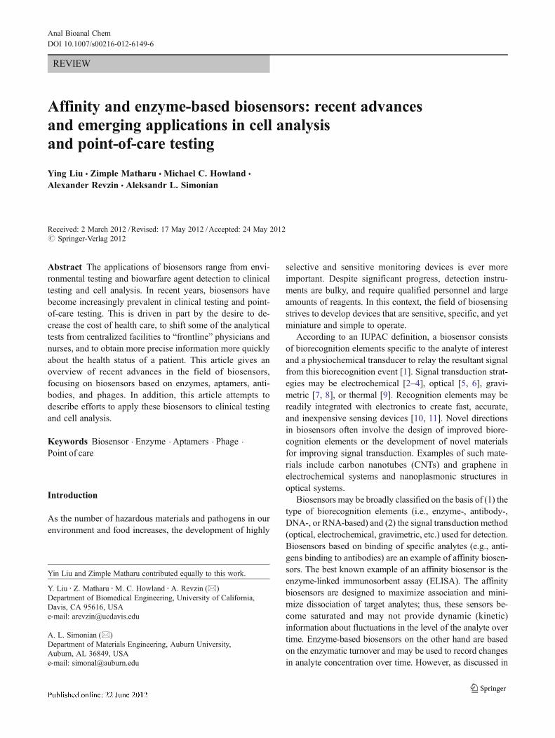
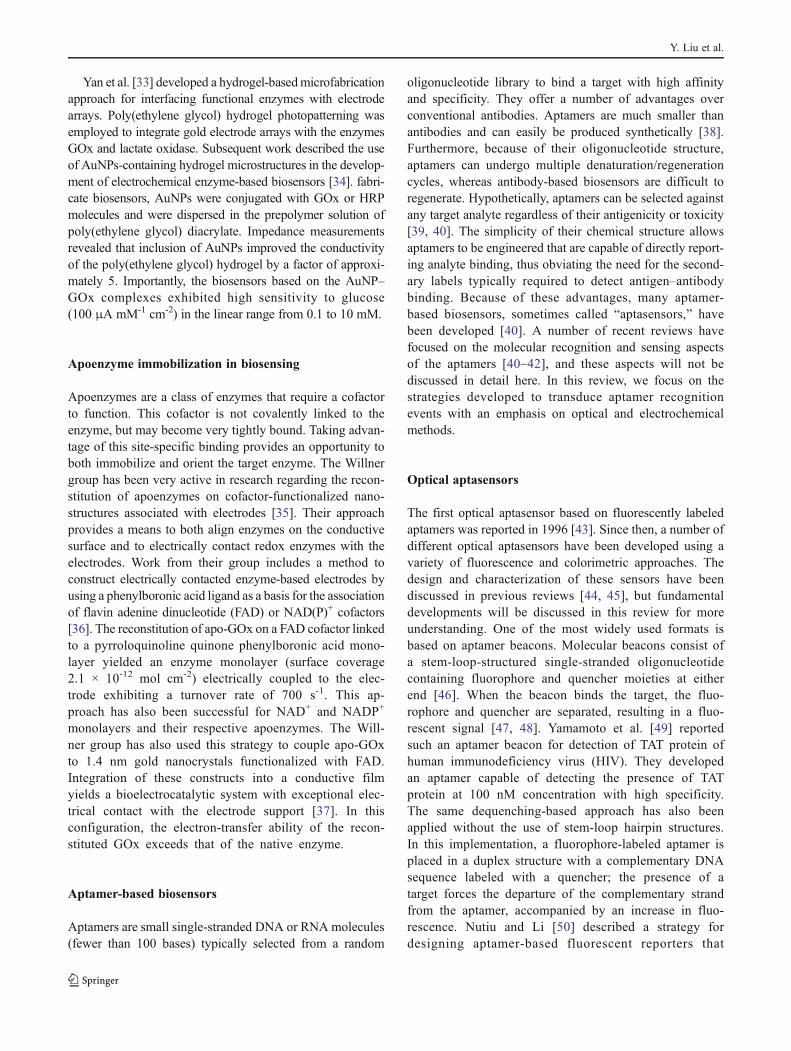
Utilizing layer-by-layer (LBL) assembly, Mantha et al.[11] demonstrated that a sandwich-like LBL structure pro-vides a suitable microenvironment to retain the molecularactivity of incorporated biopolymers. They used the electro-static interaction of anionic/cationic biomolecular layersstructured with multiwalled CNTs to build up the hybridcatalytic interfaces (Fig. 1). The electrodes generated by thisapproach were easy to fabricate and had excellent sensitivityand excellent electrochemical response.
Recent research efforts have shown graphene, a structuralelement of CNTs, to be an excellent matrix for biomoleculeimmobilization. The biocompatibility and excellent electro-chemical properties of this material may prove beneficial indeveloping electrochemical enzyme-based biosensors. In arecent study, Liu et al. [26] demonstrated a highly efficientenzyme-based electrode using covalent attachment of
Y. Liu et al.
carboxy-functionalized graphene oxide sheets and GOx.The resulting biosensor exhibited a broad linear range upto 28 mM per square millimeter of glucose with a sensitivityof 8.045 mA cm-2 M-1. Kang et al. [27] observed directelectron transfer in GOx immobilized on graphene–chitosannanocomposite films. The immobilized enzyme exhibited asurface-confined, reversible two-proton and two-electrontransfer reaction with an electron transfer rate constant of2.83 s−1. This nanocomposite sensor had glucose sensitivityof 37.93 μA mM−1 cm−2 and a detection limit of 0.02 mM.
Sol-gels and hydrogels for enzyme immobilization
Sol-gel/hydrogel chemistry has been used extensively toprepare metal oxide, silica, and organosiloxane materialsfor sensors, coatings, and catalysts [28]. Sol-gel technologymay be used to prepare a three-dimensional network for the
encapsulation of biomolecules [29] and for the creation ofbio-inorganic conductive matrices for immobilization ofenzymes. In this context, Ivnitski et al. [30] showed directelectron transfer by entrapping GOx in a silica/CNT nano-composite prepared on a screen-printed carbon electrode.Ramanathan et al. [31] described a rapid method for enzymeimmobilization directly on a waveguide surface by encap-sulation in a silica matrix. OPH, an enzyme that catalyticallyhydrolyzes organophosphates, was used as a model to dem-onstrate the utility of lysozyme-mediated silica formationfor enzyme stabilization. Silica-encapsulated OPH retainedits catalytic activity for nearly 60 days, with a detection limitfor paraoxon, a common organophosphate, of 35 μM. Jia etal. [32] have fabricated gold nanoparticles (AuNPs) contain-ing a sol-gel network for HRP immobilization. The resultingbiosensor exhibited a fast amperometric response to H2O2,
with a detection limit of 2.0 μmol L-1, and the linear rangewas 5.0 μmol L-1 to 10.0 mmol L-1.
MWNT-PEINegatively Charged slide
MW
NT-
DN
A
LBL assembly
LBL assembly
MWNT-OPH
MWNT-DNA
MWNT-PEI
(bilayers)
(n layers)
Fig. 1 LBL interface design. The initial layers of poly(ethylene imine)-functionalized multiwalled carbon nanotubes (MWNT-PEI) and DNA-functionalized multiwalled carbon nanotubes (MWNT-DNA) (fourbilayers as shown in the scanning electron microscope image) provide
support for subsequent layers of multiwalled carbon nanotubes func-tionalized with organophosphorus hydrolase (MWNT-OPH) andMWNT-DNA (nine bilayers are shown in the scanning electron micro-scope image). (From [11])
Affinity and enzyme-based biosensors
Yan et al. [33] developed a hydrogel-basedmicrofabricationapproach for interfacing functional enzymes with electrodearrays. Poly(ethylene glycol) hydrogel photopatterning wasemployed to integrate gold electrode arrays with the enzymesGOx and lactate oxidase. Subsequent work described the useof AuNPs-containing hydrogel microstructures in the develop-ment of electrochemical enzyme-based biosensors [34]. fabri-cate biosensors, AuNPs were conjugated with GOx or HRPmolecules and were dispersed in the prepolymer solution ofpoly(ethylene glycol) diacrylate. Impedance measurementsrevealed that inclusion of AuNPs improved the conductivityof the poly(ethylene glycol) hydrogel by a factor of approxi-mately 5. Importantly, the biosensors based on the AuNP–GOx complexes exhibited high sensitivity to glucose(100 μA mM-1 cm-2) in the linear range from 0.1 to 10 mM.
Apoenzyme immobilization in biosensing
Apoenzymes are a class of enzymes that require a cofactorto function. This cofactor is not covalently linked to theenzyme, but may become very tightly bound. Taking advan-tage of this site-specific binding provides an opportunity toboth immobilize and orient the target enzyme. The Willnergroup has been very active in research regarding the recon-stitution of apoenzymes on cofactor-functionalized nano-structures associated with electrodes [35]. Their approachprovides a means to both align enzymes on the conductivesurface and to electrically contact redox enzymes with theelectrodes. Work from their group includes a method toconstruct electrically contacted enzyme-based electrodes byusing a phenylboronic acid ligand as a basis for the associationof flavin adenine dinucleotide (FAD) or NAD(P)+ cofactors[36]. The reconstitution of apo-GOx on a FAD cofactor linkedto a pyrroloquinoline quinone phenylboronic acid mono-layer yielded an enzyme monolayer (surface coverage2.1 × 10-12 mol cm-2) electrically coupled to the elec-trode exhibiting a turnover rate of 700 s-1. This ap-proach has also been successful for NAD+ and NADP+
monolayers and their respective apoenzymes. The Will-ner group has also used this strategy to couple apo-GOxto 1.4 nm gold nanocrystals functionalized with FAD.Integration of these constructs into a conductive filmyields a bioelectrocatalytic system with exceptional elec-trical contact with the electrode support [37]. In thisconfiguration, the electron-transfer ability of the recon-stituted GOx exceeds that of the native enzyme.
Aptamer-based biosensors
Aptamers are small single-stranded DNA or RNA molecules(fewer than 100 bases) typically selected from a random
oligonucleotide library to bind a target with high affinityand specificity. They offer a number of advantages overconventional antibodies. Aptamers are much smaller thanantibodies and can easily be produced synthetically [38].Furthermore, because of their oligonucleotide structure,aptamers can undergo multiple denaturation/regenerationcycles, whereas antibody-based biosensors are difficult toregenerate. Hypothetically, aptamers can be selected againstany target analyte regardless of their antigenicity or toxicity[39, 40]. The simplicity of their chemical structure allowsaptamers to be engineered that are capable of directly report-ing analyte binding, thus obviating the need for the second-ary labels typically required to detect antigen–antibodybinding. Because of these advantages, many aptamer-based biosensors, sometimes called “aptasensors,” havebeen developed [40]. A number of recent reviews havefocused on the molecular recognition and sensing aspectsof the aptamers [40–42], and these aspects will not bediscussed in detail here. In this review, we focus on thestrategies developed to transduce aptamer recognitionevents with an emphasis on optical and electrochemicalmethods.
Optical aptasensors
The first optical aptasensor based on fluorescently labeledaptamers was reported in 1996 [43]. Since then, a number ofdifferent optical aptasensors have been developed using avariety of fluorescence and colorimetric approaches. Thedesign and characterization of these sensors have beendiscussed in previous reviews [44, 45], but fundamentaldevelopments will be discussed in this review for moreunderstanding. One of the most widely used formats isbased on aptamer beacons. Molecular beacons consist ofa stem-loop-structured single-stranded oligonucleotidecontaining fluorophore and quencher moieties at eitherend [46]. When the beacon binds the target, the fluo-rophore and quencher are separated, resulting in a fluo-rescent signal [47, 48]. Yamamoto et al. [49] reportedsuch an aptamer beacon for detection of TAT protein ofhuman immunodeficiency virus (HIV). They developedan aptamer capable of detecting the presence of TATprotein at 100 nM concentration with high specificity.The same dequenching-based approach has also beenapplied without the use of stem-loop hairpin structures.In this implementation, a fluorophore-labeled aptamer isplaced in a duplex structure with a complementary DNAsequence labeled with a quencher; the presence of atarget forces the departure of the complementary strandfrom the aptamer, accompanied by an increase in fluo-rescence. Nutiu and Li [50] described a strategy fordesigning aptamer-based fluorescent reporters that
Y. Liu et al.
function by switching structures from the DNA/DNAduplex to the DNA/target complex. The duplex isformed between a fluorophore-labeled DNA aptamerand a small oligonucleotide modified with a quenchingmoiety (denoted QDNA).
In addition to dequenching, other fluorescence-basedmethods have been used as detection schemes. Yamana etal. [51] demonstrated that fluorophores specially selected tobe sensitive to the local environment are also suitable for usein beacons. They made use of a bispyrene-labeled aptamerwherein the emission of the fluorophore shifts from that ofthe pyrene monomer to that of the excimer upon binding ofthe target.
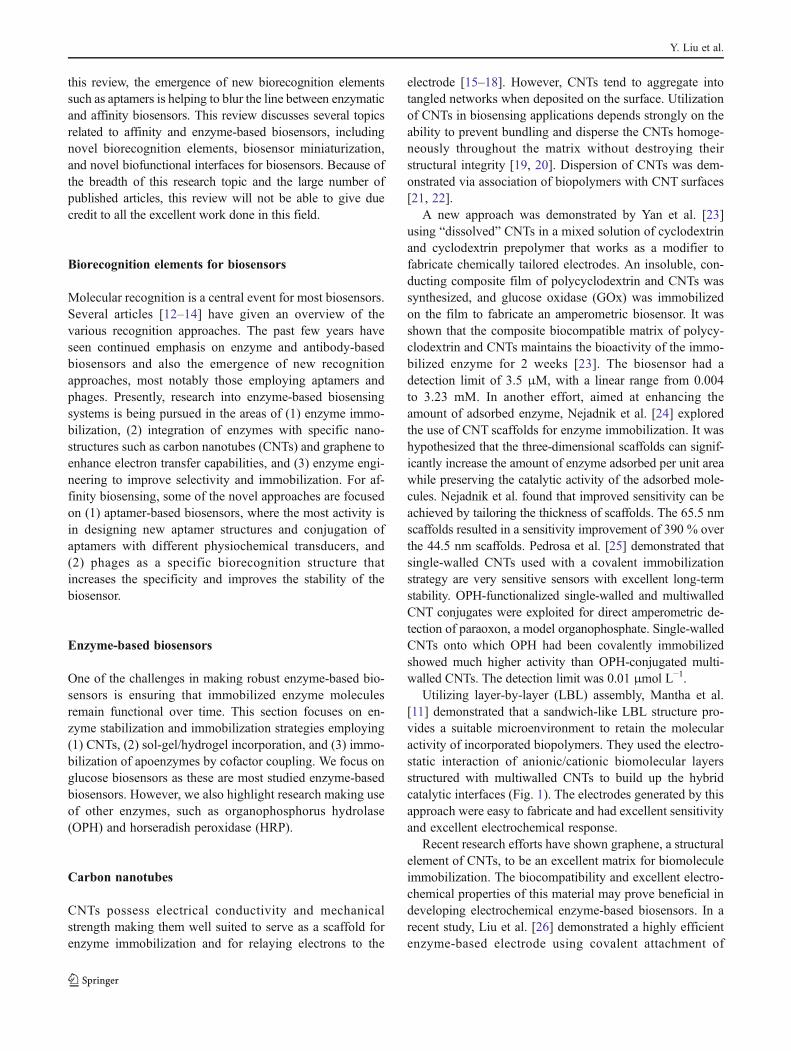
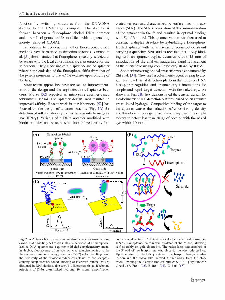
More recent approaches have focused on improvementsin both the design and the sophistication of aptamer bea-cons. Morse [52] reported an interesting aptamer-basedtobramycin sensor. The aptamer design used resulted inimproved affinity. Recent work in our laboratory [53] hasfocused on the design of aptamer beacons (Fig. 2A) fordetection of inflammatory cytokines such as interferon gam-ma (IFN-γ). Variants of a DNA aptamer modified withbiotin moieties and spacers were immobilized on avidin-
coated surfaces and characterized by surface plasmon reso-nance (SPR). The SPR studies showed that immobilizationof the aptamer via the 3′ end resulted in optimal bindingwith Kd of 3.44 nM. This aptamer variant was then used toconstruct a duplex structure by hybridizing a fluorophore-labeled aptamer with an antisense oligonucleotide strandcarrying a quencher. SPR studies revealed that IFN-γ bind-ing with an aptamer duplex occurred within 15 min ofintroduction of the analyte, suggesting rapid replacementof the quencher-carrying complementary strand by IFN-γ.
Another interesting optical aptasensor was constructed byZhi et al. [54]. They used a colorimetric agent-caging hydro-gel as a novel visual detection platform that relies on DNAbase-pair recognition and aptamer–target interactions forsimple and rapid target detection with the naked eye. Asshown in Fig. 2B, they demonstrated the general design fora colorimetric visual detection platform based on an aptamercross-linked hydrogel. Competitive binding of the target tothe aptamer causes the reduction of cross-linking densityand therefore induces gel dissolution. They used this simplesystem to detect less than 20 ng of cocaine with the nakedeye within 10 min.
Substrate
Target
Linker aptamer
PS-A
PS-B
Quenching strand
Fluorophore labeled aptamer
PEG gel
Glass slide
PEG gel
Glass slide
Add IFN-γ
IFN-γ
Aptamer duplex, low fluorescence due to FRET
Aptamer in complex with IFN-γ, high fluorescence
Add IFN-γe-
Potential
Curre
nt
Potential
Curre
nt
Redoxmolecule
Aptamer
(A) (B)
(C)
Enzyme
e-
Fig. 2 A Aptamer beacons were immobilized inside microwells usingavidin–biotin binding. A beacon molecule consisted of a fluorophore-labeled DNA aptamer and a quencher-labeled complementary strand.In duplex, fluorescence of an aptamer was quenched owing to thefluorescence resonance energy transfer (FRET) effect resulting fromthe proximity of the fluorophore-labeled aptamer to the acceptor-carrying complementary strand. Binding of interferon gamma (IFN-γ)disrupted the DNA duplex and resulted in a fluorescent signal.BWorkingprinciple of DNA cross-linked hydrogel for signal amplification
and visual detection. C Aptamer-based electrochemical sensor forIFN-γ. The aptamer hairpin was thiolated at the 5′ end, allowingself-assembly on gold electrodes. The redox label was attached atthe 3′ end of the hairpin and was close to the electrode surface.Upon addition of the IFN-γ aptamer, the hairpin changed confir-mation and the redox label moved further away from the elec-trode, lowering the electron-transfer efficiency. PEG poly(ethyleneglycol). (A From [53], B from [54], C from [68])
Affinity and enzyme-based biosensors
Electrochemical aptasensors
In comparison with the use of fluorescence, electrochemicaltransduction was implemented in aptamer-based biosensorsconsiderably later, with the first report appearing in 2004[55]. Although optical and electrochemical detection meth-ods have a number of advantages and disadvantages, it maybe argued that electrochemistry offers a less expensivemeans of reading the signal. If the electrochemical reportersand the electrolyte are chosen correctly, the electrical signalis likely to be stabler over time and may have less interfer-ences compared with optical detection. Recent reviews havediscussed fabrication of electrochemical aptasensors. Hianikand Wang [56] reviewed electrochemical aptasensorsaccording to their transduction mode, including amperomet-ric and impedimetric devices as well as a field effect tran-sistor and a recently reported potentiometric aptasensor.Willner and Zayats [38] summarized recent accomplish-ments in developing electronic aptasensors, which includemany of these methods as well as microgravimetric quartzcrystal microbalance sensors. The review of Willner andZayats further describes methods to develop amplified apta-sensor devices and label-free aptasensors. In the currentreview, we discuss some of the recent work on electrochem-ical impedance spectroscopy (EIS)-based aptasensors.
A label-free aptamer-based sensor using EIS as a detec-tion technique was reported by Xu et al. [57]. EIS is atechnology typically used for studying biomolecular inter-actions [58]. Their approach was based on an aptamer forhuman IgE, containing a hairpin loop responsible for thetarget recognition, and three other nonspecific oligomerswith some sequence changes, all immobilized on a goldelectrode array via self-assembly and all of them containinga loop structure of the same size. The results indicated thatonly the targeted aptamer recognizes IgE, demonstrating thestrong dependence of the affinity not only on the loop sizebut also on the sequence. Rodriguez et al. [59] reportedanother example of label-free impedance-based aptasensors.In this case, thiol-modified aptamers were self-assembled ona gold electrode and target binding was measured by anincrease of electron transfer resistance in the presence ofthe [Fe(CN)6]
3-/4- redox couple. The increase in electrontransfer resistance can be attributed to the repulsion betweenthe negatively charged aptamer and redox couple. Similarprotocols have also been developed for a number of othertarget analytes, including platelet-derived growth factor(PDGF) [60], interferon [61], and human IgE [62]. Thereported detection limits ranged from 0.1 nM for thrombinto 100 fM for interferon.
In addition to EIS, binding-induced conformational switch-ing has also been used in recent years in the development of alarge number of electrochemical aptasensors. In particular,aptasensors based on “signal on,” wherein the presence of
the target increases the signal strength upon target binding,and “signal off,”wherein the presence of the target reduces thesignal strength upon target binding, have recently been studied[63, 64]. The Heeger and Plaxco groups have pioneered theuse of these aptasensors. In their implementation an immobi-lized aptamer is either partially [65] or entirely [66] unfolded.Upon analyte binding, these aptamer probes fold and hold theredox labels closer to or farther from the electrode surface,respectively, thus increasing or decreasing the measured fara-daic current. Different electrochemically active redox labelshave been employed to relay the electrical signal resultingfrom aptamer–analyte binding. Of particular note is the workof Lubin and Plaxco [63], who modified electrodes withmethylene blue (MB)-containing aptamers and demonstratedelectrochemical detection (signal “off”) of a range of analytes.Lai et al. [67] developed an aptasensor for the direct detectionof PDGF in blood serum (signal “on”). Their approachemployed alternating current voltammetry to monitor target-induced folding in an MB-modified, PDGF-binding aptamer.They detected the BB variant of PDGF at 1 nM directly inundiluted, unmodified blood serum and at 50 pM in serumdiluted twofold with aqueous buffer. Our group described thedevelopment of an electrochemical DNA aptamer-based bio-sensor for detection of IFN-γ based on the “signal-off” sens-ing mechanism (Fig. 2c) [68]. Binding of IFN-γ caused theaptamer hairpin to unfold, pushingMB redox molecules awayfrom the electrode and decreasing the electron-transfer effi-ciency. The change in redox current was quantified usingsquare wave voltammetry and was highly sensitive to theIFN-γconcentration. The limit of detection for the optimizedbiosensor was 0.06 nM, with a linear response up to 10 nM.
A reagentless “signal-on” aptasensor for adenosine triphos-phate (ATP) detection has also been described [69]. Aferrocene-labeled thiolated aptamer in its duplex form wasself-assembled on a gold electrode surface. Upon binding ofATP, a stabilized rigid tertiary aptamer structure stabilizes andliberates the complementary DNA strand, which in turn bringsthe ferrocene moiety close to the electrode surface, thus gen-erating an enhanced square wave voltammetry peak current.
Phages as biorecognition elements
Phages represent a promising new direction in the develop-ment of affinity biosensors. There are several reasons for usingphages as biorecognition elements [70, 71]:
1. There are important analytes against which antibodiescannot be obtained easily [72].
2. Some applications, for example, environmental moni-toring, may require extraction of samples from soil orgroundwater that are soluble in organic solvents only.Such solvents may denature antibodies but not phages.
Y. Liu et al.
A phage, a threadlike virus, has a specific recognitionpeptide on its surface that binds to its host/target with highaffinity and specificity [73]. Peptide-bearing phages can beselected from large phage libraries and can serve as recep-tors for detection of various bioanalytes. As opposed tosynthesis of antibodies, which involves in vivo immuniza-tion of animals, phages can be easily synthesized by cheapgeneric fermentation processes. Besides this, they are morecost-effective, give high throughput, and have excellentstability at high temperatures (below 80 °C) and in harshenvironmental conditions such as acidic or basic pH [74].
Phages can be immobilized via physical adsorption [75,76], covalent binding [77, 78], affinity binding [79], andLangmuir–Blodgett deposition [80]. Cross-linking agentssuch as N-ethyl-N-(dimethylaminopropyl)carbodiimide/N-hydroxysuccinimide [81] and glutaraldehyde [82] have beenused for covalent immobilization of phages onto the desiredsensor surfaces. The strong affinity between biotin andavidin/streptavidin has also been widely used by researchersfor development of high-performance phage-based biosen-sors [79]. It has been found that sensors based on affinity(e.g., avidin–biotin) and covalent attachments of phagesperform better than sensors based on physical adsorption[79, 83]. The sensing event (phage–target interaction) hasbeen frequently investigated using several electroanalytical,optical, and mass-sensitive tools. Some of the recent work isdiscussed below
Electrochemical phage-based biosensors
Jia et al. [82] prepared a phage-modified light-addressablepotentiometric sensor for label-free detection of cancer cells.The sensor surface was prepared by covalent immobilizationof phage probes onto a silane-modified Si3N4 surface viaglutaraldehyde. This sensor could detect human phospha-tase of regenerating liver 3 in the concentration range from0.04 to 400 nM, and mammary adenocarcinoma cells inconcentrations from 0 to 105 mL. Other electroanalyticaltechniques such as EIS have also been employed for aspecific label-free biosensor using phages covalently immo-bilized onto the sensor surface [81, 84]. Shabani et al. [81]showed detection of Escherichia coli by a sensor surfacecontaining covalently immobilized T4 phages on screen-printed carbon electrode microarrays. An increase in elec-trolyte resistance and a decrease in charge-transfer resis-tance were observed owing to binding of bacteria ofincreasing concentration. This effect is contrary to the usu-ally observed increase in charge-transfer resistance withincreasing concentration of intact bacteria at the surface.Shabani et al. attributed this to onset of lysis (starts after20 min of incubation), which involves the breakup of thebacterial cell, owing to which a large amount of ionic
material is released and causes increased conductivity.Shabani et al. found a detection limit of 104 cfu mL-1. Recent-ly, Mejri et al. [85] utilized the effect of bacteria bindingbefore and after the start of the lysis process and proposed adual-signal-based detection of bacteria. The specific capturegenerates an initial increase in impedance, followed by animpedance decrease due to phage-induced lysis. Mejri et al.found a limit of detection of 104 cfu mL-1 with minimalinterference from nontarget lactobacillus.
Optical phage-based biosensors
Balasubramanian et al. [86] reported SPR-based label-freedetection of Staphylococcus aureus using lytic phages thatshowed a detection limit of 104 cfu mL-1. However, most ofthe SPR-based phage biosensors [87, 88] have utilizedphysical adsorption for immobilization of phages on thesensor surface. This method takes a long time for completeattachment of the biorecognition elements. Also, leaching ofphages from the surface does not allow complete regenera-tion of the sensor. In this context, covalent immobilizationhas aroused much interest as it may provide improvedattachment of the phages to the sensor surface in less timeand exhibits a wide dynamic range. Zhu et al. [77] devel-oped a sensitive and inexpensive optofluidic ring resonatorbiosensor for biomolecule detection. They observed that lesstime is required for complete immobilization by the covalentbinding method as compared with physical adsorption. Theyattributed this to the high surface-to-volume ratio in thecircular optofluidic ring resonator’s microfluidics channel,which promotes faster phage diffusion to the sensing surfacefrom the bulk solution.
Mass-sensing-based phage-based biosensors
A piezoelectric biosensor based on a quartz crystal micro-balance for detection of β-galactosidase from E. coli wasdeveloped by Nanduri et al. [89]. The sensor had a detectionlimit of a few nanomoles per liter over the range from 0.003to 210 nM. Horikawa et al. [78] fabricated magnetoelasticphage biosensors based on differently functionalized goldsurfaces. They found that surface functionalization has alarge effect on the surface phage coverage. The appropriatesurface can be utilized to pattern the phage probe layer toenhance the detection capabilities of magnetoelastic biosen-sors. Wan et al. [90] demonstrated detection of Bacillusanthracis spores using physical adsorption of phages on amagnetoelastic sensor surface.
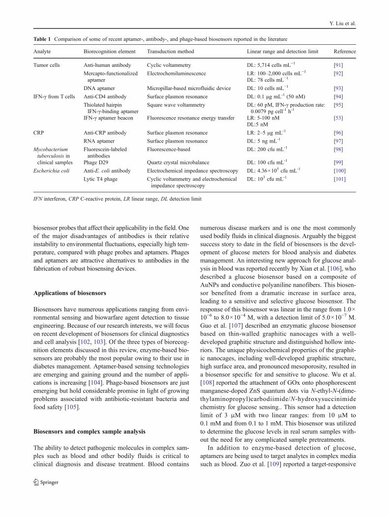
Some of the recent biosensors based on aptamers, anti-bodies, and phages are listed in Table 1. Despite their highaffinity and specificity, antibodies have limitations as
Affinity and enzyme-based biosensors
biosensor probes that affect their applicability in the field. Oneof the major disadvantages of antibodies is their relativeinstability to environmental fluctuations, especially high tem-perature, compared with phage probes and aptamers. Phagesand aptamers are attractive alternatives to antibodies in thefabrication of robust biosensing devices.
Applications of biosensors
Biosensors have numerous applications ranging from envi-ronmental sensing and biowarfare agent detection to tissueengineering. Because of our research interests, we will focuson recent development of biosensors for clinical diagnosticsand cell analysis [102, 103]. Of the three types of biorecog-nition elements discussed in this review, enzyme-based bio-sensors are probably the most popular owing to their use indiabetes management. Aptamer-based sensing technologiesare emerging and gaining ground and the number of appli-cations is increasing [104]. Phage-based biosensors are justemerging but hold considerable promise in light of growingproblems associated with antibiotic-resistant bacteria andfood safety [105].
Biosensors and complex sample analysis
The ability to detect pathogenic molecules in complex sam-ples such as blood and other bodily fluids is critical toclinical diagnosis and disease treatment. Blood contains
numerous disease markers and is one the most commonlyused bodily fluids in clinical diagnosis. Arguably the biggestsuccess story to date in the field of biosensors is the devel-opment of glucose meters for blood analysis and diabetesmanagement. An interesting new approach for glucose anal-ysis in blood was reported recently by Xian et al. [106], whodescribed a glucose biosensor based on a composite ofAuNPs and conductive polyaniline nanofibers. This biosen-sor benefited from a dramatic increase in surface area,leading to a sensitive and selective glucose biosensor. Theresponse of this biosensor was linear in the range from 1.0×10⁻⁶ to 8.0×10⁻⁴ M, with a detection limit of 5.0×10⁻⁷ M.Guo et al. [107] described an enzymatic glucose biosensorbased on thin-walled graphitic nanocages with a well-developed graphitic structure and distinguished hollow inte-riors. The unique physicochemical properties of the graphit-ic nanocages, including well-developed graphitic structure,high surface area, and pronounced mesoporosity, resulted ina biosensor specific for and sensitive to glucose. Wu et al.[108] reported the attachment of GOx onto phosphorescentmanganese-doped ZnS quantum dots via N-ethyl-N-(dime-thylaminopropyl)carbodiimide/N-hydroxysuccinimidechemistry for glucose sensing.. This sensor had a detectionlimit of 3 μM with two linear ranges: from 10 μM to0.1 mM and from 0.1 to 1 mM. This biosensor was utilizedto determine the glucose levels in real serum samples with-out the need for any complicated sample pretreatments.
In addition to enzyme-based detection of glucose,aptamers are being used to target analytes in complex mediasuch as blood. Zuo et al. [109] reported a target-responsive
Table 1 Comparison of some of recent aptamer-, antibody-, and phage-based biosensors reported in the literature
Analyte Biorecognition element Transduction method Linear range and detection limit Reference
Tumor cells Anti-human antibody Cyclic voltammetry DL: 5,714 cells mL−1 [91]
Mercapto-functionalizedaptamer
Electrochemiluminescence LR: 100–2,000 cells mL−1 [92]DL: 78 cells mL−1
DNA aptamer Micropillar-based microfluidic device DL: 10 cells mL−1 [93]
IFN-γ from T cells Anti-CD4 antibody Surface plasmon resonance DL: 0.1 μg mL-1 (50 nM) [94]
Thiolated hairpinIFN-γ-binding aptamer
Square wave voltammetry DL: 60 pM, IFN-γ production rate:0.0079 pg cell-1 h-1
[95]
IFN-γ aptamer beacon Fluorescence resonance energy transfer LR: 5-100 nM [53]DL:5 nM
CRP Anti-CRP antibody Surface plasmon resonance LR: 2–5 μg mL-1 [96]
RNA aptamer Surface plasmon resonance DL: 5 ng mL-1 [97]
Mycobacteriumtuberculosis inclinical samples
Fluorescein-labeledantibodies
Fluorescence-based DL: 200 cfu mL-1 [98]
Phage D29 Quartz crystal microbalance DL: 100 cfu mL-1 [99]
Escherichia coli Anti-E. coli antibody Electrochemical impedance spectroscopy DL: 4.36×10⁵ cfu mL-1 [100]
Lytic T4 phage Cyclic voltammetry and electrochemicalimpedance spectroscopy
DL: 103 cfu mL-1 [101]
IFN interferon, CRP C-reactive protein, LR linear range, DL detection limit
Y. Liu et al.
electrochemical aptamer switch for reagentless detection ofstandard and cellular ATP with high sensitivity and selectivity.The approach required neither exogenous reagents nor label-ing of the target. The sensor functioned well when challengedwith cell lysate, a complex, contaminant-ridden medium. Inanother study, Huang and Liu [110] combined flow cytometrywith aptamer-functionalized magnetic microparticles fordetection of adenosine in serum. In this implementation,the flow cytometer serves to isolate the magnetic mi-croparticle sensors from the serum and detect the pres-ence of the fluorescent signal. The antibiotic gentamicinhas also been detected in serum using an RNA aptamer[111]. Stability issues necessitated the modification ofthe aptamer against resistance breakdown by nucleases.These modifications resulted in an unacceptable de-crease in sensitivity and the researchers were eventuallyforced to filter the serum to achieve acceptable stabilityand sensitivity.
Recent advancements to improve the sensitivity ofaptamers in small-molecule detection have seen a shift backtoward the sandwich assay format originally used withantibody-based assays such as the enzyme-linked immuno-sorbent assay. The Plaxco group [112] recently reporteddetection of ATP from cell lysate and cocaine from serumusing such an approach. By separating the redox reporterfrom the target-binding strand, they were able to increase thesignal gain sixfold. In another sandwich-type assaystudy, cocaine was detected from banknotes left in sim-ulated real-world environments with large amounts ofcontaminants [113]. This assay utilized aptamers andelectrochemiluminescence. In this instance as well, thesandwich-type assay provided a higher signal and better sen-sitivity than a conventional aptamer-based electrochemilumi-nescence assay. The downside of the sandwich approach,however, is that the assayrequires the redox strand be presentas a reagent.
Another advancement of note is the use of small mole-cules not as targets but as binding agents in biosensors. ThePlaxco group [114] demonstrated the use of a nucleotide-based biosensor with a biotin moiety conjugated to onestrand. Upon binding streptavidin, the electrical signalchanges as it would in a typical aptamer biosensor. Thesensor was insensitive to contaminants in a number ofcomplex media, including serum, soil, and beer. Their workportends the possibility of adapting biosensor technologiesto probe protein–small molecule interactions.
A number of phage-based biosensors reported recentlywere used for detection in real samples. Lakshmanan et al.[76] detected Salmonella typhimurium in fat-free milk usinga magnetoelastic sensor immobilized with phages. Jia et al.[82] tested a phage-modified light-addressable potentiomet-ric sensor for analysis of human phosphatase of regeneratingliver 3 in blood samples.
Biosensors for cellular analysis
Biosensors for studying living systems, i.e., cells, tissue,organs, and organisms, have received a great deal of atten-tion in the past few decades. Cell-based biosensors canmonitor physiological changes in cells exposed to patho-gens, pollutants, biomolecules, and drugs [115].
To obtain the information hidden inside the cell, somevery refined procedures and sensing techniques are requiredthat do not disrupt the cell membrane and maintain cellviability for a long time. Microfabrication and microfluidicsystems show great potential is this area owing to theirunique properties, such as controllable transport, immobili-zation, and facile manipulation of biological molecules andcells. With use of these techniques, various systems havebeen developed for analysis of intracellular parameters andto detect the presence of cell metabolites, even at the single-cell level [116, 117]. Electrochemical measurements usingmicroelectrodes are powerful tools for spatiotemporal mon-itoring of electroactive chemicals, particularly when inte-grated into microfluidic systems. This allows many samplemanipulations to be integrated into the microsystem andthus monitoring can be performed in a simple and automat-ed way [118]. The electrochemical methods could serve asan excellent transducing system for invasive but nondestruc-tive cell analysis. Moreover, in combination with opticalprobes and imaging techniques, electroanalytical methodsshow great potential for the development of multianalytesystems to monitor cellular dynamics. This section exem-plifies biosensors for detection of various cell-secretedproducts.
Biosensors for cell-secreted products
The monitoring of cell-secreted products is of wide interestin the area of biomedical and health care as this process is anessential physiological function [119]. A variety of chem-icals, including signal molecules such as hormones, neuro-transmitters [120], trophic factors, and metabolic products,are released from cells upon stimulation. The responses ofcells to physical or chemical cues have typically been mea-sured in microfluidic devices [121] via optical [122] orelectrochemical [123] means.
Detecting cellular release of neurotransmitters
Measurements of catecholamine and glutamate exocytosishave received tremendous interest in the past few decades.Disturbance of their controlled secretion may lead to manyneurological disorders, such as Parkinson’s disease [124,125] and Alzheimer’s disease [126, 127]. Glutamate plays
Affinity and enzyme-based biosensors
an important role in long-term changes in synaptic efficacy,forming thebasis for learningandmemory.Castillo et al. [128]described a glutamate sensor based on bienzyme (glutamateoxidase and HRP) redox hydrogel capable of detecting therelease of this excitatory neurotransmitter from adherentlygrowingcells uponstimulation.Thesensorwasable tooperateat low working potential, which obviated the possibility ofinterference by easily oxidizable compounds present in com-plex biological samples. The sensor had a low detection limitof 0.5μMglutamate, a response timeof about 35 s, anda linearrange of up to 60μM.Another amperometric sensor based onbienzymes for direct monitoring of L-glutamate in a flowinjection system was reported by Belay et al. [129]. Thebienzyme electrodeswere constructed by coating solid graph-ite rods with a premixed solution containing glutamate oxi-dase andHRP cross-linkedwith a redox polymer formed frompoly(1-vinylimidazole). The sensor detected in the range from0.3 and 250μMglutamate concentration, with a sensitivity of88.36 ±0.14 μA mM-1 cm-2, a detection limit of 0.3 μM, andresponse time of less than 10 s.
Efforts were made toward electrochemical measurementof release of catecholamines from pheochromocytoma(PC12) cells [130]. Microfluidics was used to transportand trap a single cell while the stimulants were introducedfrom the microchannel and a carbon fiber microelectrodewas positioned over the cell for amperometric measure-ments. Li et al. [131] used collagen coating in the micro-channels to attach PC12 cells. They were able to measureexocytosis from a large population of cells with a micromolded carbon ink electrode. Huang et al. [132] developed apolydimethylsiloxane/glass microfluidic system for trans-port of a single PC12 cell to a microvial. Shi et al. [133]developed a novel microfluidic electrochemical sensor witha CNT-modified indium tin oxide microelectrode for releaseof dopamine from a single living rat PC12 cell. The sensi-tivity of the electrochemical sensor after CNT modificationwas more than that of the unmodified electrode by 2.5 tothree orders of magnitude. Lin et al. [134] fabricated amicrosensor by electrochemically depositing a film contain-ing overoxidized polypyrrole and multiwalled CNTs onto acarbon fiber microelectrode. The sensor was utilized in invivo microdialysis with electrochemical microsensing deter-mination of dopamine in striatum of freely moving rats. Thesensor exhibited linear detection in the concentration rangefrom 5.0 nM to 10 μM, and the detection limit (at a signal-to-noise ratio of 3) was 0.5 nM.
Biosensors have been developed for monitoring D-ser-ine, which is the predominant D-amino acid in the mam-malian central nervous system and has been recentlyrelated to several neurological and psychiatric diseases.Pernot et al. [135] reported a microbiosensor based oncylindrical platinum microelectrodes, covered with a mem-brane of poly(m-phenylenediamine) and a layer of
immobilized D-amino acid oxidase from the yeast Rhodo-torula gracilis. By detecting the hydrogen peroxide pro-duced by enzymatic degradation of D-serine, thismicrobiosensor exhibited a detection limit of 16 nM anda mean response time of 2 s.
Detection of reactive oxygen species secreted from cells
Inflammatory processes play a crucial role in a number ofdiseases, including diabetes [136], cancer [137], and tissuefibrosis [138]. Molecules secreted by immune cells duringinflammation are critical in combating pathogens. Macro-phages, immune cells residing in the tissue, are first torespond to invading pathogens. These cells produce inflam-matory markers, including cytokines and reactive oxygenspecies (ROS). ROS include superoxide anion (O2
·−) hydro-gen peroxide (H2O2), and hydroxyl radical (.OH). H2O2 isone of the major indicators of oxidative stress and thestablest of ROS compounds. Our group [139] has developedelectrochemical biosensors based on enzyme-containinghydrogel for detection of H2O2 secreted from macrophages.Enzyme-containing hydrogel was coated onto micropat-terned gold electrodes and the hydrogel was cross-linkedwith UV exposure. The macrophages were seeded into ply(ethylene glycol) microwells and stimulated with phorbol12-myristate 13-acetate. Polydimethylsiloxane was put onthe micropatterned slides to make the electrochemical setupfor amperometric measurements. Fluorescence microscopywas used to sense the H2O2 released from the immune cells[140]. The production of H2O2 after mitogenic stimulationof macrophages resulted in the appearance of fluorescencein the HRP-containing hydrogel microstructures, with thefluorescence intensity being a strong function of analyteconcentration. Amatore et al. [141] studied release of ROSand reactive nitrogen species by electrochemistry within amicrofluidic device. Macrophages were cultured in adetection chamber containing a three-electrode systemand were stimulated by the microinjection of a calciumionophore. Cheah et al. [142] developed a microfluidicdevice for heart tissue perfusion with real-time electro-chemical monitoring of ROS release. Sun et al. [143]reported determination of ROS in a single erythrocyte byusing a simple crossed-channel glass chip with integratedoperational functions such as docking, lysing, and capillaryelectrophoretic separation with laser-induced-fluorescencedetection.
Detection of other cell-secreted products
An optical approach for monitoring allergic response wasdemonstrated by Matsubara et al. [144]. A rat basophilic
Y. Liu et al.
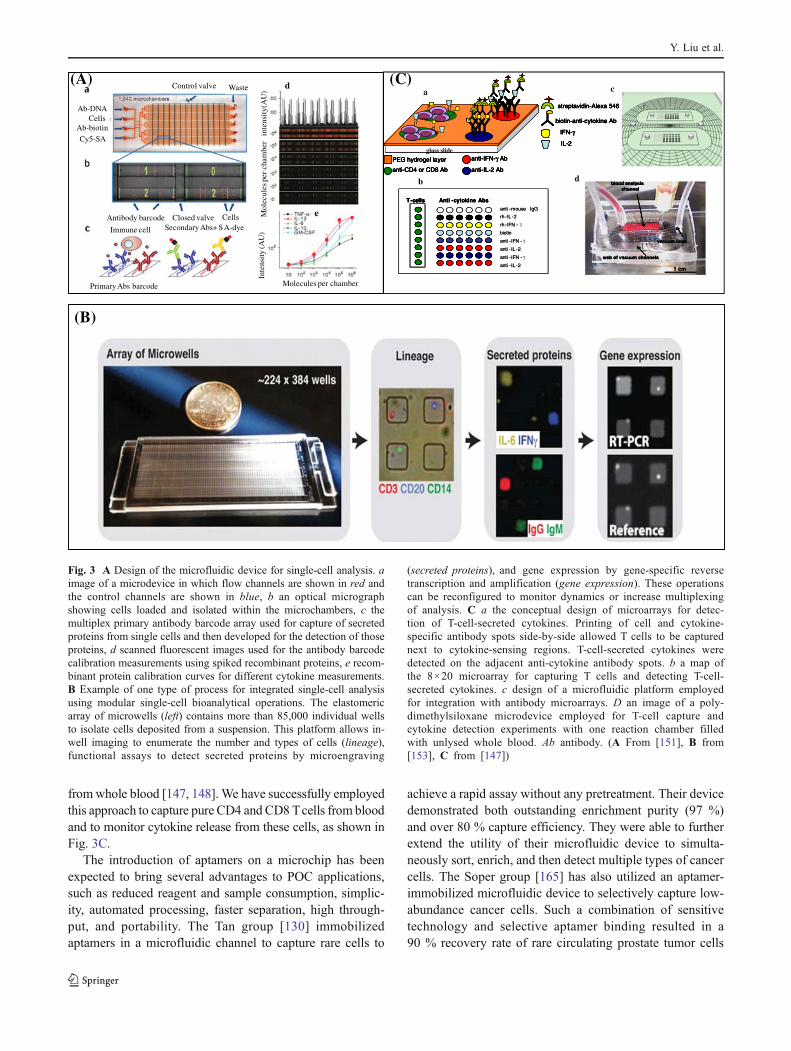
leukemia cell line (RBL-2 H3), a tumor analog of rat mu-cosal mast cells, was used as a model to observe the allergicresponse upon antigenic stimulus. When exocytosis eventsoccurred, the microfluidic system detected the fluorescentsignal of quinacrine, which was released from RBL-2 H3cells. The Kennedy group [145, 146] has developed micro-fluidic devices for high-throughput, automated, and onlinemonitoring of insulin secretion from individual islets inparallel. This chip consists of 15 channel networks eachcapable of superfusing a single islet and mixing superfusatefrom each islet online with fluorescein isothiocyanate la-beled insulin and anti-insulin antibody for a competitiveimmunoassay. The resulting continuous reaction streamswere periodically injected onto parallel electrophoresischannels, where the mixtures were separated. The chipwas used to demonstrate that free fatty acid induced lip-otoxicity in islets eliminates pulsatile insulin secretion. Ourgroup [147–149] has developed biosensors for the detectionof molecules secreted or taken up by cells. We also devel-oped antibody- or aptamer-based biosensors for detection ofinflammatory cytokines released by activated immune cells[68, 94, 149, 150]. In addition to these optical and electro-chemical methods, the application of SPR-based techniquescould contribute extensively to cellular analysis. We alsodescribed a strategy combining antibody-based affinity cellseparation and SPR for capturing human CD4 T cells andfor label-free detection of cell-secreted IFN-γ.
Recent advancements in cell-secretion analysis havegreatly improved the throughput of these biosensors.Work by the Heath group [151] (Fig. 3A) demonstrateda microfluidic platform designed for highly multiplexed(more than ten proteins), reliable, sample-efficient (ap-proximately 1×10⁴ cells), and quantitative measure-ments of secreted proteins from single cells. Theplatform was tested by assessment of multiple inflam-matory cytokines from lipopolysaccharide-stimulated hu-man macrophages before being applied to quantificationof T-cell polyfunctional diversity from tumor antigen-specific cytotoxic T lymphocytes responding to a tumorand compared against the response of cells from healthydonor controls. Their results showed a large degree ofheterogeneity in function between cells with similarsurface markers, highlighting the importance of secre-tion analysis. In another study, Love et al. [152, 153]presented a soft lithographic method based on intaglioprinting to generate microarrays comprising the secretedproducts of single cells. These engraved arrays enable arapid (less than 12 h) and high-throughput (100,000individual cells) system for identification, recovery, andclonal expansion of cells producing antigen-specificantibodies. This method shown in Fig. 3B can be adap-ted, in principle, to detect any secreted product in sucha multiplexed manner.
Biosensors at the point of care
Bringing diagnostics to the point of care (POC) could allowsome form of preliminary self-screening or sorting and evenbasic treatment by front-line nursing staff, therefore reducingthe burden on practitioners and hospitals and allowing spe-cialist care to be dedicated to those most in need [154, 155].Microfluidics has been reviewed recently in this context[156], and applications in this field are increasing rapidly.Miniaturizing biosensor inside microfluidic devices hasgrown tremendously and rapidly, sustained by the promise itoffers to revolutionize conventional laboratory handling, pro-cessing, and analytical techniques. One particularly interestingmicrofluidic device for POC applications is the integratedblood barcode chip [157]. This device has been devel-oped to address the need for microchips that integrateon-chip plasma separations from microliter quantities ofwhole blood with rapid in situ measurements of multi-ple plasma proteins. The versatility of this barcodeimmunoassay has been demonstrated by detecting humanchorionic gonadotropin from human serum over a 105 con-centration range and by stratifying 22 cancer patients viamultiple measurements of a dozen blood protein biomarkersfor each patient.
Another important area where microfluidic-based biosen-sors is having an impact is the monitoring of HIV andacquired immune deficiency syndrome (AIDS). Advancesin the field have enabled rapid, inexpensive, and reliableapproaches for detecting and quantifying the HIV and AIDSstatus in patients. One way to evaluate the AIDS status inHIV-infected patients is to measure the absolute number ofCD4+ T lymphocytes in blood. Handheld, reliable, and low-cost CD4 counting devices for use in resource-scarceregions of the world are needed. As a result, considerableeffort has been directed toward the development of minia-ture devices for simple and inexpensive leukocyte analysis[158–162]. The Toner group [163] has developed a simpleCD4-counting microfluidic device that uses cell affinitychromatography operated under differential shear flow tospecifically isolate CD4+ T lymphocytes with high efficien-cy directly from 10 μL of unprocessed, unlabeled wholeblood. CD4 counts are obtained under an optical microscopein a rapid, simple, and label-free fashion. Our group [164]has also developed methods for capturing and counting Tcells using microfluidics in combination with printed micro-arrays of antibody spots. These methods capture cells withhigh purity (more than 94 %) and allow the quantitation ofsubset populations purely on the basis of the capture loca-tion. Our results were shown to be in good agreement withthose from flow cytometry.
Building on these cell capture studies, our laboratory hasdeveloped methods for using antibody microarrays insidemicrofluidic devices for immunophenotyping of leukocytes
Affinity and enzyme-based biosensors
fromwhole blood [147, 148]. We have successfully employedthis approach to capture pure CD4 and CD8 Tcells from bloodand to monitor cytokine release from these cells, as shown inFig. 3C.
The introduction of aptamers on a microchip has beenexpected to bring several advantages to POC applications,such as reduced reagent and sample consumption, simplic-ity, automated processing, faster separation, high through-put, and portability. The Tan group [130] immobilizedaptamers in a microfluidic channel to capture rare cells to
achieve a rapid assay without any pretreatment. Their devicedemonstrated both outstanding enrichment purity (97 %)and over 80 % capture efficiency. They were able to furtherextend the utility of their microfluidic device to simulta-neously sort, enrich, and then detect multiple types of cancercells. The Soper group [165] has also utilized an aptamer-immobilized microfluidic device to selectively capture low-abundance cancer cells. Such a combination of sensitivetechnology and selective aptamer binding resulted in a90 % recovery rate of rare circulating prostate tumor cells
Mol
ecul
es p
er c
ham
ber
inte
nsit
y(A
U)
Inte
nsit
y (A
U)
Molecules per chamberPrimary Abs barcode
Immune cell Secondary Abs+ SA-dyeAntibody barcode Closed valve Cells
Control valve Waste
Ab-DNACells
Ab-biotinCy5-SA
d(A)
e
(B)
PEG hydrogel layer
anti-IL-2 Ab
anti-IFN-γ Ab
anti-CD4 or CD8 Ab
biotin-anti-cytokine Ab
streptavidin-Alexa 546
IL-2
IFN-γ
glass slide
PEG hydrogel layer
anti-IL-2 Ab
anti-IFN-γ Ab
anti-CD4 or CD8 Ab
PEG hydrogel layerPEG hydrogel layer
anti-IL-2 Abanti-IL-2 Ab
anti-IFN-γ Abanti-IFN-γ Ab
anti-CD4 or CD8 Abanti-CD4 or CD8 Ab
biotin-anti-cytokine Ab
streptavidin-Alexa 546
IL-2
IFN-γ
biotin-anti-cytokine Abbiotin-anti-cytokine Ab
streptavidin-Alexa 546streptavidin-Alexa 546
IL-2IL-2
IFN-γIFN-γ
glass slideglass slide
anti -mouse IgG
rh -IL -2
rh -IFN - γ
biotin
anti -IFN - γ
anti -IL -2
anti -IFN - γ
anti -IL -2
Anti -cytokine AbsT-cellsanti -mouse IgG
rh -IL -2
rh -IFN -
biotin
anti -IFN -
anti -IL -2
anti -IFN -
anti -IL -2
Anti -cytokine AbsT-cells Anti -cytokine AbsT-cellsT-cells
web of vacuum channels
vacuum lines
blood analysis channel
1 cm
web of vacuum channels
vacuum lines
blood analysis channel
web of vacuum channels
vacuum lines
blood analysis channel
1 cm
(C)a
b
c
d
Fig. 3 A Design of the microfluidic device for single-cell analysis. aimage of a microdevice in which flow channels are shown in red andthe control channels are shown in blue, b an optical micrographshowing cells loaded and isolated within the microchambers, c themultiplex primary antibody barcode array used for capture of secretedproteins from single cells and then developed for the detection of thoseproteins, d scanned fluorescent images used for the antibody barcodecalibration measurements using spiked recombinant proteins, e recom-binant protein calibration curves for different cytokine measurements.B Example of one type of process for integrated single-cell analysisusing modular single-cell bioanalytical operations. The elastomericarray of microwells (left) contains more than 85,000 individual wellsto isolate cells deposited from a suspension. This platform allows in-well imaging to enumerate the number and types of cells (lineage),functional assays to detect secreted proteins by microengraving
(secreted proteins), and gene expression by gene-specific reversetranscription and amplification (gene expression). These operationscan be reconfigured to monitor dynamics or increase multiplexingof analysis. C a the conceptual design of microarrays for detec-tion of T-cell-secreted cytokines. Printing of cell and cytokine-specific antibody spots side-by-side allowed T cells to be capturednext to cytokine-sensing regions. T-cell-secreted cytokines weredetected on the adjacent anti-cytokine antibody spots. b a map ofthe 8×20 microarray for capturing T cells and detecting T-cell-secreted cytokines. c design of a microfluidic platform employedfor integration with antibody microarrays. D an image of a poly-dimethylsiloxane microdevice employed for T-cell capture andcytokine detection experiments with one reaction chamber filledwith unlysed whole blood. Ab antibody. (A From [151], B from[153], C from [147])
Y. Liu et al.
from a peripheral blood matrix. As another interesting de-velopment in aptamer-based microfluidics, Swensen et al.[166] introduced a microfluidic, electrochemical, aptamer-based sensor chip by integrating target-specific DNA. Thesystem was applied to achieve continuous, real-time moni-toring of cocaine in blood serum at the physiologicallyrelevant concentration and with physiologically relevanttime resolution.
Apart from aptamers, phage-based systems have recentlyemerged as promising tools for POC testing [155, 167]. Thephage probes provide high throughput, adaptability to thetesting environment, and are cheaper than antibodies. Jia et al.[82] reported a novel phage-modified light-addressable poten-tiometric sensor system for cancer cell monitoring. In recentwork, Mi et al. [99] fabricated a multichannel quartz crystalmicrobalance based biosensor for detection of Mycobacteriumtuberculosis in clinical samples using phage D29, which infectsM. tuberculosis. The sensor had a detection limit of100 cfu mL-1. Several other cell-specific peptide ligands iden-tified from phage display libraries have shown binding withdifferent tumor types [155].
Infectious agents also have an indirect effect on humanhealth via agricultural and other related commodities [168].Recent biological terrorism threats and outbreaks of micro-bial pathogens clearly emphasize the need for biosensorsthat can quickly and accurately identify infectious agents.Phages are novel innovative affinity-based recognition ele-ments that are becoming increasingly important for food andenvironmental sensors because of their exceptional charac-teristics, such as their high affinity and specificity for theirtargets, their fast, cheap and animal-friendly production,their stability, and their high resistance against environmen-tal stress. Phage probes are more amenable than antibodiesto manipulation at the molecular level to improve theirinteraction with the analyte. A recent review by Dorst etal. [168] has discussed application of a phage biosensor forfood and environmental monitoring.
Phage probes have also been used for detection of food-and water-borne pathogens in real samples such as waterand fat-free milk [169]. Edgar et al. [170] used a biotin-tagged lytic T7 phage to form quantum dot complexes fordetection of E. coli in river water. Thus, application of thesebiorecognition elements can lead to the fabrication of vari-ous POC devices.
Conclusions
This articles has presented an overview of recent advancesin the development and application of affinity and enzyme-based biosensors. Some of recent applications of these bio-sensors in clinical sample testing and cell analysis have beenhighlighted. Moving forward, we envision biosensors
increasing their foothold in the areas of cell analysis andclinical diagnostics. We are particularly excited about newsensing technologies allowing continuous monitoring of cellfunction in vitro and in vivo with high spatiotemporal res-olution. We predict cell function analysis (the types and thelevels of molecules produced by cells) will become morewidespread in disease diagnosis and foresee novel biosen-sors playing an important role in this trend.
At the beginning of this article, we underscored thedifferences between enzyme-based and affinity biosensorsand mentioned that the latter sensors cannot be regeneratedand may not be used for long-term analyte monitoring. Thisstatement holds true for antibody-based affinity biosensors,but the outlook is quite different for aptamer-based sensors.Because aptamers are chemically stable and may be easilyregenerated by denaturation of the target analyte, we envi-sion aptamer-based biosensors being used for continuousmonitoring. Unlike the natural turnover of enzyme-basedbiosensors, aptasensors will be regenerated chemically andwill likely be coupled with microfluidics to automate regen-eration process.
In addition, the field of phage-based biosensors is likelyto grow dynamically in the immediate future. This growthwill be fueled by the increasing prevalence of hospital-acquired infection and antibiotic-resistant bacteria. Currenttechnologies for bacterial detection are inexpensive but slow(based on cell growth) or rapid but expensive (PCR-based).Phages evolved to detect and invade bacteria and thereforeprovide a natural biorecognition element for bacterial detec-tion. Because phages multiply/replicate within bacterialhosts, there is also a natural amplification effect to phage-based detection of bacteria. Finally, we envision that con-tinued innovation in biosensors will come not only from thetraditional biosensing disciplines of biorecognition engi-neering and transducer development but also from nano-technology and microfluidics.
Acknowledgments Financial support for this study was provided byNational Science Foundation Emerging Frontiers in Research andInnovation grant #0937997 awarded to A.R. Additionally, A.L.S.acknowledges working at the National Science Foundation duringpreparation of this manuscript. The views expressed in this article arethose of the authors, and do not reflect the views of the NationalScience Foundation.
References
1. Chambers JP, Arulanandam BP, Matta LL, Weis A, Valdes JJ(2008) Curr Issues Mol Biol 10:1–12
2. Grieshaber D, MacKenzie R, Voros J, Reimhult E (2008) Sensors8:1400–1458
3. Pohanka M, Skladai P (2008) J Appl Biomed 6:57–644. Ronkainen NJ, Halsall HB, Heineman WR (2010) Chem Soc Rev
39:1747–1763
Affinity and enzyme-based biosensors
5. Gauglitz G (2010) Anal Bioanal Chem 398:2363–23726. Lee K, Povlich LK, Kim J (2010) Analyst 135:2179–21897. Ko W, Yim C, Jung N, Joo J, Jeon S, Seo H, Lee SS, Park JC
(2011) Nanotechnology 22:4055028. Walton PW, O’Flaherty MR, Butler ME, Compton P (1993)
Biosens Bioelectron 8:401–4079. Mosbach K (1991) Biosens Bioelectron 6:179–182
10. Chovan T, Guttman A (2002) Trends Biotechnol 20:116–12211. Mantha S, Pedrosa VA, Olsen EV, Davis VA, Simonian AL
(2010) Langmuir 26:19114–1911912. Matharu Z, Bandodkar AJ, Gupta V, Malhotra BD (2012) Chem
Soc Rev 41:1363–140213. Collings AF, Caruso F (1997) Rep Prog Phys 60:1397–144514. Vo-Dinh T, Cullum B (2000) Fresenius J Anal Chem 366:540–
55115. Jacobs CB, Peairs MJ, Venton BJ (2010) Anal Chim Acta
662:105–12716. Shao Y, Wang J, Wu H, Liu J, Aksay IA, Lin Y (2010) Electro-
analysis 22:1027–103617. Lee D, Cui TH (2009) IEEE Sens J 9:449–45618. Wang YD, Joshi PP, Hobbs KL, Johnson MB, Schmidtke DW
(2006) Langmuir 22:9776–978319. Sun Y, Fu K, Lin Y, Huang W (2002) Acc Chem Res 35:1096–
110420. Chen RJ, Zhang Y, Wang D, Dai HJ (2001) Am Chem Soc
123:3838–383921. Bandyopadhyaya R, Nativ-Roth E, Regev O, Yerushalmi-Rozen
R (2002) Nano Lett 2:2522. O'Connell MJ, Boul P, Ericson LM, Huffman C, Wang Y, Haroz
E (2001) Chem Phys Lett 342:26523. Yan HP, Zhu YF, Chen DC, Li CH, Chen SG, Ge ZC (2010)
Biosens Bioelectron 26:295–29824. Nejadnik MR, Deepak FL, Garcia CD (2011) Electroanalysis
23:1462–146925. Pedrosa VA, Paliwal S, Balasubramanian S, Nepal D, Davis V,
Wild J, Ramanculov E, Simonian A (2010) Colloids Surf BBiointerfaces 77:69–74
26. Liu Y, Yu D, Zeng C, Miao Z, Dai L (2010) Langmuir 26:6158–6160
27. Kang X, Wang J, Wu H, Aksay IA, Liu J, Lin Y (2009) BiosensBioelectron 25:901–905
28. Gill I, Ballesteros A (2000) Trends Biotechnol 18:282–29629. Hench LL, Valentine JK (1990) Chem Rev 90:33–7230. Ivnitski D, Artyushkova K, Rincn RA, Atanassov P, Luckarift
HR, Johnson GR (2008) Small 4:357–36431. Ramanathan M, Luckarift HR, Sarsenova A, Wild JR, Ramanculov
EK, Olsen EV, Simonian AL (2009) Colloids Surf B Biointerfaces73:58–64
32. Jia J, Wang B, Wu A, Cheng G, Li Z, Dong S (2002) Anal Chem74:2217–2223
33. Yan J, Pedrosa VA, Simonian AL, Revzin A (2010) Appl MaterInterfaces 2:748–755
34. Pedrosa VA, Yan J, Simonian AL, Revzin A (2011) Electroanal-ysis 23:1142–1149
35. Yehezkeli O, Tel-Vered R, Reichlin S, Willner I (2011) ACSNano 5:2385–2391
36. Zayats M, Katz E, Willner I (2002) J Am Chem Soc 124:14724–14735
37. Xiao Y, Patolsky F, Katz E, Hainfeld JF, Willner I (2003) Science299:1877–1881
38. Willner I, Zayats M (2007) Angew Chem Int Ed 46:6408–641839. Tombelli S, Minunni M, Mascini M (2005) Biosens Bioelectron
20:2424–243440. Cho EJ, Lee JW, Ellington AD (2009) Annu Rev Anal Chem
2:241–26441. XuY, ChengG,He P, Fang Y (2009) Electroanalysis 21:1251–1259
42. Liu J, Cao Z, Lu Y (2009) Chem Rev 109:1948–199843. Davis KA, Abrams B, Lin Y, Jayasena SD (1996) Nucleic Acids
Res 24:702–70644. Nutiu R, Li YF (2005) Methods 37:16–2545. Sassolas A, Blum LJ (2011) Leca-Bouvier BD 26:3725–373646. Tyagi S, Bratu DP, Kramer FR (1998) Nat Biotechnol 16:49–5347. Santangelo PJ, Nix B, Tsourkas A, Bao G (2004) Nucleic Acids
Res 3248. Bernacchi S, Mély Y (2001) Nucleic Acids Res 29:e6249. Yamamoto R, Baba T, Kumar PK (2000) Genes Cells 5:389–39650. Nutiu R, Li YF (2003) J Am Chem Soc 125:4771–477851. Yamana K, Ohtani Y, Nakano H, Saito I (2003) Bioorg Med
Chem Lett 13:3429–343152. Morse DP (2007) Biochem Biophys Res Commun 359:94–10153. Tuleuova N, Jones CN, Yan J, Ramanculov E, Yokobayashi Y,
Revzin A (2010) Anal Chem 82:1851–185754. Zhi Z, Cuichen W, Haipeng L, Yuan Z, Xiaoling Z, Huaizhi
K, Yang CJ, Weihong T (2010) Angew Chem Int Ed49:1052–1056
55. Ikebukuro K, Kiyohara C, Sode K (2004) Anal Lett 37:2901–2909
56. Hianik T, Wang J (2009) Electroanalysis 21:1223–123557. Xu DK, Xu DW, Yu XB, Liu ZH, He W, Ma ZQ (2005) Anal
Chem 77:5107–511358. Sadik OA, Xu H (2002) Anal Chem 74:3142–315059. Rodriguez MC, Kawde AN, Wang J (2005) Chem Commun
4267-426960. Liao W, Cui XT (2007) Biosens Bioelectron 23:218–22461. Min K, Cho M, Han SY, Shim YB, Ku J, Ban C (2008) Biosens
Bioelectron 23:1819–182462. Xu D, Han H, He W, Liu Z, Xu D, Liu X (2006) Electroanalysis
18:1815–182063. Lubin AA, Plaxco KW (2010) Acc Chem Res 43:496–50564. Li D, Song S, Fan C (2010) Acc Chem Res 43:631–64165. Baker BR, Lai RY, Wood MS, Doctor EH, Heeger AJ, Plaxco
KW (2006) J Am Chem Soc 128:3138–313966. Xiao Y, Piorek BD, Plaxco KW, Heeger AJ (2005) J Am Chem
Soc 127:17990–1799167. Lai RY, Plaxco KW, Heeger AJ (2007) Anal Chem 79:229–23368. Liu Y, Tuleouva N, Ramanculov E, Revzin A (2010) Anal Chem
82:8131–813669. Zuo XL, Song SP, Zhang J, Pan D, Wang LH, Fan CH (2007) J
Am Chem Soc 129:1042–104370. Smartt AE, Ripp S (2011) Anal Bioanal Chem 400:991–100771. Smartt AE, Xu T, Jegier P, Carswell JJ, Blount SA, Sayler GS,
Ripp S (2012) Anal Bioanal Chem 402:3127–1346. doi:10.1007/s00216-011-5555-5
72. Goldman ER, Pazirandeh MP, Mauro JM, King KD, Frey JC,Anderson GP (2000) J Mol Recognit 13:382–387
73. Weiss GA, Penner RM (2008) Anal Chem 80:3082–308974. Wan J, Shu H, Huang S, Fiebor B, Chen I-H, Petrenko VA, Chin
BA (2007) IEEE Sens J 7:470–47775. Lakshmanan RS, Guntupalli R, Hu J, Kim D-J, Petrenko VA,
Barbaree JM, Chin BA (2007) J Microbiol Methods 71:55–6076. Lakshmanan RS, Guntupalli R, Hu J, Petrenko VA, Barbaree JM,
Chin BA (2007) Sens Actuators B 126:544–55077. Zhu H, White IM, Suter JD, Fan X (2008) Biosens Bioelectron
24:461–46678. Horikawa S, Bedi D, Li S, Shen W, Huang S, Chen I-H, Chai Y,
Auad ML, Bozacke MJ, Barbaree JM, Petrenko VA, Chin BA(2011) Biosens Bioelectron 26:2361–2367
79. Gervais L, Gel M, Allain B, Tolba M, Brovko L, Zourob M,Mandeville R, Griffiths M, Evoy S (2007) Sens Actuators B12:615–621
80. Guntupalli R, Sorokulova I, Long R, Olsen E, Neely W, VodyanoyV (2011) Colloids Surf B Biointerfaces 82:182–189
Y. Liu et al.
81. Shabani A, Zourob M, Allain B, Marquette CA, Lawrence MF,Mandeville R (2008) Anal Chem 80:9475–9482
82. Jia Y, Qin M, Zhang H, Niu W, Li X, Wang L, Li X, Bai Y, Cao Y,Feng X (2007) Biosens Bioelectron 22:3261–3266
83. Singh A, Glass N, Tolba M, Brovko L, Griffiths M, Evoy S(2009) Biosens Bioelectron 24:3645–3651
84. Shabani A, Zourob M, Allain B, Lawrence M, Mandeville R(2007) Signals Syst Electron 2:45–47
85. Mejri MB, Baccar H, Baldrich E, Del Campo FJ, Helali S, KtariT, Simonian A, Aouni M, Abdelghani A (2010) Biosens Bioelec-tron 26:1261–1267
86. Balasubramanian S, Sorokulova IB, Vodyanoy VJ, Simonian AL(2007) Biosens Bioelectron 22:948–955
87. Nanduri V, Balasubramanian S, Sista S, Vodyanoy VJ, SimonianAL (2007) Anal Chim Acta 589:166–172
88. Nanduri V, Bhunia AK, Tu SI, Paoli GC, Brewster JD (2007)Biosens Bioelectron 23:248–252
89. Nanduri V, Sorokulova IB, Samoylov AM, Simonian AL,Petrenko VA, Vodyanoy V (2007) Biosens Bioelectron22:986–992
90. Wan J, Johnson ML, Guntupalli R, Petrenko VA, Chin BA (2007)Sens Actuators B 127:559–566
91. Muñiz A, Espinel C, Freitas B, Fernández A, Costa M, MerkociA (2009) Anal Chem 81:10268–10274
92. Yu F, Li G, Mao C (2011) Electrochem Commun 13:1244–124793. Sheng W, Chen T, Kamath R, Xiong X, Tan W, Fan ZH (2012)
Anal Chem 84:4199–420694. Stybayeva G, Kairova M, Ramanculov E, Simonian AL, Revzin
A (2010) Colloids Surf B Biointerfaces 80:251–25595. Liu Y, Yan J, Howland MC, Kwa T, Revzin A (2011) Anal Chem
83:8286–829296. Meyer MHF, Hartmann M, Keusgen M (2006) Biosens Bioelec-
tron 21:1987–199097. Bini A, Centi S, Tombelli S, Minunni M, Mascini M (2008) Anal
Bioanal Chem 390:1077–108698. Kim J-H, YeoW-H, Shu Z, Soelberg SD, Inoue S, Kalyanasundaram
D, Ludwig J, Furlong CE, Riley JJ, Weigel KM, CangelosiGA, Oh K, Lee K-H, Gao D, Chung J-H (2012) Lab Chip12:1437–1440
99. Mi X, He F, Xiang M, Lian Y, Yi S (2012) Anal Chem 84:939–946100. Yang L, Li Y, Erf GF (2004) Anal Chem 76:1107–1113101. Mejri MB, Tlili A, Abdelghani A (2011) Int J Electrochem.
doi:10.4061/2011/421387102. Justino CIL, Rocha-Santos TA, Duarte AC (2010) Trends Anal
Chem 29:1172–1183103. Dittrich PS, Manz A (2006) Nat Rev Drug Discov 5:210–218104. Iliuk AB, Hu LH, Tao WA (2011) Anal Chem 83:4440–4452105. Maura D, Debarbieux L (2011) Appl Microb Biotechnol 90
(3):851–859106. Xian YZ, Hu Y, Liu F, Xian Y, Wang HT, Jin LT (2006) Biosens
Bioelectron 21:1996–2000107. Guo CX, Sheng ZM, Shen YQ, Dong ZL, Li CM (2010) ACS
Appl Mater Interfaces 2:2481–2484108. Wu P, He Y, Wang HF, Yan XP (2010) Anal Chem 82:1427–1433109. Zuo X, Song S, Zhang J, Pan D, Wang L, Fan C (2007) J Am
Chem Soc 129:1042–1043110. Huang PJ, Liu J (2010) Anal Chem 82:4020–4026111. Rowe AA, Miller EA, Plaxco KW (2010) Anal Chem 82:7090–
7095112. Zuo XL, Xiao Y, Plaxco KW (2009) J Am Chem Soc 131:6944113. Cai QHCQH, Chen LF, Luo F, Qiu B, Lin ZY, Chen GN (2011)
Anal Bioanal Chem 400:289–294114. Cash KJ, Ricci F, Plaxco KW (2009) J Am Chem Soc 131:6955115. Yeon JH, Park JK (2007) Biochip J 1:17–27116. Huang WH, Ai F, Wang ZL, Cheng JK (2008) J Chromatogr B
Anal Technol Biomed Life Sci 866:104–122
117. Zare RN, Kim S (2010) Annu Rev Biomed Eng 12:187–201118. Woods LA, Powell PR, Paxon TL, Ewing AG (2005) Electro-
analysis 17:1192–1197119. Konry T, Dominguez-Villar M, Baecher-Allan C, Hafler DA,
Yarmush ML (2011) Biosens Bioelectron 26:2707–2710120. Peterman MC, Noolandi J, Blumenkranz MS, Fishman HA
(2004) Proc Natl Acad Sci USA 101:9951–9954121. El-Ali J, Sorger PK, Jensen KF (2006) Nature 442:403–411122. Yi CQ, Zhang Q, Li CW, Yang J, Zhao JL, Yang MS (2006) Anal
Bioanal Chem 384:1259–1268123. Spegel C, Heiskanen A, Skjolding LHD, Emneus J (2008) Elec-
troanalysis 20:680–702124. Cookson MR (2005) Annu Rev Biochem 74:29–52125. Blandini F, Porter RHP, Greenamyre JT (1996) Mol Neurobiol
12:73–94126. Storga D, Vrecko K, Birkmayer JGD, Reibnegger G (1996)
Neurosci Lett 203:29–32127. Hynd MR, Scott HL, Dodd PR (2004) Neurochem Int 45:583–
595128. Castillo J, Blöchl A, Dennison S, Schuhmann W, Csöregi E
(2005) Biosens Bioelectron 20:2116–2119129. Belay A, Collins A, Ruzgas T, Kissinger PT, Gorton L, Csöregi E
(1999) J Pharm Biomed Anal 19:93–105130. Xu Y, Phillips JA, Yan JL, Li QG, Fan ZH, Tan WH (2009) Anal
Chem 81:7436–7442131. Li MW, Spence DM, Martin RS (2005) Electroanalysis 17:1171–
1180132. Huang WH, Cheng W, Zhang Z, Pang DW, Wang Zl, Cheng JK,
Cui DF (2004) Anal Chem 76:483–488133. Shi BX, Wang Y, Zhang K, Lam TL, Chan HLW (2011) Biosens
Bioelectron 26:2917–2921134. Lin L, Cai Y, Lin R, Yu L, Song C, Gao H, Li X (2011) Micro-
chim Acta 172:217–223135. Pernot P, Mothet JP, Schuvailo O, Soldatkin A, Pollegioni L,
Pilone M, Adeline M, Cespuglio R, Marinesco S (2008) AnalChem 80:1589–1597
136. Simmons R (2006) Free Radic Biol Med 40:917–922137. Halliwell B (2007) Biochem J 401:1–11138. Picardi A, D’Avola D, Gentilucci UV, Galati G, Fiori E, Spataro
S, Afeltra A (2006) Diabetes Metab Res Rev 22:274139. Yan J, Valber A, Perdosa V, Enomoto J, Simonian A, Revzin A
(2011) Biomicrofluidics 5(3):032008140. Yan J, Sun Y, Zhu H, Marcu L, Revzin A (2009) Biosens Bio-
electron 24:2604–2610141. Amatore C, Arbault S, Chen Y, Crozatier C, Tapsoba I (2007) Lab
Chip 7:233–238142. Cheah L, Dou YH, Seymour AML, Dyer CE, Haswell SJ, Wad-
hawan JD, Greenman J (2010) Lab Chip 10:2720–2726143. Sun Y, Yin XF, Ling YY, Fang ZL (2005) Anal Bioanal Chem
382:1472–1476144. Matsubara Y, Murakami Y, Kobayashi M, Morita Y, Tamiya E
(2004) Biosens Bioelectron 19:741–747145. Dishinger JF, Reid KR, Kennedy RT (2009) Anal Chem
81:3119–3127146. Shackman JG, Dahlgren GM, Peters JL, Kennedy RT (2005) Lab
Chip 5:56–63147. Zhu H, Stybayeva G, Macal M, Ramanculov E, George MD,
Dandekar S, Revzin A (2008) Lab Chip 8:2197–2205148. Zhu H, Stybayeva G, Silangcruz J, Yan J, Ramanculov E, Dandekar
S, George MD, Revzin A (2009) Anal Chem 81:8150–8156149. Tuleuova N, Revzin A (2010) Cell Mol Bioeng 3:337–344150. Lee JY, Shah SS, Yan J, Howland MC, Parikh AN, Pan T, Revzin
A (2009) Langmuir 25:3880–3886151. Ma C, Fan R, Ahmad H, Shi Q, Comin-Anduix B, Chodon T,
Koya RC, Liu C-C, Kwong GA, Radu CG, Ribas A, Heath JR(2011) Nat Med 17:738–743
Affinity and enzyme-based biosensors
152. Love JC, Ronan JL, Grotenbreg GM, van der Veen AG, PloeghHL (2006) Nat Biotechnol 24:703–707
153. Love JC (2010) AICHE J 56:2496–2502154. Yager P, Domingo GJ, Gerdes J (2008) Ann Rev Biomed Eng
10:107–144155. Soper SA, Brown K, Ellington A, Frazier B, Garcia-Manero G,
Gau V, Gutman SI, Hayes DF, Korte B, Landers JL, Larson D,Ligler F, Majumdar A, Mascini M, Nolte D, Rosenzweig Z, WangJ, Wilson D (2006) Biosens Bioelectron 21:1932–1942
156. Yeo LY, Chang HC, Chan PPY, Friend JR (2011) Small 7:12–48157. Fan R, Vermesh O, Srivastava A, Yen BKH, Qin LD, Ahmad H,
Kwong GA, Liu CC, Gould J, Hood L, Heath JR (2008) NatBiotechnol 26:1373–1378
158. Jokerst JV, Floriano PN, Christodoulides N, Simmons GW,McDevitt JT (2008) Lab Chip 8:2079–2090
159. Cheng X, Gupta A, Chen C, Tompkins RG, Rodriguez W, TonerM (2009) Lab Chip 9:1357–1364
160. Stybayeva G, Mudanyali O, Seo S, Silangcruz J, Macal M,Ramanculov E, Dandekar S, Erlinger A, Ozcan A, Revzin A(2010) Anal Chem 82:3736–3744
161. Wang JH, Wang CH, Lin CC, Lei HY, Lee GB (2011) Micro-fluidics Nanofluidics 10:531–541
162. Wang Z, Chin SY, Chin CD, Sarik J, Harper M, Justman J, Sia SK(2010) Anal Chem 82:36–40
163. Cheng X, Irimia D, Dixon M, Sekine K, Demirci U, ZamirL, Tompkins RG, Rodriguez W, Toner M (2007) Lab Chip7:170–178
164. Zhu H, Macal M, Jones CN, George MD, Dandekar S, Revzin A(2008) Anal Chim Acta 608:186–196
165. Dharmasiri U, Balamurugan S, Adams AA, Okagbare PI,Obubuafo A, Soper SA (2009) Electrophoresis 30:3289–3300
166. Swensen JS, Xiao Y, Ferguson BS, Lubin AA, Lai RY, HeegerAJ, Plaxco KW, Soh HT (2009) J Am Chem Soc 131:4262–4266
167. Bohunicky B, Mousa SA (2011) Nanotechnol Sci Appl41:1–10
168. Dorsta BV, Mehta J, Bekaert K, Rouah-Martina E, Coena WD,Dubruel P, Blusta R, Robbens J (2010) Biosens Bioelectron26:1178–1194
169. Li S, Li Y, Chen H, Horikawa S, Shen W, Simonian A, Chin BA(2010) Biosens Bioelectron 26:1313–1319
170. Edgar R, McKinstry M, Hwang J, Oppenheim AB, Fekete RA,Giulian G, Merril C, Nagashima K, Adhya S (2006) Proc NatlAcad Sci USA 103:4841–4845
Y. Liu et al.


![MB-JASS 2006 Nathalie Munnikes, TUM...Biosensors and Bioelectronics 20, 2005 [5] Enzyme inhibition-based biosensors for food safety and environmental monitoring A. Amine, H. Mohammadi,](https://img.pdfslide.net/doc/110x75/60d900078a7b5d08b05d9583/mb-jass-2006-nathalie-munnikes-biosensors-and-bioelectronics-20-2005-5-enzyme.jpg)