Embed Size (px)
Citation preview
Glat et al. Journal of Nanobiotechnology 2013, 11:32http://www.jnanobiotechnology.com/content/11/1/32
RESEARCH Open Access
Age-dependent effects of microglial inhibitionin vivo on Alzheimer’s disease neuropathologyusing bioactive-conjugated iron oxidenanoparticlesMicaela Glat1†, Hadas Skaat2†, Noa Menkes-Caspi1, Shlomo Margel2 and Edward A Stern1,3*
Abstract
Background: Tau dysfunction is believed to be the primary cause of neurodegenerative disorders referred to astauopathies, including Alzheimer’s disease, Pick’s disease, frontotemporal dementia and Parkinsonism. The role ofmicroglial cells in the pathogenesis of tauopathies is still unclear. The activation of microglial cells has beencorrelated with neuroprotective effects through the release of neurotrophic factors and through clearance of celldebris and phagocytosis of cells with intracellular inclusions. In contrast, microglial activation has also been linkedwith chronic neuroinflammation contributing to the development of neurodegenerative diseases such astauopathies. Microglial activation has been recently reported to precede tangle formation and the attenuation oftau pathology occurs after immunosuppression of transgenic mice.
Methods: Here we report the specific inhibition of microglial cells in rTg4510 tau-mutant mice by using fibrinγ377-395 peptide conjugated to iron oxide (γ-Fe2O3) nanoparticles of 21 ± 3.5 nm diameter.
Results: Stabilization of the peptide by its covalent conjugation to the γ-Fe2O3 nanoparticles significantly decreasedthe number of the microglial cells compared to the same concentration of the free peptide. The specific microglialinhibition induces different effects on tau pathology in an age dependent manner. The reduction of activation ofmicroglial cells at an early age increases the number of neurons with hyperphosphorylated tau in transgenic mice.In contrast, reduction of activation of microglial cells reduced the severity of the tau pathology in older mice. Thenumber of neurons with hyperphosphorylated tau and the number of neurons with tangles are reduced than thosein animals not receiving the fibrin γ377-395 peptide-nanoparticle conjugate.
Conclusions: These results demonstrate a differential effect of microglial activity on tau pathology using the fibrinγ377-395 peptide-nanoparticle conjugate, depending on age and/or stage of the neuropathological accumulationand aggregation.
Keywords: Tau, Microglia, Tangle, Fibrin γ377-395 peptide, Iron oxide nanoparticles
* Correspondence: [email protected]†Equal contributors1Gonda Multidisciplinary Brain Research Center, Bar-Ilan University,Ramat-Gan 52900, Israel3MassGeneral Institute of Neurodegenerative Disease, Massachusetts GenralHospital and Harvard Medical School, Charlestown, MA 02129, USAFull list of author information is available at the end of the article
© 2013 Glat et al.; licensee BioMed Central Ltd. This is an Open Access article distributed under the terms of the CreativeCommons Attribution License (http://creativecommons.org/licenses/by/2.0), which permits unrestricted use, distribution, andreproduction in any medium, provided the original work is properly cited.
Glat et al. Journal of Nanobiotechnology 2013, 11:32 Page 2 of 12http://www.jnanobiotechnology.com/content/11/1/32
IntroductionTau protein is present in phosphorylated and aggregatedform in Alzheimer’s disease (AD) and in a group of neuro-degenerative disorders collectively termed tauopathies [1].Tau is a microtubule-associated protein (MAP) that
under normal physiological conditions is involved in micro-tubule assembly and stabilization [2,3]. In the normal adulthuman brain, six isoforms of tau are produced from a singlegene by alternative mRNA splicing in exons 2, 3, and 10 ofthe tau gene located on chromosome 17 [4,5]. Tau occursmainly in axons, whereas another MAP protein, MAP2, islocalized to the somatodendritic compartment [6].Tauopathies are characterized by an abnormal hyper-
phosphorylation of the tau protein in sites not normallyphosphorylated and later assembled into neurofibrillarytangles (NFTs) in neuronal cell bodies and sometimes inglial cells [7]. Early hyperphosphorylation is facilitated bykinases such as glycogen synthase kinase 3 (GSK3), cyclin-dependent kinase 5 (cdk5) and c-Jun N-terminal kinase(JNK) [8,9]. Hyperphosphorylation and the formation ofNFTs create conditions in which the tau protein is unableto bind with microtubules, producing impairments inaxonal trafficking and profound effects on the function andviability of neurons, contributing to synaptic dysfunctionand neurodegeneration [10-12].Microglial cells are the resident immune cells of the
CNS [13,14]. Under physiological conditions, residentialmicroglial cells are quiescent and scattered throughoutthe CNS [15], and are characterized by a small cell bodyand a ramified morphology. Occasionally, microglia willbecome moderately activated in order to play the classicrole as “scavengers” for the maintenance and restorationof the CNS. They begin to proliferate, changing theirmorphology into an amoeboid shape and phagocytosecells that are pathologically damaged or developmentallyunnecessary [16]. These functions of microglia are con-trolled by communication with cytokines, chemokines,trophic factors, and other neuromodulating moleculesamong neurons, astrocytes and microglia [17].Several studies have shown that microglial cells are bene-
ficial for the proper function of the CNS through phagocyt-osis of cell debris [18-20]. It has been suggested that thisphagocytic activity plays a fundamental role in facilitatingreorganization of neuronal circuits and triggering repair inneurodegenerative diseases. Furthermore, it was shownthat insufficient clearance by microglia, prevalent in taupathology and declining with age, is associated with an in-adequate regenerative response [21]. Activated microglialcells have also been reported to possess neuroprotective/neurotrophic effects in vitro [22]. Activated microglia couldsecrete some neurotrophic factors such as NGF, NT-3 andBDNF which have been demonstrated to be neuroprotective[23]. However, other studies have shown that inflammationplays a key role in the progression of neurodegenerative
diseases such as AD. In an animal model of tauopathies,early microglial activation was associated with loss of synap-ses preceding tangle formation, suggesting that microglialactivation is a cause of neuronal degeneration rather than aconsequence of it [24-26]. Therefore, controversy existsabout the role microglial function plays in the developmentof the progression of neurodegenerative diseases, driving theneed for a more precise method of testing microglial func-tion. To do so, we have utilized a specific inhibitor of micro-glial cells, the fibrinogen-derived γ377-395 peptide, for theinhibition of microglial cells at various stages of thetauopathy disease.The fibrinogen-derived γ377-395 peptide has been re-
cently reported as a specific inhibitor of microglial acti-vation via the MAC-1 receptor (αMβ2, CD11b/CD18)[27,28]. The main disadvantage of fibrin γ377-395 peptideis its short in vivo half-life time due to rapid enzymaticdegradation, leading to loss of biological activity andfunctions. It requires, therefore, frequent direct injec-tions of the peptide in order to maintain its bioavaila-bility necessary for microglia inactivation. However,repeated direct intracranial administration may lead toundesirable systematic effects and toxicity. To obtain asteady administration over-time, peptides are adsorbedonto, or encapsulated within, nano-materials to protecttheir stability and biological activity in a sustained andcontrollable manner [29].Magnetic iron oxide (maghemite, γ-Fe2O3) nanoparticles
are particularly promising due to their high surface area tovolume ratio, magnetic properties, biocompatibility, rela-tive non-toxicity, and biodegradability. The use of ironoxide magnetic nanoparticles for various biomedical appli-cations, e.g., hyperthermia, diagnosis, cell-labeling andsorting, DNA separation, MRI contrast agents and drugdelivery, have already been demonstrated [30-35].In this study, we present a novel approach for specific
inhibition of microglial cells in rTg4510 tau-transgenicmice by using fibrin γ377-395 peptide-conjugated γ-Fe2O3
nanoparticles of 21 ± 3.5 nm diameter. The stabilizationof the peptide by its conjugation to these nanoparticlessignificantly decreased the number of the microglial cellscompared to the same concentration of the free peptide.Furthermore, the specific inhibition of microglial cells,attained using the γ377-395 peptide-conjugated γ-Fe2O3
nanoparticles, was found to have a dual effect on taupathology depending on the age of the mice used in thestudy.
ResultsSpecific inhibition of microglial cells using fibrin γ377-395
peptide-conjugated γ-Fe2O3 nanoparticlesThe fibrin derived γ377-395 peptide has been shown tospecifically inhibit microglial activity in vivo [28] andwas therefore chosen to facilitate the specific inhibition
Glat et al. Journal of Nanobiotechnology 2013, 11:32 Page 3 of 12http://www.jnanobiotechnology.com/content/11/1/32
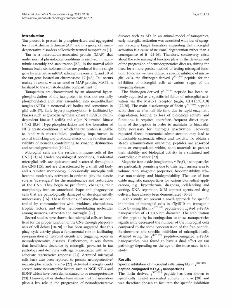
of microglial activity. This peptide, however, possesses ashort half-life when administered in saline solution andneeds to be administered constantly as a result. Tocounteract this issue, we have conjugated the peptide toγ-Fe2O3 nanoparticles. The transmission electron micro-scope (TEM) image of the fibrin γ377-395 peptide-conjugated γ-Fe2O3 nanoparticles shown in Figure 1demonstrates that these nanoparticles are stable againstagglomeration and possess a diameter of 21 ± 3.5 nm.These nanoparticles have been shown to enhance effi-cacy of delivery of bioactive material and to provideprotection against biodegradation [36]. Fibrin γ377-395
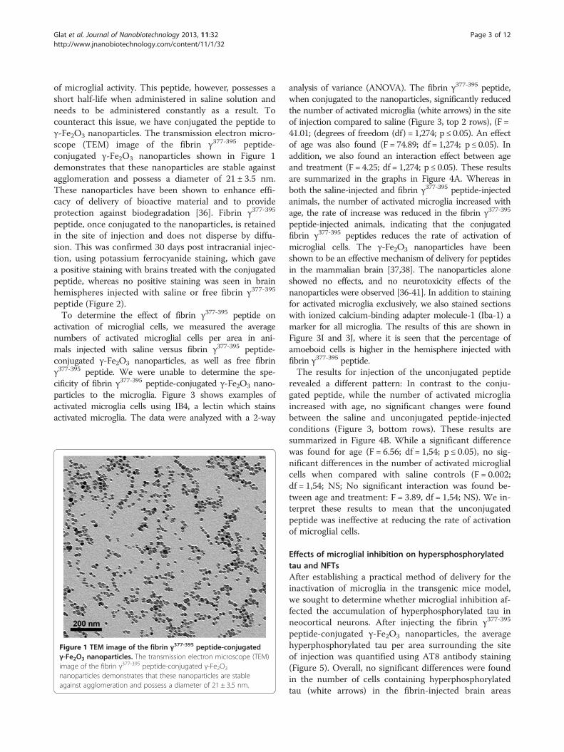
peptide, once conjugated to the nanoparticles, is retainedin the site of injection and does not disperse by diffu-sion. This was confirmed 30 days post intracranial injec-tion, using potassium ferrocyanide staining, which gavea positive staining with brains treated with the conjugatedpeptide, whereas no positive staining was seen in brainhemispheres injected with saline or free fibrin γ377-395
peptide (Figure 2).To determine the effect of fibrin γ377-395 peptide on
activation of microglial cells, we measured the averagenumbers of activated microglial cells per area in ani-mals injected with saline versus fibrin γ377-395 peptide-conjugated γ-Fe2O3 nanoparticles, as well as free fibrinγ377-395 peptide. We were unable to determine the spe-cificity of fibrin γ377-395 peptide-conjugated γ-Fe2O3 nano-particles to the microglia. Figure 3 shows examples ofactivated microglia cells using IB4, a lectin which stainsactivated microglia. The data were analyzed with a 2-way
Figure 1 TEM image of the fibrin γ377-395 peptide-conjugatedγ-Fe2O3 nanoparticles. The transmission electron microscope (TEM)image of the fibrin γ377-395 peptide-conjugated γ-Fe2O3
nanoparticles demonstrates that these nanoparticles are stableagainst agglomeration and possess a diameter of 21 ± 3.5 nm.
analysis of variance (ANOVA). The fibrin γ377-395 peptide,when conjugated to the nanoparticles, significantly reducedthe number of activated microglia (white arrows) in the siteof injection compared to saline (Figure 3, top 2 rows), (F =41.01; (degrees of freedom (df) = 1,274; p ≤ 0.05). An effectof age was also found (F = 74.89; df = 1,274; p ≤ 0.05). Inaddition, we also found an interaction effect between ageand treatment (F = 4.25; df = 1,274; p ≤ 0.05). These resultsare summarized in the graphs in Figure 4A. Whereas inboth the saline-injected and fibrin γ377-395 peptide-injectedanimals, the number of activated microglia increased withage, the rate of increase was reduced in the fibrin γ377-395
peptide-injected animals, indicating that the conjugatedfibrin γ377-395 peptides reduces the rate of activation ofmicroglial cells. The γ-Fe2O3 nanoparticles have beenshown to be an effective mechanism of delivery for peptidesin the mammalian brain [37,38]. The nanoparticles aloneshowed no effects, and no neurotoxicity effects of thenanoparticles were observed [36-41]. In addition to stainingfor activated microglia exclusively, we also stained sectionswith ionized calcium-binding adapter molecule-1 (Iba-1) amarker for all microglia. The results of this are shown inFigure 3I and 3J, where it is seen that the percentage ofamoeboid cells is higher in the hemisphere injected withfibrin γ377-395 peptide.The results for injection of the unconjugated peptide
revealed a different pattern: In contrast to the conju-gated peptide, while the number of activated microgliaincreased with age, no significant changes were foundbetween the saline and unconjugated peptide-injectedconditions (Figure 3, bottom rows). These results aresummarized in Figure 4B. While a significant differencewas found for age (F = 6.56; df = 1,54; p ≤ 0.05), no sig-nificant differences in the number of activated microglialcells when compared with saline controls (F = 0.002;df = 1,54; NS; No significant interaction was found be-tween age and treatment: F = 3.89, df = 1,54; NS). We in-terpret these results to mean that the unconjugatedpeptide was ineffective at reducing the rate of activationof microglial cells.
Effects of microglial inhibition on hypersphosphorylatedtau and NFTsAfter establishing a practical method of delivery for theinactivation of microglia in the transgenic mice model,we sought to determine whether microglial inhibition af-fected the accumulation of hyperphosphorylated tau inneocortical neurons. After injecting the fibrin γ377-395
peptide-conjugated γ-Fe2O3 nanoparticles, the averagehyperphosphorylated tau per area surrounding the siteof injection was quantified using AT8 antibody staining(Figure 5). Overall, no significant differences were foundin the number of cells containing hyperphosphorylatedtau (white arrows) in the fibrin-injected brain areas
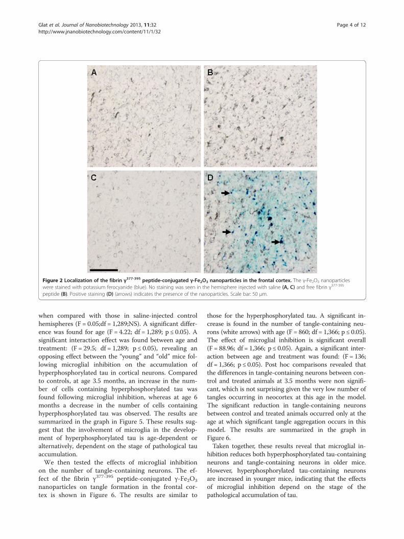
Figure 2 Localization of the fibrin γ377-395 peptide-conjugated γ-Fe2O3 nanoparticles in the frontal cortex. The γ-Fe2O3 nanoparticleswere stained with potassium ferocyanide (blue). No staining was seen in the hemisphere injected with saline (A, C) and free fibrin γ377-395
peptide (B). Positive staining (D) (arrows) indicates the presence of the nanoparticles. Scale bar: 50 μm.
Glat et al. Journal of Nanobiotechnology 2013, 11:32 Page 4 of 12http://www.jnanobiotechnology.com/content/11/1/32
when compared with those in saline-injected controlhemispheres (F = 0.05;df = 1,289;NS). A significant differ-ence was found for age (F = 4.22; df = 1,289; p ≤ 0.05). Asignificant interaction effect was found between age andtreatment: (F = 29.5; df = 1,289; p ≤ 0.05), revealing anopposing effect between the “young” and “old” mice fol-lowing microglial inhibition on the accumulation ofhyperphosphorylated tau in cortical neurons. Comparedto controls, at age 3.5 months, an increase in the num-ber of cells containing hyperphosphorylated tau wasfound following microglial inhibition, whereas at age 6months a decrease in the number of cells containinghyperphosphorylated tau was observed. The results aresummarized in the graph in Figure 5. These results sug-gest that the involvement of microglia in the develop-ment of hyperphosphorylated tau is age-dependent oralternatively, dependent on the stage of pathological tauaccumulation.We then tested the effects of microglial inhibition
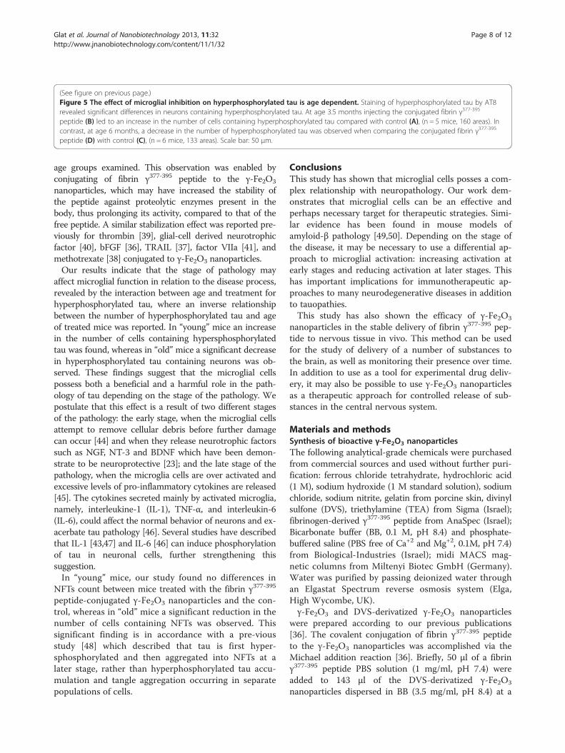
on the number of tangle-containing neurons. The ef-fect of the fibrin γ377-395 peptide-conjugated γ-Fe2O3
nanoparticles on tangle formation in the frontal cor-tex is shown in Figure 6. The results are similar to
those for the hyperphosphorylated tau. A significant in-crease is found in the number of tangle-containing neu-rons (white arrows) with age (F = 860; df = 1,366; p ≤ 0.05).The effect of microglial inhibition is significant overall(F = 88.96; df = 1,366; p ≤ 0.05). Again, a significant inter-action between age and treatment was found: (F = 136;df = 1,366; p ≤ 0.05). Post hoc comparisons revealed thatthe differences in tangle-containing neurons between con-trol and treated animals at 3.5 months were non signifi-cant, which is not surprising given the very low number oftangles occurring in neocortex at this age in the model.The significant reduction in tangle-containing neuronsbetween control and treated animals occurred only at theage at which significant tangle aggregation occurs in thismodel. The results are summarized in the graph inFigure 6.Taken together, these results reveal that microglial in-
hibition reduces both hyperphosphorylated tau-containingneurons and tangle-containing neurons in older mice.However, hyperphosphorylated tau-containing neuronsare increased in younger mice, indicating that the effectsof microglial inhibition depend on the stage of thepathological accumulation of tau.
Figure 3 Fibrin γ377-395 peptide-conjugated γ-Fe2O3 nanoparticles decreased the number of activated microglia. rTg4510 mice at ages3.5 (A, B, E, F) and 6 months (C, D, G, H) were injected given a single intracranial injection unilaterally with saline (left column) or fibrin γ377-395
peptide-conjugated γ-Fe2O3 nanoparticles (B, D) or free fibrin γ377-395 peptide (F, H) and were sacrificed after 30 days. Activated microglial cellswere stained with the lectin IB4 (white arrows). DAPI was used for nuclear staining. Statistical analysis (see Figure 3) revealed a significantreduction in the number of activated microglia following injection with fibrin γ377-395 peptide-conjugated γ-Fe2O3 nanoparticles (compare B andD with A and C). Scale bar: 50 μm. I, J; Iba-1 staining demonstrates resting and activated microglia. I: saline injection; J: following injection withfibrin γ377-395 peptide-conjugated γ-Fe2O3 nanoparticles. The differences in shape of the microglia are clearly seen. Scale bar: 25 μm.
Glat et al. Journal of Nanobiotechnology 2013, 11:32 Page 5 of 12http://www.jnanobiotechnology.com/content/11/1/32
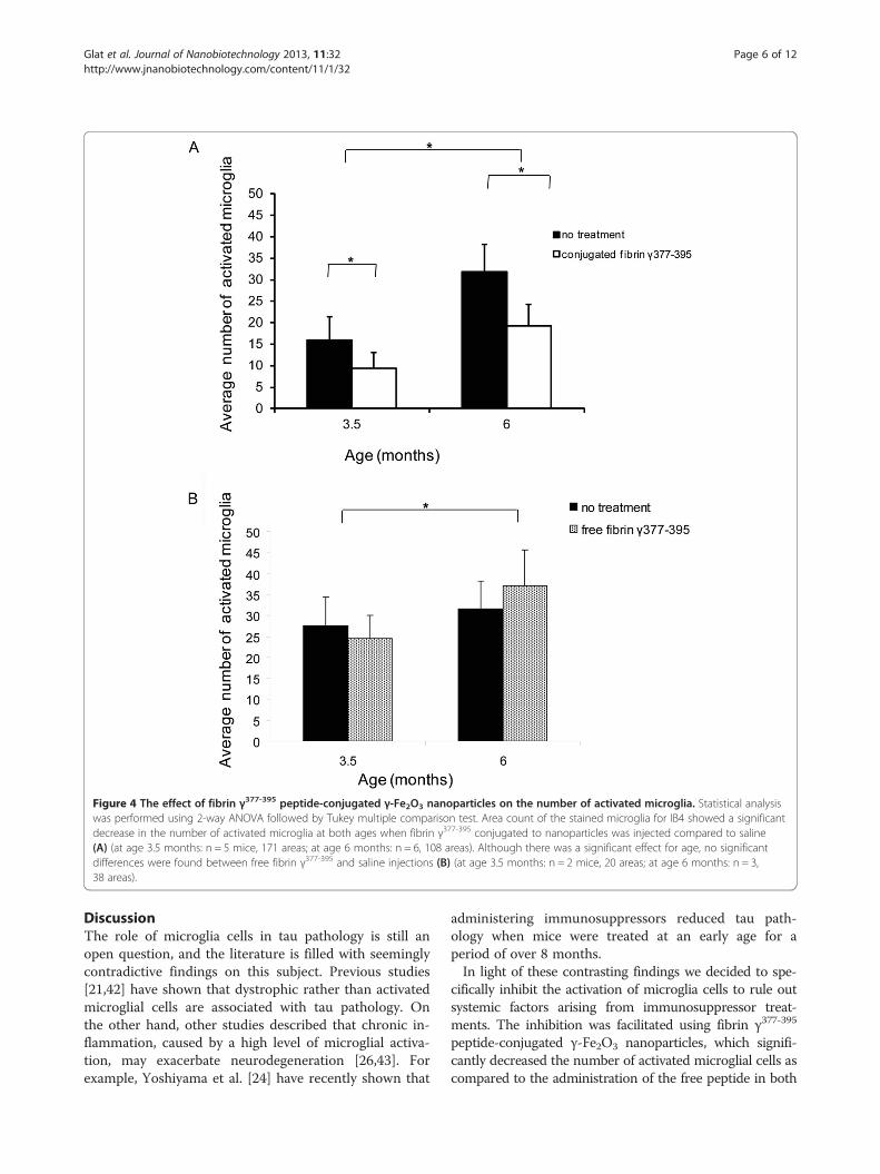
Figure 4 The effect of fibrin γ377-395 peptide-conjugated γ-Fe2O3 nanoparticles on the number of activated microglia. Statistical analysiswas performed using 2-way ANOVA followed by Tukey multiple comparison test. Area count of the stained microglia for IB4 showed a significantdecrease in the number of activated microglia at both ages when fibrin γ377-395 conjugated to nanoparticles was injected compared to saline(A) (at age 3.5 months: n = 5 mice, 171 areas; at age 6 months: n = 6, 108 areas). Although there was a significant effect for age, no significantdifferences were found between free fibrin γ377-395 and saline injections (B) (at age 3.5 months: n = 2 mice, 20 areas; at age 6 months: n = 3,38 areas).
Glat et al. Journal of Nanobiotechnology 2013, 11:32 Page 6 of 12http://www.jnanobiotechnology.com/content/11/1/32
DiscussionThe role of microglia cells in tau pathology is still anopen question, and the literature is filled with seeminglycontradictive findings on this subject. Previous studies[21,42] have shown that dystrophic rather than activatedmicroglial cells are associated with tau pathology. Onthe other hand, other studies described that chronic in-flammation, caused by a high level of microglial activa-tion, may exacerbate neurodegeneration [26,43]. Forexample, Yoshiyama et al. [24] have recently shown that
administering immunosuppressors reduced tau path-ology when mice were treated at an early age for aperiod of over 8 months.In light of these contrasting findings we decided to spe-
cifically inhibit the activation of microglia cells to rule outsystemic factors arising from immunosuppressor treat-ments. The inhibition was facilitated using fibrin γ377-395
peptide-conjugated γ-Fe2O3 nanoparticles, which signifi-cantly decreased the number of activated microglial cells ascompared to the administration of the free peptide in both
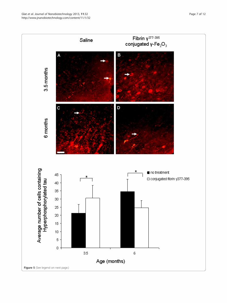
Figure 5 (See legend on next page.)
Glat et al. Journal of Nanobiotechnology 2013, 11:32 Page 7 of 12http://www.jnanobiotechnology.com/content/11/1/32
(See figure on previous page.)Figure 5 The effect of microglial inhibition on hyperphosphorylated tau is age dependent. Staining of hyperphosphorylated tau by AT8revealed significant differences in neurons containing hyperphosphorylated tau. At age 3.5 months injecting the conjugated fibrin γ377-395
peptide (B) led to an increase in the number of cells containing hyperphosphorylated tau compared with control (A), (n = 5 mice, 160 areas). Incontrast, at age 6 months, a decrease in the number of hyperphosphorylated tau was observed when comparing the conjugated fibrin γ377-395
peptide (D) with control (C), (n = 6 mice, 133 areas). Scale bar: 50 μm.
Glat et al. Journal of Nanobiotechnology 2013, 11:32 Page 8 of 12http://www.jnanobiotechnology.com/content/11/1/32
age groups examined. This observation was enabled byconjugating of fibrin γ377-395 peptide to the γ-Fe2O3
nanoparticles, which may have increased the stability ofthe peptide against proteolytic enzymes present in thebody, thus prolonging its activity, compared to that of thefree peptide. A similar stabilization effect was reported pre-viously for thrombin [39], glial-cell derived neurotrophicfactor [40], bFGF [36], TRAIL [37], factor VIIa [41], andmethotrexate [38] conjugated to γ-Fe2O3 nanoparticles.Our results indicate that the stage of pathology may
affect microglial function in relation to the disease process,revealed by the interaction between age and treatment forhyperphosphorylated tau, where an inverse relationshipbetween the number of hyperphosphorylated tau and ageof treated mice was reported. In “young” mice an increasein the number of cells containing hypersphosphorylatedtau was found, whereas in “old” mice a significant decreasein hyperphosphorylated tau containing neurons was ob-served. These findings suggest that the microglial cellspossess both a beneficial and a harmful role in the path-ology of tau depending on the stage of the pathology. Wepostulate that this effect is a result of two different stagesof the pathology: the early stage, when the microglial cellsattempt to remove cellular debris before further damagecan occur [44] and when they release neurotrophic factorssuch as NGF, NT-3 and BDNF which have been demon-strate to be neuroprotective [23]; and the late stage of thepathology, when the microglia cells are over activated andexcessive levels of pro-inflammatory cytokines are released[45]. The cytokines secreted mainly by activated microglia,namely, interleukine-1 (IL-1), TNF-α, and interleukin-6(IL-6), could affect the normal behavior of neurons and ex-acerbate tau pathology [46]. Several studies have describedthat IL-1 [43,47] and IL-6 [46] can induce phosphorylationof tau in neuronal cells, further strengthening thissuggestion.In “young” mice, our study found no differences in
NFTs count between mice treated with the fibrin γ377-395
peptide-conjugated γ-Fe2O3 nanoparticles and the con-trol, whereas in “old” mice a significant reduction in thenumber of cells containing NFTs was observed. Thissignificant finding is in accordance with a pre-viousstudy [48] which described that tau is first hyper-sphosphorylated and then aggregated into NFTs at alater stage, rather than hyperphosphorylated tau accu-mulation and tangle aggregation occurring in separatepopulations of cells.
ConclusionsThis study has shown that microglial cells posses a com-plex relationship with neuropathology. Our work dem-onstrates that microglial cells can be an effective andperhaps necessary target for therapeutic strategies. Simi-lar evidence has been found in mouse models ofamyloid-β pathology [49,50]. Depending on the stage ofthe disease, it may be necessary to use a differential ap-proach to microglial activation: increasing activation atearly stages and reducing activation at later stages. Thishas important implications for immunotherapeutic ap-proaches to many neurodegenerative diseases in additionto tauopathies.This study has also shown the efficacy of γ-Fe2O3
nanoparticles in the stable delivery of fibrin γ377-395 pep-tide to nervous tissue in vivo. This method can be usedfor the study of delivery of a number of substances tothe brain, as well as monitoring their presence over time.In addition to use as a tool for experimental drug deliv-ery, it may also be possible to use γ-Fe2O3 nanoparticlesas a therapeutic approach for controlled release of sub-stances in the central nervous system.
Materials and methodsSynthesis of bioactive γ-Fe2O3 nanoparticlesThe following analytical-grade chemicals were purchasedfrom commercial sources and used without further puri-fication: ferrous chloride tetrahydrate, hydrochloric acid(1 M), sodium hydroxide (1 M standard solution), sodiumchloride, sodium nitrite, gelatin from porcine skin, divinylsulfone (DVS), triethylamine (TEA) from Sigma (Israel);fibrinogen-derived γ377-395 peptide from AnaSpec (Israel);Bicarbonate buffer (BB, 0.1 M, pH 8.4) and phosphate-buffered saline (PBS free of Ca+2 and Mg+2, 0.1M, pH 7.4)from Biological-Industries (Israel); midi MACS mag-netic columns from Miltenyi Biotec GmbH (Germany).Water was purified by passing deionized water throughan Elgastat Spectrum reverse osmosis system (Elga,High Wycombe, UK).γ-Fe2O3 and DVS-derivatized γ-Fe2O3 nanoparticles
were prepared according to our previous publications[36]. The covalent conjugation of fibrin γ377-395 peptideto the γ-Fe2O3 nanoparticles was accomplished via theMichael addition reaction [36]. Briefly, 50 μl of a fibrinγ377-395 peptide PBS solution (1 mg/ml, pH 7.4) wereadded to 143 μl of the DVS-derivatized γ-Fe2O3
nanoparticles dispersed in BB (3.5 mg/ml, pH 8.4) at a
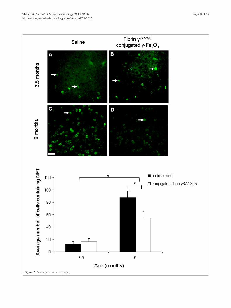
Figure 6 (See legend on next page.)
Glat et al. Journal of Nanobiotechnology 2013, 11:32 Page 9 of 12http://www.jnanobiotechnology.com/content/11/1/32
(See figure on previous page.)Figure 6 The effect of microglial inhibition on NFTs. Staining of NFTs by Thioflavin-S revealed a reduction in neurons containing NFT in miceaged 6 months when comparing saline (C) with fibrin γ377-395 peptide-conjugated γ-Fe2O3 nanoparticles (D) (n = 8 mice, 124 areas). Nodifferences in NFTs were found in mice aged 3.5 months when comparing saline (A) with the treatment hemisphere (B) (n = 4 mice, 246 areas).Scale bar: 50 μm.
Glat et al. Journal of Nanobiotechnology 2013, 11:32 Page 10 of 12http://www.jnanobiotechnology.com/content/11/1/32
[nanoparticles]/[γ377-395] weight ratio of 10. The reactionmixture was then shaken at room temperature for 18 h.Blocking of the residual double bonds was then accom-plished by adding 1% glycine (w/v) and then shaking foran additional hour. The obtained fibrin γ377-395 peptide-conjugated γ-Fe2O3 nanoparticles were than washedfrom non-magnetic waste with PBS using the high gradi-ent magnetic field (HGMF) technique [34]. The concen-tration of the γ377-395 peptide conjugated to thenanoparticles was determined by measuring the un-bound peptide using the Bradford assay and subtractingit from the initial concentration [51].
Animal modelrTg4510 mice were used in this study. Santa Cruz et al.[52] created these transgenic mice using a CaMII alphapromoter driven by a tetracycline operator to focus hu-man mutant P301L tau over-expression in the forebrain(hippocampus and higher cortical layers) of the mice.Nine rTg4510 mice aged 3.5 months (“young”) and 7
rTg4510 aged 6 months (“old”) were used in this study.Mice weight, on average, was 25g. All procedures wereperformed in accordance with the NIH Guide for theCare and Use of Laboratory Animals, and with the Bar-Ilan University guidelines for the use and care of labora-tory animals in research, approved and supervised by theInstitutional Animal Care and Use Committee.
Intracranial injections3.5 and 6 month old rTg4510 mice were injected intra-cranially with the fibrin γ377-395 peptide-conjugated γ-Fe
2O3 nanoparticles and free fibrin γ377-395 peptide (1:3weight ratio). Briefly, mice were anaesthetized beforesurgery with a ketamine:xylazine at 150 and 12 mg/kg,respectively [53]. The animals were then placed into astereotaxic apparatus with a heating pad to maintainbody temperature. Stereotaxic coordinates were bregma +1.5 mm anterior-posterior, 1.5 mm lateral and 1.8 mmvertical for frontal cortex. Subsequently, 10 μL of the fi-brin γ377-395 peptide-conjugated γ-Fe2O3 nanoparticlesdispersed in PBS (2 mg/ml, 10 ug bound fibrin γ377-395
per injection) and free fibrin γ377-395 peptide dissolvedin a PBS solution (3 mg/ml), were intracranially injectedin the right and left hemispheres, respectively. The in-jection holes were then filled with bone wax. The micewere stitched and then returned to their cages. All miceused in our experiments were then sacrificed 30 days
post injection via an intracardial perfusion described inthe next section.
Tissue preparationThe animals were given a lethal dose of thiopental so-dium solution and were perfused intracardially with iso-tonic PBS (pH 7.3) followed by 4% paraformaldehydesolution in 0.1M PBS. Once the perfusions werecomplete, the brains were rapidly removed from theskulls and were placed in a 4% paraformaldehyde solu-tion for post-fixation of at least 24 h at 4°C. After 24 h,the brains were transferred to a 30% sucrose solution fortwo days. The brains were then frozen in dry ice andplaced in −80°C over night. Before sectioning the brainswith a cryostat, the brains were covered using an opti-mal cutting temperature (OCT) solution and wereplaced in −20°C. Once the OCT solidified, the brainswere placed in the cryostat with the frontal side up. Tis-sue samples were selected from areas of cortex adjacentto the injection sites. The brains were sliced to 25 μmcoronal sections. Each sample consisted of 5 slices. Con-trols were the contralateral areas. Sampling within theslices was performed at 3 sites, each 400 × 340 μm. Thebrain sections were then individually picked up using apaint brush and were transferred to a well filled with0.1M PBS. Slices were then either stained immediatelyfollowing the appropriate protocol or stored in a cryo-protectant solution at −20°C until further use.
Staining methodsLectin stainingThe slices were first introduced into a PBS solution inorder to remove the OCT from the tissue. The sliceswere treated with FITC-conjugated Bandeiraea simpli-cifolia isolectin B4 (IB-4; 1:50; Sigma-Aldrich) for 1 h,and then were washed three times for 5 min each inPBS. The slices were then mounted on slides and storedin the dark to air-dry. A drop of DAPI (1:2000; Sigma-Aldrich) solution was added to sections after dryingusing a Pasteur pipette for 1 min and washed twice for 1min each in PBS. The slides were then sealed using acover slip.
Iron stainingThe slices were mounted on slides and placed in 2% po-tassium ferrocyanide solution with 2% HCl solution for30 min. Afterwards, the slides were washed three timesin distilled water and sealed with a cover slip.
Glat et al. Journal of Nanobiotechnology 2013, 11:32 Page 11 of 12http://www.jnanobiotechnology.com/content/11/1/32
ImmunohistochemistryThe slices were first introduced into a PBS solution inorder to remove the OCT from the tissue. The sliceswere incubated in 0.5% Triton for 20 min and thenwashed three times for 5 min each in PBS. The sliceswere then incubated in a blocking solution (NGS, 5% inPBS; from Jackson Immuno Research) for 1 h. After theblocking stage, slices were treated with Anti-Human-PHF-Tau Monoclonal Antibody (AT8; 1:1000 in normalgoat serum; Thermo Scientific) and then left overnightat 4°C. Following incubation, the slices were washedthree times for 5 min each in PBS. The slices weretreated with Cy3 donkey anti-mouse (Cy3 1:500, JacksonImmuno Research). After 1 h, the slices were washedthree times in PBS. The slices were then mounted onslides, stored in the dark to air-dry and then sealed usinga cover slip. If NFTs staining was performed as well, adrop of 0.05% Thio-S solution (Sigma-Aldrich) wasadded to sections after drying using a Pasteur pipette.The sections were then incubated for 8 min in the dark,followed by two washes for 10 sec in 80% ethanol solu-tion and one wash in distilled water. The slides werethen stored in the dark to air-dry and sealed using acover slip.To study microglia morphology, Iba-1 staining was
performed. The endogenous peroxidase was blocked by10% H2O2, 0.01% Triton X-100 in PBS, and 3% Metha-nol. After 30 minutes blocking was performed by 20%NGS for 1 h. Slices were then incubated overnight inrabbit polyclonal anti-IBA1 antibody (1:250; WakoChemicals) with 2% NGS. Following incubation the sec-tions were treated with a secondary antibody, Anti rabbitIgG Peroxidase (1:200; Santa Cruz) for 1 h followed byincubation with ABC solution (Vector) for 30 minutes.The peroxidase labeling was visualized by incubationwith commercial DAB Substrate Kit (Vector) for a fewminutes. The reaction was stopped by placing the slicesin PBS. Slices were then mounted, dehydrated, andcover-slipped.
Statistical analysisTwo to three different areas surrounding the site of injec-tion from five slices from each mouse were defined for theanalysis of activated microglial cells and cells containinghypersphosphorylated tau and NFTs. Statistical analysiswas performed using 2-way-ANOVAs followed by Tukeymultiple comparison tests. A significance level of p ≤ 0.05was set for all statistical tests.
Competing interestsThe authors declare that they have no competing interests.
Authors’ contributionsMG participated in the design of the study, conducted the experiments,analyzed the data, and wrote the manuscript. HS participated in the designof the study, synthesized the bioactive nanoparticles and helped to draft the
manuscript. NMC conducted the experiments. SM and EAS conceived theidea of the project, SM supervised the synthesis of the bioactivenanoparticles, and EAS designed and supervised the biological experiments,analysis and the writing of the manuscript. All authors read and approvedthe final manuscript. All authors have reviewed the manuscript.
AcknowledgementsWe thank Professor George A. Carlson and Ms. Rose Pitstick, McLaughlinResearch Institute, Montana State University, for supplying the breeders forthe rTg4510 mouse colony.
FundingThis work was supported by the Elias, Genevieve and Georgianna AtolCharitable Trust.
Author details1Gonda Multidisciplinary Brain Research Center, Bar-Ilan University,Ramat-Gan 52900, Israel. 2Department of Chemistry, Bar-Ilan Institute ofNanotechnology and Advanced Materials, Ramat-Gan 52900, Israel.3MassGeneral Institute of Neurodegenerative Disease, Massachusetts GenralHospital and Harvard Medical School, Charlestown, MA 02129, USA.
Received: 21 May 2013 Accepted: 16 September 2013Published: 23 September 2013
References1. Spillantini MG, Goedert M: Tau protein pathology in neurodegenerative
diseases. Trends Neurosci 1998, 21:428–433.2. Weingarten MD, Lockwood AH, Hwo SY, Kirschner MW: A protein factor
essential for microtubule assembly. Proc Natl Acad Sci U S A 1975,72:1858–1862.
3. Drubin D, Kobayashi S, Kirschner M: Association of tau protein withmicrotubules in living cells. Ann N Y Acad Sci 1986, 466:257–268.
4. Neve RL, Harris P, Kosik KS, Kurnit DM, Donlon TA: Identification of cDNAclones for the human microtubule-associated protein tau andchromosomal localization of the genes for tau and microtubule-associated protein 2. Brain Res 1986, 387:271–280.
5. Goedert M, Spillantini MG, Jakes R, Rutherford D, Crowther RA: Multipleisoforms of human microtubule-associated protein tau: sequences andlocalization in neurofibrillary tangles of Alzheimer’s disease. Neuron 1989,3:519–526.
6. Kosik KS: The molecular and cellular pathology of Alzheimerneurofibrillary lesions. J Gerontol 1989, 44:55–58.
7. Arrasate M, Perez M, Avila J: Tau dephosphorylation at tau-1 sitecorrelates with its association to cell membrane. Neurochem Res 2000,25:43–50.
8. Kawamata T, Taniguchi T, Mukai H, Kitagawa M, Hashimoto T, Maeda K, OnoY, Tanaka C: A protein kinase, PKN, accumulates in Alzheimerneurofibrillary tangles and associated endoplasmic reticulum-derivedvesicles and phosphorylates tau protein. J Neurosci 1998, 18:7402–7410.
9. Alvarez A, Toro R, Caceres A, Maccioni RB: Inhibition of tauphosphorylating protein kinase cdk5 prevents beta-amyloid-inducedneuronal death. FEBS Lett 1999, 459:421–426.
10. Zhang YJ, Xu YF, Liu YH, Yin J, Wang JZ: Nitric oxide induces tauhyperphosphorylation via glycogen synthase kinase-3beta activation.FEBS Lett 2005, 579:6230–6236.
11. Lee HG, Perry G, Moreira PI, Garrett MR, Liu Q, Zhu X, Takeda A, NunomuraA, Smith MA: Tau phosphorylation in Alzheimer’s disease: pathogen orprotector? Trends Mol Med 2005, 11:164–169.
12. Spires TL, Orne JD, SantaCruz K, Pitstick R, Carlson GA, Ashe KH, Hyman BT:Region-specific dissociation of neuronal loss and neurofibrillarypathology in a mouse model of tauopathy. Am J Pathol 2006,168:1598–1607.
13. Kreutzberg GW: Microglia: a sensor for pathological events in the CNS.Trends Neurosci 1996, 19:312–318.
14. Gonzalez-Scarano F, Baltuch G: Microglia as mediators of inflammatoryand degenerative diseases. Annu Rev Neurosci 1999, 22:219–240.
15. Thomas WE: Brain macrophages: evaluation of microglia and theirfunctions. Brain Res Brain Res Rev 1992, 17:61–74.
16. Aloisi F: Immune function of microglia. Glia 2001, 36:165–179.
Glat et al. Journal of Nanobiotechnology 2013, 11:32 Page 12 of 12http://www.jnanobiotechnology.com/content/11/1/32
17. Hanisch UK: Microglia as a source and target of cytokines. Glia 2002,40:140–155.
18. Cullheim S, Thams S: The microglial networks of the brain and their rolein neuronal network plasticity after lesion. Brain Res Rev 2007, 55:89–96.
19. Trapp BD, Wujek JR, Criste GA, Jalabi W, Yin X, Kidd GJ, Stohlman S,Ransohoff R: Evidence for synaptic stripping by cortical microglia. Glia2007, 55:360–368.
20. Neumann H, Kotter MR, Franklin RJ: Debris clearance by microglia: anessential link between degeneration and regeneration. Brain 2009,132:288–295.
21. Streit WJ, Braak H, Xue QS, Bechmann I: Dystrophic (senescent) rather thanactivated microglial cells are associated with tau pathology and likelyprecede neurodegeneration in Alzheimer’s disease. Acta Neuropathol2009, 118:475–485.
22. Li L, Lu J, Tay SS, Moochhala SM, He BP: The function of microglia, eitherneuroprotection or neurotoxicity, is determined by the equilibriumamong factors released from activated microglia in vitro. Brain Res 2007,1159:8–17.
23. Nakajima K, Honda S, Tohyama Y, Imai Y, Kohsaka S, Kurihara T:Neurotrophin secretion from cultured microglia. J Neurosci Res 2001,65:322–331.
24. Yoshiyama Y, Higuchi M, Zhang B, Huang SM, Iwata N, Saido TC, Maeda J,Suhara T, Trojanowski JQ, Lee VM: Synapse loss and microglial activationprecede tangles in a P301S tauopathy mouse model. Neuron 2007,53:337–351.
25. Gorlovoy P, Larionov S, Pham TT, Neumann H: Accumulation of tauinduced in neurites by microglial proinflammatory mediators. FASEB J2009, 23:2502–2513.
26. Kitazawa M, Oddo S, Yamasaki TR, Green KN, LaFerla FM:Lipopolysaccharide-induced inflammation exacerbates tau pathology bya cyclin-dependent kinase 5-mediated pathway in a transgenic model ofAlzheimer’s disease. J Neurosci 2005, 25:8843–8853.
27. Ugarova TP, Solovjov DA, Zhang L, Loukinov DI, Yee VC, Medved LV, PlowEF: Identification of a novel recognition sequence for integrin alphaMbeta2 within the gamma-chain of fibrinogen. J Biol Chem 1998,273:22519–22527.
28. Adams RA, Bauer J, Flick MJ, Sikorski SL, Nuriel T, Lassmann H, Degen JL,Akassoglou K: The fibrin-derived gamma377-395 peptide inhibitsmicroglia activation and suppresses relapsing paralysis in centralnervous system autoimmune disease. J Exp Med 2007, 204:571–582.
29. Slomkowski S, Gosecki M: Progress in nanoparticulate systems forpeptide, proteins and nucleic acid drug delivery. Curr Pharm Biotechnol2011, 12:1823–1839.
30. de Vries IJM, Lesterhuis WJ, Barentsz JO, Verdijk P, van Krieken JH, BoermanOC, Oyen WJG, Bonenkamp JJ, Boezeman JB, Adema GJ: Magneticresonance tracking of dendritic cells in melanoma patients formonitoring of cellular therapy. Nat Biotechnol 2005, 23:1407–1413.
31. Hergt R, Hiergeist R, Hilger I, Kaiser WA, Lapatnikov Y, Margel S, Richter U:Maghemite nanoparticles with very high AC-losses for application in RF-magnetic hyperthermia. J Magn Magn Mater 2004, 270:345–357.
32. Scherer F, Anton M, Schillinger U, Henke J, Bergemann C, Kruger A,Gansbacher B, Plank C: Magnetofection: enhancing and targeting genedelivery by magnetic force in vitro and in vivo. Gene Ther 2002, 9:102–109.
33. Rudge SR, Kurtz TL, Vessely CR, Catterall LG, Williamson DL: Preparation,characterization, and performance of magnetic iron-carbon compositemicroparticles for chemotherapy. Biomaterials 2000, 21:1411–1420.
34. Margel S, Gura S: Nucleation and growth magnetic metal oxidenanoparticles and its use. Israel patent 2006. No. WO9962079.
35. Perlstein B, Ram Z, Daniels D, Ocherashvilli A, Roth Y, Margel S, Mardor Y:Convection-enhanced delivery of maghemite nanoparticles: increasedefficacy and MRI monitoring. Neuro Oncol 2008, 10:153–161.
36. Skaat H, Ziv O, Shahar A, Margel S: Enhancement of the growth anddifferentiation of nasal olfactory mucosa cells by the conjugation ofgrowth factors to functional nanoparticles. Bioconjugate Chem 2011,22:2600–2610.
37. Margel S, Perlstein B, Brodie C: Polymer nanoparticles coated by magneticmetal oxide and uses thereof. US patent 2009. No. WO 2009/040811.
38. Corem-Salkmon E, Ram Z, Daniels D, Perlstein B, Last D, Salomon S, TamarG, Shneor R, Guez D, Margel S: Convection-enhanced delivery ofmethotrexate-loaded maghemite nanoparticles. Int J Nanomedicine 2011,6:1595–1602.
39. Ziv-Polat O, Topaz M, Brosh T, Margel S: Enhancement of incisional woundhealing by thrombin conjugated iron oxide nanoparticles. Biomaterials2010, 31:741–747.
40. Green-Sadan T, Kuttner Y, Lublin-Tennenbaum T, Kinor N, Boguslavsky Y,Margel S, Yadid G: Glial cell line-derived neurotrophic factor-conjugatednanoparticles suppress acquisition of cocaine self-administration in rats.Exp Neurol 2005, 194:97–105.
41. Shafir G, Galperin A, Margel S: Synthesis and characterization ofrecombinant factor VIIa-conjugated magnetic iron oxide nanoparticlesfor hemophilia treatment. J Biomed Mater Res Part A 2009, 91:1056–1064.
42. Streit WJ: Microglia and neuroprotection: implications for Alzheimer’sdisease. Brain Res Brain Res Rev 2005, 48:234–239.
43. Li Y, Liu L, Barger SW, Griffin WS: Interleukin-1 mediates pathologicaleffects of microglia on tau phosphorylation and on synaptophysinsynthesis in cortical neurons through a p38-MAPK pathway. J Neurosci2003, 23:1605–1611.
44. Rogers J, Strohmeyer R, Kovelowski CJ, Li R: Microglia and inflammatorymechanisms in the clearance of amyloid beta peptide. Glia 2002,40:260–269.
45. Morales I, Farias G, Maccioni RB: Neuroimmunomodulation in thepathogenesis of Alzheimer’s disease. Neuroimmunomodulation 2010,17:202–204.
46. Quintanilla RA, Orellana DI, Gonzalez-Billault C, Maccioni RB: Interleukin-6induces Alzheimer-type phosphorylation of tau protein by deregulatingthe cdk5/p35 pathway. Exp Cell Res 2004, 295:245–257.
47. Mrak RE, Griffin WS: Potential inflammatory biomarkers in Alzheimer’sdisease. J Alzheimers Dis 2005, 8:369–375.
48. Avila J, Lucas JJ, Perez M, Hernandez F: Role of tau protein in bothphysiological and pathological conditions. Physiol Rev 2004, 84:361–384.
49. Garcia-Alloza M, Ferrara BJ, Dodwell SA, Hickey GA, Hyman BT, Bacskai BJ: Alimited role for microglia in antibody mediated plaque clearance in APPmice. Neurobiol Dis 2007, 28:286–292.
50. Koenigsknecht-Talboo J, Meyer-Luehmann M, Parsadanian M, Garcia-AllozaM, Finn MB, Hyman BT, Bacskai BJ, Holtzman DM: Rapid microglialresponse around amyloid pathology after systemic anti-Abeta antibodyadministration in PDAPP mice. J Neurosci 2008, 24:14156–14164.
51. Bradford MM: A rapid and sensitive method for the quantitation ofmicrogram quantities of protein utilizing the principle of protein-dyebinding. Anal Biochem 1976, 72:248–254.
52. Santacruz K, Lewis J, Spires T, Paulson J, Kotilinek L, Ingelsson M, GuimaraesA, DeTure M, Ramsden M, McGowan E, Forster C, Yue M, Orne J, Janus C,Mariash A, Kuskowski M, Hyman B, Hutton M, Ashe KH: Tau suppression ina neurodegenerative mouse model improves memory function. Science2005, 309:476–481.
53. Stern EA, Bacskai BJ, Hickey GA, Attenello FJ, Lombardo JA, Hyman BT:Cortical synaptic integration in vivo is disrupted by amyloid-betaplaques. J Neurosci 2004, 24:4535–4540.
doi:10.1186/1477-3155-11-32Cite this article as: Glat et al.: Age-dependent effects of microglialinhibition in vivo on Alzheimer’s disease neuropathology usingbioactive-conjugated iron oxide nanoparticles. Journal ofNanobiotechnology 2013 11:32.
Submit your next manuscript to BioMed Centraland take full advantage of:
• Convenient online submission
• Thorough peer review
• No space constraints or color figure charges
• Immediate publication on acceptance
• Inclusion in PubMed, CAS, Scopus and Google Scholar
• Research which is freely available for redistribution
Submit your manuscript at www.biomedcentral.com/submit