Embed Size (px)
Citation preview

Age-related macular degeneration
Understanding

2
Contact usWe’re here to answer any questions you have about your eye condition or treatment. If you need further information about age-related macular degeneration or on coping with changes in your vision, then our Helpline is there for you.
Just give us a call on 0303 123 9999 or email us at [email protected] and we’ll be happy to speak with you.
RNIB’s Understanding seriesThe Understanding series is designed to help you, your friends and family understand a little bit more about your eye condition.
The series covers a range of eye conditions, and is available in audio, print and braille formats.

3
Contents4 What is age-related macular
degeneration?
6 Why have I developed AMD?
8 What are the symptoms and when should I seek help?
10 What is the macula?
12 What are the different types of AMD?
16 How is AMD diagnosed?
22 What is the treatment for AMD?
28 Coping
30 Further help and support

4
What is age-related macular degeneration?Age-related macular degeneration (AMD) affects a tiny part of the retina at the back of your eye, called the macula. AMD causes changes to the macula, which leads to problems with your central vision. AMD doesn’t cause pain, and doesn’t lead to the total loss of sight.
AMD affects the vision you use when you’re looking straight at something, for example when you’re reading, looking at photos or watching television. Your central vision can become distorted or blurry, and over time, a blank patch may appear in the centre of your vision.

5

6
Why have I developed AMD?The exact cause for AMD is not known. Some things are thought to make it more likely you’ll develop AMD, such as:
• Your age: AMD develops as people grow older and while it’s most often seen in those aged over 65, it can also develop in people who are in their forties and fifties.
• Your gender: more women have AMD than men, probably because women tend to live longer than men.
• Your genes: certain genes have been found which seem to be linked to the development of AMD in some people. This has been discovered by looking at families with more than one member who has AMD. However, not all AMD is thought to be inherited.
• Your lifestyle: high blood pressure and lack of exercise have been identified as possible risk factors for AMD; therefore, maintaining a healthy weight and living an active lifestyle with regular exercise is recommended.
• Smoking: smoking greatly increases your risk of developing AMD – you can reduce this risk if you stop smoking.

7
• Sunlight: some studies have suggested that exposure to high levels of sunlight (particularly the UV light contained in sunlight) throughout your life may increase your risk of developing AMD, but this has not been proven. However, wearing sunglasses to protect your eyes from the UV light in sunlight is a good idea for everyone throughout their life.
• What you eat: a diet high in fat and low in omega 3 and 6, vitamins and minerals have also been associated with AMD. At the moment, there isn’t an agreement on how much of a risk factor diet can be.
In general, protecting your eyes from the sun, eating a balanced diet with plenty of fresh fruit and vegetables and stopping smoking, keeping active and maintaining a healthy weight and blood pressure may all help to keep your eyes as healthy as possible.
Unfortunately, because the exact cause of AMD is not known, you may develop AMD even if you don’t have any of these risk factors.

8
What are the symptoms and when should I seek help?Everyone can have slightly different symptoms, but usually the first thing you’ll notice is that it’s harder to see detail, such as small print. You may find that your vision has a small blurred area in the centre. Straight lines may look distorted or wavy, or like there’s a little bump in them. You may also find that you’re more sensitive to bright light.

9
You should have your eyes tested by an optometrist (also known as an optician) if you experience any of these in one or both eyes:
• You have difficulty reading small print despite wearing reading glasses.
• Straight lines start to look wavy or distorted. You can check this by looking at door and window frames, or tiles in your home.
• Your vision isn’t as clear as it used to be.
Your optometrist can measure any changes in your vision and look at the back of your eye. They may see “drusen” when they examine your retina. Drusen are small deposits under the retina, which they can see as yellow dots. Drusen can be present as a normal part of ageing and are not always a sign that someone has AMD. Your optometrist might say you have early AMD if you have larger drusen in one or both your eyes.
If your optometrist finds any changes to your macula or any cause for concern, they’ll send a letter to your GP or sometimes directly to the hospital. Based on your optometrist’s letter, the hospital will judge how quickly you need to be seen by the ophthalmologist (also known as a hospital eye doctor), and arrange an appointment for you.
10
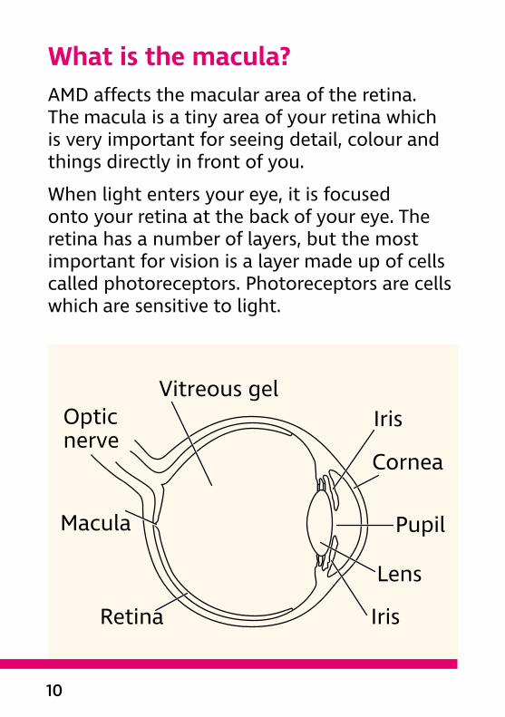
What is the macula?AMD affects the macular area of the retina. The macula is a tiny area of your retina which is very important for seeing detail, colour and things directly in front of you.
When light enters your eye, it is focused onto your retina at the back of your eye. The retina has a number of layers, but the most important for vision is a layer made up of cells called photoreceptors. Photoreceptors are cells which are sensitive to light.
Retina
Macula
Opticnerve
Vitreous gelIris
Iris
Cornea
Pupil
Lens

11
The macula contains a few million specialised photoreceptor cells called cone cells. These cone cells work best in bright light and allow you to see fine detail for activities like reading and writing and recognising colours. When someone develops AMD, the cone cells in the macular area become damaged and stop working as well as they should.
The peripheral retina is further away from the central macula. It is mostly made up of the other type of photoreceptors called rod cells. They allow us to see when light is dim and provide peripheral vision (also known as side vision) outside of the main line of sight. AMD does not affect the peripheral retina, meaning that side vision remains good. AMD does not cause you to lose all your sight.

12
What are the different types of AMD?There are two main types of AMD – “wet” AMD and “dry” AMD. They are called “wet” and “dry” because of what happens inside your eye and what the ophthalmologist sees when examining the inside of your eye, not because of how your eye feels or whether you have a watery or dry eye.
Dry AMDDry AMD is the more common type of AMD. It develops very slowly and causes a gradual change in your central vision. Dry AMD usually takes a long time – sometimes years, to get to its final stage. At its worst, dry AMD causes a blank patch in the centre of your vision in both of your eyes. It doesn’t affect your peripheral vision, so it never leads to total blindness.
Wet AMDAbout 10 to 15 per cent of people who develop AMD have wet AMD, often having had dry AMD to begin with. You develop wet AMD when the cells of the macula stop working correctly and your body starts growing new

13
blood vessels to fix the problem. As these blood vessels grow in the wrong place, they cause swelling and bleeding underneath the macula – this is why it’s called “wet” AMD. This new blood vessel growth is medically known as neovascularisation. It causes more damage to your macula and eventually leads to scarring. Both the new blood vessels and the scarring damage your central vision, and may lead to a blank patch in the centre of your sight.
Wet AMD can develop very quickly, causing serious changes to your central vision in a short period of time, over days or weeks. Treatment is available for wet AMD, which stops the new blood vessels from growing and damaging your macula. This treatment usually needs to be given quickly before the new blood vessels do too much damage to your macula. If the blood vessels are left to grow, the scarring and the sight loss they cause is usually permanent. Wet AMD doesn’t affect your peripheral vision, so it doesn’t lead to total blindness.
Both types of AMDWet and dry AMD have a few things in common. They usually affect both your eyes, though sometimes one eye may be affected long

14
before the other. Both wet and dry AMD only affect your central vision and won’t affect your peripheral vision. Neither type of AMD will cause you to lose all your sight.
Some people diagnosed with dry AMD may find that, with time, new blood vessels grow and they develop wet AMD. If you have dry AMD and you notice a sudden change in either of your eyes, you should let the hospital know as soon as possible. This is because dry AMD can develop into wet AMD, and if this happens, sight-saving treatment may be possible.

15
Some people may have wet AMD in one eye, and dry AMD in the other, which doesn’t develop into wet AMD. Most people, however, have the same type of AMD in both eyes.
People who have had wet AMD for a long time, causing bad scarring on their retina, may be told that their wet AMD has “dried up”. This usually means that there are no new blood vessels growing and that your macula has been badly scarred. At this stage of wet AMD, the treatments available wouldn’t help.
AMD is not painful and it never leads to a complete loss of vision. Most people with AMD keep their peripheral vision, which means that you should still be able to get around on your own and make use of this vision every day.
Some people who have lost a lot of vision because of AMD or another eye condition may experience visual hallucinations – they may see shapes, colours or figures that aren’t really there. This condition is known as Charles Bonnet syndrome (CBS). You can find more information on CBS on RNIB’s website, or by calling our Helpline on 0303 123 9999 to ask for a leaflet.
16
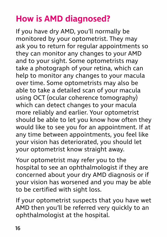
How is AMD diagnosed?If you have dry AMD, you’ll normally be monitored by your optometrist. They may ask you to return for regular appointments so they can monitor any changes to your AMD and to your sight. Some optometrists may take a photograph of your retina, which can help to monitor any changes to your macula over time. Some optometrists may also be able to take a detailed scan of your macula using OCT (ocular coherence tomography) which can detect changes to your macula more reliably and earlier. Your optometrist should be able to let you know how often they would like to see you for an appointment. If at any time between appointments, you feel like your vision has deteriorated, you should let your optometrist know straight away.
Your optometrist may refer you to the hospital to see an ophthalmologist if they are concerned about your dry AMD diagnosis or if your vision has worsened and you may be able to be certified with sight loss.
If your optometrist suspects that you have wet AMD then you’ll be referred very quickly to an ophthalmologist at the hospital.

17
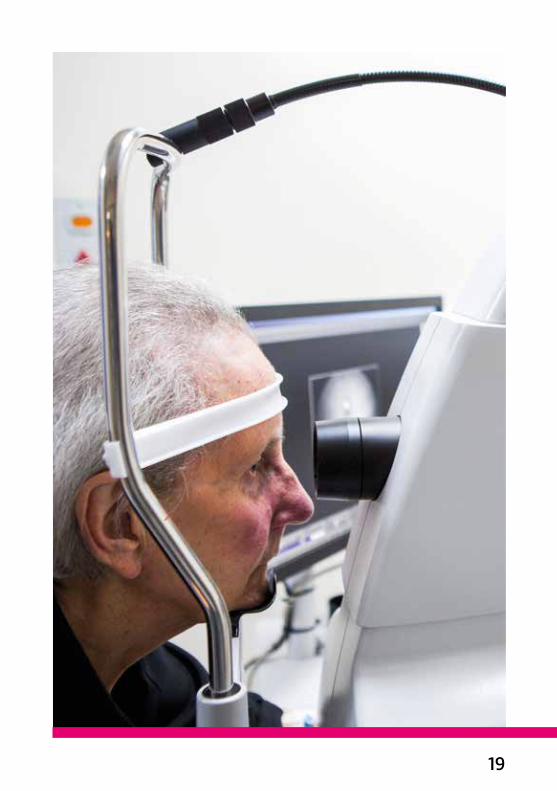
When you are seen at the hospital for your AMD, your vision will be checked by reading the eye chart and your pupils will be dilated (made bigger) by putting in eye drops. This allows the ophthalmologist to look at your macula at the back of the eye and spot any changes that AMD may have caused. Your pupils are dilated with drops that take about 30 minutes to work. The drops will make you sensitive to light and cause your vision to become blurry, but they allow the ophthalmologist to see the inside of your eye more easily. The effect of the drops usually wears off after three to six hours, although sometimes, it may take until the next morning for your vision to feel normal again. It is not safe to drive until the effects have worn off, so you should not drive yourself to your appointment.
Sometimes the ophthalmologist can tell you whether or not you have AMD from just looking at your macula. However, you may need a test called optical coherence tomography (OCT), and sometimes, a fluorescein angiogram, to confirm you have AMD or to find out whether you have wet or dry AMD.

18
Optical coherence tomography (OCT) An OCT is a scanner that provides a cross section image of the retina, showing the layers of the retina and giving a detailed picture of your macula. This helps the ophthalmologist to find out how much fluid is in your macula, to see if your macula is thicker than it should be and to check for any signs of changes.
You’ll need to have some drops to dilate your pupil before the scan. You’ll then be asked to sit in front of the OCT machine, look at a light and keep still while your eye is scanned by the machine. It’s a painless and very quick procedure, which doesn’t involve any physical contact with your eye, and it only takes a couple of minutes to complete.
Another test that the ophthalmologist may use to help in diagnosing as well as monitoring AMD is a fluorescein angiogram.
19

20
Fluorescein angiogram The network of blood vessels underneath your retina can’t usually be seen by looking at the back of your eye. The ophthalmologist can see the damage to your retina, but they can’t see the detail of the blood vessels. A fluorescein angiogram is a way of taking pictures of these blood vessels, which allows the ophthalmologist to see if there are any changes to them which could be causing problems.
The test is carried out using a yellow dye called fluorescein that is injected into your arm. The fluorescein travels through your blood stream to your eye. This usually isn’t painful, but it can make some people feel nauseous or be sick. This dye makes the blood vessels in your eye show up on the pictures taken.
When the dye has been injected, you’ll be asked to look at a special machine. The machine takes pictures of the back of your eye as the dye is travelling through the blood vessels. There will be a series of flashing lights as the pictures are taken, but the test isn’t painful. It usually takes about 10 minutes.
It’s a very common test, and very few people have any major side effects – the most serious

21
one is an allergic reaction, but this rarely occurs. The injection may make your skin look slightly yellow from the dye for up to a day or two. Your urine may also appear a darker yellow than normal (possibly for up to two to three days) but it often fades more quickly than that. Some people are dazzled for a while from the flashing lights, but most people find the test straightforward.
These tests show the blood flow through your vessels and will reveal any bleeding, as well as the type and location of the bleed in your eye. It will also help the ophthalmologist to determine which type of AMD you have, and to decide if any treatment is possible.
22
What is the treatment for AMD?
Treating dry AMDUnfortunately, at the moment there is no way to treat dry AMD. Although research is going on to try and find out why the cells of the macula stop working, this has yet to lead to a proven treatment.
There’s some evidence that high doses of vitamin A, C, E, the minerals zinc and copper and the micro nutrient lutein when taken together may help slow down the progression of dry AMD, particularly if AMD has already caused vision changes in one eye. However, there is no evidence that taking high doses of these vitamins can prevent you from developing AMD in the first place.
There are a number of vitamin products available which have been designed for people with dry AMD and you can usually buy these over the counter or from your pharmacist. A balanced diet with plenty of fresh fruit and vegetables is good for your general health and may also help your eye health.

23
You can find more information about nutrition and the eye on RNIB’s website or by calling our Helpline for a factsheet on 0303 123 9999.

24
Treating wet AMDAnti-VEGFThe treatment available on the NHS for wet AMD is a group of medications called anti-vascular endothelial growth factor (anti-VEGF) drugs. As new blood vessels form in your eye, your body produces a chemical which encourages further new blood vessel growth. Anti-VEGF drugs interfere with this chemical and stop the vessels from growing, minimising further damage to your sight.
The medication is injected into the vitreous, which is a gel-like substance inside your eye. This is called an intravitreal injection. This injection needs to be given in an operating theatre or a “clean room” to avoid infection. A clean room is a sterile room which may not have the full facilities of an operating theatre.
Before the injection, you’ll be given anaesthetic eye drops to make your eye numb, and an antibiotic drop to help prevent you from getting an infection.
The injection shouldn’t be painful thanks to the anaesthetic, but your eye may be a little sore after the anaesthetic wears off. There is a slight chance that the pressure inside your

25
eye may rise a little, but it shouldn’t cause you any pain or change your vision.
The sight in your treated eye may be blurry because of the treatment, but this should wear off within a day. You may notice a small black circle in the lower part of your vision. This is caused by a small air bubble which will disappear after a few hours. You may also have slight swirls in your vision for a few days following the injection, but this doesn’t always happen to everyone. Your eye may water a bit

26
more after the injection and it may be slightly red or irritated. This normally gets better after a few days. If your eye becomes very painful or very red and hot to touch, or if you notice any worsening of your vision, then you should let your hospital know as soon as possible.
The main complications of this treatment are the chance of a rise in pressure in your eye, retinal detachment (where the retina at the back of the eye peels away from its normal position) and eye infections. These complications are rare, happening to less than one per cent of people having the injections. There are treatments available if any of these complications happen to you. If you’re worried about your eye after the injection, then let your hospital know.
Normally, a course of three injections, one a month for three months, is given to start with. Once you’ve had these three injections, your eyes will be checked at the hospital every four to eight weeks. You may be given further injections if your ophthalmologist thinks they are needed. It’s quite common for people to have more injections after the first three.

27
There are different types of anti-VEGF medications and each may require different monitoring schedules. Monitoring and treatment can go on for several years depending on your level of vision and whether you still have active wet AMD. Your ophthalmologist will let you know how often you need to have your eyes checked.
Anti-VEGF treatments usually have a high success rate and in most cases, they stop your sight from getting worse in the short term. In the long term, your vision may gradually worsen even with treatment, but much more slowly than those who have not been treated.
It is important to remember that the main aim of anti-VEGF treatments is to control the swelling and bleeding under the macula by stopping the growth of new blood vessels. It cannot repair any underlying damage that may already be there, which is why you should start your treatment for wet AMD as soon as possible.
You can find more information about anti-VEGF treatment on RNIB’s website or by calling our Helpline on 0303 123 9999.

28
CopingIt’s completely natural to be upset when you’ve been diagnosed with AMD and it’s normal to find yourself worrying about the future and how you will manage with a change in your vision.
It can sometimes be helpful to talk about these feelings with someone outside of your circle of friends or family. At RNIB, we can help with our telephone Helpline and our Sight Loss Counselling team. Your GP or social worker may also find a counsellor for you if you feel this might help.
Your eye clinic may also have a sight loss adviser (also known as an Eye Clinic Liaison Officer or ECLO), who can be on hand to provide you with further practical and emotional support about your eye condition.
The Macular Society has local groups which meet throughout the country and also offer a telephone counselling service. Sometimes, it can help to talk about your feelings or share your experience with people who may have had similar experiences.

29

30
Further help and supportBoth types of AMD can cause severe problems with your central vision. However, most people with AMD have some vision and there are a lot of things you can do to make the most of your remaining vision. This may mean making things bigger, using brighter lighting, or using colour to make things easier to see. We have a series of leaflets with helpful information on living with sight loss, including how to make the most of your sight. You can find out more about our range of titles by calling our Helpline.
Ask your ophthalmologist, optometrist or GP about low vision aids, such as a magnifier, and ask for a referral to your local low vision service. Your low vision service or the Macular Society can also provide eccentric viewing training. Eccentric viewing is a technique of reading using an area in your peripheral vision when you have central vision loss. There is further information on low vision and eccentric training on our website or from the Macular Society.

31
You should also ask your ophthalmologist whether you’re eligible to register as sight impaired (partially sighted) or severely sight impaired (blind). Registration can act as your passport to expert help and sometimes to financial concessions. Even if you aren’t registered, a lot of this support is still available to you.
Local social services should be able to give you information on staying safe in your home and getting out and about safely. They should also be able to offer you some practical mobility training to give you more confidence when you are out.
Being diagnosed with AMD does not automatically mean you are no longer able to drive a car. Speak to your optometrist or ophthalmologist about whether you can still continue to drive, and if you need to report your eye condition to the Driver and Vehicle Licensing Authority (DVLA).

32
If you have questions about anything you’ve read in this publication, please get in touch with us.
Our Helpline is your direct line to the support, advice and services you need. Whether you want to know more about your eye condition, buy a product from our shop, join our library, find out about possible benefit entitlements, or be put in touch with a trained counsellor, we’re only a call away.
It’s also a way for you to join RNIB Connect, our community for anyone affected by sight loss. RNIB Connect is free to join and you’ll have the chance to meet other people with similar experiences in our helpful, welcoming and supportive community.
Give us a call today to find out how we can help you.
RNIB Helpline 0303 123 9999 [email protected]
We’re ready to answer your call Monday to Friday 8am to 8pm and Saturday 9am to 1pm.

33
You can also get in touch by post or by visiting our website:
RNIB 105 Judd Street London WC1H 9NE rnib.org.uk
Other useful contactsMacular SocietyPO Box 1870Andover SP10 9AD0300 3030 111macularsociety.org
Driver and Vehicle Licensing Authority (DVLA)Drivers’ Medical EnquiriesSwansea SA99 1TU0300 790 6806 www.dvla.gov.uk

34
We value your feedbackYou can help us improve this publication by letting us know what you think about it. Please complete and return the form opposite to:
RNIB Eye Health Information105 Judd StreetLondonWC1H 9NE
You can also email us at [email protected]
Please include your contact details if you’re requesting information.

35
1. Where did you receive your copy of this publication?
2. Did you find the information easy to read and understand? Please give details of anything you feel could be improved.

36
3. Is there any information you would have found helpful, that was missing?
4. Do you have any other comments about this publication or any aspect of your contact with RNIB?
UAMD 1006/07/2016

37
Information sourcesRNIB and The Royal College of Ophthalmologists do all we can to ensure that the information we supply is accurate, up to date and in line with the latest research and expertise.
This publication uses information from:
• The Royal College of Ophthalmologists’ guidelines for treatment
• clinical research and studies obtained through literature reviews
• specific support groups for individual conditions
• medical text books• RNIB publications and research.
For a full list of references and information sources used in the compilation of this publication, email [email protected].

38
About The Royal College of OphthalmologistsThe Royal College of Ophthalmologists champions excellence in the practice of ophthalmology and is the only professional membership body for medically qualified ophthalmologists.
The College is unable to offer direct advice to patients. If you’re concerned about the health of your eyes, you should seek medical advice from your GP or ophthalmologist.
rcophth.ac.uk

39
©RNIB reg charity in England and Wales (226227), Scotland (SC039316), Isle of Man (1226). Also operating in Northern Ireland.SE
1808
04
If you or someone you know is living with sight loss, we’re here to help.
RNIB Helpline0303 123 [email protected]
Ask RNIB is the simple and easy way to find the answers to your questions online – try it today at rnib.org.uk/ask
This publication has been produced jointly by RNIB and The Royal College of Ophthalmologists.
Produced date February 2019 Review date February 2022
PR10006 ISBN 978-1-85878-697-1 Ed 3


![Precision medicine for age-related macular degeneration ... · 3 Age-related macular degeneration (AMD) is the primary cause of irreversible blindness in developed countries [1].4](https://img.pdfslide.net/doc/110x75/5f4c3164835c03225a766e33/precision-medicine-for-age-related-macular-degeneration-3-age-related-macular.jpg)