Embed Size (px)
Citation preview
American Journal of Medical Genetics 34:266-267 (1989)
Letter to the Editor Agnathia, Holoprosencephaly, and Situs Inversus
To The Editor:
Leech et al. [1988] were first to document a case of agnathia with both holoprosencephaly and situs in- versus. As they pointed out, agnathia with holoprosen- cephaly and agnathia with situs inversus have been previously reported [Pauli et al., 19811, but their case displaying the triad was unique.
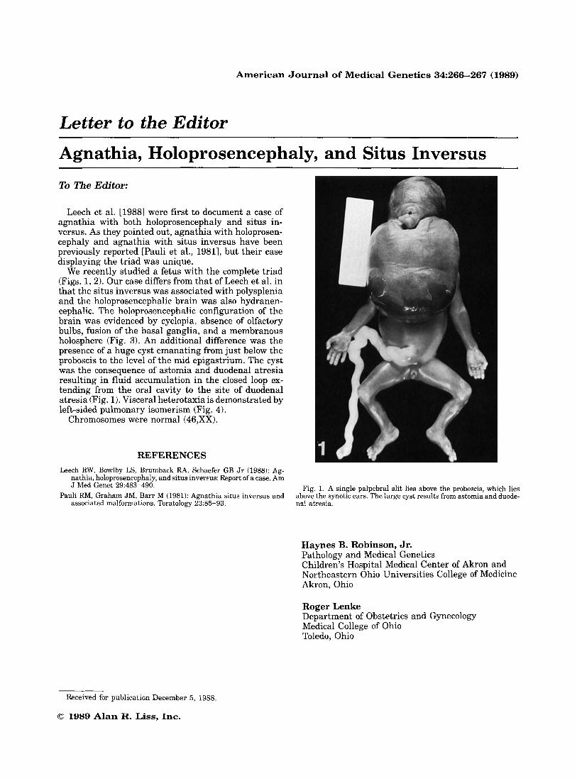
We recently studied a fetus with the complete triad (Figs. 1,2). Our case differs from that of Leech et al. in that the situs inversus was associated with polysplenia and the holoprosencephalic brain was also hydranen- cephalic. The holoprosencephalic configuration of the brain was evidenced by cyclopia, absence of olfactory bulbs, fusion of the basal ganglia, and a membranous holosphere (Fig. 3). An additional difference was the presence of a huge cyst emanating from just below the proboscis to the level of the mid epigastrium. The cyst was the consequence of astomia and duodenal atresia resulting in fluid accumulation in the closed loop ex- tending from the oral cavity to the site of duodenal atresia (Fig. 1). Visceral heterotaxia is demonstrated by left-sided pulmonary isomerism (Fig. 4).
Chromosomes were normal (46,XX).
REFERENCES Leech RW, Bowlby LS, Brumback RA, Schaefer GB Jr (1988): Ag-
nathia, holoprosencephaly, and situs inversus: Report of a case. Am J Med Genet 29:483-490.
Pauli RM, Graham JM, Barr M (1981): Agnathia situs inversus and associated malformations. Teratology 23:85-93.
Fig. 1. A single palpebral slit lies above the proboscis, which lies above the synotic ears. The large cyst results from astomia and duode- nal atresia.
Haynes B. Robinson, Jr. Pathology and Medical Genetics Children’s Hospital Medical Center of Akron and Northeastern Ohio Universities College of Medicine Akron, Ohio
Roger Lenke Department of Obstetrics and Gynecology Medical College of Ohio Toledo, Ohio
Received for publication December 5, 1988.
0 1989 Alan R. Liss, Inc.
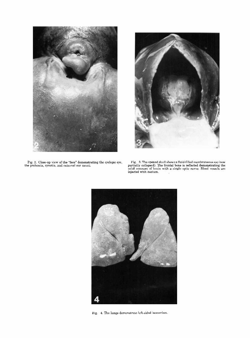
Fig. 2. Close-up view of the “face” demonstrating the cyclopic eye, Fig. 3. The opened skull shows a fluid-filled membraneous sac (now partially collapsed). The frontal bone is reflected demonstrating the solid remnant of brain with a single optic nerve. Blood vessels are injected with barium.
the proboscis, synotia, and external ear meati.
Fig. 4. The lungs demonstrate left-sided isomerism.