Embed Size (px)
Citation preview
CE: Namrta; AIDS-D-13-01409; Total nos of Pages: 12;
AIDS-D-13-01409
Highly pathogenic adapte
d HIV-1 strains limit hostimmunity and dictate rapid disease progressionJudith Dalmaua,M, Margalida Rotgerb,M, Itziar Erkiziaa, Andri Rauchc,
Pedro Reched, Maria Pinoa, Anna Estevef, Eduard Paloug,
Christian Brandera,i, Roger Paredesa, Pham Phunge,
Bonaventura Cloteta,h, Amalio Telentib, Javier Martinez-Picadoa,h,i,
Julia G. Pradoa, The CoRP Study Group
Copyright © L
aAIDS Research InBarcelona, BadalocUniversity ClinicComplutense de MEpidemiologics sohUniversitat de Vi
Correspondence toCanyet S/N, 0891
Tel: +34 93 465 6�
Judith Dalmau a
Received: 18 Dec
DOI:10.1097/QAD
IS
Objective: The study of HIV-1 rapid progressors has been limited to specific casereports. Nevertheless, identification and characterization of the viral and host factorsinvolved in rapid progression are crucial when attempting to uncover the correlates ofrapid disease outcome.
Design: We carriedout comparative functional analyses in rapid progressors (n¼46) andstandard progressors (n¼46) early after HIV-1 seroconversion (�1 year). The viral traitstested were viral replicative capacity, co-receptor usage, and genomic variation. HostCD8þ T-cell responses, humoral activity, and HLA immunogenetic markers were alsodetermined.
Results: Our data demonstrate an unusual convergence of highly pathogenic HIV-1strains in rapid progressors. Compared with standard progressors, rapid progressorviral strains show higher in-vitro replicative capacity (81.5 vs. 67.9%; P¼0.025) andgreater X4/DM co-receptor usage (26.3 vs. 2.8%; P¼0.006) in early infection.Limited or absent functional HIV-1 CD8þ T-cell responses and neutralizing activitywere measured in rapid progressors. Moreover, the increase in common HLAallele-restricted CD8þ T-cell escape mutations in rapid progressors acts as a signatureof uncontrolled HIV-1 replication and early impairment of adaptive cellularresponses.
Conclusion: Our data support a dominant role for viral factors in rapid progressors.Robust HIV-1 replication and intrinsic viral properties limit host adaptive immuneresponses, thus driving rapid disease progression.
� 2014 Wolters Kluwer Health | Lippincott Williams & Wilkins
AIDS 2014, 28:000–000
Keywords: HIV-1, host factors, pathogenesis, rapid progressors, viralfactors
ippincott Williams & Wilkins. Unauthorized reproduction of this article is prohibited.
stitute -IrsiCaixa-, Institut d’Investigacio en Ciencies de la Salut Germans Trias i Pujol, Universitat Autonoma dena, Spain, bInstitute of Microbiology, University Hospital Center and University of Lausanne, Lausanne,of Infectious Diseases, University Hospital Bern and University of Bern, Bern, Switzerland, dUniversidadadrid, Madrid, Spain, eMonogram Biosciences, South San Francisco, California, USA, fCentre d’Estudis
bre les Infeccions de Transmissio Sexual i Sida de Catalunya (CEEISCAT), Badalona, gBanc de Sang i Teixits,c, and iInstitucio Catalana de Recerca i Estudis Avancats (ICREA), Barcelona, Spain.
Julia G. Prado, PhD, AIDS Research Institute – IrsiCaixa – Hospital Universitari Germans Trias i Pujol, Crta6 Badalona, Barcelona, Spain.
3 74 ext 171; e-mail: [email protected]
nd Margalida Rotger contributed equally to the writing of this article.
ember 2013; revised: 26 March 2014; accepted: 27 March 2014.
.0000000000000293
SN 0269-9370 Q 2014 Wolters Kluwer Health | Lippincott Williams & Wilkins 1
Co
CE: Namrta; AIDS-D-13-01409; Total nos of Pages: 12;
AIDS-D-13-01409
2 AIDS 2014, Vol 00 No 00
Introduction
The clinical outcome of HIV-1-infected patients variesconsiderably. In the absence of antiretroviral therapy, themedian time to development of AIDS has been estimatedto vary from 7.7 to 11.0 years, depending on age at sero-conversion [1]. Nevertheless, some individuals are able tocontrol HIV-1 replication for longer periods (HIV-1 con-trollers and elite controllers), whereas others progressquickly to AIDS or meet the criteria for initiation ofantiretroviral treatment within the first 3 years after sero-conversion (rapid progressors). Thus, research intoextreme phenotypes can provide us with invaluable dataon the contribution of viral and host factors to HIV-1disease outcome [2–4].
Most studies on extreme HIV-1 phenotypes have focusedon HIV-1 controllers and elite controllers as models fordefining the correlates of protective immunity and controlof viral replication [3,5]. The study of rapid progressors, onthe contrary, has proved particularly challenging, becauseidentification of this phenotype requires a reliable estima-tion of the seroconversion date within a small observationwindow [4]. This limitation is even more remarkable if weconsider that multiple biological samples need to be takenfor analysis. Consequently, very few core studies haveanalysed rapid progressors, and the data reported thus farare heterogeneous [6–8]. Nevertheless, the factors contri-buting to rapid disease progression must be uncovered toimprove the clinical management of these individuals.
The present study is based on a previous case report studyanalysing extremely severe progression of HIV-1 infection[7] and attempts to broadly characterize rapidprogressors infunctional terms by assessing viral and host factors. Ourobjective was to provide new insights into the causes ofrapid disease progression. We recruited anunprecedentedlylarge group of rapid progressors and systematically com-pared them to HIV-1-infected patientswith average diseaseprogression. Our selection criteria for rapid progressorswere based on the reduction in CD4þ T-cell counts to lessthan 350 cells/ml within 3 years after documentedseroconversion in the absence of antiretroviral treatment,as previously validated by our genomic studies [4].
Our findings reveal a profile of HIV-1-specific traits (e.g.replicative capacity, co-receptor usage, and virus varia-bility) that is common to rapid progressors. Additionaldifferences between groups were observed in cellular andhumoral immune profiles and in HLA class I immuno-genetics.
Material and methods
Cohort description and study groupsWe established two progression cohorts: one fromIrsiCaixa (Barcelona, Spain) and one from the Swiss
pyright © Lippincott Williams & Wilkins. Unautho
HIV Cohort (Lausanne, Switzerland). The cohorts weredefined based on an HIV-1 seroconversion window of lessthan 1 year and documented negative and positiveserology result. Disease progression stage was establishedduring the 3 years after seroconversion (Fig S1, http://links.lww.com/QAD/A517). Patients in the first cohort(CoRP, n¼ 106) had two or more CD4þ T-cell countsbelow 350 cells/ml with no subsequent increase in theabsence of antiretroviral treatment. Individuals withaverage progression or standard progressors (CoSP,n¼ 84) maintained CD4þ T-cell counts above350 cells/ml over the same period in the absence ofantiretroviral treatment. From these two cohorts, weselected two study groups: the rapid progressors (n¼ 46)and the standard progressors (n¼ 46). These groups wereselected based on clinical data and sample availability earlyafter HIV-1 seroconversion at baseline (median �1 year)and a median of 1.5 years from initial sampling post-baseline. Samples were distributed and prioritizedaccording to functional analysis (for details, see Fig S1,http://links.lww.com/QAD/A517). The characteristicsof both groups are summarized in Table 1. Differences inthe mode of HIV-1 transmission between the groups weredue to an increase in the number of intravenous drug users(IVDUs) in rapid progressors. Consistency of the resultswas demonstrated by sensitivity analyses in all data setsregardless of the presence of IVDUs in rapid progressors.The project was approved by the institutional reviewboards of Hospital Germans Trias i Pujol and theUniversity Hospital of Lausanne. Participants gave theirwritten informed consent to participate.
HIV-1 isolation, replicative capacity, andco-receptor usageViral isolates were obtained from plasma samples usinganti-CD44 beads (Miltenyi Biotec, Germany [7,9]. Viralisolates from cryopreserved PBMCs were obtained byco-culture as described in reference [7]. Virus replicativecapacity was determined for all isolates. Viral growth wasmonitored by p24 ELISA in supernatants (Perkin Elmer,Spain), and replicative capacity was calculated by the slopeof p24 production in the exponential growth phase [10].Tropism from viral isolates was measured in U87-immortalized cell lines expressing CCR5 or CXCR4[11]. Virus tropism was assessed in parallel in plasmasamples using the enhanced sensitivity tropism assay(ESTA).
HLA typing and assessment of HIV-1-specificCD8R T-cell responsesHigh-resolution HLA class I typing for alleles A, B, andCw was performed by sequence-based typing methods.Comprehensive HIV-1 epitope screening of optimalresponses was carried out using the interferon-g (IFN-g)ELISpot assay in a subset of patients, as previouslydescribed [12] (Table S1, http://links.lww.com/QAD/A517). Wells were considered positive if they contained atleast 50 spot-forming cells per 106 PBMCs above the
rized reproduction of this article is prohibited.
CE: Namrta; AIDS-D-13-01409; Total nos of Pages: 12;
AIDS-D-13-01409
Rapid progression of HIV-1 infection Dalmau et al. 3
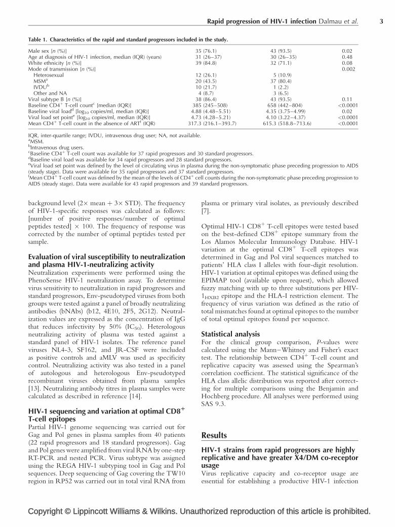
Table 1. Characteristics of the rapid and standard progressors included in the study.
Male sex [n (%)] 35 (76.1) 43 (93.5) 0.02Age at diagnosis of HIV-1 infection, median (IQR) (years) 31 (26–37) 30 (26–35) 0.48White ethnicity [n (%)] 39 (84.8) 32 (71.1) 0.08Mode of transmission [n (%)] 0.002
Heterosexual 12 (26.1) 5 (10.9)MSMa 20 (43.5) 37 (80.4)IVDUb 10 (21.7) 1 (2.2)Other and NA 4 (8.7) 3 (6.5)
Viral subtype B [n (%)] 38 (86.4) 43 (93.5) 0.11Baseline CD4þ T-cell countc [median (IQR)] 385 (245–508) 658 (442–804) <0.0001Baseline viral loadd [log10 copies/ml, median (IQR)] 4.88 (4.48–5.51) 4.35 (3.75–4.99) 0.02Viral load set pointe [log10 copies/ml, median (IQR)] 4.73 (4.28–5.21) 4.10 (3.22–4.37) <0.0001Mean CD4þ T-cell count in the absence of ARTf (IQR) 317.3 (216.1–393.7) 615.3 (518.8–713.6) <0.0001
IQR, inter-quartile range; IVDU, intravenous drug user; NA, not available.aMSM.bIntravenous drug users.cBaseline CD4þ T-cell count was available for 37 rapid progressors and 30 standard progressors.dBaseline viral load was available for 34 rapid progressors and 28 standard progressors.eViral load set point was defined by the level of circulating virus in plasma during the non-symptomatic phase preceding progression to AIDS(steady stage). Data were available for 35 rapid progressors and 37 standard progressors.fMean CD4þ T-cell count was defined by the mean of the levels of CD4þ cell counts during the non-symptomatic phase preceding progression toAIDS (steady stage). Data were available for 43 rapid progressors and 39 standard progressors.
background level (2� mean þ 3� STD). The frequencyof HIV-1-specific responses was calculated as follows:[number of positive responses/number of optimalpeptides tested] � 100. The frequency of response wascorrected by the number of optimal peptides tested persample.
Evaluation of viral susceptibility to neutralizationand plasma HIV-1-neutralizing activityNeutralization experiments were performed using thePhenoSense HIV-1 neutralization assay. To determinevirus sensitivity to neutralization in rapid progressors andstandard progressors, Env-pseudotyped viruses from bothgroups were tested against a panel of broadly neutralizingantibodies (bNAbs) (b12, 4E10, 2F5, 2G12). Neutral-ization values are expressed as the concentration of IgGthat reduces infectivity by 50% (IC50). Heterologousneutralizing activity of plasma was tested against astandard panel of HIV-1 isolates. The reference panelviruses NL4-3, SF162, and JR-CSF were includedas positive controls and aMLV was used as specificitycontrol. Neutralizing activity was also tested in a panelof autologous and heterologous Env-pseudotypedrecombinant viruses obtained from plasma samples[13]. Neutralizing antibody titres in plasma samples werecalculated as described in reference [14].
HIV-1 sequencing and variation at optimal CD8R
T-cell epitopesPartial HIV-1 genome sequencing was carried out forGag and Pol genes in plasma samples from 40 patients(22 rapid progressors and 18 standard progressors). Gagand Pol genes were amplified from viral RNA by one-stepRT-PCR and nested PCR. Virus subtype was assignedusing the REGA HIV-1 subtyping tool in Gag and Polsequences. Deep sequencing of Gag covering the TW10region in RP52 was carried out in total viral RNA from
Copyright © Lippincott Williams & Wilkins. Unaut
plasma or primary viral isolates, as previously described[7].
Optimal HIV-1 CD8þ T-cell epitopes were tested basedon the best-defined CD8þ epitope summary from theLos Alamos Molecular Immunology Database. HIV-1variation at the optimal CD8þ T-cell epitopes wasdetermined in Gag and Pol viral sequences matched topatients’ HLA class I alleles with four-digit resolution.HIV-1 variation at optimal epitopes was defined using theEPIMAP tool (available upon request), which allowedfuzzy matching with up to three substitutions per HIV-1HXB2 epitope and the HLA-I restriction element. Thefrequency of virus variation was defined as the ratio oftotal mismatches found at optimal epitopes to the numberof total optimal epitopes found per sequence.
Statistical analysisFor the clinical group comparison, P-values werecalculated using the Mann–Whitney and Fisher’s exacttest. The relationship between CD4þ T-cell count andreplicative capacity was assessed using the Spearman’scorrelation coefficient. The statistical significance of theHLA class allelic distribution was reported after correct-ing for multiple comparisons using the Benjamin andHochberg procedure. All analyses were performed usingSAS 9.3.
Results
HIV-1 strains from rapid progressors are highlyreplicative and have greater X4/DM co-receptorusageVirus replicative capacity and co-receptor usage areessential for establishing a productive HIV-1 infection
horized reproduction of this article is prohibited.
Co
CE: Namrta; AIDS-D-13-01409; Total nos of Pages: 12;
AIDS-D-13-01409
4 AIDS 2014, Vol 00 No 00
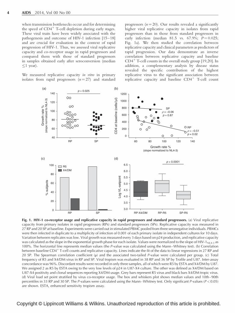
when transmission bottlenecks occur and for determiningthe speed of CD4þ T-cell depletion during early stages.These viral traits have been widely associated with thepathogenesis and outcome of HIV-1 infection [15–18]and are crucial for evaluation in the context of rapidprogression of HIV-1. Thus, we assessed viral replicativecapacity and co-receptor usage in rapid progressors andcompared them with those of standard progressorsin samples obtained early after seroconversion (median�1 year).
We measured replicative capacity in vitro in primaryisolates from rapid progressors (n¼ 27) and standard
pyright © Lippincott Williams & Wilkins. Unautho
(a)
100
Gro
wth
rat
e %
(slo
pe n
orm
aliz
ed to
NL4
-3)
120
140
80
60
40RP
p = 0.025
SP
100
(c)
R5X4/DM
80
% s
ubje
cts
60
40
20
0RP SP
(b)
CD
4 at
bas
elin
e (c
ells
//µl)
120
100
80
60
40
20
(d)
6
6
5
5
Log
VL-
setp
oint
4
4
3
3
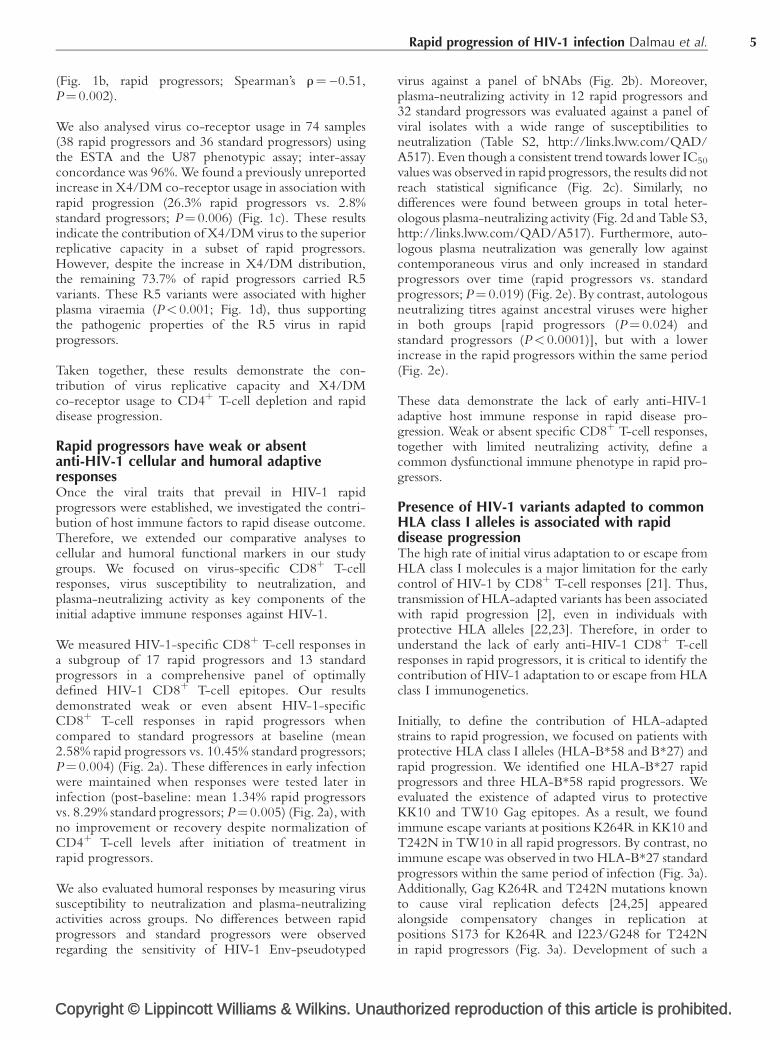
Fig. 1. HIV-1 co-receptor usage and replicative capacity in rapicapacity from primary isolates in rapid progressors (RPs) and stand27 RP and 20 SP at baseline. Experiments were carried out in stimulawere then infected in duplicate to a multiplicity of infection of 0.00Variation between replicates was low. Viral growth was measured ewas calculated as the slope in the exponential growth phase for each100%. The horizontal line represents median values (the P-value wbetween baseline CD4þ T-cell counts and replicative capacity. Line20 SP. The Spearman correlation coefficient (r) and the associafrequency of R5 and X4/DM virus in RP and SP. Viral tropism wasconcordance was 96%. Discordant results were recorded in only thrWe assigned 2 as R5 by ESTA owing to the very low levels of p24 iU87-X4 positivity and clonal sequences reporting X4/DM usage. G(d) Viral load set point stratified by virus co-receptor usage. Thepercentiles in 33 RP and 30 SP. The P-values were calculated usingare shown. ESTA, enhanced sensitivity tropism assay.
progressors (n¼ 20). Our results revealed a significantlyhigher viral replicative capacity in isolates from rapidprogressors than in those from standard progressors inearly infection (median 81.5 vs. 67.9%; P¼ 0.025;Fig. 1a). We then studied the correlation betweenreplicative capacity and clinical parameters as predictors ofrapid progression. Our data demonstrate an inversecorrelation between replicative capacity and baselineCD4þ T-cell counts in the overall study group [19,20]. Inaddition, a complementary analysis by disease statusrevealed the specific contribution of the highestreplicative virus to the significant association betweenreplicative capacity and baseline CD4þ T-cell count
rized reproduction of this article is prohibited.
100
Growth rate %(slope normalized to NL4-3)
120 1408060
SPρ = –0.06p = 0.87
RPρ = –0.51p = 0.02
0
0
0
0
0
0
0
RP-X4/DM RP-R5 SP-R5
p < 0.0001.5
.0
.5
.0
.5
.0
.5
.0
d progressors and standard progressors. (a) Viral replicativeard progressors (SPs). Replicative capacity was measured inted PBMC pooled from three seronegative individuals. PBMCs1 of each primary isolate in independent cultures for 10 days.very 3 days based on p24 production, and replicative capacityisolate. Values were normalized to the slope of HIV-1NL4-3 asas calculated using the Mann–Whitney test). (b) Correlations indicate the fit of the data to linear regressions in 27 RP and
ted two-tailed P-value were calculated per group. (c) Totalevaluated in 38 RP and 36 SP by Trofile and U87. Inter-assayee samples, all of which were R5 by ESTA and X4/DM by U87.n U87-X4 culture. The other was defined as X4/DM based onrey bars represent R5 virus and black bars X4/DM tropic virus.box and whiskers plot shows median values and 10th–90ththe Mann–Whitney test. Only significant P-values (P<0.05)
CE: Namrta; AIDS-D-13-01409; Total nos of Pages: 12;
AIDS-D-13-01409
Rapid progression of HIV-1 infection Dalmau et al. 5
(Fig. 1b, rapid progressors; Spearman’s r¼ –0.51,P¼ 0.002).
We also analysed virus co-receptor usage in 74 samples(38 rapid progressors and 36 standard progressors) usingthe ESTA and the U87 phenotypic assay; inter-assayconcordance was 96%. We found a previously unreportedincrease in X4/DM co-receptor usage in association withrapid progression (26.3% rapid progressors vs. 2.8%standard progressors; P¼ 0.006) (Fig. 1c). These resultsindicate the contribution of X4/DM virus to the superiorreplicative capacity in a subset of rapid progressors.However, despite the increase in X4/DM distribution,the remaining 73.7% of rapid progressors carried R5variants. These R5 variants were associated with higherplasma viraemia (P< 0.001; Fig. 1d), thus supportingthe pathogenic properties of the R5 virus in rapidprogressors.
Taken together, these results demonstrate the con-tribution of virus replicative capacity and X4/DMco-receptor usage to CD4þ T-cell depletion and rapiddisease progression.
Rapid progressors have weak or absentanti-HIV-1 cellular and humoral adaptiveresponsesOnce the viral traits that prevail in HIV-1 rapidprogressors were established, we investigated the contri-bution of host immune factors to rapid disease outcome.Therefore, we extended our comparative analyses tocellular and humoral functional markers in our studygroups. We focused on virus-specific CD8þ T-cellresponses, virus susceptibility to neutralization, andplasma-neutralizing activity as key components of theinitial adaptive immune responses against HIV-1.
We measured HIV-1-specific CD8þ T-cell responses ina subgroup of 17 rapid progressors and 13 standardprogressors in a comprehensive panel of optimallydefined HIV-1 CD8þ T-cell epitopes. Our resultsdemonstrated weak or even absent HIV-1-specificCD8þ T-cell responses in rapid progressors whencompared to standard progressors at baseline (mean2.58% rapid progressors vs. 10.45% standard progressors;P¼ 0.004) (Fig. 2a). These differences in early infectionwere maintained when responses were tested later ininfection (post-baseline: mean 1.34% rapid progressorsvs. 8.29% standard progressors; P¼ 0.005) (Fig. 2a), withno improvement or recovery despite normalization ofCD4þ T-cell levels after initiation of treatment inrapid progressors.
We also evaluated humoral responses by measuring virussusceptibility to neutralization and plasma-neutralizingactivities across groups. No differences between rapidprogressors and standard progressors were observedregarding the sensitivity of HIV-1 Env-pseudotyped
Copyright © Lippincott Williams & Wilkins. Unaut
virus against a panel of bNAbs (Fig. 2b). Moreover,plasma-neutralizing activity in 12 rapid progressors and32 standard progressors was evaluated against a panel ofviral isolates with a wide range of susceptibilities toneutralization (Table S2, http://links.lww.com/QAD/A517). Even though a consistent trend towards lower IC50
values was observed in rapid progressors, the results did notreach statistical significance (Fig. 2c). Similarly, nodifferences were found between groups in total heter-ologous plasma-neutralizing activity (Fig. 2d and Table S3,http://links.lww.com/QAD/A517). Furthermore, auto-logous plasma neutralization was generally low againstcontemporaneous virus and only increased in standardprogressors over time (rapid progressors vs. standardprogressors; P¼ 0.019) (Fig. 2e). By contrast, autologousneutralizing titres against ancestral viruses were higherin both groups [rapid progressors (P¼ 0.024) andstandard progressors (P< 0.0001)], but with a lowerincrease in the rapid progressors within the same period(Fig. 2e).
These data demonstrate the lack of early anti-HIV-1adaptive host immune response in rapid disease pro-gression. Weak or absent specific CD8þ T-cell responses,together with limited neutralizing activity, define acommon dysfunctional immune phenotype in rapid pro-gressors.
Presence of HIV-1 variants adapted to commonHLA class I alleles is associated with rapiddisease progressionThe high rate of initial virus adaptation to or escape fromHLA class I molecules is a major limitation for the earlycontrol of HIV-1 by CD8þ T-cell responses [21]. Thus,transmission of HLA-adapted variants has been associatedwith rapid progression [2], even in individuals withprotective HLA alleles [22,23]. Therefore, in order tounderstand the lack of early anti-HIV-1 CD8þ T-cellresponses in rapid progressors, it is critical to identify thecontribution of HIV-1 adaptation to or escape from HLAclass I immunogenetics.
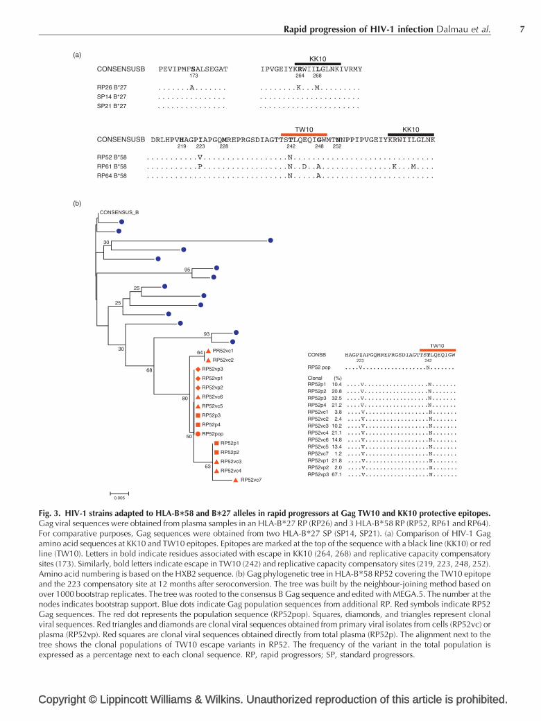
Initially, to define the contribution of HLA-adaptedstrains to rapid progression, we focused on patients withprotective HLA class I alleles (HLA-B�58 and B�27) andrapid progression. We identified one HLA-B�27 rapidprogressors and three HLA-B�58 rapid progressors. Weevaluated the existence of adapted virus to protectiveKK10 and TW10 Gag epitopes. As a result, we foundimmune escape variants at positions K264R in KK10 andT242N in TW10 in all rapid progressors. By contrast, noimmune escape was observed in two HLA-B�27 standardprogressors within the same period of infection (Fig. 3a).Additionally, Gag K264R and T242N mutations knownto cause viral replication defects [24,25] appearedalongside compensatory changes in replication atpositions S173 for K264R and I223/G248 for T242Nin rapid progressors (Fig. 3a). Development of such a
horized reproduction of this article is prohibited.

Copyright © Lippincott Williams & Wilkins. Unauthorized reproduction of this article is prohibited.
CE: Namrta; AIDS-D-13-01409; Total nos of Pages: 12;
AIDS-D-13-01409
6 AIDS 2014, Vol 00 No 00
30
25
20
15
10
5
0
35
30
25
20
15
10
5
0
0
25
50
75
100
0
0
50
100
150250
750
1250
20
40
60
80
800
1600
2400
RP
p = 0.004 p = 0.005
SP
RP SP
RP SP RP
p = 0.019
p = 0.024
p < 0.0001
SP RP SP RP SPRP SP
BLv-BLp BLv-BLp
IC50
(1/
dilu
tion)
ove
rtim
e
BLv-PpPv-Pp
IC50
(1/
dilu
tion)
aga
inst
BL
viru
ses
BLv-Pp
RP SP RP SP RP SP RP SP RP SP RP SP
Sensitive Moderate Resistantb12
IC50
(m
g/m
l) ag
ains
t BL
viru
ses
IC50
(1/
dilu
tion)
from
sub
ject
pla
smas
4E10 2F5 2G12
BL
Fre
quen
cy o
f HIV
-1IN
F-g
ELI
Spo
t res
pons
es (
%)
PRP SP
(a)
(b) bNAb (c) Virus panel
(d) Heterologous neutralization (e) Autologous neutralization
Fig. 2. HIV-1-specific CD8R T-cell responses, virus sensitivity to neutralization and plasma-neutralizing activity in rapidprogressors and standard progressors. (a) Frequency of IFN-g HIV-1-specific CD8þ T-cell responses at baseline (BL) and post-baseline (P) were measured in PBMCs against a panel of optimally defined HLA class I HIV-1 epitopes in 17 rapid progressors (RPs)and 13 standard progressors (SPs). The box and whiskers plot shows median values and 10th and 90th percentiles. The P-valueswere calculated using the Mann–Whitney test. Only significant P-values (P<0.05) are shown. (b) Sensitivity to neutralization ofEnv-pseudotyped virus derived from the plasma of 24 RP and 31 SP against a panel of broadly neutralizing antibodies (bNAbs)including b12, 4E10, 2F5, and 2G12. (c) Heterologous plasma-neutralizing activity at baseline against a standard panel of viralisolates from various subtypes and with various ranges of susceptibilities to neutralization (sensitive, moderate, and resistant).(d) Heterologous cross-reactive virus-neutralizing activity in plasma samples, at baseline (BLp) and post-baseline (Pp), against apanel of baseline patient-derived Env-pseudotyped viruses (BLv). (e) Autologous neutralizing activity in plasma and Env-pseudotyped virus pairs. All P-values were calculated using the Mann–Whitney test. Only significant P-values (P<0.05) arerepresented. BLp, baseline plasma; BLv, baseline virus; Pp, post-baseline plasma; Pv, post-baseline virus.
Copyright © Lippincott Williams & Wilkins. Unauthorized reproduction of this article is prohibited.
CE: Namrta; AIDS-D-13-01409; Total nos of Pages: 12;
AIDS-D-13-01409
Rapid progression of HIV-1 infection Dalmau et al. 7
RP52p1RP52p2RP52p3RP52p4RP52vc1RP52vc2RP52vc3RP52vc4RP52vc6RP52vc5RP52vc7RP52vp1RP52vp2RP52vp3
0.005
10.4Clonal
RP52 pop
CONSB
CONSENSUS_B
(b)
30
95
25
25
93
30
68
64
80
50
63
(%)
20.832.521.2
3.82.4
10.221.114.813.4
1.221.8
2.067.1
PR52vc1
RP52vc2
RP52vp3
RP52vp1
RP52vp2
RP52vc6
RP52vc5
RP52p3
RP52p4
RP52pop
RP52p1
RP52p2
RP52vc3
RP52vc4
RP52vc7
(a)
RP26 B*27
SP14 B*27
SP21 B*27
CONSENSUSB
CONSENSUSB
KK10
KK10TW10
173 264
219 223 228 242 248 252
268
RP52 B*58
RP61 B*58
RP64 B*58
Fig. 3. HIV-1 strains adapted to HLA-BM58 and BM27 alleles in rapid progressors at Gag TW10 and KK10 protective epitopes.Gag viral sequences were obtained from plasma samples in an HLA-B�27 RP (RP26) and 3 HLA-B�58 RP (RP52, RP61 and RP64).For comparative purposes, Gag sequences were obtained from two HLA-B�27 SP (SP14, SP21). (a) Comparison of HIV-1 Gagamino acid sequences at KK10 and TW10 epitopes. Epitopes are marked at the top of the sequence with a black line (KK10) or redline (TW10). Letters in bold indicate residues associated with escape in KK10 (264, 268) and replicative capacity compensatorysites (173). Similarly, bold letters indicate escape in TW10 (242) and replicative capacity compensatory sites (219, 223, 248, 252).Amino acid numbering is based on the HXB2 sequence. (b) Gag phylogenetic tree in HLA-B�58 RP52 covering the TW10 epitopeand the 223 compensatory site at 12 months after seroconversion. The tree was built by the neighbour-joining method based onover 1000 bootstrap replicates. The tree was rooted to the consensus B Gag sequence and edited with MEGA.5. The number at thenodes indicates bootstrap support. Blue dots indicate Gag population sequences from additional RP. Red symbols indicate RP52Gag sequences. The red dot represents the population sequence (RP52pop). Squares, diamonds, and triangles represent clonalviral sequences. Red triangles and diamonds are clonal viral sequences obtained from primary viral isolates from cells (RP52vc) orplasma (RP52vp). Red squares are clonal viral sequences obtained directly from total plasma (RP52p). The alignment next to thetree shows the clonal populations of TW10 escape variants in RP52. The frequency of the variant in the total population isexpressed as a percentage next to each clonal sequence. RP, rapid progressors; SP, standard progressors.
Co
CE: Namrta; AIDS-D-13-01409; Total nos of Pages: 12;
AIDS-D-13-01409
8 AIDS 2014, Vol 00 No 00
complex mutational pattern is rare in early infection[23,26,27] and likely represents a marker of HIV-1transmission events. This observation is reinforced by thepresence of KK10 escape mutants in non-B�27 rapidprogressors (Fig. 3a) and by the homogeneous clonalpopulations of TW10 escape mutants found in an HLA-B�58 rapid progressors (Fig. 3b). Overall, these findingsare consistent with the low level of viral genetic variabilitydescribed in recent infection and probably identifytransmission events in rapid progression.
With the aim of generalizing previous observations tospecific immunogenetic profiles in rapid progressors, weperformed an exploratory analysis of HLA class Idistribution in 122 HIV-1-infected individuals (74 rapidprogressors and 48 standard progressors). Our datademonstrate an increase in the frequency of commonCaucasian HLA haplotypes, including HLA-A�02:01,B�07:02, and B�08:01 in rapid progressors (75.6% rapidprogressors vs. 50% standard progressors) to the detrimentof protective ones (6.76% rapid progressors vs. 22.9%standard progressors) Fig. 4a. In addition, a modestincrease was observed in risk alleles. Similar trends acrossdata analyses were supported by individual HLA typeanalyses (Table S4, http://links.lww.com/QAD/A517).
In order to evaluate the contribution of virus adaptationor escape to the immunogenetics of rapid progressors, wefocused on HLA-A�02:01 carriers because of their higherprevalence in the study population. Thus, we determinedHIV-1 variation at the immunodominant A�02:01-restricted SL9-Gag epitope. We compared HIV-1 SL9-Gag variation in HLA-A�02:01-positive individuals(A�02 : 02þve) early after seroconversion (Fig. 4b).Our data demonstrated preferential variation at position3 (Y3F), but not position 8, of the SL9-Gag epitope inA�02:01þve rapid progressors when compared toA�02:01þve standard progressors (62.5% rapid progres-sors vs. 0% standard progressors; P¼ 0.031) (Fig. 4b).These differences revealed a specific increase in SL9escape variants in rapid progressors. Furthermore, toextend these findings to complete viral proteins, wemeasured total variation in Gag and Pol regions andlimited the analyses to those epitopes recognized by eachpatient’s HLA. Our data indicated a significant increase intotal Gag epitope variation in viral strains from rapidprogressors when compared to standard progressors (1rapid progressors vs. 0.63 standard progressors; P¼ 0.008)(Fig. 4c), but not in Pol. Moreover, these differences inepitope variation were not equally distributed acrossfunctional domains in Gag, but significantly accumulatedin the p24 region (0.87 rapid progressors vs. 0.35 standardprogressors; P¼ 0.0084) (Fig. 4d). These results supportthe association between HLA class I adaptation or escapeof HIV-1 variants and rapid disease progression. Thisassociation is marked by a high prevalence of commonCaucasian haplotypes and higher mutational changesagainst p24 epitopes.
pyright © Lippincott Williams & Wilkins. Unautho
Discussion
The time required for the development of AIDS afterHIV-1 infection is associated with the temporal equi-librium between viral and host factors. This dynamicinterplay can be disrupted and present as extreme diseasethat either contributes to prolonged periods of HIV-1control or drives rapid disease progression. In this context,rapid progression remains one of the least understoodextreme HIV-1 phenotypes, yet it must be understood ifwe are to tackle the epidemic. Consequently, the presentstudy overcomes previous study limitations through theidentification and functional characterization of thelargest group of rapid progressors to date. For the firsttime, we provide evidence of the contribution of intrinsicviral properties to rapid disease progression in a studygroup.
Our data demonstrate how viral factors, including robustHIV-1 replication, higher X4/DM co-receptor usage,and an increased number of common HLA footprints,could lead to uncontrolled virus replication. Such a robustreplicative profile will likely limit the generation ofadaptive responses and precipitate rapid disease pro-gression. The pathogenic HIV-1 strains found in rapidprogressors are rare at the onset of infection, anddiscrepancies in viral factors are most apparent with regardto viral tropism. Thus, the reported prevalence of X4/DM virus ranged from 5 to 10% in primary infection[17,28], compared with above 25% in rapid progressors.However, differences in the selection criteria according torapid disease progression might partially account for thedivergence between studies. Although X4/DM virusesare more pathogenic than R5 viruses [29], R5 variants inrapid progressors were equally associated with highreplicative capacity and viral load set point. Therefore,these data highlight a previously unnoticed contributionof R5 strains to disease progression.
Intrinsic viral properties may also contribute to thelimited adaptive immunity found in rapid progressors,which is key to controlling HIV-1 replication upontransmission. We observed defective HIV-1-specificCD8þ cellular responses and no major role of neutralizingantibodies, as previously reported [30,31]. However, thegeneralized static immune phenotype in rapid progressorsdoes not reflect a pre-existing immunodeficiency [2], butrather an outstanding loss of CD4þ T cells in primaryinfection. The dramatic loss of CD4þ T cells willirreversibly affect CD4þ homeostasis and thus compro-mise CD4þ T-cell helper responses and CD8þ T-cell [32]and B-cell function [33,34]. In addition, subsequentlimited anti-HIV-1-adaptive immunity established earlyon could add to the lack of control of viral replication anddisease progression.
In this study, we observed early disruption of hostimmunity in favour of viral replication. This phenomenon
rized reproduction of this article is prohibited.

CE: Namrta; AIDS-D-13-01409; Total nos of Pages: 12;
AIDS-D-13-01409
Rapid progression of HIV-1 infection Dalmau et al. 9
(a)
(b)
RP52 RP48RP27RP6
CONSENSUS B SLYNTVATL
SL9
RP58 RP59 RP65 RP69
SP3 SP14 SP15 SP17 SP39 SP49
p = 0.0083.5
3.0
2.5
2.0
1.5
Fre
q of
var
iatio
n in
Gag
(N m
ism
atch
/N e
pito
pes
foun
d)
1.0
0.5
0.0
RP SP
p = 0.00843.5
3.0
2.5
2.0
1.5
Fre
q of
var
iatio
n pe
r G
ag d
omai
n(N
mis
mat
ch/N
epi
tope
s fo
und)
1.0
0.5
0.0
RP SP
P17
RP SP
P24
90 p = 0.031
(%)
SL9
var
iatio
n in
HLA
-A*0
2:01
+ve
0
P3
RP
P8
SP80
70
60
50
40
30
20
10
RP
SP
80
Phe
noty
pe fr
eque
ncy
(%)
0
≥1 common ≥1 protective ≥1 risk
60
40
20
Fig. 4. HLA class I immunogenetics and HIV-1 variation at HLA-matched epitopes in rapid progressors and standardprogressors. (a) Phenotypic frequency (number of patients with a defined haplotype/number of total individuals in the group� 100) in pooled analyses for HLA class I alleles. Patients in groups (74 RP and 48 SP) were included if they had at least onecommon allele (A�02:01, B�07:02 and B�08:01), a protective allele (B�57:01, B�14:01 and B�27:05), or a risk allele (B�35:02,B�35:03, B�35:04 and B�53:01). Only those alleles with a frequency above 3% in the overall population were taken into accountfor data representation. (b) HIV-1 SL9 epitope alignment in HLA-A�02:01þve RP and SP; P3 and P8 indicate amino acid sites inSL9. The bar graph shows the frequency of epitope variation at P3 and P8 sites in HLA-A�02:01þve SP and RP. (c) Frequency ofepitope variation in HIV-1 Gag. Partial sequencing was carried out in a total of 40 patients (22 RP and 18 SP). (d) Frequency of HLA-matched epitope variation in HIV-1 Gag per functional domains p17 and p24. Values in (c) and (d) are expressed as mean and SEM.The P-values were calculated using the Mann–Whitney test. Only significant P-values (P<0.05) are represented. RP, rapidprogressors; SP, standard progressors.
seems to be associated with the increase in HLA class I virusadaptation or escape. HIV-1 adapts to the most frequentalleles in a population [21]. Therefore, the probability oftransmission of adapted viruses will increase among
Copyright © Lippincott Williams & Wilkins. Unaut
common HLA carriers, thus reducing the chances for apotent anti-HIV-1 CD8þ T-cell response [21,35].Consistent with this hypothesis, our data identified theaccumulation of common Caucasian alleles, including
horized reproduction of this article is prohibited.
Co
CE: Namrta; AIDS-D-13-01409; Total nos of Pages: 12;
AIDS-D-13-01409
10 AIDS 2014, Vol 00 No 00
A�02:01, B�07:02, and B�08:01, in more than 75% ofrapid progressors. Our results confirm and extend those ofprevious studies showing a disadvantage of A2 and B7supertypes in control of HIV-1 infection [35,36].According to this immunogenetics profile, the likelihoodof having adapted or escaped variants should increase inrapid progression of HIV-1. This observation is consistentwith the presence of well adapted and well compensatedviruses in HLA-B�58 and HLA-B�27 patients andreinforced by the spread of SL9 escaped variants inHLA-A�02:01-positive rapid progressors. In addition, theoverall increase in HIV-1 Gag-p24 mutations matched toHLA class I epitopes in rapid progressors, but not instandard progressors, points to the existence of adapted orescape variants during the first year after seroconversion.Despite the fact that we cannot directly associate specificamino acid changes in Gag-p24 epitopes with HIV-1escape, multiple intra-epitope variation might interferewith the generation of particularly protective Gag CD8þ
T-cell responses [3,37–39]. Although our study demon-strated an increase in HLA class I-adapted HIV-1 variantswith rapid progression, we were unable to determinewhether viruses were transmitted as homogeneous adaptedfounder strains or whether escape mutations quicklyemerge through the robust replicative profile in acuteinfection. Nevertheless, the presence of viruses withcomplex mutational patterns found only at late stages ofdisease [23,26,40] and homogeneous populations of escapemutants in early infection seem to indicate thattransmission events are the origin of these pathogenicstrains. Thus, rapid progression could originate fromacquisition of HIV-1 from chronically infected untreatedindividuals [41]. These source patients increase theprobability of transmission of X4/DM or pathogenicR5 variants [42], which are fully replicative and adapted tohost HLA through long-term viral replication.
The limitations of the present study include the lack oftransmission pairs to clarify the contribution of founderviruses to rapid progression. Moreover, ideally, compari-sons between groups should be made during acute stages,which might minimize differences at baseline. However,this would reduce the number of study participants andthus make it difficult to draw conclusions. Additionally,availability of biological material was limited owing to thesize and heterogeneity of the samples included in somesub-analyses.
In conclusion, our findings suggest that rapid progressionresults from the acquisition of highly pathogenic strainsalready adapted to HLA class I alleles. These strainsavoid the initial control of HIV-1 replication mediatedby adaptive CD8þ T-cell responses, leading to rapidprogression, and might help to explain the increasedvirulence of HIV-1 observed over the course of theepidemic [43]. Therefore, it would be advisable todevelop strategies for early identification of rapidprogressors, specific clinical guidelines, and robust
pyright © Lippincott Williams & Wilkins. Unautho
prevention policies in order to provide adequate careand to prevent the spread of highly pathogenic viralstrains in the population.
Acknowledgements
We thank all the participants in the study and the clinicalteam of the Fundacio Lluita contra la SIDA. This studywas supported by the European Union’s FP7/2007–2013under the ‘Collaborative HIV and Anti-HIV DrugResistance Network (CHAIN)’ Project Grant agreement223131 and the ‘Catalonian HIV-1 Vaccine Program’(HIVACAT program). Work at J.M.P.’s laboratory issupported by the Spanish Department of Science,Development and Innovation through grant SAF2010-21224. Work at A.T.’s laboratory is supported by the SwissNational Science Foundation within the framework ofthe Swiss HIV Cohort Study (grant # 33CS30_134277).Work at J.G.P.’s laboratory is supported by grants CP09/00279 and PI11/00249 from the FIS-ISCIII. We thank D.Ouchi for statistical support. We thank Z. Brumme foradvice on HLA data analysis. We thank G. Fernandez forcritical comments on the manuscript.
Author contributions: J.D., M.R., A.T., J.M.P. and J.G.P.were responsible for patient selection, study design, datainterpretation, and manuscript writing. P.P. performedexperiments on humoral responses. J.D., I.E., M.P., andJ.G.P. conceived and performed experiments on virusisolation, co-receptor usage, replication, sequencing, andcellular responses. P.R. developed the EPIMAP tool andreviewed the manuscript. E.P. was responsible for HLAdata generation. A.E. provided statistical support andreviewed the manuscript. A.T., C.B., R.P., and B.C.contributed to data interpretation and writing and reviewof the manuscript. All the authors approved the finalmanuscript.
Conflicts of interestJ.G.P. holds a Miguel Servet Contract (MS09/00279)funded by FIS-ISCIII (Spanish Government). Thefunders had no role in study design, data collectionand analysis, decision to publish, or preparation ofthe manuscript.
The authors have no conflicting financial interests.
References
1. Time from HIV-1 seroconversion to AIDS and death beforewidespread use of highly-active antiretroviral therapy: a col-laborative re-analysis. Collaborative Group on AIDS Incuba-tion and HIV Survival including the CASCADE EU ConcertedAction Concerted Action on SeroConversion to AIDS andDeath in Europe. Lancet 2000; 355:1131–1137.
rized reproduction of this article is prohibited.
CE: Namrta; AIDS-D-13-01409; Total nos of Pages: 12;
AIDS-D-13-01409
Rapid progression of HIV-1 infection Dalmau et al. 11
2. Dalmau J, Puertas MC, Azuara M, Marino A, Frahm N, Mothe B,et al. Contribution of immunological and virological factors toextremely severe primary HIV type 1 infection. Clin Infect Dis2009; 48:229–238.
3. Pereyra F, Addo MM, Kaufmann DE, Liu Y, Miura T, Rathod A,et al. Genetic and immunologic heterogeneity among personswho control HIV infection in the absence of therapy. J Infect Dis2008; 197:563–571.
4. Rotger M, Dalmau J, Rauch A, McLaren P, Bosinger SE,Martinez R, et al. Comparative transcriptomics of extremephenotypes of human HIV-1 infection and SIV infection insooty mangabey and rhesus macaque. J Clin Invest 2011;121:2391–2400.
5. Pereyra F, Jia X, McLaren PJ, Telenti A, de Bakker PIW, WalkerBD, et al. The major genetic determinants of HIV-1 controlaffect HLA class I peptide presentation. Science 2010;330:1551–1557.
6. Casado C, Colombo S, Rauch A, Martınez R, Gunthard HF,Garcia S, et al. Host and viral genetic correlates of clinicaldefinitions of HIV-1 disease progression. PLoS One 2010; 5:e11079.
7. Dalmau J, Codoner FM, Erkizia I, Pino M, Pou C, Paredes R, et al.In-depth characterization of viral isolates from plasma andcells compared with plasma circulating quasispecies in earlyHIV-1 infection. PLoS One 2012; 7:e32714.
8. Markowitz M, Mohri H, Mehandru S, Shet A, Berry L, Kalyanara-man R, et al. Infection with multidrug resistant, dual-tropicHIV-1 and rapid progression to AIDS: a case report. Lancet2005; 365:1031–1038.
9. Prado JG, Prendergast A, Thobakgale C, Molina C, Tudor-Williams G, Ndung’u T, et al. Replicative capacity of humanimmunodeficiency virus type 1 transmitted from mother tochild is associated with pediatric disease progression rate.J Virol 2010; 84:492–502.
10. Prado JG, Honeyborne I, Brierley I, Puertas MC, Martinez-Picado J, Goulder PJR. Functional consequences of humanimmunodeficiency virus escape from an HLA-BM13-restrictedCD8R T-cell epitope in p1 Gag protein. J Virol 2009; 83:1018–1025.
11. Bjorndal A, Deng H, Jansson M, Fiore JR, Colognesi C, KarlssonA, et al. Coreceptor usage of primary human immunodefi-ciency virus type 1 isolates varies according to biologicalphenotype. J Virol 1997; 71:7478–7487.
12. Addo MM, Yu XG, Rathod A, Cohen D, Eldridge RL, Strick D,et al. Comprehensive epitope analysis of human immunodefi-ciency virus type 1 (HIV-1)-specific T-cell responses directedagainst the entire expressed HIV-1 genome demonstratebroadly directed responses, but no correlation to viral load.J Virol 2003; 77:2081–2092.
13. Schweighardt B, Liu Y, Huang W, Chappey C, Lie YS,Petropoulos CJ, Wrin T. Development of an HIV-1 referencepanel of subtype B envelope clones isolated from the plasma ofrecently infected individuals. J Acquir Immune Defic Syndr2007; 46:1–11.
14. Frost SDW, Wrin T, Smith DM, Kosakovsky Pond SL, Liu Y,Paxinos E, et al. Neutralizing antibody responses drive theevolution of human immunodeficiency virus type 1 envelopeduring recent HIV infection. Proc Natl Acad Sci U S A 2005;102:18514–18519.
15. Ferbas J, Daar ES, Grovit-Ferbas K, Lech WJ, Detels R, Giorgi JV,Kaplan AH. Rapid evolution of human immunodeficiency virusstrains with increased replicative capacity during the serone-gative window of primary infection. J Virol 1996; 70:7285–7289.
16. Mohri H, Markowitz M. In vitro characterization of multi-drug-resistant HIV-1 isolates from a recently infectedpatient associated with dual tropism and rapid diseaseprogression. J Acquir Immune Defic Syndr 2008; 48:511–521.
17. Raymond S, Delobel P, Mavigner M, Cazabat M, Encinas S,Souyris C, et al. CXCR4-using viruses in plasma and peri-pheral blood mononuclear cells during primary HIV-1infection and impact on disease progression. AIDS 2010;24:2305–2312.
18. Richman DD, Bozzette SA. The impact of the syncytium-indu-cing phenotype of human immunodeficiency virus on diseaseprogression. J Infect Dis 1994; 169:968–974.
Copyright © Lippincott Williams & Wilkins. Unaut
19. Barbour JD, Hecht FM, Wrin T, Segal MR, Ramstead CA, LieglerTJ, et al. Higher CD4R T cell counts associated with low viralpol replication capacity among treatment-naive adults in earlyHIV-1 infection. J Infect Dis 2004; 190:251–256.
20. Daar ES, Kesler KL, Wrin T, Petropoulo CJ, Bates M, Lail A, et al.HIV-1 pol replication capacity predicts disease progression.AIDS 2005; 19:871–877.
21. Kawashima Y, Pfafferott K, Frater J, Matthews P, Payne R, AddoM, et al. Adaptation of HIV-1 to human leukocyte antigen classI. Nature 2009; 458:641–645.
22. Crawford H, Lumm W, Leslie A, Schaefer M, Boeras D, PradoJG, et al. Evolution of HLA-BM5703 HIV-1 escape mutations inHLA-BM5703-positive individuals and their transmission reci-pients. J Exp Med 2009; 206:909–921.
23. Goulder PJ, Brander C, Tang Y, Tremblay C, Colbert RA,Addo MM, et al. Evolution and transmission of stable CTLescape mutations in HIV infection. Nature 2001; 412:334–338.
24. Martinez-Picado J, Prado JG, Fry EE, Pfafferott K, Leslie A, ChettyS, et al. Fitness cost of escape mutations in p24 Gag inassociation with control of human immunodeficiency virustype 1. J Virol 2006; 80:3617–3623.
25. Schneidewind A, Brockman MA, Yang R, Adam RI, Li B, Le GallS, et al. Escape from the dominant HLA-B27-restricted cyto-toxic T-lymphocyte response in Gag is associated with a dra-matic reduction in human immunodeficiency virus type 1replication. J Virol 2007; 81:12382–12393.
26. Feeney ME, Tang Y, Roosevelt KA, Leslie AJ, McIntosh K,Karthas N, et al. Immune escape precedes breakthrough humanimmunodeficiency virus type 1 viremia and broadening of thecytotoxic T-lymphocyte response in an HLA-B27-positive long-term-nonprogressing child. J Virol 2004; 78:8927–8930.
27. Schneidewind A, Brumme ZL, Brumme CJ, Power KA, Reyor LL,O’Sullivan K, et al. Transmission and long-term stability ofcompensated CD8 escape mutations. J Virol 2009; 83:3993–3997.
28. Chalmet K, Dauwe K, Foquet L, Baatz F, Seguin-Devaux C, VanDer Gucht B, et al. Presence of CXCR4-using HIV-1 in patientswith recently diagnosed infection: correlates and evidence fortransmission. J Infect Dis 2012; 205:174–184.
29. Koot M, Keet IP, Vos AH, de Goede RE, Roos MT, Coutinho RA,et al. Prognostic value of HIV-1 syncytium-inducing phenotypefor rate of CD4R cell depletion and progression to AIDS. AnnIntern Med 1993; 118:681–688.
30. Hay CM, Ruhl DJ, Basgoz NO, Wilson CC, Billingsley JM,DePasquale MP, et al. Lack of viral escape and defectivein vivo activation of human immunodeficiency virus type 1-specific cytotoxic T lymphocytes in rapidly progressive infec-tion. J Virol 1999; 73:5509–5519.
31. Richman DD, Wrin T, Little SJ, Petropoulos CJ. Rapid evolutionof the neutralizing antibody response to HIV type 1 infection.Proc Natl Acad Sci U S A 2003; 100:4144–4149.
32. Nakanishi Y, Lu B, Gerard C, Iwasaki A. CD8(R) T lymphocytemobilization to virus-infected tissue requires CD4(R) T-cellhelp. Nature 2009; 462:510–513.
33. Lindqvist M, van Lunzen J, Soghoian DZ, Kuhl BD, RanasingheS, Kranias G, et al. Expansion of HIV-specific T follicular helpercells in chronic HIV infection. J Clin Invest 2012; 122:3271–3280.
34. Petrovas C, Yamamoto T, Gerner MY, Boswell KL, Wloka K,Smith EC, et al. CD4 T follicular helper cell dynamics duringSIV infection. J Clin Invest 2012; 122:3281–3294.
35. Trachtenberg E, Korber B, Sollars C, Kepler TB, Hraber PT,Hayes E, et al. Advantage of rare HLA supertype in HIV diseaseprogression. Nat Med 2003; 9:928–935.
36. Peterson TA, Kimani J, Wachihi C, Bielawny T, Mendoza L,Thavaneswaran S, et al. HLA class I associations with rates ofHIV-1 seroconversion and disease progression in the PumwaniSex Worker Cohort. Tissue Antigens 2013; 81:93–107.
37. Kiepiela P, Ngumbela K, Thobakgale C, Ramduth D, Honey-borne I, Moodley E, et al. CD8R T-cell responses to differentHIV proteins have discordant associations with viral load. NatMed 2007; 13:46–53.
38. Saez-Cirion A, Lacabaratz C, Lambotte O, Versmisse P, UrrutiaA, Boufassa F, et al. HIV controllers exhibit potent CD8 T cellcapacity to suppress HIV infection ex vivo and peculiar cyto-toxic T lymphocyte activation phenotype. Proc Natl Acad SciU S A 2007; 104:6776–6781.
horized reproduction of this article is prohibited.
Co
CE: Namrta; AIDS-D-13-01409; Total nos of Pages: 12;
AIDS-D-13-01409
12 AIDS 2014, Vol 00 No 00
39. Zuniga R, Lucchetti A, Galvan P, Sanchez S, Sanchez C,Hernandez A, et al. Relative dominance of Gag p24-specific cytotoxic T lymphocytes is associated with humanimmunodeficiency virus control. J Virol 2006; 80:3122–3125.
40. Kelleher AD, Long C, Holmes EC, Allen RL, Wilson J,Conlon C, et al. Clustered mutations in HIV-1 gag areconsistently required for escape from HLA-B27-restrictedcytotoxic T lymphocyte responses. J Exp Med 2001; 193:375–386.
pyright © Lippincott Williams & Wilkins. Unautho
41. Hollingsworth T, Anderson R, Fraser C. HIV-1 transmission,by stage of infection. J Infect Dis 2008; 198:687–693.
42. Brumme ZL, Goodrich J, Mayer HB, Brumme CJ, Henrick BM,Wynhoven B, et al. Molecular and clinical epidemiology ofCXCR4-using HIV-1 in a large population of antiretroviral-naive individuals. J Infect Dis 2005; 192:466–474.
43. Herbeck JT, Muller V, Maust BS, Ledergerber B, Torti C, DiGiambenedetto S, et al. Is the virulence of HIV changing? Ameta-analysis of trends in prognostic markers of HIV diseaseprogression and transmission. AIDS 2012; 26:193–205.
rized reproduction of this article is prohibited.