Embed Size (px)
Citation preview
Aim: How is the cell cycle regulated?
Do Now:1.Worksheet
HW: Study for test tomorrow
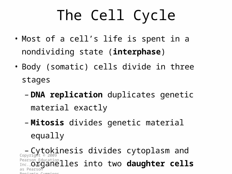
The Cell Cycle
• Most of a cell’s life is spent in a
nondividing state (interphase)
• Body (somatic) cells divide in three stages
–DNA replication duplicates genetic
material exactly
–Mitosis divides genetic material equally
– Cytokinesis divides cytoplasm and
organelles into two daughter cellsCopyright © 2009 Pearson Education, Inc., publishing as Pearson Benjamin Cummings

A Cell’s Life Cycle
Figure 3–23 The Cell Life Cycle.
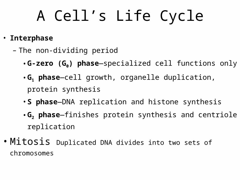
A Cell’s Life Cycle• Interphase
– The non-dividing period
• G-zero (G0) phase—specialized cell functions only
• G1 phase—cell growth, organelle duplication,
protein synthesis
• S phase—DNA replication and histone synthesis
• G2 phase—finishes protein synthesis and centriole
replication
• Mitosis Duplicated DNA divides into two sets of
chromosomes

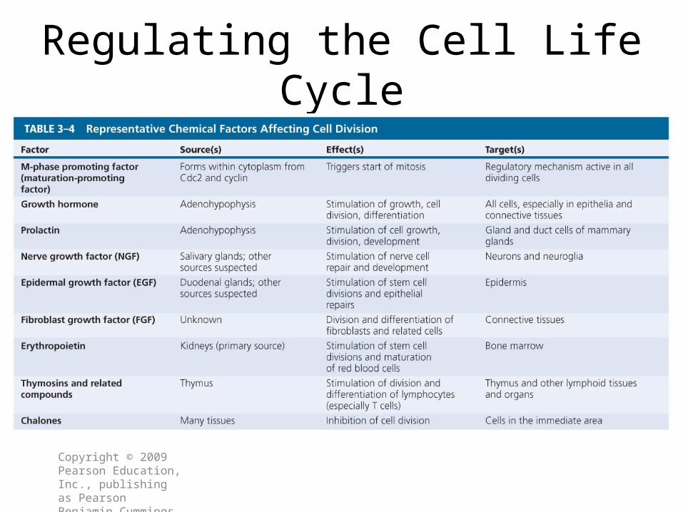
Regulating the Cell Life Cycle
• Normally, cell division balances cell loss• Increased cell division– Internal factors (M-phase promoting factor,
MPF) – Extracellular chemical factors (growth
factors)• Decreased cell division– Repressor genes (faulty repressors cause
cancers)–Worn out telomeres (terminal DNA segments)
Copyright © 2009 Pearson Education, Inc., publishing as Pearson Benjamin Cummings

Control of Cell Destiny•MPF- Maturation promoting factor (MPF)–Signal that induces cell division.–Consists of group of enzymes called cdc2 proteins (and proteins called cyclins.
•Cyclins build up during interphase and then activates cdc2 proteins which activates MPF which results in cell division.
•Apoptosis –Programmed cell death
Tumor Suppressor Genes• Produce proteins that normally inhibit
cell division.– Loss or alternation of p53 gene
leads to breast cancer, colon cancer, and other tumors.
–Normal p53 gene protein arrests a cell in G1 which prevents cell division.• Repair of damaged DNA and induces apoptosis in the cells where repair was not successful
Oncogenes
• Most oncogenes are mutations of certain normal genes called proto-oncogenes.
• When a proto-oncogene mutates (changes) into an oncogene, it becomes a "bad" gene that can become permanently turned on or activated when it is not supposed to be.
1
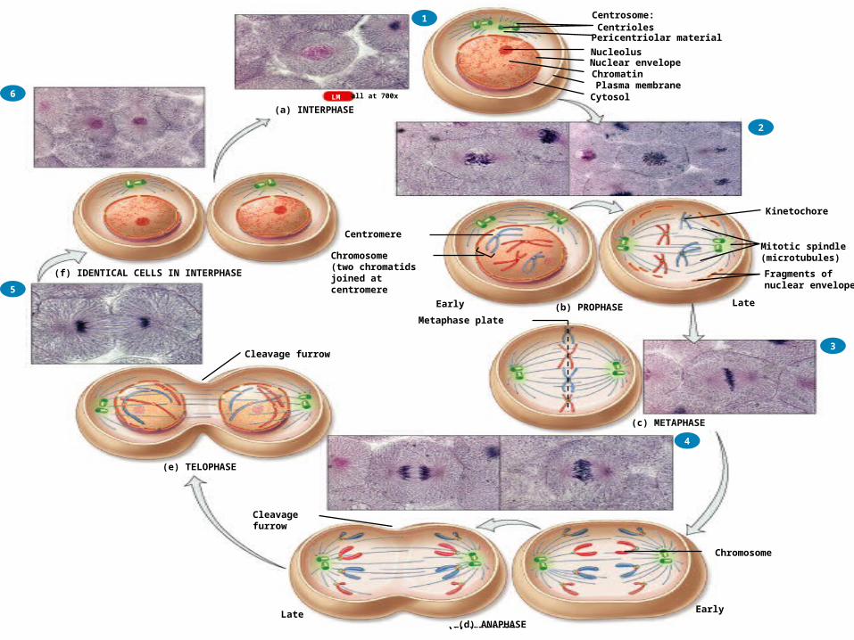
Pericentriolar material
NucleolusNuclear envelopeChromatinPlasma membraneCytosol
(a) INTERPHASE
CentriolesCentrosome:
all at 700xLM
1
LateEarly
Pericentriolar material
NucleolusNuclear envelopeChromatinPlasma membraneCytosol
Chromosome(two chromatidsjoined atcentromere
(a) INTERPHASE
(b) PROPHASE
CentriolesCentrosome:
Fragments ofnuclear envelope
Mitotic spindle(microtubules)
Kinetochore
2
all at 700xLM
Centromere
1
Pericentriolar material
NucleolusNuclear envelopeChromatinPlasma membraneCytosol
Metaphase plate
(a) INTERPHASE
CentriolesCentrosome:
(c) METAPHASE
2
3
LateEarly (b) PROPHASE
Fragments ofnuclear envelope
Mitotic spindle(microtubules)
Kinetochore
all at 700xLM
Chromosome(two chromatidsjoined atcentromere
Centromere
1
EarlyLate(d) ANAPHASE
Pericentriolar material
NucleolusNuclear envelopeChromatinPlasma membraneCytosol
Chromosome
(a) INTERPHASE
CentriolesCentrosome:
(c) METAPHASE
2
3
4
Cleavage furrow
LateEarly (b) PROPHASE
Fragments ofnuclear envelope
Mitotic spindle(microtubules)
Kinetochore
Metaphase plate
all at 700xLM
Chromosome(two chromatidsjoined atcentromere
Centromere
1
EarlyLate(d) ANAPHASE
Pericentriolar material
NucleolusNuclear envelopeChromatinPlasma membraneCytosol
(a) INTERPHASE
CentriolesCentrosome:
Cleavage furrow
(e) TELOPHASE
(c) METAPHASE
2
3
4
5
Cleavage furrow
LateEarly (b) PROPHASE
Fragments ofnuclear envelope
Mitotic spindle(microtubules)
Kinetochore
Metaphase plate
Chromosome
all at 700xLM
Chromosome(two chromatidsjoined atcentromere
Centromere
1
EarlyLate(d) ANAPHASE
Pericentriolar material
NucleolusNuclear envelopeChromatinPlasma membrane
Cytosol(a) INTERPHASE
CentriolesCentrosome:
(f) IDENTICAL CELLS IN INTERPHASE
Cleavage furrow
(e) TELOPHASE
(c) METAPHASE
Cleavage furrow
2
3
4
5
6
LateEarly (b) PROPHASE
Fragments ofnuclear envelope
Mitotic spindle(microtubules)
Kinetochore
Metaphase plate
Chromosome
all at 700xLM
Centromere
Chromosome(two chromatidsjoined atcentromere
–Rate of cell division
• Slower mitotic rate means longer
cell life
–Muscle cells, neurons rarely divide
–Exposed cells (skin and digestive
tract) live only days or hours.
Copyright © 2009 Pearson Education, Inc., publishing as Pearson Benjamin Cummings
Mitotic Rate
Tumors and Cancer
• Cancer develops in steps
– Abnormal cell
– Primary tumor
–Metastasis
– Secondary tumor
Copyright © 2009 Pearson Education, Inc., publishing as Pearson Benjamin Cummings
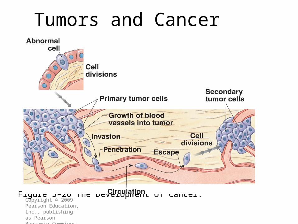
Tumors and Cancer
Figure 3–26 The Development of Cancer.Copyright © 2009 Pearson Education, Inc., publishing as Pearson Benjamin Cummings

Summary
• Why is interphase important to a cell?• Distinguish mitosis and meiosis.• Define apoptosis• Why are tumor suppressor genes important?
Regulating the Cell Life Cycle
Copyright © 2009 Pearson Education, Inc., publishing as Pearson Benjamin Cummings