Embed Size (px)
Citation preview

Cobalt Inhalation Cancer Potency Values
i
Cobalt and Cobalt
Compounds
Cancer Inhalation Unit
Risk Factors
Technical Support Document for
Cancer Potency Factors
Appendix B
Public Comment Draft
March 2019
Air, Community, and Environmental Research Branch
Office of Environmental Health Hazard Assessment
California Environmental Protection Agency
OFFICE OF ENVIRONMENTAL HEALTH HAZARD ASSESSMENT
Air Toxics Hot Spots Program
Cobalt Inhalation Cancer Potency Values Public Review Draft March 2019
Page Intentionally Left Blank

Cobalt Inhalation Cancer Potency Values
i
Cobalt and Cobalt Compounds
Cancer Inhalation Unit Risk Factors
Technical Support Document for Cancer Potency Factors
Appendix B
Prepared by the
Office of Environmental Health Hazard Assessment
Lauren Zeise, Ph.D., Director
Authors
Daryn E. Dodge, Ph.D.
Rona M. Silva, Ph.D.
Technical Reviewers
David M. Siegel, Ph.D.
John D. Budroe, Ph.D.
Public Comment Draft March 2019

Cobalt Inhalation Cancer Potency Values Public Review Draft March 2019
Table of Contents
Introduction .......................................................................................................... iii I. PHYSICAL AND CHEMICAL PROPERTIES ....................................................... 1
II. HEALTH ASSESSMENT VALUES ...................................................................... 1
III. CARCINOGENICITY ........................................................................................ 2
NTP Carcinogenicity Bioassays .............................................................................. 4
Cobalt Metal ......................................................................................................... 4
Cobalt Sulfate Heptahydrate ................................................................................ 9
Other Supporting Cancer Bioassays ..................................................................... 13
Inhalation ........................................................................................................... 13
Intratracheal instillation ...................................................................................... 13
Subcutaneous, intraperitoneal and intramuscular administration ....................... 14
Toxicokinetics ........................................................................................................ 15
Human Toxicokinetics and Comparison to other Mammalian Species .............. 15
NTP Tissue Burden Studies of Cobalt Metal in Rats and Mice .......................... 16
Cellular Toxicokinetics of Cobalt Nanoparticles ................................................. 17
Epidemiological Studies ........................................................................................ 18
Genotoxicity ........................................................................................................... 21
Soluble and Insoluble Cobalt Compounds, Not Including Cobalt Metal ............. 21
DNA strand-break and cross-linking tests ...................................................... 22
Oxidative DNA damage tests ......................................................................... 24
Tests for reduction in DNA replication and repair ........................................... 25
Bacterial and mammalian cell gene mutation tests ........................................ 26
Chromosomal damage ................................................................................... 26
Cobalt Metal, Including Comparisons with Soluble and Insoluble Cobalt Compounds ....................................................................................................... 33
DNA strand break tests .................................................................................. 33
Bacterial and mammalian cell gene mutation tests ........................................ 35
Chromosomal damage ................................................................................... 36
Gene Mutation Analysis ................................................................................. 37
Genotoxicity tests in workers exposed to cobalt metal ................................... 37
Morphological Cell Transformation and Tumor Suppressor Protein Induction....... 40
Toxicogenomics .................................................................................................... 41
IV. CANCER HAZARD EVALUATION ................................................................. 42
V. QUANTITATIVE CANCER RISK ASSESSMENT .......................................... 45
Cobalt Metal .......................................................................................................... 45
Effective Tumor Incidences ................................................................................ 45
Calculation of Single- and Multi-Site Tumor CSFs ............................................. 48
Inhalation Unit Risk Factor ................................................................................. 53
Cobalt Sulfate Heptahydrate ................................................................................. 54
Effective Tumor Incidences ................................................................................ 54
Calculation of Single- and Multi-Site Tumor CSFs ............................................. 55
Inhalation Unit Risk Factor ................................................................................. 59
VI. CONCLUSIONS ............................................................................................. 59
VII. REFERENCES ............................................................................................... 61

Cobalt Inhalation Cancer Potency Values Public Review Draft March 2019
iii
Introduction
This document summarizes the carcinogenicity and derivation of cancer inhalation unit
risk factors (IURs) for cobalt and cobalt compounds. Cancer unit risk factors are used to
estimate lifetime cancer risks associated with inhalation exposure to a carcinogen.
The Office of Environmental Health Hazard Assessment (OEHHA) is required to develop
guidelines for conducting health risk assessments under the Air Toxics Hot Spots
Program (Health and Safety Code Section 44360 (b) (2)). In implementing this
requirement, OEHHA develops cancer inhalation unit risk factors for carcinogenic air
pollutants listed under the Air Toxics Hot Spots program. The cobalt and cobalt
compounds IURs were developed using the most recent “Air Toxics Hot Spots Program
Technical Support Document for Cancer Potency Factors”, finalized by OEHHA in 2009.
Literature summarized and referenced in this document covers the relevant published
literature for cobalt and cobalt compounds through the fall of 2018.
Several government agencies or programs currently list cobalt metal and cobalt
compounds as carcinogens. Cobalt metal and soluble cobalt(II) salts are listed
separately by the International Agency for Research on Cancer (IARC) as Group 2B
carcinogens, i.e., possibly carcinogenic to humans (IARC, 2006). The National
Toxicology Program (NTP) listed cobalt and cobalt compounds that release cobalt ions in
vivo in the 14th Report on Carcinogens, which identifies substances that either are known
to be human carcinogens or are reasonably anticipated to be human carcinogens, and to
which a significant number of persons residing in the United States are exposed (NTP,
2016). Under the California Proposition 65 program, cobalt metal powder, cobalt sulfate,
cobalt sulfate heptahydrate, and cobalt(II) oxide are listed as chemicals known to the
state to cause cancer (OEHHA, 2018a).
NTP conducted inhalation carcinogenicity bioassays with cobalt sulfate heptahydrate, a
soluble cobalt compound, in rats and mice of both sexes in 1998 (NTP, 1998). NTP
subsequently conducted inhalation carcinogenicity bioassays with cobalt metal in rats
and mice of both sexes in 2014 (NTP, 2014). These studies provided evidence of
carcinogenicity for cobalt sulfate heptahydrate and for cobalt metal in rats and mice of
both sexes. Due to chemical, physical, and toxicological differences between cobalt
metal and various cobalt compounds, separate IURs were derived for water soluble
cobalt compounds (based on studies with cobalt sulfate heptahydrate) and cobalt metal
and insoluble cobalt compounds (based on studies with cobalt metal).
Most cobalt is used industrially in the form of cobalt metal powder as an alloying
component and in the preparation of cobalt salts (HSDB, 2016; NTP, 2016). Cobalt salts
and oxides are used as pigments in the glass and ceramics industries, as catalysts in the
oil and chemical industries, as paint and printing ink driers, and as trace metal additives

Cobalt Inhalation Cancer Potency Values Public Review Draft March 2019
iv
in agriculture and medicine. Other significant cobalt uses are as a catalyst or component
in green energy technologies (e.g., solar panels), and as a primary component in lithium-
and nickel-based rechargeable batteries. The presence of cobalt in some electric and
electronic devices may also result in exposure to cobalt in the E-waste recycling industry
(Leyssens et al., 2017).
Cobalt occurs naturally in the Earth’s crust but is usually in the form of arsenides and
sulfides (Baralkiewicz and Siepak, 1999). Natural levels of cobalt in air generally range
from 0.0005 to 0.005 nanograms per cubic meters (ng/m3). In major industrial cities,
levels of cobalt may reach as high as 6 ng/m3. The California Air Resources Board
collects air monitoring data for numerous pollutants found in urban areas, including
cobalt and other metals (CARB, 2018). In southern California, mean cobalt
concentrations at air monitoring sites in 2017 ranged from 1.3 to 1.97 ng/m3, with
maximum levels between 2.9 and 5.6 ng/m3. However, cobalt concentrations were often
below the limit of detection (1.3 ng/m3).
Emissions estimates of cobalt in California are collected and presented in the California
Toxics Inventory, or CTI (CARB, 2013). Potential sources include stationary (point and
aggregated point), area-wide, on-road mobile (gasoline and diesel), off-road mobile
(gasoline, diesel, and other), and natural sources. The primary emission source for
cobalt in 2010 was area-wide sources, at 55.2 tons per year. Stationary point sources
released 2.2 tons of cobalt per year while the remaining sources were small or negligible.
The CTI estimates total organic gas and particulate matter for area, mobile, and natural
sources. Speciated emissions for each source category are then reconciled with
reported stationary point source toxics data to establish a complete inventory. Area-wide
sources include source categories associated with human activity, and emissions take
place over a wide geographic area. Such sources are consumer products, fireplaces,
farming operations and unpaved roads. Stationary sources include point sources
provided by facility operators and/or districts pursuant to the Air Toxics “Hot Spots”
Program (AB 2588).

Cobalt Inhalation Cancer Potency Values Public Review Draft March 2019
v
List of Acronyms
8-OHdG 8-hydroxydeoxyguanosine AIC Akaike Information Criterion BMDL05 The 95% lower confidence
bound at the 5% response rate
BMD Benchmark dose BMD05 BMD 5% response rate BMDS Benchmark dose modelling
software BMR Benchmark response BNMN Binucleated micronucleated BR Breathing rate BW Body weight CEBS Chemical effects in biological
systems CF Conversion factor CKE Cystic keratinizing
epithelioma Co Cobalt CoSO4·7H2O Cobalt sulfate heptahydrate CPF Cancer potency factor CSF Cancer slope factor CTI California Toxics Inventory DMSO Dimethyl sulfoxide DNA Deoxyribonucleic acid Fpg Formamido-pyrimidine
glycosylate GSD Geometric standard deviation H2O2 Hydrogen peroxide HL Human lymphocyte hOOG1 Human 8-hydroxyguanine
DNA-glycosylate 1 IARC International Agency for
Research on Cancer
IUR Inhalation unit risk IR Inhalation rate LDH Lactate dehydrogenase MMAD Mass median aerodynamic diameter µg/L Micrograms per liter µg/ml Micrograms per milliliter µm Micrometer µM Micromole per liter mg/m3 Milligrams per cubic meter mg/kg-BW Milligrams per kilogram of bodyweight mM Millimole per liter NCE Normochromatic erythrocytes NP Nanoparticle NTP National Toxicology Program O2
- Superoxide radical OECD Organisation for Economic Co- operation and Development OEHHA Office of Environmental Health Hazard Assessment PCE Polychromatic erythrocytes ROS Reactive oxygen species SHE Syrian hamster embryo SIR Standardized incidence rate SMR Standardized mortality ratio SPF Specific pathogen free TWA Time-weighted average UV Ultraviolet US EPA United States Environmental Protection Agency

Cobalt Inhalation Cancer Potency Values Public Review Draft March 2019
vi
Page Intentionally Left Blank

Cobalt Inhalation Cancer Potency Values Public Review Draft March 2019
1
COBALT AND COBALT COMPOUNDS
I. PHYSICAL AND CHEMICAL PROPERTIES
(Kyono et al., 1992; Hillwalker and Anderson, 2014; NTP, 2016)
Molecular formula Co (elemental form) Molecular weight 58.93 Description Gray, hard, magnetic, ductile, somewhat
malleable metal Density 8.92 g/cm3 Boiling point 2927ºC Melting point 1495ºC Vapor pressure Not applicable Odor Cobalt metal powder or fumes are odorless Solubility Metallic cobalt particles in the micrometer size
range or larger are considered poorly water soluble. Soluble in dilute acids.
Conversion factor Not applicable
II. HEALTH ASSESSMENT VALUES
Cobalt metal and water-insoluble cobalt compounds
Unit Risk Factor 7.8 × 10⁻³ (µg/m3)-1
Inhalation Slope Factor 27 (mg/kg-day)-1
Water-soluble cobalt compounds (normalized to cobalt content)
Unit Risk Factor 8.0 × 10⁻⁴ (µg/m3)-1
Inhalation Slope Factor 2.8 (mg/kg-day)-1
Insoluble/poorly soluble cobalt compounds are defined here as having a water solubility
of ≤100 mg/L at 20˚C and would use the IUR of 7.8 × 10⁻³ (µg/m3)-1 for risk assessment.
This definition of water solubility has been used by other organizations (MAK, 2007;
USP, 2015). This document will refer to insoluble/poorly soluble cobalt compounds
simply as insoluble cobalt compounds from this point on. Cobalt compounds that have a
water solubility of >100 mg/L at 20˚C are considered water-soluble and would use the
IUR of 8.0 × 10⁻⁴ (µg Co/m3)-1. It should be noted that this definition of solubility is only
applicable to this document for regulatory purposes, and does not apply to other OEHHA
documents and programs.

Cobalt Inhalation Cancer Potency Values Public Review Draft March 2019
2
III. CARCINOGENICITY
Bioaccessibility of the cobalt ion following inhalation is considered to be the primary
factor for cancer risk (NTP, 2016). Thus, any cobalt compound inhaled that releases the
cobalt ion in pulmonary fluids presents an inhalation cancer risk. Water-soluble cobalt
compounds reaching the alveoli following inhalation will dissolve in the alveolar lining
fluid and release the cobalt ion (Kreyling et al., 1986; Stopford et al., 2003). Water-
insoluble cobalt compounds (e.g., cobalt oxides) and cobalt metal reaching distal airways
and alveoli may dissolve intracellularly in the acidic environment of lysosomes (pH 4.5 to
5) following uptake via endocytosis by macrophages and other epithelial cells (Kreyling
et al., 1990; Ortega et al., 2014).
The IUR values derived by OEHHA apply to metallic cobalt, water-soluble cobalt
compounds, and water-insoluble cobalt compounds that will have some solubility in
pulmonary physiologic fluids (i.e., alveolar lining fluid, interstitial fluid and the acidic
environment of cellular lysosomal fluid). The IURs and cancer slope factors are intended
for use in the evaluation of cancer risk due to the inhalation of cobalt and cobalt
compounds. They are not intended to be used for the evaluation of cancer risk due to
cobalt and cobalt compound exposure by the oral route. There is currently inadequate
evidence for carcinogenicity of cobalt and cobalt compounds by the oral route of
exposure. Commercially significant cobalt compounds include, but are not limited to, the
oxide, hydroxide, chloride, sulfate, nitrate, carbonate, acetate, and oxalate forms (Table
1). The cobalt IURs do not apply to cobalt alloys (e.g., cobalt-tungsten hard metal dust)
or the cobalt-containing essential nutrient vitamin B12.

Cobalt Inhalation Cancer Potency Values Public Review Draft March 2019
3
Table 1. Some cobalt compounds of commercial importance that are bioaccessible in interstitial, alveolar and/or lysosomal lung fluidsa (IARC, 1991; Hillwalker and Anderson, 2014; NTP, 2016; Lison et al., 2018)
Molecular Formula
Molecular Weight
Form of Cobalt (Metal or Cobalt Compound)
CAS # Solubility
Co 58.9 Cobalt metal particles/dust
7440-48-4 insoluble in water; soluble in lysosomal fluid
CoSO4 281.1 Sulfate (heptahydrate) 10026-24-1 soluble in water and lung physiologic fluids
CoCl2 129.9 Chloride (hexahydrate) 7646-79-9 soluble in water and lung physiologic fluids
Co(C2H2O2)2 249.1 Acetate (tetrahydrate) 71-48-7 soluble in water and lung physiologic fluids
CoN2O6 182.9 Nitrate (hexahydrate) 10141-05-6 soluble in water and lung physiologic fluids
C8H16O2:1/2Co
344.9 Octoate 136-52-7 soluble in water and lung physiologic fluids
CoO 74.9 Oxide (II) 1307-96-6 insoluble in water; soluble in lysosomal fluid
Co3O4 240.8 Oxide (II,III) 1308-06-1 insoluble in water; slightly soluble in lysosomal fluid
CoCO3 118.9 Carbonate 513-79-1 insoluble in water; soluble in lysosomal fluid
Co(OH)2 93.0 Hydroxide 21041-93-0 slightly soluble in water b; soluble in lysosomal fluid
CoS 91.0 Sulfide 1317-42-6 slightly soluble in water b; slightly soluble in lysosomal fluid
CoC2O4 147.0 Oxalate 814-89-1 insoluble in water; soluble in lysosomal fluid
a The IUR value derived from cobalt metal applies to cobalt metal and poorly water-soluble cobalt compounds, and the IUR value for cobalt sulfate heptahydrate (normalized to cobalt content) applies to water-soluble cobalt compounds. b These cobalt compounds have a water solubility of ≤100 mg/L at 20˚C and would use the IUR for insoluble cobalt compounds
The mechanism of action for cobalt genotoxicity and carcinogenicity probably involves
release of cobalt ions leading to cobalt-mediated generation of free radicals and cellular
oxidative stress (Hanna et al., 1992; Lison, 1996; Valko et al., 2005). Cobalt-generated
reactive oxygen species (ROS) result in oxidative damage to deoxyribonucleic acid
(DNA) and inhibition of DNA repair. Cobalt and several other transition metals, such as
copper, vanadium, and chromium, likely participate in ROS generation (e.g., hydroxyl
radical formation) through a Fenton-type reaction (Valko et al., 2005). Work by Green et
al. (2013) found that lung cells have a high tolerance (i.e., delayed apoptosis and cell
death) for cobalt loading (as cobalt chloride), when compared to nickel (Ni2+), another
soluble transition metal. High cobalt loading of the cells led to accumulation of genetic
and epigenetic abnormalities. Exposure of lung cells to Ni2+ led to comparatively greater

Cobalt Inhalation Cancer Potency Values Public Review Draft March 2019
4
overall cell death and apoptosis and less genotoxicity. These investigators proposed
that lung carcinogenicity may result from tolerance to cobalt cell loading, which allows
cell replication and survival despite the presence of cobalt-mediated accumulation of
genetic damage.
NTP Carcinogenicity Bioassays Cobalt Metal
NTP conducted lifetime rodent inhalation carcinogenicity studies for cobalt metal (NTP,
2014a). The mass median aerodynamic diameter (MMAD) ± geometric standard
deviation (GSD) of the inhaled particles, recorded monthly, was in the range of 1.4-2.0
micrometers (µm) ± 1.6-1.9. This particle size was noted by NTP to be within the
respirable range of the rodents. Groups of F-344/NTac rats and B6C3F1/N mice
(50/group/sex/species) were exposed to the cobalt metal aerosol via whole-body
inhalation at concentrations of 0, 1.25, 2.5 or 5 milligrams per cubic meter (mg/m3), for
6.2 hrs/day, 5 days/week for up to 105 weeks. These nominal concentrations were
within 1% of the analytical concentrations. The daily exposures include the 6 hr
exposure time at a uniform aerosol concentration plus the ramp-up time of 12 min (0.2
hrs/day) to achieve 90% of the target concentration after the beginning of aerosol
generation. The decay time to 10% of the target concentration at the end of the
exposures was about 9.4 min.
In rats, body weights of males and females in the 2.5 and 5 mg/m3 groups were reduced
(≥10%) compared to controls. In the 5 mg/m3 groups, body weights were reduced
starting after weeks 12 and 21 for males and females, respectively. In the 2.5 mg/m3
groups, body weights were reduced after weeks 99 and 57 in males and females,
respectively. Survival was significantly reduced in the mid-dose 2.5 mg/m3 female rats
compared to controls (p=0.038, life table pairwise comparison) (NTP, 2014a). However,
significant differences in survival between the 2.5 mg/m3 group and controls were not
apparent until after week 85 of the study. Most of the female rats in the 2.5 mg/m3 group
had died with treatment-related tumors (42 of 50 (84%)), many of which were considered
the primary cause of death (13 of 50 [26%]).
The statistically significant and/or biologically noteworthy tumor incidences in male and
female rats are shown in Table 2. The incidences of pulmonary alveolar/bronchiolar
adenoma, alveolar/bronchiolar carcinoma, and alveolar/bronchiolar adenoma or
carcinoma (combined) were statistically significantly increased in nearly all cobalt-
exposed groups. Positive trends for these tumors, both individually and combined, were
observed in both males and females.
The rats also exhibited a generally increasing trend of multiple alveolar/bronchiolar
adenoma and carcinoma with increasing exposure concentration. Squamous cell

Cobalt Inhalation Cancer Potency Values Public Review Draft March 2019
5
neoplasms of the lung, which were predominantly cystic keratinizing epitheliomas (CKE),
were observed in several cobalt-exposed females and in two cobalt-exposed males, but
did not reach statistical significance in either sex. CKE is a rare chemically-induced
pulmonary tumor that has been observed in rats exposed to certain particulate
compounds (Behl et al., 2015). CKE originates from a different lung cell type from that of
alveolar/bronchiolar adenoma and carcinoma, and is considered separately for tumor
dose-response analysis (McConnell et al., 1986; Brix et al., 2010). One female rat in the
high exposure group had a squamous cell carcinoma, which is believed to be part of the
continuum of lesions progressing from CKE. NTP considered the increase in squamous
cell neoplasms of the lung to be a treatment-related effect in female rats due to its rarity
and exceedance in incidence when compared to the historical control range for all routes
of administration. The incidence of lung squamous cell neoplasms in male rats was
lower, resulting in an equivocal finding of carcinogenicity by NTP (2014a).
Increased incidences of benign and malignant pheochromocytoma, and benign or
malignant pheochromocytoma (combined) of the adrenal medulla were observed in male
and female rats. The incidences of these adrenal medulla neoplasms, both individually
and combined, were statistically significantly increased at 2.5 and 5 mg/m3 in male rats.
The same was true for female rats, with the exception of a lack of increased incidence in
malignant pheochromocytoma at 2.5 mg/m3. NTP (2014a) also noted a trend-related
increased incidence of bilateral pheochromocytoma, both benign and malignant, in male
and female rats.
In male rats, a positive trend for pancreatic islet cell carcinoma, and pancreatic islet cell
adenoma or carcinoma (combined), was observed following cobalt metal exposure. A
borderline positive trend (p=0.0501) for pancreatic islet cell adenoma was noted. At 2.5
mg/m3, the incidence of adenoma was significantly increased compared to controls. A
significantly greater incidence of adenoma or carcinoma (combined) was observed at
both 2.5 and 5 mg/m3. In female rats the incidence of islet cell neoplasms was slightly
increased at 5 mg/m3 (two rats with a carcinoma, and one with an adenoma and a
carcinoma), but was not statistically significant. However, islet cell tumor incidence in
high exposure females did exceed the historical control incidences for all routes of
administration. Thus, NTP concluded there was equivocal evidence of pancreatic islet
cell carcinoma in female rats due to the absence of statistically significant trends or
pairwise comparisons. NTP stated this was the first time that the pancreas was a target
organ of carcinogenicity in NTP inhalation studies.
Standard kidney evaluation, in which only one section of each kidney is microscopically
examined, revealed a slightly increased incidence of renal tubule adenoma or carcinoma
(combined) in 5 mg/m3 male rats. Although not statistically significant, this finding
suggested a treatment-related effect due to exceedance of historical control ranges for
all routes of administration. An extended evaluation of the kidneys with step-sectioning

Cobalt Inhalation Cancer Potency Values Public Review Draft March 2019
6
at 1 mm intervals subsequently revealed more tumors in the 5 mg/m3 rats but also more
in the control group. Thus, pairwise test comparison was still not significant. In addition,
no supporting nonneoplastic lesions were found in the kidneys. Nevertheless, NTP
concluded that due to the relative rarity of these tumors, there is equivocal evidence that
these tumors are related to cobalt exposure.
Lastly, female rats had an increased incidence of mononuclear cell leukemia in all
exposure groups. NTP considered the increased incidence of this leukemia to be related
to cobalt exposure.

Cobalt Inhalation Cancer Potency Values Public Review Draft March 2019
7
Table 2. Tumor incidencesa in male and female rats in the two-year NTP (2014a) inhalation studies of cobalt metal Tumor Cobalt Concentration (mg/m3)
0 1.25 2.5 5.0
Male Rats Lung Alveolar/bronchiolar adenoma Alveolar/bronchiolar carcinoma Alveolar/bronchiolar adenoma or carcinoma
Cystic keratinizing epithelioma Adrenal medulla Benign pheochromocytoma Malignant pheochromocytoma Benign or malignant pheochromocytoma Pancreatic Islets Adenoma Carcinoma Adenoma or carcinoma Kidney Adenoma or carcinoma standard evaluation standard + extended evaluation Female Rats Lung Alveolar/bronchiolar adenoma Alveolar/bronchiolar carcinoma Alveolar/bronchiolar adenoma or carcinoma
Squamous cell tumors (predominantly cystic keratinizing epithelioma)b Adrenal medulla Benign pheochromocytoma Malignant pheochromocytoma Benign or malignant pheochromocytoma Pancreatic Islets Adenoma or carcinoma Immunologic System Mononuclear cell leukemia
2/50† 0/50‡ 2/50‡
0/50 15/50‡ 2/50‡ 17/50‡ 0/50 2/50† 2/50‡ 0/50 3/50† 2/50‡ 0/50‡ 2/50‡
0/50 6/50‡ 0/50‡ 6/50‡ 1/50 16/50
10/50* 16/50** 25/50**
1/50 23/50 2/50 23/50 1/50 1/50 2/50 1/50 1/50 7/50 9/50** 15/50**
4/50 12/50 2/50 13/50 0/50 29/50**
10/50* 34/50** 39/50**
0/50 37/50** 9/50* 38/50** 6/48* 5/48 10/48* 0/50 1/50 9/50* 17/50** 20/50**
1/50 22/50** 3/50 23/50** 0/50 28/50*
14/50** 36/50** 44/50**
1/50 34/50** 16/50** 41/50** 3/49 6/49 9/49* 4/50 7/50 13/50** 30/50** 38/50**
3/50 36/50** 11/50** 40/50** 3/50 27/50*
Tumor type and incidence data in italics: equivocal finding of carcinogenicity by NTP (2014a)
* p<0.05, ** p<0.01 for statistical difference from control, poly-3 test † p<0.05, ‡ p<0.01 for positive trend for tumor type, poly-3 test conducted by NTP a Denominator represents number of animals examined b Includes one squamous cell carcinoma in the 5 mg/m3 group

Cobalt Inhalation Cancer Potency Values Public Review Draft March 2019
8
Nonneoplastic findings in the rats included various pulmonary lesions (alveolar
epithelium hyperplasia, alveolar proteinosis, chronic active inflammation and bronchiole
epithelium hyperplasia), which were observed in the animals at all exposure levels (data
not shown). A spectrum of nonneoplastic nasal lesions was also observed in all exposed
groups.
In the mouse lifetime exposure studies, body weights of males and females at the
highest exposure were reduced ≥10% compared to controls. The body weights in these
groups were reduced starting after weeks 85 and 21 for males and females, respectively.
Survival of male mice was significantly reduced in the 2.5 and 5 mg/m3 males compared
to controls. However, most of the male mice in the two groups died late in the study
resulting in mortality rates that were not significantly different than controls until after
week 85. Most of the male mice in these two exposed groups died with treatment-
related lung tumors (43/50 (86%) and 47/50 (94%) in the 2.5 and 5 mg/m3 groups,
respectively). For the males that died prior to terminal sacrifice, the primary cause of
death were lung tumors in most cases (13 of 21 (62%) at 2.5 mg/m3 and 25 of 28 (89%)
at 5 mg/m3).
The tumor incidences resulting from two-year exposure to cobalt metal in mice are
presented in Table 3. Treatment-related tumors in mice were confined to the lungs. The
incidences of pulmonary alveolar/bronchiolar carcinoma and alveolar/bronchiolar
adenoma or carcinoma (combined) were statistically significantly increased in both males
and females in all cobalt-exposed groups, and showed positive trends with exposure in
both sexes (Table 3). Statistically significantly increased alveolar/bronchiolar adenomas
were observed in male mice in the 2.5 mg/m3 group, and in female mice in the 5 mg/m3
group. The incidences of multiple alveolar/bronchiolar carcinomas were statistically
significantly increased in both males and females in all cobalt-exposed groups.

Cobalt Inhalation Cancer Potency Values Public Review Draft March 2019
9
Table 3. Tumor incidencesa in male and female mice in the two-year NTP (2014a) inhalation studies of cobalt metal
Tumor
Cobalt Concentration (mg/m3)
0 1.25 2.5 5.0
Male Mice Lung Alveolar/bronchiolar adenoma Alveolar/bronchiolar carcinoma Alveolar/bronchiolar adenoma or carcinoma Female Mice Lung Alveolar/bronchiolar adenoma Alveolar/bronchiolar carcinoma Alveolar/bronchiolar adenoma or carcinoma
7/50 11/50‡ 16/50‡ 3/49† 5/49‡ 8/49‡
11/49 38/49** 41/49** 9/50 25/50** 30/50**
15/50* 42/50** 43/50** 8/50 38/50** 41/50**
3/50 46/50** 47/50** 10/50* 43/50** 45/50**
* p<0.05, ** p<0.01 for statistical difference from control, poly-3 test † p<0.05, ‡ p<0.01 for positive trend for tumor type, poly-3 test conducted by NTP a Denominator represents number of animals examined
Nonneoplastic findings in the mice were mainly confined to the lungs, including
alveolar/bronchiolar epithelium hyperplasia and cytoplasmic vacuolization, alveolar
epithelium hyperplasia, proteinosis, and infiltration of cellular histiocytes within alveolar
spaces, which were observed at all exposure levels (data not shown). The incidences of
bronchiole epithelium hyperplasia, bronchiole epithelium erosion, and suppurative
inflammation occurred at mid- and/or high-exposure levels in one or both sexes.
Additionally, nonneoplastic lesions in the nose, larynx and trachea were observed in
males and females in all exposed groups.
Overall, NTP (2014a) concluded there was clear evidence of carcinogenic activity of
cobalt metal in male and female rats and mice. The lung was the primary site for
carcinogenicity in rats and mice exposed to cobalt metal, with concentration-related
increases in alveolar/bronchiolar adenoma and carcinoma, including multiple adenomas
and carcinomas, observed in males and females of both species.
Cobalt Sulfate Heptahydrate
Groups of F-344/N rats and B6C3F1 mice (50 group/sex/species) were exposed to 0,
0.3, 1.0 or 3.0 mg/m3 cobalt sulfate heptahydrate aerosol via whole-body inhalation for
6.2 hrs/day, 5 days/week, for 105 weeks (NTP, 1998a; Bucher et al., 1999). The MMAD,
recorded monthly, was within the range of 1 to 3 µm. The daily exposures included the 6
hr exposure time at a uniform aerosol concentration plus the ramp-up time of 12 min (0.2
hr/day) to achieve 90% of the target concentration after the beginning of aerosol
Cobalt Inhalation Cancer Potency Values Public Review Draft March 2019
10
generation. The decay time to 10% of the target concentration at the end of the
exposures was in the range of 11-13 min.
In rats, survival and body weights of cobalt sulfate heptahydrate-exposed animals
remained similar to that of controls throughout the studies. The statistically significant
and/or biologically noteworthy tumor incidences in male and female rats are shown in
Table 4. The tumor incidence of alveolar/bronchiolar adenoma or carcinoma (combined)
was statistically significantly increased in male rats exposed to 3.0 mg/m3, and showed a
positive trend with exposure. In addition, the incidence of alveolar/bronchiolar adenoma
at 3.0 mg/m3, and alveolar/bronchiolar carcinoma at 1.0 mg/m3 exceeded historical
control ranges in the males. Female rats at the two highest exposures showed
statistically significantly increased incidences of alveolar/bronchiolar adenoma,
alveolar/bronchiolar carcinoma, and alveolar/bronchiolar adenoma or carcinoma
(combined). A positive trend for these lung tumors was also present in the female rats.
One female rat in each of the 1.0 and 3.0 mg/m3 exposure groups had a squamous cell
carcinoma in the lungs at terminal necropsy. These tumors were included with the
alveolar/bronchiolar adenoma or carcinoma (combined) for determination of the effective
tumor incidence. Squamous cell carcinoma generally arises from a lung tissue different
from that of alveolar/bronchiolar adenoma and carcinoma. However, NTP (1998a) noted
that squamous lesion differentiation was a variable component of other
alveolar/bronchiolar proliferative lesions, including the fibroproliferative lesions (some of
which were diagnosed as alveolar/bronchiolar carcinomas) observed in this study.
Therefore, NTP combined the two squamous cell carcinomas identified in cobalt-
exposed female rats with the observed alveolar/bronchiolar adenomas and carcinomas
in assessing treatment-related lung tumors.
A significant increase (p = 0.045) in the incidence in the adrenal medulla of benign,
complex or malignant pheochromocytoma (combined), was observed in 1.0 mg/m3 male
rats. There was also some evidence for an increased incidence of bilateral
pheochromocytoma in the cobalt sulfate heptahydrate-exposed male rats. However,
lack of increased severity of hyperplasia and lack of increased neoplasms in the 3.0
mg/m3 group led to an equivocal finding of carcinogenicity in male rats by NTP. In
female rats, statistically significantly increased incidences of benign pheochromocytoma,
and benign, complex or malignant pheochromocytoma (combined) were observed in the
3.0 mg/m3 exposure group. Positive trends were observed for both benign
pheochromocytoma and for the combined adrenal medulla neoplasms.

Cobalt Inhalation Cancer Potency Values Public Review Draft March 2019
11
Table 4. Tumor incidencesa in male and female rats in the two-year NTP (1998) inhalation studies of cobalt sulfate heptahydrate Tumor Type
CoSO4·7H2O Concentration (mg/m3)
0 0.3 1.0 3.0
Male Rats Lung Alveolar/bronchiolar adenoma Alveolar/bronchiolar carcinoma Alveolar/bronchiolar adenoma or carcinoma Adrenal medulla Benign pheochromocytomab Benign, complex or malignant pheochromocytomab Benign bilateral pheochromocytoma Female Rats Lung Alveolar/bronchiolar adenoma Alveolar/bronchiolar carcinoma Alveolar/bronchiolar adenoma, carcinoma, or squamous cell carcinoma Adrenal medulla Benign pheochromocytoma Benign, complex or malignant pheochromocytoma
1/50 0/50 1/50† 14/50 15/50 1/50 0/50‡ 0/50† 0/50‡ 2/48‡ 2/48‡
4/50 0/50 4/50 19/50 19/50 4/50 1/49 2/49 3/49 1/49 1/49
1/48 3/48 4/48 23/49 25/49* 6/49 10/50** 6/50* 16/50** 3/50 4/50
6/50 1/50 7/50* 20/50 20/50 5/50 9/50** 6/50* 16/50** 8/48* 10/48*
Tumor type and incidence data in italics: equivocal finding of carcinogenicity by NTP (1998) * p<0.05, ** p<0.01 for statistical difference from control † p<0.05, ‡ p<0.01 for positive trend for tumor type, logistic regression test conducted by NTP a Denominator represents number of animals examined b Includes benign bilateral pheochromocytoma
Nonneoplastic pulmonary lesions (alveolar epithelium metaplasia, proteinosis,
granulomatous inflammation, and interstitial fibrosis) were observed in nearly all cobalt
sulfate heptahydrate-exposed rats of both sexes, and the severity generally increased
with dose (data not shown). Squamous metaplasia of the larynx and a spectrum of
nonneoplastic lesions in the nose were also observed in all cobalt-exposed groups.
In mice, two-year exposure to cobalt sulfate heptahydrate aerosol did not affect the
survival rate. Body weights of 3.0 mg/m3 males were slightly reduced compared to
controls starting at week 96. Body weights of cobalt sulfate heptahydrate-exposed
female mice were similar to, or slightly greater, than body weights of controls.
Neoplastic findings in mice included statistically significantly increased incidences of
alveolar/bronchiolar adenoma and alveolar/bronchiolar carcinoma in both 3.0 mg/m3
males and females (Table 5). The incidences of alveolar/bronchiolar adenoma or
carcinoma (combined) were statistically significantly increased in both 3.0 mg/m3 males

Cobalt Inhalation Cancer Potency Values Public Review Draft March 2019
12
and females, and also in 1.0 mg/m3 females. Positive trends were observed for these
pulmonary neoplasms, both individually and combined.
The incidence of hemangiosarcoma was increased above the historical control range in
all cobalt sulfate heptahydrate-exposed male mice, and was significantly increased (p =
0.050) above control mice in the 1.0 mg/m3 group. However, the presence of
Helicobacter hepaticus infection in the males, and in some females, compromised the
liver tumor findings in these studies, leading to equivocal findings of carcinogenicity by
NTP.
Table 5. Tumor incidencesa in male and female mice in the two-year NTP (1998) inhalation studies of cobalt sulfate heptahydrate
Tumor
CoSO4·7H2O Concentration (mg/m3)
0 0.3 1.0 3.0
Male Mice Lung Alveolar/bronchiolar adenoma Alveolar/bronchiolar carcinoma Alveolar/bronchiolar adenoma or carcinoma Liver Hemangiosarcoma Female Mice Lung Alveolar/bronchiolar adenoma Alveolar/bronchiolar carcinoma Alveolar/bronchiolar adenoma or carcinoma Liver Hemangiosarcoma
9/50† 4/50‡ 11/50‡ 2/50 3/50† 1/50‡ 4/50‡ 1/50
12/50 5/50 14/50 4/50 6/50 1/50 7/50 0/50
13/50 7/50 19/50 8/50* 9/50 4/50 13/50* 3/50
18/50* 11/50* 28/50** 7/50 10/50* 9/50** 18/50** 0/50
Tumor type and incidence data in italics: equivocal finding of carcinogenicity by NTP (1998) * p≤0.05, ** p≤0.01 for statistical difference from control † p≤0.05, ‡ p≤0.01 for positive trend for tumor type, logistic regression test conducted by NTP a Denominator represents number of animals examined
Non-neoplastic lesions of the bronchi, nasal tissue and larynx were observed either in the two highest exposure groups or in all exposed groups in both studies (data not shown). Similar to rats, squamous metaplasia of the larynx was observed in mice, and was considered one of the most sensitive tissue responses to cobalt sulfate heptahydrate exposure. Overall, NTP (1998a) concluded that there is “clear evidence” for a treatment-related
increase in carcinogenic activity in female rats exposed to cobalt sulfate heptahydrate
due to the increased lung and adrenal tumors. The weaker tumor response in cobalt

Cobalt Inhalation Cancer Potency Values Public Review Draft March 2019
13
sulfate heptahydrate-exposed male rats resulted in a lower finding of “some evidence”
for carcinogenic activity in male rats. In mice, NTP concluded there was “clear evidence”
for treatment-related lung tumors in both males and females.
Other Supporting Cancer Bioassays
Inhalation
In an early chronic inhalation study, male Syrian golden hamsters (51/group) were
exposed whole-body to 0 or 10.1 mg/m3 aerosolized cobalt(II) oxide 7 hr/day, 5
days/week for their life span (Wehner et al., 1979). The particle size was 0.45 µm ± 1.9
(MMAD ± GSD). Exposures began at 2 months of age. No difference in survival was
observed between the two groups throughout the study. However, approximately 50% of
the animals in both groups had died by 15-16 months of age, and the maximum survival
was about 22 months. The normal average life span of Syrian golden hamsters is 2 to
2.5 years. Noncancer effects due to cobalt(II) oxide exposure included interstitial
pneumonitis, diffuse granulomatous pneumonia, and emphysema. No differences were
observed in the total incidence of neoplasms between cobalt(II) oxide-exposed animals
(3/51) and control animals (3/51), which IARC (1991) suggested may be partly related to
the overall poor survival rate. Only one of these tumors was specifically identified as a
lung tumor (adenoma in the control group) by the authors. Compared to rats, Syrian
golden hamsters appear to be more resistant to respiratory tract tumors following
exposure to carcinogenic metals (e.g., nickel) (Wehner et al., 1979; NTP, 1996; 2014a).
Intratracheal instillation
Two additional sets of chronic exposure studies exposed the respiratory tract of animals
via intratracheal instillation. Groups of male and female hamsters (25/sex/group)
received weekly doses of 0 or 4 mg cobalt(II, III) oxide powder suspended in
gelatin/saline vehicle via intratracheal administration for 30 weeks (Farrell and Davis,
1974). The animals were then observed for another 68 weeks. The size range of the
particles were described as 0.5 to 1.0 µm. Two of 50 hamsters receiving cobalt oxide
developed pulmonary alveolar tumors, and one of 50 hamsters receiving gelatin-saline
control developed a tracheal tumor.
Steinhoff and Mohr (1991) administered cobalt(II) oxide to specific pathogen free (SPF)-
bred male and female Sprague Dawley rats by intratracheal instillation every 2-4 weeks
over a period of two years. Exposure groups in these studies consisted of 50
rats/sex/dose given either nothing (untreated control), saline (vehicle control), 2 mg/kg-
body weight (BW) cobalt(II) oxide (total dose 78 mg/kg), or 10 mg/kg-BW cobalt(II) oxide
(total dose 390 mg/kg). Approximately 80% of the cobalt particles instilled were said to
be in the range of 5-40 µm. In males, no pulmonary tumors were found in the untreated
controls or the saline controls, one benign squamous epithelial lung tumor was found in

Cobalt Inhalation Cancer Potency Values Public Review Draft March 2019
14
the low dose group, and 2 bronchioalveolar adenomas, 1 bronchioalveolar
adenocarcinoma, and 2 adenocarcinomas (cell type not specified) were observed in the
high dose group. The increase in combined pulmonary tumors in the high dose group
was statistically significant (p = 0.02) by pairwise comparison with controls. The authors
concluded that under the conditions of this study, cobalt(II) oxide is weakly carcinogenic
by the intratracheal instillation route. In females, no pulmonary tumors were found in the
untreated controls or the saline controls, one bronchoalveolar adenoma was found in the
low dose group and one bronchoalveolar carcinoma was found in the high dose group.
Subcutaneous, intraperitoneal and intramuscular administration
Subcutaneous and intraperitoneal injections of rats with cobalt(II) oxide resulted in local
tumors (Steinhoff and Mohr, 1991). In SPF male Sprague Dawley rats (10/group),
subcutaneous injection of saline (control), 2 milligrams per kilogram of bodyweight
(mg/kg-BW) cobalt(II) oxide five times per week, or 10 mg/kg-BW cobalt(II) oxide once
per week over a two-year period resulted in no tumors in controls and 9/20 malignant
tumors in treated rats (p<0.001). In the intraperitoneal injection study, male and female
SPF rats (10/sex/dose) were injected with saline (control) or 200 mg cobalt(II) oxide 3
times at intervals of 2 months. Tumors were reported for males and females combined
at the end of two years: 1/20 control rats developed malignant tumors (1 malignant
histiocytoma) compared to 14/20 cobalt-treated rats (10 histiocytomas, 3 sarcomas, 1
mesothelioma) (p<0.001).
Using a rodent implantation model, ten male Sprague-Dawley rats were implanted
bilaterally with cobalt nanoparticles (NPs) (surface area to volume ratio: 5 × 104 mm-1)
intramuscularly, and bulk cobalt particles (surface area to volume ratio: 4.73 mm-1)
subcutaneously on the contralateral side (Hansen et al., 2006). The specific cobalt
compound was not identified by the authors, but was likely cobalt metal. On the cobalt
NP side, malignant mesenchymal tumors were found in one of four rats sacrificed after
six months of exposure, and in five out of six of the remaining rats sacrificed after eight
months of exposure. On the cobalt bulk material side, inflammation was observed after
six months, and one preneoplastic lesion out of six rats after eight months. The authors
concluded that the physical properties of cobalt (NP vs. bulk form) could have a
significant influence on the acceleration of the neoplastic process.
Earlier non-inhalation studies summarized by IARC (1991) also suggest that cobalt metal
and cobalt compounds are carcinogenic by the subcutaneous and intramuscular routes
of administration, mainly producing local sarcomas.

Cobalt Inhalation Cancer Potency Values Public Review Draft March 2019
15
Toxicokinetics
Human Toxicokinetics and Comparison to other Mammalian Species
For cobalt metal and salts, good correlations were found between the cobalt
concentration in the breathing zone of workers and the concentration of cobalt in post-
shift urine and blood (Swennen et al., 1993; Lison et al., 1994; Hutter et al., 2016). For
every 1 mg/m3 cobalt in air, there was an excretion of approximately 200 micrograms per
liter (µg/L) in urine of cobalt workers (Hutter et al., 2016). In cobalt oxide workers,
concentrations of cobalt in blood and urine were higher than in non-exposed subjects,
but no correlation with air concentration was found with post-shift urine and blood
concentrations (Lison et al., 1994) The authors suggested the lack of correlation was
due to lower pulmonary absorption of cobalt oxides compared to more soluble cobalt
compounds.
Inhalation studies in workers and volunteers exposed to cobalt metal or cobalt oxides
have shown that cobalt elimination from the lungs is multiphasic with reported half-lives
of 2 to 44 hrs, 10 to 78 days, and a long-term phase lasting many months to years
(Newton and Rundo, 1971; Foster et al., 1989; Apostoli et al., 1994; Beleznay and
Osvay, 1994). These elimination phases likely involve an initial rapid elimination from
the tracheobronchial region via mucociliary clearance, an intermediate phase of
macrophage-mediated clearance, and long-term retention and clearance probably due to
cobalt bound to cellular components in the lung. Approximately 1 to 10% of the inhaled
cobalt deposited in lung is subject to long-term retention and is predominantly cleared by
translocation to blood (Bailey et al., 1989). The pattern of elimination appears to be
independent of the level of exposure (Apostoli et al., 1994).
In a study of human volunteers (n=4) inhaling cobalt(II,III) oxide (as 57Co3O4), about 20%
of the initial lung burden was eliminated after 10 days (Foster et al., 1989). However,
about 40% of the initial lung burden was still retained in the body 100 days following
exposure. The clearance half-time of the slow phase, a result of lung to blood
translocation of cobalt, was in the range of 150-250 days. Two volunteers each had
inhaled cobalt particles with different mass median aerodynamic diameters (MMAD) of
0.8 and 1.7 µm. Fractional deposition averaged 52% for the 0.8 µm particles and 78%
for the 1.7 µm particles. However, differences in the elimination rates and retention rates
could not be detected.
Oral intake of soluble cobalt, as cobalt chloride, by human volunteers resulted in
intermediate half-times (32 days) and long-term half-times (80-720 days) that were
consistent with intermediate and long half-times resulting from inhalation exposure to
cobalt metal or cobalt oxides (Holstein et al., 2015). An initial rapid half-time phase

Cobalt Inhalation Cancer Potency Values Public Review Draft March 2019
16
(mean = 0.71 days) following oral intake reflected loss through fecal excretion during the
first week after ingestion.
Translocation rates of inhaled radiolabeled cobalt(II,III) oxide (57Co3O4) from lung to
blood show considerable interspecies variation (Bailey et al., 1989). Excluding the initial
rapid phase of mucociliary clearance from the tracheobronchial tree, rats, mice, dogs,
hamsters, and guinea pigs exhibited 90% or greater lung clearance of cobalt six months
after exposure. However, humans and baboons showed much slower lung clearance
with only 50% and 70% cleared by six months, respectively. The translocation rate of
dissociated 57Co from the lung to the blood in humans and baboons was 0.2 to 0.6%
day-1. In other mammalian species, this translocation rate was greater (up to 2.4% day-1)
but varied considerably in some species over time. The maximum difference in the
translocation rate was up to seven-fold between rats and humans for 0.8 µm particles
(Bailey et al., 1989; Kreyling et al., 1991b). Kreyling et al. (1991b) considered that for
cobalt oxide particles retained in the lung, the rate-determining process for translocation
to blood is the intracellular particle dissolution in the macrophage since transfer of the
dissociated material to blood is fast and almost quantitative. However, interspecies
phago-lysosomal pH differences in alveolar macrophages were not found and do not
appear to be the cause of translocation rate differences among mammalian species
(Kreyling et al., 1991a).
Lung retention is generally greater for larger cobalt particles (as Co3O4) than smaller
particles (Kreyling et al., 1986; Bailey et al., 1989; Leggett, 2008). However, cobalt
metal and radiolabeled cobalt oxide (57Co) had whole body clearance times very similar
to that of clearance times from the lung indicating cobalt does not translocate or
accumulate appreciably in other tissues (Rhoads and Sanders, 1985; Bailey et al., 1989;
Patrick et al., 1994; NTP, 2014a).
For soluble cobalt compounds (as CoCl2 or Co(NO3)2), Patrick et al. (1994) found that
the fraction of instilled or inhaled cobalt remaining in the lung of mammalian species (rat,
dog, baboon, guinea pig, hamster) averaged 0.13-0.58% 100 days after exposure. This
finding suggests lung clearance of cobalt may be faster with soluble compounds
compared to poorly soluble or insoluble compounds such as Co3O4.
NTP Tissue Burden Studies of Cobalt Metal in Rats and Mice
Tissue burden and concentration were assessed by NTP (2014a) in rats and mice
exposed by inhalation to cobalt metal (1.25, 2.5 or 5 mg/m3) for up to two years. Tissue
burden (µg Co/tissue), rather than tissue concentration (µg Co/g tissue), was generally
preferred to express levels of cobalt in the organs due to significant changes in organ
weights caused by cobalt exposure. In the 2-year exposure studies, lung cobalt
concentrations and burdens in rats and mice increased with increasing cobalt

Cobalt Inhalation Cancer Potency Values Public Review Draft March 2019
17
concentrations, but appeared to reach steady state by day 184. Little change in lung
burden was observed through day 548, and then the burden steadily decreased following
cessation of cobalt exposure. The modeled pulmonary clearance of cobalt showed a
two-phase elimination. The half-life of the rapid phase ranged from 1.53 to 2.94 days in
rats and 1.1 to 5.2 days in mice. The slow clearance phase in rats produced a half-life
estimate ranging from 83 to 167 to days. In mice, the slow clearance half-life was 409,
172, and 118 days with increasing exposure concentration. The majority of the
deposited lung cobalt was cleared during the fast elimination phase (>95% in rats and
>82% in mice).
Cobalt concentrations and burdens in exposed rats and mice increased in all other
tissues examined by NTP (2014a) indicating absorption and systemic distribution occurs
by the inhalation route. In 13-week studies, blood cobalt levels increased proportionally
to exposure concentration in rats and mice. Blood cobalt in exposed groups of animals
reached steady state at the earliest time point (day 5) measured in rats, and at about day
12 in mice. In both rats and mice, blood cobalt then rapidly decreased below the level of
detection following cessation of cobalt exposure. Cobalt burdens and concentrations in
liver also increased with increasing cobalt concentration up to day 26 in both rodent
species. However, cobalt burdens by day 40 were generally lower than at day 26. Liver
cobalt burdens approached, or even exceeded, the lung cobalt burdens at days 26 and
40.
Overall, normalized lung tissue burdens (measured as µg Co/total lung per mg Co/m3)
did not increase with increasing exposure even though cobalt concentrations increased
with increasing exposure (NTP, 2014a). Cobalt concentrations (µg Co/g tissue) in rats
showed the following order: lung > liver >kidney > femur > heart > serum > blood.
Tissue cobalt burdens (µg Co/tissue) showed similar order with the exception that liver
accumulated more cobalt than lung, and the heart accumulated more cobalt than the
femur. With minor exceptions, the order for tissue concentration and burden were similar
in mice.
Cellular Toxicokinetics of Cobalt Nanoparticles
Cobalt oxide NPs are finding increasing use in commercial and industrial applications,
leading to interest in conducting genotoxicity studies to examine their effects in various
human and animal cells (Alarifi et al., 2013). NPs have diameters of 0.1 µm or less.
Compared to fine-scale particles, or micrometer-sized particles, NPs have a larger
specific surface area, higher physical and chemical activity, and thus higher biological
activity (Horie et al., 2012). For poorly soluble cobalt oxide NPs, in vitro studies in
human keratinocyte HaCaT cells suggested that the most important cytotoxic factor of
these particles is cobalt ion (Co2+) release. Horev-Azaria et al. (2011) and Cappellini et
al. (2018) came to a similar conclusion following in vitro studies with cobalt metal NPs.

Cobalt Inhalation Cancer Potency Values Public Review Draft March 2019
18
Cappellini et al. (2018) found the high genotoxic activity of cobalt metal NPs was related
to intracellular corrosion (i.e., oxidation) generating both Co2+ ions and ROS. However,
when accounting for differences in surface area, the toxicity of Co (average primary size
25 nm) and CoO (primary size 43 nm) NPs was similar based on surface area dose
rather than of mass dose.
The cellular uptake of radiolabeled cobalt(II) oxide NPs (60Co) and cobalt chloride
(57Co2+) were investigated in vitro in Balb/3T3 mouse fibroblasts (Ponti et al., 2009) and
human peripheral blood leukocytes (Colognato et al., 2008). In both studies, cobalt NPs
showed a 50- to 140-fold greater uptake compared to 57Co2+. The authors postulated a
“Trojan horse”-type mechanism was involved, in which cobalt NPs interacted with
proteins on the surface of the cells and were more readily taken up (Ponti et al., 2009).
This led to the observed increase in cytotoxicity and genotoxicity. Further research
suggests internalized cobalt metal nano- and micro-particles diffuse to subcellular
organelles and release cobalt ion in millimolar concentrations in nuclei and mitochondria
(Sabbioni et al., 2014a; Sabbioni et al., 2014b).
Epidemiological Studies
Limited information is available to assess the carcinogenic risk to workers exposed to
cobalt and cobalt compounds. Occupational exposure to combined cobalt and tungsten
carbide powders in the hard metal refinery industry has resulted in excess lung cancer
cases, and is also known to cause a severe noncarcinogenic lung disease known as
hard metal disease (Hogstedt and Alexandersson, 1987; Lasfargues et al., 1994; Lison,
1996; Moulin et al., 1998; Wild et al., 2000). Mixed tungsten carbide-cobalt hard metal
powders are categorized by IARC (2006) in Group 2A (probably carcinogenic to
humans). The cobalt metal powder content used in the presintering process usually
ranges from 5-15% while tungsten carbide usually exceeds 80% (Keane et al., 2002).
Co-exposure to other pulmonary system carcinogens (nickel, hexavalent chromium,
asbestos) in the hard metal industry has been reported. Nickel is sometimes added as a
binding agent for the sintering of hard metal, but is normally found in only trace amounts
in tungsten (Yamada et al., 1987; Scansetti et al., 1998).
Studies suggest an interaction between cobalt and tungsten carbide that produces
activated oxygen species that is markedly greater than that produced by cobalt metal
alone. Tungsten carbide alone appears to have no carcinogenic action or ability to
generate ROS (Lison, 1996). The genotoxicity of tungsten carbide-cobalt powder is also
considerably greater than cobalt metal alone (Anard et al., 1997; Lloyd et al., 1997; Van
Goethem et al., 1997; De Boeck et al., 2003). Zanetti and Fubini (1997) suggest that the
two metals together act like a new compound with different physico-chemical properties
from those of cobalt and tungsten carbide alone. Clinical and epidemiological evidence
support this interaction of cobalt and tungsten leading to pulmonary injury, while cobalt

Cobalt Inhalation Cancer Potency Values Public Review Draft March 2019
19
metal on its own is not as potent (Lison, 1996). Consequently, OEHHA recommends
that a cancer potency factor for cobalt and cobalt compounds not be applied in
estimating risks from cobalt-tungsten carbide exposure related to the hard metal refinery
industry, as it may underestimate the cancer risk resulting from this metal-on-metal
interaction.
In studies of workers primarily exposed to cobalt compounds, Mur et al. (1987)
performed a retrospective mortality investigation of 1,143 workers at a French
electrochemical plant producing cobalt and sodium. Of these, 110 workers had at least
one year of service between 1950 and 1980 in the facility producing cobalt metal and
some cobalt oxides and salts. Using male mortality in France as a reference, a
Standardized Mortality Ratio (SMR) of 1.29 was found for the cobalt worker cohort. The
relative high death rate was attributed, in part, to higher lung cancer cases (SMR=4.66,
p<0.05, 4 cases). However, this study had several limitations or confounders, including
too small a number of lung cancer cases to reliably establish a link to occupational risk,
no smoking assessment, possible (but not quantified) co-exposure to the carcinogenic
metals arsenic and nickel, and no findings of nonneoplastic pulmonary diseases usually
associated with cobalt exposure.
Follow-up by Moulin et al. (1993) at the French electrochemical plant extended mortality
surveillance from 1981-1988. The study did not find excess mortality due to lung cancer
in the cobalt worker cohort (SMR= 0.85 including all cobalt workers; SMR=1.16 including
only French-born workers). This disparate finding was due to the lack of additional lung
cancer deaths during the 1981-1988 follow-up and improved collection of causes of
death by examining death certificates. Use of death certificates rather than medical
records lowered the proportion of unknown causes from 20% observed by Mur et al.
(1987) to 11% in the later study, though the number of lung cancer cases did not
increase. Neither Mur et al. (1987) nor Moulin et al. (1993) provided an estimate of the
worker airborne cobalt exposure concentrations.
The incidence of lung cancer among Danish women plate painters was investigated in a
retrospective study at two porcelain factories in which workers sprayed cobalt blue dye
onto plates (Tuchsen et al., 1996). Only trace exposure to other carcinogenic metals
was said to have occurred. Participation for the study entailed employment between
1943 and 1987 at Factory 1 (n=382), and 1962 and 1987 at Factory 2 (n=492). The last
year of follow-up occurred for both factories in 1992. A referent group consisted of 520
women working in another part of Factory 1 without exposure to cobalt. Cancer
incidence rates for all Danish women were used to calculate the expected number of
cancer cases.
Exposure at the porcelain factories was to insoluble cobalt-aluminate spinel with a cobalt
content of 25%. The factories switched over to soluble cobalt silicate dye in 1972

Cobalt Inhalation Cancer Potency Values Public Review Draft March 2019
20
(Factory 1) and 1989 (Factory 2). The authors reported the latency period was too short
and number exposed too low to assess cancer risk for the soluble dye exclusively
(Tuchsen et al., 1996). Christensen and Poulsen (1994) observed that exposure of
porcelain plate painters to the insoluble dye resulted in lower levels and slower excretion
of cobalt in urine compared to painters exposed to the soluble dye. Limited personal
sampling in 1982, before improvements in industrial hygiene, showed a mean airborne
cobalt concentration of 1,356 nanomoles per cubic meter (0.08 mg/m3) for painters
exposed to the soluble silicate dye. Since 1982, personal exposures were 372-593
nmol/m3 (0.03-0.04 mg/m3).
Tuchsen et al. (1996) found a statistically significant increase in lung cancer incidence
for the exposed group (8 observed, 3.41 expected, standardized incidence rate [SIR]
2.35) compared to all Danish women. Lung cancer in the reference group was also
elevated, although not significantly (7 observed, 3.51 expected, SIR=1.99), compared to
all Danish women. Comparison of the exposed group with the reference group resulted
in a relative risk of 1.2. No association was found between length of employment and
lung cancer incidence. The authors noted that smoking information was incomplete, but
suggested the increased risk was likely not due to differences in smoking. However, the
women plate sprayers consisted of unskilled manufacturing workers, who are known to
have a higher lung cancer incidence rate compared to the general Danish population.
The authors concluded follow-up is needed to determine if there is a true effect of lung
cancer in the cobalt-exposed painters.
Stopford et al. (2003) found that in vitro bioaccessibility of cobalt aluminate spinel was
very low in all physiological fluids tested, including artificial interstitial, alveolar and
lysosomal fluids. Thus, the equivocal increased cancer risk noted by Tuchsen et al. may
be related to the lack of significant in vivo release of cobalt ion from cobalt aluminate
spinel (Suh et al., 2016). Presently, no cancer assessment for exposure exclusively to
the soluble cobalt silicate dye, currently in use in the factories, has been performed.
In a recent retrospective study by Sauni et al. (2017), male workers at a Finnish cobalt
plant (n = 995) were assessed for cancer incidence during 1968-2004. Workers were
employed at the plant at least one year and the mean follow-up was 26.2 years. Cancer
incidence was determined as SIRs that compared the observed worker cancer incidence
to the expected incidence of the population in the same region using the Finnish Cancer
Registry, a population-based nationwide database. The cohort was also subdivided into
low, moderate, high and variable exposure groups based on exposure by department.
Respirators were available for use, but not mandatory, during the study period. Airborne
levels of cobalt and other compounds were consistently measured several times per year
over the study period (Linna et al., 2003; Sauni et al., 2017).

Cobalt Inhalation Cancer Potency Values Public Review Draft March 2019
21
Highest cobalt exposures were in the reduction and powder production departments and
sulfating-roasting department where mean cobalt levels during 1968-2003 were between
0.06 and 0.10 mg/m3 (Sauni et al., 2017). In the roasting department, dust in the
ambient air contained 15-20% iron, 1% zinc, 0.4% cobalt and 0.2% nickel, with cobalt
and nickel in the form of water-soluble sulfates. The concentration of nickel was usually
≤0.04 mg/m3. In the reduction and powder production facility, cobalt was mainly in the
form of cobalt powder and fine powder. Moderate exposures (0.02-0.03 mg/m3) to cobalt
as sulfates, carbonates, oxides and hydroxides occurred in the chemical department,
whereas low exposure (≤0.02 mg/m3) to cobalt sulfides and sulfates occurred in the
leaching and solution purification building. Nickel compounds (as sulfates, carbonates,
oxides and hydroxides) were also present in the chemical department, but at lower levels
compared to the cobalt compounds (Linna et al., 2003).
Neither total cancer risk incidence (SIR 1.00; 95% CI 0.81-1.22) nor lung cancer
incidence (SIR 0.50; CI 0.18-1.08) were increased in this cohort of Finnish cobalt
workers (Sauni et al., 2017). For workers with over five years of exposure, the total
cancer risk (SIR 1.08; 95% CI 0.85-1.34) and lung cancer incidence (SIR 0.52; 95% CI
0.17-1.22) were likewise not significantly elevated. In addition, none of the exposure
subgroups with over one year of employment had lung cancer SIRs significantly different
from 1.0. Three cases of tongue cancer were observed in the cobalt worker group,
which was significantly greater than expected (SIR 7.39; 95% CI 1.52-21.6). However,
all were smokers. The authors suggested a synergistic action of cobalt exposure with
smoking, although the excess may have occurred through chance alone. Bladder
cancer among the workers was nearly twice the expected number (SIR 1.88; 95% CI
0.86-3.56), but not statistically significant. Six out of the nine total cases were in the low
exposure group, only one of which was a non-smoker. The authors noted that the SIR of
0.5 for lung cancer was likely not a result of lower smoking prevalence because the
cobalt worker smoking prevalence (31.8%) was greater than the control population
prevalence (18 to 25%, depending on educational class). The authors concluded that at
the cobalt levels measured, lung cancer risk and overall cancer risk is not increased in
the cobalt workers.
Genotoxicity
Soluble and Insoluble Cobalt Compounds, Not Including Cobalt Metal
Early studies examined the genotoxicity of soluble cobalt(II) compounds, since it was
thought that bioavailable cobalt ions were a leading cause of genetic damage (IARC,
1991; Lison, 1996). More recent studies compared the genotoxicity of soluble and
insoluble cobalt compounds (particularly NP cobalt). Thus, soluble and insoluble cobalt
studies are presented together in this section. In in vitro mammalian cell systems,
soluble and insoluble cobalt compounds were found to produce altered DNA bases, DNA

Cobalt Inhalation Cancer Potency Values Public Review Draft March 2019
22
strand breaks, DNA crosslinks, micronuclei, chromosomal aberrations, aneuploidy, gene
mutations, and inhibition of DNA repair. However, in vivo studies show mixed results for
induction of chromosomal aberrations in bone marrow cells.
DNA strand-break and cross-linking tests
The comet assay is a commonly used method to identify DNA lesions (e.g., breaks or
alkali-labile sites) following exposure of an isolated cell culture with a genotoxin. When
DNA lesions are present, this electrophoretic technique at high pH results in streaming of
cellular DNA towards the anode giving the appearance of a comet. The comet effect is
only seen when DNA contains breaks, or when DNA lesions are converted to breaks
under alkaline conditions. This assay measures premutagenic lesions, which, in intact
cells, can be removed by DNA repair processes, if the repair occurs prior to DNA
replication. Thus, positive assay data for a given compound do not necessarily indicate
that the compound will induce mutations.
De Boeck et al. (1998) showed that cobalt chloride (0.3 to 6.0 micrograms per milliliter
[µg/ml] Co-equivalents) induced DNA damage in isolated human lymphocytes (HLs) from
three donors by the alkaline comet assay. DNA damage occurred in both a dose- and
time-dependent manner.
Cobalt chloride induced DNA double strand breaks in a cancer-derived H460 human
lung epithelial cell line (Patel et al., 2012). Increased double strand break formation was
determined by examining histone H2AX phosphorylation in Western blot analysis. The
production of double strand breaks correlated with the intracellular generation of ROS; a
2.5-fold induction of ROS at 300 µM cobalt chloride resulted in a measurable increase in
double strand break formation. Pretreatment of the cells with N-acetyl cysteine to inhibit
ROS generation reduced the production of double strand breaks.
Cobalt sulfate produced DNA double strand breaks in E. coli as measured by the pulse
field gel electrophoresis method (Kumar et al., 2017). However, generation of ROS
could not be detected using two different ROS-sensing dyes (2’,7’-
dichlorodihydrofluorescein diacetate and dihydroethidium) in E. coli cultured with cobalt
sulfate, suggesting to the authors that oxidative stress did not cause the DNA damage.
Cobalt chloride was found to inhibit the removal of pyrimidine dimers in HeLa cells
exposed to UV light, even though cobalt chloride by itself does not induce these DNA
lesions (Hartwig et al., 1991). This suggested to the authors that cobalt interferes with
DNA repair processes. At a cobalt chloride concentration that did not cause strand
breaks (100 µM), nucleoid sedimentation showed a greater accumulation of strand
breaks when UV irradiation was combined with cobalt chloride treatment. Chromatin
structures are repaired 3-5 hrs after UV alone, but the process was delayed with

Cobalt Inhalation Cancer Potency Values Public Review Draft March 2019
23
combined cobalt chloride-UV treatment indicating an interference with the completion of
repair events.
Non-cytotoxic doses of cobalt chloride (50 to 200 µM as Co(II), or 3.0 to 12 µg Co/ml)
were used to investigate DNA repair of lesions induced by low UVC rays (200 to 280 nm
in wavelength) in cultured human fibroblasts (Kasten et al., 1997). Employing the
alkaline unwinding technique, cobalt was observed to inhibit both the incision and
polymerization step of nucleotide excision repair, but did not interfere with the ligation
step.
The comet assay was also used to examine the genotoxicity of cobalt(II, III) oxide NPs (5
to 15 µg/ml) in human hepatocarcinoma (HepG2) cells (Alarifi et al., 2013). A dose- and
time-related increase in DNA damage, measured as increased percentage of tail DNA
and increased olive tail moment, was observed in the HepG2 cells. The authors
confirmed that a small percentage of Co2+ ions were released from cobalt(II, III) oxide in
the suspensions, which is considered to be the factor responsible for genotoxicity.
Similar levels of soluble cobalt chloride (10 and 15 µg/ml as Co2+) in cell suspension also
produced a statistically significant increase in DNA damage, although the genotoxic
response was less than that of cobalt(II, III) oxide. The authors also observed that
cobalt(II, III) oxide NPs caused a reduction of glutathione in HepG2 cells with a
concomitant increase in lipid hydroperoxides, ROS generation, and increased
superoxide dismutase and catalase activity.
In a similar in vitro study using HLs, cobalt(II, III) oxide NPs caused a significant increase
in percentage tail DNA damage in the comet assay (Rajiv et al., 2016). The level of
exposure used (100 µg/ml for 24 hrs) also led to a significant reduction in cell viability
(<30% viability), and increases in cellular LDH leakage and ROS levels.
Cobalt NPs (likely as cobalt(II) oxide) and cobalt chloride were compared in their ability
to cause DNA damage in human peripheral blood leukocytes by means of the comet
assay (Colognato et al., 2008). Incubation time was 2 hrs and subtoxic concentrations
used were 10, 50 and 100 µM (0.6, 3 and 6 µg/ml as Co2+). A dose-dependent increase
in percent tail DNA was observed for cobalt NPs, which was significantly greater (p <
0.05) than controls at the two highest doses. Cobalt chloride did not induce significant
changes over control levels, which the authors thought could be a result of the short
incubation time used and the longer uptake time needed for cobalt ions.
Ponti et al. (2009) compared cobalt(II) oxide NPs and cobalt chloride for induction of
DNA damage in Balb/3T3 mouse fibroblast cells by the comet assay at doses of 1, 5,
and 10 µM 0.075, 0.37 and 0.75 µg/ml for cobalt(II) oxide; 0.13, 0.65 and 1.3 µg/ml for
cobalt chloride) and a 2 hr incubation time. A comparable genotoxic response was
observed for the two cobalt forms, including formation of single- and double-strand

Cobalt Inhalation Cancer Potency Values Public Review Draft March 2019
24
breaks. However, a dose-dependent increase in DNA damage was only seen for cobalt
chloride, probably a result of increased cytotoxicity at the higher doses of cobalt NPs,
which masked the genotoxic potential. Differences in results compared to work by
Colognato et al. (2008) were suggested by the authors to be related to the sparse data
on NP cobalt and different in vitro models used.
The genotoxicity of cobalt(II, III) oxide NPs was investigated by use of the comet assay
in four different human cell lines: A549 lung carcinoma cells, HepG2 hepatocarcinoma
cells, Caco-2 colorectal adenocarcinoma cells, and SH-SY5Y neuroblastoma cells
(Abudayyak et al., 2017). DNA damage was induced only in the A549 lung cell line, and
was induced in a concentration-dependent manner over a range of 0.1 to 100 µg/ml.
Additionally, cell viability was tested in all four cell types and only A549 cell viability was
decreased by cobalt(II, III) oxide NPs (IC50 = 409.2 µg/ml). Oxidative cellular damage
was also demonstrated in A549, HepG2, and SH-SY5Y cell lines (but not in Caco-2
cells) resulting in increased malondialdehyde and 8-hydroxydeoxyguanosine levels and
decreased GSH levels. The authors concluded that A549 lung carcinoma cells were the
most sensitive cell line to DNA damage from cobalt(II, III) oxide NPs.
In isolated salmon sperm DNA exposed to a Fenton-type oxygen radical-generating
system, including cobalt sulfate (25 micromoles per liter (µM) to 1 millimole per liter
(mM), or 1.5 to 59 µg Co/ml) with hydrogen peroxide, bulky DNA lesions were produced
suggestive of free radical-mediated intrastrand cross-linking (Lloyd et al., 1997).
However, unlike other transition metals tested by the authors, cobalt sulfate did not
cause DNA strand breaks up to 1 mM.
Oxidative DNA damage tests
Neither superoxide radical (O2-) nor hydrogen peroxide (H2O2) reacts chemically with
DNA. However, a number of transition metal ions catalyze hydroxyl radical (•OH)
formation in the presence of O2- and H2O2, which can modify purine and pyrimidine
bases and cause strand breaks. In the presence of H2O2, cobalt sulfate (25 µM) was
observed to cause DNA damage to isolated chromatin from human K562 cells
(Nackerdien et al., 1991). The altered base products (e.g., cytosine glycol,
formamidopyrimidines, 8-hydroxypurines) were typical of hydroxyl radical attack
suggesting that the hydroxyl radical was generated in a Fenton-type reaction with cobalt
and H2O2. Addition of scavengers of hydroxyl radical, mannitol and dimethyl sulfoxide
(DMSO), led to a partial inhibition of DNA damage.
Human A549 alveolar adenocarcinoma cells and bronchial BEAS-2B normal cells were
exposed to concentrations of 1 to 40 µg/ml cobalt(II, III) oxide NPs in vitro to investigate
differences in cyto-genotoxic effects (Cavallo et al., 2015). No cytotoxicity was found in
A549 cells, whereas BEAS-2B cells showed reduced viability at 40 µg/ml. In A549 cells,

Cobalt Inhalation Cancer Potency Values Public Review Draft March 2019
25
direct and oxidative DNA damage occurred at 20 µg/ml and greater as measured by the
Formamido-pyrimidine glycosylate (Fpg)-modified comet assay. The Fpg enzyme
recognizes and cuts the oxidized DNA bases for evaluation of oxidative DNA damage.
BEAS-2B cells showed peak oxidative DNA damage at a lower concentration of 5 µg/ml,
but direct DNA damage only at 40 µg/ml. The authors suggested that the transformed
A549 cells are more resistant to cytotoxicity, but have a lower capacity for DNA repair
compared to BEAS-2B cells.
Induction of DNA strand breaks and oxidative DNA lesions by cobalt octoate (50, 200
and 800 µg/ml of original substance mass) and cobalt sulfate heptahydrate (800 µg/ml of
original substance mass) were determined in A549 cells using the human 8-
hydroxyguanine DNA-Glycosylate 1 (hOGG1) modified comet assay (Kirkland et al.,
2015). Oxidative DNA-base modifications can only be detected in the comet assay if
lesion-specific repair enzymes (e.g., hOGG1) are incorporated. For the assay, the cobalt
compounds were suspended in artificial alveolar fluid to simulate dissolution in
physiological environments. Cobalt octoate showed high solubility in this fluid. Both
cobalt compounds induced a significant increase in mean tail intensity in the absence of
hOGG1. In the presence of hOGG1, mean tail intensity was further enhanced indicating
induction of oxidative DNA-base lesions. However, DNA strand breaks and oxidative
DNA-base lesions seemed to coincide with cytotoxic activity (i.e., reduced cell number).
The results suggested to the authors that cobalt solubility and the cobalt cations are
important determinants of the observed DNA damaging effect.
Soluble cobalt acetate administered intraperitoneally in rats at a dose of 50 or 100
µmol/kg produced oxidative DNA damage in renal, hepatic and pulmonary chromatin
(Kasprzak et al., 1994). The altered DNA bases (e.g., 5-hydroxycytosine) were the same
as those found due to hydroxyl radical attack on DNA, supporting ROS generation by
cobalt in vivo. Some of the altered bases, including 5-(hydroxymethyl)uracil and 7,8-
dihydro-8-oxoguanine, have been shown to be promutagenic.
Tests for reduction in DNA replication and repair
Kumar et al. (2017) observed that cobalt sulfate produced double strand breaks in E.
coli, but could not demonstrate the generation of ROS and subsequent oxidative stress
by the methods used. Rather, cobalt sulfate was found to reduce the rate of DNA
replication in E. coli and inhibited the SOS repair pathway, which is the bacteria’s
response to DNA damage. The authors proposed that direct binding of cobalt to the
DNA caused conformational changes in the DNA, as measured in a circular dichroism
experiment, and led to replication fork stalling and the DNA damage observed.

Cobalt Inhalation Cancer Potency Values Public Review Draft March 2019
26
Bacterial and mammalian cell gene mutation tests
A number of early prokaryotic assays were performed with soluble cobalt(II) salts and
are reported in IARC (1991). Most of these studies were negative for mutagenicity,
which may be related to poor bioavailability of cobalt(II) in these in vitro systems
(Beyersmann and Hartwig, 1992; Kirkland et al., 2015).
NTP (1998a) evaluated the genotoxicity of cobalt sulfate heptahydrate in bacteria, using
Salmonella typhimurium strains (TA98, TA100, TA1535) either in buffer or S9 mix
obtained from Arochlor 1254-induced liver of Sprague-Dawley rats or Syrian hamsters.
Cobalt sulfate heptahydrate (3 to 10,000 µg/mL) was mutagenic in S. typhimurium
TA100 with and without S9, but was not mutagenic in TA98 or TA1535 strains with or
without S9.
To resolve inconsistencies in previous bacterial mutation assays, cobalt chloride was
tested in strain TA97a and cobalt sulfate was tested in strain TA100 independently in
three different laboratories using Organisation for Economic Co-operation and
Development (OECD)-recommended guidelines (Kirkland et al., 2015). Neither soluble
cobalt compound produced a mutagenic response up to 5000 µg per plate, with and
without S9, using either plate incorporation or pre-incubation methodology. Negative
assay results in five strains of S. typhimurium (TA98, TA100, TA102, TA1535 and
TA1537, with and without S9) were also found with two poorly soluble cobalt
salts/compounds, cobalt acetyl acetonate and cobalt resinate.
Cobalt hydroxide and cobalt oxalate were tested for induction of Hprt mutations in mouse
lymphoma L5178Y cells (Kirkland et al., 2015). The cobalt compounds were incubated
for 3 hr either in the absence or presence of S9. An extended 24 hr treatment in the
absence of S9 was also performed with cobalt sulfate, cobalt sulfide and cobalt(II) oxide
in order to detect any mutagenic effects that might only manifest after being in contact
with the cells for a full cell cycle. Hprt enzyme activity is important for DNA synthesis.
Incubation of the cells with a chemical that leads to mutations, which destroy the
functionality of the Hprt gene and/or protein are detected by positive selection using a
toxic analogue. Hprt-negative mutants are seen as viable colonies. Although some
equivocal results were obtained, overall it was concluded by the authors that the cobalt
compounds did not induce Hprt mutations when tested in the absence or presence of S9.
Chromosomal damage
Micronucleus tests
The frequency of micronucleated cells was examined following exposure of human blood
leukocytes in vitro to 0.01 to 0.5 µg/ml (0.0006 to 0.03 µM as Co) cobalt chloride
(Capomazza and Botta, 1991). In general, the micronucleus test can detect DNA lesions

Cobalt Inhalation Cancer Potency Values Public Review Draft March 2019
27
that have survived at least one mitotic cycle. Micronuclei rates increased with cobalt
chloride concentration up to a maximal level of 77 micronuclei per 1000 binucleated
cells, corresponding to a dose of 0.1 µg/ml. This dose was considered subtoxic due to a
marginal 10-20% decrease of the mitotic index. In the in vitro micronucleus test in Syrian
hamster embryo (SHE) cells, cobalt sulfate heptahydrate also tested positive (Gibson et
al., 1997). A dose-dependent, significant increase in the percentage of binucleated
micronucleated (BNMN) cells occurred at multiple concentrations (1.0 to 4.0 µg/ml) of
cobalt sulfate heptahydrate.
Chromosomal aberrations
Human primary fibroblasts were examined for translocations and aneuploidy following
24-hour in vitro exposure to cobalt chloride at concentrations of 1.3, 25 and 50 ppb
(0.005, 0.105 and 0.210 µM) (Figgitt et al., 2010). Aneuploidy occurs during cell division
when the chromosomes do not separate properly between the two cells. The lowest
concentration tested was considered a physiological concentration, as it resulted in a
cobalt ion concentration equivalent to that found in patients with well-functioning metal-
on-metal cobalt chrome alloy hip implants. Cobalt chloride caused a dose-dependent
increased incidence of total chromosomal aberrations that was statistically significant
(p<0.05) at the lowest dose compared to controls. The types of chromosomal
aberrations present were predominantly numerical (aneuploidy). Structural aberrations
(translocations) were not observed.
Figgitt et al. (2010) also investigated the delayed effects of cobalt chloride up to 30 days
post-exposure in order to monitor the repair of any lesions induced in the human primary
fibroblasts. Simple aneuploidy (cells displaying gains or losses involving only 3
chromosomes) was increased (p<0.001) one-day post-exposure in the 25 and 50 ppb
groups, but had resolved by Day 10 post-exposure. Complex aneuploidy (numerical
aberrations in excess of 49 chromosomes) was observed only at the highest dose one-
day post-exposure, and none of the cobalt treatments led to chromosome fragments.
In human lung fibroblast cells, the genotoxic and cytotoxic potency of soluble cobalt
chloride and insoluble cobalt(II) oxide (average size of 1 µm) was compared in vitro
(Smith et al., 2014). Genotoxicity was determined by treating cell cultures with varying
concentrations of the soluble cobalt (50 to 500 µM, or 3 to 30 µg Co/ml) or insoluble
cobalt (0.1 to 5 µg/cm2), and then harvesting for metaphases to look for chromosome
aberrations. Using intracellular cobalt ion levels for comparison, both soluble and
insoluble cobalt induced similar levels of aberrations per 100 metaphases that were
dose-dependent. The most common aberrations for both cobalt forms were simple
chromatid lesions, with no complex lesions such as dicentrics and chromatid exchanges.
However, soluble cobalt induced cell cycle arrest indicated by a lack of metaphases at
much lower intracellular concentrations compared to insoluble cobalt. The authors

Cobalt Inhalation Cancer Potency Values Public Review Draft March 2019
28
concluded that both cobalt forms have similar levels of genotoxicity, but that soluble
cobalt induces more cytotoxicity than insoluble cobalt.
Further in vitro research by Smith et al. (2014) observed that uptake of cobalt(II) oxide
particulate by human lung fibroblast cells requires particle-cell contact, indicating that the
primary mechanism for cobalt ion release is from the internal dissolution of phagocytized
particles rather than uptake of extracellular ions. The researchers concluded that
solubility appears to play a role in cobalt-induced lung cell genotoxicity and suggests
soluble and insoluble forms of cobalt may have different carcinogenicity potentials.
The genotoxicity and cytotoxicity of cobalt(II) oxide (0.1 to 5 µg/cm2) and cobalt chloride
(100 to 250 µM) were investigated in normal primary human bronchial epithelial cells (Xie
et al., 2016). Both cobalt compounds induced a concentration-dependent increase in
cytotoxicity and chromosomal aberrations, most commonly seen as chromatid lesions.
However, based on intracellular cobalt concentrations, cobalt chloride induced more
chromosome damage than cobalt(II) oxide in the cells. In terms of cytotoxicity,
intracellular levels of cobalt indicated no significant difference between the two cobalt
compounds. The difference in genotoxicity between the two cobalt compounds was
suggested by the authors to be a result of insoluble cobalt taking a longer time to reach
genotoxic intracellular concentrations compared to soluble cobalt. In comparing similar
work by the researchers with human lung fibroblast cells (Smith et al., 2014), the primary
human bronchial epithelial cells were less efficient in taking up cobalt ions than
fibroblasts. However, chromosome damage was similar after soluble cobalt treatment
despite lower intracellular cobalt levels in the epithelial cells.
The chromosomal aberration test was conducted in vitro with cobalt acetyl acetonate,
cobalt resinate, and cobalt oxyhydroxide suspended separately in cultures of HLs
(Kirkland et al., 2015). Cobalt acetyl acetonate induced a clastogenic response in the
cells in both the absence (34 to 150 µg/ml) and presence (17 to 100 µg/ml) of S9.
Cobalt resinate induced chromosomal aberrations with S9 (75 to 300 µg/ml), although
cobalt precipitation may have been a confounding factor. The biological relevance of the
results for cobalt oxyhydroxide was unclear due to the presence of a persistent cobalt
compound precipitate on the cell layer resulting in cytotoxic and genotoxic effects.
Due to urinary elimination being the main route of excretion for absorbed cobalt, a
human urothelial cell line (hTUI-38) was used by (Speer et al., 2017) to investigate the
genotoxicity and cytotoxicity of cobalt(II) oxide and cobalt chloride. Based on
intracellular cobalt ion levels, both compounds induced similar levels of chromosomal
aberrations primarily in the form of chromatid breaks and chromatid gaps. However,
cobalt chloride was more cytotoxic at similar intracellular levels and induced cell cycle
arrest that was not observed after treatment with insoluble cobalt. The authors

Cobalt Inhalation Cancer Potency Values Public Review Draft March 2019
29
concluded that both cobalt compounds were cytotoxic and genotoxic to human urothelial
cells and solubility may play a role in cobalt-induced toxicity.
Cobalt chloride (10 to 100 µM, or 0.6 to 6 µg Co/ml) enhanced the number of UV-
induced sister chromatid exchanges in V79 Chinese hamster cells (Hartwig et al., 1991).
The increase was significantly higher than the expected values from individual
treatments of UV irradiation or cobalt alone.
Nanoparticle chromosomal damage tests
The frequency of BNMN cells was examined following exposure of human peripheral
blood leukocytes to cobalt NPs (likely as cobalt(II) oxide) and cobalt chloride by means
of the cytokinesis-block micronucleus assay (Colognato et al., 2008). Both cobalt NPs
and cobalt chloride increased the frequency of BNMN with increasing dose, which was
statistically significant at 400 µM (24 µg Co/ml). Ponti et al. (2009) compared cobalt(II)
oxide NPs and cobalt chloride in vitro with Balb/3T3 mouse fibroblast cells using the
micronucleus test at doses of 1, 5, and 10 µM (0.06, 0.3 and 0.6 µg Co/ml),
corresponding to 50% plating efficiency (a measure of cytotoxicity). Cobalt NPs caused
chromosomal aberrations at all concentrations, although not dose-dependently, while
cobalt chloride was not genotoxic under the conditions used.
Exposure of HLs to cobalt(II, III) oxide NPs (100 µg/ml) in vitro has also resulted in
increased chromosomal aberrations in the form of greater numbers of chromosome
breaks and deletions compared to controls (Rajiv et al., 2016). Oxidative stress was
measured in the cells in the form of increased ROS and lipid peroxidation, and depletion
of catalase, reduced glutathione, and superoxide dismutase. The authors concluded
oxidative stress resulting from cobalt(II, III) oxide NP exposure led to DNA damage and
chromosomal aberrations in the HLs.
In vivo chromosomal damage tests
Chromosomal aberrations in bone marrow cells of mice have been induced by cobalt
chloride in vivo (Palit et al., 1991). Mice orally administered cobalt chloride at high doses
of 20, 40 and 80 (1/10 LD50) mg/kg body weight resulted in a dose-related increase in
aberrations, including chromosomes with and without gaps and breaks per cell.
Farah (1983) administered daily injections of cobalt chloride to male Syrian hamsters
intraperitoneally over nine days (total dose: 0.04 g/100 g body weight). Bone marrow
cells showed a significant increase (p<0.001) in pseudodiploidy and hyperdiploidy. An
increased frequency (p<0.01) of meiotic cells with abnormal chromosome numbers
during metaphase 1 was found in testicular preparations of the male hamsters.

Cobalt Inhalation Cancer Potency Values Public Review Draft March 2019
30
Cobalt resinate and cobalt acetyl acetonate were tested for induction of micronuclei in
vivo in mouse bone marrow cells (Kirkland et al., 2015). Mice were administered the
cobalt compounds up to the maximally tolerated dose by oral gavage (1500 and 500
mg/kg-d for cobalt resinate and cobalt acetyl acetonate, respectively) on two occasions
24 hr apart. Polychromatic erythrocytes (PCE) from bone marrow were counted to
determine the micronucleus frequency, and total erythrocyte count was used to
determine the ratio of PCE to normochromatic erythrocytes (NCE). Neither cobalt
compound produced significant increases in micronucleus frequency up to the maximally
tolerated dose, although cobalt resinate caused bone marrow toxicity with a significant
decrease in PCE:NCE ratio.
Kirkland et al. (2015) also conducted an in vivo bone marrow chromosomal aberration
study in rats with single-dose and multi-dose oral administration (3 dose levels each per
sex) of cobalt sulfate, cobalt(II) oxide, and tricobalt tetroxide. In the single dose study,
no increase in chromosomal aberrations was seen in the bone marrow with any cobalt
compound up to the maximally tolerated dose (1000 or 2000 mg/kg-d). In the multi-dose
phase of the study, rats were orally administered the same cobalt compounds daily for
up to 5 days. The authors found no biologically significant induction of chromosome
aberrations in bone marrow with any cobalt compound at or above the maximally
tolerated dose. Kirkland et al. (2015) also orally administered daily doses of cobalt
chloride (3, 10 and 30 mg/kg/day) to male rats for 28 days up to the maximally tolerated
dose to look for chromosomal aberrations in spermatogonia. No reduction in the mitotic
index was found and there was no increase in the frequency of chromosomal
aberrations.

Cobalt Inhalation Cancer Potency Values Public Review Draft March 2019
31
Table 6. Genotoxicity testing summary for soluble and insoluble cobalt compounds, not including cobalt metal
Cell type or species/strain
Cobalt compound
Metabolic Activation Reference
without with DNA strand-break tests (comet assay, or other DNA damage assay)
Human lymphocytes Cobalt chloride + NA De Boeck et al. (1998)
H460 human lung epithelial cells
Cobalt chloride + NA Patel et al. (2012)
Human HepG2 cells Cobalt chloride Cobalt(II, III) oxide NP
+ +
NA NA
Alarifi et al., 2013
Human lymphocytes Cobalt(II, III) oxide NP + NA Rajiv et al. (2016)
Human peripheral blood leukocytes
Cobalt(II) oxide NP Cobalt chloride
+ -
NA NA
Colognato et al. (2008)
Balb/3T3 mouse fibroblast cells
Cobalt(II) oxide NP Cobalt chloride
+ +
NA NA
Ponti et al. (2009)
Human cell lines: A549 lung carcinoma HepG2 hepatocarcinoma Caco-2 colorectal SH-SY5Y neuroblastoma
Cobalt(II, III) oxide NP (A549) (HepG2) (Caco-2) (SH-SY5Y)
+ - - -
NA NA NA NA
Abudayyak et al. (2017)
HeLa cells; enhanced UV-caused strand breaks
Cobalt chloride + NA Hartwig et al. (1991)
Human fibroblasts Inhibit DNA repair by UVC
Cobalt chloride + NA Kasten et al. (1997)
Salmon sperm DNA Cobalt sulfate + NA Lloyd et al. (1997)
Human K562 cells Cobalt sulfate + NA Nackerdien et al. (1991).
E.coli bacteria Cobalt sulfate + NA Kumar et al., 2017
Oxidative DNA Damage Tests
Human A549 alveolar adenocarcinoma and bronchial BEAS-2B cells
Cobalt(II, III) oxide NP + NA Cavallo et al. (2015)
Human A549 cells Cobalt octoate Cobalt sulfate heptahydrate
+ +
NA NA
Kirkland et al. (2015)
Rat in vivo IP - renal, hepatic and pulmonary chromatin
Cobalt acetate + NA Kasprzak et al. (1994)
Test for Reduction in DNA Replication and Repair
E.coli bacteria Cobalt sulfate + NA Kumar et al., 2017
+/-: equivocal NA: not applicable NP: nanoparticles

Cobalt Inhalation Cancer Potency Values Public Review Draft March 2019
32
Table 6. Genotoxicity testing summary for soluble and insoluble cobalt compounds, not including cobalt metal (continued)
Cell type or species/strain
Cobalt compound
Metabolic Activation Reference
without with
Bacterial and mammalian cell gene mutation tests (continued)
S. typhimurium TA100, TA98, TA1535
Cobalt sulfate 7H2O (TA100) (TA98, TA1535)
+ -
+ -
NTP (1998)
S. typhimurium TA97a S. typhimurium TA100 S. typhimurium TA98, TA100, TA 102, TA1535, TA1537
Cobalt chloride Cobalt sulfate Cobalt acetyl acetonate and cobalt resinate
- - -
- - -
Kirkland et al. (2015)
L5178Y mouse lymphoma cells
Cobalt hydroxide Cobalt oxalate Cobalt sulfate Cobalt sulfide Cobalt(II) oxide
- - - - -
- - - - -
Kirkland et al. (2015)
Chromosomal Damage – Micronucleus test
Human blood leukocytes Cobalt chloride + NA Capomazza and Botta (1991)
SHE cells Cobalt sulfate heptahydrate
+ NA Gibson et al. (1997)
Chromosomal Damage – Chromosomal Aberrations Human peripheral blood leukocytes
Cobalt(II) oxide NP Cobalt chloride
+ +
NA NA
Colognato et al. (2008)
Balb/3T3 mouse fibroblast cells
Cobalt(II) oxide NP Cobalt chloride
+ -
NA NA
Ponti et al. (2009)
Mouse bone marrow cells (in vivo)
Cobalt resinate Cobalt acetyl acetonate
- -
NA NA
Kirkland et al. (2015)
Human primary fibroblasts cells
Cobalt chloride + NA Figgitt et al. (2010)
Human lung fibroblast cells
Cobalt chloride Cobalt(II) oxide
+ +
NA NA
Smith et al. (2014)
Human lung bronchial epithelial cells
Cobalt chloride Cobalt(II) oxide
+ +
NA NA
Xie et al. (2016)
Human lymphocytes Cobalt acetyl acetonate Cobalt resinate Cobalt oxyhydroxide
+ - +/-
+ + +/-
Kirkland et al. (2015)
Human urothelial cells Cobalt chloride Cobalt(II) oxide
+ +
NA NA
Speer et al. (2017)
Human lymphocytes Cobalt(II, III) oxide NP + NA Rajiv et al. (2016)
+/-: equivocal NA: not applicable NP: nanoparticles

Cobalt Inhalation Cancer Potency Values Public Review Draft March 2019
33
Table 6. Genotoxicity testing summary for soluble and insoluble cobalt compounds, not including cobalt metal (continued)
Cell type or species/strain
Cobalt compound
Metabolic Activation Reference
without with
Chromosomal Damage – Chromosomal Aberrations (continued)
Mouse bone marrow cells (in vivo)
Cobalt chloride + NA Palit et al. (1991)
Syrian hamster bone marrow cells (in vivo)
Cobalt chloride + NA Farah (1983)
Rat bone marrow cells (in vivo)
Cobalt sulfate Cobalt(II) oxide Tricobalt tetroxide
- - -
NA NA NA
Kirkland et al. (2015)
Male rat spermatogonia (in vivo)
Cobalt chloride - NA Kirkland et al. (2015)
Chinese hamster V79 cells Enhanced UV-induced sister chromatid exchanges
Cobalt chloride + NA Hartwig et al. (1991)
+/-: equivocal NA: not applicable NP: nanoparticles
Cobalt Metal, Including Comparisons with Soluble and Insoluble Cobalt
Compounds
Investigation of the metal form of cobalt for genotoxicity has been explored more
recently. Some of these studies compared the genotoxicity of cobalt metal with a soluble
cobalt compound. Cobalt metal produced mixed results for mutations in bacterial tests.
In in vitro mammalian cell systems, cobalt metal caused DNA strand breaks,
chromosomal aberrations, gene mutations, and inhibition of DNA repair. Cobalt metal
did not cause chromosomal aberrations in vivo in HLs or murine bone marrow.
DNA strand break tests
Employing the alkaline comet assay, cobalt metal (median particle size 4 µm) induced a
dose-dependent increase in tail lengths and moments (0.6 to 6.0 µg/ml) in isolated HLs
that were statistically significant at 4.5 µg/ml (Anard et al., 1997). These changes
occurred without a significant effect on cell viability. A modified alkaline elution assay
also showed a dose-dependent increase (1.5 to 15 µg/ml) in production of DNA breaks
in isolated lymphocytes exposed to cobalt metal. The increase in number of breaks was
statistically significant starting at 3 µg/ml. The alkaline elution assay measures the rate
of DNA elution through a filter membrane and the amount of DNA single strand breaks or
lesions converted to breaks under alkaline conditions. DNA breaks are estimated by the
increase in DNA elution rate. When cobalt chloride was substituted for cobalt metal in
the alkaline elution assay, production of DNA breaks was not different from controls.

Cobalt Inhalation Cancer Potency Values Public Review Draft March 2019
34
Similar strand break results as that obtained with HLs were observed when mouse 3T3
fibroblast cells were exposed to cobalt metal in the alkaline elution assay.
In a similar study, De Brock et al. (1998) examined the ability of cobalt metal (median
particle size: 4 µm) and cobalt chloride to induce DNA damage in isolated HLs from three
donors using the alkaline comet assay. In this case, however, both cobalt compounds
over a range of 0 to 6.0 µg Co-equivalent/ml showed comparable responses in inducing
DNA damage in a dose-dependent and time-dependent manner. Relatively large inter-
experimental and inter-donor variability in the response was observed. This finding plus
differences in comet assay methodologies may explain the different results obtained by
Anard et al. (1997).
Cobalt metal may also have an indirect genotoxic action through inhibition of DNA repair.
Isolated HLs were exposed to methyl methanesulphonate (MMS) followed by a 2-hour
post-incubation recovery period, or MMS followed by a 2-hour treatment with a non-
genotoxic dose of 1.2 µg/ml cobalt metal particles (De Boeck et al., 1998). Cobalt metal
was observed to inhibit the repair of MMS-induced DNA damage.
The genotoxicity of cobalt metal, cobalt(II) oxide, and cobalt(II, III) oxide NPs were
compared in vitro using human lung cells in culture (Cappellini et al., 2018). All three
cobalt-containing NPs showed efficient uptake in A549 type II epithelial cells, although
only cobalt metal NPs were cytotoxic after 24 hrs. Cobalt metal NPs significantly
increased DNA damage in A549 (at 40 µg/ml) and HBEC (human bronchial epithelial
cells) (20 and 40 µg/ml) cell types by the alkaline comet assay. Cobalt(II) oxide NPs also
produced significant DNA damage in these cell types at the higher dose of 60 µg/ml.
Cobalt(II, III) oxide NPs caused no DNA damage at any concentration. The Fpg assay
used in A549 cells showed that DNA damage caused by cobalt metal and cobalt(II) oxide
NPs was related to oxidative stress. The Fpg assay was negative for cobalt(II, III) oxide
NPs.
Cappellini et al. (2018) also tested the three cobalt-containing NPs in the ToxTracker
reporter assay to investigate the mechanisms of genotoxicity. The ToxTracker assay is
a mouse embryonic stem cell-based genotoxicity assay employing six green fluorescent
protein reporters specific for DNA damage, oxidative stress, protein damage, and cellular
stress response. Cobalt metal NPs, and to a lesser extent cobalt(II) oxide NPs, caused
an induction of the Srxn1-GFP reporter related to generation of ROS that can lead to
DNA single strand breaks during the repair of oxidative DNA lesions. Cobalt metal and
cobalt(II) oxide NPs also activated the Rtkn-GFP genotoxicity reporter that is associated
with induction of DNA strand breaks. Cobalt(II, III) oxide NPs were inactive. Overall, the
authors concluded that the primary mechanism of genotoxicity by cobalt metal and
cobalt(II) oxide NPs, but not cobalt(II,III) oxide, was induction of oxidative stress that can
lead to DNA strand breaks.

Cobalt Inhalation Cancer Potency Values Public Review Draft March 2019
35
Wan et al. (2017) intratracheally instilled 50 µg NPs containing cobalt (85-90% metal
cobalt and 10-15% cobalt(II, III) oxide) per mouse to determine if these NPs result in
DNA damage and DNA mutation. Results on DNA mutation are discussed in “Gene
Mutation Analysis”. DNA damage was measured four months after treatment by
immunohistochemical staining for γ-H2AX, which is involved in DNA repair activities for
specific types of DNA damage such as double-strand breaks, and as levels of 8-
hydroxydeoxyguanosine (8-OHdG), a biomarker of oxidative DNA damage caused by
ROS. At day 7 following exposure, during the acute inflammatory phase of pulmonary
injury, treated mice showed a significant increase in γ-H2AX-positive nuclei as compared
to saline-instilled controls. An increase in γ-H2AX-positive nuclei was still apparent at 4
months following exposure, during which the pulmonary injury in the treated mice
progressed to pulmonary interstitial fibrosis and continued infiltration of inflammatory
cells. The level of 8-OHdG was also significantly higher in genomic DNA of lung tissues
of treated mice compared to saline controls. 8-OHdG formation was suggested by the
authors to result in the G:C to T:A transversion, which was observed at higher
frequencies in cobalt-treated tissues (also discussed in “Gene Mutation Analysis”).
Bacterial and mammalian cell gene mutation tests
NTP (2014a) evaluated the genotoxicity of cobalt metal particulate (1.7-1.8 µm) in
bacteria, using Salmonella typhimurium strains (TA98, TA100) and Escherichia coli
(WP2 uvrA/pKM101) either in buffer or S9 mix obtained from Arochlor 1254-induced liver
of rats. Five doses of cobalt metal (100 to 5000 µg/plate) were examined, with the
highest concentration resulting in toxicity. Without S9, cobalt produced an equivocal
response with S. typhimurium TA100, but was weakly mutagenic with the TA98 strain.
With S9, no mutagenic activity was observed in either S. typhimurium strain. No
mutagenic activity was observed, with or without S9, in E. coli. Hong et al. (2015)
suggested the lack of mutagenicity in S. typhimurium with S9 could be related to radical
scavenging enzymes (e.g., glutathione peroxidase) contained within the S9 mix and/or
binding of cobalt to S9 proteins.
Kirkland et al. (2015) conducted the Ames test with cobalt metal following OECD-
recommended guidelines. Cobalt metal powder (median diameter: 2.9 µm) was
suspended in DMSO and tested in strain TA98 independently in three different
laboratories up to maximum test concentrations of 1000 to 5000 µg/plate. A mutagenic
response was not produced, with and without S9, using either plate incorporation or pre-
incubation methodology.
Cobalt metal powder (median diameter: 3.4 µm) was tested for induction of Hprt
mutations in mouse lymphoma L5178Y cells (Kirkland et al., 2015). The metal was
incubated for 3 hr both in the absence and presence of S9 at treatment concentrations
ranging from 0 to 250 µg/ml. An extended 24 hr treatment in the absence of S9 was also

Cobalt Inhalation Cancer Potency Values Public Review Draft March 2019
36
performed with cobalt metal powder extract in order to detect any mutagenic effects that
might only manifest after being in contact with the cells for a full cell cycle. Although
some equivocal results were obtained, overall it was concluded by the authors that
cobalt metal and the metal extract did not induce Hprt mutations when tested in the
absence or presence of S9. Undissolved cobalt metal was present in the culture
medium of the cobalt metal experiment, but it was unclear how this influenced the toxic
and mutagenic responses.
Chromosomal damage
The ability of cobalt to induce micronuclei in vivo in normochromatic erythrocytes (NCEs)
of male and female B6C3F1/N mice was determined by NTP (2014a) following inhalation
exposure to cobalt metal particulate (MMAD 1.7-1.8 µm) for 14 weeks. The percentage
of circulating polychromatic erythrocytes (reticulocytes) was also scored as a measure of
bone marrow toxicity. Peripheral blood samples were collected from mice (5
animals/sex/group) exposed to cobalt by inhalation at concentrations of 0, 0.625, 1.25,
2.5, 5, and 10 mg/m3 (6 hrs/day, 5 days/week). No increases in the frequencies of
NCEs, or significant alterations in the percentages of reticulocytes, were observed.
Under the conditions examined, NTP concluded cobalt metal did not cause bone marrow
toxicity.
The chromosomal damaging capacity of cobalt metal powder (median particle size: 4
µm) was assessed by the cytokinesis-blocked micronucleus test in vitro on isolated
human leukocytes (Van Goethem et al., 1997). The cytokinesis-block micronucleus
assay is a sensitive and simple indicator of chromosome damage, both chromosome
loss and chromosome breakage, and provides information on cell cycle progression and
cytotoxicity. Cobalt metal induced a dose-dependent and statistically significant increase
(p<0.05) in micronucleated cytokinesis-blocked cells at all concentrations tested (0.6 to
6.0 µg/ml). Cell cycle delay and/or cytotoxicity were also observed at all doses tested. A
concurrently run alkaline Comet assay on the same cell type by the authors showed a
dose-dependent increase of DNA breaks characterized by increased tail lengths and/or
moments that was statistically significant from control at all doses (0.3 to 12.0 µg/ml).
Combined, these two genotoxicity tests (i.e.,Comet assay and cytokinesis-block
micronucleus assay) show that a significant amount of DNA breakage translated into
chromosome damage and/or gene mutations.

Cobalt Inhalation Cancer Potency Values Public Review Draft March 2019
37
Gene Mutation Analysis
A mutation analysis of the lung neoplasms observed in rats and mice in the 2-year cobalt
metal NTP studies was performed to look for the most commonly altered genes, the
Kras, Egfr and Tp53 genes, which are found in human lung cancer (NTP, 2014a; Hong
et al., 2015). The most frequent mutation found in the mouse alveolar/bronchiolar
carcinomas was in the Kras gene (67%, 46/69). None of these mutations (Kras, Egfr or
Tp53) were present in alveolar/bronchiolar tumors examined in the control mice,
although Kras gene mutations are observed in historical controls (27%, 34/124). The
majority of the Kras mutations were within codon 12 of lung carcinomas in metal cobalt-
exposed mice, where mutations are also frequently localized in spontaneous
alveolar/bronchiolar carcinomas of mice. The difference is that G→T transversions were
primarily found in cobalt metal-exposed mice (80%, 24/30), while G→A transversions are
most common in historical spontaneous carcinomas examined (70%, 14/20). The G→T
transversion is seen in chemically-induced lung tumors, including cobalt sulfate
heptahydrate (NTP, 1998a) and is thought to be related to ROS generation.
In rats, the most frequent mutation found in alveolar/bronchiolar carcinomas from cobalt
metal treated animals was also in the Kras gene (31%, 15/48) (Hong et al., 2015). Kras
mutations were not found in spontaneous lung tumors of controls (0/10). Similar to mice,
the majority of Kras mutations in cobalt-exposed rats were within codon 12, with the
most common being G→T transversions.
Cobalt metal NPs were instilled intratracheally (50 µg) in guanine phosphoribosyl-
transferase (gpt) delta transgenic mice to determine if exposure results in DNA damage
and DNA mutation in the lung (Wan et al., 2017). The transgenic mice carry about 80
copies of the transgene, lambda EG10 DNA, on each chromosome 17. Four months
after exposure, mutation frequencies of gpt genes in the lungs were significantly greater
compared with saline instilled controls. The most common mutation was G:C to T:A
transversion, which can be increased due to oxidative stress.
Genotoxicity tests in workers exposed to cobalt metal
Genotoxic endpoints were evaluated in workers (n = 35) exposed exclusively to cobalt
metal dust in hard metal refineries (De Boeck et al., 2000). Matched control workers (n =
35) were recruited from the same plants. A third group consisted of workers (n = 29)
exposed to both cobalt and tungsten carbide dust. Exposure to cobalt was characterized
as moderate, with a mean cobalt urine concentration of 21.5 µg/g creatinine on Friday at
the end of the work week. Based on previous epidemiological studies by the authors, a
urine concentration at this level equates to a time-weighted average (TWA) exposure of
20 µg/m3 of cobalt. Lymphocytes from the blood of the workers were examined for DNA
damage by the comet assay and for chromosomal aberrations by the micronucleus test.

Cobalt Inhalation Cancer Potency Values Public Review Draft March 2019
38
In addition, the urine of the workers was examined for altered DNA (i.e., 8-OHdG). No
significant increase in genotoxic effects could be detected in workers exposed to cobalt
dust alone or combined cobalt-hard metal dusts. The authors suggested that the cobalt
exposure may have been too low to detect genotoxic changes, and that analysis of
respiratory cells that are in direct contact with inhaled particles may be more appropriate
to examine for genotoxic effects.
In another study, sister chromatid exchange in blood lymphocytes was evaluated in 24
workers in a metal powder producing factory that were matched against 23 control
workers by age and smoking status (Gennart et al., 1993). Urinary cobalt levels in
exposed workers were 23.6 µg/g creatinine, which was similar to that observed in hard
metal refinery workers by De Boeck et al. (2000). However, it was not specified when
the samples were collected during the week. The workers were also exposed to
chromium and nickel powders and showed significantly higher levels of these metals in
urine compared to controls. The mean sister-chromatid exchange score in lymphocytes
was significantly greater (p < 0.05) in the exposed workers. Considering the weak
carcinogenic action of cobalt, the researchers believed that the small amounts of
chromium and nickel that were solubilized and absorbed caused the increased score.
The levels of serum tumor markers (carcinoembryonic antigen and polypeptide antigen)
were also increased in exposed workers, but did not reach statistical significance.
Exfoliated cells were collected from the buccal and nasal mucosa of electroplate workers
that were exposed to cobalt (specific cobalt compounds not discussed) and hexavalent
chromium to look for genotoxic effects (Wultsch et al., 2017). The workers (n = 42) were
matched with a control group (n = 43) with regard to gender, age, body mass index,
alcohol consumption and smoking. No induction of micronuclei was detected in the
collected cells that would suggest chromosomal aberrations. However, the
electroplaters wore masks while working and blood levels of cobalt (0.85 µg/L plasma)
were not statistically significantly elevated from the controls (0.80 g/L plasma). Air levels
of cobalt were below the detectable limit (0.12 ng/m3) while the mean level of hexavalent
chromium was 0.2 µg/m3.

Cobalt Inhalation Cancer Potency Values Public Review Draft March 2019
39
Table 7. Genotoxicity testing summary for cobalt metal, including comparisons with soluble and insoluble cobalt compounds
Cell type or species/strain
Cobalt metal/compound
Metabolic Activation Reference
without with DNA strand-break tests (comet assay or other DNA damage assay)
Human lymphocytes (HL) and mouse 3T3 fibroblast cells (3T3)
Cobalt metal (HL) (3T3) Cobalt chloride (HL)
+
+
-
NA NA NA
Anard et al. (1997)
Human lymphocytes Cobalt metal Cobalt chloride
+ +
NA NA
De Broeck et al. (1998)
Human leukocytes Cobalt metal + NA Van Goethem et al. (1997)
Blood lymphocytes of exposed workers (in vivo)
Cobalt metal NA De Boeck et al. (2000)
Lung tissue of gpt delta transgenic mice (in vivo)
Cobalt metal NPs NA Wan et al. (2017)
Human A549 and HBEC cells
Cobalt metal NPs Cobalt(II) oxide NPs Cobalt(II, III) oxide NPs
+ + -
Cappellini et al. (2018)
Oxidative DNA damage test (8-hydroxy-deoxyguanosine)
Lung tissue of gpt delta transgenic mice (in vivo)
Cobalt metal NPs NA Wan et al. (2017)
Blood lymphocytes of exposed workers (in vivo)
Cobalt metal NA De Boeck et al. (2000)
Bacterial and mammalian cell gene mutation tests
S. typhimurium TA98, TA100 and E. coli (WP2 uvrA/pKM101)
Cobalt metal TA98 TA100 E. coli
+ +/- -
- - -
NTP (2014a)
S. typhimurium TA98 Cobalt metal - - Kirkland et al. (2015)
L5178Y mouse lymphoma cells
Cobalt metal Cobalt metal extract
- -
- -
Kirkland et al. (2015)
Chromosomal damage
Human leukocytes Cobalt metal + NA Van Goethem et al. (1997)
B6C3F1/N mouse reticulocytes (in vivo)
Cobalt metal NA NTP (2014a)
Blood lymphocytes of exposed workers (in vivo)
Cobalt metal NA De Boeck et al. (2000)
Buccal and nasal mucosa cells of exposed workers (in vivo)
Cobalt metal (likely) NA Wultsch et al. (2017)
+/-: equivocal NA: not applicable NP: Nanoparticle

Cobalt Inhalation Cancer Potency Values Public Review Draft March 2019
40
Table 7. Genotoxicity testing summary for cobalt metal, including comparisons with soluble and insoluble cobalt compounds (continued)
Cell type or species/strain
Cobalt metal/compound
Metabolic Activation Reference
without with
Gene mutation analysis Kras gene mutations B6C3F1/N mice and F-344/NTac rats (in vivo)
Cobalt metal (Mice) (Rats)
NA NA
NTP (2014a); Hong et al. (2015)
gpt gene mutation Lung tissue of gpt delta transgenic mice (in vivo)
Cobalt metal NPs NA Wan et al. (2017)
+/-: equivocal NA: not applicable NP: Nanoparticle
Morphological Cell Transformation and Tumor Suppressor Protein Induction
Positive cell transformation assays are suggestive of carcinogenic potential (Creton et
al., 2012). Cobalt sulfate was positive in the SHE cell transformation assay (Kerckaert et
al., 1996). SHE cells have been used to evaluate the potential carcinogenicity of a wide
variety of chemical and physical agents. SHE cells display a multistage pattern of
progression to cancer following acute (24 hr exposure in this study) carcinogen exposure
that is similar to the multistage progression of in vivo carcinogenesis. At all
concentrations tested (0.125 to 1 µg/ml), cobalt sulfate statistically significantly (p<0.05)
increased the number of transformed colonies per total colonies, although a dose-
response trend was not observed.
Crystalline cobalt sulfide (CoS2) and amorphous cobalt sulfide (CoS) particles (1.25 to 2
µm) were observed to increase the incidence of morphological transformation in SHE
cells (1 to 20 µg/ml), with the crystalline form showing a greater potency for cell-
transforming activity (Costa et al., 1982). Compared to findings of crystalline and
amorphous nickel sulfides, the authors postulated that the crystalline cobalt sulfide form
is more actively phagocytized by cells resulting in greater intracellular dissolution and
ROS formation, and subsequently leading to greater cell transformation.
Balb/3T3 mouse fibroblast cells were used to evaluate the morphological transforming
ability of cobalt(II) oxide NPs (1 to 30 µM) and cobalt chloride (1 to 70 µM) (Ponti et al.,
2009). Cobalt NPs were cytotoxic, but also increased morphological transformation at
nearly all concentrations tested. Cobalt chloride was also cytotoxic but did not lead to
morphological transformation at any concentration tested.
Wild-type mouse embryonic fibroblast cells (MEF Ogg1+/+) and its isogenic Ogg1
knockout partner (MEF Ogg1-/-) were exposed in vitro to low, subtoxic doses (0.05 and

Cobalt Inhalation Cancer Potency Values Public Review Draft March 2019
41
0.1 µg/ml) of cobalt NPs for 12 weeks (Annangi et al., 2015). MEF Ogg1-/- cells are
unable to maintain genomic integrity by effectively repairing oxidative DNA damage
lesions, such as 8-OH-dG lesions on DNA. At five weeks of exposure, there was an
increased number of colonies formed by MEF Ogg1-/- cells. After 10 weeks of exposure,
significantly increased colony formation was observed for both cell types. Additionally,
cancer-like phenotypic hallmarks were also observed in the exposed cells, including
morphological cell changes, significant increases in the secretion of metalloproteinases,
and anchorage-independent cell growth ability, with MEF Ogg1-/- cells showing greater
sensitivity to these changes. The cobalt NP compound used was not specified, but was
likely a cobalt oxide.
Balb/3T3 mouse fibroblast cells were used to evaluate the in vitro morphological
transforming ability of cobalt metal NPs and microparticles and cobalt chloride (Sabbioni
et al., 2014a). Both cobalt NPs and microparticles were cytotoxic and significantly
positive (p<0.05) at most concentrations tested (1 to 10 µM) for morphological
transformation, measured as the increase of type III foci. Cobalt chloride added to the
culture medium displayed lower cytotoxicity and did not cause an increase in
morphological transformation in the cells. Cobalt microparticles were more efficient than
NPs in inducing both morphological transformation and oxidative stress, which conforms
with the finding of greater cellular uptake of cobalt microparticles by the cells. The
authors concluded that a high degree of internalization of cobalt particles and/or
dissolution within cells could play an important role in inducing morphological
transformation. On the other hand, cobalt ions released from soluble cobalt chloride do
not become bioavailable to cells until after saturation of binding with culture medium
components (>40 µM).
The NCTC 929 cell line derived from mouse fibroblast cells was treated in vitro with
cobalt sulfate (1 to 100 µg/ml) to determine if there is a resulting induction of p53 protein
(Duerksen-Hughes et al., 1999). The p53 protein is a tumor-suppressor protein that
increases following DNA damage. The protein prevents replication of damaged DNA,
either by causing the cell to undergo a reversible growth arrest or by initiating a cell’s
apoptotic pathway. Cobalt sulfate strongly induced p53 at 6 hrs (50 and 100 µg/ml) and
17 hrs (20 and 50 µg/ml) with subtoxic doses. Cytotoxicity was evident in cells exposed
to 100 µg/ml cobalt sulfate for 17 hrs.
Toxicogenomics
Mateuca et al. (2005) evaluated polymorphisms responsible for reduced DNA repair
capacity among cobalt-only and hard metal workers to look for associations with
genotoxic endpoints resulting from cobalt-generated ROS. The gene variations
examined were involved in base-excision (hOGGI, XRCC1) and double strand break
(XRCC3) DNA repair. Lymphocytes were collected for genotyping from 21 cobalt-

Cobalt Inhalation Cancer Potency Values Public Review Draft March 2019
42
exposed, 26 hard metal-exposed and 26 matched control male workers. The alkaline
comet assay was used to look for DNA single strand breaks and the cytokinesis-block
micronucleus test was used to look for chromosomal rearrangements. The presence of
8-OHdG in urine, suggestive of oxidative DNA damage, was also investigated. The only
significant genotoxic endpoint found was a higher frequency of micronucleated
mononucleates (p=0.01) in hard metal-exposed workers with the variant hOGG1326
genotype, which leads to a reduced ability to excise 8-OHdG and has been associated
with increased risk of esophageal, lung and prostate cancers.
Multivariate analysis was also performed with a number of independent variables on
cobalt and hard metal workers combined (Mateuca et al., 2005). The presence of the
variant XRCC1280 genotype was associated with higher Comet assay DNA breakage
(p=0.053), and having both XRCC3241 and hOGG1326 variant genotypes was associated
with greater micronucleated mononucleate frequency (p=0.020). Smoking status and
type of plant (cobalt or hard metal) was also shown to have a significant impact on
genotoxicity endpoints. The authors noted that the small number of subjects was a
weakness of this study.
IV. CANCER HAZARD EVALUATION
The differences in physical and chemical properties among cobalt metal and cobalt
compounds indicate that they should initially be placed into more than one category for
cancer assessment.
Limited or inadequate epidemiology studies exist that assessed the carcinogenic risk of
cobalt to humans. A comprehensive retrospective study by Sauni et al. (2017) did not
find an increased total cancer risk or lung cancer incidence among workers exposed to
cobalt metal powder and cobalt compounds. OEHHA notes that the mean cobalt levels
of 0.06 to 0.10 mg/m3 the workers were exposed to were below the lowest cobalt sulfate
heptahydrate concentration (0.3 mg/m3) used in the NTP (1998a) rodent studies - a
concentration that did not result in a statistically significant increase at the p = 0.05 level
in tumor incidence in the animals by pairwise comparison. In addition, the workers in the
Sauni et al. study had respiratory protection available, although the level of use was not
specified.
The cancer hazard of cobalt inhalation was assessed by NTP in separate chronic rodent
studies of the water-soluble cobalt compound, cobalt sulfate heptahydrate (NTP, 1998a),
and cobalt metal (NTP, 2014a) in male and female rats and mice. Based on the results
of these NTP studies, cobalt exhibits carcinogenicity in multiple species, which reflects
the greatest potential to induce tumors in other species including humans (Tennant and
Spalding, 1996; NTP, 2014a; Behl et al., 2015). Cobalt induced tumors at one or more
sites in both rats and mice, and induced tumors at the same site (i.e., lung) that are of

Cobalt Inhalation Cancer Potency Values Public Review Draft March 2019
43
the same histogenic type in both species. Similar toxicity results for cobalt metal and
cobalt sulfate heptahydrate in the NTP studies point to a common mechanism of action.
The cobalt ion is considered the primary factor for cancer risk following inhalation
exposure. To compare cancer potencies of the two cobalt forms, the exposure levels for
the studies were calculated based on cobalt content alone (Behl et al., 2015). Thus,
chamber concentrations of 0, 0.3, 1.0 and 3.0 mg/m3 cobalt sulfate (CoSO4) corresponds
to 0, 0.11, 0.38 and 1.14 mg/m3 Co, respectively. Comparing the two sets of NTP
studies in this way, cobalt metal exposure at the lowest concentration (1.25 mg/m3 Co)
produced a greater incidence of pulmonary tumors in the rodents (except in female rats)
and proportionally more pulmonary carcinomas than adenomas at a comparable cobalt
sulfate concentration (1.14 mg/m3 Co). The pulmonary alveolar/bronchiolar adenomas
and carcinomas produced in these studies are similar to those that occur spontaneously
through a progression of lung lesions (NTP, 2014a; Behl et al., 2015). However, cobalt
metal appears to be more effective than cobalt sulfate at inducing lung tumors.
Also in the lung, the rare chemically-induced squamous cell neoplasms (predominantly
CKE neoplasms) were found only in rats exposed to cobalt metal. Pancreatic islet
tumors in male rats were observed only with exposure to cobalt metal, although at
comparatively higher Co concentrations (2.5 mg/m3) than those used in the cobalt sulfate
studies. In addition, an increased incidence of mononuclear cell leukemia in female rats
was observed only with exposure to cobalt metal.
Some similarities between cobalt sulfate and cobalt metal were found with the adrenal
gland tumors. In rats, cobalt sulfate at the highest exposure (1.14 mg/m3 Co) produced
approximately the same number of benign, malignant and benign/complex/malignant
pheochromocytomas (combined) as that produced by cobalt metal at a comparable
exposure concentration (1.25 mg/m3 Co).
Regarding the finding of pheochromocytomas in both studies, NTP has noted an
association with the generation of these tumors in other inhalation studies that also
produced extensive chronic non-neoplastic lung lesions (Ozaki et al., 2002; NTP, 2014a;
Behl et al., 2015). It is hypothesized that large space-occupying tumors and
nonneoplastic lesions, including fibrosis and chronic inflammation, may lead to systemic
hypoxemia. This in turn chronically stimulates catecholamine secretion from the adrenal
medulla causing endocrine hyperactivity. The result may be hyperplasia and neoplasia
of adrenal gland tissue. However, NTP (2014a) notes that it is still unclear if
pheochromocytoma is a secondary response to hypoxia, or a directly acting chemical
response to cobalt exposure.
No conclusive inhalation carcinogenicity studies have been performed for water-insoluble
cobalt particulate compounds (e.g., cobalt oxides), although exposure to these

Cobalt Inhalation Cancer Potency Values Public Review Draft March 2019
44
compounds is prevalent in occupational settings. However, water-insoluble cobalt
compounds have been shown to release cobalt ions in pulmonary fluids, which then
reach the bloodstream (Bailey et al., 1989; Foster et al., 1989; Kreyling et al., 1991b;
Lison et al., 1994), so are considered to pose an inhalation cancer risk by OEHHA. A
cobalt(II) oxide carcinogenicity inhalation study in hamsters has been performed
(Wehner et al., 1979), but drawbacks with the experimental animal choice (i.e., resistant
to lung tumor development, unusually short life-span) prevent any conclusions regarding
the carcinogenic potential for cobalt oxide. Intratracheal instillation, subcutaneous
injection and intraperitoneal injection studies with cobalt(II) oxide in animals suggest that
this cobalt form is carcinogenic (IARC, 1991; Steinhoff and Mohr, 1991).
Many in vitro studies have shown cobalt oxide compounds to be genotoxic. Both
cobalt(II, III) oxide and cobalt(II) oxide particles have been shown to cause DNA damage
and chromosomal aberrations in human lung or lymphocyte cells (Alarifi et al., 2013;
Smith et al., 2014; Rajiv et al., 2016; Xie et al., 2016; Abudayyak et al., 2017; Cappellini
et al., 2018). Additionally, cobalt(II) oxide and cobalt sulfide particles have resulted in
morphological cell transformation in mammalian cells (SHE cells and Balb/3T3 mouse
fibroblast cells) in vitro (Costa et al., 1982; Ponti et al., 2009).
Several in vitro studies have compared the genotoxicity of soluble cobalt compounds
with insoluble cobalt compounds. In general, these studies showed qualitatively similar
results between soluble and insoluble forms, but in some cases were quantitatively
different in the genotoxic response. Differences in the potency of the genotoxic
response suggest that they have different carcinogenic potentials, possibly due to
differences in ROS generation and differences in how cobalt enters the cell and
subsequently accumulates and distributes within the cell (Sabbioni et al., 2014b; Smith et
al., 2014).
Overall, cobalt in its various forms has been found to be genotoxic, particularly by in vitro
DNA-breaking tests and chromosomal aberration tests. Several studies have pointed to
ROS generation being involved in these types of genotoxicity studies. Positive
morphological cell transformation findings in mammalian cells indicate a mutagenic
action for cobalt metal and cobalt compounds. Recent rigorous in vivo studies (oral
gavage and inhalation exposure) in cobalt-exposed rodents by Kirkland et al. (2015) and
NTP (2014a) did not find evidence of chromosomal damage in bone marrow or
erythrocytes, although in vivo chromosomal damage assays are regarded to be less
sensitive than in vitro assays. The few genotoxicity tests conducted on blood
lymphocytes of workers exposed to cobalt have been negative. Kirkland et al. (2015)
suggest that protective processes that exist in whole animals compared to single cells
are sufficient to prevent DNA damage resulting from ROS. Thus, other processes may
be involved (e.g., inhibition of DNA repair) in the genotoxicity of cobalt. However, cells
exposed to cobalt at the point of contact (i.e., pulmonary cells with inhalation exposure),

Cobalt Inhalation Cancer Potency Values Public Review Draft March 2019
45
as suggested by De Boeck et al. (2000), may be a better approach to investigate
genotoxic damage caused in vivo. Cobalt metal NPs intratracheally instilled into lungs of
mice have resulted in evidence of DNA damage in the lung cells (Wan et al., 2017). In
addition, the in vivo NTP (NTP, 1998a; 2014a) cobalt inhalation studies performed a
mutation analysis of the lung neoplasms in the exposed rodents and observed a greater
proportion of G→T transversions, which are thought to be chemically-induced and
related to ROS generation.
In vitro and in vivo studies with cobalt NPs indicate that they are also genotoxic and
possibly carcinogenic. However, the level of exposure to cobalt NPs in the general
population is unclear since it appears to be largely limited to occupational exposure. In
comparison studies with soluble cobalt compounds, cobalt NPs induced more
cytotoxicity than cobalt ions while cobalt ions induced more micronuclei but fewer strand
breaks than cobalt NPs (Colognato et al., 2008; Ponti et al., 2009). A separate in vitro
study observed that soluble cobalt compounds induced more cytotoxicity than
microparticles of water-insoluble cobalt compounds but with similar levels of genotoxicity
(Smith et al., 2014). Finally, an in vitro comparison of cobalt metal NPs and
microparticles found that the cobalt metal microparticles are more efficient than NPs in
inducing both morphological cell transformation and oxidative stress, which supported
the finding of greater cellular uptake of cobalt metal microparticles compared to cobalt
metal NPs (Sabbioni et al., 2014a; Sabbioni et al., 2014b). However, soluble cobalt
compounds showed considerably lower cellular uptake than either cobalt metal NPs or
microparticles, and induced no oxidative stress or morphological cell transformation.
The available carcinogenicity and genotoxicity data indicate that separate cancer slope
factors (CSFs) and IURs should be used for water-soluble cobalt compounds and cobalt
metal. Toxicity data are limited for poorly water-soluble cobalt compounds, but due to a
similar particle uptake mechanism and intracellular distribution of cobalt ions released
from these water-insoluble cobalt compounds, a CSF based on cobalt metal can also
represent water-insoluble cobalt compounds. Similarities in how cells treat cobalt nano-
and micro-particles indicate that a cobalt metal CSF based on microparticle exposure will
also be relevant for exposure to cobalt metal NPs.
V. QUANTITATIVE CANCER RISK ASSESSMENT
Cobalt Metal
Effective Tumor Incidences
The effective tumor incidences in rats (Table 8) and mice (Table 9) were used to
calculate the cancer potency factor (CPF) for cobalt metal. The effective tumor
incidence is the number of tumor-bearing animals (numerator) over the number of
animals alive at the time of first occurrence of the tumor (denominator). This method of

Cobalt Inhalation Cancer Potency Values Public Review Draft March 2019
46
tallying tumor incidence removes animals from the assessment that died before they are
considered at risk for tumor development. For example, effective tumor incidences of
tumor types that were only observed near the end of the rodents’ lifespan will generally
have smaller denominators as a result of early deaths occurring before first appearance
of the tumor. The NTP individual animal pathology data from the cobalt inhalation
studies were obtained from the Chemical Effects in Biological Systems (CEBS) database
(NTP, 2014b).
No treatment-related effects of cobalt metal on survival were observed in the male rat
study. In the female rat study, a reduction in survival was observed in the 2.5 mg/m3
exposure group compared to controls. However, significant survival differences in this
group were not apparent until late in the study (after week 85). Thus, use of effective
tumor incidences for cancer dose-response modeling were judged appropriate for both
the male and female rat studies.

Cobalt Inhalation Cancer Potency Values Public Review Draft March 2019
47
Table 8. Effective tumor incidences (number of animals alive at day of first tumor) of treatment-related lesions in rats in the two-year inhalation studies of cobalt metal (NTP, 2014a) Tumor Cobalt Concentration (mg/m3)
0 1.25 2.5 5.0
Male Rats Lung Alveolar/bronchiolar adenoma Alveolar/bronchiolar carcinoma Alveolar/bronchiolar adenoma or carcinoma Adrenal medulla Benign pheochromocytoma Malignant pheochromocytoma Benign or malignant pheochromocytoma Pancreatic Islets Adenoma Carcinoma Adenoma or carcinoma Female Rats Lung Alveolar/bronchiolar adenoma Alveolar/bronchiolar carcinoma Alveolar/bronchiolar adenoma or carcinoma
Cystic keratinizing epitheliomaa Adrenal medulla Benign pheochromocytoma Malignant pheochromocytoma Benign or malignant pheochromocytoma Immunologic System Mononuclear cell leukemia
2/47‡ 0/47‡ 2/47‡ 15/46‡ 2/37‡ 17/46‡ 0/38 2/38† 2/38‡ 2/45‡ 0/48‡ 2/48‡
0/45 6/45‡ 0/36‡ 6/45‡ 16/50†
10/48* 16/48** 25/48** 23/48 2/37 23/48 1/39 1/39 2/39 7/47 9/49** 15/49**
4/43 12/47 2/27 13/47 29/50**
10/50* 34/50** 39/50** 37/49** 9/39* 38/49** 6/40* 5/40 10/40* 9/46* 17/48** 20/48**
1/40 22/46** 3/25 23/46** 28/50*
14/49** 36/49** 44/49** 34/46** 16/38** 41/46** 3/39 6/39 9/39* 13/44** 30/50** 38/50**
3/40 36/44** 11/30** 40/44** 27/50*
* p<0.05, ** p<0.01 for difference from control by Fisher’s exact test (calculated by OEHHA) † p<0.05, ‡ p<0.01 positive trend for tumor type by the Cochran-Armitage trend test (calculated by
OEHHA a Includes one squamous cell carcinoma in the 5 mg/m3 group
No treatment-related effects of cobalt metal on survival were observed in the female
mouse study. In the male mouse study significant reductions in survival were observed in
the 2.5 and 5 mg/m3 exposure groups, but the animal deaths occurred late in the study
(after week 85). Thus, the use of effective tumor incidences for cancer dose-response
modeling were appropriate for both the male and female mouse studies.

Cobalt Inhalation Cancer Potency Values Public Review Draft March 2019
48
Table 9. Effective tumor incidences (number of animals alive at day of first tumor) of treatment-related lesions in mice in the two-year inhalation studies of cobalt metal (NTP, 2014a)
Tumor
Cobalt Concentration (mg/m3)
0 1.25 2.5 5.0
Male Mice Lung Alveolar/bronchiolar adenoma Alveolar/bronchiolar carcinoma Alveolar/bronchiolar adenoma or carcinoma Female Mice Lung Alveolar/bronchiolar adenoma Alveolar/bronchiolar carcinoma Alveolar/bronchiolar adenoma or carcinoma
7/49 11/50† 16/50† 3/46 5/47† 8/47†
11/48 38/49** 41/49** 9/49 25/49** 30/49**
15/43* 42/49** 43/49** 8/49 38/50** 41/50**
3/44 46/49** 47/49** 10/48* 43/49** 45/49**
* p<0.05, ** p<0.01 for statistical difference from control by Fisher’s exact test (calculated by OEHHA) † Positive trend (p<0.01) for tumor type by the Cochran-Armitage trend test (calculated by OEHHA)
Calculation of Single- and Multi-Site Tumor CSFs
For the derivation of the CSF, cobalt metal chamber concentrations of 0, 1.25, 2.5 and
5.0 mg/m3 were time-adjusted (6.2 hrs/24 hrs × 5 days/7 days) to extrapolate from the
intermittent chamber exposure conditions to a continuous exposure over the life span of
the animals (i.e., to simulate an annualized average air concentration). The time-
adjusted concentrations were 0, 0.2307, 0.4613, and 0.9226 mg/m3.
The average daily dose, in mg/kg BW-day, is used for calculating the cancer potencies.
To calculate the daily dose, the average body weight of the rats and mice over the
duration of the study is used to determine the inhalation rate (IR). The weighted average
lifetime body weights for control animals in each study were calculated from data of
group mean body weights reported every 1 to 4 weeks during the 2-year exposure
period. The average body weights were 453.8, 276.0, 48.5, and 52.7 g for the control
male rats, female rats, male mice and female mice, respectively.
A comprehensive analysis of rat minute volume data was undertaken by OEHHA
(2018b) to update the IR equation by Anderson (1983) and is shown below (Eq. 6-1a).
The analysis incorporates studies since 1988 that more accurately reflect true resting IRs
of rats. For mice, the IRs were determined using the equation (Eq. 6-1b) by Anderson
(1983). These formulas reflect proportional differences of body weight (BW2/3) on the
respiratory rate within a species:
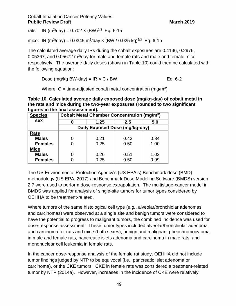
Cobalt Inhalation Cancer Potency Values Public Review Draft March 2019
49
rats: IR (m3/day) = 0.702 × (BW)2/3 Eq. 6-1a
mice: IR (m3/day) = 0.0345 m3/day × (BW / 0.025 kg)2/3 Eq. 6-1b
The calculated average daily IRs during the cobalt exposures are 0.4146, 0.2976,
0.05367, and 0.05672 m3/day for male and female rats and male and female mice,
respectively. The average daily doses (shown in Table 10) could then be calculated with
the following equation:
Dose (mg/kg BW-day) = IR × C / BW Eq. 6-2
Where: C = time-adjusted cobalt metal concentration (mg/m3)
Table 10. Calculated average daily exposed dose (mg/kg-day) of cobalt metal in the rats and mice during the two-year exposures (rounded to two significant figures in the final assessment).
Species sex
Cobalt Metal Chamber Concentration (mg/m3)
0 1.25 2.5 5.0
Daily Exposed Dose (mg/kg-day)
Rats Males Females Mice Males Females
0 0
0 0
0.21 0.25
0.26 0.25
0.42 0.50
0.51 0.50
0.84 1.00
1.02 0.99
The US Environmental Protection Agency’s (US EPA’s) Benchmark dose (BMD)
methodology (US EPA, 2017) and Benchmark Dose Modeling Software (BMDS) version
2.7 were used to perform dose-response extrapolation. The multistage-cancer model in
BMDS was applied for analysis of single-site tumors for tumor types considered by
OEHHA to be treatment-related.
Where tumors of the same histological cell type (e.g., alveolar/bronchiolar adenomas
and carcinomas) were observed at a single site and benign tumors were considered to
have the potential to progress to malignant tumors, the combined incidence was used for
dose-response assessment. These tumor types included alveolar/bronchiolar adenoma
and carcinoma for rats and mice (both sexes), benign and malignant pheochromocytoma
in male and female rats, pancreatic islets adenoma and carcinoma in male rats, and
mononuclear cell leukemia in female rats.
In the cancer dose-response analysis of the female rat study, OEHHA did not include
tumor findings judged by NTP to be equivocal (i.e., pancreatic islet adenoma or
carcinoma), or the CKE tumors. CKE in female rats was considered a treatment-related
tumor by NTP (2014a). However, increases in the incidence of CKE were relatively

Cobalt Inhalation Cancer Potency Values Public Review Draft March 2019
50
small (0/45, 4/43, 1/40, 3/40 for control, low-, mid-, and high-dose groups, respectively)
compared with increases in other treatment-related tumors, and were not statistically
significant by trend test or pairwise comparison of cobalt-exposed group tumor incidence
with controls.
The NTP (2014a) concluded that exposure to cobalt metal led to an increased incidence
of mononuclear cell leukemia in female rats, although the trend test applied by the NTP
(based on total number of animals examined) did not reach statistical significance (p =
0.118). Lack of a positive trend was likely a result of a plateau response for all non-
control cobalt exposures. When converted to an effective tumor incidence by OEHHA, a
significant positive trend (p = 0.0426) was observed with the Cochran-Armitage trend
test supplied in the BMDS, version 2.7 (US EPA, 2017). Thus, BMD analysis was
performed for the leukemia tumor data.
For large datasets such as those by NTP, the BMD recommended by OEHHA (2008) is
the 95% lower confidence bound on the effective dose producing 5% response
(BMDL05). The multistage-cancer polynomial model was fit to the data, which fits most
tumor data sets well. First- and second-degree polynomial multistage models were run
for all tumor incidence data sets, and the most appropriate model was chosen based on
BMD guidance (U.S. EPA, 2016). Briefly, a goodness-of-fit p-value > 0.05 indicates that
the model fits the data well, and in cases where more than one model provides an
adequate fit, the model with the lowest Akaike Information Criterion (AIC) value is often
selected as the best fitting model. The BMD05 and BMDL05 are shown in Table 11. The
degree of polynomial chosen was 1 in all cases, except for adrenal medulla tumors in
female rats where a 2nd degree polynomial provided the best fit to the data.
Male and female rats developed tumors in several organ systems following cobalt metal
exposure. Basing cancer risk on only one tumor type may underestimate the
carcinogenic potential of a chemical that induces tumors at multiple sites. Multisite tumor
CSFs were calculated in both male and female rats using MS Combo Model (US EPA,
2017). The BMDS procedure for summing risks over several tumor sites uses the profile
likelihood method. In this method, the maximum likelihood estimates (MLEs) for the
multistage model parameters (qi) for each tumor type are added together (i.e., Σԛ0, Σԛ1,
Σԛ2), and the resulting model is used to determine a combined BMD. A confidence
interval for the combined BMD is then calculated by computing the desired percentile of
the chi-squared distribution associated with a likelihood ratio test having one degree of
freedom.
For male rats, multisite tumor analysis was conducted for lung (alveolar/bronchiolar
adenoma or carcinoma combined), adrenal medulla (benign or malignant
pheochromocytoma combined), and pancreatic islet tumors (pancreatic islets adenoma
and carcinoma). In female rats, multisite tumor analysis was conducted for lung

Cobalt Inhalation Cancer Potency Values Public Review Draft March 2019
51
(alveolar/bronchiolar adenoma or carcinoma combined), adrenal medulla (benign or
malignant pheochromocytoma combined), and mononuclear cell leukemia. Some
evidence suggests that pheochromocytoma of the adrenal medulla may be dependent
on tumor formation in the lungs (see Cancer Hazard Evaluation section), although NTP
(2014a) noted that the evidence is not clear. OEHHA therefore uses the health
protective assumption that these two tumor types are independent and considered the
lung and adrenal tumors as separate sites in the multi-site analysis. The treatment-
related female rat CKE tumor data were not included in the dose-response analysis as
they were judged not to contribute significantly to the CSF, based on the relatively small
increased incidence (0/45, 4/43, 1/40, 3/40 for control, low-, mid-, and high-dose groups,
respectively) compared with increases in other treatment-related tumors, and the
absence of any apparent dose-related trend.
For male and female mice, single-site tumor analyses were conducted for lung
(alveolar/bronchiolar adenoma or carcinoma combined) tumors.
At the effective dose producing a 5% tumor response, the CSF is calculated as
0.05/BMDL05 and is in units of (mg/kg-day)-1 (Table 11). The rodent (a) CSFs were then
converted to human (h) equivalents using body weight (BW3/4) scaling:
CSF(h) = CSF(a) × (BW(h) / BW(a))1/4 Eq. 6-3
Using this interspecies scaling factor is preferred by OEHHA because it is assumed to
account not only for pharmacokinetic differences (e.g., breathing rate, metabolism), but
also for pharmacodynamic considerations, i.e., tissue responses to chemical exposure
(U.S. EPA, 2005). Lifetime body weights for control rats and mice of both sexes were
calculated from the NTP (2014a) study as described above. The default body weight for
humans is 70 kg. The body weight scaling factor assumes that mg/surface area/day is
an equivalent dose between species (OEHHA, 2009).
Comparison of the single-site and multisite CSFs in Table 11 shows that the lung tumor
human CSF of 27 (mg/kg-day)-1 based on male mice to be the most sensitive estimate of
cancer risk (CSF rounded to two significant figures in the final assessment).

Cobalt Inhalation Cancer Potency Values Public Review Draft March 2019
52
Table 11. BMD05, BMDL05, rodent CSFs, and human CSFs for single-site and multi-site tumors in rats and mice resulting from 2-year inhalation exposure to cobalt metal Tumor type AICa p-value BMD05
(mg/kg-day)a
BMDL05
(mg/kg-day)
CSF -Rodent (mg/kg-day)-1
CSF - Human (mg/kg-day)-1
Rats
Alveolar/bronchiolar Males Females
Adrenal medulla Males Females
Pancreatic Islet cell Males
Mononuclear cell leukemia Females
Multisite: lung-adrenal-pancreatic tumors combined Males
Multisite: lung-adrenal-leukemia combined Females
Mice
Alveolar/bronchiolar Males Females
173.14 202.56
217.47 187.45
126.34
277.81
NAb
NA
167.47 188.20
0.54 0.54
0.27 0.75
0.16
0.065
NA
NA
0.12 0.57
0.01647 0.04162
0.02451 0.1287
0.1636
0.1297
0.009291
0.02828
0.01446 0.01868
0.01364 0.03363
0.01853 0.04931
0.1029
0.06698
0.007947
0.01867
0.01122 0.01506
3.66 1.49
2.70 1.01
0.49
0.74
6.29
2.68
4.46 3.32
12.91 5.96
9.51 4.03
1.71
2.95
22.17
10.70
27.49 20.04
a Akaike Information Criterion b Not applicable
Multistage model fits to the data and the resulting BMD and BMDL are shown in Figure 1
for alveolar/bronchiolar lung tumors in male rats, and in Figure 2 for alveolar/bronchiolar
lung tumors in male mice.

Cobalt Inhalation Cancer Potency Values Public Review Draft March 2019
53
Figure 1. Multistage model fit to the male rat lung tumor data for cobalt metal. (The benchmark used is the exposure concentration producing 5% tumor response (BMD) with the 95% lower confidence bound (BMDL) on the BMD).
Figure 2. Multistage model fit to the male mouse lung tumor data for cobalt metal.
(The benchmark used is the exposure concentration producing 5% tumor response
(BMD) with the 95% lower confidence bound (BMDL) on the BMD.)
Inhalation Unit Risk Factor
The Inhalation Unit Risk (IUR) describes the excess cancer risk associated with an
inhalation exposure to a concentration of 1 µg/m3 and is derived from the cobalt metal
0
0.2
0.4
0.6
0.8
1
0 0.1 0.2 0.3 0.4 0.5 0.6 0.7 0.8
Frac
tion
Affe
cted
dose
Multistage Cancer Model, with BMR of 5% Extra Risk for the BMD and 0.95 Lower Confidence Limit for the BMDL
08:41 03/02 2018
BMDBMDL
Multistage Cancer
Linear extrapolation
0.2
0.3
0.4
0.5
0.6
0.7
0.8
0.9
1
0 0.2 0.4 0.6 0.8 1
Frac
tion
Affe
cted
dose
Multistage Cancer Model, with BMR of 5% Extra Risk for the BMD and 0.95 Lower Confidence Limit for the BMDL
10:10 12/12 2016
BMDBMDL
Multistage Cancer
Linear extrapolation

Cobalt Inhalation Cancer Potency Values Public Review Draft March 2019
54
CSF. Using a human breathing rate (BR) of 20 m3/day, an average human body weight
(BW) of 70 kg, and a mg to µg conversion factor (CF) of 1,000, the IUR is calculated as:
IUR = (CSF x BR) / (BW x CF) Eq. 6-4
Use of the equation above with the cobalt metal CSF of 27 (mg/kg-day)-1 results in a
calculated IUR of 0.0078 (µg/m3)-1 or 7.8 × 10⁻³ (µg/m3)-1. Thus, the extra cancer risk
associated with continuous lifetime exposure to 1 µg/m3 cobalt metal is 7.8 in one
thousand, or 7800 in a million.
Cobalt Sulfate Heptahydrate
Effective Tumor Incidences
The effective tumor incidences (number of tumor-bearing animals over the number of
animals alive at the time of first occurrence of the tumor) for treatment-related tumors
observed in the NTP studies conducted in rats and mice are shown in Tables 12 and 13,
respectively. The NTP individual animal pathology data used to determine the tumor
incidences for cobalt sulfate heptahydrate were obtained from the CEBS database (NTP,
1998b).
Table 12. Effective tumor incidences (number of animals alive at day of first tumor) of treatment-related lesions in rats in the two-year inhalation studies of cobalt sulfate hyptahydrate NTP (1998a) Tumor Type
CoSO4·7H2O Concentration (mg/m3)
0 0.3 1.0 3.0
Male Rats Lung Alveolar/bronchiolar adenoma Alveolar/bronchiolar carcinoma Alveolar/bronchiolar adenoma or carcinoma Female Rats Lung Alveolar/bronchiolar adenoma Alveolar/bronchiolar carcinoma Alveolar/bronchiolar adenoma, carcinoma, or squamous cell carcinoma Adrenal medulla Benign pheochromocytoma Benign, complex or malignant pheochromocytoma
1/43† 0/24 1/43† 0/39‡ 0/44† 0/44‡ 2/39‡ 2/39‡
4/44 0/28 4/44 1/33 2/41 3/41 1/37 1/37
1/43 3/34 4/43 10/37** 6/42* 16/42** 3/38 4/38
6/40 1/25 7/40* 9/35** 6/46* 16/46** 8/38 10/39*
* p<0.05, ** p<0.01 for statistical difference from control by Fisher’s exact test (calculated by OEHHA) † p<0.05, ‡ p<0.01 for positive trend for tumor type by the Cochran-Armitage trend test (calculated by OEHHA) a Includes benign bilateral pheochromocytoma

Cobalt Inhalation Cancer Potency Values Public Review Draft March 2019
55
Table 13. Effective tumor incidences (number of animals alive at day of first tumor) of treatment-related lesions in mice in the two-year inhalation studies of cobalt sulfate heptahydrate NTP (1998a)
Tumor
CoSO4·7H2O Concentration (mg/m3)
0 0.3 1.0 3.0
Male Mice Lung Alveolar/bronchiolar adenoma Alveolar/bronchiolar carcinoma Alveolar/bronchiolar adenoma or carcinoma Female Mice Lung Alveolar/bronchiolar adenoma Alveolar/bronchiolar carcinoma Alveolar/bronchiolar adenoma or carcinoma
9/49† 4/50† 11/50‡ 3/40† 1/49‡ 4/49‡
12/50 5/50 14/50 6/47 1/49 7/49
13/49 7/49 19/49 9/42 4/49 13/49*
18/48* 11/48* 28/48** 10/39* 9/45** 18/45**
* p<0.05, ** p<0.01 for statistical difference from control by Fisher’s exact test (calculated by OEHHA) † p<0.05, ‡ p<0.01 for positive trend for tumor type by the Cochran-Armitage trend test (calculated by OEHHA)
Calculation of Single- and Multi-Site Tumor CSFs
For the derivation of the CSF, cobalt sulfate heptahydrate chamber concentrations of 0,
0.3, 1.0 and 3.0 mg/m3, were time-adjusted (6.2 hrs/24 hrs x 5 days/7 days) to
extrapolate from the intermittent lab exposure conditions to a continuous exposure over
the life span of the animals (i.e., to simulate an annualized average air concentration).
The time-adjusted cobalt sulfate heptahydrate concentrations of 0, 0.055, 0.18, and 0.55
mg/m3 were used to calculate the average daily dose in mg/kg BW-day.
To calculate the daily dose, the average body weight of the rats and mice over the
duration of the study is used to determine the IR. The weighted average lifetime body
weights for control animals in each study were calculated from the data o group mean
body weights reported every 1 to 4 weeks during the 2-year exposure period. The
average body weights were 435.8, 263.3, 41.7 and 40.2 g for the control male rats,
female rats, male mice and female mice, respectively.
The IRs were estimated the same as that shown in Eq 6-1a and 6-1b above, where:
For rats, IR (m3/day) = 0.702 (BW)2/3 (OEHHA, 2018b)
For mice, IR (m3/day) = 0.0345 m3/day (BW / 0.025 kg)2/3 (Anderson, 1983)

Cobalt Inhalation Cancer Potency Values Public Review Draft March 2019
56
The calculated average daily IRs during the cobalt exposures are 0.4035, 0.2884,
0.04852, and 0.04735 m3/day for male and female rats and male and female mice,
respectively. The IR multiplied by the time-adjusted exposure concentration and divided
into the animal body weight gives the dose (Eq. 6-2) in mg/kg BW-day (Table 14).
Table 14. Calculated average daily exposed dose (mg/kg-day) of cobalt sulfate heptahydrate in the rats and mice during the two-year exposures (rounded to two significant figures in the final assessment)
Species Sex
CoSO4·7H2O Chamber Concentration (mg/m3)
0 0.3 1.0 3.0
Daily Exposed Dose (mg/kg-day)
Rats Males Females Mice Males Females
0 0 0 0
0.051 0.061 0.064 0.065
0.17 0.20 0.21 0.22
0.51 0.61 0.64 0.65
US EPA (2017) BMD methodology (BMDS version 2.7) was applied for single-site
tumors using the multistage-cancer model. US EPA BMD guidance (U.S. EPA, 2016)
was used to choose the most appropriate model among multistage 1st, and 2nd degree
polynomial models, similar as that described above for cobalt metal. Tumor incidences
in the low dose groups of both rats and mice were very near or below a 5% tumor
response. Combined with the large group sizes (n=48 to 50), a benchmark of 5% tumor
response (BMD05) is appropriate for determining the cancer potency (OEHHA, 2009).
The BMD05 and BMDL05 were determined for treatment-related tumors in each of the
studies. Specifically, these values were determined for lung alveolar/bronchiolar
adenoma, carcinoma, or squamous cell carcinoma (combined) for rats of both sexes, for
lung alveolar/bronchiolar adenoma or carcinoma (combined) for mice of both sexes, and
for benign or malignant adrenal medulla tumors (combined) in female rats (Table 15).
The incidence of these tumors showed a statistically significant increase above control
values at one or more dose levels, and also exhibited a statistically significant positive
trend across dose levels (See Tables 12 and 13).
Cobalt sulfate heptahydrate induced tumors at two sites in female rats (tumors in the
lung and adrenal medulla). To avoid the potential underestimation of the true
carcinogenic risk using a single tumor-site approach, a multi-site tumor risk analysis for
female rats was performed, which included both alveolar/bronchiolar adenoma,
carcinoma, or squamous cell carcinoma (combined) and benign, complex, or malignant
pheochromocytoma (combined). The multi-site tumor CSFs were calculated using MS
Combo Model (US EPA, 2017). A description of the MS Combo Model is provided in the
cobalt metal CSF derivation above.

Cobalt Inhalation Cancer Potency Values Public Review Draft March 2019
57
Some evidence suggests that pheochromocytoma of the adrenal medulla may be
dependent on tumor formation in the lungs (see Cancer Hazard Evaluation section),
although NTP (2014a) noted that the evidence is not clear. OEHHA therefore uses the
health protective assumption that these two tumor types are independent and considered
separate tumor sites for multi-site analysis.
Modeling the single-site tumor incidence data, in all cases the selected models were first
degree polynomials either because the model defaulted to polynomial = 1 or because
this degree of polynomial provided the best fit to the data (i.e., lowest AIC value). The
multistage polynomial model fit the tumor data well (goodness of fit p-value p > 0.05),
except for the lung tumor incidence in female rats. The female rat lung tumor data
exhibited a plateau response at the two highest dose groups, resulting in the lack of
model fit (p = 0.0065). The high dose group was subsequently removed and the three
remaining dose groups were rerun using the multistage model. An acceptable model fit
to the data was achieved (p = 0.57) with these three dose groups (Figure 3). For
comparison, a plot showing the multistage model fit to the male mice lung tumor data is
given in Figure 4.
At the effective dose producing a 5% tumor response, the cancer slope factor (CSF) is
calculated as 0.05/BMDL05 and is in units of (mg/kg-day)-1 (Table 15). The animal (a)
CSFs were then converted to human (h) equivalents using body weight (BW)3/4 scaling
as shown above in Eq. 6-3. Lifetime body weights for control rats and mice of both
sexes were calculated from NTP (2015) as described above. The default body weight
for humans is 70 kg.
Comparison of the single-site and multi-site human CSFs in Tables 15 shows the human
CSF of 13.41 (mg/kg-day)-1 based on the female rat multi-site tumor data to be the most
sensitive indicator of cancer risk for cobalt sulfate heptahydrate. The cobalt ion is
considered to be the primary factor for cancer risk. Normalizing the cobalt sulfate
heptahydrate CSF to the content of cobalt by dividing the molecular weight of cobalt into
the molecular weight of cobalt sulfate heptahydrate (58.9 Co / 281.1 CoSO4 • 7H2O =
0.2095) and multiplying by 13.41 (mg/kg-day)-1 results in an adjusted CSF of 2.8 (mg
Co/kg-day)-1.
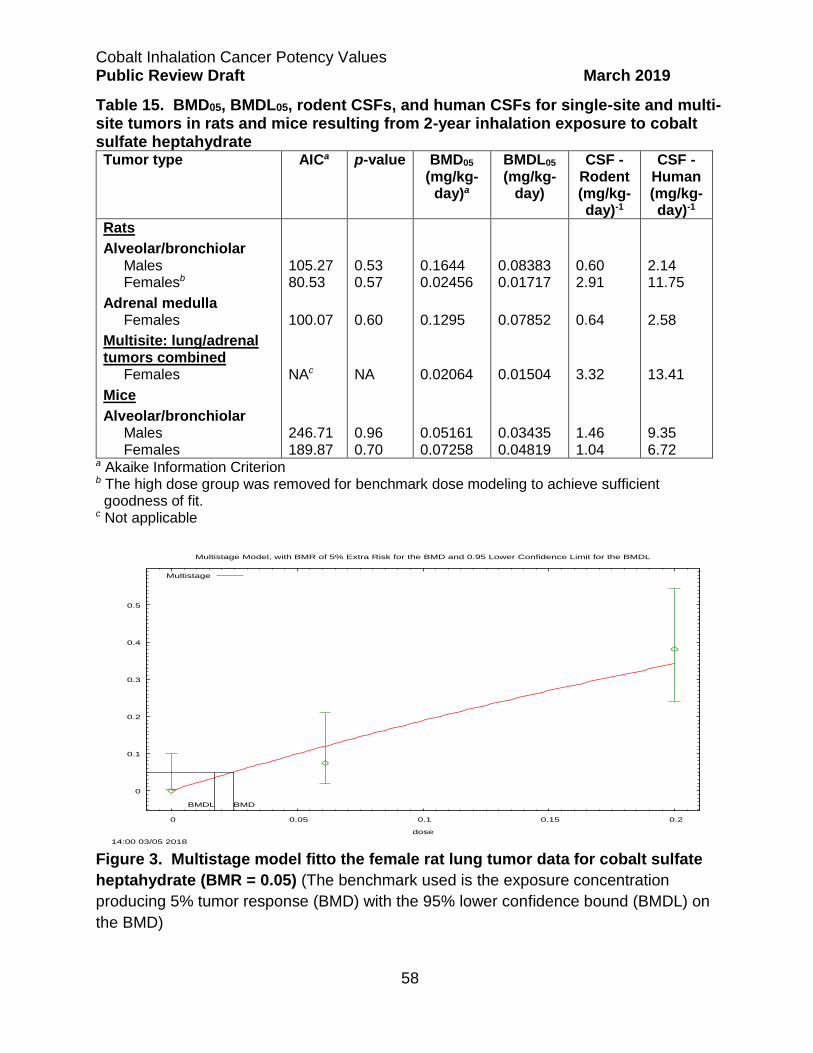
Cobalt Inhalation Cancer Potency Values Public Review Draft March 2019
58
Table 15. BMD05, BMDL05, rodent CSFs, and human CSFs for single-site and multi-site tumors in rats and mice resulting from 2-year inhalation exposure to cobalt sulfate heptahydrate Tumor type AICa p-value BMD05
(mg/kg-day)a
BMDL05
(mg/kg-day)
CSF -Rodent (mg/kg-day)-1
CSF - Human (mg/kg-day)-1
Rats
Alveolar/bronchiolar Males Femalesb
Adrenal medulla Females
Multisite: lung/adrenal tumors combined Females
Mice
Alveolar/bronchiolar Males Females
105.27 80.53
100.07
NAc
246.71 189.87
0.53 0.57
0.60
NA
0.96 0.70
0.1644 0.02456
0.1295
0.02064
0.05161 0.07258
0.08383 0.01717
0.07852
0.01504
0.03435 0.04819
0.60 2.91
0.64
3.32
1.46 1.04
2.14 11.75
2.58
13.41
9.35 6.72
a Akaike Information Criterion b The high dose group was removed for benchmark dose modeling to achieve sufficient goodness of fit. c Not applicable
Figure 3. Multistage model fitto the female rat lung tumor data for cobalt sulfate
heptahydrate (BMR = 0.05) (The benchmark used is the exposure concentration
producing 5% tumor response (BMD) with the 95% lower confidence bound (BMDL) on
the BMD)
0
0.1
0.2
0.3
0.4
0.5
0 0.05 0.1 0.15 0.2
Frac
tion
Affe
cted
dose
Multistage Model, with BMR of 5% Extra Risk for the BMD and 0.95 Lower Confidence Limit for the BMDL
14:00 03/05 2018
BMDBMDL
Multistage

Cobalt Inhalation Cancer Potency Values Public Review Draft March 2019
59
Figure 4. Multistage model fit to the male mice lung tumor data for cobalt sulfate
heptahydrate (BMR = 0.05) (The benchmark used is the exposure concentration
producing 5% tumor response (BMD) with the 95% lower confidence bound (BMDL) on
the BMD.)
Inhalation Unit Risk Factor
The Inhalation Unit Risk (IUR) describes the excess cancer risk associated with an
inhalation exposure to a concentration of 1 µg/m3 and is derived from the cobalt sulfate
heptahydrate CSF. Using a human breathing rate of 20 m3/day, an average human BW
of 70 kg, and a mg to µg conversion factor of 1,000, the IUR was calculated as shown in
Eq. 6-4 (see above).
Using the cobalt normalized CSF of 2.8 (mg Co/kg-day)-1 results in a calculated IUR of
0.00080 (µg Co/m3)-1 or 8.0 10-4 (µg Co/m3)-1. Thus, the extra cancer risk associated
with continuous lifetime exposure to 1 µg/m3 cobalt sulfate heptahydrate normalized to
the cobalt content is 8.0 in ten thousand, or 800 in a million.
VI. CONCLUSIONS
Carcinogenicity studies conducted by NTP established clear evidence of carcinogenicity
for cobalt metal and cobalt sulfate heptahydrate. The lungs were the primary site of
tumor formation in both rats and mice. Cobalt metal and cobalt sulfate heptahydrate also
caused tumors at multiple sites in rats. Carcinogens that produce tumors in more than
one species have the greatest potential to induce tumors in other species, including
humans. For each cobalt compound, the CSF was based on the most sensitive species
0.1
0.2
0.3
0.4
0.5
0.6
0.7
0 0.1 0.2 0.3 0.4 0.5 0.6
Frac
tion
Affe
cted
dose
Multistage Cancer Model, with BMR of 5% Extra Risk for the BMD and 0.95 Lower Confidence Limit for the BMDL
07:54 01/10 2017
BMDBMDL
Multistage Cancer
Linear extrapolation

Cobalt Inhalation Cancer Potency Values Public Review Draft March 2019
60
and sex. Derivation of a CSF for cobalt metal (27 [mg/kg-day]-1) is based on lung tumor
formation in male mice. The CSF derivation for cobalt sulfate heptahydrate (13 [mg/kg-
day]-1) is based on a multi-site analysis of lung and adrenal medulla tumors observed in
female rats.
The cobalt ion is considered the primary factor for cancer risk for both cobalt metal and
cobalt compounds, and both cobalt metal and cobalt sulfate heptahydrate induced
tumors of the same histogenic type in lungs. Consequently, nearly comparable CSF
values for both forms of cobalt is not unexpected. However, cobalt metal produced a
greater incidence of pulmonary tumors (except in female rats) and proportionally more
pulmonary carcinomas than adenomas in the chronic rodent studies at comparable
cobalt ion concentrations. Thus, cobalt metal appears to be more effective than cobalt
sulfate heptahydrate at inducing lung tumors. CKE neoplasms were found at a greater
incidence in rats exposed to cobalt metal than to cobalt sulfate heptahydrate, and an
increased incidence of mononuclear cell leukemia in female rats were observed only with
exposure to cobalt metal.
Additionally, in vitro studies suggest differences in how the cells internalize cobalt metal
particles and water-insoluble cobalt compounds compared to cobalt ions (released by
water-soluble cobalt compounds), which are then distributed within the cells. This may
explain some of the different genotoxicity results observed for cobalt metal and insoluble
cobalt compounds as compared to those observed for soluble cobalt compounds. The in
vitro studies also suggest that insoluble cobalt compounds, such as cobalt oxides, are
internalized and distributed in cells in a manner similar to that of cobalt metal particles.
With the available information, OEHHA recommends that the CSF derived from cobalt
metal (27 [mg/kg-day]-1) be used for cobalt metal exposure and for cobalt compounds,
such as cobalt oxides, that are poorly water-soluble or insoluble but are bioavailable in
pulmonary fluids following inhalation. The CSF derived for cobalt sulfate heptahydrate
(13 [mg/kg-day]-1) is recommended exclusively for water-soluble cobalt compounds,
such as the chloride, acetate, and nitrate salts of cobalt.

Cobalt Inhalation Cancer Potency Values Public Review Draft March 2019
61
VII. REFERENCES
Abudayyak M, Gurkaynak TA and Ozhan G (2017). In vitro evaluation of cobalt oxide nanoparticle-induced toxicity. Toxicol Ind Health 33(8): 646-654.
Alarifi S, Ali D, Y AO, Ahamed M, Siddiqui MA and Al-Khedhairy AA (2013). Oxidative stress contributes to cobalt oxide nanoparticles-induced cytotoxicity and DNA damage in human hepatocarcinoma cells. Int J Nanomedicine 8: 189-199.
Anard D, Kirsch-Volders M, Elhajouji A, Belpaeme K and Lison D (1997). In vitro genotoxic effects of hard metal particles assessed by alkaline single cell gel and elution assays. Carcinogenesis 18(1): 177-84.
Anderson EL (1983). Quantitative approaches in use to assess cancer risk. Risk Analysis 3(4): 277-295.
Annangi B, Bach J, Vales G, Rubio L, Marcos R and Hernandez A (2015). Long-term exposures to low doses of cobalt nanoparticles induce cell transformation enhanced by oxidative damage. Nanotoxicology 9(2): 138-47.
Apostoli P, Porru S and Alessio L (1994). Urinary cobalt excretion in short time occupational exposure to cobalt powders. Sci Total Environ 150(1-3): 129-32.
Bailey MR, Kreyling WG, Andre S, Bachelor A, Collier CG, Drosselmeyer E, Ferron GA, Foster PP, Haider B, Hodgson A, Masse R, Metivier H, Morgan A, Muller H-L, Patrick G, Pearman I, Pickering S, Ramsden D, Stirling C and Talbot RJ (1989). An interspecies comparison of the lung clearance of inhaled monodisperse cobalt oxide particles-Part I: Objectives and summary of results. J Aerosol Sci 20(2): 169-188.
Baralkiewicz D and Siepak J (1999). Chromium, nickel and cobalt in environmental samples and existing legal norms. Polish J Environ Studies 8(4): 201-208.
Behl M, Stout MD, Herbert RA, Dill JA, Baker GL, Hayden BK, Roycroft JH, Bucher JR and Hooth MJ (2015). Comparative toxicity and carcinogenicity of soluble and insoluble cobalt compounds. Toxicology 333: 195-205.
Beleznay E and Osvay M (1994). Long-term clearance of accidentally inhaled 60Co aerosols in humans. Health Phys 66(4): 392-9.
Beyersmann D and Hartwig A (1992). The genetic toxicology of cobalt. Toxicol Appl Pharmacol 115(1): 137-45.
Brix AE, Hardisty JF and McConnell EE (2010). Combining neoplasms for evaluation of rodent carcinogenesis studies. In: Cancer Risk Assessment, C-H Hsu and T Stedeford eds., John Wiley & Sons, Inc. pp. 619-715.

Cobalt Inhalation Cancer Potency Values Public Review Draft March 2019
62
Bucher JR, Hailey JR, Roycroft JR, Haseman JK, Sills RC, Grumbein SL, Mellick PW and Chou BJ (1999). Inhalation toxicity and carcinogenicity studies of cobalt sulfate. Toxicol Sci 49(1): 56-67.
Capomazza C and Botta A (1991). Cobalt chloride induces micronuclei in human lymphocytes. Med Sci Res 19: 219-220.
Cappellini F, Hedberg Y, McCarrick S, Hedberg J, Derr R, Hendriks G, Odnevall Wallinder I and Karlsson HL (2018). Mechanistic insight into reactivity and (geno)toxicity of well-characterized nanoparticles of cobalt metal and oxides. Nanotoxicology 12(6): 602-620.
CARB (2013). California Air Resources Board California Toxics Inventory. Online at: http://www.arb.ca.gov/toxics/cti/cti.htm.
CARB (2018). California Air Resources Board California Toxics Inventory. Online at: https://www.arb.ca.gov/adam/toxics/sitesubstance.html.
Cavallo D, Ciervo A, Fresegna AM, Maiello R, Tassone P, Buresti G, Casciardi S, Iavicoli S and Ursini CL (2015). Investigation on cobalt-oxide nanoparticles cyto-genotoxicity and inflammatory response in two types of respiratory cells. J Appl Toxicol 35(10): 1102-13.
Christensen JM and Poulsen OM (1994). A 1982-1992 surveillance programme on Danish pottery painters. Biological levels and health effects following exposure to soluble or insoluble cobalt compounds in cobalt blue dyes. Sci Total Environ 150(1-3): 95-104.
Colognato R, Bonelli A, Ponti J, Farina M, Bergamaschi E, Sabbioni E and Migliore L (2008). Comparative genotoxicity of cobalt nanoparticles and ions on human peripheral leukocytes in vitro. Mutagenesis 23(5): 377-82.
Costa M, Heck JD and Robison SH (1982). Selective phagocytosis of crystalline metal sulfide particles and DNA strand breaks as a mechanism for the induction of cellular transformation. Cancer Res 42(7): 2757-63.
Creton S, Aardema MJ, Carmichael PL, Harvey JS, Martin FL, Newbold RF, O'Donovan MR, Pant K, Poth A, Sakai A, Sasaki K, Scott AD, Schechtman LM, Shen RR, Tanaka N and Yasaei H (2012). Cell transformation assays for prediction of carcinogenic potential: state of the science and future research needs. Mutagenesis 27(1): 93-101.
De Boeck M, Lardau S, Buchet JP, Kirsch-Volders M and Lison D (2000). Absence of significant genotoxicity in lymphocytes and urine from workers exposed to moderate levels of cobalt-containing dust: a cross-sectional study. Environ Mol Mutagen 36(2): 151-60.
De Boeck M, Lison D and Kirsch-Volders M (1998). Evaluation of the in vitro direct and indirect genotoxic effects of cobalt compounds using the alkaline comet assay. Influence of interdonor and interexperimental variability. Carcinogenesis 19(11): 2021-9.

Cobalt Inhalation Cancer Potency Values Public Review Draft March 2019
63
De Boeck M, Lombaert N, De Backer S, Finsy R, Lison D and Kirsch-Volders M (2003). In vitro genotoxic effects of different combinations of cobalt and metallic carbide particles. Mutagenesis 18(2): 177-86.
Duerksen-Hughes PJ, Yang J and Ozcan O (1999). p53 induction as a genotoxic test for twenty-five chemicals undergoing in vivo carcinogenicity testing. Environ Health Perspect 107(10): 805-12.
Farah SB (1983). The in vivo effect of cobalt chloride on chromosomes. Rev Brasil Genet 3: 433-442.
Farrell RL and Davis GW (1974). The effects of particulates on respiratory carcinogenesis by diethylnitrosamine. ln: Karb, E. & Paris, J .R., eds, Experimental Lung Cancer: Carcinogenesis and Bioassays, Springer-Verlag Berlin, pp. 219-233.
Figgitt M, Newson R, Leslie IJ, Fisher J, Ingham E and Case CP (2010). The genotoxicity of physiological concentrations of chromium (Cr(III) and Cr(VI)) and cobalt (Co(II)): an in vitro study. Mutat Res 688(1-2): 53-61.
Foster PP, Pearman I and Ramsden D (1989). An interspecies comparison of the lung clearance of inhaled monodisperse cobalt oxide particles - Part II: Lung clearance of inhaled cobalt oxide particles in man. J Aerosol Sci 20(2): 189-204.
Gennart JP, Baleux C, Verellen-Dumoulin C, Buchet JP, De Meyer R and Lauwerys R (1993). Increased sister chromatid exchanges and tumor markers in workers exposed to elemental chromium-, cobalt- and nickel-containing dusts. Mutat Res 299(1): 55-61.
Gibson DP, Brauninger R, Shaffi HS, Kerckaert GA, LeBoeuf RA, Isfort RJ and Aardema MJ (1997). Induction of micronuclei in Syrian hamster embryo cells: comparison to results in the SHE cell transformation assay for National Toxicology Program test chemicals. Mutat Res 392(1-2): 61-70.
Green SE, Luczak MW, Morse JL, DeLoughery Z and Zhitkovich A (2013). Uptake, p53 pathway activation, and cytotoxic responses for Co(II) and Ni(II) in human lung cells: implications for carcinogenicity. Toxicol Sci 136(2): 467-77.
Hanna PM, Kadiiska MB and Mason RP (1992). Oxygen-derived free radical and active oxygen complex formation from cobalt(II) chelates in vitro. Chem Res Toxicol 5(1): 109-15.
Hansen T, Clermont G, Alves A, Eloy R, Brochhausen C, Boutrand JP, Gatti AM and Kirkpatrick CJ (2006). Biological tolerance of different materials in bulk and nanoparticulate form in a rat model: sarcoma development by nanoparticles. J R Soc Interface 3(11): 767-75.
Hartwig A, Snyder RD, Schlepegrell R and Beyersmann D (1991). Modulation by Co(II) of UV-induced DNA repair, mutagenesis and sister-chromatid exchanges in mammalian cells. Mutat Res 248(1): 177-85.

Cobalt Inhalation Cancer Potency Values Public Review Draft March 2019
64
Hillwalker WE and Anderson KA (2014). Bioaccessibility of metals in alloys: evaluation of three surrogate biofluids. Environ Pollut 185: 52-8.
Hogstedt C and Alexandersson R (1987). Mortality among hard-metal workers in Sweden. Scand J Work Environ Health 13: 177-178.
Holstein H, Ranebo Y and Raaf CL (2015). Human metabolism of orally administered radioactive cobalt chloride. J Environ Radioact 143: 152-8.
Hong HH, Hoenerhoff MJ, Ton TV, Herbert RA, Kissling GE, Hooth MJ, Behl M, Witt KL, Smith-Roe SL, Sills RC and Pandiri AR (2015). Kras, Egfr, and Tp53 mutations in B6C3F1/N mouse and F344/NTac rat alveolar/bronchiolar carcinomas resulting from chronic inhalation exposure to cobalt metal. Toxicol Pathol 43(6): 872-82.
Horev-Azaria L, Kirkpatrick CJ, Korenstein R, Marche PN, Maimon O, Ponti J, Romano R, Rossi F, Golla-Schindler U, Sommer D, Uboldi C, Unger RE and Villiers C (2011). Predictive toxicology of cobalt nanoparticles and ions: comparative in vitro study of different cellular models using methods of knowledge discovery from data. Toxicol Sci 122(2): 489-501.
Horie M, Fujita K, Kato H, Endoh S, Nishio K, Komaba LK, Nakamura A, Miyauchi A, Kinugasa S, Hagihara Y, Niki E, Yoshida Y and Iwahashi H (2012). Association of the physical and chemical properties and the cytotoxicity of metal oxide nanoparticles: metal ion release, adsorption ability and specific surface area. Metallomics 4(4): 350-60.
HSDB (2016). Hazardous Substances Data Bank. Hexamethylene Diisocyanate, Chemical/Physical Properties. National Library of Medicine, Bethesda, MD. Available at: http://toxnet.nlm.nih.gov.
Hutter HP, Wallner P, Moshammer H and Marsh G (2016). Dust and Cobalt Levels in the Austrian Tungsten Industry: Workplace and Human Biomonitoring Data. Int J Environ Res Public Health 13(9).
IARC (1991). IARC Monographs on the Evaluation of Carcinogenic Risks to Humans. Chlorinated Drinking-water; Chlorination By-products; Some Other Halogenated Compounds; Cobalt and Cobalt Compounds. World Health Organization. Vol. 52. Online at: http://monographs.iarc.fr/ENG/Monographs/vol52/index.php.
IARC (2006). IARC Monographs on the Evaluation of Carcinogenic Risks to Humans - Cobalt in Hard Metals and Cobalt Sulfate, Gallium Arsenide, Indium Phosphide and Vanadium Pentoxide. International Agency for Research on Cancer (IARC). World Health Organization. Vol. 86. Online at: http://monographs.iarc.fr/ENG/Monographs/vol86/mono86-6.pdf.
Kasprzak KS, Zastawny TH, North SL, Riggs CW, Diwan BA, Rice JM and Dizdaroglu M (1994). Oxidative DNA base damage in renal, hepatic, and pulmonary chromatin of rats after intraperitoneal injection of cobalt(II) acetate. Chem Res Toxicol 7(3): 329-35.

Cobalt Inhalation Cancer Potency Values Public Review Draft March 2019
65
Kasten U, Mullenders LH and Hartwig A (1997). Cobalt(II) inhibits the incision and the polymerization step of nucleotide excision repair in human fibroblasts. Mutat Res 383(1): 81-9.
Keane MJ, Hornsby-Myers JL, Stephens JW, Harrison JC, Myers JR and Wallace WE (2002). Characterization of hard metal dusts from sintering and detonation coating processes and comparative hydroxyl radical production. Chem Res Toxicol 15(8): 1010-6.
Kerckaert GA, Brauninger R, LeBoeuf RA and Isfort RJ (1996). Use of the Syrian hamster embryo cell transformation assay for carcinogenicity prediction of chemicals currently being tested by the National Toxicology Program in rodent bioassays. Environ Health Perspect 104 Suppl 5: 1075-84.
Kirkland D, Brock T, Haddouk H, Hargeaves V, Lloyd M, Mc Garry S, Proudlock R, Sarlang S, Sewald K, Sire G, Sokolowski A and Ziemann C (2015). New investigations into the genotoxicity of cobalt compounds and their impact on overall assessment of genotoxic risk. Regul Toxicol Pharmacol 73(1): 311-38.
Kreyling WG, Ferron GA and Haider B (1986). Metabolic fate of inhaled Co aerosols in beagle dogs. Health Phys 51(6): 773-95.
Kreyling WG, Godleski JJ, Kariya ST, Rose RM and Brain JD (1990). In vitro dissolution of uniform cobalt oxide particles by human and canine alveolar macrophages. Am J Respir Cell Mol Biol 2(5): 413-22.
Kreyling WG, Nyberg K, D. NC, Collier CG, Camner P, Heilmann P, Lirsac PN, Lundborg M and Matejkova E (1991a). Interspecies comparison of phagolysosomal pH in alveolar macrophages. Inhal Toxicol 3: 91-100.
Kreyling WG, S. AC, Collier CG, Ferron GA, Metivier H and Schumann G (1991b). lnterspecies comparison of lung clearance after inhalation of monodisperse, solid cobalt oxide aerosol particles. J Aerosol Sci 22: 509-535.
Kumar V, Mishra RK, Kaur G and Dutta D (2017). Cobalt and nickel impair DNA metabolism by the oxidative stress independent pathway. Metallomics 9(11): 1596-1609.
Kyono H, Kusaka Y, Homma K, Kubota H and Endo-Ichikawa Y (1992). Reversible lung lesions in rats due to short-term exposure to ultrafine cobalt particles. Ind Health 30(3-4): 103-18.
Lasfargues G, Wild P, Moulin JJ, Hammon B, Rosmorduc B, Rondeau du Noyer C, Lavandier M and Moline J (1994). Lung cancer mortality in a French cohort of hard-metal workers. Am J Ind Med 26(5): 585-95.
Leggett RW (2008). The biokinetics of inorganic cobalt in the human body. Sci Total Environ 389(2-3): 259-69.

Cobalt Inhalation Cancer Potency Values Public Review Draft March 2019
66
Leyssens L, Vinck B, Van Der Straeten C, Wuyts F and Maes L (2017). Cobalt toxicity in humans-A review of the potential sources and systemic health effects. Toxicology 387: 43-56.
Linna A, Oksa P, Palmroos P, Roto P, Laippala P and Uitti J (2003). Respiratory health of cobalt production workers. Am J Ind Med 44(2): 124-32.
Lison D (1996). Human toxicity of cobalt-containing dust and experimental studies on the mechanism of interstitial lung disease (hard metal disease). Crit Rev Toxicol 26(6): 585-616.
Lison D, Buchet JP, Swennen B, Molders J and Lauwerys R (1994). Biological monitoring of workers exposed to cobalt metal, salt, oxides, and hard metal dust. Occup Environ Med 51(7): 447-50.
Lison D, van den Brule S and Van Maele-Fabry G (2018). Cobalt and its compounds: update on genotoxic and carcinogenic activities. Crit Rev Toxicol: 1-18.
Lloyd DR, Phillips DH and Carmichael PL (1997). Generation of putative intrastrand cross-links and strand breaks in DNA by transition metal ion-mediated oxygen radical attack. Chem Res Toxicol 10(4): 393-400.
MAK (2007). MAK Value Documentations: The MAK-Collection Part 1. Cobalt and its Compounds (as inhalable dusts aerosols). DFG, Deutsche Forschungsgemeinschaft, Wiley‐VCH Verlag GmbH & Co. KGaA. Online at: https://onlinelibrary.wiley.com/doi/10.1002/3527600418.mb744048e0023. 23.
Mateuca R, Aka PV, De Boeck M, Hauspie R, Kirsch-Volders M and Lison D (2005). Influence of hOGG1, XRCC1 and XRCC3 genotypes on biomarkers of genotoxicity in workers exposed to cobalt or hard metal dusts. Toxicol Lett 156(2): 277-88.
McConnell EE, Solleveld HA, Swenberg JA and Boorman GA (1986). Guidelines for combining neoplasms for evaluation of rodent carcinogenesis studies. J Natl Cancer Inst 76(2): 283-9.
Moulin JJ, Wild P, Mur JM, Fournier-Betz M and Mercier-Gallay M (1993). A mortality study of cobalt production workers: an extension of the follow-up. Am J Ind Med 23(2): 281-8.
Moulin JJ, Wild P, Romazini S, Lasfargues G, Peltier A, Bozec C, Deguerry P, Pellet F and Perdrix A (1998). Lung cancer risk in hard-metal workers. Am J Epidemiol 148(3): 241-8.
Mur JM, Moulin JJ, Charruyer-Seinerra MP and Lafitte J (1987). A cohort mortality study among cobalt and sodium workers in an electrochemical plant. Am J Ind Med 11(1): 75-81.

Cobalt Inhalation Cancer Potency Values Public Review Draft March 2019
67
Nackerdien Z, Kasprzak KS, Rao G, Halliwell B and Dizdaroglu M (1991). Nickel(II)- and cobalt(II)-dependent damage by hydrogen peroxide to the DNA bases in isolated human chromatin. Cancer Res 51(21): 5837-42.
Newton D and Rundo J (1971). The long-term retention of inhaled cobalt-60. Health Phys 21(3): 377-84.
NTP (1996). Toxicology and Carcinogenesis Studies of Nickel Subsulfide (CAS No. 12035-72-2) in F344/N Rats and B6C3F1 Mice (Inhalation Studies). NTP TR 453. Research Triangle Park, NC.
NTP (1998a). NTP Technical Report on the Toxicology and Carcinogenesis Studies of Cobalt Sulfate Heptahydrate (CAS No. 10026-24-1) in F344/N Rats and B6C3F1 Mice (Inhalation Studies). NTP TR 471. Research Triangle Park, NC.
NTP (1998b). TR-471: Cobalt sulfate heptahydrate (10026-24-1). Chemical Effects in Biological Systems (CEBS). Research Triangle Park, NC (USA): National Toxicology Program (NTP). Accessed November 2017. https://manticore.niehs.nih.gov/cebssearch/publication/TR-47.
NTP (2014a). NTP Technical Report on the Toxicology Studies of Cobalt Metal (CAS No. 7440-48-4) in F344/N Rats and B6C3F1/N Mice and Toxicology and Carcinogenesis Studies of Cobalt Metal in F344/NTac Rats and B6C3F1/N Mice (Inhalation Studies). NTP TR 581. Research Triangle Park, NC.
NTP (2014b). TR-581: Cobalt (7440-48-4). Chemical Effects in Biological Systems (CEBS). Research Triangle Park, NC (USA): National Toxicology Program (NTP). Accessed November, 2017. https://manticore.niehs.nih.gov/cebssearch/publication/TR-581.
NTP (2016). National Toxicology Program. Cobalt and Cobalt Compounds That Release Cobalt Ions In Vivo, CAS No. 7440-48-4 (Cobalt metal). Report on Carcinogens, Fourteenth Edition. 8 p.
OEHHA (2008). Air Toxics Hot Spots Program Risk Assessment Guidelines. Technical Support Document for the Derivation of Noncancer Reference Exposure Levels. California Environmental Protection Agency, Office of Environmental Health Hazard Assessment, Oakland, CA. Online at: http://www.oehha.ca.gov/air/hot_spots/rels_dec2008.html.
OEHHA (2009). Air Toxics Hot Spots Program Risk Assessment Guidelines. Technical Support Document for Cancer Potency Factors: Methodologies for derivation, listing of available values, and adjustments to allow for early life stage exposures. California Environmental Protection Agency Office of Environmental Health Hazard Assessment. Online at: http://oehha.ca.gov/air/crnr/technical-support-document-cancer-potency-factors-2009.

Cobalt Inhalation Cancer Potency Values Public Review Draft March 2019
68
OEHHA (2018a). Office of Environmental Health Hazard Assessment. The Proposition 65 List. Online at: https://oehha.ca.gov/proposition-65/proposition-65-list.
OEHHA (2018b). Calculation of rat breathing rate based on bodyweight. Office of Environmental Health Hazard Assessment. California Environmental Protection Agency Office of Environmental Health Hazard Assessment. Online at: https://oehha.ca.gov/media/downloads/crnr/calcuratbreathingrate092818.pdf.
Ortega R, Bresson C, Darolles C, Gautier C, Roudeau S, Perrin L, Janin M, Floriani M, Aloin V, Carmona A and Malard V (2014). Low-solubility particles and a Trojan-horse type mechanism of toxicity: the case of cobalt oxide on human lung cells. Part Fibre Toxicol 11: 14.
Ozaki K, Haseman JK, Hailey JR, Maronpot RR and Nyska A (2002). Association of adrenal pheochromocytoma and lung pathology in inhalation studies with particulate compounds in the male F344 rat--the National Toxicology Program experience. Toxicol Pathol 30(2): 263-70.
Palit S, Sharma A and Talukder G (1991). Chromosomal aberrations induced by cobaltous chloride in mice in vivo. Biol Trace Elem Res 29(2): 139-45.
Patel E, Lynch C, Ruff V and Reynolds M (2012). Co-exposure to nickel and cobalt chloride enhances cytotoxicity and oxidative stress in human lung epithelial cells. Toxicol Appl Pharmacol 258(3): 367-75.
Patrick G, Stirling C, Kreyling WG, Poncy J-L, Duserre C, Collier CG, Godlesk iJ and Brain JD (1994). Interspecies comparison of the clearance of ionic cobalt from the lungs. Inhal Toxicol 6(3): 225-240.
Ponti J, Sabbioni E, Munaro B, Broggi F, Marmorato P, Franchini F, Colognato R and Rossi F (2009). Genotoxicity and morphological transformation induced by cobalt nanoparticles and cobalt chloride: an in vitro study in Balb/3T3 mouse fibroblasts. Mutagenesis 24(5): 439-45.
Rajiv S, Jerobin J, Saranya V, Nainawat M, Sharma A, Makwana P, Gayathri C, Bharath L, Singh M, Kumar M, Mukherjee A and Chandrasekaran N (2016). Comparative cytotoxicity and genotoxicity of cobalt (II, III) oxide, iron (III) oxide, silicon dioxide, and aluminum oxide nanoparticles on human lymphocytes in vitro. Hum Exp Toxicol 35(2): 170-83.
Rhoads K and Sanders CL (1985). Lung clearance, translocation, and acute toxicity of arsenic, beryllium, cadmium, cobalt, lead, selenium, vanadium, and ytterbium oxides following deposition in rat lung. Environ Res 36(2): 359-78.
Sabbioni E, Fortaner S, Farina M, Del Torchio R, Olivato I, Petrarca C, Bernardini G, Mariani-Costantini R, Perconti S, Di Giampaolo L, Gornati R and Di Gioacchino M (2014a). Cytotoxicity and morphological transforming potential of cobalt nanoparticles,

Cobalt Inhalation Cancer Potency Values Public Review Draft March 2019
69
microparticles and ions in Balb/3T3 mouse fibroblasts: an in vitro model. Nanotoxicology 8(4): 455-64.
Sabbioni E, Fortaner S, Farina M, Del Torchio R, Petrarca C, Bernardini G, Mariani-Costantini R, Perconti S, Di Giampaolo L, Gornati R and Di Gioacchino M (2014b). Interaction with culture medium components, cellular uptake and intracellular distribution of cobalt nanoparticles, microparticles and ions in Balb/3T3 mouse fibroblasts. Nanotoxicology 8(1): 88-99.
Sauni R, Oksa P, Uitti J, Linna A, Kerttula R and Pukkala E (2017). Cancer incidence among Finnish male cobalt production workers in 1969-2013: a cohort study. BMC Cancer 17(1): 340.
Scansetti G, Maina G, Botta GC, Bambace P and Spinelli P (1998). Exposure to cobalt and nickel in the hard-metal production industry. Int Arch Occup Environ Health 71(1): 60-3.
Smith LJ, Holmes AL, Kandpal SK, Mason MD, Zheng T and Wise JP, Sr. (2014). The cytotoxicity and genotoxicity of soluble and particulate cobalt in human lung fibroblast cells. Toxicol Appl Pharmacol 278(3): 259-65.
Speer RM, The T, Xie H, Liou L, Adam RM and Wise JP, Sr. (2017). The Cytotoxicity and Genotoxicity of Particulate and Soluble Cobalt in Human Urothelial Cells. Biol Trace Elem Res.
Steinhoff D and Mohr U (1991). On the question of a carcinogenic action of cobalt-containing compounds. Exp Pathol 41(4): 169-74.
Stopford W, Turner J, Cappellini D and Brock T (2003). Bioaccessibility testing of cobalt compounds. J Environ Monit 5(4): 675-80.
Suh M, Thompson CM, Brorby GP, Mittal L and Proctor DM (2016). Inhalation cancer risk assessment of cobalt metal. Regul Toxicol Pharmacol 79: 74-82.
Swennen B, Buchet JP, Stanescu D, Lison D and Lauwerys R (1993). Epidemiological survey of workers exposed to cobalt oxides, cobalt salts, and cobalt metal. Br J Ind Med 50(9): 835-42.
Tennant RW and Spalding J (1996). Predictions for the outcome of rodent carcinogenicity bioassays: identification of trans-species carcinogens and noncarcinogens. Environ Health Perspect 104 Suppl 5: 1095-100.
Tuchsen F, Jensen MV, Villadsen E and Lynge E (1996). Incidence of lung cancer among cobalt-exposed women. Scand J Work Environ Health 22(6): 444-50.
U.S. EPA (2016). Benchmark Dose Software (BMDS) User Manual. United States Environmental Protection Agency. Online at: https://www.epa.gov/sites/production/files/2015-11/documents/bmds_manual.pdf.

Cobalt Inhalation Cancer Potency Values Public Review Draft March 2019
70
US EPA (2017). Benchmark Dose Software, Version 2.7. United States Environmental Protection Agency. National Center for Environmental Assessment. Online at: http://www.epa.gov/bmds.
USP (2015). The Pharmacopeia of the United States of America. General Notices and Requirements. Thirty-Eighth Revision and the National Formulary, Thirty-Third Edition.
Valko M, Morris H and Cronin MT (2005). Metals, toxicity and oxidative stress. Curr Med Chem 12(10): 1161-208.
Van Goethem F, Lison D and Kirsch-Volders M (1997). Comparative evaluation of the in vitro micronucleus test and the alkaline single cell gel electrophoresis assay for the detection of DNA damaging agents: genotoxic effects of cobalt powder, tungsten carbide and cobalt-tungsten carbide. Mutat Res 392(1-2): 31-43.
Wan R, Mo Y, Zhang Z, Jiang M, Tang S and Zhang Q (2017). Cobalt nanoparticles induce lung injury, DNA damage and mutations in mice. Part Fibre Toxicol 14(1): 38.
Wehner AP, Stuart BO and Sanders CL (1979). Inhalation studies with Syrian golden hamsters. Prog Exp Tumor Res 24: 177-98.
Wild P, Perdrix A, Romazini S, Moulin JJ and Pellet F (2000). Lung cancer mortality in a site producing hard metals. Occup Environ Med 57(8): 568-73.
Wultsch G, Nersesyan A, Kundi M, Misik M, Setayesh T, Waldherr M, Vodicka P, Vodickova L and Knasmuller S (2017). Genotoxic and Cytotoxic Effects in Exfoliated Buccal and Nasal Cells of Chromium and Cobalt Exposed Electroplaters. J Toxicol Environ Health A: 1-10.
Xie H, Smith LJ, Holmes AL, Zheng T and Pierce Wise J, Sr. (2016). The cytotoxicity and genotoxicity of soluble and particulate cobalt in human lung epithelial cells. Environ Mol Mutagen 57(4): 282-7.
Yamada Y, Kido T, Honda R, Ishizaki M, Tsuritani I, Yamaya H and Nogawa K (1987). Analysis of dusts and evaluation of dust exposure in a hard metal factory. Ind Health 25(1): 1-10.
Zanetti G and Fubini B (1997). Surface interaction between metallic cobalt and tungsten carbide particles as a primary cause of hard metal lung disease. J Mater Chem 7: 1647–1654.