Embed Size (px)
Citation preview
Kuranishi et al. Respiratory Research (2015) 16:55 DOI 10.1186/s12931-015-0213-7
RESEARCH Open Access
Airway-centered interstitial fibrosis: etiology,clinical findings and prognosisLilian Tiemi Kuranishi1*, Kevin O Leslie2, Rimarcs Gomes Ferreira3, Ester Aparecida Ney Coletta3,Karin Mueller Storrer1, Maria Raquel Soares1 and Carlos Alberto de Castro Pereira1
Abstract
Background: Airway-centered Interstitial Fibrosis (ACIF) is a common pathologic pattern observed in our practice.
Objectives: The objectives of this study are to describe the causes associated with ACIF in a large sample of patients andits effect on survival.
Methods: A retrospective study in three centers of interstitial lung disease in São Paulo, between January of 1995 andDecember of 2012. The surgical lung biopsy specimens were reviewed by three pathologists. The clinical, functional andtomographic findings were analyzed by a standardized protocol.
Results: There were 68 cases of ACIF, most of them women. The mean age was 57 ± 12 yr. Dyspnea, cough, restrictivepattern at spirometry and oxygen desaturation at exercise were common. A reticular pattern with peribronchovascularinfiltrates was found in 79% of the cases. The etiologies of ACIF were hypersensitivity pneumonitis in 29 (42.6%),gastroesophageal reflux disease in 17 (25.0%), collagen vascular disease in 4 (5.9%), a combination of them in 15cases and idiopathic in 3 (4.4%). The median survival was 116 months (95% CI = 58.5 – 173.5). Lower values ofoxygen saturation at rest, presence of cough and some histological findings - organizing tissue in the airways, fibroblasticfoci and microscopic honeycombing - were predictors of worse survival.
Conclusions: ACIF is an interstitial lung disease with a better survival when compared with IPF. The main etiologies areHP and GERD. The oxygen saturation at rest, the presence of cough and some histological findings are predictors ofsurvival.
Keywords: Interstitial lung disease, Hypersensitivity pneumonia, Gastroesophageal reflux, Pulmonary fibrosis
IntroductionSince 2002 a new interstitial pneumonia centered on smallairways and no granulomas has been described in smallseries [1-4]. This entity has been variably called centrilobu-lar fibrosis, bronchiolocentric interstitial pneumonia,airway-centered interstitial fibrosis, and peribronchiolarmetaplasia [3-6]. Lung biopsies from these cases showed adistinctive pattern of interstitial fibrosis centered and ex-tending around the bronchioles, often with bronchiolarmetaplasia of the epithelium. Due to involvement of smallas well large airways [5,7], the best name seems to beairway-centered interstitial fibrosis (ACIF). The prognosis isunknown due to small number of cases described [2-4].The etiology in these series was unclear. Similar pathologic
* Correspondence: [email protected] Department, Federal University of São Paulo, Sao Paulo, BrazilFull list of author information is available at the end of the article
© 2015 Kuranishi et al.; licensee BioMed CentrCommons Attribution License (http://creativecreproduction in any medium, provided the orDedication waiver (http://creativecommons.orunless otherwise stated.
findings have been described in hypersensitivity pneumon-itis (HP) [8-15] or be secondary to gastroesophageal refluxdisease (GERD), isolated or associated with connective tis-sue diseases [16,17]. In our clinical practice this pathologicpattern is common in multidisciplinary case discussions.The objectives of the present study were to describe
the causes, the clinical, tomographic, functional andpathologic findings and their influence on survival in alarge number of patients with histologic diagnosis ofairway-centered interstitial fibrosis.
Material and methodsSelection of casesA retrospective cohort of 68 adult patients with ACIFwas evaluated. The medical records of 2716 patientswith interstitial lung diseases, seen between January of1995 and December of 2012 at three facilities in the city
al. This is an Open Access article distributed under the terms of the Creativeommons.org/licenses/by/4.0), which permits unrestricted use, distribution, andiginal work is properly credited. The Creative Commons Public Domaing/publicdomain/zero/1.0/) applies to the data made available in this article,

Kuranishi et al. Respiratory Research (2015) 16:55 Page 2 of 8
of São Paulo, Brazil, were reviewed. From this register600 patients were submitted to surgical lung biopsies.Cases with any findings of possible ACIF or describingbronchiolar involvement without a specific diagnosis ofbronchiolitis were reevaluated. In this database, 115 pa-tients had a diagnosis of possible ACIF by surgical lungbiopsy. From this sample, 36 cases were excluded due toa another diagnosis after a review of the slides (mainlybronchiolitis, characterized by bronchiolar inflammationand/or fibrosis in the absence of peribronchiolar involve-ment as well absence of reticular pattern on HRCT); sixcases were excluded due to incomplete data and five dueto honeycombing (an exclusion criteria due to possiblenon representative biopsies in advanced disease) ob-served on a HRCT scan. None of the patients died afterlung biopsy. Biopsies were done by small thoracotomiesor VATS, involving one or more sites, but this informa-tion was not available in final analysis. The final samplecomprised 68 patients.This study was approved by ethics committee of the
Federal University of São Paulo (register number 2079-09).
Histological findingsPatients were selected primarily by a revision of lungbiopsies by two pathologists dedicated to lung pathology,and with a large experience in interstitial lung disease(ILD). The cases were selected initially by one of themand 48 of them were sent to KOL for confirmation. Heagreed with diagnoses and described and classified thefindings in each case. The remaining twenty cases werediagnosed by consensus between our two pathologists,who also followed the same classification of findings.Concordance by kappa was not calculated. The diagnosiswas based on previously suggested criteria [4-6]. Themain diagnostic criteria included a fibrosis predomin-antly bronchiolocentric associated with bronchiolar orperibronchiolar inflammation and peribronchiolar meta-plasia. Cases with granulomas and foreign material wereexcluded. Incidental findings like such as fibromyxoidtissue foci in airways, fibroblastic foci, honeycombing,giant cells, cholesterol clefts, respiratory bronchiolitis,features of acute injury (tissue edema and the presenceof fibrin), and focal areas of heterogeneous or homoge-neous fibrosis (usual interstitial pneumonia (UIP) andnod-specific interstitial pneumonia (NSIP) like, respect-ively) were recorded.
Clinical analysis and HRCTA standardized protocol for investigation of interstitiallung diseases was used for all patients. Dyspnoea wasassessed by Magnitude of Task of Basal Dyspnea Index(BDI) [18]. Total BDI score was not considered becausefunctional impairment and magnitude of effort do notinvolve the same activities in different patients. The data
related to environmental exposures were recorded. Adiagnosis of HP was based on the exposure and the ab-sence of other potential causes of ACIF. Precipitins testsand cellular analysis of bronchoalveolar lavage were notavailable. Possible connective tissue disease (CTD) wasinvestigated by clinical and complementary tests in allcases. The diagnostic criteria for CTD were those previ-ously described [19].Symptoms of GERD (heartburn, regurgitation) were
recorded in all cases. An ambulatory 24-hour esophagealpH measurement with a dual sensor was performed ac-cording to standardized techniques [20]. The diagnosticcriteria for an abnormal proximal [21] and distal [22]reflux have been previously described.After biopsies selection, the final clinical diagnoses were
retrospectively determined by multidisciplinary discussion.The diagnoses were classified as HP, GERD, CTD, a com-bination of two or more categories or idiopathic.Pulmonary function tests were conducted according to
the American Thoracic Society guidelines [23]. The nor-mal values for spirometry were recalculated according2007 values derived for the Brazilian population [24].Normal values for DLco were from Crapo [25]. The per-ipheral oxygen saturation was evaluated at rest and aftera 4-minute self-paced step test [26].All CT scans were read by experienced radiologists
and pulmonologists through a systematic analysis of thefindings. The final findings were selected by consensus.Scores for extension of disease were not calculated. Ex-piratory views were not done systematically.The drugs used for at least three months for ACIF and
GERD treatment were recorded. Antigen avoidance andabatement procedures were recommended when expos-ure was present.
SurvivalSurvival was assessed from the day of the biopsy throughDecember 2012. Deaths were identified by a follow-up con-tact or through telephone notification by relatives. Deathswere considered ACIF-related if they were due to respira-tory failure, pneumonia or pulmonary fibrosis. One patientwas censored at the time of lung transplantation.
Statistical analysisAll of the data analyses were performed using the SPSSprogram, version 19. According distribution, data wereexpressed as mean ± SD, or as median and range. The con-tinuous data with a normal distribution were comparedusing t-tests. A chi-square test was used for comparisons ofproportions. The impact of clinical, functional, tomographicand pathologic data on survival was calculated by univariateCox regression ant by Kaplan-Meyer curves. Two-sidedp values <0.05 were considered to be statistically
Kuranishi et al. Respiratory Research (2015) 16:55 Page 3 of 8
significant. The study design was approved by the eth-ics committees of the hospitals involved.
ResultsThe study sample comprised 68 cases, with a predomin-ance of non-smokers, and females. The baseline charac-teristics are shown in Table 1. The main clinical featureswere cough (in 78%) and dyspnea (100%). The functionalprofile was typical of diffuse restrictive lung disease, withreduced FVC, FEV1, DLCO and a decrease of SaO2 upon
Table 1 Baseline characteristics of patients withairway-centered interstitial fibrosis (n = 68)
Characteristic Number
Sex, male/female 29/39
Age, years (mean ± SD) 57 ± 12
Duration of symptoms, in months, median (range) 24 (3–132)
Smoking
Never/ex-smoker/current smoker, n 39/28/1
Dyspnea
Major/moderate/light tasks, n 22/33/13
Coughing, n (%) 53 (78%)
Clubbing, n (%) 12 (18%)
Velcro crackles, n (%) 29 (43%)
Exposure to organic particles, n (%) 42 (62%)
Molds/birds/both 24/ 4/ 14
Gastroesophageal reflux (GERD) symptoms
None/past/current 30/ 12/ 26
Connective tissue disease (CTD), n (%)† 12 (17%)
Lung function tests
FVC, % predicted (mean ± SD) 66 ± 18
FEV1, % predicted (mean ± SD) 69 ± 18
FEV1/FVC, (mean ± SD) 0.85 ± 0,08
DLCO, % predicted (mean ± SD) (n = 43) 59 ± 17
Oxygen saturation at rest, % (mean ± SD) 94 ± 4
Oxygen saturation during exercise, % (mean ± SD) (n = 59) 87 ± 7
HRCT findings
Reticular infiltrate, n (%) 68 (100%)
Predominance: Upper lobe/lower lobe/diffuse, n 10/38/20
Predominance: Central/peripheral/both, n 9/14/45
Ground-glass opacities, n (%) 57 (84%)
Peribronchovascular, n (%) 54 (79%)
Bronchiectasis, n (%) 43 (63%)
Mosaic pattern/airtrapping, n (%) 25 (37%)
Centrilobular nodules, n (%) 14 (21%)†Systemic sclerosis (2), Rheumatoid arthritis (3), Mixed connective tissuedisease (3), Dermatomyositis (2), Antisynthetase syndrome (1), Systemicsclerosis and Sjögren’s syndrome (1), FEV1 = forced expiratory volume in 1 s;FVC = forced vital capacity; DLCO =monoxide carbon lung diffusion. HRCT =high resolution chest tomography.
exercise. A reticular pattern, suggesting fibrosis, waspresent in all of the cases. Central and peribronchovas-cular distributions were seen in 79% of the cases, but anassociated peripheral distribution was common.Exposure to birds, molds or both was present in 42 (62%)
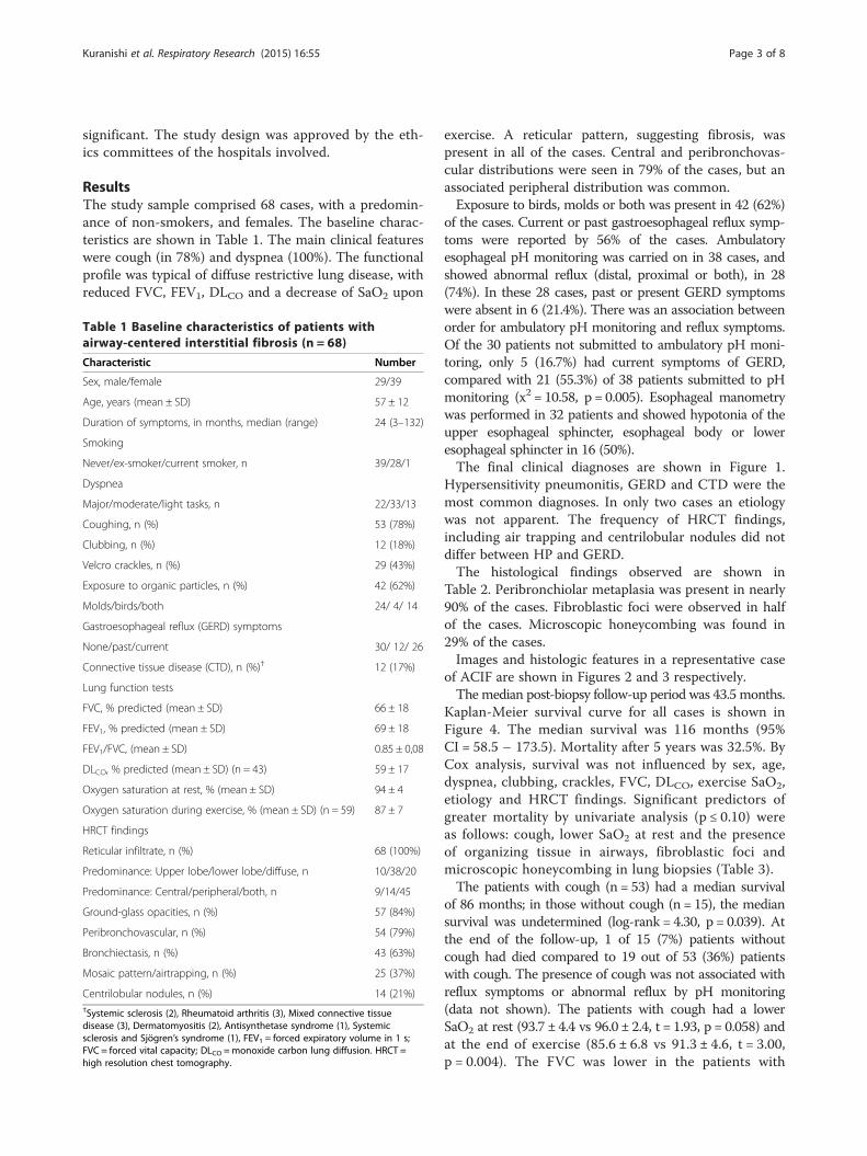
of the cases. Current or past gastroesophageal reflux symp-toms were reported by 56% of the cases. Ambulatoryesophageal pH monitoring was carried on in 38 cases, andshowed abnormal reflux (distal, proximal or both), in 28(74%). In these 28 cases, past or present GERD symptomswere absent in 6 (21.4%). There was an association betweenorder for ambulatory pH monitoring and reflux symptoms.Of the 30 patients not submitted to ambulatory pH moni-toring, only 5 (16.7%) had current symptoms of GERD,compared with 21 (55.3%) of 38 patients submitted to pHmonitoring (x2 = 10.58, p = 0.005). Esophageal manometrywas performed in 32 patients and showed hypotonia of theupper esophageal sphincter, esophageal body or loweresophageal sphincter in 16 (50%).The final clinical diagnoses are shown in Figure 1.
Hypersensitivity pneumonitis, GERD and CTD were themost common diagnoses. In only two cases an etiologywas not apparent. The frequency of HRCT findings,including air trapping and centrilobular nodules did notdiffer between HP and GERD.The histological findings observed are shown in
Table 2. Peribronchiolar metaplasia was present in nearly90% of the cases. Fibroblastic foci were observed in halfof the cases. Microscopic honeycombing was found in29% of the cases.Images and histologic features in a representative case
of ACIF are shown in Figures 2 and 3 respectively.The median post-biopsy follow-up period was 43.5 months.
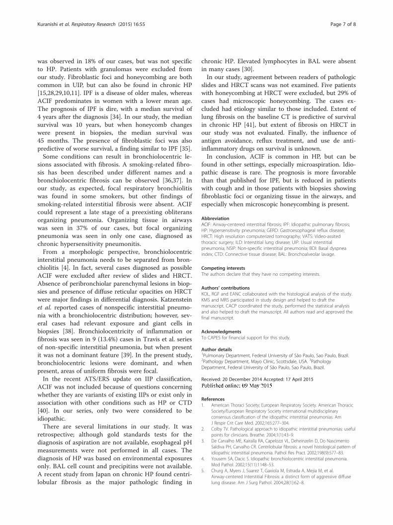
Kaplan-Meier survival curve for all cases is shown inFigure 4. The median survival was 116 months (95%CI = 58.5 – 173.5). Mortality after 5 years was 32.5%. ByCox analysis, survival was not influenced by sex, age,dyspnea, clubbing, crackles, FVC, DLCO, exercise SaO2,etiology and HRCT findings. Significant predictors ofgreater mortality by univariate analysis (p ≤ 0.10) wereas follows: cough, lower SaO2 at rest and the presenceof organizing tissue in airways, fibroblastic foci andmicroscopic honeycombing in lung biopsies (Table 3).The patients with cough (n = 53) had a median survival
of 86 months; in those without cough (n = 15), the mediansurvival was undetermined (log-rank = 4.30, p = 0.039). Atthe end of the follow-up, 1 of 15 (7%) patients withoutcough had died compared to 19 out of 53 (36%) patientswith cough. The presence of cough was not associated withreflux symptoms or abnormal reflux by pH monitoring(data not shown). The patients with cough had a lowerSaO2 at rest (93.7 ± 4.4 vs 96.0 ± 2.4, t = 1.93, p = 0.058) andat the end of exercise (85.6 ± 6.8 vs 91.3 ± 4.6, t = 3.00,p = 0.004). The FVC was lower in the patients with
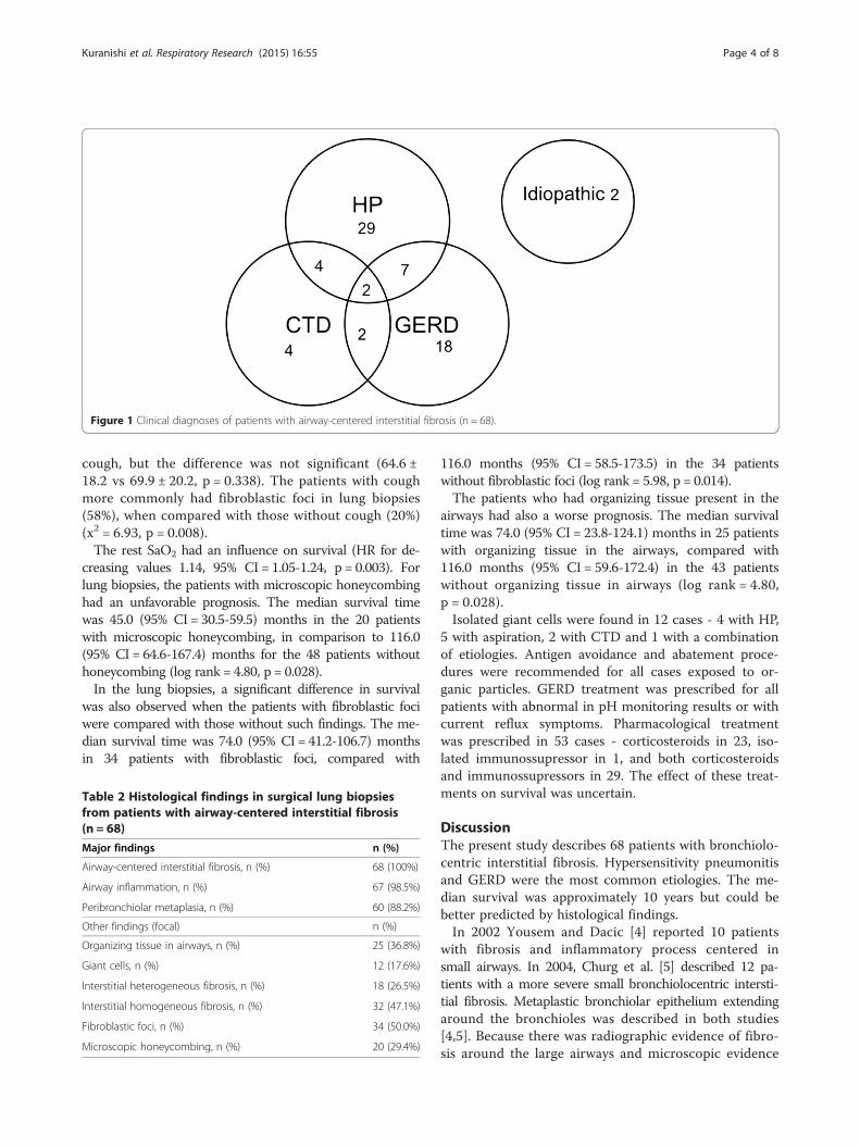
Figure 1 Clinical diagnoses of patients with airway-centered interstitial fibrosis (n = 68).
Kuranishi et al. Respiratory Research (2015) 16:55 Page 4 of 8
cough, but the difference was not significant (64.6 ±18.2 vs 69.9 ± 20.2, p = 0.338). The patients with coughmore commonly had fibroblastic foci in lung biopsies(58%), when compared with those without cough (20%)(x2 = 6.93, p = 0.008).The rest SaO2 had an influence on survival (HR for de-
creasing values 1.14, 95% CI = 1.05-1.24, p = 0.003). Forlung biopsies, the patients with microscopic honeycombinghad an unfavorable prognosis. The median survival timewas 45.0 (95% CI = 30.5-59.5) months in the 20 patientswith microscopic honeycombing, in comparison to 116.0(95% CI = 64.6-167.4) months for the 48 patients withouthoneycombing (log rank = 4.80, p = 0.028).In the lung biopsies, a significant difference in survival
was also observed when the patients with fibroblastic fociwere compared with those without such findings. The me-dian survival time was 74.0 (95% CI = 41.2-106.7) monthsin 34 patients with fibroblastic foci, compared with
Table 2 Histological findings in surgical lung biopsiesfrom patients with airway-centered interstitial fibrosis(n = 68)
Major findings n (%)
Airway-centered interstitial fibrosis, n (%) 68 (100%)
Airway inflammation, n (%) 67 (98.5%)
Peribronchiolar metaplasia, n (%) 60 (88.2%)
Other findings (focal) n (%)
Organizing tissue in airways, n (%) 25 (36.8%)
Giant cells, n (%) 12 (17.6%)
Interstitial heterogeneous fibrosis, n (%) 18 (26.5%)
Interstitial homogeneous fibrosis, n (%) 32 (47.1%)
Fibroblastic foci, n (%) 34 (50.0%)
Microscopic honeycombing, n (%) 20 (29.4%)
116.0 months (95% CI = 58.5-173.5) in the 34 patientswithout fibroblastic foci (log rank = 5.98, p = 0.014).The patients who had organizing tissue present in the
airways had also a worse prognosis. The median survivaltime was 74.0 (95% CI = 23.8-124.1) months in 25 patientswith organizing tissue in the airways, compared with116.0 months (95% CI = 59.6-172.4) in the 43 patientswithout organizing tissue in airways (log rank = 4.80,p = 0.028).Isolated giant cells were found in 12 cases - 4 with HP,
5 with aspiration, 2 with CTD and 1 with a combinationof etiologies. Antigen avoidance and abatement proce-dures were recommended for all cases exposed to or-ganic particles. GERD treatment was prescribed for allpatients with abnormal in pH monitoring results or withcurrent reflux symptoms. Pharmacological treatmentwas prescribed in 53 cases - corticosteroids in 23, iso-lated immunossupressor in 1, and both corticosteroidsand immunossupressors in 29. The effect of these treat-ments on survival was uncertain.
DiscussionThe present study describes 68 patients with bronchiolo-centric interstitial fibrosis. Hypersensitivity pneumonitisand GERD were the most common etiologies. The me-dian survival was approximately 10 years but could bebetter predicted by histological findings.In 2002 Yousem and Dacic [4] reported 10 patients
with fibrosis and inflammatory process centered insmall airways. In 2004, Churg et al. [5] described 12 pa-tients with a more severe small bronchiolocentric intersti-tial fibrosis. Metaplastic bronchiolar epithelium extendingaround the bronchioles was described in both studies[4,5]. Because there was radiographic evidence of fibro-sis around the large airways and microscopic evidence
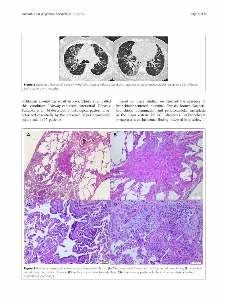
Figure 2 Radiologic findings of a patient with ACIF, showing diffuse ground glass opacities on peribronchovascular region, reticular infiltratesand traction bronchiectasias.
Kuranishi et al. Respiratory Research (2015) 16:55 Page 5 of 8
of fibrosis around the small airways, Churg et al. calledthis condition “Airway-centered Interstitial Fibrosis.Fukuoka et al. [6] described a histological pattern char-acterized essentially by the presence of peribronchiolarmetaplasia in 15 patients.
Figure 3 Histologic features of airway-centered interstitial fibrosis. (A) Aircentrilobular fibrosis from figure a. (C) Peribronchiolar alveolar metaplasiaorganization on airways.
Based on these studies, we selected the presence ofbronchiolar-centered interstitial fibrosis, bronchiolar/peri-bronchiolar inflammation and peribronchiolar metaplasiaas the major criteria for ACIF diagnosis. Peribronchiolarmetaplasia is an incidental finding observed in a variety of
way-centered fibrosis with obliteration of bronchioles, (B) a detailed. (D) Inflammatory peribronchiolar infiltration, obliterative focal
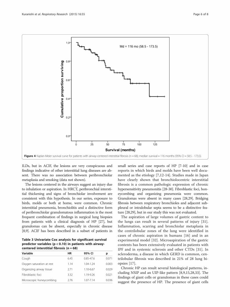
Figure 4 Kaplan-Meier survival curve for patients with airway-centered interstitial fibrosis (n = 68); median survival = 116 months (95% CI = 58.5 - 173.5).
Kuranishi et al. Respiratory Research (2015) 16:55 Page 6 of 8
ILDs, but in ACIF, the lesions are very conspicuous andfindings indicative of other interstitial lung diseases are ab-sent. There was no association between peribronchiolarmetaplasia and smoking (data not shown).The lesions centered in the airways suggest an injury due
to inhalation or aspiration. In HRCT, peribronchial intersti-tial thickening and signs of bronchiolar involvement areconsistent with this hypothesis. In our series, exposure tobirds, molds or both at home, were common. Chronicinterstitial pneumonia, bronchiolitis and a distinctive formof peribronchiolar granulomatous inflammation is the mostfrequent combination of findings in surgical lung biopsiesfrom patients with a clinical diagnosis of HP [27], butgranulomas can be absent, especially in chronic disease[8,9]. ACIF has been described in a subset of patients in
Table 3 Univariate Cox analysis for significant survivalpredictor variables (p < 0.10) in patients with airway-centered interstitial fibrosis (n = 68)
Variable HR 95% CI p
Cough 6.45 0.85-47.6 0.071
Oxygen saturation at rest 1.14 1.04-1.24 0.003
Organizing airway tissue 2.71 1.10-6.67 0.029
Fibroblastic foci 3.32 1.19-9.26 0.021
Microscopic honeycombing 2.76 1.07-7.14 0.036
small series and case reports of HP [7-10] and in casereports in which birds and molds have been well docu-mented as the etiology [7,12-14]. Studies made in Japanhave clearly shown that bronchiolocentric interstitialfibrosis is a common pathologic expression of chronichypersensitivity pneumonitis [28-30]. Fibroblastic foci, hon-eycombing and organizing pneumonia were common.Granulomas were absent in many cases [28,29]. Bridgingfibrosis between respiratory bronchioles and adjacent sub-pleural or intralobular septa seems to be a distinctive fea-ture [28,29], but in our study this was not evaluated.The aspiration of large volumes of gastric content to
the lungs can result in several patterns of injury [31].Inflammation, scarring and bronchiolar metaplasia inthe centrilobular zones of the lung were identified incases of chronic aspiration in humans [16] and in anexperimental model [32]. Microaspiration of the gastriccontents has been extensively evaluated in patients withIPF and in systemic sclerosis and other CTDs [31]. Inscleroderma, a disease in which GERD is common, cen-trilobular fibrosis was described in 21% of 28 lung bi-opsies [17].Chronic HP can result several histological patterns, in-
cluding NSIP and an UIP-like pattern [8,9,15,28,33]. Thefindings of giant cells or granulomas in these cases couldsuggest the presence of HP. The presence of giant cells
Kuranishi et al. Respiratory Research (2015) 16:55 Page 7 of 8
was observed in 18% of our cases, but was not specificto HP. Patients with granulomas were excluded fromour study. Fibroblastic foci and honeycombing are bothcommon in UIP, but can also be found in chronic HP[15,28,29,10,11]. IPF is a disease of older males, whereasACIF predominates in women with a lower mean age.The prognosis of IPF is dire, with a median survival of4 years after the diagnosis [34]. In our study, the mediansurvival was 10 years, but when honeycomb changeswere present in biopsies, the median survival was45 months. The presence of fibroblastic foci was alsopredictive of worse survival, a finding similar to IPF [35].Some conditions can result in bronchiolocentric le-
sions associated with fibrosis. A smoking-related fibro-sis has been described under different names and abronchiolocentric fibrosis can be observed [36,37]. Inour study, as expected, focal respiratory bronchiolitiswas found in some smokers, but other findings ofsmoking-related interstitial fibrosis were absent. ACIFcould represent a late stage of a preexisting obliteransorganizing pneumonia. Organizing tissue in airwayswas seen in 37% of our cases, but focal organizingpneumonia was seen in only one case, diagnosed aschronic hypersensitivity pneumonitis.From a morphologic perspective, bronchiolocentric
interstitial pneumonia needs to be separated from bron-chiolitis [4]. In fact, several cases diagnosed as possibleACIF were excluded after review of slides and HRCT.Absence of peribronchiolar parenchymal lesions in biop-sies and presence of diffuse reticular opacities on HRCTwere major findings in differential diagnosis. Katzensteinet al. reported cases of nonspecific interstitial pneumo-nia with a bronchiolocentric distribution; however, sev-eral cases had relevant exposure and giant cells inbiopsies [38]. Bronchiolocentricity of inflammation orfibrosis was seen in 9 (13.4%) cases in Travis et al. seriesof non-specific interstitial pneumonia, but when presentit was not a dominant feature [39]. In the present study,bronchiolocentric lesions were dominant, and whenpresent, areas of uniform fibrosis were focal.In the recent ATS/ERS update on IIP classification,
ACIF was not included because of questions concerningwhether they are variants of existing IIPs or exist only inassociation with other conditions such as HP or CTD[40]. In our series, only two were considered to beidiopathic.There are several limitations in our study. It was
retrospective; although gold standards tests for thediagnosis of aspiration are not available, esophageal pHmeasurements were not performed in all cases. Thediagnosis of HP was based on environmental exposuresonly. BAL cell count and precipitins were not available.A recent study from Japan on chronic HP found centri-lobular fibrosis as the major pathologic finding in
chronic HP. Elevated lymphocytes in BAL were absentin many cases [30].In our study, agreement between readers of pathologic
slides and HRCT scans was not examined. Five patientswith honeycombing at HRCT were excluded, but 29% ofcases had microscopic honeycombing. The cases ex-cluded had etiology similar to those included. Extent oflung fibrosis on the baseline CT is predictive of survivalin chronic HP [41], but extent of fibrosis on HRCT inour study was not evaluated. Finally, the influence ofantigen avoidance, reflux treatment, and use de anti-inflammatory drugs on survival is unknown.In conclusion, ACIF is common in HP, but can be
found in other settings, especially microaspiration. Idio-pathic disease is rare. The prognosis is more favorablethan that published for IPF, but is reduced in patientswith cough and in those patients with biopsies showingfibroblastic foci or organizing tissue in the airways, andespecially when microscopic honeycombing is present.
AbbreviationACIF: Airway-centered interstitial fibrosis; IPF: Idiopathic pulmonary fibrosis;HP: Hypersensitivity pneumonia; GERD: Gastroesophageal reflux disease;HRCT: High resolution computerized tomography; VATS: Video-assitedthoracic surgery; ILD: Interstitial lung disease; UIP: Usual interstitialpneumonia; NSIP: Non-specific interstitial pneumonia; BDI: Basal dyspneaindex; CTD: Connective tissue disease; BAL: Bronchoalveolar lavage.
Competing interestsThe authors declare that they have no competing interests.
Authors’ contributionsKOL, RGF and EANC collaborated with the histological analysis of the study.KMS and MRS participated in study design and helped to draft themanuscript. CACP coordinated the study, performed the statistical analysisand also helped to draft the manuscript. All authors read and approved thefinal manuscript.
AcknowledgmentsTo CAPES for financial support for this study.
Author details1Pulmonary Department, Federal University of São Paulo, Sao Paulo, Brazil.2Pathology Department, Mayo Clinic, Scottsdale, USA. 3PathologyDepartment, Federal University of São Paulo, Sao Paulo, Brazil.
Received: 20 December 2014 Accepted: 17 April 2015
References1. American Thoraci Society; European Respiratory Society. American Thoracic
Society/European Respiratory Society international multidisciplinaryconsensus classification of the idiopathic interstitial pneumonias. AmJ Respir Crit Care Med. 2002;165:277–304.
2. Colby TV. Pathological approach to idiopathic interstitial pneumonias: usefulpoints for clinicians. Breathe. 2004;1(1):43–9.
3. De Carvalho ME, Kairalla RA, Capelozzi VL, Deheinzelin D, Do NascimentoSaldiva PH, Carvalho CR. Centrilobular fibrosis: a novel histological pattern ofidiopathic interstitial pneumonia. Pathol Res Pract. 2002;198(9):577–83.
4. Yousem SA, Dacic S. Idiopathic bronchiolocentric interstitial pneumonia.Mod Pathol. 2002;15(11):1148–53.
5. Churg A, Myers J, Suarez T, Gaxiola M, Estrada A, Mejia M, et al.Airway-centered Interstitial Fibrosis: a distinct form of aggressive diffuselung disease. Am J Surg Pathol. 2004;28(1):62–8.
Kuranishi et al. Respiratory Research (2015) 16:55 Page 8 of 8
6. Fukuoka J, Franks TJ, Colby TV, Flaherty KR, Galvin JR, Hayden D, et al.Peribronchiolar metaplasia: a common histologic lesion in diffuse lungdisease and a rare cause of interstitial lung disease: clinicopathologicfeatures of 15 cases. Am J Surg Pathol. 2005;29(7):948–54.
7. Colombat M, Groussard O, Taillé C, Marrash-Chahla R, Brugière O, Mal H,et al. Lung transplantation in a patient with bronchiolocentric fibrosis. AmJ Surg Pathol. 2004;28(11):1540–2.
8. Lima MS, Coletta EN, Ferreira RG, Jasinowodolinski D, Arakaki JS, RodriguesSC, et al. Subacute and chronic hypersensitivity pneumonitis:histopathological patterns and survival. Respir Med. 2009;103(4):508–15.
9. Takemura T, Akashi T, Ohtani Y, Inase N, Yoshizawa Y. Pathology ofhypersensitivity pneumonitis. Curr Opin Pulm Med. 2008;14(5):440–54.
10. Churg A, Sin DD, Everett D, Brown K, Cool C. Pathologic patterns andsurvival in chronic hypersensitivity pneumonitis. Am J Surg Pathol.2009;33(12):1765–70.
11. Gaxiola M, Buendía-Roldán I, Mejía M, Carrillo G, Estrada A, Navarro MC,et al. Morphologic diversity of chronic pigeon breeder’s disease: clinicalfeatures and survival. Respir Med. 2011;105(4):608–14.
12. Muñoz A, Aranda I, Pascual J, Ferrando C. Idiopathic bronchiolocentricinterstitial pneumonia: a new idiopathic interstitial pneumonia. ArchBronconeumol. 2007;43(8):464–6.
13. Fenton ME, Cockcroft DW, Wright JL, Churg A. Hypersensitivity pneumonitisas a cause of bronchiolocentric interstitial fibrosis. Ann Allergy AsthmaImmunol. 2007;99(5):465–6.
14. Blanc AL, Delhaes L, Copin MC, Stach B, Faivre JB, Wallaert B. Interstitial lungdisease due to domestic moulds. Rev Mal Respir. 2011;28(7):913–8.
15. Trahan S, Hanak V, Ryu JH, Myers JL. Role of surgical lung biopsy inseparating chronic hypersensitivity pneumonia from usual interstitialpneumonia/idiopathic pulmonary fibrosis: analysis of 31 biopsies from 15patients. Chest. 2008;134(1):126–32.
16. Yousem SA, Faber C. Histopathology of aspiration pneumonia notassociated with food or other particulate matter: a clinicopathologic studyof 10 cases diagnosed on biopsy. Am J Surg Pathol. 2011;35(3):426–31.
17. De Souza RB, Borges CT, Capelozzi VL, Parra ER, Jatene FB, Kavakama J, et al.Centrilobular fibrosis: an underrecognized pattern in systemic sclerosis.Respiration. 2009;77(4):389–97.
18. Mahler DA, Weinberg DH, Wells CK, Feinstein AR. The measurement ofdyspnea. Contents, interobserver agreement, and physiologic correlates oftwo new clinical indexes. Chest. 1984;85(6):751–8.
19. De Lauretis A, Veeraraghavan S, Renzoni E. Aspects of interstitial lungdisease: connective tissue disease-associated interstitial lung disease: howdoes it differ from IPF? How should the clinical approach differ? ChronRespir Dis. 2011;8(1):53–82.
20. Dhiman RK, Saraswat VA, Naik SR. Ambulatory esophageal pH monitoring:technique, interpretations, and clinical indications. Dig Dis Sci.2002;47(2):241–50.
21. Dobhan R, Castell DO. Normal and abnormal proximal esophageal acidexposure: results of ambulatory dual-probe pH monitoring Am.J Gastroenterol. 1993;88(1):25–9.
22. Johnson LF, Demeester TR. Twenty-four-hour pH monitoring of the distalesophagus. A quantitative measure of gastroesophageal reflux. AmJ Gastroenterol. 1974;62(4):325–32.
23. Standardization of Spirometry. 1994 Update. American Thoracic Society. AmJ Respir Crit Care Med. 1995;152:1107–36.
24. Pereira CA, Sato T, Rodrigues SC. New reference values for forcedspirometry in white adults in Brazil. J Bras Pneumol. 2007;33(4):397–406.
25. Crapo RO, Morris AH. Standardized single breath normal values for carbonmonoxide diffusing capacity. Am Rev Respir Dis. 1981;123:185–9.
26. Corso SD, Duarte SR, Neder JA, Malaguti C, de Fuccio MB, de Castro PereiraCA, et al. A step test to assess exercise-related oxygen desaturation ininterstitial lung disease. Eur Respir J. 2006;29:330–6.
27. Coleman A, Colby TV. Histologic diagnosis of extrinsic allergic alveolitis. AmJ Surg Pathol. 1988;12(7):514–8.
28. Akashi T, Takemura T, Ando N, Eishi Y, Kitagawa M, Takizawa T, et al.Histopathologic analysis of sixteen autopsy cases of chronic hypersensitivitypneumonitis and comparison with idiopathic pulmonary fibrosis/usualinterstitial pneumonia. Am J Clin Pathol. 2009;131(3):405–15.
29. Takemura T, Akashi T, Kamiya H, Ikushima S, Ando T, Oritsu M, et al.Pathological differentiation of chronic hypersensitivity pneumonitis fromidiopathic pulmonary fibrosis/usual interstitial pneumonia. Histopathology.2012;61(6):1026–35.
30. Okamoto T, Miyazaki Y, Ogura T, Chida K, Kohno N, Kohno S, et al. Anationwide epidemiological survey of chronic hypersensitivity pneumonitisin Japan. Respir Investig. 2013;51(3):191–9.
31. Meyer KC, Raghu G. GER and aspiration in interstitial lung disease. In: MeyerKC, Raghu G, editors. Gastroesophageal reflux and lung, respiratorymedicine series. New York: Springer; 2013. p. 175–98.
32. Appel 3rd JZ, Lee SM, Hartwig MG, Li B, Hsieh CC, Cantu 3rd E, et al.Characterization of the innate immune response to chronic aspiration in anovel rodent model. Respir Res. 2007;8:87.
33. Ohtani Y, Saiki S, Kitaichi M. Chronic bird fancier’s lung: histopathologicaland clinical correlation: an application of the 2002 ATS/ERS consensusclassification of the idiopathic interstitial pneumonias. Thorax.2005;60(8):665–71.
34. Raghu G, Collard HR, Egan JJ, Martinez FJ, Behr J, Brown KK, et al. ATS/ERS/JRS/ALAT Committee on Idiopathic Pulmonary Fibrosis. An official ATS/ERS/JRS/ALAT statement: idiopathic pulmonary fibrosis:evidence-basedguidelines for diagnosis and management. Am J Respir Crit Care Med.2011;183:788–824.
35. King Jr TE, Schwarz MI, Brown K, Tooze JA, Colby TV, Waldron Jr JA, et al.Idiopathic pulmonary fibrosis: relationship between histopathologic featuresand mortality. Am J Respir Crit Care Med. 2001;164(6):1025–32.
36. Katzenstein AL. Smoking-related interstitial fibrosis (SRIF): pathologic findingsand distinction from other chronic fibrosing lung diseases. J Clin Pathol.2013;66(10):882–7.
37. Reddy TL, Mayo J, Churg A. Respiratory bronchiolitis with fibrosis.High-resolution computed tomography findings and correlation withpathology. Ann Am Thorac Soc. 2013;10(6):590–601.
38. Katzenstein AL, Fiorelli RF. N Nonspecific interstitial pneumonia/fibrosis.Histologic features and clinical significance. Am J Surg Pathol.1994;18(2):136–47.
39. Travis WD, Hunninghake G, King Jr TE, Lynch DA, Colby TV, Galvin JR, et al.Idiopathic nonspecific interstitial pneumonia: report of an AmericanThoracic Society project. Am J Respir Crit Care Med. 2008;177(12):1338–47.
40. Travis WD, Costabel U, Hansell DM, King Jr TE, Lynch DA, Nicholson AG,et al. An official American Thoracic Society/European Respiratory Societystatement: update of the international multidisciplinary classification of theidiopathic interstitial pneumonias. Am J Respir Crit Care Med.2013;188(6):733–48.
41. Walsh SL, Sverzellati N, Devaraj A, Wells AU, Hansell DM. Chronichypersensitivity pneumonitis: high resolution computed tomographypatterns and pulmonary function indices as prognostic determinants. EurRadiol. 2012;22(8):1672–9.
Submit your next manuscript to BioMed Centraland take full advantage of:
• Convenient online submission
• Thorough peer review
• No space constraints or color figure charges
• Immediate publication on acceptance
• Inclusion in PubMed, CAS, Scopus and Google Scholar
• Research which is freely available for redistribution
Submit your manuscript at www.biomedcentral.com/submit


![Airway-centered interstitial fibrosis: etiology, clinical findings ......findings have been described in hypersensitivity pneumon-itis (HP) [8-15] or be secondary to gastroesophageal](https://img.pdfslide.net/doc/110x75/60b3e633c7ec66496d24f92d/airway-centered-interstitial-fibrosis-etiology-clinical-findings-findings.jpg)