Embed Size (px)
Citation preview
Kidney International, Vol. 58 (2000), pp. 1219–1227
Aldosterone modulates plasminogen activator inhibitor-1and glomerulosclerosis in vivo
NANCY J. BROWN, SHINYA NAKAMURA, LIJUN MA, IKUKO NAKAMURA, ELLEN DONNERT,MICHAEL FREEMAN, DOUGLAS E. VAUGHAN, and AGNES B. FOGO
Departments of Medicine and Pharmacology, Pathology, and Radiology, Vanderbilt University Medical Center,Nashville, Tennessee, USA
correlation between the degree of sclerosis and the level ofAldosterone modulates plasminogen activator inhibitor-1 andPAI-1 immunostaining within individual rats (R 2 5 0.97, P ,glomerulosclerosis in vivo.0.0001).Background. Aldosterone promotes nephrosclerosis in sev-
Conclusion. This study is, to our knowledge, the first toeral rat models, whereas aldosterone receptor antagonism bluntsdemonstrate that aldosterone regulates PAI-1 expression inthe effect of activation of the renin-angiotensin-aldosteronevivo, and supports the hypothesis that aldosterone inducessystem (RAAS) on nephrosclerosis, independent of effects onrenal injury through its effects on PAI-1 expression.blood pressure. Based on recent findings linking activation of
the RAAS with impaired fibrinolytic balance, we hypothesizedthat aldosterone induces sclerosis through effects on plasmino-gen activator inhibitor-1 (PAI-1), the major physiological in-
The renin-angiotensin-aldosterone system (RAAS)hibitor of plasminogen activation.has long been implicated in the progression of glomerulo-Methods. We examined the effect of aldosterone antago-
nism on the development of sclerosis and on renal PAI-1 ex- sclerosis [1, 2]. Angiotensin II (Ang II) regulates vascularpression following radiation injury in the rat. Following a single tone by inducing contraction of vascular smooth muscledose of 12 Gy to the kidneys, male Sprague-Dawley rats were cells (VSMCs) or mesangial cells and also promotes cel-treated with placebo, the aldosterone antagonist spironolac-
lular proliferation and extracellular matrix (ECM) syn-tone (4.5 mg/day by time-release subcutaneous pellet), the angio-thesis through direct effects or via induction of growthtensin type 1 receptor antagonist L158-809 (AT1RA; 80 mg/L
drinking water), or combined spironolactone and AT1RA. factors [3–7]. Interruption of the RAAS attenuates theResults. Rats treated with placebo developed significant pro- development of glomerulosclerosis and interstitial fibrosis
teinuria and nephrosclerosis 12 weeks following radiation asso- in experimental animal models, regardless of effects onciated with hypertension. Kidney PAI-1 mRNA expressionblood pressure [2, 5, 8]. Ang II also stimulates aldoste-was increased eightfold (P , 0.001 vs. nonradiated controls).rone synthesis [9]. Aldosterone has been thought to con-Spironolactone alone had no effect on blood pressure (systolic
blood pressure 149.0 6 5.4 mm Hg) compared with placebo tribute to the deleterious effects of activation of the(151.6 6 11.2 mm Hg, P 5 NS), whereas AT1RA alone (107.7 6 RAAS by increasing reabsorbtion of sodium and water8.9 mm Hg, P 5 0.013 vs. placebo) or in combination therapy
in the kidney collecting duct and thereby increasing(102.1 6 6.2 mm Hg, P 5 0.001 vs. placebo) lowered bloodblood pressure and volume [10]. However, there is sub-pressure. Both the AT1RA and spironolactone decreased pro-
teinuria following radiation (P , 0.001 vs. placebo for either stantial evidence that aldosterone plays a direct role indrug), and the combination of AT1RA 1 spironolactone had vascular toxicity and fibrosis, independent of its indirecta greater effect on proteinuria than spironolactone alone (P 5 volume homeostasis and hemodynamic effects [11–16].0.003). Aldosterone antagonism significantly decreased (P 5
Thus, aldosterone promotes myocardial and aortic fi-0.016 vs. placebo) and AT1RA virtually abolished (P 5 0.001brosis and nephrosclerosis in a variety of animal models,vs. placebo) the development of sclerosis. Spironolactone sig-
nificantly decreased PAI-1 mRNA expression in the kidneys while aldosterone receptor antagonism reverses theseof radiated animals (PAI-1 mRNA/GAPDH ratio 0.39 6 0.13 processes. These effects cannot be directly attributed tovs. placebo 0.84 6 0.05, P 5 0.006), and there was a significant blood pressure effects.
One possible mechanism through which activation ofthe RAAS promotes progression of glomerulosclerosisKey words: fibrosis, renin, angiotensin, spironolactone, renal injury,
nephrosclerosis, progressive renal disease. and interstitial fibrosis involves the interaction of theRAAS and fibrinolytic systems. Plasminogen activatorReceived for publication December 6, 1999inhibitor-1 (PAI-1) is the major physiological inhibitorand in revised form April 4, 2000
Accepted for publication April 14, 2000 of plasminogen activators (tissue-type plasminogen acti-vator and urokinase-type plasminogen activator) in vivo 2000 by the International Society of Nephrology
1219
Brown et al: Aldosterone, GS, and PAI-1 in vivo1220
[17] and has been implicated in ECM accumulation by given by drinking water at a concentration of 80 mg/L(N 5 3). Placebo pellets were implanted subcutaneously;its effects to inhibit matrix degradation [18–20]. Ang II
and aldosterone stimulate expression of PAI-1 in vitro in (Group 3) spironolactone and AT1RA were administeredat the same doses as in groups 1 and 2 (N 5 5); (Group 4)a number of cell types [21–24], indicating the existence of
hemodynamic-independent effects that promote PAI-1 placebo tablets were implanted subcutaneously (N 5 5).These radiated animals were then compared with nonra-and ECM accumulation. Activation of the RAAS in hu-
mans is associated with increased morning plasma PAI-1 diated, age-matched controls (N 5 5). The average dailydose of spironolactone was calculated to be 22 mg/kg atconcentrations, whereas interruption of the RAAS by
angiotensin I-converting enzyme (ACE) inhibition re- the start of the study and 15 mg/kg at the end of thestudy. This dose of spironolactone is higher than theduces PAI-1 antigen and activity [25]. Increased PAI-1
expression has been demonstrated in humans in athero- dose of 10 mg/kg, which has been shown previously toabolish the effect of aldosterone on urinary Na/K excre-sclerotic plaque [26], in the glomeruli in thrombotic mi-
croangiopathy [27], and in lupus nephritis [28]. tion and to inhibit [3H]aldosterone binding in kidneytissue in vivo by 95% [32]. In addition, similar doses ofRecently, we have demonstrated increased PAI-1 ex-
pression in the rat radiation injury model of nephrosclero- spironolactone have been shown to prevent aldosterone-induced myocardial fibrosis [14] and to decrease signifi-sis [29]. PAI-1 overexpression was specifically localized
to sites of injury. Previous data suggest that impaired cantly the development of malignant nephrosclerotic andcebrovascular lesions and death in stroke prone sponta-vascular function, rather than direct radiation injury to
parenchymal cells, underlies the parenchymal cell loss neously hypertensive rats (SHRs) [15]. AT1RA was ad-ministered at a dose fourfold higher than the minimumcharacteristic of late radiation injury [29, 30]. Thus, endo-
thelial cell injury and thrombosis in capillaries precede antihypertensive dose (20 mg/L drinking water) so as tomaximize the antisclerotic effect, as previously showninterstitial fibrosis and glomerulosclerosis. Ang II has been
implicated in the development of renal injury in this [29]. All studies were approved by the Institutional Ani-mal Care and Use Committee.model. Both ACE inhibition and Ang II type 1 receptor
antagonism (AT1RA) attenuate the progression of glo- Blood pressure and 24-hour urine protein excretionwere assessed at 4, 8, and 12 weeks following radiation.merulosclerosis [29, 31]. Significantly, these agents also
attenuate radiation injury-induced PAI-1 expression [29]. After the measurement of blood pressure at 12 weeks,the rats were sacrificed. Rats were first anesthetized withTaken together, these data support the hypothesis that
Ang II promotes glomerulosclerosis in part through ef- Nembutal. The kidneys were cross-clamped. The aortawas exposed, and blood was obtained for measurementfects on PAI-1 expression. The purpose of the present
study was to test the hypothesis that endogenous aldoste- of creatinine and aldosterone. The kidneys were thenprepared for histology, immunostaining, and in situ hy-rone also contributes to both injury-induced increases
in PAI-1 expression and glomerulosclerosis. bridization, as outlined later in this article. All animalswere sacrificed at the same time of day.
METHODS Blood pressure measurementsExperimental design Unanesthetized rats were prewarmed for 15 minutes
before they were placed in the blood pressure chamber.Two-hundred-gram male Sprague-Dawley rats (CharlesRiver, TN, USA) were studied. Rats were housed under Systolic blood pressure (SBP) was measured using tail-
cuff plethysmography (IITC; Life Science Inc., Wood-normal conditions with a 12-hour light/dark cycle, 708F,with 40% humidity and 12 air exchanges/hour. Rats re- land Hills, CA, USA) at an ambient temperature of
298C. The tail was passed through a miniaturized cuffceived normal rat chow and water ad libitum (“5001” diet,Purina Laboratory Rodent diet, 23.4% protein, 4.5% fat, connected to an amplifier. The amplified pulse was re-
corded during automatic inflation and deflation of the6.0% fiber, 0.40% sodium). At the time of radiation,animals were anesthetized with intraperitoneal Nembu- cuff. Tail-cuff SBP was defined as the inflation pressure
at which the waveform became indistinguishable fromtal, and kidney location was palpated. A dose of 12 Gyradiation was delivered with a 60 g cobalt irradiator to baseline noise. Final SBP readings were obtained by
averaging three successful readings. Three training ses-a band across the abdomen that included both kidneys.Rats were then assigned to one of the four following sions were performed over one week before the first
measurements were recorded for experiments.groups, with treatment starting at the time of radiation:(Group 1) spironolactone, an aldosterone receptor antag-
Renal function measurementsonist, was delivered by subcutaneous tablet (InnovativeResearch of America, Sarasota, FL, USA) at a dose of Serum creatinine was measured by Vitros CREA slides
(Johnson & Johnson Clinical Diagnostics Inc., Roches-4.5 mg/day (N 5 5); (Group 2) AT1RA (L158-809, a giftof Merck Research Laboratories, Rahway, NJ, USA) was ter, NY, USA). Animals were placed in metabolic cages
Brown et al: Aldosterone, GS, and PAI-1 in vivo 1221
for 24 hours for urine collection. Urinary protein concen- and cloned using TA cloning kit (Invitrogen, Carlsbad,tration was measured by using the Bio-Rad Protein CA, USA) and harvested and purified from EscherichiaAssay kit (Bio-Rad Laboratories, Hercules, CA, USA). coli as previously described [29]. The cDNA product was
confirmed by sequence analysis. Transforming growthSclerosis factor-b1 (TGF-b1) was a gift from Dr. H.L. Moses. A
Tissue was immersion fixed in 4% paraformaldehyde commercial human glyceraldehyde 3-phosphate dehy-phosphate-buffered saline (PBS) solution and routinely drogenase (GAPDH) cDNA probe was used (Promega,processed, and 4m paraffin sections were prepared and Madison, WI, USA). cDNA probes were labeled withstained with periodic acid-Schiff. Sclerosis was defined [32P] deoxycytidimine triphosphate (dCTP; New Englandas collapse and/or obliteration of the glomerular capillary Nuclear, Boston, MA, USA) by random primer method.tuft and increase of matrix. Glomerular sclerosis was
RNA isolation and Northern blot hybridizationassessed by scoring severity of sclerosis on all glomerulion a single section of the kidney. The severity of sclerosis Total RNA was extracted from the left kidney byfor each glomerulus was graded from 0 to 41 as follows: RNAzoleB method (Cinna Biotecx, Houston, TX, USA).0 for no sclerosis, 1 for ,25% of the glomerular tuft RNA pellets were resuspended by diethyl pyrocarbo-involved with sclerosis, 2 for 25 to ,50%, 3 for 50 to nate-treated water, and their concentration was deter-,75%, and 4 for 75 to 100% sclerosis. A whole-kidney mined by absorbance at 260 nm. RNA (15 mg) was sizeaverage sclerosis index (SI) was obtained by averaging fractionated on 1.0% formaldehyde agarose gels. Equalscores from all glomeruli on one section (more than 50 loading of RNA was confirmed by visual examination ofglomeruli evaluated per rat). All sections were examined ribosomal RNA using ethidium bromide staining. RNAwithout knowledge of the treatment protocol. was transferred to nylon membrane (Hybond N; Amer-
sham, Picataway, NJ, USA) and cross-linked by ultravio-Immunohistochemistrylet irradiation. The membranes were incubated in prehy-
Sections were treated by 3% hydrogen peroxidase 10 bridization buffer for two hours and hybridized withminutes, power block (BioGenex Laboratories, San Ra- cDNA probes labeled with [32P] dCTP (New Englandmon, CA, USA) for 45 minutes, and then incubated with Nuclear) for 18 to 24 hours at 658C in hybridization buffer20 mL/mL rabbit anti-rat PAI-1 antibody overnight (Amer- [4 3 single strand conformational polymorphism (SSCP),ican Diagnostica Inc., Greenwich, CT, USA). After rins- 1 3 Denhardt’s, 1 3 sodium dodecyl sulfate (SDS), 100ing twice with PBS, supersensitive rabbit link for mouse/
mg/mL denatured salmon sperm DNA, and 10% dextranrat tissue biotinylated goat anti-rabbit Ig (BioGenex)sulfate]. Membranes were washed twice in 2 3 standardwas added, incubated for 45 minutes, followed by rinsingsaline citrate (SSC), 0.1% SDS for 10 minutes at roomtwo times, and supersensitive label peroxidase conjugatedtemperature, once in 0.1% SDS for 20 minutes at 658C,streptavidin (BioGenex) for 45 minutes. After rinsingand once in 0.1 3 SSC, 0.1% SDS for 20 minutes atthree times with PBS, diaminobenzidine was added as a658C. Membranes were air dried and exposed to XARchromagen. Slides were counterstained with hematoxy-film (Kodak Co., Rochester, NY, USA) in intensifyinglin. Negative control without primary antibody showedscreens at 2708C for three to five days. Autoradiographsno staining.were scanned by image scanner JX-330 (Sharp, Osaka,A semiquantitative score of PAI-1 immunostain wasJapan), and intensity of signals was measured by NIHused to evaluate the degree of PAI-1 protein expression.Image (National Institutes of Health, Bethesda, MD,PAI-1 protein was assessed by scoring staining intensityUSA). The ratio of specific message to the housekeepingon all glomeruli, or up to 50 consecutive glomeruli, ongene GAPDH was used to quantitate expression.a single section of the kidney. Staining intensity for each
glomerulus was graded from 0 to 4: 0 no staining, 11 In situ hybridizationtrace staining, 21 staining in ,10% of the glomerular
35S-labeled sense and antisense riboprobes for PAI-1tuft, 31 staining in 10 to 25% of the glomerulus, andwere prepared by transcription of the pCReII plasmid41 for .25% of the glomerulus staining. In each kidney,with insertion of the cDNA fragment by SP6 or T7 RNAthe average immunostain score was then calculated andpolymerase (Promega). 35S-labeled sense and antisensecompared with average SI for those same glomeruli. Inriboprobes for TGF-b1 were prepared by transcriptionaddition, average PAI-1 immunostaining for each gradeof the pmTGFb1-A plasmid from Dr. H.L. Moses, withof sclerosis was calculated for all glomeruli from all ani-insertion of cDNA fragment by SP6 or T7 RNA polymer-mals. All sections were examined without the knowledgease. Sections were dewaxed in xylene and hydrated inof the treatment protocol.graded ethanols and then 4% paraformaldehyde. After
cDNA probes treatment by proteinase K and triethanolamine/aceticanhydride, sections were dehydrated in ethanol and airThe cDNA fragment for mouse PAI-1 mRNA was pre-
pared by reverse transcription-polymerase chain reaction dried. Hybridization was done in buffer [50% form-
Brown et al: Aldosterone, GS, and PAI-1 in vivo1222
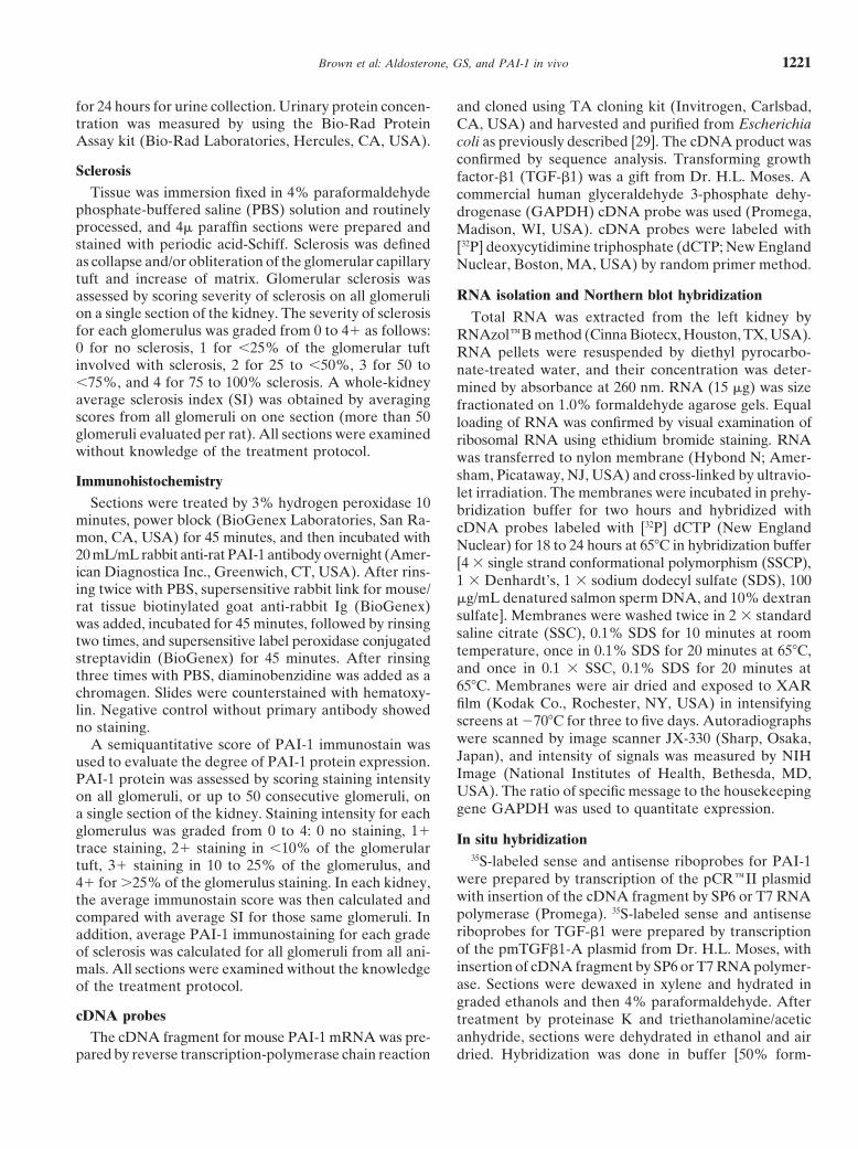
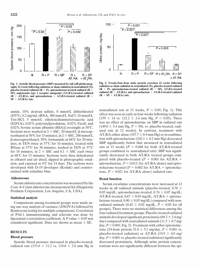
Fig. 2. Twenty-four–hour urine protein excretion 12 weeks followingFig. 1. Systolic blood pressure (SBP) measured by tail cuff plethysmog-radiation or sham radiation in nonradiated (N), placebo-treated radiatedraphy 12 weeks following radiation or sham radiation in nonradiated (N),(R 1 P), spironolactone-treated radiated (R 1 SP), AT1RA-treatedplacebo-treated radiated (R 1 P), spironolactone-treated radiated (R 1radiated (R 1 AT1RA), and spironolactone 1 AT1RA-treated radiatedSP), angiotensin type 1 receptor antagonist (AT1RA)-treated radiated(R 1 SP 1 AT1RA) rats.(R 1 AT1RA), and spironolactone 1 AT1RA-treated radiated (R 1
SP 1 AT1RA) rats.
nonradiated rats at 12 weeks, P , 0.05; Fig. 1). Thisamide, 10% dextran sulfate, 8 mmol/L dithiothreitoleffect was seen as early as four weeks following radiation(DTT), 0.2 mg/mL tRNA, 300 mmol/L NaCl, 10 mmol/L(159 6 14 vs. 121.2 6 2.4 mm Hg, P , 0.05). ThereTris-HCl, 5 mmol/L ethylenediaminetetraacetic acidwas no effect of spironolactone on SBP in radiated rats(EDTA), 0.02% polyvinylpyrolidone, 0.02% Ficoll, and(149.0 6 5.4 mm Hg, P 5 NS, vs. placebo-treated, radi-0.02% bovine serum albumin (BSA)] overnight at 508C.ated rats at 12 weeks). In contrast, treatment withSections were washed in 5 3 SSC, 20 mmol/L b-mercap-AT1RA either alone (107.7 6 8.9 mm Hg) or in combina-toethanol at 508C for 15 minutes, in 2 3 SSC, 200 mmol/Ltion with spironolactone (102.1 6 6.2 mm Hg) decreasedb-mercaptoethanol, 50% formamide at 688C for 20 min-SBP significantly below that measured in nonradiatedutes, in TEN twice at 378C for 10 minutes, treated withrats at 12 weeks (P 5 0.048 for both AT1RA-treatedRNase at 378C for 30 minutes, washed in TEN at 378Cgroups combined vs. nonradiated rats). SBP was signifi-for 10 minutes, in 2 3 SSC and 0.1 3 SSC each twicecantly decreased in both AT1RA treated groups com-at 688C for 15 minutes. Sections were then dehydratedpared with placebo-treated (P 5 0.001 for AT1RA 1in ethanol and air dried, dipped in photographic emul-spironolactone, P 5 0.013, for AT1RA alone) and spiro-sion, and exposed at 48C for 14 days. The sections were
developed with D-19 developer (Kodak) and counter- nolactone-treated (P 5 0.002 for AT1RA 1 spironolac-stained with toluidine blue. tone, P 5 0.021 for AT1RA alone) radiated rats.
Aldosterone Renal functionSerum aldosterone concentration was measured by the Serum creatinine concentrations were increased at 12
Coat-A-Count aldosterone measurement kit (Diagnostic weeks in all radiated animals (placebo-treated, 0.78 6Products Corporation, Los Angeles, CA, USA). 0.05 mg/dL; spironolactone-treated, 0.74 6 0.07 mg/dL;
AT1RA-treated, 0.67 6 0.03 mg/dL; AT1RA 1 spirono-Statistical analysislactone-treated, 0.80 6 0.05 mg/dL) compared with non-
Comparisons among treatment groups were made us- radiated animals (0.42 6 0.02 mg/dL, P , 0.05 for alling one-way analysis of variance (ANOVA) followed by
groups). There were no statistical differences among theBonferroni testing for multiple comparisons. Correlationfour radiated treatment groups. Placebo-treated radiatedof PAI-1 immunostaining and sclerosis was done byanimals developed significant proteinuria (69.2 6 2.4 mg/Spearman’s correlation coefficient. A P value , 0.05 wasday) compared with nonradiated animals (11.5 6 0.7 mg/considered significant. Data are shown as mean 6 SE.day, P , 0.001; Fig. 2). Treatment with either spironolac-tone (24-hour protein 31.8 6 5.1 mg/day, P , 0.001 vs.
RESULTS placebo-treated radiation) or AT1RA (19.9 6 4.0 mg/Blood pressure day, P , 0.001 vs. placebo-treated radiation) significantly
decreased proteinuria. Although urine protein concen-Systolic blood pressure increased in placebo-treated,radiated rats (151.6 6 11.2 vs. 118.8 6 2.6 mm Hg in trations were not significantly different between the spi-
Brown et al: Aldosterone, GS, and PAI-1 in vivo 1223
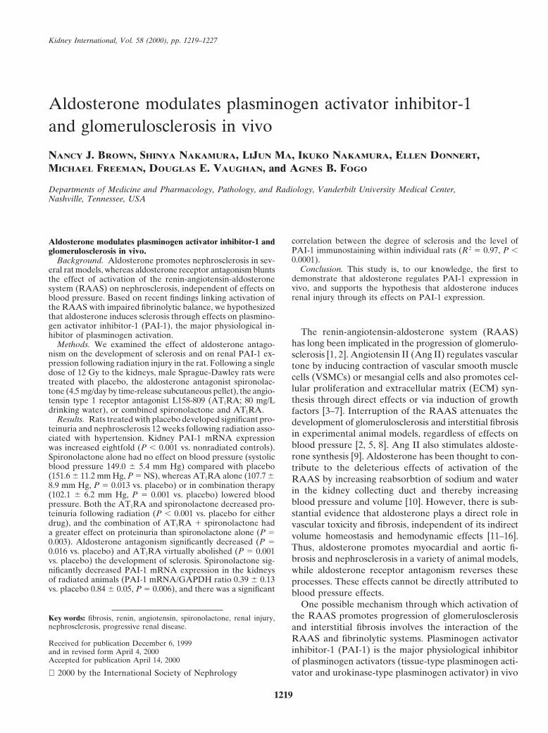
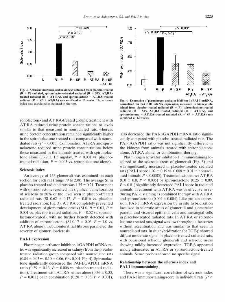
Fig. 3. Sclerosis index assessed in kidneys obtained from placebo-treated(R 1 P) radiated, spironolactone-treated radiated (R 1 SP), AT1RA-treated radiated (R 1 AT1RA), and spironolactone 1 AT1RA-treatedradiated (R 1 SP 1 AT1RA) rats sacrificed at 12 weeks. The sclerosis Fig. 4. Expression of plasminogen activator inhibitor-1 (PAI-1) mRNA,index was calculated as outlined in the text. normalized for GAPDH mRNA expression, measured in kidneys ob-
tained from placebo-treated radiated (R 1 P), spironolactone-treatedradiated (R 1 SP), AT1RA-treated radiated (R 1 AT1RA), andspironolactone 1 AT1RA-treated radiated (R 1 SP 1 AT1RA) ratssacrificed at 12 weeks.
ronolactone- and AT1RA-treated groups, treatment withAT1RA reduced urine protein concentrations to levelssimilar to that measured in nonradiated rats, whereasurine protein concentration remained significantly higher also decreased the PAI-1/GAPDH mRNA ratio signifi-
cantly compared with placebo-treated radiated rats. Thein the spironolactone-treated rats compared with nonra-diated rats (P 5 0.001). Combination AT1RA and spiro- PAI-1/GAPDH ratio was not significantly different in
the kidneys from animals treated with spironolactonenolactone reduced urine protein concentrations belowthose measured in the animals treated with spironolac- alone, AT1RA alone, or combination therapy.
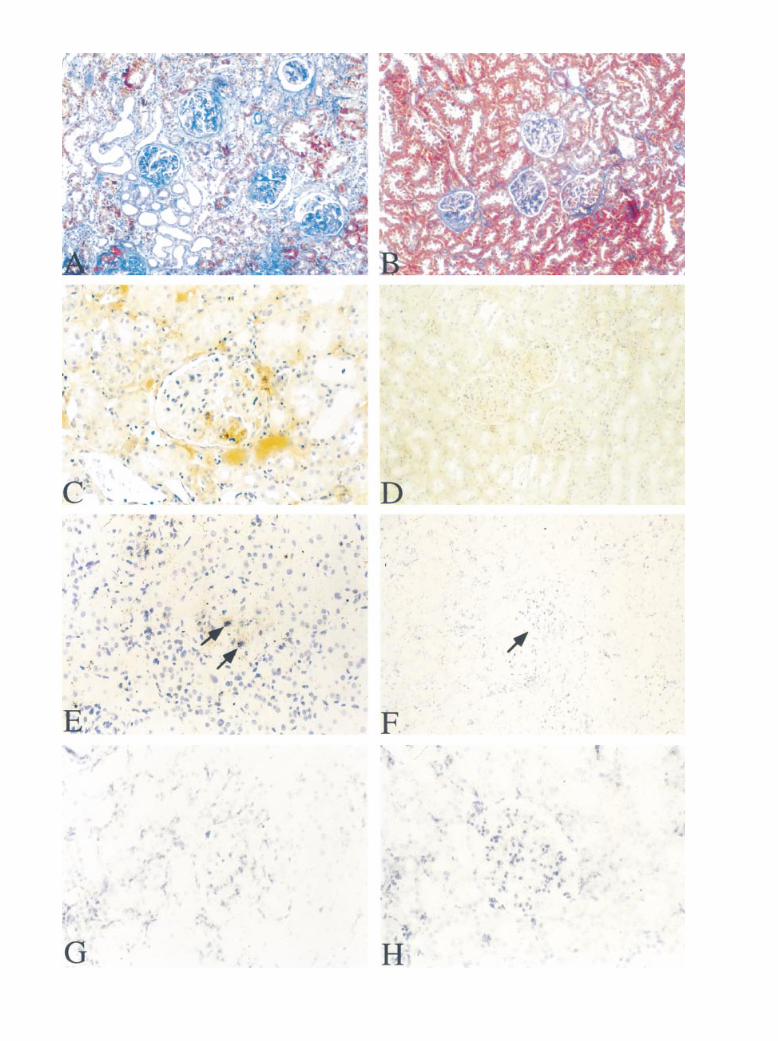
Plasminogen activator inhibitor-1 immunostaining lo-tone alone (13.2 6 1.3 mg/day, P , 0.001 vs. placebo-treated radiation, P 5 0.003 vs. spironolactone alone). calized to the sclerotic areas of glomeruli (Fig. 5) and
was significantly increased in placebo-treated radiatedSclerosis index rats (PAI-1 score 1.02 6 0.19 vs. 0.008 6 0.01 in nonradi-
ated animals, P , 0.0005). Treatment with either AT1RAAn average of 153 glomeruli was examined on eachsection for each rat (range 79 to 238). The average SI in (0.0 6 0.0, P , 0.005) or spironolactone (0.25 6 0.09,
P , 0.01) significantly decreased PAI-1 score in radiatedplacebo-treated radiated rats was 1.35 6 0.21. Treatmentwith spironolactone resulted in a significant amelioration animals. Treatment with AT1RA was as effective in re-
ducing PAI-1 staining as combined therapy with AT1RAof sclerosis to 50% of the level seen in placebo-treatedradiated rats (SI 0.62 6 0.17, P 5 0.016 vs. placebo- and spironolactone (0.004 6 0.004). Like protein expres-
sion, PAI-1 mRNA expression by in situ hybridizationtreated radiation; Fig. 3). AT1RA completely preventeddevelopment of glomerulosclerosis (SI 0.19 6 0.03, P 5 localized in sclerotic areas of glomeruli and glomerular
parietal and visceral epithelial cells and mesangial cells0.001 vs. placebo-treated radiation, P 5 0.52 vs. spirono-lactone-treated), with no further benefit detected with in placebo-treated radiated rats. In AT1RA or spirono-
lactone-treated rats, signal was low throughout the cortexaddition of spironolactone (SI 0.17 6 0.03, P 5 1.0 vs.AT1RA alone). Tubulointerstitial fibrosis paralleled the without accentuation and was similar to that seen in
nonradiated rats. In situ hybridization for TGF-b showedseverity of glomerulosclerosis.diffuse moderate signal in placebo-treated radiated rats,
PAI-1 expression with occasional sclerotic glomeruli and sclerotic areasshowing mildly increased expression. TGF-b appearedPlasminogen activator inhibitor-1/GAPDH mRNA ra-
tio was significantly increased in kidneys from the placebo- mildly attenuated in AT1RA or spironolactone-treatedanimals. Sense probes showed no specific signal.treated radiation group compared with nonradiated rats
(0.84 6 0.05 vs. 0.10 6 0.06, P , 0.001; Fig. 4). Spironolac-Relationship between the sclerosis index andtone significantly decreased the PAI-1/GAPDH mRNAPAI-1 immunostainingratio (0.39 6 0.13, P 5 0.006 vs. placebo-treated radia-
tion). Treatment with AT1RA, either alone (0.36 6 0.33, There was a significant correlation of sclerosis indexand PAI-1 immunostaining score in individual rats (P ,P 5 0.011) or in combination (0.20 6 0.03, P , 0.001),
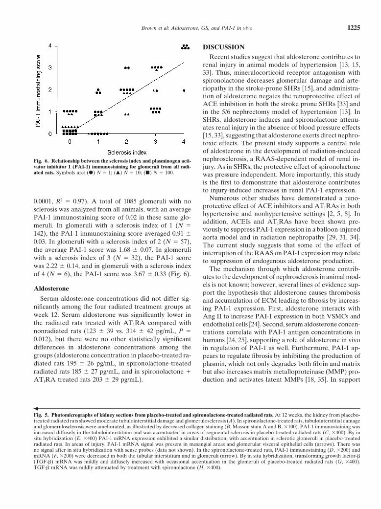
Brown et al: Aldosterone, GS, and PAI-1 in vivo 1225
DISCUSSION
Recent studies suggest that aldosterone contributes torenal injury in animal models of hypertension [13, 15,33]. Thus, mineralocorticoid receptor antagonism withspironolactone decreases glomerular damage and arte-riopathy in the stroke-prone SHRs [15], and administra-tion of aldosterone negates the renoprotective effect ofACE inhibition in both the stroke prone SHRs [33] andin the 5/6 nephrectomy model of hypertension [13]. InSHRs, aldosterone induces and spironolactone attenu-ates renal injury in the absence of blood pressure effects[15, 33], suggesting that aldosterone exerts direct nephro-toxic effects. The present study supports a central roleof aldosterone in the development of radiation-inducednephrosclerosis, a RAAS-dependent model of renal in-Fig. 6. Relationship between the sclerosis index and plasminogen acti-
vator inhibitor 1 (PAI-1) immunostaining for glomeruli from all radi- jury. As in SHRs, the protective effect of spironolactoneated rats. Symbols are: (d) N 5 1; (m) N 5 10; (j) N 5 100. was pressure independent. More importantly, this study
is the first to demonstrate that aldosterone contributesto injury-induced increases in renal PAI-1 expression.
Numerous other studies have demonstrated a reno-0.0001, R2 5 0.97). A total of 1085 glomeruli with noprotective effect of ACE inhibitors and AT1RAs in bothsclerosis was analyzed from all animals, with an averagehypertensive and nonhypertensive settings [2, 5, 8]. InPAI-1 immunostaining score of 0.02 in these same glo-addition, ACEIs and AT1RAs have been shown pre-
meruli. In glomeruli with a sclerosis index of 1 (N 5viously to suppress PAI-1 expression in a balloon-injured
142), the PAI-1 immunostaining score averaged 0.91 6 aorta model and in radiation nephropathy [29, 31, 34].0.03. In glomeruli with a sclerosis index of 2 (N 5 57), The current study suggests that some of the effect ofthe average PAI-1 score was 1.68 6 0.07. In glomeruli interruption of the RAAS on PAI-1 expression may relatewith a sclerosis index of 3 (N 5 32), the PAI-1 score to suppression of endogenous aldosterone production.was 2.22 6 0.14, and in glomeruli with a sclerosis index The mechanism through which aldosterone contrib-of 4 (N 5 6), the PAI-1 score was 3.67 6 0.33 (Fig. 6). utes to the development of nephrosclerosis in animal mod-
els is not known; however, several lines of evidence sup-Aldosteroneport the hypothesis that aldosterone causes thrombosis
Serum aldosterone concentrations did not differ sig- and accumulation of ECM leading to fibrosis by increas-nificantly among the four radiated treatment groups at ing PAI-1 expression. First, aldosterone interacts withweek 12. Serum aldosterone was significantly lower in Ang II to increase PAI-1 expression in both VSMCs andthe radiated rats treated with AT1RA compared with endothelial cells [24]. Second, serum aldosterone concen-nonradiated rats (123 6 39 vs. 314 6 42 pg/mL, P 5 trations correlate with PAI-1 antigen concentrations in0.012), but there were no other statistically significant humans [24, 25], supporting a role of aldosterone in vivodifferences in aldosterone concentrations among the in regulation of PAI-1 as well. Furthermore, PAI-1 ap-groups (aldosterone concentration in placebo-treated ra- pears to regulate fibrosis by inhibiting the production ofdiated rats 195 6 26 pg/mL, in spironolactone-treated plasmin, which not only degrades both fibrin and matrixradiated rats 185 6 27 pg/mL, and in spironolactone 1 but also increases matrix metalloproteinase (MMP) pro-
duction and activates latent MMPs [18, 35]. In supportAT1RA treated rats 203 6 29 pg/mL).
b
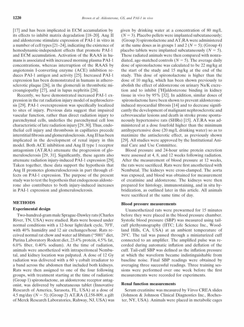
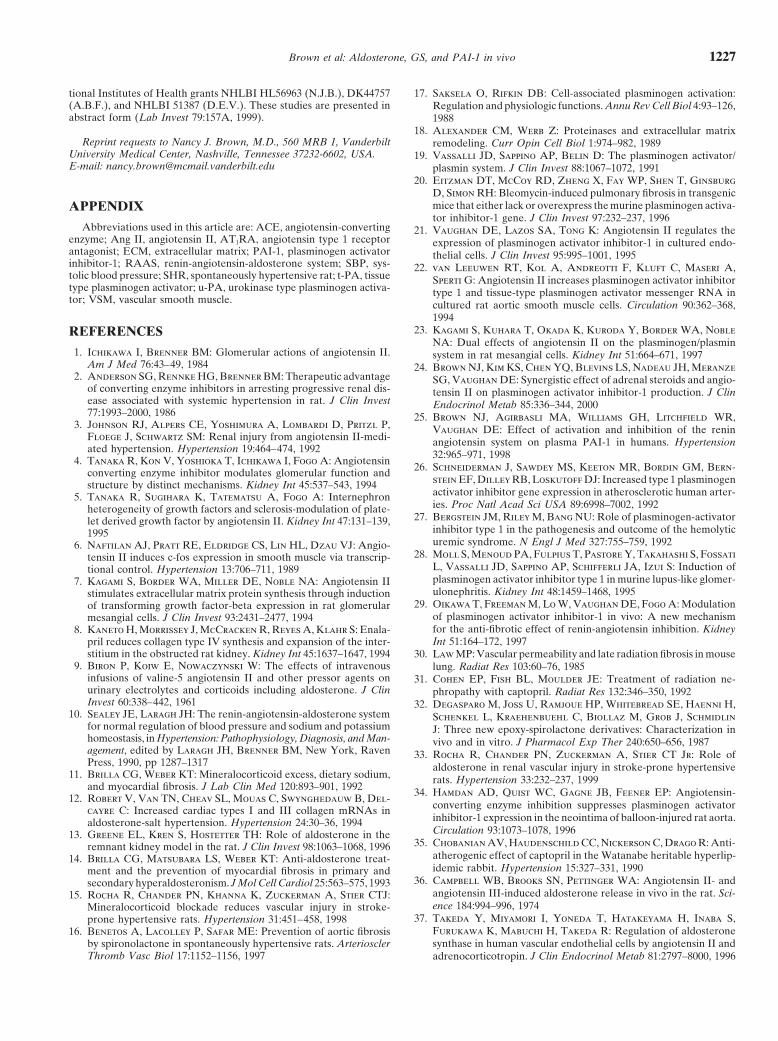
Fig. 5. Photomicrographs of kidney sections from placebo-treated and spironolactone-treated radiated rats. At 12 weeks, the kidney from placebo-treated radiated rats showed moderate tubulointerstitial damage and glomerulosclerosis (A). In spironolactone-treated rats, tubulointerstitial damageand glomerulosclerosis were ameliorated, as illustrated by decreased collagen staining (B; Masson stain A and B, 3100). PAI-1 immunostaining wasincreased diffusely in the tubulointerstitium and was accentuated in areas of segmental sclerosis in placebo-treated radiated rats (C, 3400). By insitu hybridization (E, 3400) PAI-1 mRNA expression exhibited a similar distribution, with accentuation in sclerotic glomeruli in placebo-treatedradiated rats. In areas of injury, PAI-1 mRNA signal was present in mesangial areas and glomerular visceral epithelial cells (arrows). There wasno signal after in situ hybridization with sense probes (data not shown). In the spironolactone-treated rats, PAI-1 immunostaining (D, 3200) andmRNA (F, 3200) were decreased in both the tubular interstitium and in glomeruli (arrow). By in situ hybridization, transforming growth factor-b(TGF-b) mRNA was mildly and diffusely increased with occasional accentuation in the glomeruli of placebo-treated radiated rats (G, 3400).TGF-b mRNA was mildly attenuated by treatment with spironolactone (H, 3400).
Brown et al: Aldosterone, GS, and PAI-1 in vivo1226
of a role for PAI-1 in the regulation of fibrosis, Eitzman expression colocalized with areas of sclerosis in the sameglomeruli, and glomeruli with greater sclerosis had pro-et al have reported that bleomycin-induced pulmonary
fibrosis is increased in transgenic mice overexpressing gressively higher PAI-1 immunostaining scores. In addi-tion, there was a correlation between the extent to whichPAI-1 and decreased in PAI-1 knockout mice relative
to controls [20]. each drug ameliorated sclerosis and the decrease in PAI-1expression. Nevertheless, studies in PAI-1–deficient ani-In the present study, spironolactone and AT1RA, both
alone and in combination, decreased proteinuria, nephro- mals are needed to determine whether PAI-1 contributesto aldosterone and Ang II-induced nephrosclerosis.sclerosis, and PAI-1 expression. Although there was little
effect on serum creatinine, creatinine is an insensitive While not statistically significant, there was a numeri-cally greater reduction of PAI-1 mRNA with combinedmarker of the extent of renal injury at this relatively early
stage of injury in animals with two kidneys. Interestingly, AT1RA and spironolactone than with AT1RA alone.However, the effects of AT1RA, alone and in combina-only AT1RA reduced urinary protein excretion to nor-
mal. This differential effect of aldosterone receptor an- tion on sclerosis were equivalent. In addition, the correla-tion between sclerosis and PAI-1 immunostaining seemedtagonism and AT1RA may be a consequence of the dif-
ferent hemodynamic effects of the two drugs; blood to be better than the correlation between sclerosis andPAI-1 mRNA. This raises the hypothesis that aldoste-pressure was lower than normal in the animals treated
with AT1RA, while radiated rats treated with either spi- rone affects post-transcriptional regulation of PAI-1. Al-though we cannot address this hypothesis in the presentronolactone or placebo exhibited an approximately 50%
increase in SBP. In addition, Ang II is known to promote model, studies in endothelial cells indicate that aldoste-rone-induced PAI-1 expression is blocked by inhibitorsproliferation and ECM protein synthesis via production
of growth factors such as platelet-derived growth factor of transcription [24], suggesting that aldosterone regu-lates PAI-1 expression at a transcriptional level.and TGF-b [5, 7]. Hence, it is likely that Ang II exerts
specific aldosterone-independent effects on glomerular One limitation of the present study is that the statusof the circulating RAAS was not well defined. Serumstructure and function. The effect of aldosterone on
growth factors such as TGF-b has not been studied exten- aldosterone concentrations were measured in anesthe-tized animals, in which the effects of radiation and drugsively; however, in the present study, TGF-b did not
specifically localize to areas of glomerulosclerosis, and treatment on circulating aldosterone concentrations maybe obscured [36]. Thus, there were no differences in aldo-its expression was similarly mildly attenuated by either
AT1RA or spironolactone. Thus, although TGF-b modu- sterone concentrations among radiation-treated groups.In particular, radiation nephropathy was not associatedlation may have contributed to the effects of AT1RA
and spironolactone, its expression did not appear to be with increased circulating concentrations of aldosterone.Taken together with the finding that spironolactone de-tightly associated with injury.
Whereas coadministration of AT1RA significantly en- creased the development of radiation-induced protein-uria and sclerosis, this suggests either that sensitivity tohanced the effect of sprionolactone on blood pressure
and the development of proteinuria, coadministration aldosterone is increased in radiation nephropathy or thatcirculating concentrations of aldosterone do not reflectof aldosterone receptor antagonist did not enhance the
effect of AT1RA on sclerosis, proteinuria, or PAI-1 ex- local tissue levels of aldosterone. In support of the latterhypothesis, regulated extra-adrenal synthesis of aldoste-pression in this study. This may be attributable to the
fact that the dose of AT1RA used nearly completely atten- rone has been demonstrated [37]. For these reasons, thedemonstration that aldosterone antagonism attenuatesuated the development of sclerosis, preventing detection
of any additive or synergistic effects. Alternatively, aldo- the development of sclerosis provides the best evidencefor a role of endogenous aldosterone in this model ofsterone may not alter PAI-1 expression in the absence
of an Ang II effect. Indeed, studies in both VSMCs and sclerosis.In summary, this study using the radiation-inducedendothelial cells suggest that the effect of aldosterone
on PAI-1 expression is Ang II dependent [24]. Further nephrosclerosis model confirms data from other animalmodels that suggest that aldosterone plays a role in thestudies using submaximal doses of AT1RA or delayed
onset of intervention may help clarify the pathophysiology progression of glomerulopathy. It provides the first evi-dence, to our knowledge, that aldosterone regulatesof the interaction between Ang II and aldosterone in vivo.
The finding that spironolactone and AT1RA decrease PAI-1 expression in vivo. The study supports the hypoth-esis that aldosterone induces renal injury through itsPAI-1 expression and decrease sclerosis does not prove
a causal relationship between Ang II or aldosterone- effects on PAI-1 expression.induced PAI-1 synthesis and renal injury. However, thedata offer indirect evidence for this relationship. We ACKNOWLEDGMENTSobserved a strong correlation between sclerosis and PAI-1 Portions of these studies were supported by an Established Investi-
gator Award from the American Heart Association (A.B.F.) and Na-expression for each animal. Furthermore, increased PAI-1
Brown et al: Aldosterone, GS, and PAI-1 in vivo 1227
tional Institutes of Health grants NHLBI HL56963 (N.J.B.), DK44757 17. Saksela O, Rifkin DB: Cell-associated plasminogen activation:(A.B.F.), and NHLBI 51387 (D.E.V.). These studies are presented in Regulation and physiologic functions. Annu Rev Cell Biol 4:93–126,abstract form (Lab Invest 79:157A, 1999). 1988
18. Alexander CM, Werb Z: Proteinases and extracellular matrixReprint requests to Nancy J. Brown, M.D., 560 MRB 1, Vanderbilt remodeling. Curr Opin Cell Biol 1:974–982, 1989
University Medical Center, Nashville, Tennessee 37232-6602, USA. 19. Vassalli JD, Sappino AP, Belin D: The plasminogen activator/E-mail: [email protected] plasmin system. J Clin Invest 88:1067–1072, 1991
20. Eitzman DT, McCoy RD, Zheng X, Fay WP, Shen T, GinsburgD, Simon RH: Bleomycin-induced pulmonary fibrosis in transgenicmice that either lack or overexpress the murine plasminogen activa-APPENDIXtor inhibitor-1 gene. J Clin Invest 97:232–237, 1996
Abbreviations used in this article are: ACE, angiotensin-converting 21. Vaughan DE, Lazos SA, Tong K: Angiotensin II regulates theenzyme; Ang II, angiotensin II, AT1RA, angiotensin type 1 receptor expression of plasminogen activator inhibitor-1 in cultured endo-antagonist; ECM, extracellular matrix; PAI-1, plasminogen activator thelial cells. J Clin Invest 95:995–1001, 1995inhibitor-1; RAAS, renin-angiotensin-aldosterone system; SBP, sys- 22. van Leeuwen RT, Kol A, Andreotti F, Kluft C, Maseri A,tolic blood pressure; SHR, spontaneously hypertensive rat; t-PA, tissue Sperti G: Angiotensin II increases plasminogen activator inhibitortype plasminogen activator; u-PA, urokinase type plasminogen activa- type 1 and tissue-type plasminogen activator messenger RNA intor; VSM, vascular smooth muscle. cultured rat aortic smooth muscle cells. Circulation 90:362–368,
199423. Kagami S, Kuhara T, Okada K, Kuroda Y, Border WA, NobleREFERENCES
NA: Dual effects of angiotensin II on the plasminogen/plasmin1. Ichikawa I, Brenner BM: Glomerular actions of angiotensin II. system in rat mesangial cells. Kidney Int 51:664–671, 1997
Am J Med 76:43–49, 1984 24. Brown NJ, Kim KS, Chen YQ, Blevins LS, Nadeau JH, Meranze2. Anderson SG, Rennke HG, Brenner BM: Therapeutic advantage SG, Vaughan DE: Synergistic effect of adrenal steroids and angio-
of converting enzyme inhibitors in arresting progressive renal dis- tensin II on plasminogen activator inhibitor-1 production. J Clinease associated with systemic hypertension in rat. J Clin Invest Endocrinol Metab 85:336–344, 200077:1993–2000, 1986 25. Brown NJ, Agirbasli MA, Williams GH, Litchfield WR,
3. Johnson RJ, Alpers CE, Yoshimura A, Lombardi D, Pritzl P, Vaughan DE: Effect of activation and inhibition of the reninFloege J, Schwartz SM: Renal injury from angiotensin II-medi- angiotensin system on plasma PAI-1 in humans. Hypertensionated hypertension. Hypertension 19:464–474, 1992 32:965–971, 19984. Tanaka R, Kon V, Yoshioka T, Ichikawa I, Fogo A: Angiotensin 26. Schneiderman J, Sawdey MS, Keeton MR, Bordin GM, Bern-converting enzyme inhibitor modulates glomerular function and
stein EF, Dilley RB, Loskutoff DJ: Increased type 1 plasminogenstructure by distinct mechanisms. Kidney Int 45:537–543, 1994activator inhibitor gene expression in atherosclerotic human arter-5. Tanaka R, Sugihara K, Tatematsu A, Fogo A: Internephronies. Proc Natl Acad Sci USA 89:6998–7002, 1992heterogeneity of growth factors and sclerosis-modulation of plate-
27. Bergstein JM, Riley M, Bang NU: Role of plasminogen-activatorlet derived growth factor by angiotensin II. Kidney Int 47:131–139,inhibitor type 1 in the pathogenesis and outcome of the hemolytic1995uremic syndrome. N Engl J Med 327:755–759, 19926. Naftilan AJ, Pratt RE, Eldridge CS, Lin HL, Dzau VJ: Angio-
28. Moll S, Menoud PA, Fulpius T, Pastore Y, Takahashi S, Fossatitensin II induces c-fos expression in smooth muscle via transcrip-L, Vassalli JD, Sappino AP, Schifferli JA, Izui S: Induction oftional control. Hypertension 13:706–711, 1989plasminogen activator inhibitor type 1 in murine lupus-like glomer-7. Kagami S, Border WA, Miller DE, Noble NA: Angiotensin IIulonephritis. Kidney Int 48:1459–1468, 1995stimulates extracellular matrix protein synthesis through induction
29. Oikawa T, Freeman M, Lo W, Vaughan DE, Fogo A: Modulationof transforming growth factor-beta expression in rat glomerularof plasminogen activator inhibitor-1 in vivo: A new mechanismmesangial cells. J Clin Invest 93:2431–2477, 1994for the anti-fibrotic effect of renin-angiotensin inhibition. Kidney8. Kaneto H, Morrissey J, McCracken R, Reyes A, Klahr S: Enala-Int 51:164–172, 1997pril reduces collagen type IV synthesis and expansion of the inter-
stitium in the obstructed rat kidney. Kidney Int 45:1637–1647, 1994 30. Law MP: Vascular permeability and late radiation fibrosis in mouse9. Biron P, Koiw E, Nowaczynski W: The effects of intravenous lung. Radiat Res 103:60–76, 1985
infusions of valine-5 angiotensin II and other pressor agents on 31. Cohen EP, Fish BL, Moulder JE: Treatment of radiation ne-urinary electrolytes and corticoids including aldosterone. J Clin phropathy with captopril. Radiat Res 132:346–350, 1992Invest 60:338–442, 1961 32. Degasparo M, Joss U, Ramjoue HP, Whitebread SE, Haenni H,
10. Sealey JE, Laragh JH: The renin-angiotensin-aldosterone system Schenkel L, Kraehenbuehl C, Biollaz M, Grob J, Schmidlinfor normal regulation of blood pressure and sodium and potassium J: Three new epoxy-spirolactone derivatives: Characterization inhomeostasis, in Hypertension: Pathophysiology, Diagnosis, and Man- vivo and in vitro. J Pharmacol Exp Ther 240:650–656, 1987agement, edited by Laragh JH, Brenner BM, New York, Raven 33. Rocha R, Chander PN, Zuckerman A, Stier CT Jr: Role ofPress, 1990, pp 1287–1317 aldosterone in renal vascular injury in stroke-prone hypertensive
11. Brilla CG, Weber KT: Mineralocorticoid excess, dietary sodium, rats. Hypertension 33:232–237, 1999and myocardial fibrosis. J Lab Clin Med 120:893–901, 1992 34. Hamdan AD, Quist WC, Gagne JB, Feener EP: Angiotensin-12. Robert V, Van TN, Cheav SL, Mouas C, Swynghedauw B, Del-
converting enzyme inhibition suppresses plasminogen activatorcayre C: Increased cardiac types I and III collagen mRNAs ininhibitor-1 expression in the neointima of balloon-injured rat aorta.aldosterone-salt hypertension. Hypertension 24:30–36, 1994Circulation 93:1073–1078, 199613. Greene EL, Kren S, Hostetter TH: Role of aldosterone in the
35. Chobanian AV, Haudenschild CC, Nickerson C, Drago R: Anti-remnant kidney model in the rat. J Clin Invest 98:1063–1068, 1996atherogenic effect of captopril in the Watanabe heritable hyperlip-14. Brilla CG, Matsubara LS, Weber KT: Anti-aldosterone treat-idemic rabbit. Hypertension 15:327–331, 1990ment and the prevention of myocardial fibrosis in primary and
36. Campbell WB, Brooks SN, Pettinger WA: Angiotensin II- andsecondary hyperaldosteronism. J Mol Cell Cardiol 25:563–575, 1993angiotensin III-induced aldosterone release in vivo in the rat. Sci-15. Rocha R, Chander PN, Khanna K, Zuckerman A, Stier CTJ:ence 184:994–996, 1974Mineralocorticoid blockade reduces vascular injury in stroke-
37. Takeda Y, Miyamori I, Yoneda T, Hatakeyama H, Inaba S,prone hypertensive rats. Hypertension 31:451–458, 1998Furukawa K, Mabuchi H, Takeda R: Regulation of aldosterone16. Benetos A, Lacolley P, Safar ME: Prevention of aortic fibrosissynthase in human vascular endothelial cells by angiotensin II andby spironolactone in spontaneously hypertensive rats. Arterioscler
Thromb Vasc Biol 17:1152–1156, 1997 adrenocorticotropin. J Clin Endocrinol Metab 81:2797–8000, 1996