Embed Size (px)
Citation preview

GALILEO, University System of Georgia
G A L I L E O Open Learning Materials
Nursing and Health Sciences Open Textbooks Nursing and Health Sciences
Spring 2015
Physical Therapy Applications for Individuals with Neurologic Dysfunction Charlotte Chatto Augusta University, [email protected]
Jeff Mastromonico Augusta University, [email protected]
Follow this and additional works at: https://oer.galileo.usg.edu/health-textbooks
Part of the Physical Therapy Commons
Recommended Citation Chatto, Charlotte and Mastromonico, Jeff, "Physical Therapy Applications for Individuals with Neurologic Dysfunction" (2015) . Nursing and Health Sciences Open Textbooks. 2. https://oer.galileo.usg.edu/health-textbooksZ2
This Open Textbook is brought to you for free and open access by the Nursing and Health Sciences at G A L I L E O Open Learning Materials. It has been
accepted for inclusion in Nursing and Health Sciences Open Textbooks by an authorized administrator of G A L I L E O Open Learning Materials. For
more information, please contact [email protected].

Physical Therapy Applications for Individuals with Neurologic Dysfunction
Dr. Charlotte Chatto. PT, PhD
1
C H A P T E R 1
Key assessment and Interventions: Focus on Patients with Guillain Barre and Spinal Cord Injury
In this chapter, the primary physical therapy examination techniques for patients with neurologic dysfunction wi l l be described and demonstrated. Although many of these techniques are equally appropriate for patients with other diagnoses, such as medical or orthopaedic conditions, the focus wi l l be on possible outcomes when a person's nervous system has been damaged by injury or disease.

2
S E C T I O N 1
Strength, Reflexes, and Flexibility Key Points
1. In persons w i t h damaged nervous systems, these tests may be context-dependent.
2. It is important to test and re-test in the same environment and in test positions.
3. Recognize that the results of these tests may not automatically translate into what y o u might predict a patient's performance w i l l be during a functional activity.
General considerations
Safety
Although the therapist is assessing the musculoskeletal system, in a patient w i t h neurologic dysfunction, cognitive issues resulting in a decreased awareness of overall safety must be considered. In addition, in the presence of weakness or sensory loss, body alignment and joint protection is often one of the therapist's most important tasks. Providing external stability through manual contacts and support from the sitting surface w i l l not only protect the patient, but also al low for the most reliable results of the tests.
Posture and Positioning
Posture and alignment of body segments, must not be overlooked in the indiv idual w i t h
neurologic dysfunction. In order to function effectively and efficiently, the h u m a n body must be able to respond to changes in gravity secondary to alterations in the alignment of body segments.
The therapist needs to consider:
• Both movement and the relationship between how the muscles are functioning and the patient's biomechanical alignment.
• The init ial postural alignment of the patient before movement.
• The alignment of body segments throughout the movement.
Motor Control
The control of a patient's movement is often more critical to consider than his or her ability to produce force. For example, in a patient w i t h a

3
cerebellar lesion, strength w i l l be typically be intact, however functional activities that involve the coordination of this strength may have significant deficits.
The therapist needs to consider:
• The muscles used to complete the desired movement.
• The patient's ability to relax antagonists a l lowing the agonists to contract effectively.
• The velocity and acceleration of movements desired to efficiently complete the task
• Compensations in other body segments
Range of motion Use care w h e n performing of joint range of motion ( R O M ) tests and activities in the presence of spasticity or flaccidity. Spasticity may interfere w i t h range measurement because it may change depending on the relationship of the trunk or extremity to gravity, therapists' hand positions and technique.
Instructions to the patient: Make sure that your commands to the patient are consistent w i t h the type of R O M y o u are assessing (or performing as a treatment intervention). Some examples of instructions follow:
• Passive: "Relax and let me move y o u . "
• Active Assisted: " I want y o u to try to move through the motion and I w i l l help y o u if y o u need assistance." W h e n y o u ask the person to try to move, but there is no movement noted, this w i l l still be considered active-assisted not passive, because of the
commands given. Documentation should reflect this distinct difference. For example, a therapist may document: "Act ive Assisted R O M was attempted, but no motion noted."
• Active: " I want to y o u to move through the motion as far as y o u can."
• Hand placement: The technique used whi le performing R O M may promote hyperactivity in the muscle being elongated. Examples include: D u r i n g R O M at the ankle, a hand on the ball of the foot may produce hyperactivity in the gastrocnemius-soleus and prevent ful l movement into dorsiflexion; during wris t extension, palmar contact may promote finger and wris t flexion l imit ing elongation of the long finger flexors.
• Speed of technique: The speed of the technique may produce spasticity -- the faster the movement the more l ikely the muscles are to respond w i t h involuntary contraction (see tone below)
• Protecting the joints: In the presence of flaccidity care must be taken, as the joints have no protection from the normal reaction of muscles to stretch. Overstretching of joint structures may occur. The shoulder is particular vulnerable. Remember to a lways have one hand on the scapula w h e n performing R O M .
Flexibility Individuals w i t h neurologic impairment generally demonstrate deficits in voluntary movement, whether secondary to muscle weakness/ imbalance, spasticity, or posturing. Alterations in muscle tone secondary to neurologic insult may lead to changes in the muscle properties itself. These changes in the architecture of

4
the muscle cause a reduction in the flexibility of the muscle, or the ability of the muscle to lengthen appropriately.
As w i t h testing joint R O M or f lexibil i ty in a person w i t h a neurologic injury, remember to move the l imb s lowly to avoid eliciting spasticity that may reduce accuracy of your tests. The fol lowing body parts/segments that are important to assess are listed below, each w i t h a particular critical aspect of functioning that requires that particular f lexibi l i ty .
• Cervical flexibility in all planes of movement for head righting and equil ibrium reactions
• Pectoral muscle length for posture and balance
• External rotators of shoulders and supination of forearms for upper extremity support in the absence of triceps
• Wrist extensors for upper extremity support
• Trunk elongation for general posture, balance, and mobility
• H i p l e x o r s for prone assumption and gait
• Hamstr ing length for upright activities and gait
• H i p adductor length for A D L s and hygiene, as w e l l as general mobility
• H i p external rotation w i t h hip f l e x i o n for dressing
• H i p internal rotators for scooting and gait
• Plantarflexor length for gait and balance reactions
Reflexes
Examinat ion of the deep tendon reflex ( D T R ) is an unconscious motor response to a sensory stimulus. It gives information about the location of the neurologic lesion by assessment of the reflex arc. Performance of this test requires a reflex hammer to provide stimulation of sensory receptors (the muscle spindle), w h i c h evokes action potentials that are conducted into spinal cord. The sensory neuron synapses w i t h an association neuron, w h i c h synapses w i t h somatic motor neuron, w h i c h conducts impulses to muscle and stimulates a reflex contraction. Remember that the brain is not directly involved.
Interactive 1.1 Complete the chart in preparation for practicing MMT skills
Self Directed Lab Activity 1.1 For each of the muscles and activities listed above, role play as if y o u lack significant flexibility and then try to perform the associated activity to experience the difficulty in movement
• Hyperact ive tendon responses suggest lesions of the U M N , (corticospinal tracts).
• Pendular tendon re lexes are suggestive of cerebellar ( C b ) dysfunction.
• Absent tendon re lexes suggest L M N lesions. (Compare w i t h decreased responses in an orthopedic evaluation.)

5
Examinat ion may include any or all of the fol lowing reflexes, each representing a particular myotome, w h i c h is the group of muscles a specific nerve root(s): (There are videos for reflex testing and other testing on www.neuroexam.com.. .it is open access, but y o u cannot download the videos. We could insert the l ink for n o w and maybe we take our o w n later)
• Achil les tendon (S1,S2)
• Biceps (C5, C6)
• Brachioradialis (C6)
• Quadriceps (L3 , L4)
• Triceps (C7)
Strength Manual muscle tests ( M M T ) are used to evaluate strength and can be misleading in the neurologically impaired indiv idual . These individuals certainly have weakness, but it is a more complex phenomenon than weakness that results from the patients w i t h intact nervous systems. Lesions of the upper motor neuron affect the number, type and discharge frequency of motor units recruited during performance. E v e n if there is min imal or no loss of cross-sectional area in the muscle fibers, force generation is impaired. Over time, secondary changes can also occur in muscle w i t h selective atrophy of fast twitch fibers.
• The nervous system adopts a variety of synergistic muscle activation patterns to control the body's degrees of freedom, or available planes of movement. A neurologically impaired person
often has less control and fewer synergistic patterns are available. T h i s makes it difficult for an indiv idual to isolate a muscle or muscle group for a reliable manual muscle test. The opposite also holds true that individuals unable to isolate a muscle contraction as required for a muscle strength test, might exhibit good force production in a gross or mass synergy pattern.
• Results of strength testing on a person w i t h an upper motor neuron lesion cannot be reliably interpreted without knowing details of the testing procedure. E v e n then, the test results may not tell y o u what the person is capable of doing functionally.
• The muscle itself may be capable of producing force, but the strength may test lower secondary to recruitment problems, problems of co-contraction, t iming of contractions or sequence of firing of muscles involved in the motor plan

6
• An indiv idua l might produce a grade 0 or 1 on an isolated movement test but perform at a 3 / 5 or better on a task where it acts synergistically w i t h others. Th is is an example of strength or force production being task specific.
• This weakness might not be improved by specific exercises to strengthen the muscle.
Cl in ica l T i p s :
• As a rule of thumb, muscle testing is only valuable if the patient is able to produce an isolated muscle contraction.
• Documentation for motor ability, if M M T grades are not appropriate, should qualitatively describe the percentage or fraction of movement throughout the range of motion being tested.

7
S E C T I O N 2
Functional Mobility Training for Patients with Spinal Cord Injury
Key Points
• For as long as needed for preventing orthostatic hypotension, make sure the patient has the following donned, before moving to an upright position:
• Thigh-high compression stockings, such as T E D ™ hose
• Elastic wraps , such as A C E ™ wraps over the stockings
• An abdominal binder w i l l assist w i t h blood pressure, but should be used for any patient without fu l l abdominal innervation to assist w i t h the length-tension relationship of the diaphragm. This w i l l al low the patient to take a deeper breath.
• If y o u are planning to help the patient transfer to a chair, make sure y o u have a wheelchair w i t h appropriate
amount of support (for example, head support for a patient w i t h high tetraplegia) and a pressure distributing cushion.
• For a patient w h o does not have fu l l innervation of his hand, make sure y o u protect your patient's tenodesis by never al lowing wris t extension and finger extension
• If the hamstring range of motion ( R O M ) is not at least 90 degrees, be very careful not to have the patient assume positions in w h i c h his knees are extending in long sitting and his back is in a flexed position. Th is w i l l provide an excessive stretch to the back extensors and compromise the passive stability of the trunk. Once bilateral hamstring ( R O M ) is at least at 90 degrees, then long sitting can be assumed w i t h extended knees.
Transfer 1 Therapist Over Patient's Back

8
Transfer 2 Therapist Under Patient's Arm Mobility 1 Independent Mat Mobility For Patient With C5 Spinal Cord Injury
Transfer 3 Therapist Using A Stool Mobility 2 Mobility and Gait With Lower Extremity Orthodics

9
C H A P T E R 2
Respitory Assesment and Interventions
The therapeutic interventions you choose to address recovery of motor function in your patient with neurologic dysfunction all have a theoretical foundation. Key theories wi l l be presented and video demonstrations of interventions wi l l help you begin to fill your toolbox with options to drive neuroplasticity and facilitate your patients' recovery.

10
S E C T I O N 1
Assisted Breathing Techniques Key Points
1. A thorough auscultation examination w i l l provide a baseline to help determine w h i c h techniques to use and to determine effectiveness of the techniques
2. Assisted Breathing and Coughing Techniques can only be performed w i t h patients w h o have a stable spine and no rib fractures.
Diaphragmatic Breathing (facilitation of the diaphragm):
E x p l a i n the purpose and goals of the exercise. Position the patient supine w i t h legs flexed at 45 degrees in a relaxed and w e l l
supported fashion. Demonstrate and explain the technique. Place hand or hands at the costophrenic angle. Squeeze d o w n and i n w a r d gently during exhalation. A l l o w the patient to breathe into your hand during inspiration. After several respiratory cycles the therapist w i l l ask the patient if he feels a difference and should try to perform the maneuver independently using his o w n hand. H o w can y o u modify this technique for the indiv idua l without upper extremity control?
Diaphragmatic Breathing (facilitation of the diaphragm)
Inhibition of the diaphragm: Supine Position
Inhibition of the diaphragm:
• Supine position: the heel of the therapist's hand is placed lightly on the patient's
abdomen below the base of the xiphoid process. As the patient exhales the therapist gently allows
his hand to follow the diaphragm up and in . W h e n the exhalation is complete, the therapist w i l l keep his hand in this position. At the next exhalation the therapist w i l l move his hand further in and after two or three cycles, the therapist keeps his hand in one position and observes the patient. The patient w i l l unconsciously alter his breathing pattern to use accessory muscles. W h e n this has happened, the therapist w i l l explain what has occurred and ask the patient to try to

11
reproduce the breathing pattern. The therapist w i l l gradually disengage his hand, observing carefully for the maintenance of the new breathing pattern by the patient. If the patient cannot maintain the pattern, the therapist w i l l help by reapplying the pressure unt i l the patient regains control of the pattern.
• Prone position: the patient is positioned prone on elbows. This position w i l l effectively compress the excursion of the diaphragm, forcing the patient to utilize accessory muscles. The patient can then practice the preferred breathing pattern as w e l l as incorporating it into other activities such as head and neck movement, reaching and single arm support. Note. . . this position is m u c h more threatening to the patient than the supine position and should not be attempted as an early training technique. W h e n is it l ikely y o u w o u l d desire an inhibition of the diaphragm? W h e n w o u l d this technique be indicated?
Inhibition of the diaphragm: Prone Position
Upper chest breathing using the pectorals:
the patient is positioned in supine w i t h arms at their sides. The therapist place the heels of her hands close to the sternum and aligns the fingers along the diagonal to the shoulders. As the patient begins to breathe up and Upper chest breathing using the
pectorals
into the therapist's hands the therapist applies a quick manual stretch (down and in toward the sternum) thus facilitating the pectoral muscles and expanding the upper chest.
Counter-rotation assisted breathing:
the patient is placed in a sidelying position. The therapist stands/ kneels behind the patient at a 45 degree angle to the head of the plinth. The cephalic hand is placed along the inferior border of the scapula and caudal hand is placed on the A S I S . As the patient inspires the therapist w i l l assist this movement by pushing the shoulder forward and d o w n whi le pul l ing the hip backward and down. At the end of inspiration, the therapist w i l l smoothly and gently move her cephalic hand to the gluteal fossa. As the patient expires the therapist assists this movement by pul l ing the two hands together. In both assists, the hands move in a counterrotation or wringing fashion. It is important in performing this technique to apply pressure through the palms instead of fingertips and palms, w h i c h can be painful to the patient.
Counter-rotation assisted breathing
Glossopharyngeal breathing:
no diaphragm action is required for this. An excellent technique to teach a patient if he or she requires ventilator assist. If a patient knows how to do this, then there w o u l d not be a fear of loss of electricity or power. The patient open his mouth wide ly and

12
abruptly creates a negative area of pressure w h i c h air fills. The patient then closes his l ips and " s w a l l o w s " the air d o w n into the lungs. It looks l ike a frog gulping air. The patient w i l l report a feeling as though his lungs were about to burst if the technique is being performed correctly. A feeling of nausea and indigestion indicates that air is being sucked into the stomach instead of the lungs. Init ial training can be very tiring. In addition to providing an alternative method of breathing if a stimulator or ventilator malfunctions, G P B can also act to (a) increase vi ta l capacity or produce a more effective cough, (b) assist in a longer and stronger phonation, and ( c) act as an internal mobilizer for the chest w a l l . Problems w h i c h interfere w i t h a patient's ability to perform this activity include an open nasal passage or glottis that al lows the air to escape, incorrect shaping of the mouth, incoordinated backward movement of the tongue and swal lowing of air into the stomach.
13
S E C T I O N 2
Assisted Coughing Techniques Key Points
1. A thorough auscultation examination w i l l provide a baseline to help determine w h i c h techniques to use and to determine effectiveness of the techniques
2. Assisted Breathing and Coughing Techniques can only be performed w i t h patients w h o have a stable spine and no rib fractures.
For each of the following techniques, be prepared to d iscuss the indications, precautions and contraindications. Techniques done in supine:
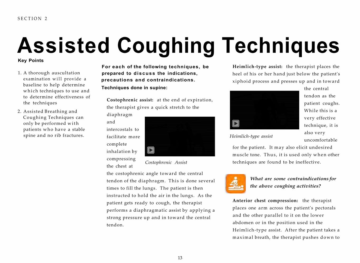
Costophrenic assist: at the end of expiration, the therapist gives a quick stretch to the diaphragm and intercostals to facilitate more complete inhalation by compressing the chest at the costophrenic angle toward the central tendon of the diaphragm. This is done several times to fill the lungs. The patient is then instructed to hold the air in the lungs. As the patient gets ready to cough, the therapist performs a diaphragmatic assist by applying a strong pressure up and in toward the central tendon.
Costophrenic Assist
Heimlich-type assist: the therapist places the heel of his or her hand just below the patient's x iphoid process and presses up and in toward
the central tendon as the patient coughs. While this is a very effective technique, it is also very uncomfortable
for the patient. It may also elicit undesired muscle tone. T h u s , it is used only w h e n other techniques are found to be ineffective.
Heimlich-type assist
What are some contraindications for the above coughing activities?
Anterior chest compression: the therapist places one arm across the patient's pectorals and the other parallel to it on the lower abdomen or in the position used in the Heimlich-type assist. After the patient takes a maximal breath, the therapist pushes d o w n to

14
help the patient cough. The greatest force is applied through the lower chest during expulsion.
Anterior chest compression Techniques done in sidelying:
• Costophrenic assist: in sidelying, the excursion of the anterior chest w a l l is now done w i t h gravity-eliminated whi le lateral
excursion becomes anti-gravity In addition, greater balance is required making this a good progression for w o r k i n g on trunk stability for the patient moving from supine to upright T h i s technique is asymmetrical W h i c h segment of the lung is
this technique effective for?
Costophrenic Assist
• Heimlich-type assist: this is the same as previously described. The only variation involves positioning the patient w i t h both hips and knees flexed. In this reflex inhibiting posture, the chances of eliciting high muscle tone are diminished.
Combined costophrenic and Heimlich-type assist
• Combined costophrenic and Heimlich-type assist: the therapist uses both hands at the same time, one to provide a costophrenic assist to lateral excursion and one to provide a H e i m l i c h assist to anterior excursion. Because two planes of respiration are used to remove excretions this method is more effective than either one used alone.
• Massery counter-rotation assist: the therapist provides a quick stretch facilitation to the pectorals and the hip hikers at the end
of expiration to maximize the next few inhalations. The therapist then facilitates inspiration by stretching along the inferior border of the scapula in an oblique and u p w a r d manner whi le pul l ing the hip in a d o w n w a r d and posterior fashion. After several
breaths have been taken to fill the lungs as m u c h as possible. The therapist positions her hands as she w o u l d for the quick stretch but compresses on exhalation. The patient's chest is squeezed in all three planes of respiration making this a very effective method of assistive cough. It is important to follow a true diagonal in both the physical assistance and the compression of the chest so that air is forced out of the lungs. T h i s method has an additional advantage because the rotation component w i l l act to inhibit the development of abnormal muscle tone. Finally, because it does not require active participation on the part of the patient, this technique can be used w i t h incoherent or unresponsive patients.
Massery counter-rotation assist
15
Techniques done in prone:
Head Flexion assist: the patient is positioned prone on his or her elbows. The patient then extend his head and neck up and back as far as possible, whi le taking a deep breath. Then , the patient is instructed to cough out as hard as possible whi le throwing his head forward and down. Because the diaphragm is inhibited in this position this cough w i l l be a fairly w e a k one and thus should not be the only method of assisted coughing uti l ized.
Techniques done in sitting:
• Quad long sitting assist: the patient sits in a long sitting position w i t h UE support. The therapist instructs the patient to breathe in as deeply as possible whi le extending neck and upper back ful ly onto his or her arms. The patient is to cough out forcefully whi le throwing his entire upper body into a flexed position. A p i l low on his legs w i l l prevent injury whi le falling forward onto his legs.
• Para long sitting assist: the technique is the same as described above only more vigorous because of the action of the innervated trunk musculature of the indiv idua l w i t h paraplegia
• Short-sitting self-assist: The patient is positioned in a short-sitting posture. Both hands are held in the lap w i t h one hand over the other at the wrist . The patient w i l l extend his trunk whi le inhaling deeply. As the patient coughs, both hands are moving up to the abdomen, compressing the diaphragm in a Heimlich-type maneuver. Most patients can learn to use this technique independently although individuals w i t h tetraplegia
w i l l need trunk support to prevent them from falling too far forward.
Technique done in hands-and-knees:
The patient is positioned in an all-fours position. He is instructed to rock forward into a ful ly extended position whi le inhaling deeply and then to cough out whi le moving forcefully back onto his heels w i t h a flexed head posture.
T h i n k about what deficits predispose each of the patients in the cases to respiratory complications. W h i c h of the preceding techniques, if any, w o u l d be appropriate for each of them? Be prepared to state your rationale for your choices. What w i l l your evaluation and treatment consist of during the early stages of care?
Physical Therapy Applications for Individuals with Neurologic Dysfunction
Dr. Charlotte Chatto, PT, PhD

1
C H A P T E R 1
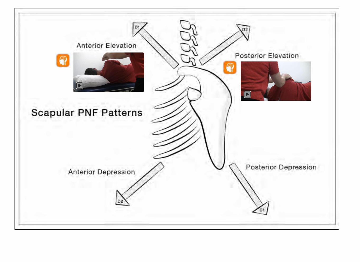
Proprioceptive Neuromuscular Facilitation
The therapeutic interventions you choose to address recovery of motor function in your patient with neurologic dysfunction all have a theoretical foundation. Key theories wi l l be presented and video demonstrations of interventions wi l l help you begin to fill your toolbox with options to drive neuroplasticity and facilitate your patients' recovery.
2
Proprioceptive Neuromuscular Facilitation S E C T I O N 1
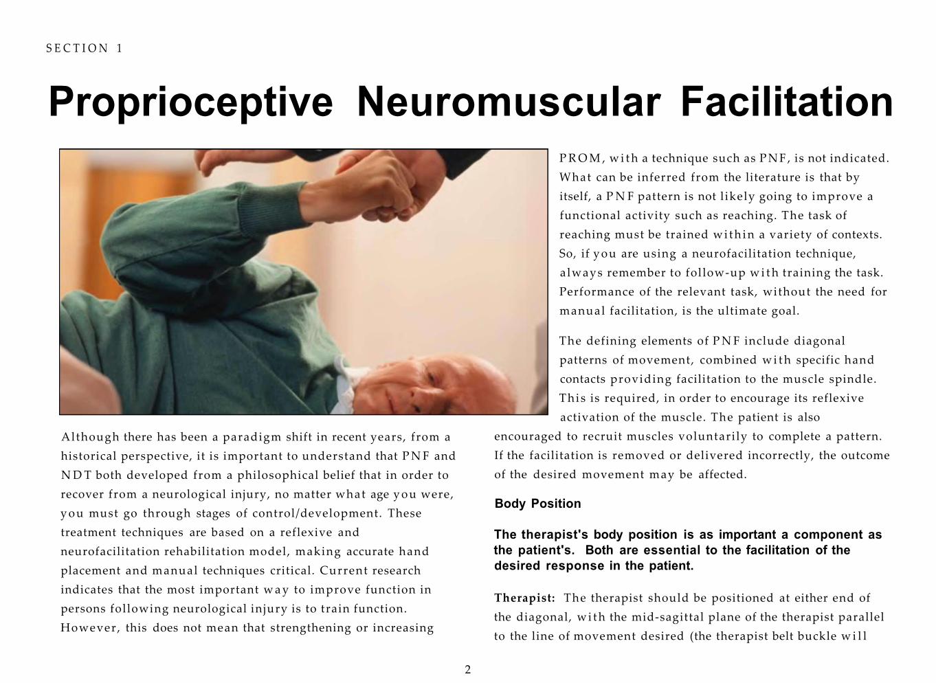
Although there has been a paradigm shift in recent years, from a historical perspective, it is important to understand that P N F and N D T both developed from a philosophical belief that in order to recover from a neurological injury, no matter what age y o u were, y o u must go through stages of control/development. These treatment techniques are based on a reflexive and neurofacilitation rehabilitation model, making accurate hand placement and manual techniques critical. Current research indicates that the most important w a y to improve function in persons fol lowing neurological in jury is to train function. However , this does not mean that strengthening or increasing
P R O M , w i t h a technique such as P N F , is not indicated. What can be inferred from the literature is that by itself, a P N F pattern is not l ikely going to improve a functional activity such as reaching. The task of reaching must be trained w i t h i n a variety of contexts. So, if y o u are using a neurofacilitation technique, a lways remember to fol low-up w i t h training the task. Performance of the relevant task, without the need for manual facilitation, is the ultimate goal.
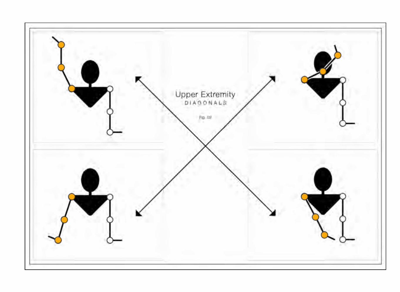
The defining elements of P N F include diagonal patterns of movement, combined w i t h specific hand contacts providing facilitation to the muscle spindle. T h i s is required, in order to encourage its reflexive activation of the muscle. The patient is also
encouraged to recruit muscles voluntari ly to complete a pattern. If the facilitation is removed or delivered incorrectly, the outcome of the desired movement may be affected.
Body Position
The therapist's body position is as important a component as the patient's. Both are essential to the facilitation of the desired response in the patient.
Therapist: The therapist should be positioned at either end of the diagonal, w i t h the mid-sagittal plane of the therapist parallel to the line of movement desired (the therapist belt buckle w i l l

3
face the motion). The choice of w h i c h end is based on: best direction of force reception, the amount of space, best position for v isual input, or the indiv idua l therapist's preference. The therapist needs to resist each pattern in a line parallel to her or his m i d -sagittal plane. M O V E M E N T S H O U L D A L W A Y S B E D I R E C T L Y I N T O O R O U T O F T H E C E N T E R O F G R A V I T Y O F T H E T H E R A P I S T . With the upper extremity patterns, it is often easier to position the therapist at the shoulder and go through a 180o pivot as the motion passes the mid-point of its ' arc. It is still important to get the therapists' mid-sagittal plane facing the motion at the beginning and end of the motion.
Patient: The patient should position themselves w i t h body parts in the most comfortable position and w i t h all parts in as close to proper alignment as possible for normal physiological movement. Support the normal curves of the body, e.g.. lumbar lordosis in supine, cervical spine in side ly ing , waist in side ly ing . For extremity patterns done in supine, it w o r k s better to get the patient as close to the edge of the support surface and is safe.
Body Mechanics
The movement of the therapist directly influences the response of the patient and should enable the patient to move correctly and freely in the diagonal. Key points to remember:
• The therapist's spine is most stable in neutral rotation and lateral f l e x i o n : A V O I D R O T A T I O N O R L A T E R A L F L E X I O N O F T H E S P I N E D U R I N G P N F !
• The majority of the movement should come from the legs and hips
• The therapist should a lways move or shift weight in the direction of movement desired from the patient
• A r m s and hands should stay relaxed, and the majority of the resistance should come from the therapist's trunk and pelvis
• The therapist's center of gravity stays even w i t h the arc of movement of the patient; if the center of gravity of the therapist becomes lower than the movement of the patient, there is more therapist w o r k due to increase in patient's mechanical advantage.
Clinical Tips
• Manual contacts may v a r y between individuals . The best contact is that w h i c h facilitates the correct response in the desired direction.
• Remember, gravity alone may be enough resistance to facilitate the desired response.
Manual Contacts:
"Appropriate" hand placement is essential to facilitate the desired response in the patient. Manual contacts LEAD the desired motion and must oppose the desired motion. Manual contacts need be specific and follow one of two rules. The contact surface needs to be the most precise and direct surface to resist the desired motion or it needs to contact the specific

4
agonist muscle, or muscle group in order to facilitate that muscle and be on a surface that resists the desired motion. The role of the manual contact is to increase the stimulation of the skin and other receptors to enhance the desired motor response.
Lumbrical Grip: T h i s manual contact utilizes the intrinsic muscles of the hand, a l lowing for a comfortable, specific, and secure contact. The key contact points are the pa lm of the hand, specifically the thenar and hypothenar eminences, the entire palmar surface of the fingers, and the F I N G E R P A D S , not the tips! R E M E M B E R : Point pressure produces P A I N & W I L L I N H I B I T T H E D E S I R E D R E S P O N S E !
Quick Stretch: Immediately prior to commands for muscle contractions, a quick stretch of the agonist muscle enhances the agonistic muscle contraction. The therapist does the quick stretch at the end of the lengthened range of the agonist. The therapist applies the stretch to as many of the agonistic muscles in the given pattern as practical. Th is quick stretch is intended to trigger a reflex muscle contraction that coincides w i t h the initiation of the client's voluntary contraction. The therapist times the quick stretch w i t h the client's contraction by giving a verbal cue ["and....pull (or push)"] in w h i c h the quick stretch immediately precedes the "pul l " or "push" commands the voluntary contraction. The therapist fine-tunes the client's response by adjusting the delay between the quick stretch and the "pul l " or "push".
Appropriate Resistance:
The amount of resistance should allow for smooth, coordinated, and appropriate speed of contraction throughout the available range of motion. Over-resisting leads to halting or too slow motion. The amount of resistance w i l l v a r y through the range to facilitate smooth flow of the desired response. Resistance w i l l be greater where the patient has most strength and mechanical advantage and less where the motion is weaker.
Use resistance for any of the following:
• strengthening
• increasing range of motion
• increasing stabilization
• enhancing relaxation
• enabling appropriate speed of contraction
• increasing coordination
Isotonic: concentric, eccentric, maintained; the command and intent is a lways for movement though at times little movement may occur (commands are " p u s h ! " or " p u l l ! " ) .
Isometric: the command and intent is to maintain the position in space (command is "hold ! " )
Verbal and Visual Cues:
Verbal commands assist in developing communication between the therapist and patient. The brevity, specificity, and t iming of

5
verbal cues are critical to optimal patient response. Visual input is key to developing coordinated use of the body, especially in cases of sensory loss. One or two w o r d commands are B E S T ; let your hand placement and tactile cues communicate the rest of the instructions. Make sure that the patient W A T C H E S their motion whi le learning each pattern.
Both are used to:
• Identify the desired direction of movement
• Facilitate the amount and type of response desired
• Direct the movement of the body w i t h the movement pattern desired
TECHNIQUES:
The fol lowing key techniques w i l l be described, w i t h video demonstrations of the techniques w i t h the use of different patterns.
• Quick Stretch
• Rhythmic Initiation
• Repeated Contractions
• Slow Reversals
• H o l d Relax
• Contract Relax
• Rhythmic Stabilization
• Alternating Isometrics
• Resisted Progression
Remember that these techniques can facilitate the desired response for most movements of the l imb girdles, extremities, or whole body.
Rhythmic Initiation:
Begins w i t h passive motion to familiarize the client w i t h the desired direction and sequence of motion. Rhythmic initiation progresses to active motion to teach the client to do the motion. F inal ly rhythmic initiation progresses to resisted movements that enable strengthening the motion to functional levels. It usual ly w o r k s best to have the client initiate the movement w i t h the distal components first. That w a y the therapist can use their distal hand better to guide the motion. T h i s technique is a lways uni-directional in a P N F pattern. Use Rhythmic Initiation to teach the desired pattern of movement and to assess the range of motion and gross strength. C a n w o r k effectively to improve:
• speed of movement
• direction of movement
• quality of movement
• strength or endurance
Repeated Contractions:
Used w i t h isotonic contractions that are weaker at some point(s) in the range of motion. The therapist repeats the quick stretch and the

6
verbal cue whenever the motion slows below the desired pace or weakens. T h i s enhances the active control of the motion and strength. At the point in the range of motion where the contraction weakens, repeat the initial quick stretch and verbal command (i.e. "and pul l farther" or " p u l l harder") and reduce your resistance. W h e n the motion slows too much, reapply your quick stretch, reduce y o u resistance, and command " p u l l faster". Be sure that y o u al low the motion to be fast enough by "y ie lding your resistance" at the correct speed.
Reversals of Antagonists:
Reversal of Antagonists includes contraction of the agonist followed by contraction of antagonist. T h i s facilitates coordinated "changes of direction" in a movement pattern for daily function. T h i s technique first commands motion in one direction of a diagonal pattern then (after a quick change in therapist manual contact) commands the opposite direction of motion. It is a repeating cycle of alternating contractions of antagonistic muscle groups. Slow reversal w o r k s on isotonic contractions and promotes smooth reversal of motion. Rhythmic stabilization w o r k s on isometric contractions and promotes stability at the target joint.
1. Slow reversals: Reversal of isotonic contractions through all or part of the available R O M . Commands might be (therapist uses manual contact to resist agonists) "now p u s h " , client goes through one direction of the motion; (therapist changes manual contacts to resist antagonists). Therapist commands " n o w p u l l " and client returns in the opposite direction of the motion. Used to improve:
• dynamic strength
• coordination
• kinesthetic awareness
• endurance
• active range of motion
2. Rhythmic stabilization: Isometric contraction of antagonistic muscle groups either simultaneously or alternately. Commands w o u l d be (therapist uses manual contact to resist agonists) "now hold"...(therapist changes manual contacts to resist antagonists)..."now hold" . To achieve a maximal response, it is necessary to resist both the diagonal and rotational components. Compression into the proximal joints w i l l further facilitate stability.
Used to improve:
• stability
• control of posture
• balance
• relaxation and pain reduction
• range of motion
Combination of Isotonics:
Combination of Isotonics involves concentric (shortening) and eccentric (lengthening) contractions in one direction, without any relaxation. T h e y promote controlled strong movements through some or all of the available R O M . There are three types of

7
contractions: concentric, eccentric, and maintained isotonics (alternate concentric and eccentric). Commands might be "pul l your hand up let me p u l l it d o w n s lowly" repeat this for several cycles as appropriate to the target A D L . D a i l y function consists of the coordination of al l three types of muscle contractions. Combination of isotonics is particularly helpful w h e n patients are l imited in strength or smooth control in specific points in the range of motion. The ultimate goal is re-integration into functional activity. Generally used to improve:
• strength in a focused part of the R O M
• the ability to alternate between concentric and eccentric contractions
• better control of more range of motion
• initiation of movement in different parts of available range
Many activities do not occur throughout the entire available range of motion. For example, sit to stand from a high chair, or w a l k i n g d o w n stairs. An indiv idua l may have no functional problems in mid-range, but may not function w e l l at the shortened or lengthened extremes of muscle range of motion. An example is the common difficulty patients have in controlling descent into a low chair. The quads just get beyond their optimal length for eccentric control and the client "p lops" into the chair. In this case the therapist might do combination of isotonics in the range of hip and knee motion associated w i t h a low chair. T h e n the patient w o u l d progress to practicing controlled sitting on lower and lower chairs.
Hold Relax:
T h i s technique is effective for increasing R O M due to muscle tightness and also works w e l l in the presence of m i l d to moderate pain. The key to effective hold relax technique is applying and releasing the resistance in a smooth and gradual w a y to minimize patient discomfort. T h i s w a y the therapist can regulate the resistance so that it does not increase the pain. If pain persists w i t h gentle contractions or prevents significant relaxation, try gentle active assistive exercise first. H o l d Relax description: Take the extremity through the available R O M to where the restriction begins, but client is still comfortable. Gently resist all components of the tight muscle pattern, w i t h emphasis on rotation. Commands might be "hold gently..now a little stronger...hold...now let go some...now relax completely". M A K E S U R E T H E C L I E N T R E L A X E S F U L L Y , this is critical. Next the therapist moves the extremity further into the well-tolerated restricted range (but not to the point of pain) and repeats the hold. . . .relax. The therapist repeats the cycles unt i l there is no further gain in R O M . Gentle traction during the contractions helps minimize joint pain. C a n do H o l d Relax by contracting the "t ight" muscles (providing improved direct relaxation after the contraction) or by contracting the "opposite" muscles (enabling improved relaxation of the tight muscles indirectly, by reciprocal inhibition).
H o l d Relax is useful for:
increasing range of motion (wi th and without m i l d to moderate pain)
initiation of movement
Contract Relax:

8
Contract Relax uses a concentric contraction of the tight muscle (direct relaxation), or of the opposing muscles (indirect relaxation) and is otherwise much like H o l d Relax. Resist all components of the pattern w i t h concentration on rotational component. Do not al low any significant motion through the R O M . The contraction is followed by complete relaxation of the body part, and active or passive movement in the direction that lengthens the restricted soft tissues. Commands might be "pull . . .pull . . .pull . . .now relax...let go completely". T H I S T E C H N I Q U E I S N O T Preferred W H E N P A I N I S P R E S E N T I N T H E M O V E M E N T P A T T E R N . (The reason that H o l d Relax w o r k s better in the presence of pain is that the therapist controls the gradual onset of contraction and can m u c h better K E E P the contractions gentle enough. In Contract Relax, w h e n a therapist gives a " P u l l " or " P u s h " command, the client determines h o w m u c h he/she w i l l pu l l or push. In the presence of pain, clients have muscle guarding and find it difficult to push or p u l l just a " l i t t le" . )
Contract Relax is useful for:
range of motion (only without pain)
initiation of movement
The fol lowing chart provides a summary of P N F in relation to Margaret Rood's (one of the foremothers of physical therapy) Stages of Control. Al though originally describing only developmental stages of motor control, the concepts were
expanded to the recovery of movement in adults. With current evidence, it is understood that the nervous system of adults recover differently than the nervous system develops in children.
However , Rood has provided a conceptual f ramework into w h i c h the application of the different elements of P N F can be organized.

9
Table 1.1.1 Stages of Motor Control Associated with Proprioceptive Neuromuscular Facilitation
STAGES OF C O N T R O L G E N E R A L G O A L S PNF T E C H N I Q U E S SAMPLE TREATMENT
A C T I V I T I E S
M O B I L I T Y •Increase R O M
•Increase initiation of range of motion.
•Hold Relax (HR) , Contract Relax
(CR), Rhythmic stabilizations (RS),
Rhythmic rotation, Joint mobilization
•HR-active movement, Repeated
Contractions
For a patient wi th decrease in shoulder
joint mobility, incorporate Rhythmic
Rotation into P R O M exercises to
facilitate relaxation, especially in a
patient with pain and hypertonia.
For a patient wi th a limitation in R O M ,
particularly when pain is
accompanying, Hold Relax can be used
which includes an isometric
contraction at the end point of the
range, followed by relaxation and
movement to new point of limitation.
S T A B I L I T Y
•Sustained isometric contractions in
shortened range for increasing
duration
•Coordinated isometric contractions in
midline or weight bearing postures
•Shortened-held resisted contractions
(SHRC)
•Alternating Isometrics, RS
For a patient who has difficulty
standing maintaining LE extension,
S H R C as a Quad and Glut set in
supine, held for 10 seconds to gain
stability across the joints in non-weight
bearing to prepare for weight-bearing.
For a patient with difficulty stabilizing
in standing or sitting and in order to
facilitate smooth performance of
isometric contractions in all three
planes simultaneously, you could use
RS at the trunk.

10
Figure 1.1 Stages of Motor Control Associated with Proprioceptive Neuromuscular Facilitation (cont)
STAGES OF C O N T R O L G E N E R A L G O A L S PNF T E C H N I Q U E S SAMPLE TREATMENT
A C T I V I T I E S
C O N T R O L L E D M O B I L I T Y
•Weight-shifting in weight bearing
postures: AP, lateral, rotation. Also,
reversal of antagonists or concentric-
eccentric contractions; trunk rotations
•Slow-reversal hold, slow reversal,
agonistic reversals
For the patient who shows difficulty in
eccentric contractions in stand to sit,
you could have the patient in the
position and have the patient "make
you work at pushing him/her down"
into sitting using the technique of
agonistic reversals.
S K I L L
•Proximal dynamic stability
•Normal timing and sequencing of
movement
•Trunk counter-rotation
•Locomotion and Manipulation
( A D L s )
•Communication
•Shortened-held resisted contractions
(SHRC)
•Alternating Isometrics, RS
For a patient who has difficulty
standing maintaining LE extension,
S H R C as a Quad and Glut set in
supine, held for 10 seconds to gain
stability across the joints in non-weight
bearing to prepare for weight-bearing.
For a patient with difficulty stabilizing
in standing or sitting and in order to
facilitate smooth performance of
isometric contractions in all three
planes simultaneously, you could use
RS at the trunk.

Click a touchpoint for more information

Lower Extremity D1 Flexion and Extension demonstrating rhythmic initiation, quick stretch and reversal of agonists
Lower Extremity D2 Flexion and Extension demonstrating rhythmic initiation, quick stretch and reversal of agonists

Upper Extremity D1 Flexion and Extension demonstrating rhythmic initiation, quick stretch and reversal of agonists
Upper Extremity D2 Flexion and Extension demonstrating rhythmic initiation, quick stretch and reversal of agonists