Embed Size (px)
Citation preview
Algae as nutritional and functional food sources: revisitingour understanding
Mark L. Wells1 & Philippe Potin2& James S. Craigie3 & John A. Raven4,5
&
Sabeeha S. Merchant6 & Katherine E. Helliwell7,8 & Alison G. Smith7&
Mary Ellen Camire9 & Susan H. Brawley1
Received: 6 June 2016 /Revised and accepted: 25 September 2016# The Author(s) 2016. This article is published with open access at Springerlink.com
Abstract Global demand for macroalgal and microalgalfoods is growing, and algae are increasingly being consumedfor functional benefits beyond the traditional considerations ofnutrition and health. There is substantial evidence for thehealth benefits of algal-derived food products, but there re-main considerable challenges in quantifying these benefits,as well as possible adverse effects. First, there is a limitedunderstanding of nutritional composition across algal species,geographical regions, and seasons, all of which can substan-tially affect their dietary value. The second issue is quantifyingwhich fractions of algal foods are bioavailable to humans, andwhich factors influence how food constituents are released,ranging from food preparation through genetic differentiationin the gut microbiome. Third is understanding how algal nu-tritional and functional constituents interact in human metab-olism. Superimposed considerations are the effects of harvest-ing, storage, and food processing techniques that can dramat-ically influence the potential nutritive value of algal-derivedfoods. We highlight this rapidly advancing area of algal sci-
ence with a particular focus on the key research required toassess better the health benefits of an alga or algal product.There are rich opportunities for phycologists in this emergingfield, requiring exciting new experimental and collaborativeapproaches.
Keywords Algal foods . Antioxidants . Arsenosugars .
Experimental design .Microalgal supplements . Nutritionalminerals . Omega-3-fatty acids . Polysaccharides . Seavegetables . Vitamins
Introduction
Algae have been part of the human diet for thousands of years,based on archaeological evidence from 14,000 yBP in Chile(Dillehay et al. 2008) and early written accounts (e.g., inChina, 300 A.D.; in Ireland, 600 A.D.; Newton 1951;Tseng 1981; Aaronson 1986; Turner 2003; Gantar and
Electronic supplementary material The online version of this article(doi:10.1007/s10811-016-0974-5) contains supplementary material,which is available to authorized users.
* Susan H. [email protected]
1 School of Marine Sciences, University of Maine, Orono, ME 04469,USA
2 Integrative Biology of Marine Models, Station Biologique Roscoff,CNRS-Université Pierre et Marie Curie, Place Georges Teissier,29680 Roscoff, France
3 National Research Council of Canada, 1411 Oxford Street,Halifax, NS B3H 3Z1, Canada
4 Division of Plant Sciences, University of Dundee (James HuttonInst), Invergowrie, Dundee DD2 5DA, Scotland, UK
5 Plant Functional Biology and Climate Change Cluster, University ofTechnology Sydney, Ultimo, NSW 2007, Australia
6 Department of Chemistry & Biochemistry, University ofCalifornia-Los Angeles, 607 Charles E. Young Dr., East, LosAngeles, CA 90095-1569, USA
7 Department of Plant Sciences, University of Cambridge, DowningSt., Cambridge CB2 3EA, UK
8 Present address: Marine Biological Association of the UK, CitadelHill, Plymouth PL1 2PB, UK
9 School of Food and Agriculture, University of Maine,Orono, ME 04469, USA
J Appl PhycolDOI 10.1007/s10811-016-0974-5

Svircev 2008; Craigie 2010). In North America, the TsimshianFirst Nations’ people named the month of May for the time ofyear when they harvested the important food crop of Pyropia(Fig. 1). More contemporaneously, the global harvest of sea-weeds in 2013was estimated at US $6.7 billion, and over 95%was produced in mariculture, with China and Indonesia beingthe top producers (FAO 2015). In addition to macroalgae,some microalgae are cultivated for foods and food additives(Switzer 1980; Jassby 1988; Fournier et al. 2005; Gantar andSvircev 2008; Chacón-Lee and González-Mariño 2010; FAO2016). The FAO (2014) estimated that 38% of the 23.8milliont of seaweeds in the 2012 global harvest was eaten by humansin forms recognizable to them as seaweeds (e.g., kelps,nori/laver), not counting additional consumption of hydrocol-loids (e.g., agars, alginates, carrageenans) used as thickeningagents in foods and beverages. Human consumption of algalfoods varies by nation, with Japanese diets representing a re-cent (2010–2014) annual per capita consumption ranging from9.6 (2014) to 11.0 (2010) g macroalgae day−1 (MHLW 2014).
Overall, the trend towards increasing nutritional demand foralgal products on a global basis stems from a greater focus onhealth and wider use of food additives.
In addition to their nutritional value, algae increasingly arebeing marketed as Bfunctional foods^ or Bnutraceuticals^;these terms have no legal status in many nations but describefoods that contain bioactive compounds, or phytochemicals,that may benefit health beyond the role of basic nutrition (e.g.,anti-inflammatories, disease prevention; Bagchi 2006;Hafting et al. 2012). The path from algal research to thelaunching of new food products or dietary supplements isstrongly affected by industrial, regulatory, and nutritional con-siderations (e.g., see Borowitzka 2013a; Finley et al. 2014).The widespread interest in algal foods and/or their functionalfood potential is evident in numerous recent reviews (Warrand2006; MacArtain et al. 2007; Kulshreshtha et al. 2008;Bocanegra et al. 2009; Mendes et al. 2009; Cottin et al.2011; Harnedy and FitzGerald 2011; Holdt and Kraan 2011;Lordan et al. 2011; Pangestuti and Kim 2011; Stengel et al.2011; Cornish et al. 2015; Hafting et al. 2015) and books(Rhatigan 2009; Mouritsen 2013; Tiwari and Troy 2015;Fleurence and Levine 2016). Many studies report the potentialnutritional or bioactive content of different algae but manyfewer studies quantify the bioavailability of nutrients and phy-tochemicals from algal foods. Our purpose is to review andassess what is known about different food components (i.e.,proteins, polysaccharides, lipids, vitamins, minerals, and anti-oxidants, potential toxicants) in the context of improvingknowledge about the efficacy of algal foods. There are richopportunities for phycologists to collaborate with other scien-tists and clinicians in this emerging field from algalBprospecting^ to defining nutritional value, bioaccessibility,and subsequent bioactivity, to the design and construction ofmid-large cultivation systems for production of commercial-scale product.
Digestion and bioavailability
In this article we use the term bioavailability, as defined byCarbonell-Capella et al. (2014) Bas a combination of bioactiv-ity and bioaccessibility,^ where bioaccessibility refers to therelease from the food matrix, transformations during diges-tion, and transport across the digestive epithelium, while bio-activity encompasses uptake into tissues, metabolism, andphysiological effects. Because of the difficulties, both practi-cal and ethical in terms of measuring bioactivity, the fractionof a given compound or its metabolite that reaches the system-ic circulation (Holst and Williamson 2008) can be consideredbioaccessible, but not necessarily bioactive. Most publishedevaluations of bioactivity of algal foods are based on short-term in vitro tests using algal extracts that frequently are of ill-defined composition and purity, so a clear understanding of
Fig 1 a Pyropia spp. being dried in squares in the intertidal zone by FirstNations’ people at Pearce Island, British Columbia (2009). Harvesterswould traditionally lay the seaweed out to dry on warm rocks whilewaiting for those fishing to return with the canoes (photo credit, AmyDeveau). b Checking the seaweed squares after transfer to cedar racks forfinal drying (photo credit, Victoria Wyllie-Echeverria)
J Appl Phycol

their food value is highly constrained. Particularly lacking isinformation on the behavior of algal food components in thegut. For example, can the purported active metabolites identi-fied in in vitro studies be transferred from the gut lumen intothe body? Likewise, are observed in vivo biological effects theconsequence of biological uptake or instead indirect outcomesstemming from improved functionality or composition of theintestinal microbiome? It is important then to consider theprocess of digestion and transformation in the human system.
Digestion refers to the physical and biochemical degrada-tion of foods and the nutrients therein in preparation for ab-sorption into the body. Digestion begins in the mouth withchewing, which reduces particle size and mixes food withsaliva (Lovegrove et al. 2015). The predominant salivary en-zyme is alpha (α)-amylase, which is specific for α(1→4) glu-cose linkages, and human salivary amylase is more active thanthat from other primates (Boehlke et al. 2015). Hardy et al.(2015) hypothesized that cooking to increase digestibility andsensory quality of starch-rich foods helped drive human evo-lution by providing more glucose to growing brains. Studiesof the effect of human saliva on algae and specifically algalstarch are lacking, however. The relative importance of sali-vary versus pancreatic amylase in starch digestion also is notclear (Lovegrove et al. 2015). Pepsin and the pepsinogensbegin protein digestion in the stomach, aided by hydrochloricacid that denatures proteins and releases nutrients from thefood matrix. Lipases produced in the mouth and stomach be-gin the process of digesting triacylglycerols. The stomach alsoreleases intrinsic factor that is essential for vitamin B12 absorp-tion in the small intestine. Gastric peristalsis further reducesfood particle size, preparing macronutrients for additionalchemical breakdown and absorption in the small intestine.The pancreas discharges a mixture of trypsin, chymotrypsin,carboxypeptidases, α-amylase, lipase, and other enzymes thatrespectively digest proteins and peptides, starches, triacylglyc-erols, and other compounds in the small intestine (Gropperand Smith 2013). The mixture of proteases, amylase, and li-pase are collectively known as pancreatin; porcine pancreatinis often used to model human digestion in in vitro systems.The small intestine itself releases a variety of enzymes actingon peptides, amino acids, monoacyglycerols, disaccharides,and α(1→4) and α(1→6) linkages in oligosaccharides, dex-trins, and polysaccharides such as starch. Micronutrients suchas vitamins and minerals also are absorbed in the small intes-tine once they are solubilized from the food matrix.Fucoxanthin, a key algal carotenoid, may be better absorbedif other lipids are present (Peng et al. 2011).
Humans lack the ability to digest β(1→4) linkages in glu-can polysaccharides, as in cellulose and hemicelluloses suchas xyloglucan, and this indigestible material is referred to asdietary fiber. The undigested materials continue on to the largeintestine (colon) where microbial co-metabolism fermentssubstrates such as non-starch polysaccharides, resistant starch,
and oligosaccharides to short-chain fatty acids, and proteinsinto a wider variety of compounds. These bacterial-dependentenzymatic processes are not considered Bdigestion,^ althoughthe fermentation products can provide nutritional or functionalbenefits either by being absorbed and transported via thebloodstream or by shaping more healthful gut microbiomesand chemical conditions in the colon (MacFarlane andMacFarlane 2012). Indigestible, fermentable carbohydratesand sugar alcohols are referred to as FODMAP (fermentable,oligo-, di- mono-saccharides and polyols) (Gropper and Smith2013). Algal proteins and carbohydrates that escape completedigestion in the small intestine may benefit humans by stimu-lating immune response indirectly via promotion of microbialresponses (Cian et al. 2015). Dietary modulation of the colon-ic flora and the impact of bacterial fermentation products onhuman health are rapidly evolving areas of research (Duffyet al. 2015) and are likely to be especially important consid-erations in assessing the health benefits of algal-derived foods.
Not all human gut microbiomes have equal competencies,as algal polysaccharide fermentation differs among humansfrom different regions. The arsenal of polysaccharide-degrading enzymes exhibited in the common gut bacterium(Bacteroides plebeius) of Japanese people, but not Americans,appears to result from horizontal gene transfer (HGT) fromZobellia galactanivorans (Bacteroidetes), a marine bacteriuminhabiting the surfaces of algae such as nori (Hehemann et al.2010). HGTalso may explain the presence of a gene cluster inJapanese gut Bacteroides that enables fermentation of algi-nates in brown algal cell walls (Thomas et al. 2012).Similarly, a small cohort of Spaniards possesses gutmicrobiomes with apparently HGT-provided porphyranasesand agarases (Hehemann et al. 2012). Such striking differ-ences emphasize the complex interactions among food cus-toms, dietary history, and gut microbiomes that complicatestudy of the nutritional and functional benefits of algal foods(Paulsen and Barsett 2005; Costello et al. 2012; Gordon 2012;Nicholson et al. 2012).
The importance of assessing the biological availability ofnutritional and functional food components cannot beunderestimated. Bioavailability has critical relevance to boththe proportional digestion and uptake of nutrients and func-tional food components, but also the degree of fermentationand nature of the host-microbial co-metabolism in the colon.While there exists a vast literature on the food content ofmicroalgal and macroalgal foods and supplements, extrapolat-ing these findings to assess their quantitative contribution tohuman health is more tenuous. The analytically determinedconcentration of constituents in food can differ, sometimessubstantially, from that actually crossing from the digestivetract into the blood (i.e., the bioaccessible fraction).Moreover, current analytical approaches give even less insightto the complexity of interacting effects that regulate the bac-terial flora of the colon, and hence the nature of fermentation
J Appl Phycol
products. Confounding issues stem from the food itself (e.g.,the presence and nature of intact cell walls, soluble fiber char-acteristics, and the presence of other substances that may inhibitor facilitate the uptake of metabolites), the harvest season (e.g.,altered metabolite and biomass composition, environmental var-iability of essential precursors, and anthropogenic factors), andthe food preparation methods (Sensoy 2014). Analyticalmethods such as simulated gastrointestinal digestion (Moreda-Pineiro et al. 2011; Maehre et al. 2014), xenobiotic animalmodels, and molecular biological and genetic techniques canprovide a sound basis for improved assessment of bioavailabil-ity; however, their use is not yet widespread in the study of foodsof algal origin. As a consequence, and despite highly accurateand precise analytical determinations of food content, currentknowledge of the nutritional or functional food value of algalproducts remains largely qualitative. The development of appro-priate model systems and use of rigorous experimental designthus is essential in order to verify the bioavailability of nutrition-al and functional components of algae used in all foods.
Proteins
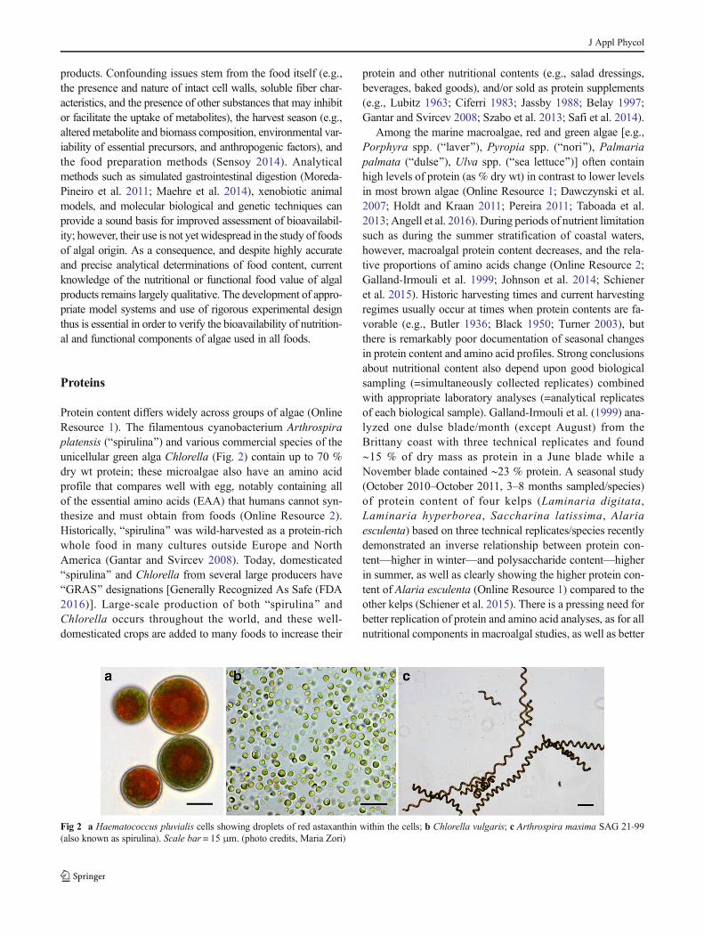
Protein content differs widely across groups of algae (OnlineResource 1). The filamentous cyanobacterium Arthrospiraplatensis (Bspirulina^) and various commercial species of theunicellular green alga Chlorella (Fig. 2) contain up to 70 %dry wt protein; these microalgae also have an amino acidprofile that compares well with egg, notably containing allof the essential amino acids (EAA) that humans cannot syn-thesize and must obtain from foods (Online Resource 2).Historically, Bspirulina^ was wild-harvested as a protein-richwhole food in many cultures outside Europe and NorthAmerica (Gantar and Svircev 2008). Today, domesticatedBspirulina^ and Chlorella from several large producers haveBGRAS^ designations [Generally Recognized As Safe (FDA2016)]. Large-scale production of both Bspirulina^ andChlorella occurs throughout the world, and these well-domesticated crops are added to many foods to increase their
protein and other nutritional contents (e.g., salad dressings,beverages, baked goods), and/or sold as protein supplements(e.g., Lubitz 1963; Ciferri 1983; Jassby 1988; Belay 1997;Gantar and Svircev 2008; Szabo et al. 2013; Safi et al. 2014).
Among the marine macroalgae, red and green algae [e.g.,Porphyra spp. (Blaver^), Pyropia spp. (Bnori^), Palmariapalmata (Bdulse^), Ulva spp. (Bsea lettuce^)] often containhigh levels of protein (as % dry wt) in contrast to lower levelsin most brown algae (Online Resource 1; Dawczynski et al.2007; Holdt and Kraan 2011; Pereira 2011; Taboada et al.2013; Angell et al. 2016). During periods of nutrient limitationsuch as during the summer stratification of coastal waters,however, macroalgal protein content decreases, and the rela-tive proportions of amino acids change (Online Resource 2;Galland-Irmouli et al. 1999; Johnson et al. 2014; Schieneret al. 2015). Historic harvesting times and current harvestingregimes usually occur at times when protein contents are fa-vorable (e.g., Butler 1936; Black 1950; Turner 2003), butthere is remarkably poor documentation of seasonal changesin protein content and amino acid profiles. Strong conclusionsabout nutritional content also depend upon good biologicalsampling (=simultaneously collected replicates) combinedwith appropriate laboratory analyses (=analytical replicatesof each biological sample). Galland-Irmouli et al. (1999) ana-lyzed one dulse blade/month (except August) from theBrittany coast with three technical replicates and found∼15 % of dry mass as protein in a June blade while aNovember blade contained ∼23 % protein. A seasonal study(October 2010–October 2011, 3–8 months sampled/species)of protein content of four kelps (Laminaria digitata,Laminaria hyperborea, Saccharina latissima, Alariaesculenta) based on three technical replicates/species recentlydemonstrated an inverse relationship between protein con-tent—higher in winter—and polysaccharide content—higherin summer, as well as clearly showing the higher protein con-tent of Alaria esculenta (Online Resource 1) compared to theother kelps (Schiener et al. 2015). There is a pressing need forbetter replication of protein and amino acid analyses, as for allnutritional components in macroalgal studies, as well as better
Fig 2 a Haematococcus pluvialis cells showing droplets of red astaxanthin within the cells; b Chlorella vulgaris; c Arthrospira maxima SAG 21-99(also known as spirulina). Scale bar = 15 μm. (photo credits, Maria Zori)
J Appl Phycol

definition of the natural intertidal or commercial sites fromwhich analyzed samples were obtained (N.B. Shuuluka et al.2013 as example). Some new sea vegetable products will ben-efit from complementary food constituents (Woolf et al. 2011;see vProtein software).
Protein concentration in algae is often estimated using atotal nitrogen-to-protein (NTP) conversion factor (6.25) basedon the assumption that most N in the sample occurs as protein.This conversion factor, however, often over-estimates the pro-tein content because of the presence of variable amounts ofnon-protein-N in the sample (Lourenço et al. 2002; Safi et al.2013; Angell et al. 2016). For example, the conversion factorcalculated for crude biomass for Chlorella vulgaris (walled)was 6.35, whereas it was 5.96 based on direct protein extracts(Safi et al. 2013). Similar studies of 19 tropical marine algaeyielded even lower average factors of 4.59 (red algae), 5.13(green algae), and 5.38 (brown algae) (Lourenço et al. 2002),perhaps related to seasonally lower N inputs to tropical surfacewaters. Zhou et al. (2012 [see her Tables 3–5]) reported similarfindings. These conversion factors certainly will vary withseason based upon varying amino acid composition, empha-sizing the need for protein and amino acid studies to determinethe seasonal optima for harvest among algal foods. Angellet al. (2016) argued for a new universal conversion factor, afterfinding a median nitrogen-to-protein value of 5 in a literature-based meta-analysis of 103 macroalgae; however, the range ofvalues in their analysis was high (see their Fig. 4). The algaehave polyphyletic origins and this, too, is reflected in the ab-sence of a universal N to protein conversion factor.
In most analyses of amino acid composition in marine al-gae, glutamic acid, and aspartic acid represent the highestproportions of amino acids (e.g., Fleurence 1999b; Lourençoet al. 2002; Online Resources 1, 2; Holdt and Kraan 2011).These amino acids occur as protein constituents and as freeamino acids or their salts. For humans, glutamate is the majorcomponent of the savory, the fifth basic taste called umamifrom its characterization in kelp (Ninomiya 2002; Mouritsen2013). Glutamic acid content may decrease after several suc-cessive harvests of Pyropia yezoensis (nori; Niwa et al. 2008).Other amino acids (alanine and glycine) also contribute todistinctive flavors of some marine algae (e.g., see Holdt andKraan 2011).
The non-proteinaceous amino acid taurine is especiallyabundant in marine red algae (e.g., ∼1–1.3 g taurine per100 g DWof nori, Niwa et al. 2008). Although taurine is notan EAA for adults, it is a component of bile acids that complexand lower cholesterol in the bloodstream (Medeiros andWildman 2015).
In general, protein in most algae is digested less completelythan reference proteins such as casein (a milk protein) inin vitro model systems containing digestive enzymes such aspepsin, pronase, and pancreatin, with evidence that this is dueespecially to inhibitory soluble fibers (e.g., Fujiwara-Arasaki
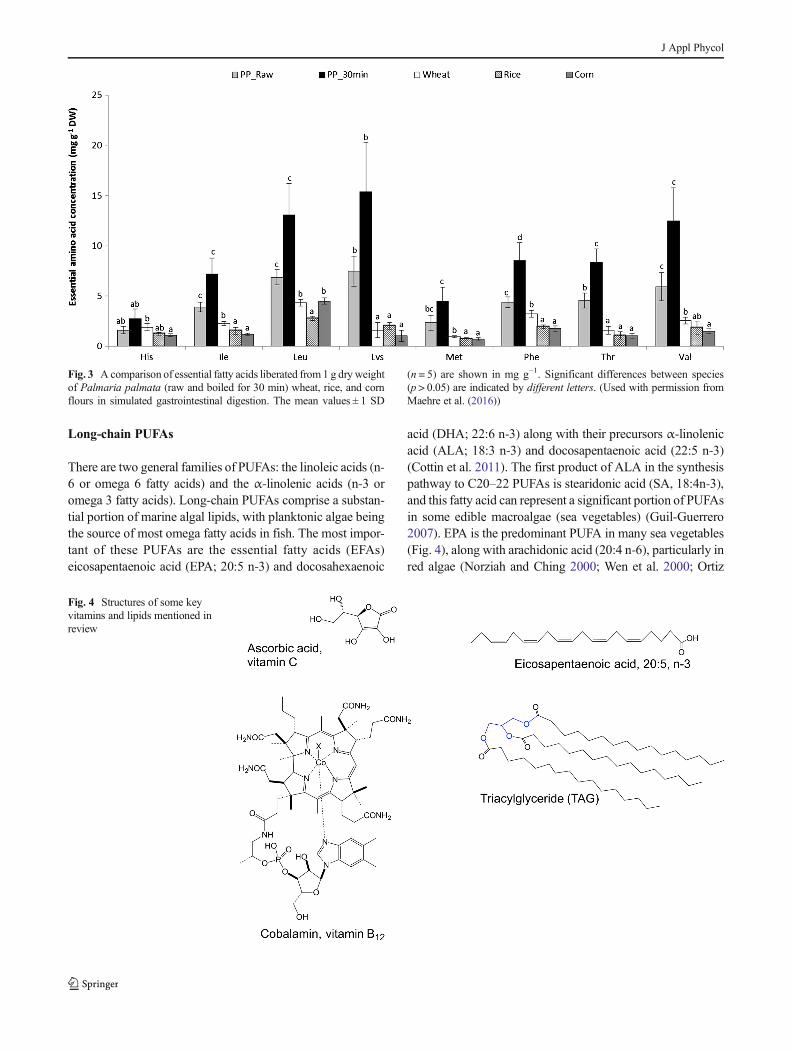
et al. 1984; Fleurence 1999a; Urbano and Goni 2002; Marrionet al. 2003, 2005; Wong and Cheung 2003; De Marco et al.2014). Inclusion of pre-analytical steps such as freezing, mill-ing, digestion of crude sample with polysaccharide-digestingenzymes, and/or osmotic rupture of cells to free intracellularcompounds is an active area of research (e.g., Harnedy andFitzGerald 2013; Safi et al. 2014; Ursu et al. 2014; andreferences therein). Importantly, a recent study (Maehre et al.2016) with excellent biological and technical replicationshows the beneficial effect of cooking on amino acid avail-ability from dried dulse (Online Resource 3); however,cooking did not significantly increase the total amino acidsmeasured from Alaria (Online Resource 3). Furthermore,Maehre et al. (2016) demonstrated that the apparent aminoacid bioaccessibility from both raw and 30 min-boiled dulsewas higher than froman equivalent dryweight ofwheat, rice,or corn flour in a simulated in vitro gastrointestinal digestionmodel with analysis at each sequential digestive step (amy-lase/saliva buffer; pepsin/gastric buffer; pancreatin/duodenalbuffer) (Fig. 3). Future research on microalgal and macroalgalprotein bioavailability might incorporate measures such as theprotein digestibility-corrected amino acid score (PDCAAS),which involves urinary and fecal determinations of N absorp-tion in rats, as well as the FAO recommended replacement ofPDCAAS by the digestible indispensable amino acid score(DIAAS) (Medeiros and Wildman 2015; Rutherfurd et al.2015).
Lipids
Lipids are essential for all living organisms as components ofmembranes, energy storage compounds, and as cell signalingmolecules (Eyster 2007). Although humans and other mam-mals synthesize lipids, some essential lipids must be obtainedfrom dietary oils or fats. Phospho- and glycolipids, importantfor membrane function, contain a polar head group with twofatty acid chains, while the triacylglyceroles (TAGs), impor-tant energy stores in the cell, are non-polar (neutral) lipidscontaining three fatty acid chains (Fig. 4). Lipid membranescontain sterols such as fucosterol and β-sitosterol (Fahy et al.2005) that also have reported health benefits (Arul et al. 2012).Embedded in algal lipid fractions are the nutritionally valuablecarotenoid pigments that will be discussed in theBphytochemicals^ section (below). TAGs have attracted greatattention in recent years as a source for biodiesel, with somemicroalgae accumulating up to 40–60 % of their dry weightas TAGs (Georgianna and Mayfield 2012). However, marinemacrophytes typically do not exceed 2–4.5 % dry wt as lipids,mainly as phospholipids and glycolipids (Holdt and Kraan2011). Of these, the long-chain polyunsaturated fatty acids(PUFAs) and carotenoids are most noteworthy as functionalfoods (Holdt and Kraan 2011).
J Appl Phycol
Long-chain PUFAs
There are two general families of PUFAs: the linoleic acids (n-6 or omega 6 fatty acids) and the α-linolenic acids (n-3 oromega 3 fatty acids). Long-chain PUFAs comprise a substan-tial portion of marine algal lipids, with planktonic algae beingthe source of most omega fatty acids in fish. The most impor-tant of these PUFAs are the essential fatty acids (EFAs)eicosapentaenoic acid (EPA; 20:5 n-3) and docosahexaenoic
acid (DHA; 22:6 n-3) along with their precursors α-linolenicacid (ALA; 18:3 n-3) and docosapentaenoic acid (22:5 n-3)(Cottin et al. 2011). The first product of ALA in the synthesispathway to C20–22 PUFAs is stearidonic acid (SA, 18:4n-3),and this fatty acid can represent a significant portion of PUFAsin some edible macroalgae (sea vegetables) (Guil-Guerrero2007). EPA is the predominant PUFA in many sea vegetables(Fig. 4), along with arachidonic acid (20:4 n-6), particularly inred algae (Norziah and Ching 2000; Wen et al. 2000; Ortiz
Fig. 4 Structures of some keyvitamins and lipids mentioned inreview
Fig. 3 A comparison of essential fatty acids liberated from 1 g dry weightof Palmaria palmata (raw and boiled for 30 min) wheat, rice, and cornflours in simulated gastrointestinal digestion. The mean values ± 1 SD
(n = 5) are shown in mg g−1. Significant differences between species(p > 0.05) are indicated by different letters. (Used with permission fromMaehre et al. (2016))
J Appl Phycol

et al. 2009) where EPA comprises up to 50 % of the total fattyacid content (e.g., Palmaria palmata, van Ginneken et al.2011). Humans and other animals cannot convert ALA toEPA and DHA at required levels, so dietary sources of theseEFAs are critically important for the health of humans (Cottinet al. 2011) and many animals (Li et al. 2009).
Numerous epidemiological and controlled interventionaltrials (N.B., the excellent reviews of Conquer and Holub1996; Holub 2009; Cottin et al. 2011) support the health ben-efits to humans of DHA and EPA long-chain omega-3 fattyacids from fish oils and algal sources (mainly extracts). Incontrast to most other algal food constituents, the bioaccessi-bility of DHA and EPA in algal-derived oils and extracts iswell quantified for humans, ranging from ∼50 to 100 % de-pending on the matrix (Haug et al. 2011; Schuchardt et al.2011). While clinical research to date strongly supports a nu-tritional need for oils that are enriched in DHA and EPA, thereis more understanding about the bioactivity of DHA than ofEPA (Conquer and Holub 1996; Holub 2009; Cottin et al.2011). There is a considerable literature (Cottin et al. 2011)on the cardioprotective effects of DHA-containing TAG fromCrypthecodinium cohnii (a dinoflagellate, Mendes et al. 2009)and Schizochytrium sp. (a thraustochytrid stramenopile, Liet al. 2009; Barclay et al. 2010), and as a consequence, infantformula, infant foods, and certain other food categories (dairy,bakery, eggs, and non-alcoholic beverages) and marketed nu-tritional products now are supplemented with algal-derivedDHA. There is evidence that enhanced DHA intake may im-prove infant cognitive performance and enhance visual acuity(Jensen et al. 2005, 2010; Imhoff-Kunsch et al. 2011), al-though more recent data raises question about this linkage(Delgado-Noguera et al. 2015). There also is some under-standing about the bioaccessibility of DHA in different algalproducts. Algal oil capsules based on a patented commercialsource (Martek) and cooked salmon are reported to representnutritionally equivalent sources of DHA (see in Cottin et al.2011). A similar human trial showed that DHA from twodifferent strains of Schizochytrium sp. (DHASCO-T andDHASCO-S) supplied in capsules generated equivalentdose-dependent DHA levels in plasma phospholipids anderythrocytes (Arterburn et al. 2008). Fortified snack bars alsodelivered equivalent amounts of DHA on a DHA dose basis(Arterburn et al. 2007). A systematic review of plant omega-3fatty sources by Lane et al. (2014) concluded that further re-search on algal sources was warranted based on promisingpreliminary work.
Nonetheless, the relative health benefits of commercial al-gal supplements that tend to be DHA-rich versus natural fishoils that contain both DHA and EPA are uncertain. Cottin et al.(2011) found that BRecent evidence from randomized con-trolled trials has produced equivocal results. Heterogeneityof the studies in terms of dosage, duration, population target,sample size, as well as the relative amounts of EPA and DHA
used in supplements could account for the variability of theresults.^ Even so, important trends stand out. While both EPAand DHA reduce TAG levels in humans (Wang et al. 2006;Bernstein et al. 2012), DHA appears responsible for the bloodpressure and heart rate-lowering effect of fish oils (Valera et al.2014). DHA also seems to be beneficial for endothelial andplatelet function, although a direct role for EPA in regulatingTAGs has not been ruled out. Algal DHA extracts can produceother cardiovascular protective effects in humans by alteringplasma lipoproteins at reasonably small doses (2 g algalDHA day−1 over 4.5 months: Neff et al. 2011). The healthbenefits of algal DHA supplements for subgroups such asvegetarians, who otherwise may have low essential fatty acidintakes, remains a high research priority (Geppert et al. 2005;Cottin et al. 2011).
Fish oils also have demonstrated anti-inflammatory andinsulin-sensitizing properties in vitro and in animal studies(Nauroth et al. 2010; Cottin et al. 2011); however, humantrials often yield conflicting findings. Neither EPA nor DHAalone showed any effects on inflammation in double-blindtrials with cystic fibrosis patients (Van Blervliet et al. 2008)or insulin sensitivity in human subjects, despite indications forpotency in vitro (critically reviewed in Cottin et al. 2011).Without better quantification of the biological uptake of EPAor DHA, the reason for this discrepancy remains unknown.
Microalgae are the primary sources of DHA and EPA forzooplankton, fish, and other multicellular organisms, andthese essential fatty acids (EFAs) become increasingly con-centrated up the food web (e.g., Legezynska et al. 2014).Therefore, fish oils are rich in both DHA and EPA becausethey represent the trophic integration of DHA-rich flagellatesand EPA-rich diatoms in the food web (Viso andMarty 1993).There is emerging evidence that ocean acidification, the resultof changing coastal processes and increased atmospheric CO2,can negatively change the supply of these essential fatty acidsto higher trophic levels (Rossoll et al. 2012). This and otherfactors affecting EFA production in algal assemblages willbe an important area of future research (Chrismadha andBorowitzka 1994; Pasquet et al. 2014).
Concern over the sustainable supply of fish oils and thecommercial dominance of algal-based DHA-only supple-ments has led to a large industry effort towards developingalternatives to fish oil-derived EPA (Zeller 2005). One exam-ple is LovazaTM, a prescription pharmaceutical containingpurified DHA and EPA synthesized from fish oils that report-edly have anti-hyperlipidemic properties (Weintraub 2014),although there are some negative indicators for this product(Spindler et al. 2014). A new promising biotechnologicalsource of EPA has been proposed by Řezanka et al. (2010)from the Eustigmatophyceae Trachydiscus minutus; however,its commercial production is not developed yet. Other biotech-nological production of EPA is provided by the diatomsPhaeodac t y l um t r i co rnu tum g rown in tubu l a r
J Appl Phycol
photobioreactors (Chrismadha and Borowitzka 1994) orOdontella aurita co-cultivated in raceway ponds with thered macroalga Chondrus crispus in France by the Innovalgcompany (Braud 2006). Commercial production of DHA andEPA is one of the main targets of producers and has benefitedfrom the development of microalgal cultivation via fermenta-tion technology (Branger et al. 2003; Barclay et al. 2013).
Several recent studies analyzed the constituent fatty acidsof large numbers of red, brown, and green macroalgae frompolar (Graeve et al. 2002, 20 species), temperate (Schmid et al.2014, 16 species; McCauley et al. 2015, 10 species), andtropical (Kumari et al. 2010, 27 species; Kumar et al. 2011,22 species) habitats, and, despite some species variability, red(Rhodophyta) and brown (Phaeophyceae) macroalgae had ahigh proportion of total FAs in EPA and arachidonic acidacross latitudes, whereas the green (Chlorophyta) algae hadlow EPA (as % of total FA) but some DHA, and, wereenriched in C18 LC PUFA. Phytoplankton contain morePUFA, as expected, when grown at low temperature (e.g.,DHA in Crypthecodinium, Jiang and Chen 2000), and highertemperatures good for maximal biomass production can belowered for as little as 12 h to induce maximal EPA contentin the diatom Phaeodactylum (Jiang and Gao 2004).
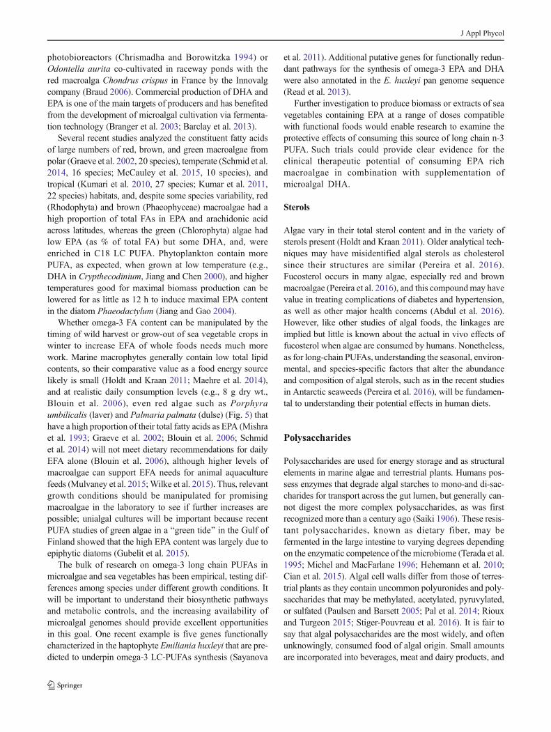
Whether omega-3 FA content can be manipulated by thetiming of wild harvest or grow-out of sea vegetable crops inwinter to increase EFA of whole foods needs much morework. Marine macrophytes generally contain low total lipidcontents, so their comparative value as a food energy sourcelikely is small (Holdt and Kraan 2011; Maehre et al. 2014),and at realistic daily consumption levels (e.g., 8 g dry wt.,Blouin et al. 2006), even red algae such as Porphyraumbilicalis (laver) and Palmaria palmata (dulse) (Fig. 5) thathave a high proportion of their total fatty acids as EPA (Mishraet al. 1993; Graeve et al. 2002; Blouin et al. 2006; Schmidet al. 2014) will not meet dietary recommendations for dailyEFA alone (Blouin et al. 2006), although higher levels ofmacroalgae can support EFA needs for animal aquaculturefeeds (Mulvaney et al. 2015;Wilke et al. 2015). Thus, relevantgrowth conditions should be manipulated for promisingmacroalgae in the laboratory to see if further increases arepossible; unialgal cultures will be important because recentPUFA studies of green algae in a Bgreen tide^ in the Gulf ofFinland showed that the high EPA content was largely due toepiphytic diatoms (Gubelit et al. 2015).
The bulk of research on omega-3 long chain PUFAs inmicroalgae and sea vegetables has been empirical, testing dif-ferences among species under different growth conditions. Itwill be important to understand their biosynthetic pathwaysand metabolic controls, and the increasing availability ofmicroalgal genomes should provide excellent opportunitiesin this goal. One recent example is five genes functionallycharacterized in the haptophyte Emiliania huxleyi that are pre-dicted to underpin omega-3 LC-PUFAs synthesis (Sayanova
et al. 2011). Additional putative genes for functionally redun-dant pathways for the synthesis of omega-3 EPA and DHAwere also annotated in the E. huxleyi pan genome sequence(Read et al. 2013).
Further investigation to produce biomass or extracts of seavegetables containing EPA at a range of doses compatiblewith functional foods would enable research to examine theprotective effects of consuming this source of long chain n-3PUFA. Such trials could provide clear evidence for theclinical therapeutic potential of consuming EPA richmacroalgae in combination with supplementation ofmicroalgal DHA.
Sterols
Algae vary in their total sterol content and in the variety ofsterols present (Holdt and Kraan 2011). Older analytical tech-niques may have misidentified algal sterols as cholesterolsince their structures are similar (Pereira et al. 2016).Fucosterol occurs in many algae, especially red and brownmacroalgae (Pereira et al. 2016), and this compoundmay havevalue in treating complications of diabetes and hypertension,as well as other major health concerns (Abdul et al. 2016).However, like other studies of algal foods, the linkages areimplied but little is known about the actual in vivo effects offucosterol when algae are consumed by humans. Nonetheless,as for long-chain PUFAs, understanding the seasonal, environ-mental, and species-specific factors that alter the abundanceand composition of algal sterols, such as in the recent studiesin Antarctic seaweeds (Pereira et al. 2016), will be fundamen-tal to understanding their potential effects in human diets.
Polysaccharides
Polysaccharides are used for energy storage and as structuralelements in marine algae and terrestrial plants. Humans pos-sess enzymes that degrade algal starches to mono-and di-sac-charides for transport across the gut lumen, but generally can-not digest the more complex polysaccharides, as was firstrecognized more than a century ago (Saiki 1906). These resis-tant polysaccharides, known as dietary fiber, may befermented in the large intestine to varying degrees dependingon the enzymatic competence of the microbiome (Terada et al.1995; Michel and MacFarlane 1996; Hehemann et al. 2010;Cian et al. 2015). Algal cell walls differ from those of terres-trial plants as they contain uncommon polyuronides and poly-saccharides that may be methylated, acetylated, pyruvylated,or sulfated (Paulsen and Barsett 2005; Pal et al. 2014; Riouxand Turgeon 2015; Stiger-Pouvreau et al. 2016). It is fair tosay that algal polysaccharides are the most widely, and oftenunknowingly, consumed food of algal origin. Small amountsare incorporated into beverages, meat and dairy products, and
J Appl Phycol

fillers (Cofrades et al. 2008; Gupta and Abu-Ghannam 2011a,b; Griffin 2015) at levels generally deemed to be beneficialand safe by regulatory agencies (extensively reviewed inMabeau and Fleurence 1993; MacArtain et al. 2007; Watson2008; Holdt and Kraan 2011; Barlow et al. 2015; Fleurenceand Levine 2016).
Edible macroalgae contain unusually high amounts of die-tary fiber, ranging from 23.5 % (Codium reediae) to 64.0 % ofdry weight in Gracilaria spp., values that frequently exceedthose for wheat bran (Ruperez and Saura-Calixto 2001;McDermid et al. 2005; Benjama and Masniyom 2012). Thenomenclature of food-derived fiber is confusing because thereis no consensus on its definition among scientists andregulatory agencies. Dietary fiber, considered a nutrient inthe USA under the Nutrition and Education Act of 1990(Thomas.loc.gov/ H.R. 3562.ENR), comprises Bnondigestiblecarbohydrates and lignin that are intrinsic in intact plants.^Some fraction of this human-inert matter is considered bysome as Functional fiber; that fraction of isolated, non-digested carbohydrates having apparent beneficial physiolog-ical effects beyond nutrition in humans (Institute-of-Medicine2005;Medeiros andWildman 2015). In this case, Total fiber isthe sum of dietary and functional fiber (Institute-of-Medicine2005; Medeiros and Wildman 2015). In contrast, theEuropean Food Safety Authority, following the CODEXAlimentarius Commission definition of dietary fiber (Jones2014), acknowledges that benefits beyond nutrition can occur
but does not formally distinguish functional from dietary fiberbecause no analytical methods exist for this differentiation(EFSA 2010). Regardless of these semantics, non- or partiallyfermented fiber that generates physiological benefits, througheither physical or chemical pathways, meets the definition ofdietary fiber (Jones 2014).
BSoluble fiber^ comprises 52–56 % of total fiber in com-monly used green and red macroalgae and 67–85 % in brownmacroalgae (Lahaye 1991). Much of it can be fermented toshort-chain fatty acids (SCFAs) such as acetate, propionate,and butyrate (see Table 1 in Michel and MacFarlane 1996;Cantarel et al. 2012) which both nourish the epithelia of thelarge intestine and offer other benefits to the host (Terada et al.1995; Michel and MacFarlane 1996). For example, acetateand propionate are transported in the blood to many organswhere they are oxidized for energy or utilized in signaling tohelp regulate aspects of energy homeostasis and immunefunction (reviewed by Nicholson et al. 2012). The fermenta-tion process and SCFA products also nourish and modify themicrobial consortia in the large intestine, thereby exerting pre-biotic effects and influencing digestive outcomes (e.g.,Fernando et al. 2008; O’Sullivan et al. 2010; Cian et al.2015). Investigating the coupling of algal (and other) polysac-charides to the health of intestinal microbiomes and their an-imal and human hosts is an active and needed area of research(Bäckhed et al. 2005; Hehemann et al. 2010; Cantarel et al.2012). These beneficial responses may include reduced risk of
Fig. 5 Sea vegetables used inEuropean cuisine includemarinated kelp (Alaria esculenta)in a cannelloni bean salad (a),laver/nori (Porphyra umbilicalis/Pyropia yezoensis) in chocolatemolasses meringues (b), thetraditional Welsh laver-breadcakes, with dulse (Palmariapalmata) crisps (c), anddulse-cheese scones (d). Theseadditions add texture, protein,vitamins and minerals, and flavor.(Used with permission of PrannieRhatigan from The Irish SeaweedKitchen)
J Appl Phycol
diabetes, hypertension, and cardiac heart disease (Institute-of-Medicine 2005). However, the complexity of interactionsamong functional and dietary fiber and the intestinalmicrobiome challenges efforts to demonstrate the functionalfood and biomedical benefits of algal polysaccharides (deJesus Raposo et al. 2015; Dhargalkar 2015).
The evidence for bioactivity of algal polysaccharides de-rives largely from in vitro experiments using isolated oligo-mers/polymers, with fewer data on testing any whole alga inanimal or human trials. Compositional analysis of Chlorellaand similar microalgae began more than 60 years ago, and animpressive number of biological processes are now reportedto be influenced by ingestion of whole algae or polysaccharideextracts as food or supplements (Pulz and Gross 2004; Plazaet al. 2009; Chacón-Lee and González-Mariño 2010; Lordanet al. 2011; Vo et al. 2011). Microalgal genera (Fig. 2) com-monly considered as beneficial dietary supplements includeChlore l la , Arthrosp ira (sp i ru l ina ) , Dunal ie l la ,Haematococcus, Scenedesmus, Aphanizomenon, Odontella,and Porphyridium, with species ofChlorella being recognizedas particularly rich in polysaccharides (Chacón-Lee andGonzález-Mariño 2010). This putative bioactivity includesanticancer properties, cytokine modulation, anti-inflammatory effects, macrophage activation, and inhibitionof protein tyrosine phosphatase (Hasegawa et al. 1997;Cheng et al. 2004; Kralovec et al. 2005; Sheng et al. 2007;Hsu et al. 2010). Algal polysaccharide extracts can possessstrong immunomodulating effects both in vitro and in vivo(Watanabe and Seto 1989; Pasco and Pugh 2010; Suáreze t a l . 2010 ) . Kwak e t a l . ( 2012 ) obse rved animmunostimulatory effect in 30 Korean volunteers fed5 g day−1 Chlorella vs. placebo in a double-blinded 8-weektrial. Acidic polysaccharide extracts from Chlorellapyrenoidosa have been patented (Chlon A and RespondinTM)as potentially useful anti-tumor and immunostimulating sup-plements (Umezawa and Komiyama 1985; Komiyama et al.1986; Kralovec 2005; Kralovec et al. 2005). Even so, themolecular structures responsible for such observed physio-logical functions are poorly understood because of fragmen-tary and sometimes conflicting information on the chemistryof these large, highly complex cell wall polymers (Řezanakaand Sigler 2007). Research also has focused on strikinglyfew algal species, leaving a broad window of opportunityfor more expansive assessment of potential sources of bio-active compounds (Admassu et al. 2015).
The study of extracted polymer sub-fractions of structuralpolysaccharides provides a useful exploratory tactic forassessing the potential functional benefits of consumingmacroalgal foods, and it establishes a quantitative means todetermine the seasonal or environmental effects on food qual-ity (Stengel et al. 2011; Mak et al. 2013). The predominantalgal polysaccharides are the alginates in brown macroalgae,and the sulfate-esterified polysaccharides of macro- and
microalgae that are widespread in red, brown, and green sea-weeds (Aquino et al. 2005; Popper et al. 2011). The cellularquantities and compositions of these polysaccharides varyamong species and with seasonal and environmental changes(Bourgougnon and Stiger-Pouvreau 2011; Mak et al. 2013).
Alginate
Alginate is the major polysaccharide of brown algae, compris-ing 14–40% of its dry mass (cf. Ramberg et al. 2010), and wasfirst isolated in 1881 as algin from kelp (Laminaria sp.) by E.C. C. Stanford. The direct consumption of brown algae ashuman food is of long standing (Tseng 1981; Druehl 1988;Dharmananda 2002; McHugh 2003). The purported benefi-cial effects of alginate include its ability to absorb toxins,decrease cholesterol uptake, alter the colonic bacterial pro-files, and generate SCFAs (Brownlee et al. 2005). The metalchelating abilities of alginates makes them valuable scaven-gers of toxic elements in the human gut, but this scavengingalso may lead to nutritional deficiencies of essential di- orpolyvalent trace metals (Hollriegl et al. 2004; Brownleeet al. 2005). Most studies have investigated the effects ofpolysaccharide extracts rather than consumption of intact sea-weeds. Although the extent of alginate dissociation from algalcell walls after ingestion is not well studied, there is little or nodigestion of sodium alginate from Ascophyllum nodosum inhumans (Percival and McDowell 1967; Painter 1983; Aarstadet al. 2012). Dietary alginates also provide a sense of satietyand so have been explored as a weight control measure, al-though there remains uncertainty about its efficacy in this role(Yavorska 2012).
Sulfated heteroglycans—ulvans
The abundant, heavily sulfated ulvans are extracted frommembers of the Ulvales. They are the best studied of the greenseaweed polysaccharides, in part because the high productionof Ulva spp. in eutrophic coastal waters has sparked researchfor new uses of these algae (Alves et al. 2013). Ulvans owetheir bioactive properties to their unusual hydrophilicpolyanionic features and structural analogies with animal gly-cosaminoglycan regulators (dermatan sulfate, heparin/heparinsulfates) and L-rhamnose specific lectins in humans. The re-ported bioactivities of ulvan extracts in vitro include antibac-terial, anticoagulant, antioxidant, antiviral, anti-tumor, anti-hyperlipidemic, and immunoregulatory effects (Kaeffer et al.1999; Yu et al. 2003; Mao et al. 2006, 2008; Leiro et al. 2007;Zhang et al. 2008, 2010; Lee et al. 2010; Holdt and Kraan2011; Matloub et al. 2013).
Although the ingestion of green macroalgae by humans israther widespread, the potential health benefits of food sup-plements of native ulvans or their chemically modified deriv-atives, let alone the direct consumption of the whole algae, are
J Appl Phycol

not well understood (Taboada et al. 2010; Wijesekara et al.2011). Fermentation of Ulva and ulvan by human colonicbacteria was slight (16.6 and 8.9 % of organic matter, respec-tively) (Durand et al. 1997), indicating that they would bepoor sources of SCFA production in the colon (Bobin-Dubigeon et al. 1997). However, these results cannot be gen-eralized because only two individuals provided the bacterialinocula, and their prior dietary history relating to algal foodswas unknown. A cautionary note here though is thatUlva canbe rich in free sulfate which is readily converted to sulfideduring fermentation, so consumption of more than 20 g day-1 of the dry, unprocessed seaweed may have adverse (andodiferous) health effects (Durand et al. 1997).
Sulfated galactans—carrageenans
Red algal polysaccharides include the nutritionally importantfloridean starch, and their sulfated galactans are known toinitiate or modulate a large number of biological activities ofsignificance to human health (Prajapati et al. 2014). The moststudied are the sulfated agarocolloids and the carrageenansderived from macroalgae in the orders Gelidiales,Gigartinales, and Gracilariales. Anti-viral activities includethose against herpes simplex, herpes zoster, dengue-2, vaccin-ia, rabies, and vesicular stomatitis virus with patents and somecommercial projects resulting (Richards et al. 1978; Baba etal. 1988; Vedros 1993; Bourgougnon 2003; Eccles et al. 2010;Talarico et al. 2011; Levendosky et al. 2015; Luo et al. 2015).Whether consumption of the relevant red algae or their ex-tracts in foods is protective against viruses does not appearto be known and deserves study. Carrageenan extracts that aredepolymerized to various degrees have potential as tumor in-hibitors and as immunostimulators in cancer therapy.Oligomers from acid hydrolyzed κ-carrageenan injected intomice increased macrophage phagocytosis and stimulated sev-eral immune-related markers while significantly inhibiting thegrowth of sarcoma S180 cells (Yuan et al. 2006).Phosphorylation or further sulfation of these oligomers in-creased the activity of natural killer cells, the cytotoxic lym-phocytes critical to immune system function (Yuan et al.2011). Similarly, transplanted human sarcoma S180 tumorswere inhibited significantly in mice fed fractionated λ-carrageenan extracts of Chondrus ocellatus (200 mg kg−1 dai-ly) (Zhou et al. 2004). Although seaweeds containing carra-geenans act as prebiotics when supplied as supplements inboth poultry and rat diets (Kulshreshtha et al. 2014; Liuet al. 2015), the potential for sulfated galactans from algae tobenefit human health remains to be established.
Carrageenans have the potential to be harmful (Tobacman2001). Carrageenan extracts generate proinflammatory agentsin mice (Hansra et al. 2000), and the resulting public healthconcerns have led to several actions regarding carrageenans infood products (Watson 2008). Carrageenan is prohibited in the
EU for use in infant formulas, and, although it is permitted inthe USA, it must be of high molecular mass (i.e., >100 kDawith <5% of 50 kDa fragments). High doses of lowmolecularmass carrageenan cause ulceration in the guinea pig colon(Watson 2008) and lead to marked increases in the chemokineinterleukin-8 and B-cell CLL/lymphoma 10 activities in thenormal human colonic mucosal epithelial NCM460 cell line(Bhattacharyya et al. 2010). Oral introduction of undegradedλ-κ carrageenan in drinking water of 12-week-old mice alsocaused significantly higher peak glucose levels in the blood,leading to concern that carrageenan-induced insulin resistancemight contribute to human diabetes (Bhattacharyya et al.2012). However, a comprehensive examination of in vivo di-etary κ-carrageenan effects in rats revealed no effects on bloodglucose (Weiner et al. 2007). More recent appraisals of carra-geenans as food additives could find no hazards to humanhealth as they are currently used (McKim 2014; Weiner 2014;Barlow et al. 2015; Weiner et al. 2015). The potential benefitsand negative effects of including algae or their refined productsin the diet require continuing research on a case-by-case basis.
Beta-(1-3)-glucans—laminarans
The main polysaccharides after the alginates in brown algaeinclude β-glucans (laminarans), cellulose, and heteroglycans,the first being an energy reserve while the others are structuralcomponents of the cell wall, fitting the definitions of dietaryfiber (Jones 2014). The concentrations and composition of theβ-glucans vary substantially with season and growth rates(Rioux et al. 2009). The most studied β-glucans are thosefrom cereals and fungi, but these differ significantly in struc-ture from those of algal origin (Rioux et al. 2010). The biolog-ical responses elicited by algal β-glucans depend strongly ondetails of their primary structures (Bohn and BeMiller 1995;Mueller et al. 2000; Williams et al. 2005; Hofer and Pospíšil2011). For example, brown algal M-series laminaran showedsignificant hepatoprotective effects when ingested orally by rats(Neyrinck et al. 2007). The protection was organ specific andappeared to act via the Kupffer cells in the liver, thereby estab-lishing an immunoregulatory function of this orally ingestedfunctional fiber. These and other biological effects of β-glucans have been reviewed (Novak and Vetvica 2008;Ramberg et al. 2010; Lehtovaara and Gu 2011; Kadam et al.2015), and certain cautions have been expressed about the func-tional effects of soluble and particulate forms of these com-pounds (Young andCastranova 2005; Hofer and Pospíšil 2011).
Sulfated fucose-containing polysaccharides—fucoidans
The fucoidans are a subset of marine fucose-containing poly-saccharides (FCPs) found in brown algae (Painter 1983) thatare now attracting widespread interest (Shanmugam andMody 2000; Berteau and Mulloy 2003; Kusaykin et al.
J Appl Phycol

2008; Li et al. 2008; Pomin andMourão 2008; Courtois 2009;Pomin 2009, 2012; Fitton 2011; Jiao et al. 2011; Kim and Li2011; Kim and Wijesekara 2011; Wijesinghe et al. 2011;Wijesinghe and Jeon 2012). Double-blind clinical trials withfucoidan extracts show anti-aging effects on skin and otherbenefits in cosmetic applications (Fitton et al. 2015). A com-mon source of FCPs used in experimental studies is Fucusvesiculosus, but fucoidans also are found in edible speciessuch as Cladosiphon okamuranus, Saccharina japonica (asLaminaria japonica), and Undaria pinnatifida (Fitton 2011).The highly sulfated nature and molecular weights of FCPsappear to be responsible for many demonstrated biologicalactivities in vitro (Croci et al. 2011; Ustyuzhanina et al.2014). The FCP structures are species-dependent and can bemodified by environmental variables and the developmentalstatus of the seaweed fronds, all of which can affect theirbioactivities (Honya et al. 1999; Zvyagintseva et al. 2003;Rioux et al. 2009; Pielesz and Biniaś 2010; Skriptsova et al.2010; Stengel et al. 2011; Anastyuk et al. 2012; Mak et al.2013). More recently, in vitro studies have provided insightinto some structure-function relationships of FCPs (Cumashiet al. 2007; Ushakova et al. 2009; Ustyuzhanina et al. 2013,2014).
It can be concluded that knowledge of the beneficial effectsof algae and their extracts as food additives for humans lagsfar behind that on which diets have been formulated for com-mercially important species in aquaculture and agriculture.The number of species exhibiting benefits is wide rangingfrom invertebrates (nematodes, shrimp, abalone) and finfish(sea bream to salmon) to farm animals including poultry andmammals (both ruminants and monogastric species) (reviews:Craigie 2010; O’Sullivan et al. 2010; Rajauria 2015; Heuzéet al. 2016; Makkar et al. 2016). Algal-based productsTasco™ from Ascophyllum nodosum and Ocean Feed™ (ablend of brown, green and red macroalgae) are commerciallymarketed as feed additives to improve performance, stimulateimmune reactions, mitigate sea lice damage in salmonids, andother benefits. Notable is the Alternative Feeds Initiative todevelop alternative dietary ingredients (NOAA 2011). In ad-dition to conventional methods of measuring animal perfor-mance, molecular techniques have been applied to buttressclaims of efficacy (cf. Kulshreshtha et al. 2014; Liu et al.2015). Bearing in mind ethical considerations, similar ap-proaches may be adapted to facilitate the assessment of thebenefits of macroalgal ingestion by humans.
Vitamins
Vitamins are essential organic micronutrients, which an organ-ism cannot synthesize directly in sufficient quantities and soinstead must obtain from the diet. Well-known humanvitamin-deficiency diseases include beriberi (deficiency in
thiamine, vitamin B1), pellagra (niacin), pernicious anemia(cobalamin, vitamin B12), and scurvy (ascorbic acid, vitaminC) (Stabler and Allen 2004; Martin et al. 2011). These com-pounds serve as precursors for essential enzyme cofactors andare needed for essential metabolic functions (Fig. 4). Sinceanimals have lost the capacity to synthesize these cofactors,they must obtain them from external sources. Algal foods arerich in vitamins. Several sea vegetables—laver (Porphyraumbilicalis), sea spaghetti (Himanthalia elongata), andGracilaria changii—contain levels of vitamin C comparableto common vegetables such as tomatoes and lettuce (Norziahand Ching 2000; Ferraces-Casais et al. 2012), while the vita-min C content described for the brown seaweed Eiseniaarborea (34.4 mg (100 g)−1 dry wt) approaches those reportedfor mandarin oranges (Hernandez-Carmona et al. 2009). Thevitamin content of individual algal species discussed in thissection, including details of sample origin and handling, iscompiled in Online Resource 4.
Sea vegetables also are a good source of B-group vitamins(particularly B1, B12), as well as the lipophilic vitamin A(derived from the carotenoid β-carotene) and vitamin E (to-copherol). Kelp (Macrocystis pyrifera) can contain levels ofα-tocopherol (the most biologically active form of vitamin E)at par with plant oils rich in this vitamin, such as palm, sun-flower seed, and soybean oils (Ortiz et al. 2009; Skrovankova2011). Moreover, values of β-carotene (pro-vitamin A) foundin the seaweeds Codium fragile and Gracilaria chilensis canexceed those measured in carrots (Ortiz et al. 2009). The vi-tamin composition of microalgae can be equally remarkable.Fabregas and Herrero (1990) showed that Tetraselmis suecica,Isochrysis galbana, Dunaliella tertiolecta, and Chlorellastigmatophora were particularly rich in lipid-soluble (A andE) and B-group vitamins [including vitamins B1, B2 (ribofla-vin), B6 (pyridoxal), and B12]. The concentrations of severalvitamins, including E, B1, and β-carotene, exceeded those inconventional foods considered to be rich sources of thesecompounds (Fabregas and Herrero 1990). It is clear then thatalgal foods can be an excellent source for a wide range of theseessential micronutrients.
Even so, variability between samples can make direct com-parisons among studies difficult (e.g., Chan et al. 1997;McDermid and Stuercke 2003; Hernandez-Carmona et al.2009). Part of the variability may lie in the sample processingmethods (Skrovankova 2011) as observed for other phyto-chemicals (Ling et al. 2015); for example, analysis of freeze-dried and oven dried samples of Sargassum hemiphyllumyielded substantially different vitamin C contents (Chanet al. 1997). But differences also can be due to environmentaland seasonal factors. For instance, there are notable variationsin the levels of β-carotene and vitamin C between samples ofUlva fasciata collected from different sites (McDermid andStuercke 2003) (Online Resource 4). Monthly quantitation ofvitamins C, B2, B1, and A concentrations in Eisenia arborea
J Appl Phycol

over a 12-month period revealed levels were lowest in thesummer months (June, July, August) and reached the highestconcentrations in April/September (for vitamins C, B2, B1)and January (for provitamin A) (Hernandez-Carmona et al.2009). The proximate cause for these patterns is unknown,as is the effect of growth conditions on the content and com-position of vitamins in algal foods, so this is an important topicfor future research.
Algal foods offer one of the few vegetarian alternatives forcobalamin (vitamin B12) in the diet. Cobalamin is not requiredor synthesized by higher plants (Croft et al. 2005) so fruits andvegetables are poor sources of vitamin B12, which explainswhy vitamin B12-deficiency is common among people follow-ing strict vegetarian or vegan diets (Haddad et al. 1999;Waldmann et al. 2004; Allen 2008). Over half of microalgalspecies surveyed have a metabolic requirement for B12, andcontain large amounts (Online Resource 4), but they cannotsynthesize it (Croft et al. 2005; Helliwell et al. 2011).Cobalamin is synthesized only by prokaryotes (Warren et al.2002), and it has been shown that B12-synthesizing bacteriaare closely associated with or reside on eukaryotic algal sur-faces (Croft et al. 2005; Wagner-Döbler et al. 2010). Pyropiayezoensis (nori) contains up to ∼0.06 mg vitamin B12
(100 g)−1 algal dry wt, comparable to that found in beef liver(Watanabe et al. 1999b; Takenaka et al. 2001). Lower levelsare found in other sea vegetables such as kelps (includingwakame) and hijiki, although reported concentrations varyamong studies, possibly reflecting differences among strains,growing conditions, or harvesting periods (Watanabe et al.1999a; Miyamoto et al. 2009). Given that the ultimate sourceof vitamin B12 is bacteria, changes in the character and mag-nitude of the epiphytic prokaryotic communities related toregion or algal physiological state may contribute to variationin vitamin content; these factors currently are poorlyquantified.
There is uncertainty about whether the magnitude of vita-min concentration in algal foods reflects their nutritional val-ue. Dagnelie et al. (1991) investigated how sea vegetablesaffected the hematological status of B12-deficient childrenand concluded that the algal-derived vitamin B12 was not bio-accessible to humans. However, their very small treatmentgroup (n = 5) may have been insufficient to draw firm conclu-sions. Takenaka et al. (2001) showed that feeding nori to vi-tamin B12-deficient rats yielded a 1.9-fold increase in hepaticlevels of total B12 compared to those without nori supplemen-tation. Similarly, increased consumption of Chlorella or noriby vegan participants prevented B12 deficiency (Rauma et al.1995). However, there are few data on which to base quanti-tative estimates of the portion of algal vitamins that areabsorbed during digestion.
One approach to assessing the availability of vitamins is todistinguish among their different chemical forms. The uptakeof cobalamin-based compounds, referred to more broadly as
corrinoids, is largely governed by the gastrointestinalabsorption system rather than their chemical liberation viadigestive chemical processes (Russell-Jones et al. 1999).Pseudovitamin B12 (or pseudocobalamin) differs from cobal-amin in its lower axial ligand (Stupperich and Krautler 1988),and this affects affinity of the mammalian B12-binding proteinintrinsic factor (IF) for the compound, thereby limiting itsabsorption in the intestine (Stupperich and Nexo 1991). Thisdifference has human health implications because commer-cially produced vitamin B12 supplements derived from thecyanobacterium Arthrospira sp. (spirulina) consist largely ofpseudovitamin B12 (Watanabe et al. 1999b; Watanabe 2007a),reducing their nutritional value. In contrast, Bgreen^ (Ulva[formerly Enteromorpha sp.]) and Bpurple^ (Pyropia [former-ly Porphyra] sp.) laver contain substantial amounts of biolog-ically available B12 (Watanabe et al. 1999b), and indeed, ratsfed purple laver improved their B12 status (Watanabe et al.1999b). A recent study has established that the vast majorityof cyanobacteria synthesize pseudocobalamin, whereas eu-karyotic algae that are dependent on B12 for growth are likeanimals in that they require cobalamin (Helliwell et al. 2016).Thus, sea vegetables are likely to be a more reliable source ofthe appropriate form of this vitamin, although again this willbe determined by the prokaryotic community associated withthe algae.
These findings highlight the need for rigorous care in theanalytical determinations of the vitamin content of algal foods.Bioassays using B12-dependent bacteria such as Lactobacillusdelbruekii ssp. lactis (ATCC7830) are inadequate because,unlike humans, these bacteria do not discriminate betweenvitamin B12 and pseudovitamin B12. An alternative radioiso-tope dilution assay (RIDA) is likely to better reflect the func-tional B12 content (Watanabe 2007a). Distinguishing amongbioavailable and non-bioavailable vitamin forms will be cru-cial (Watanabe 2007b). Complicating these analyses further isevidence that commercial processing methods can alter thevitamin chemistry sufficiently to affect uptake. For example,Yamada et al. (1999) showed that air-drying Pyropia tenera(asakusa-nori) produced B12 analogs that are biologically in-active. Drying by lyophilization might have better nutritionaloutcomes (Takenaka et al. 2001), although this has yet to berigorously demonstrated. Other factors of particular impor-tance to preserving vitamin content include washing methods,storage temperature, light, and moisture content (OnlineResource 1, Brown 1995; Jimenez-Escrig et al. 2001; Lage-Yusty et al. 2014). There is a strong need for more detailedinvestigations into how the nutritional quality of sea vegeta-bles is affected by processing methods suited for commercial-scale production.
The bioavailability of other algal-derived vitamins is alsounderexplored. Vitamin E encompasses eight different forms(tocopherols and tocotrienols) that differ in their biologicalactivity (α- and γ-tocopherols are the most active).
J Appl Phycol
Althoughmuch less is known about their relative bioavailabil-ity compared to the vitamin B12 analogs, it is clear that theirrelative contributions affect the nutritional quality of foodstuffs(Ortiz et al. 2009). An additional concern with fat-soluble vi-tamins is that theymust be consumed with lipid-rich foodstuffsto ensure efficient intestinal absorption (Skrovankova 2011).Although this co-dependence is understood, there currently arefew data on this dependence for edible-algal species.
Most studies on algae and vitamins often focus either onanalysis of vitamin concentrations in algae (e.g., Ortiz et al.2006, 2009; Hernandez-Carmona et al. 2009; Matanjun et al.2009; Ferraces-Casais et al. 2012) or testing the value of analgal product as a functional food (e.g., Dagnelie et al. 1991;Rauma et al. 1995; Takenaka et al. 2001), but not both. Ideally,studies combining these two approaches should be adopted togain meaningful insights on the true quality of algal foods asvitamin sources (Takenaka et al. 2001).
Finally, there are the ecological challenges to gaining abroad picture of algal foods as a nutritional source of vitamins.Vitamin production and metabolism can vary considerablyacross diverse algal lineages (Croft et al. 2006; Helliwellet al. 2011, 2013). One approach that may help reveal thiscomplexity would be a high-throughput screening of promis-ing algal food candidates with next-generation sequencingtechniques coupled with bioinformatics to search forvitamin-biosynthesis pathways. Nevertheless, there will becontinued the need for careful analytical characterizationsand bioavailability testing because the up- or down-regulation of gene expression almost certainly will be envi-ronmentally regulated.
Antioxidants
It is not surprising that there is a very broad literature onmarine algae as sources of antioxidant compounds for humandiets. Photosynthetic energy acquisition and transformationsnecessarily involve continuing redox disequilibria, with theproduction of reactive species that can decrease lifespan andevolutionary fitness. Microalgae and macroalgae, like otherlife forms, contain antioxidant organic compounds and en-zymes that limit this oxidative damage, which results primarilyfrom reduced states of oxygen—the Breactive oxygenspecies^—including the superoxide radical anion (O2
−·;O2 + 1e−), hydrogen peroxide (H2O2; O2 + 2e−), the hydroxylfree radical (HO·; O2 + 3e−), and singlet oxygen (1O2)(Halliwell and Gutteridge 2007).Whereas the antioxidant ben-efits of several terrestrial plant foods are established, much lessis known about whether algal foods provide similar benefits.
The reactive oxygen metabolism in marine algae is diverseand complex, given the wide range of antioxidant compounds(Cornish and Garbary 2010), but an extension to any benefi-cial response from human consumption of these substrates is
far less certain. Antioxidant activity can have two forms: theactivity of antioxidant enzymes or the production ofmolecules that serve as sacrificial scavengers of reactiveoxygen species. There also are two broad categories of anti-oxidant activity: limiting reactive oxygen species within thedigestive tract, thereby decreasing oxidative stress on the gutmicrobiome and epithelial cells, or transport into the blood fordistribution throughout the body. Evidence for direct transportis very limited, as there seems to have been no systematicstudy of digestive uptake of these compounds. In one study,Okada et al. (2009) examined the bioaccessibility ofastaxanthin extracted from the green alga Haematococcus(Fig. 2) as judged from the concentration in blood serum, asa function of the timing of the ingestion of astaxanthin relativeto a meal, and whether the subjects were smokers or non-smokers. Astaxanthin increased more in serum when the dosewas taken 10 min after a meal rather than 2 h before, evidenceof complex factors affecting its bioaccessibility. The ingestion(and topical application) of polyphenols of brown algaeinhibited UVB radiation-induced skin carcinogenesis in mice(Hwang et al. 2006), and while this bioactivity remains to bedetermined for humans, it provides evidence that algal foodshave significant functional food potential.
The foremost enzymes that restrict oxidative damage inalgae and terrestrial foods include the superoxide dismutasesthat remove superoxide radical anions, and catalases and per-oxidases, that convert hydrogen peroxide to water. Superoxidedismutases in cyanobacteria have Ni, or mixtures of Fe, Mn,and Ni, as the metal, whereas eukaryotic algae haveMn or Fe,or some combination of Fe, Mn, and Cu + Zn (Wolfe-Simonet al. 2005). Catalase has an Fe-containing heme cofactorwhile peroxidases use a reductant to convert hydrogen perox-ide to water. Of these enzyme cofactors, Cu and Zn, and par-ticularly Fe are used in numerous human metabolic pathways.Since the ingested antioxidant enzymes are digested in theintestine, the only effect the enzymes can have in the animalis through uptake of the metal cofactors across the intestinalepithelium. The possible effects on the intestinal microbiomeof any undigested enzyme, or of the released metal cofactors,have not been investigated.
There is a stronger linkage between selenium in food andantioxidant capacity in metazoans such as mammals. Seleniumis an essential metal in metazoans and some algae for the pro-duction of Se-requiring glutathione peroxidase, used to metab-olize hydrogen peroxide and lipid hydroperoxides (Halliwelland Gutteridge 2007; Perez et al. 2007; Gobler et al. 2011).Analyses of the elemental contents of microalgae (Quigg et al.2011) and macroalgae (Tuzen et al. 2009; Pereira 2011) rarelyinclude Se, even though it is present in both (Fournier et al.2005). Se readily bioaccumulates in algae (Cases et al. 2001;Fournier et al. 2005), and Se-deficiency in rats can be alleviat-ed by oral supplementation with Se-rich Arthrospira (spiru-lina), as indicated by increased activity of (Se-containing)
J Appl Phycol

glutathione peroxidase in the kidneys and liver (Cases et al.2001). However, increases in this enzyme activity were greaterin rats supplied selenite or selenomethionine (more reactivespecies) than with the same dosage of Se-rich cyanobacterium,likely due to lower bioavailability of the cyanobacterial Se.The factors regulating Se content of algal foods and its avail-ability are prime research topics for the future.
Under normal metabolic conditions, the production of hy-droxyl radicals and singlet oxygen cause almost immediatedamage, essentially reacting with the first oxidizable moleculethat they encounter. In these cases, Bsacrificial^ scavengers (ofHO·) and quenchers (of 1O2) often are the only recourse forlimiting damage once the free radicals are produced (Smirnoffand Cumber 1989; Telfer et al. 1994a, b; Sunda et al. 2002;Ledford and Niyogi 2005; Halliwell and Gutteridge 2007;Ledford et al. 2007). Algae contain a wide range of moleculescapable of free radical scavenging activity in vitro and in vivo.These include the water-soluble ascorbate (vitamin C) and cer-tain compatible solutes (osmoprotectants), and the lipid-solubleα-tocopherol (vitamin E) and carotenoids such as astaxanthin(Halliwell and Gutteridge 2007). Mycosporine-like aminoacids, mainly considered as UV screening compounds, are alsoantioxidants (Oren and Gunde-Cimerman 2007) as are a rangeof other solutes that act as scavengers and quenchers of reactiveoxygen species in algae (Cornish and Garbary 2010). HO·
scavengers include glycerol (Smirnoff and Cumber 1989),mannitol (Smirnoff and Cumber 1989; Shen et al. 1997;Larson et al. 2002), L-proline (Smirnoff and Cumber 1989),dimethylsulfoniopropionate (Sunda et al. 2002), andfloridoside and isofloridoside (Li et al. 2010), although glycinebetaine (or betaine: trimethylglycine) does not have this prop-erty (Smirnoff and Cumber 1989; Shen et al. 1997). Given thatalgal osmoprotectants are necessarily present in high concen-trations (≥ 0.1 mol L−1) in metabolically diverse compartments(cytosol, plastid stroma, and mitochondrial matrix), there ispotential for them to have functional food roles. However,preliminary experiments showed that none of these compoundsinteract with O2
−· (Smirnoff and Cumber 1989), unlike β-carotene and other carotenoids such as fucoxanthin that quench1O2 as well as scavenging HO· and O2
−· (Halliwell andGutteridge 2007; Sachindra et al. 2007). Other algal compo-nents that scavenge free radicals are phenolic compounds(Ragan and Globitza 1986) including halophenols (Li et al.2011) and phlorotannins (Shibata et al. 2007) and, as notedabove, alginate (Zhao et al. 2012; Zhou et al. 2012) and sulfat-ed polysaccharides (Barahona et al. 2012).
Most studies of the bioavailability of algal antioxidantproducts remain at the entry level with respect to human ef-fects: in vitro testing of extract bioactivity on cell lines. Nwosaet al. (2011) confirmed and extended previous work showingthe antioxidant activities of polyphenolic extracts from fourspecies of edible marine algae in inhibiting Caco-2 colon can-cer cell proliferation and α-glucosidase activity (see below):
the green alga,Ulva lactuca, the brown algae Alaria esculentaand Ascophyllum nodosum, and the red alga Palmariapalmata. Ulva lactuca had a low yield of polyphenols relativeto the other algae, but the brown and red algal polyphenolicextracts performed as well as antioxidants. However, Nwosaet al. (2011) illustrated that the method of preparing the ex-tracts from marine algae can significantly alter their antioxi-dant efficacy (see also Ling et al. 2015), highlighting the needfor caution in comparisons of antioxidant performance amongstudies. With this possible caveat, most work on antioxidantactivity of algal phenols has involved red algae; somebromophenols from the marine red alga Rhodomelaconfervoides have greater in vitro antioxidant activity thanascorbate (Li et al. 2011). Olsen et al. (2013) showed thatbromophenols extracted from the red alga Vertebrata lanosasignificantly inhibited oxidant effects and lipid peroxidation incultures of human fetal lung (MTC-5) and human hepatocel-lular liver carcinoma (HepG2). In this case, it was shown thatbromophenol can enter cells, and thus potentially can movefrom the gut lumen into the blood stream. Overall, there is astrong need for more work on the in vivo effects of the anti-oxidant properties of phenols and other algal food constituentsin mammals, and humans in particular.
Instead of serving to facilitate the control of reactive oxygenspecies, some algal components can inhibit their production,but most studies do not adequately distinguish between thedecreased production and increased removal of oxidants. Forexample, dietary ingestion of phycocyanin, taken up from thegut as the chromophore component phycocyanobilin, and relat-ed bile pigment metabolites inhibits the generation by NADPHoxidase of O2
−, which has a key role in numerous diseasesyndromes, e.g., antigen expression, angioplasty, cancers, gly-cemia and lipidemia, hypertension, immunostimulation, andage-related maculopathy (reviewed by McCarty 2007). Thisindustry-sponsored but balanced and authoritative reviewshows that Spirulina spp. (most now transferred toArthrospira) are a prominent cyanobacterial source of phyco-cyanobilin, a dietary supplement worthy of in-depth study.
A class of compounds attracting increasing attention are thephlorotannins found in brown algae, which have extraordinarythough inconsistent antioxidant properties (see in Wang et al.2014), in part due to the methods of extraction (Nwosa et al.2011). The vast bulk of this work was done in vitro, much of itstudying the effects of phlorotannin on carbohydrate-hydrolyzing enzymes. Nwosa et al. (2011) found that extractsof Ascophyllum and Alaria inhibited Caco-2 colon cancer cellproliferation, α-amylase activity and, to a lesser extent, α-glucosidase activity, with mass spectrometric evidence indi-cating that the active principal(s) were phlorotannins.Kawamura-Konishi et al. (2012) also found that phlorotanninextracts of four species of Sargassum significantly inhibitedthe salivary enzyme α-amylase in vitro, and that a novelphlorotannin from Sargassum patens inhibited rat pancreatic
J Appl Phycol

α-glucosidase action on amylopectin. Iwai (2008) showedthat oral administration of extracts inhibited lipid peroxidationin the plasma, red blood cells, liver, and kidney of KK-Ay
mice, indicating that the antioxidant activity of phlorotanninshad beneficial properties for reducing diabetic oxidativestress. Important recent work (Corona et al. 2016) investigatedthe effect of food grade phlorotannins from Ascophyllumnodosum in trials on human subjects. The work showed thein vitro gastrointestinal modification of phlorotannins, the oc-currence in plasma and urine of metabolites of phlorotannins,and a significant increase in cytokine IL-8. To conclude con-sideration of phlorotannins, while in vitro studies onphlorotannins are valuable, more work along the lines of thatof Iwai (2008) and Corona et al. (2016) is needed to under-stand the uptake and systemic properties of phlorotannins, andto determine whether the in vitro effects occur in vivo andrelate to their antioxidant properties (Bohn et al. 2015).
There remain substantial knowledge gaps about the effica-cy of antioxidant properties of macroalgal and microalgalfoods at all levels, from characterization among speciesthrough effects on gut microbiota and transport across thegut lumen to their impacts on human physiology. This willbe a valuable area of emerging research over the next decade.
Inorganic elements
Processed seaweeds are widely used as mineral and metalnutritional supplements (e.g., Kay 1991), but the efficacy ofthese supplements is poorly quantified. Most studies sufferfrom serious experimental limitations, including short dura-tion of the study, small sample size, and inadequate documen-tation of active ingredients. There is a comparatively smallliterature describing mineral contents of macroalgal andmicroalgal foods (Cabrita et al. 2016), and very little informa-tion about seasonal variations for naturally harvested seavegetables.
The best evidence of the human nutritional benefits of seavegetable inorganic elements is for iodine and iron, which canbe highly enriched in marine macroalgae. Nutritional general-ization about algal mineral contents is difficult because ofsometimes large seasonal, geographic, and taxonomic varia-tions in mineral contents of marine algae (e.g., Jensen 1993).For example, Indonesian green, brown, and red algae containhigh levels of potassium, calcium, and sodium, but significant-ly lower levels of iron and zinc than reported for JapanesePyropia (as Porphyra) yezoensis, Ulva (Enteromorpha)intestinalis, and Sargassum (Hijikia) fusiformis (Takeshiet al. 2005). These findings may indicate that macroalgal har-vests fromwarm equatorial areas have lowermineral nutrition-al value than higher latitude regions (e.g., Cabrita et al. 2016);however, there are remarkably few data on which to assess thevalidity of such generalizations.
Iodine
There is a long history linking seaweed consumption byhumans and the reduced incidence of goiter and other thyroiddisorders. Iodine deficiency causes hypothyroidism while ex-cess iodine uptake can induce either hyper- or hypothyroidism(Miyai et al. 2008). Seaweeds are a good nutritional source foriodine, particularly in regions where other foods are deficient,but the iodine content of commercially available sea vegeta-bles varies dramatically among species, the methods of prep-aration (many iodine compounds are water soluble), and theduration of storage (iodine may vaporize under humid condi-tions) (Teas et al. 2004a). Many macroalgae are washed anddried for storage. These processing steps did not significantlyreduce iodine content in three common species (Alariaesculenta, Palmaria palmata, andUlva intestinalis), but rehy-dration followed by boiling in water lowered iodine contentby 14–75 % (Nitschke and Stengel 2016).
Some kelps (Laminaria spp., Saccharina spp.) have highlevels of iodine, and salts that include kelp powder are avail-able commercially as a source of this vital nutrient. Not allbrown algae accumulate high levels of iodine; for example,the kelps Undaria (wakame) and Alaria (BAtlantic wakame^)have lower iodine levels that are comparable to Palmariapalmata (dulse, a red sea vegetable) (MacArtain et al. 2007;Rhatigan 2009; Holdt and Kraan 2011; Schiener et al. 2015)(Fig. 6). In contrast, high levels of iodine in other brownmacroalgae (e.g., Laminaria; Saccharina; Teas et al. 2004b;Miyai et al. 2008) have led to a strong concern that overcon-sumption of these particular sea vegetables can be unhealthy.Michikawa et al. (2012) reported that consumption of unde-fined seaweed more than 2 days per week appeared to corre-late with increased risk of thyroid cancer in Japanese postmen-opausal women, although Wang et al. (2016) did not find thisrelationship to be significant. Nevertheless, there is clear evi-dence that algal food consumption leads to elevated iodinelevels in humans. Miyai et al. (2008) measured serum levelsof thyroid hormones in conjunction with well-defined inges-tion rates of kombu (Saccharina japonica) over short (7–10 days) and longer term (∼90 days) exposure. Urinary excre-tion of iodine increased significantly over the short term withincreasing intake (15–30 g day−1 of kombu—a normal con-sumption for some Japanese) and suppressed thyroid functionfor at least 3 months. Thyroid hormones returned to normallevels when seaweed intake ceased. In this case, the absorbediodine (20–50 mg) exceeded the recommended upper dailyintake of iodine (0.2 mg day−1 by more than an order of mag-nitude; WHO 1989; Dawczynski et al. 2007; Miyai et al.2008). Food preparation (e.g., cooking, pickling) can reducethe iodine intake, but, even then, high-iodine sea vegetables,which account for only a portion of those consumed, shouldbe restricted in the diet (Teas et al. 2004a). Research is neededon how food preparation alters the bioavailability of iodine in
J Appl Phycol

different sea vegetables. Indeed, Lightowler and Davies(2002) found that established food tables (UK Departmentof Health, 1991) gave poor estimates of dietary iodine intakeand recommended developing more reliable data on iodine infoods, including the variation within food groups.
Iron
Macroalgae are a potentially rich source of iron for humandiets. Garcia-Casal et al. (2007, 2009) measured seasonal dif-ferences in the iron content of four seaweed species commonto Venezuelan waters (Ulva spp., Sargassum spp., Porphyraspp., and Gracilariopsis spp.). The Gracilariopsis spp. andSargassum spp. had substantially higher iron content withPorphyra spp. having the lowest, and there was a distinctseasonal cycle whereby iron content was highest in springand summer and lowest in fall and winter. Sargassum, Ulva,and Porphyra spp. have high iron content, and 15 g day−1 ofSargassum provided substantially higher amounts than dailyrecommended intakes (≤18 mg Fe day−1, Institute-of-
Medicine 2001). Cabrita et al. (2016) found that Fe (and othermetal) contents varied, in some cases substantially, amongmacroalgal species collected at the same sites and time, pre-sumably linked to differences in metabolic requirements.Recognizing that iron consumption in macroalgae likely doesnot equate with uptake, the researchers quantified iron incor-poration into the blood of 93 volunteers fed diets of radioac-tive (gamma-emitting) 59Fe-labeled Ulva spp., Sargassumspp., and Porphyra spp. (Garcia-Casal et al. 2007, 2009).Uptake of 59Fe was dose-dependent on sea vegetable addedto maize or wheat-based meals (Garcia-Casal et al. 2007), butgenerally was greater than the total iron content of the differ-ent macroalgal species. Thus, in addition to enhancing totaldietary iron content, the seaweeds appeared to act synergisti-cally to facilitate iron uptake from wheat and maize, possiblyrelated to high levels of ascorbic acid (vitamin C) that convertsiron to the more readily absorbed ferrous form.
The Fe content of wild algae varies seasonally and geo-graphically depending on the metal content of coastal waters,in addition to being species-specific (Garcia-Casal et al. 2007;
Fig. 6 Examples of sea vegetable use in Japanese cuisine: a BOnigiri^ isa Japanese rice ball usually wrapped by nori (Pyropia yezoensis) withseveral other ingredients: from right to left, rice wrapped in nori, withJapanese apricot (umeboshi) and preserved kombu (tsukudani,Saccharina japonica and other species of the genus Saccharina),wrapped with shredded kombu (torero-kombu), and wrapped in nori; bTempura soba with wakame (Undaria pinnatifida). Wakame is used in
Japan, noodles, soups, salads, pickles, and more. c Traditional Japanesedishes with sea vegetables: preserved kombu (tsukudani) on rice, misosoup with wakame, sliced kombu with vegetables, and sliced raw fishes(sashimi) with wakame and cucumber. d BKobu-maki^ is simmered food,often salmon or herring, wrapped in kombu, which is usually prepared forthe NewYear’s holidays. Kombu is used in several dishes and soup stock.(Courtesy of Kazuko Sato and Yoichi Sato)
J Appl Phycol

Cabrita et al. 2016). Wild harvesting must then be optimizedfor each locale known to produce algae for optimal concen-trations of inorganic nutrients. Similar constraints may applyto aquaculture crops unless they are fertilized, but there arevery few data currently available on sea vegetable nutritionalmetal content relative to species, region, or season.
Phytochemicals
The main non-acyl glyceride compounds in lipid membranesattracting nutritional and commercial interest are the caroten-oids, especially because of their dietary importance for vision(Ben-Amotz and Levy 1996). Carotenoids, among the mostwidespread pigments in prokaryotic and eukaryotic photosyn-thetic organisms (Britton et al. 2004), function as light energyharvesters, as photoprotectants, and as antioxidants (Halliwelland Gutteridge 2007, and see below). One specific keto-carot-enoid, siphonaxanthin, but not fucoxanthin, induced apoptosisin human leukemia cell lines, suggesting its potential as achemopreventative or chemotherapeutic agent (Ganesanet al. 2011), although its efficacy remains to be determinedin vivo. A subset of the carotenoids, the β-carotenes, are pre-cursors for vitamin A (Minguez-Mosquera et al. 2008), andnatural β-carotene may act as a lipophilic antioxidant in vivo,providing some protection in children exposed to radiationfrom the Chernobyl accident (Ben-Amotz et al. 1998).
Various strains of the green microalga Dunaliella salinacan accumulate about 8 % of their dry weight as β-carotene,which has been marketed as a functional food (Ben-Amotzand Levy 1996; Borowitzka 2013b). The US FDA recently(2011) had no questions about a filing for the powdered formof the microalga Dunaliella (bardawil) salina as GRAS foruse as an ingredient in food products (Agency ResponseLetter GRAS Notice No. GRN 000351, Borowitzka 2013b).Another main source of carotenoids is the green algaHaematococcus pluvialis, for which cultures have developedindustrially in several countries (Cysewski and Lorenz 2004).Carotenoid supplements, including β-carotene, are effectivein improving the carotenoid supply in breast milk at earlylactation (Nagayama et al. 2014). However, the optimal levelsof β-carotene remain controversial. Undefined β-carotenesupplements increased lung cancer risk in smokers or peopleexposed to asbestos (Druesne-Pecollo et al. 2010), while ear-lier work showed no reduction in the incidence of lung canceramong male smokers after 5 to 8 years of dietary supplemen-tation with alpha-tocopherol or β-carotene (Albanes et al.1995).
Phenolic compounds such as flavonoids and phenolic acidsserve as antioxidants. Brown macroalgae (Alaria esculenta,Ascophyllum nodosum, F. vesiculosus, Saccharina latissima)had 2–15 times the total phenolics (as mg gallic acid equiva-lents g−1 dry matter) as red species (Chondrus crispus,
Meristotheca papulosa, Palmaria palmata, Sarcodiothecagaudichaudii) (Tibbetts et al. 2016). Total phenolic contentwas inversely related (r = −0.81) to in vitro protein digestibil-ity. Phenolic compounds are challenging to extract and char-acterize, but improvements in analytical methodology are ex-pected to facilitate more detailed investigation of these anti-oxidants in algae. Drying Kappaphycus alvarezii in sunlightsignificantly reduces total phenolic, flavonoid, anthocyanin,and carotenoid content compared with samples dried in ovensor in the shade (Ling et al. 2015).
Safety
Consumption of any food is not without risk, so the promotionof algal consumption must also consider potential harm toconsumers. Possible risks associatedwith algae include excessintake of toxic metals, allergenicity, cyanotoxins, and certainlysecondary metabolites (e.g., prostaglandins, kainoids) as wellas contamination with pathogens, radioisotopes, and toxicsynthetic compounds.
Metal toxicity
Under normal, pristine conditions, metal uptake improves thenutritional quality of algal foods, but excessive uptake canlead to toxicity. Moreover, a substantial amount of metal as-sociated with macroalgae can exist as colloidal-sized particlessorbed to algal surfaces (Gadd 2009; Turner et al. 2009), sosurface chemistry and algal physical structures can affect met-al content in addition to metabolic processes. The efficacy ofmacroalgae as metal collectors is why they are effective sen-tinel organisms for detecting anthropogenic signatures incoastal waters (Melhuus et al. 1978; Leal et al. 1997; Brownet al. 1998; Mardsen and De Wreede 2000); a feature in con-flict with their use as a food source.
Despite these caveats, some general patterns appear in theliterature. Metal content tends to differ among phylogeneticgroups, with brown algae typically possessing higher levels ofmost metals in comparison with red or green algal species(Foster 1976; Munda and Hudnik 1991; Stengel and Dring2000; Al-Masri et al. 2003; Benkdad et al. 2011). But thereare exceptions. Phaneuf et al. (1999) observed that the greenalga Ulva lactuca and Ulva (as Enteromorpha) spp. in the St.Lawrence River estuary had greater concentrations of Co, Cr,and Cu, while the brown algae F. vesiculosus, Laminarialongicruris and Fucus distichus had higher As and Cd con-centrations (though all below regulatory levels). Thesespecies-specific differences among algae from the same envi-ronment may reflect differing metabolic affinities for thesemetals. Algal uptake of Cd, Cu, Co, Zn as well as Pb occursvia transporter-mediated processes, as indicated by their up-take kinetics (e.g., Garnham et al. 1992; Knauer et al. 1997;
J Appl Phycol
Mehta et al. 2002; Francois et al. 2007; Costa et al. 2011). Asnoted, however, colloidal sorption to algal surfaces can be acontributing factor, and there are few data on colloidal metalconcentrations in coastal waters (Wells 2002). Regardless,macroalgae clearly can be a vector for toxic metal transfer tohumans, especially if harvested from contaminated habitats.
Perez et al. (2007) studied the concentrations of many ele-ments in Pyropia (as Porphyra) columbina and Ulva sp. intwo Argentinian regions having different exposure to humanactivities. They found a wide range of metal content betweenthese species with Pyropia columbina being a stronger accu-mulator of As, Cd, Mo, and Se, while Ulva spp. tended toaccumulate more Cr, Pb, and Ni. There also were significantseasonal variations in metal loading with Pyropia columbinahavingmaximumCd concentrations during winter whileUlvasp. showed highest Pb concentrations during summer. Thisseasonality may result not only from metabolic controls butalso oceanographic influences. For example, Riosmena-Rodríguez et al. (2010) found elevated concentrations of Cdin several algal species during April that appeared to be relatedto local upwelling events. It is unclear to what extent thepresence of these metals could affect human health, as theirbioavailability was not assessed.
Metal accumulation will be influenced by both the geo-chemical conditions and also by metabolic control as a func-tion of ecological growth strategies or seasonally determinedproductivity (Stengel et al. 2004). Schiener et al. (2015) foundthe concentrations of major metal salts of Ca and K and thetrace element Fe to more than double during spring and sum-mer months in the brown algae Laminaria digitata, Laminariahyperborea, and Saccharina latissima, but concentrations ofother trace metals did not vary in a seasonal pattern. Althoughconcentrations of Cd, Co, Cr, Cu, Fe, Hg, Mn, Ni, Pb, or Zndiffered greatly among F. vesiculosus, Ascophyllum nodosum,Laminaria longicruris, Palmaria palmata, Ulva lactuca, andF. distichus collected from the St. Lawrence River estuary,Canada, all were at very low levels and there were no metal-related health risks (Phaneuf et al. 1999). Similarly, a study ofthe metal content of seaweeds washed up on the Braziliancoast also showed that some toxic metals were at levels con-sidered harmful, although with remediation of nearby indus-trial contamination sources these macroalgae represent a po-tential food alternative for humans (de Oliveira et al. 2009). Inone of the more comprehensive recent analyses, Dawczynskiet al. (2007) measured concentrations of six trace elements(Fe, Mn, Zn, Cu, Se, and I) and four ultra-trace elements(As, Pb, Cd, and Hg) in 34 commercially available red andbrown macroalgal products originating from China, Japan,and Korea. They found that for normal consumption, dailyintakes of Fe, Mn, Cu, and Se are well below daily intakerecommendations of the German Society of Nutrition (DGE)and the provisional tolerable weekly intake (PTWI) values ofthe World Health Organization (WHO), and the ultra-trace
elements were present at low, harmless concentrations.Turner et al. (2008) measured the uptake of the trace metals,Pd, Cd, Hg, and Pb, by Ulva lactuca along the salinity gradi-ent (S = 15–35) under well-controlled laboratory conditions.Only Cd displayed salinity-dependent uptake rates, with ratesdecreasing at higher salinity. Their findings also showed thatthe presence of environmentally relevant concentrations ofdissolved humic substances (3 mg L−1) suppressed slightlythe sorption of Pd and Hg, while moderately enhancing Pbsorption via adsorption to the algal surfaces; the uptake(internalization) of Pb was inhibited by humic substances. Itis clear that processing methods that help to eliminate metalsbound to algal cell walls/surfaces would be advantageous, butthere is little information available on this topic.
There is thus potential for some metals to reach harmfulconcentrations in edible seaweeds, but there is no informationon how bioaccessible or bioactive most algal metals are inhuman digestion. Worse, there is no consensus on a uniformor even optimal approach to quantify the bioavailability ofmetals. Recent advances using bio-digestive reactor ap-proaches (e.g., Moreda-Pineiro et al. 2012) likely providethe way forward, but this remains to be established.
Arsenic
Chronic exposure to inorganic arsenic (iAs: arsenite, As(III),arsenate, As(V)) leads to a higher incidence of several cancersincluding skin, lung, and urinary tract cancers. TheInternational Agency for Research on Cancer (IARC) clas-sifies iAs as human carcinogens (Group I), while the biolog-ical metabolites dimethylarsinic acid (DMA) andmonomethylarsonic acid (MMA) are classified as possiblycarcinogenic (Group 2B) in humans (IARC 2012). Toxiceffects of As include disruption of oxidative phosphorylation,generation of reactive oxygen stress (ROS), enzyme inhibi-tion, and epigenetic changes (IARC 2012; NRC 2013).Exposure to iAs occurs as humans drink water enriched inAs by natural geochemical processes or poor agricultural/manufacturing/mining practices and through the diet includ-ing from foods (e.g., rice) grown in contaminated soils (NRC2013; Zhu et al. 2014; Li et al. 2016). All seafoods containarsenic, which enters cells through phosphate transporters andaquaglyceroporins (Bhattacharjee et al. 2008; Cooney et al.1978; Zhao et al. 2010). The World Health Organization(WHO)’s provisional maximum level of iAs in drinking wateris 10 µg L−1 (WHO 2016).
There are more than 50 arsenic species in seafood, but theabsolute arsenic content of a seafood does not predict healthrisk because marine organisms have evolved detoxificationstrategies in which iAs is converted to methylated (organic)forms. Fish and crustaceans convert most iAs intoarsenobetaine; humans excrete arsenobetaine, and it is notconsidered to be toxic (Francesconi 2010, Molin et al. 2015
J Appl Phycol

[see their Table 1 for structures]). Algae, and mollusks that eatalgae (e.g., oysters, clams), convert most iAs to arsenosugars,and there is some evidence that when phosphate levels arelow, iAs may be converted to As-phospholipids that have arole in algal membrane function (Cooney et al. 1978; Garcia-Salgado et al. 2012). It is reassuring that fractionation studiesshow most macroalgae contain very little inorganic As incomparison to arsenosugars, although measurement of iAsin algae may be less reliable than in plants (de la Calle et al.2012; Diaz et al. 2012; Hansen et al. 2003).
One of the major, excreted metabolites from algalarsenosugars in humans is dimethylarsinic acid (DMA),which the IARC (2012) considers to be possibly carcinogenicto humans (Group 2B) (IARC 2012). In vitro trials with hu-man HepG2 cells showed that DMA toxicity occurred only atlevels that were 400× the maximum DMA found in the urineof a human volunteer during clearance of arsenosugar (Ramlet al. 2005). However, the rate of clearance of DMA and othermetabolites of an arsenosugar that was experimentallyingested appears to vary in different individuals; for example,although 4-day urinary excretion removed ≥85 % of the Asrepresented in an ingested arsenosugar in four individuals, twoother individuals excreted only 4–15%. This difference mightreflect either retention of As, or perhaps that they absorbedless arsenosugar from the digestive tract (Raml et al. 2009).
Although most algae naturally synthesize arsenosugarsfrom the iAs they take up from seawater, a few brownmacroalgae contain a significant fraction of total As as iAs(Laminaria digitata, Laminaria hyperborea: Hansen et al.2003, Taylor and Jackson 2016; Sargassum spp.: Nakamuraet al. 2008, Magura et al. 2016). The sea vegetable hijiki(Sargassum fusiforme) contains unusually high levels of iAs(e.g., 60 µg g−1 dry wt hijiki, 0.4–2.8 µg g−1 cooked hijiki) incomparison to its arsenosugar content (Francesconi 2010;Nakamura et al. 2008; Rose et al. 2007). Nevertheless, thereare at least three issues that bear upon As toxicity to humans:the chemical speciation of As in the food item, the bioacces-sibility after cooking (Devesa et al. 2008; Ichikawa et al.2006), and the metabolism of As in the individual (Choiet al. 2010; Raml et al. 2009). Ichikawa et al. (2006) reportedthat 88.7–91.5% (w/w) of As in hijiki was removed bycooking. Nakamura et al. (2008) determined inorganic As(III+V) extracted during a simulated gastric digestion (pepsin)from cooked hijiki donated by 14 families who also providedinformation on their monthly consumption of hijiki.Nakamura et al. (2008) estimated that this would result in aniAs intake of 1.1 µg kg−1 human body weight per week andcould cause a non-negligible increase in skin cancer cases bytheir model. Currently, the WHO does not have a ProvisionalTolerable Weekly Intake level of iAs, after withdrawing anearlier PTWI of 15 µg kg−1 human body weight per week(WHO 2010). More research is needed on health risks fromlower dose iAs intake following consumption of the few
brown algae that store iAs. The National Research Council(USA) recommends data-driven statistical approaches vs. lin-ear extrapolation to estimate low-dose As effects from studieswhere higher doses are used (NRC 2013). As Nakamura et al.(2008) pointed out, calculations of excess cancer risk assumethat the mechanism of carcinogenesis has no threshold dose;i.e., the incidence of cancer is linearly related to As intake in alow dose range, and this may not be the case. Inorganic As andarsenosugar content of hijiki varies geographically and withmanufacturing method (Shimoda et al. 2010). Canada and theUK advise consumers to avoid eating hijiki (CFIA 2012; UKFSA 2016).
By estimating As bioaccessibility through the human GItract using modifications of methods developed by nutrition-ists for estimating Fe uptake, Garcia-Sartal et al. (2012) con-cluded that only a fraction (7–15%) of the inorganic As andarsenosugars in cooked algae such as kombu, wakame, nori,and sea lettuce is bioaccessible. More research is needed tounderstand how different cooking processes, the particularalgal foodmatrix, and the gut microbiomemodify arsenosugarbioaccessibility and then better definition of the interaction ofother As species produced in the body with cellular metabo-lites and macromolecules before their excretion (NRC 2013;Molin et al 2015; Carlin et al 2016). Experiments at all levelsare important but it is particularly important to move beyondtests of toxicity using in vitro cell cultures in order to under-stand whether there is any risk of consuming any sea vegeta-ble. The quantities of bioaccessible arsenic as iAs and/orarsenosugars in most sea vegetables appear too low to poserisk to individuals unless there is co-exposure to much higherlevels of arsenic through high iAs-drinking water (≥10 µg L−1)or foods or environments contaminated by As pollutants.
Bromine
Less well-recognized toxic effects can arise from excess in-take of sea salt minerals, such as bromine, which can causenerve, DNA, and organ damage inmammals (e.g., Boyer et al.2002). Bromine concentrations in the urine of human femalesubjects in China, Japan, and Korea have been shown to cor-relate with their seafood intake, with macroalgae apparentlybeing a major source (Kawai et al. 2002). However, this cor-relation was skewed by the consumption of terrestrial cropstreated with methyl bromide, which can significantly increasetotal bromine intake. It is an important reminder for the needto quantify total exposure of an individual to metal intake toevaluate the effects of algal consumption.
Allergenicity and macroalgal toxins
Relatively little has been published on the subject of possibleallergenicity of algae and their products. A young man devel-oped anaphylaxis after the first-time consumption of a
J Appl Phycol
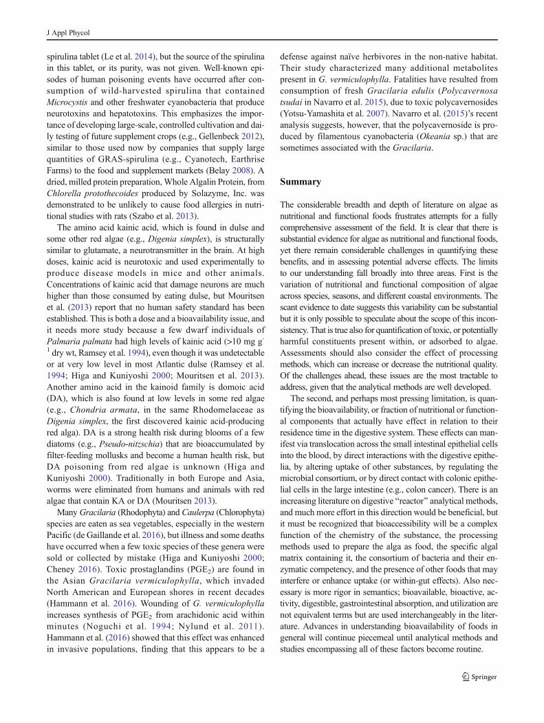
spirulina tablet (Le et al. 2014), but the source of the spirulinain this tablet, or its purity, was not given. Well-known epi-sodes of human poisoning events have occurred after con-sumption of wild-harvested spirulina that containedMicrocystis and other freshwater cyanobacteria that produceneurotoxins and hepatotoxins. This emphasizes the impor-tance of developing large-scale, controlled cultivation and dai-ly testing of future supplement crops (e.g., Gellenbeck 2012),similar to those used now by companies that supply largequantities of GRAS-spirulina (e.g., Cyanotech, EarthriseFarms) to the food and supplement markets (Belay 2008). Adried, milled protein preparation,Whole Algalin Protein, fromChlorella protothecoides produced by Solazyme, Inc. wasdemonstrated to be unlikely to cause food allergies in nutri-tional studies with rats (Szabo et al. 2013).
The amino acid kainic acid, which is found in dulse andsome other red algae (e.g., Digenia simplex), is structurallysimilar to glutamate, a neurotransmitter in the brain. At highdoses, kainic acid is neurotoxic and used experimentally toproduce disease models in mice and other animals.Concentrations of kainic acid that damage neurons are muchhigher than those consumed by eating dulse, but Mouritsenet al. (2013) report that no human safety standard has beenestablished. This is both a dose and a bioavailability issue, andit needs more study because a few dwarf individuals ofPalmaria palmata had high levels of kainic acid (>10 mg g-1 dry wt, Ramsey et al. 1994), even though it was undetectableor at very low level in most Atlantic dulse (Ramsey et al.1994; Higa and Kuniyoshi 2000; Mouritsen et al. 2013).Another amino acid in the kainoid family is domoic acid(DA), which is also found at low levels in some red algae(e.g., Chondria armata, in the same Rhodomelaceae asDigenia simplex, the first discovered kainic acid-producingred alga). DA is a strong health risk during blooms of a fewdiatoms (e.g., Pseudo-nitzschia) that are bioaccumulated byfilter-feeding mollusks and become a human health risk, butDA poisoning from red algae is unknown (Higa andKuniyoshi 2000). Traditionally in both Europe and Asia,worms were eliminated from humans and animals with redalgae that contain KA or DA (Mouritsen 2013).
ManyGracilaria (Rhodophyta) and Caulerpa (Chlorophyta)species are eaten as sea vegetables, especially in the westernPacific (de Gaillande et al. 2016), but illness and some deathshave occurred when a few toxic species of these genera weresold or collected by mistake (Higa and Kuniyoshi 2000;Cheney 2016). Toxic prostaglandins (PGE2) are found inthe Asian Gracilaria vermiculophylla, which invadedNorth American and European shores in recent decades(Hammann et al. 2016). Wounding of G. vermiculophyllaincreases synthesis of PGE2 from arachidonic acid withinminutes (Noguchi et al. 1994; Nylund et al. 2011).Hammann et al. (2016) showed that this effect was enhancedin invasive populations, finding that this appears to be a
defense against naïve herbivores in the non-native habitat.Their study characterized many additional metabolitespresent in G. vermiculophylla. Fatalities have resulted fromconsumption of fresh Gracilaria edulis (Polycavernosatsudai in Navarro et al. 2015), due to toxic polycavernosides(Yotsu-Yamashita et al. 2007). Navarro et al. (2015)’s recentanalysis suggests, however, that the polycavernoside is pro-duced by filamentous cyanobacteria (Okeania sp.) that aresometimes associated with the Gracilaria.
Summary
The considerable breadth and depth of literature on algae asnutritional and functional foods frustrates attempts for a fullycomprehensive assessment of the field. It is clear that there issubstantial evidence for algae as nutritional and functional foods,yet there remain considerable challenges in quantifying thesebenefits, and in assessing potential adverse effects. The limitsto our understanding fall broadly into three areas. First is thevariation of nutritional and functional composition of algaeacross species, seasons, and different coastal environments. Thescant evidence to date suggests this variability can be substantialbut it is only possible to speculate about the scope of this incon-sistency. That is true also for quantification of toxic, or potentiallyharmful constituents present within, or adsorbed to algae.Assessments should also consider the effect of processingmethods, which can increase or decrease the nutritional quality.Of the challenges ahead, these issues are the most tractable toaddress, given that the analytical methods are well developed.
The second, and perhaps most pressing limitation, is quan-tifying the bioavailability, or fraction of nutritional or function-al components that actually have effect in relation to theirresidence time in the digestive system. These effects can man-ifest via translocation across the small intestinal epithelial cellsinto the blood, by direct interactions with the digestive epithe-lia, by altering uptake of other substances, by regulating themicrobial consortium, or by direct contact with colonic epithe-lial cells in the large intestine (e.g., colon cancer). There is anincreasing literature on digestive Breactor^ analytical methods,and much more effort in this direction would be beneficial, butit must be recognized that bioaccessibility will be a complexfunction of the chemistry of the substance, the processingmethods used to prepare the alga as food, the specific algalmatrix containing it, the consortium of bacteria and their en-zymatic competency, and the presence of other foods that mayinterfere or enhance uptake (or within-gut effects). Also nec-essary is more rigor in semantics; bioavailable, bioactive, ac-tivity, digestible, gastrointestinal absorption, and utilization arenot equivalent terms but are used interchangeably in the liter-ature. Advances in understanding bioavailability of foods ingeneral will continue piecemeal until analytical methods andstudies encompassing all of these factors become routine.
J Appl Phycol

The third limitation lies in understanding how algal nutri-tional, and particularly functional, constituents interact in hu-man metabolism and intermediary metabolic processes. Mostinvestigators studying these questions report in vitro experi-ments or in vivo experiments employing direct introduction ofpurified algal constituents. These methods are useful probes toidentify and mechanistically understand potential effects ofthe consumption of algal-based supplements, but they are in-adequate to assess nutritional and functional foods. Theirshortcomingsmost often include unrealistic or uncertain dosesrelative to normal algal consumption, a focus on a single ornarrow range of metabolic responses, and inability to assessconcurrent, or synergistic effects arising from co-occurringconstituents during digestion. Coupling in vivo studies withmore holistic digestive research will provide for a better as-sessment of algae as nutritional and functional food sources.
We envisage a rare opportunity to develop a rich andrewarding collaboration among phycological, nutritional,medical, analytical, and industrial groups investigating algaeas nutritional and functional foods. Part of the challenge aheadfor algal scientists is understanding the complexity of mergingof basic research through clinical trials and regulatoryrequirements to create marine food products. The recentcomprehensive reviews by Finley et al. (2014) andBorowitzka (2013b) help to clarify this pathway. But the mostdramatic advances will require a rethinking of experimentaland collaborative approaches, and the impetus for this re-search will only increase as human pressures on the climatesystem lead us to turn more to the oceans for food that we canharvest and grow sustainably.
Acknowledgments We thank Eva Bradford for clerical assistance inthe initial stages of our literature review, and to Dr. Yoichi Sato (RikenFood Co., Ltd.) and Dr. Keiko Yoshinaga (Riken Vitamine Co, Ltd.) forproviding us the most recent data on Japanese macroalgal food consump-tion from The National Health and Nutrition Survey in Japan, 2004–2014(MHLW 2014). We appreciate the valuable insights Dr. Santosh P. Lall,National Research Council of Canada, provided to this work. We alsothank the anonymous reviewers and Editor for their helpful comments.AGS & KEH thank the Biotechnology and Biological Sciences ResearchCouncil (BBSRC BB/1013164/1) of the UK for funding. The Universityof Dundee is a registered Scottish charity, No. SC015096. PP is supportedby IDEALG in the frame of the stimuli program entitled BInvestissementsd’avenir, Biotechnologies-Bioressources^ (ANR-10-BTBR-04-02). Theopen access fee was supported by NSF-OCE-1435021 (MLW), DIC pro-ject 1823-06 (MEC), Maine Sea Grant (NOAA) 5405971 (SHB), NSF#11A-1355457 to Maine EPSCoR at the University of Maine (SHB), andthe listed funding to AGS and PP.
Open Access This article is distributed under the terms of the CreativeCommons At t r ibut ion 4 .0 In te rna t ional License (h t tp : / /creativecommons.org/licenses/by/4.0/), which permits unrestricted use,distribution, and reproduction in any medium, provided you giveappropriate credit to the original author(s) and the source, provide a linkto the Creative Commons license, and indicate if changes were made.
References
Aaronson S (1986) A role for algae as human food in antiquity. FoodFoodways 1:311–315
Aarstad OA, Tondervik A, Sletta H, Skjak-Braek G (2012) Alginatesequencing: an analysis of block distribution in alginates usingspecific alginate degrading enzymes. Biomacromolecules 13:106–116
Abdul QA, Choi RJ, Jung HA, Choi JS (2016) Health benefit offucosterol from marine algae: a review. J Sci Food Agric 96:1856–1866
Admassu H, Zhao W, Yang R, Gasmalla M, Alsir E (2015) Developmentof functional foods: seaweeds (algae) untouched potential and alter-native resource—a review. Int J Sci Technol Res 4:108–115
Albanes D et al (1995) Effects of alpha-tocopherol and beta-carotenesupplements on cancer incidence in the alpha-tocopherol beta-caro-tene cancer prevention study. Am J Clin Nutr 62:S1427–S1430
Allen LH (2008) Causes of vitamin B12 and folate deficiency. Food NutrBull 29:S20–S34
Al-Masri MS, Mamish S, Budier Y (2003) Radionuclides and tracemetals in eastern Mediterranean sea algae. J Environ Radioact 67:157–168
Almela C, Clemente MJ, Velez D, Montoro R (2006) Total arsenic, inor-ganic arsenic, lead and cadmium contents in edible seaweed sold inSpain. Food Chem Toxicol 44:1901–1908
Alves A, Sousa RA, Reis RL (2013) A practical perspective on ulvanextracted from green algae. J Appl Phycol 25:407–424
Anastyuk SD, Shevchenko NM, Ermakova SP, Vischuk OS, NazarenkoEL, Dmitrenok PS, Zvyagintseva TN (2012) Anticancer activityin vitro of a fucoidan from the brown alga Fucus evanescens andits low-molecular fragments, structurally characterized by tandemmass-spectrometry. Carbohydr Polym 87:186–194
Angell AR, Mata L, de Nys R, Paul NA (2016) The protein content ofseaweeds: a universal nitrogen-to-protein conversion factor of five. JAppl Phycol 28:511–524
Aquino RS, Landeira-Fernandez AM, Valente AP, Andrade LR, MourãoPAS (2005) Occurrence of sulfated galactans in marine angio-sperms: evolutionary implications. Glycobiology 15:11–20
Arterburn LM, Oken HA, Hoffman JP, Bailey-Hall E, Chung G, Rom D,Hamersley J, McCarthy D (2007) Bioequivalence ofdocosahexaenoic acid from different algal oils in capsules and in aDHA-fortified food. Lipids 42:1011–1024
Arterburn LM, Oken HA, Hall EB, Hamersley J, Kuratko CN, HoffmanJP (2008) Algal-oil capsules and cooked salmon: nutritionallyequivalent sources of docosahexaenoic acid. J Am Diet Assoc108:1204–1209
Arul AB, Al Numair K, Al Saif M, Savarimuthu I (2012) Effect of dietarybeta-sitosterol on fecal bacterial and colonic biotransformation en-zymes in 1,2-dimethylhydrazine-induced colon carcinogenesis.Turk J Med Sci 42:1307–1313
Baba M, Snoeck R, Pauwels R, deClerq E (1988) Sulfated polysaccha-rides are potent and selective inhibitors of various enveloped virus-es, including herpes simplex virus, cytomegalovirus, vesicular sto-matitis virus and human immunodeficiency virus. AntimicrobAgents Chemother 32:1742–1745
Bäckhed F, Ley RE, Sonnenburg JL, Peterson DA, Gordon JI (2005)Host-bacterial mutualism in the human intestine. Science 307:1915–1920
Bagchi D (2006) Nutraceuticals and functional foods regulations in theUnited States and around the world. Toxicology 221:1–3
Barahona T, Encinas MV, Mansilla A, Matsuhiro B, Zuniga EA (2012) Asulfated galactan with antioxidant capacity from the green variant oftetrasporic Gigartina skottsbergii (Gigartinales, Rhodophyta).Carbohydr Res 347:114–120
J Appl Phycol

Barclay W, Weaver C, Metz J, Hansen J (2010) Development ofdocosahexaenoic acid production technology using Schizochytrium.In: Cohen Z, Ratledg C (eds) Single cell oils. AOCS Press, Urbana,pp 75–96
BarclayW, Apt K, Dong X (2013) Commercial production of microalgaevia fermentation. In: Richmond A, Hu Q (eds) Handbook ofmicroalgal culture: applied phycology and biotechnology.Blackwell, Chichester, pp 134–145
Barlow S, Pascal G, Cressey P, Folmer D, Smith J (2015) Carrageenan(addendum). In: Safety evaluation of certain food additives, WHOfood additives series, vol 70. World Health Organization, Geneva
Belay A (1997)Mass culture of Spirulina outdoors—The Earthrise Farmsexperience. In: Vonshak A (ed) Spirulina platensis (Arthrospira):physiology, cell-biology and biotechnology. Taylor & Francis Ltd.,London, pp 131–158
Belay A (2008) Spirulina (Arthrospira): production and quality assur-ance. In: Gershwin ME, Belay A (eds) Spirulina in human nutritionand health. CRC Press, Boca Raton, pp 1–25
Ben-Amotz A, Levy Y (1996) Bioavailability of a natural isomer mixturecompared with synthetic all-trans beta-carotene in human serum.Am J Clin Nutr 63:729–734
Ben-Amotz A, Yatziv S, Sela M, Greenberg S, Rachmilevich B,Shwarzman M, Weshler Z (1998) Effect of natural beta-carotenesupplementation in children exposed to radiation from theChernobyl accident. Radiat Environ Biophys 37:187–193
Benjama O, Masniyom P (2012) Biochemical composition and physico-chemical properties of two red seaweeds (Gracilaria fisheri andG. tenuistipitata) from the Pattani Bay in southern Thailand.Songklanakarin J Sci Technol 34:223–230
Benkdad A, Laissaoui A, Tornero MV, Benmansour M, Chakir E,Garrido IM, Moreno JB (2011) Trace metals and radionuclides inmacroalgae from Moroccan coastal waters. Environ Monit Assess182:317–324
Bernstein AM, Ding EL, Willett WC, Rimm EB (2012) A meta-analysisshows that docosahexanaenoic acid from algal oil reduces serumtriglycerides and increases HDL-cholesterol and LDL-cholesterolin persons without coronary heart disease. J Nutr 142:99–104
Berteau O, Mulloy B (2003) Sulfated fucans, fresh perspectives: struc-tures, functions, and biological properties of sulfated fucans and anoverview of enzymes active toward this class of polysaccharide.Glycobiology 13:29R–40R
Bhattacharjee H, Mukhopadhyay R, Thiyagarajan S, Rosen BP (2008)Aquaglyceroporins: ancient channels for metalloids. J Biol 7:33.31–33.36
Bhattacharyya S, Liu H, Zhang Z, Jam M, Dudeja PK, Michel G,Linhardt RJ, Tobacman JK (2010) Carrageenan-induced innate im-mune response is modified by enzymes that hydrolyze distinctgalactosidic bonds. J Nutr Biochem 21:906–913
Bhattacharyya S, O-Sullivan I, Katyal S, Unterman T, Tobacman JK(2012) Exposure to the common food additive carrageenan leadsto glucose intolerance, insulin resistance and inhibition of insulinsignaling in HepG2 cells and C57BL/6J mice. Diabetologia 55:194–203
Black WAP (1950) The seasonal variation in weight and chemical com-position of the common British Laminariaceae. J Mar Biol AssocUK 29:45–72
Blouin N, Calder BL, Perkins B, Brawley SH (2006) Sensory and fattyacid analyses of two Atlantic species of Porphyra (Rhodophyta). JAppl Phycol 18:79–85
Bobin-Dubigeon C, LahayeM,Barry J-L (1997) Human colonic bacterialdegradability of dietary fibres from sea-lettuce (Ulva sp). J Sci FoodAgric 73:149–159
Bocanegra A, Bastida S, Benedí J, Ródenas S, Sánchez-Muniz FJ (2009)Characteristics and nutritional and cardiovascular-health propertiesof seaweeds. J Med Food 12:236–258
Boehlke C, Zierau O, Hannig C (2015) Salivary amylase—the enzyme ofunspecialized euryphagous animals. Arch Oral Biol 60:1162–1176
Bohn JA, BeMiller JN (1995) (1→3)-β-D-Glucans as biological re-sponse modifiers: a review of structure-functional activity relation-ships. Carbohydr Polym 28:3–14
Bohn T, McDougall GJ, Alegría A, Alminger M, Arrigoni E, Aura A-M,Brito C, Cilla A, El SN, Karakaya S, Martínez-Cuesta MC, SantosCN (2015) Mind the gap—deficits in our knowledge of aspectsimpacting the bioavailability of phytochemicals and their metabo-lites—a position paper focusing on carotenoids and polyphenols.Mol Nutr Food Res 59:1323
Borowitzka MA (2013a) High-value products from microalgae—theirdevelopment and commercialisation. J Appl Phycol 25:743–756
Borowitzka M (2013b) Dunaliella: biology, production, and markets. In:Richmond A, Hu Q (eds) Handbook of microalgal culture. JohnWiley & Sons, Ltd, Chichester, pp 359–368
Bourgougnon N (2003) Anti-HIV compounds from red seaweeds.Biomaterials and bioprocessing, Vol. 9. Recent advances in marinebiotechnology. Science Publishers, Enfield, pp 165–206
Bourgougnon N, Stiger-Pouvreau V (2011) Chemodiversity and bioac-tivity within red and brown macroalgae along the French coasts,metropol and overseas departments and territories. In: Kim S-E(ed) Handbook of marine macroalgae: biotechnology and appliedphycology. John Wiley & Sons, Chichester, pp 58–105
Boyer EW, Kearney S, Shannon MW, Quang L, Woolf A, Kemper K(2002) Poisoning from a dietary supplement administered duringhospitalization. Pediatrics 109:e49
Branger B, Cadudal JL, Delobel M, Ouoba H, Yameogo P, Ouedraogo D,Guerin D, Valea A, Les Personnels des C, Zombre C, Ancel P(2003) Spiruline as a food supplement in case of infant malnutritionin Burkina-Faso (La spiruline comme complement alimentaire dansla malnutrition du nourrisson au Burkina-Faso.). Arch Pediatr 10:424–431
Braud J (2006) Continuous seaweed tank culture in France: fromChondrus crispus to co-culture of macroalge and the diatomOdontella aurita. In: Critchley A, Ohno M, Largo D (eds)Seaweed resources of the world. Int. Cooperation Agency, Japan
Britton G, Liaaen-Jensen S, Pfander PH (2004) Carotenoids: handbook.Birkhauser Verlag AG, Switzerland
Brown MR (1995) Effects of storage and processing on the ascorbic-acidcontent of concentrates prepared from Chaetoceros calcitrans. JAppl Phycol 7:495–500
Brown MT, Hodgkinson WM, Hurd CL (1998) Spatial and temporalvariations in the copper and zinc concentrations of two greenseaweeds from Otago Harbour, New Zealand. Mar Environ Res47:1–10
Brownlee IA, Allen A, Pearson JP, Dettmar PW, Havler ME, AthertonMR, Onsoyen E (2005) Alginate as a source of dietary fiber. CritRev Food Sci Nutr 45:497–510
Butler MR (1936) Seasonal variations in Chondrus crispus. Biochem J30:1338–1344
Cabrita ARJ, Maia MRG, Oliveira HM, Sousa-Pinto I, Almeida AA,Pinto E, Fonseca AJM (2016) Tracing seaweeds as mineral sourcesfor farm-animals. J Appl Phycol 28:3135–3150
Canadian Food Inspection Agency (2012) Inorganic arsenic and hijikis e awe ed con s ump t i o n . h t t p : / /www. i n s p e c t i o n . g c .ca / food / in fo rmat ion- fo r -consumers / f ac t - shee t s -and-i n f og r aph i c s / s p e c i f i c - p roduc t s - and r i s k s / ch emi c a l -hazards/inorganic-arsenic/eng/1332268146718/1332268231124.(accessed 11/10/16)
Cantarel BL, Lombard V, Henrissat B (2012) Complex carbohydrateutilization by the healthy human microbiome. PLoS One 7:e28742
Carbonell-Capella JM, Buniowska M, Barba FJ, Esteve MJ, Frigola A(2014) Analytical methods for determining bioavailability and bio-accessibility of bioactive compounds from fruits and vegetables: areview. Compr Rev Food Sci Food Saf 13:155–171
J Appl Phycol

Carlin DJ, Naujokas MF, Bradham KD, Cowden J, Heacock M, HenryHF, Lee JS, Thomas DJ, Thompson C, Tokar EJ, Waalkes MP,Birnbaum LS, Suk WA (2016) Arsenic and environmental health:state of the science and future research opportunities. EnvironHealth Perspect 124:890–899
Cases J, Vacchina V, Napolitano A, Caporiccio B, Besançon P, LobinskiP, Rouanet J-M (2001) Selenium from selenium-rich Spirulina isless bioavailable than selenium from sodium selenite andselenomethionine in selenium-deficient rats. J Nutr 131:2343–2350
Chacón-Lee TL, González-Mariño GE (2010) Microalgae for Bhealthy^foods—possibilities and challenges. Comp Rev Food Sci FoodSafety 9:655–675
Chan JCC, Cheung PCK, Ang PO (1997) Comparative studies on theeffect of three drying methods on the nutritional composition ofseaweed Sargassum hemiphyllum (Turn) C Ag. J Agric FoodChem 45:3056–3059
Cheney D (2016) Toxic and harmful seaweeds. In: Fleurence J, Levine I(eds) Seaweeds in health and disease. Elsevier, Amsterdam, pp 407–421
Cheng F-C, Lin A, Feng J-J, Mizoguchi T, Takekoshi H, Kubota H, KatoY, Naoki Y (2004) Effects of Chlorella on activities of protein tyro-sine phosphatases, matrix metalloproteinases, caspases, cytokine re-lease, B and Tcell proliferations, and phorbol ester receptor binding.J Med Food 7:146–152
Choi BS, Choi SJ, Kim DW, Huang M, Kim NY, Park KS, Kim CY, LeeHM, Yum YN, Han ES, Kang TS, Yu IJ, Park JD (2010) Effects ofrepeated seafood consumption on urinary excretion of arsenic spe-cies by volunteers. Arch Environ Contam Toxicol 58:222–229
Chrismadha T, Borowitzka MA (1994) Effect of cell-density and irradi-ance on growth, proximate composition and eicosapentaenoic acidproduction of Phaeodactylum tricornutum grown in a tubularphotobioreactor. J Appl Phycol 6:67–74
Cian RE, Drago SR, deMedina FS,Martinez-Augustin O (2015) Proteinsand carbohydrates from red seaweeds: evidence for beneficial ef-fects on gut function and microbiota. Mar Drugs 13:5358–5383
Ciferri O (1983) Spirulina, the edible microorganism. Microbiol Rev 47:551–578
Cofrades S, López-López I, Solas MT, Bravo L, Jiménez-Colmenero F(2008) Influence of different types and proportions of added edibleseaweeds on characteristics of low-salt gel/emulsion meat systems.Meat Sci 79:767–776
Conquer JA, Holub BJ (1996) Supplementation with an algae source ofdocosahexaenoic acid increases (n-3) fatty acid status and altersselected risk factors for heart disease in vegetarian subjects. J Nutr126:3032–3039
Consumer-Reports (2012) Arsenic in your juice. Consumer reports mag-azine, January. ConsumerReports.org
Cooney RV, Mumma RO, Benson AA (1978) Arsonium phospholipid inalgae. Proc Natl Acad Sci U S A 75:4262–4264
CornishML, Garbary DJ (2010) Antioxidants frommacroalgae: potentialapplications to human health and nutrition. Algae 25:155–171
Cornish ML, Critchley AT, Mouritsen OG (2015) A role for dietarymacroalgae in the amelioration of certain risk factors associated withcardiovascular disease. Phycologia 54:649–666
Corona G, Ji Y, Anegboonlap P, Hotchkiss S, Gill C, Yaqoob P, SpencerJPE, Rowland I (2016) Gastrointestinal modifications and bioavail-ability of brown seaweed phlorotannins and effects on inflammatorymarkers. Brit J Nutr 115:1240–1253
Costa S, Crespo D, Henriques BMG, Pereira E, Duarte AC, Pardal AC(2011) Kinetics of mercury accumulation and its effects on Ulvalactuca growth rate at two salinities and exposure conditions.Water Air Soil Pollut 217:689–699
Costello EK, Stagaman K, Dethlefsen L, Bohannan BJM, Relman DA(2012) The application of ecological theory toward an understand-ing of the human microbiome. Science 336:1255–1261
Cottin SC, Sanders TA, Hall WL (2011) The differential effects of EPAand DHA on cardiovascular risk factors. Proc Nutr Soc 70:215–231
Courtois J (2009) Oligosaccharides from plants and algae: production andapplications in therapeutics and biotechnology. Opin Microbiol 12:261–273
Craigie JS (2010) Seaweed extract stimuli in plant science and agricul-ture. J Appl Phycol 23:371–393
Croci DO, Cumashi A, Ushakova NA, PreobrazhenskayaME, Piccoli A,Totani L, Ustyuzhanina NE, Bilan MI, Usov AI, Grachev AA,Morozevich GE, Berman AE, Sanderson CJ, Kelly M, GregorioPD, Rossi C, Tinari N, Iacobelli S, Rabinovich GA, Nifantiev NE(2011) Fucans, but not fucomannanoglucuronans, determine the bi-ological activities of sulfated polysaccharides from Laminariasaccharina brown seaweed. PLoS One 6:e17283
Croft MT, Lawrence AD, Raux-Deery E, Warren MJ, Smith AG (2005)Algae acquire vitamin B12 through a symbiotic relationship withbacteria. Nature 438:90–93
Croft MT, Warren MJ, Smith AG (2006) Algae need their vitamins.Eukaryot Cell 5:1175–1183
Cumashi A, Ushakova NA, Preobrazhenskaya ME, D’Incecco A, PiccoliA, Totani L, Tinari N,Morozevich GE, BermanAE, BilanMI, UsovAI, Ustyuzhanina NE, Grachev AA, Sanderson CJ, Kelly M,Rabinovich GA, Iacobelli S, Nifantiev NE (2007) A comparativestudy of the anti-inflammatory, anticoagulant, antiangiogenic, andantiadhesive activities of nine different fucoidans from brown sea-weeds. Glycobiology 17:541–552
Cysewski G, Lorenz R (2004) Industrial production of microalgal cell-mass and secondary products-species of high potential:Haematococcus. In: Richmond A (ed) Microalgal culture: biotech-nology and applied phycology. Blackwell Science, Oxford, pp281–288
Dagnelie PC, VanstaverenWA, Vandenberg H (1991) Vitamin B-12 fromalgae appears not to be bioavailable. Am J Clin Nutr 53:695–697
Dawczynski C, Schaefer U, Leiterer M, Jahreis G (2007) Nutritional andtoxicological importance of macro, trace, and ultra-trace elements inalgae food products. J Agric Food Chem 55:10470–10475
de Gaillande C, Payri C, Remoissenet G, Zubia M (2016) Caulerpaconsumption, nutritional value and farming in the Indo-Pacific re-gion. J Appl Phycol. doi:10.1007/s10811-016-0912-6:1-18
de Jesus Raposo FM, Bernado de Morais AM, Santos Costa de MoraisRM (2015) Marine polysaccharides from algae with potential bio-medical applications. Mar Drugs 13:2967–3028
de la Calle MB, Baer I, Robouch P, Cordeiro F, Emteborg H, Baxter MJ,Brereton N, Raber G, Velez D, Devesa V, Rubio R, Llorente-Mirandes T, Raab A, Feldmann J, Sloth JJ, Rasmussen RR,D’Amato M, Cubadda F (2012) Is it possible to agree on a valuefor inorganic arsenic in food? The outcome of IMEP-112. AnalBioanal Chem 404:2475–2488
De Marco ER, Steffolani ME, Martinez CS, Leon AE (2014)Effects of spirulina biomass on the technological and nutri-tional quality of bread wheat pasta. LWT-Food Sci Technol58:102–108
de Oliveira MN, Freitas ALP, Carvalho AFU, Sarnpaio TMT, Farias DF,Teixeira DIA, Gouveia ST, Pereira JG, de Sena M (2009) Nutritiveand non-nutritive attributes of washed-up seaweeds from the coastof Ceara, Brazil. Food Chem 115:254–259
Delgado-Noguera MF, Calvache JA, Cosp XB, Kotanidou EP,Galli-Tsinopoulou A (2015) Supplementation with long chain poly-unsaturated fatty acids (LCPUFA) to breastfeeding mothers for im-proving child growth and development. Cochrane Database SystRev. doi:10.1002/14651858.CD007901.pub3:72
Devesa V, Velez D, Montoro R (2008) Effect of thermal treatments onarsenic species contents in food. Food Chem Toxicol 46:1–8
Dhargalkar V (2015) Uses of seaweeds in the Indian diet for sustenanceand well-being. Sci Cult 80:192–202
J Appl Phycol

Dharmananda (2002) The nutritional and medicinal value of seaweedsused in Chinese medicine (2002) Institute for Traditional Medicine.http://www.imtonline.org/. Accessed 14 Oct 2016
Diaz O, Tapia Y, Munoz O, Montoro R, Velez D, Almela C (2012) Totaland inorganic arsenic concentrations in different species of econom-ically important algae harvested from coastal zones of Chile. FoodChem Toxicol 50:744–749
Dillehay TD, Ramirez C, Pino M, Collins MB, Rossen J, Pinot-NavarroJD (2008) Monte Verde: seaweed, food, medicine and the peoplingof South America. Science 320:784–789
Druehl LD (1988) Cultivated edible kelp. In: Lembi CA, Waaland JR(eds) Algae and human affairs. Cambridge University Press,Cambridge, pp 119–134
Druesne-Pecollo N, Latino-Martel P, Norat T, Barrandon E, Bertrais S,Galan P, Hercberg S (2010) Beta-carotene supplementation and can-cer risk: a systematic review and metaanalysis of randomized con-trolled trials. Int J Cancer 127:172–184
Duffy LC, Raiten DJ, Hubbard VS, Starke-Reed P (2015) Progress andchallenges in developing metabolic footprints from diet in humangut microbial cometabolism. J Nutr 145:1123S–1130S
Durand M, Beaumatin P, Bulman B, Bernalier A, Grivet JP, Serezat M,Gramet G, Lahaye M (1997) Fermentation of green alga sea-lettuce(Ulva sp.) and metabolism of its sulphate by human colonic micro-biota in a semi-continuous culture system. Reprod Nutr Dev 37:267–283
Eccles R, Meier C, Jawad M, Weinmüllner R, Grassauer A, Prieschl-Grassauer E (2010) Efficacy and safety of an antiviraliotacarrageenan nasal spray: a randomized, double-blind placebo-controlled exploratory study in volunteers with early symptoms ofthe common cold. Respir Res 11:108. doi:10.1186/1465-9921-11-108
EFSA (2010) Scientific opinion on dietary reference values for carbohy-drates and dietary fibre. Eur Food Safety Auth J 8:1–77
Eyster KM (2007) The membrane and lipids as integral participants insignal transduction: lipid signal transduction for the non-lipid bio-chemist. Adv Physiol Educ 31:5–16
Fabregas J, Herrero C (1990) Vitamin content of 4 marine microalgae—potential use as a source of vitamins in nutrition. J Indust Microbiol5:259–264
Fahy E, Subramaniam S, BrownHA,Glass CK,Merrill AH,Murphy RC,Raetz CRH, Russell DW, Seyama Y, ShawW, Shimizu T, Spener F,van Meer G, VanNieuwenhze MS, White SH, Witztum JL, DennisEA (2005) A comprehensive classification system for lipids. J LipidRes 46:839–861
FAO (2014) The state of the world fisheries and aquaculture 2014. FAO,Rome, p 223
FAO (2015) FAO Global Aquaculture Production database updated to2013—Summary information. FAO, Rome
FAO (2016) The State of the World Fisheries and Aquaculture 2016.Contribution to Food Security and Nutrition for All. Rome. 200 pp
FDA (2016) Summary: Substances Generally Regarded As Safe(Final Rule). U.S. Food and Drug Administration (HHS).http:/ /www.fda.gov/AboutFDA/ReportsManualsForms/Reports/EconomicAnalyses/ucm517103.htm. Accessed 24Oct 2016
Fernando WMADB, Ranaweera KKDS, Bamunuarachchi A, BrennanCS (2008) The influence of rice fibre fractions on the in vitro fer-mentation production of short chain fatty acids using human faecalmicro flora. Int J Food Sci Technol 43:2237–2244
Ferraces-Casais P, Lage-Yusty MA, de Quiros ARB, Lopez-Hernandez J(2012) Evaluation of bioactive compounds in fresh edible seaweeds.Food Anal Methods 5:828–834
Finley JW, Finley JW, Ellwood K, Hoadley J (2014) Launching a newfood product or dietary supplement in the United States: industrial,regulatory, and nutritional considerations. Annu Rev Nutr 34:421–447
Fitton JH (2011) Therapies from fucoidan; multifunctional marine poly-mers. Mar Drugs 9:1731–1760
Fitton J, Dell’Acqua G, Gardiner V-A, Karpiniec S, Stringer D, Davis E(2015) Topical benefits of two fucoidan-rich extracts from marinemacroalgae. Cosmetics 2:66–81
Fleurence J (1999a) The enzymatic degradation of algal cell walls: auseful approach for improving protein accessibility? J Appl Phycol11:313–314
Fleurence J (1999b) Seaweed proteins: biochemical, nutritional aspectsand potential uses. Trends Food Sci Technol 10:25–28
Fleurence J, Levine I (eds) (2016) Seaweed in health and disease preven-tion. Elsevier, Amsterdam, pp 476
Foster P (1976) Concentrations and concentration factors of heavy metalsin brown algae. Environ Pollut 10:45–54
Fournier E, Adam C,Massabuau J-C, Garnier-Laplace J (2005) Seleniumbioaccumulation in Chlamydomonas reinhardtii and subsequenttransfer to Corbicula fluminea: role of selenium speciation and bi-valve ventilation. Environ Toxicol Chem 25:2692–2699
Francesconi KA (2010) Arsenic species in seafood: origin and humanhealth implications. Pure Appl Chem 82:373–381
Francois L, Fortin C, Campbell PGC (2007) pHmodulates transport ratesof manganese and cadmium in the green alga Chlamydomonasreinhardtii through non-competitive interactions: implications foran algal BLM. Aquat Toxicol 84:123–132
Fujiwara-Arasaki T, Mino N, Kuroda M (1984) The protein value inhuman nutrition of edible marine algae in Japan. Hydrobiologia116/117:513–516
Gadd GM (2009) Biosorption: critical review of scientific rationale, en-vironmental importance and significance for pollution treatment. JChem Technol Biotechnol 84:13–28
Galland-Irmouli A-V, Fleurence J, Lamghari R, Lucon M, Rouxel C,Barbaroux O, Bronowicki J-P, Villaume C, Gueant J-L (1999)Nutritional value of proteins from edible seaweedPalmaria palmata(Dulse). J Nutr Biochem 10:353–359
Ganesan P et al (2011) Siphonaxanthin, a marine carotenoid from greenalgae, effectively induces apoptosis in human leukemia (HL-60)cells. Biochim Biophys Acta - Gen Subj 1810:497–503
Gantar M, Svircev Z (2008) Microalgae and cyanobacteria: food forthought. J Phycol 44:260–268
Garcia-Casal MN, Pereira AC, Leets I, Ramirez J, Quiroga ME (2007)High iron content and bioavailability in humans from four species ofmarine algae. J Nutr 137:2691–2695
Garcia-Casal MN, Ramirez J, Leets I, Pereira AC, Quiroga MF (2009)Antioxidant capacity, polyphenol content and iron bioavailabilityfrom algae (Ulva sp., Sargassum sp and Porphyra sp.) in humansubjects. Br J Nutr 101:79–85
Garcia-Salgado S, Raber G, Raml R, Magnes C, Francesconi KA (2012)Arsenosugar phospholipids and arsenic hydrocarbons in two speciesof brown macroalgae. Environ Chem 9:63–66
Garcia-Sartal C, Taebunpakul S, Stokes E, Barciela-Alonso M, Bermejo-Barrera P, Goenaga-Infante H (2012) Two-dimensional HPLCcoupled to ICP-MS and electrospray ionisation (ESI)-MS/MS forinvestigating the bioavailability in vitro of arsenic species from ed-ible seaweed. Anal Bioanal Chem 402:3359–3369
Garnham GW, Codd GA, Gadd GM (1992) Kinetics of uptake and intra-cellular location of cobalt, manganese and zinc in the estuarinegreen-alga Chlorella salina. Appl Microbiol Biotechnol 37:270–276
Gellenbeck KW (2012) Utilization of algal materials for nutraceutical andcosmeceutical applications—what do manufacturers need to know?J Appl Phycol 24:309–313
Georgianna DR, Mayfield SP (2012) Exploiting diversity and syntheticbiology for the production of algal biofuels. Nature 488:329–335
Geppert J, Kraft V, Demmelmair H, Koletzko B (2005) Docosahexaenoicacid supplementation in vegetarians effectively increases omega-3index: a randomized trial. Lipids 40:807–814
J Appl Phycol

Gobler CJ, Berry DL, Dyhrman ST, Wilhelm SW, Salamov A, LobanovAV, Zhang Y, Collier JL, Wurch LL, Kustka AB, Dill BD, Shah M,VerBerkmoes NC, Kuo A, Terry A, Pangilinan J, Lindquist EA,Lucas S, Paulsen IT, Hattenrath-Lehmann TK, Talmage SC,Walker EA, Koch F, Burson AM, Alejandra-Marcoval Y-ZT M,LeCleir GR, Coyne KJ, Berg GM, Bertrand EM, Saito MA,Gladyshev VN, Grigoriev IV (2011) Niche of the harmful algaAureococcus anophagefferens revealed through ecogenomics. ProcNatl Acad Sci U S A 108:4352–4357
Gordon JI (2012) Honor thy gut symbionts redux. Science 336:1251–1252
Graeve M, Kattner G, Wiencke C, Karsten U (2002) Fatty acid compo-sition of Arctic and Antarctic macroalgae: indicator of phylogeneticand trophic relationships. Mar Ecol Prog Ser 231:67–74
Griffin J (2015) An investigative study into the beneficial use of seaweedin bread and the broader food industry. Dublin Institute ofTechnology, School of Culinary Arts and Food Technology,http://arrow.dit.ie/tfschcafdis. Accessed 05 Sept 2016
Gropper SS, Smith JL (2013) Advanced human nutrition and metabo-lism, 6th edn. Wadsworth, Belmont
Gubelit YI, Makhutova ON, Sushchik NN, Kolmakova AA, KalachovaGS, Gladyshev MI (2015) Fatty acid and elemental composition oflittoral Bgreen tide^ algae from the Gulf of Finland, the Baltic Sea. JAppl Phycol 27:375–386
Guil-Guerrero JL (2007) Stearidonic acid (18:4 n-3): metabolism, nutri-tional importance, medical uses and natural sources. Eur J Lipid SciTechnol 109:1226–1236
Gupta S, Abu-Ghannam N (2011a) Bioactive potential and possiblehealth effects of edible brown seaweeds. Trends Food Sci Technol22:315–326
Gupta S, Abu-Ghannam N (2011b) Recent developments in the applica-tion of seaweeds or seaweed extracts as a means for enhancing thesafety and quality of foods. Food Sci Emerg Technol 12:600–609
Haddad EH, Berk LS, Kettering JD, Hubbard RW, Peters WR (1999)Dietary intake and biochemical, hematologic, and immune statusof vegans compared with nonvegetarians. Am J Clin Nutr 70:586S–593S
Hafting JT, Critchley AT, Cornish ML, Hubley SA, Archibald AF (2012)On-land cultivation of functional seaweed products for human us-age. J Appl Phycol 24:385–392
Hafting JT, Craigie JS, Stengel DB, Loureiro RR, BuschmannAH, YarishC, Edwards MD, Critchley AT (2015) Prospects and challenges forindustrial production of seaweed bioactives. J Phycol 51:821–837
Halliwell B, Gutteridge JMC (2007) Free radicals in biology and medi-cine, 4th edn. Clarendon, Oxford
Hammann M, Rempt M, Pohnert G, Wang G, Boo SM, Weinberger F(2016) Increased potential for wound activated production ofProstaglandin E-2 and related toxic compounds in non-native pop-ulations of Gracilaria vermiculophylla. Harmful Algae 51:81–88
Hansen HR, Raab A, Francesconi KA, Feldmann J (2003) Metabolism ofarsenic by sheep chronically exposed to arsenosugars as a normalpart of their diet. 1. Quantitative intake, uptake, and excretion.Environ Sci Technol 37:845–851
Hansra P, Moran EL, Fornasier VL, Bogoch ER (2000) Carrageenan-induced arthritis in the rat. Inflammation 24:141–155
Hardy K, Brand-Miller J, Brown KD, Thomas MG, Copeland L (2015)The importance of dietary carbohydrate in human evolution. Q RevBiol 90:251–268
Harnedy PA, FitzGerald RJ (2011) Bioactive proteins, peptides, and ami-no acids from macroalgae. J Phycol 47:218–232
Harnedy PA, FitzGerald RJ (2013) Extraction of protein from themacroalga Palmaria palmata. LWT-Food Sci Technol 51:375–382
Hasegawa T, Kimura Y, Hiromatsu K, Kobayashi N, Yamada A, MakinoM, Okuda M, Sano T, Nomoto K, Yoshikai Y (1997) Effect of hotwater extract of Chlorella vulgaris on cytokine expression patternsin mice with murine acquired immunodeficiency syndrome after
infection with Listeria monocytogenes. Immunopharmacology 35:273–282
Haug IJ, Sagmo LB, Zeiss D, Olsen IC, Draget KI, Seternes T (2011)Bioavailability of EPA and DHA delivered by gelled emulsions andsoft gel capsules. Eur J Lipid Sci Technol 113:137–145
Hehemann J-H, Correc G, Barbeyron T, Helbert W, Czjzek M, Michel G(2010) Transfer of carbohydrate-active enzymes from marine bacte-ria to Japanese gut microbiota. Nature 464:908
Hehemann JH, Kelly AG, Pudlo NA, Martens EC, Boraston AB (2012)Bacteria of the human gut microbiome catabolize red seaweed gly-cans with carbohydrate-active enzyme updates from extrinsic mi-crobes. Proc Natl Acad Sci U S A 109:19786–19791
Helliwell KE, Wheeler GL, Leptos KC, Goldstein RE, Smith AG (2011)Insights into the evolution of vitamin B12 auxotrophy from se-quenced algal genomes. Mol Biol Evol 28:2921–2933
Helliwell KE, Wheeler GL, Smith AG (2013) Widespread decay ofvitamin-related pathways: coincidence or consequence? TrendsGenet 29:469–478
Helliwell KE, Lawrence AD, Holzer A, Kudahl UJ, Sasso S, Krautler B,Scanlan DJ, Warren MJ, Smith AG (2016) Cyanobacteria and eu-karyotic algae use different chemical variants of vitamin B12. CurrBiol 26:999–1008
Hernandez-Carmona G, Carrillo-Dominguez S, Arvizu-Higuera DL,Rodriguez-Montesinos YE, Murillo-Alvarez JI, Munoz-Ochoa M,Castillo-Dominguez RM (2009) Monthly variation in the chemicalcomposition of Eisenia arborea JE Areschoug. J Appl Phycol 21:607–616
Heuzé V, Tran G, Giger-Reverdin S, LessireM, Lebas F (2016) Seaweeds(marine macroalgae). Feedipedia, a program by INRA, CIRAD,AFZ and FAO. http://www.feedipedia.org/node/78. Accessed 04Sept 2016
Higa T, Kuniyoshi M (2000) Toxins associated with medicinal and edibleseaweeds. J Toxicol Toxin Rev 19:119–137
Hofer M, Pospíšil M (2011) Modulation of animal and human hemato-poiesis by β-glucans. Molecules 16:7969–7979
Holdt SL, Kraan S (2011) Bioactive compounds in seaweed: functionalfood applications and legislation. J Appl Phycol 23:543–597
Holst B, Williamson G (2008) Nutrients and phytochemicals: from bio-availability to bioefficacy beyond antioxidants. Curr OpinBiotechnol 19:73–82
Hollriegl V, Rohmuss M, Oeh U, Roth P (2004) Strontium biokinetics inhumans: influence of alginate on the uptake of ingested strontium.Health Phys 86:193–196
Holub BJ (2009) Docosahexaenoic acid (DHA) and cardiovascular dis-ease risk factors. Prostagl Leukotr Essent Fatty Acids 81:199–204
Honya M, Mori H, Anzai M, Araki Y, Nisizawa K (1999) Monthlychanges in the content of fucans, their constituent sugars and sul-phate in cultured Laminaria japonica. Hydrobiologia 398/399:411–416
Hsu H-Y, Jeyashoke N, Yeh C-H, Song Y-J, Hua KF, Chao LK (2010)Immunostimulatory bioactivity of algal polysaccharides fromChlorella pyrenoidosa activates macrophages via toll-like receptor-4. J Agric Food Chem 58:927–936
Hwang H, Chen T, Nines RG, Shin HC, Stoner GD (2006)Photochemoprevention of UVB-induced skin carcinogenesis inSKH-1 mice by brown algae polyphenols. Int J Cancer 119:2742–2749
IARC (2012) Arsenic, metals, fibres, and dusts Vol. 100C a review ofhuman carcinogens. IARC, Lyon, pp 1–99
Ichikawa S, Kamoshida M, Hanaoka K, Hamano M, Maitani T, Kaise T(2006) Decrease of arsenic in edible brown algae Hijikia fusiformeby the cooking process. Appl Organomet Chem 20:585–590
Imhoff-Kunsch B, Stein AD, Villalpando S, Martorell R, RamakrishnanU (2011) Docosahexaenoic acid supplementation from mid-pregnancy to parturition influenced breast milk fatty acid
J Appl Phycol

concentrations at 1 month postpartum in Mexican women. J Nutr141:1762–1762
Institute-of-Medicine (2001) Iron. In: Dietary reference intakes for vita-min A, vitamin K, arsenic, boron, chromium, copper, iodine, iron,manganese, molybdenum, nickel, silicon, vanadium, and zinc.National Academies Press, Washington, DC, pp 290–378
Institute-of-Medicine (2005) Dietary, functional and total fiber. In:Dietary reference intakes for energy, carbohydrates, fiber, fat, fattyacids, cholesterol, protein, and amino acids. National AcademiesPress, Washington, DC, pp 339–421
Iwai K (2008) Antidiabetic and antioxidant effects of polyphenols inbrown algaEcklonia stolonifera in genetically diabetic kk-a(y) mice.Plant Foods Human Nutr 63:163–169
Jassby A (1988) Spirulina: a model for microalgae as food. In: LembiCA, Waaland JR (eds) Algae and human affairs. CambridgeUniversity Press, Cambridge, pp 149–179
Jensen A (1993) Present and future needs for algae and algal products.Hydrobiologia 261:15–23
Jensen CL, Voigt RG, Prager TC, Zou YLL, Fraley JK, Rozelle JC,Turcich MR, Llorente AM, Anderson RE, Heird WC (2005)Effects of maternal docosahexaenoic acid intake on visual functionand neurodevelopment in breastfed term infants. Am J Clin Nutr 82:125–132
Jensen CL, Voigt RG, Llorente AM, Peters SU, Prager TC, Zou YLL,Rozelle JC, Turcich MR, Fraley JK, Anderson RE, Heird WC(2010) Effects of early maternal docosahexaenoic acid intake onneuropsychological status and visual acuity at five years of age ofbreast-fed term infants. J Pediatrics 157:900–905
Jiang HM, Chen F (2000) Effects of temperature and temperature shift ondocosahexanenoic acid production by the marine microalgaCrypthecodinium cohnii. JAOCS 77:613–617
Jiang HM, Gao KS (2004) Effects of lowering temperature during cultureon the production of polyunsaturated fatty acids in the marine dia-tom Phaeodactylum tricornutum (Bacillariophyceae). J Phycol 40:651–654
Jiao G, Yu G, Zhang J, Ewart HS (2011) Chemical structures and bioac-tivities of sulfated polysaccharides from marine algae. Mar Drugs 9:196–123
Jimenez-Escrig A, Jimenez-Jimenez I, Pulido R, Saura-Calixto F (2001)Antioxidant activity of fresh and processed edible seaweeds. J SciFood Agric 81:530–534
Johnson RB, Kim JK, Armbruster LC, Yarish C (2014) Nitrogen alloca-tion of Gracilaria tikvahiae grown in urbanized estuaries of LongIsland Sound and New York City, USA: a preliminary evaluation ofocean farmed Gracilaria for alternative fish feeds. Algae 29:227–235
Jones JM (2014) CODEX-aligned dietary fiber definitions help to bridgethe ‘fiber gap’. Nutr J 13:14
Kadam SU, Tiwari BK, O’Donnell CP (2015) Extraction, structure andbiofunctional activities of laminarin from brown algae. Int J FoodSci Technol 50:24–31
Kaeffer B, Bénard C, Lahaye M, Blottière HM, Cherbut C (1999)Biological properties of ulvan, a new source of green seaweed sul-fated polysaccharides, on cultured normal and cancerous colonicepithelial cells. Planta Med 65:527–531
Kawai T, Zhang ZW, Moon CS, Shimbo S, Watanabe T, Matsuda-Inoguchi N, Higashikawa K, IkedaM (2002) Comparison of urinarybromide levels among people in East Asia, and the effects of dietaryintakes of cereals and marine products. Toxicol Lett 134:285–293
Kawamura-Konishi Y, Watanabe N, Saito M, Nakajima N, Sakaki T,Katayama T, Enomoto T (2012) Isolation of a new phlorotannin, apotent inhibitor of carbohydrate-hydrolyzing enzymes, from thebrown alga Sargassum patens. J Agric Food Chem 60:5565–5570
Kay RA (1991) Microalgae as food and supplement. Crit Rev Food SciNutr 30:555–573
Kim S-K, Li Y-X (2011) Medicinal benefits of sulfated polysaccharidesfrom sea vegetables. Adv Food Nutr Res 64:391–402
Kim S-K, Wijesekara I (2011) Anticoagulant effect of marine algae. AdvFood Nutr Res 64:235–244
Knauer K, Behra R, Sigg L (1997) Adsorption and uptake of copper bythe green alga Scenedesmus subspicatus (Chlorophyta). J Phycol 33:596–601
Komiyama K, Hirokawa Y, Morota T, Umezawa T (1986) An acidicpolysaccharide Chlon A, from Chlorella pyrenoidosa. 2. Anti-tumor activity and immunological responses. Chemotherapy(Tokyo) 34:302–307
Kralovec JA (2005)Chlorella composition having highmolecular weightpolysaccharides and polysaccharide complexes. US Patent 6974576B2
Kralovec JA, Power MR, Liu F, Maydanski E, Ewart HS, Watson LV,Barrow CJ, Lin TJ (2005) An aqueous Chlorella extract inhibits IL-5 production by mast cells in vitro and reduces ovalbumin-inducedeosinophil infiltration in the airways of mice in vivo. IntImmunopharmacol 5:689–698
Kulshreshtha A, Zacharia AJ, Jarouliya U, Bhadauriya P, Prasad GBKS,Bisen PS (2008) Spirulina in health care management. Curr PharmBiotechnol 9:400–405
Kulshreshtha G, Rathgeber B, Stratton G, Thomas N, Evans F, CritchleyA, Hafting J, Prithiviraj B (2014) Feed supplementation with redseaweeds, Chondrus crispus and Sarcodiotheca gaudichaudii, af-fects performance, egg quality, and gut microbiota of layer hens.Poult Sci 93:2991–3001
Kumar M, Kumari P, Trivedi N, Shukla MK, Gupta V, Reddy CRK, JhaB (2011) Minerals, PUFAs and antioxidant properties of sometropical seaweeds from Saurashtra coast of India. J Appl Phycol23:797–810
Kumari P, KumarM, Gupta V, Reddy CRK, Jha B (2010) Tropical marinemacroalgae as potential sources of nutritionally important PUFAs.Food Chem 120:749–757
Kusaykin M, Bakunina I, Sova V, Ermakova S, Kuznetsova T,Besednova N, Zaporozhets T, Zvyagintseva T (2008) Structure, bi-ological activity, and enzymatic transformation of fucoidans fromthe brown seaweeds. Biotechnol J 3:904–915
Kwak JH, Baek SH, Woo Y, Han JK, Kim BG, Kim OY, Lee JH (2012)Beneficial immunostimulatory effect of short-term Chlorella sup-plementation: enhancement of natural killer cell activity and earlyinflammatory response (randomized, double-blinded, placebo-controlled trial). Nutr J 11:8
Lage-Yusty MA, Alvarado G, Ferraces-Casais P, Lopez-Hernandez J(2014) Modification of bioactive compounds in dried edible sea-weeds. Int J Food Sci Technol 49:298–304
Lahaye M (1991) Marine algae as sources of fibres: determination ofsoluble and insoluble dietary fibre contents in some ‘sea vegetables’.J Sci Food Agric 54:587–594
Lane K, Derbyshire E, Li WL, Brennan C (2014) Bioavailability andpotential uses of vegetarian sources of omega-3 fatty acids: a reviewof the literature. Crit Rev Food Sci Nutr 54:572–579
Larson M, Webb G, Kennington S, Kellerher N, Sheppard J, Kuo J,Enisworth-White J (2002) Mannitol in cardioplegia as an oxygenfree radical scavenger measured by malondialdehyde. Perfusion 17:51–55
Le TM, Knulst AC, Rockmann H (2014) Anaphylaxis to Spirulina con-firmed by skin prick test with ingredients of Spirulina tablets. FoodChem Toxicol 74:309–310
Leal MCF, Vasconcelos MT, Sousa-Pinto I, Cabral JPS (1997)Biomonitoring with benthic macroalgae and direct assay of heavymetals in seawater of the Oporto Coast (Northwest Portugal). MarPollut Bull 34:1006–1015
Ledford HK, Niyogi KK (2005) Singlet oxygen and photo-oxidationstress management in plants and algae. Plant Cell Environ 28:1037–1045
J Appl Phycol

Ledford HK, Chin BL, Niyogi KK (2007) Acclimation to singlet oxygenstress in Chlamydomonas reinhardtii. Eukaryot Cell 6:919–930
Lee HJ, Kim HC, Vitek L, Nam CM (2010) Algae consumption and riskof type 2 diabetes: Korean national health and nutrition examinationsurvey in 2005. J Nutr Sci Vitaminol 56:13–18
Legezynska J, Kedra M, Walkusz W (2014) Identifying trophic relation-ships within the highArctic benthic community: howmuch can fattyacids tell? Mar Biol 161:821–836
Lehtovaara BC, Gu FX (2011) Pharmacological, structural, and drugdelivery properties and applications of 1,3-β-glucans. J AgricFood Chem 59:6813–6682
Leiro JM, Castro R, Arranz JA, Lamas J (2007) Immunomodulatingactivities of acidic sulphated polysaccharides obtained from the sea-weed Ulva rigida C. Agardh. Int Immunopharmacol 7:879–888
Levendosky K, Mizenina O, Martinelli E, Jean-Pierre N, Kizima L,Rodriguez A, Kleinbeck K, Bonnaire T, Robbiani M, ZydowskyTM, O’Keefe BR, Fernández-Romero JA (2015) Griffithsin andcarrageenan combination to target herpes simplex virus 2 and hu-man papillomavirus. Antimicrob Agents Chemother 59:7290–7298
Li B, Lu F, Wei X, Zhao R (2008) Fucoidan: structure and bioactivity.Molecules 13:1671–1695
Li J, Pawitwar SS, Rosen BP (2016) The organoarsenical biocycle andthe primordial antibiotic methylarsenite. Metallomics 8:1047–1055
LiMHH, Robinson EH, Tucker CS,Manning BB, Khoo L (2009) Effectsof dried algae Schizochytrium sp., a rich source of docosahexaenoicacid, on growth, fatty acid composition, and sensory quality of chan-nel catfish Ictalurus punctatus. Aquaculture 292:232–236
Li YX, Lee SH, Qiam ZJ, SKKS K (2010) Inhibitors of oxidation andmatrix metalloproteinases, floridoside and D-isofloridoside frommarine red alga Laurencia undulata. J Agric Food Chem 38:578–586
Li C, Gao Y, Xing Y, Zhu H, Shen J, Tian J (2011) Fucoidan, a sulfatedpolysaccharide from brown algae, against myocardial ischemia-reperfusion injury in rats via regulating the inflammation response.Food Chem Toxicol 49:2090–2095
Lightowler HJ, Davies GJ (2002) Assessment of iodine intake in vegans:weighed dietary record vs duplicate portion technique. Eur J ClinNutr 56:765–770
LingALM, Yasir S,Matanjun P, AbuBakarMF (2015) Effect of differentdrying techniques on the phytochemical content and antioxidantactivity of Kappaphycus alvarezii. J Appl Phycol 27:1717–1723
Liu JH, Kandasamy S, Zhang JZ, Kirby CW, Karakach T, Hafting J,Critchley AT, Evans F, Prithiviraj B (2015) Prebiotic effects of dietsupplemented with the cultivated red seaweed Chondrus crispus orwith fructo-oligo-saccharide on host immunity, colonic microbiotaand gut microbial metabolites. BMC Complement Altern Med 15:279. doi:10.1186/s12906-015-0802-5
Lordan S, Ross RP, Stanton C (2011) Marine bioactives as functionalfood ingredients: potential to reduce the incidence of chronic dis-eases. Mar Drugs 9:1056–1100
Lourenço SO, Barbarino E, De-Paula JC, Pereira LODS, Marquez UML(2002) Amino acid composition, protein content and calculation ofnitrogen-to-protein conversion factors for 19 tropical seaweeds.Phycol Res 50:233–241
Lovegrove A, Edwards C, De Noni I, Patel H, El S, Grassby T, Zielke C,Ulmius M, Nilsson L, Butterworth P, Ellis P, Shewry P (2015) Roleof polysaccharides in food, digestion and health. Crit Rev Food SciNutr. doi:10.1080/10408398.2014.939263
Lubitz J (1963) The protein quality, digestibililty and composition ofalgae, Chlorella 71105. J Food Sci 28:229–241
Luo Z, Tian D, Zhou M, Xiao W, Zhang Y, Li M, Sui B, Wang W, GuanH, Chen H, Fu ZF, Zhao L (2015) Lambda-Carrageenan P32 is apotent inhibitor of rabies virus infection. PLoS One 10(10):e0140586
Mabeau S, Fleurence J (1993) Seaweed in food products: biochemicaland nutritional aspects. Trends Food Sci Technol 4:103–107
MacArtain P, Gill CIR, Brooks M, Campbell R, Rowland IR (2007)Nutritional value of edible seaweeds. Nutr Rev 65:535–543
MacFarlane GT, MacFarlane S (2012) Bacteria, colonic fermentation,and gastrointestinal health. J AOAC Int 95:50–60
Maehre HK, Malde MK, Eilertsen KE, Elvevoll EO (2014)Characterization of protein, lipid and mineral contents in commonNorwegian seaweeds and evaluation of their potential as food andfeed. J Sci Food Agric 94:3281–3290
Maehre HK, Edvinsen GK, Eilertsen KE, Elvevoll EO (2016) Heat treat-ment increases the protein bioaccessibility in the red seaweed dulse(Palmaria palmata), but not in the brown seaweed winged kelp(Alaria esculenta). J Appl Phycol 28:581–590
Magura J,Moodley R, Jonnalagadda SB (2016) Chemical composition ofselected seaweeds from the Indian Ocean, KwaZulu-Natal coast,South Africa. J Environ Sci Health, Part B 51:525–533
Mak W, Hamid N, Liu T, Lu J, White WL (2013) Fucoidan from NewZealand Undaria pinnatifida: monthly variations and determinationof antioxidant activities. Carbohydr Polym 95:606–614
Makkar HPS, Tran G, Heuze V, Giger-Reverdin S, Lessire M, Lebas F,Ankers P (2016) Seaweeds for livestock diets: a review. Anim FeedSci Technol 212:1–17
Mao W, Zang X, Li Y, Zhang H (2006) Sulfated polysaccharides frommarine green algaeUlva conglobata and their anticoagulant activity.J Appl Phycol 18:9–14
Mao W-J, Fang F, Li H-Y, Qi X-H, Sun H-H, Chen Y, Guo S-D (2008)Heparinoid-active two sulfated polysaccharides isolated frommarine green algae Monostroma nitidum. Carbohydr Polym 74:834–839
Mardsen AD, De Wreede RE (2000) Marine macroalgal communitystructure, metal content and reproductive function near an acid minedrainage overflow. Environ Pollut 110:431–440
Marrion O, Schwertz A, Fleurence J, Gueant JL, Villaume C (2003)Improvement of the digestibility of the proteins of the red algaPalmaria palmata by physical processes and fermentation.Nahrung 47:339–344
Marrion O, Fleurence J, Schwertz A, Guéant J-L, Mamelouk L, Ksouri J,Villaume C (2005) Evaluation of protein in vitro digestibility ofPalmaria palmata and Gracilaria verrucosa. J Appl Phycol 17:99–102
Martin C, Butelli E, Petroni K, Tonelli C (2011) How can research onplants contribute to promoting human health? Plant Cell 23:1685–1699
Matanjun P, Mohamed S, Mustapha NM, Muhammad K (2009) Nutrientcontent of tropical edible seaweeds, Eucheuma cottonii, Caulerpalentillifera and Sargassum polycystum. J Appl Phycol 21:75–80
Matloub AA, El-Sherbini M, Borai IH, Ezz MK, Rizk MZ, Aly HF,Fouad GI (2013) Assessment of anti-hyperlipidemic effect andphysico-chemical characterization of water soluble polysaccharidesfrom Ulva fasciata Delile. J Appl Sci Res 9:2983–2993
McCarty MF (2007) Clinical potential of Spirulina as a source of phyco-cyanobilin. J Med Food 10:566–570
McCauley JI, Meyer BJ, Winberg PC, Ranson M, Skropeta D (2015)SelectingAustralian marinemacroalgae based on the fatty acid com-position and anti-inflammatory activity. J Appl Phycol 27:2111–2121
McDermid KJ, Stuercke B (2003) Nutritional composition of edibleHawaiian seaweeds. J Appl Phycol 15:513–524
McDermid KJ, Stuercke B, Haleakala OJ (2005) Total dietary fiber con-tent in Hawaiian marine algae. Bot Mar 48:437–440
McHugh DJ (2003) Seaweeds as human food. In: McHugh DJ (ed) Aguide to the seaweed industry, FAO Fisheries Technical Paper 411.pp 73–90
McKim JM (2014) Food additive carrageenan: part I: a critical review ofcarrageenan in vitro studies, potential pitfalls, and implications forhuman health and safety. Crit Rev Toxicol 44:211–243
J Appl Phycol
Medeiros DM, Wildman REC (2015) Advanced human nutrition. Jones& Bartlett, Burlington
Meharg AA, Sun GX, Williams PN, Adomako E, Deacon C, Zhu YG,Feldmann J, Raab A (2008) Inorganic arsenic levels in baby rice areof concern. Environ Pollut 152:746–749
Mehta SK, Singh A, Gaur JP (2002) Kinetics of adsorption and uptake ofCu2+ by Chlorella vulgaris: influence of pH, temperature, cultureage, and cations. J Environ Sci Health A 37:399–414
Melhuus A, Seip KL, Seip HM (1978) A preliminary study of the use ofbenthic algae as biological indicators of heavy metal pollution inSørfjorden, Norway. Environ Pollut 15:101–107
Mendes A, Reis A, Vasconcelos R, Guerra P, da Silva TL (2009)Crypthecodinium cohnii with emphasis on DHA production: a re-view. J Appl Phycol 21:199–214
MHLW (2014) The National Health and Nutrition Survey in Japan,2004–2014. The Ministry of Health, Labour and Welfare.http://www.mhlw.go.jp/bunya/kenkou/kenkou_eiyou_chousa.html
Michel C, MacFarlane GT (1996) Digestive fates of soluble polysaccha-rides frommarine macroalgae: involvement of the colonic microflo-ra and physiological consequences for the host. J Appl Bacteriol 80:349–369
Michikawa T, Inoue M, Shimazu T, Sawada N, Iwasaki M, Sasazuki S,Yamaji T, Tsugane S, Japan Public Hlth Ctr B (2012) Seaweedconsumption and the risk of thyroid cancer in women: the JapanPublic Health Center-based Prospective Study. Eur J Cancer Prev21:254–260
Minguez-Mosquera MI, Hornero-Mendez D, Perez-Galvez A (2008)Carotenoids and provitamin a in functional foods. In: Hurst WJ(ed) Methods of analysis for functional foods and nutraceuticals,2nd edn. CCR Press, Boca Raton, pp 277–335
Mishra VK, Temelli F, Ooraikul B, Shacklock PF, Craigie JS (1993)Lipids of the red alga, Palmaria palmata. Bot Mar 36:169–174
Miyai K, Tokushige T, Kondo M, Iodine Res G (2008) Suppression ofthyroid function during ingestion of seaweed Bkombu^ (Laminariajaponica) in normal Japanese adults. Endocr J 55:1103–1108
Miyamoto E, Yabuta Y, Kwak CS, Enomoto T, Watanabe F (2009)Characterization of vitamin B12 compounds from Korean purplelaver (Porphyra sp.) products. J Agric Food Chem 57:2793–2796
Molin M, Ulven SM, Meltzer HM, Alexander J (2015) Arsenic in thehuman food chain, biotransformation and toxicology—Review fo-cusing on seafood arsenic. J Trace Elem Med Biol 31:249–259
Moreda-Pineiro J, Moreda-Pineiro A, Romaris-Hortas V, Moscoso-PerezC, Lopez-Mahia P, Muniategui-Lorenzo S, Bermejo-Barrera P,Prada-Rodriguez D (2011) In-vivo and in-vitro testing to assessthe bioaccessibility and the bioavailability of arsenic, selenium andmercury species in food samples. Trends Anal Chem 30:324–345
Moreda-Pineiro J, Moreda-Pineiro A, Romaris-Hortas V, Dominguez-Gonzalez R, Alonso-Rodriguez E, Lopez-Mahia P, Muniategui-Lorenzo S, Prada-Rodriguez D, Bermejo-Barrera P (2012) Tracemetals in marine foodstuff: bioavailability estimation and effect ofmajor food constituents. Food Chem 134:339–345
Mouritsen OG (2013) Seaweeds: edible, available, and sustainable.University of Chicago Press, Chicago
Mouritsen OG, Dawczynski C, Duelund L, Jahreis G, Vetter W, SchroderM (2013) On the human consumption of the red seaweed dulse(Palmaria palmata (L.) Weber & Mohr). J Appl Phycol 25:1777–1791
Mueller A, Raptis J, Rice PJ, Kalbfleisch JH, Stout RD, Ensley HE,Browder W, Williams DL (2000) The influence of glucan polymerstructure and solution conformation on binding to (1→3)-β-D-glu-can receptors in a human monocyte-like cell line. Glycobiology 10:339–346
Mulvaney WJ, Jahangard S, Ingram BA, Turchini GM, Winberg PC(2015) Recovery of omega-3 profiles of cultivated abalone by die-tary macroalgae supplementation. J Appl Phycol 27:2163–2171
Munda IM, Hudnik V (1991) Trace metal content in some seaweeds fromthe northern Adriatic. Bot Mar 34:241–249
Nagayama J, Noda K, Uchikawa T, Maruyama I, Shimomura H,Miyahara M (2014) Effect of maternal Chlorella supplementationon carotenoid concentration in breast milk at early lactation. Int JFood Sci Nutr 65:573–576
Nakamura Y, Narukawa T, Yoshinaga J (2008) Cancer risk to Japanesepopulation from the consumption of inorganic arsenic in cookedhijiki. J Agric Food Chem 56:2536–2540
Nauroth JM, Liu YC, Van Elswyk M, Bell R, Hall EB, Chung G,Arterburn LM (2010) Docosahexaenoic acid (DHA) anddocosapentaenoic acid (DPA n-6) algal oils reduce inflammatorymediators in human peripheral mononuclear cells in vitro and pawedema in vivo. Lipids 45:375–384
Navarro G, Cummings S, Lee J, Moss N, Glukhov E, Valeriote FA,Gerwick L, Gerwick WH (2015) Isolation of polycavernoside dfrom amarine cyanobacterium. Environ Sci Technol Lett 2:166–170
Neff LM, Culiner J, Cunningham-Rundles S, Seidman C, Meehan D,Maturi J, Wittkowski KM, Levine B, Breslow JL (2011) Algaldocosahexaenoic acid affects plasma lipoprotein particle size distri-bution in overweight and obese adults. J Nutr 141:207–213
Newton L (1951) Seaweed utilization. Sampson Low, London
Neyrinck AM, Mouson A, Delzenne NM (2007) Dietary supplementa-tion with laminaran, a fermentable marine ß(1–3) glucan, protectsagainst hepatoxicity induced by LPS in rat by modulating immuneresponse in the hepatic tissue. Int Immunopharmacol 7:1497–1506
Nicholson JK, Holmes E, Kinross J, Burcelin R, Gibson G, Jia W,Pettersson S (2012) Host-gut microbiota metabolic interactions.Science 336:1262–1267
Ninomiya K (2002) Umami: a universal taste. Food Rev Int 18:23–38Nitschke U, Stengel DB (2016) Quantification of iodine loss in edible
Irish seaweeds during processing. J Appl Phycol. doi:10.1007/s10811-016-0868-6
Niwa K, Furuita H, Yamamoto T (2008) Changes of growth characteris-tics and free amino acid content of cultivated Porphyra yezoensisUeda (Bangiales Rhodophyta) blades with the progression of thenumber of harvests in a nori farm. J Appl Phycol 20:687–693
NOAA (2011) The future of aquafeeds. Office of Aquaculture—NOAAFisheries—National Oceanographic and AtmosphericAdministration Technical Memorandum NMFS F/SPO-124.December 2011. www.nmfs.noaa.gov/Aquaculture. Accessed 05Sept 2016
Noguchi T, Matsui T, Miyazawa K, Asakawa M, Iijima N, Shida Y, FuseM, Hosaka Y, Kirigaya C, Watabe K, Usui S, Fukagawa A (1994)Poisoning by the red-alga-ogonori (Gracilaria-verrucosa) on theNojima coast, Yokohama, Kanagawa Prefecture, Japan. Toxicon32:1533–1538
Norziah MH, Ching CY (2000) Nutritional composition of edible sea-weed Gracilaria changgi. Food Chem 68:69–76
Novak M, Vetvica V (2008) β-Glucans, history, and the present: immu-nomodulatory aspects and mechanisms of action. J Immunotoxicol5:47–57
NRC (2013) Critical aspects of EPA’s IRIS assessment of inorganic arse-nic: interim report. National Academy of Sciences, Washington, 117pp
Nwosa F, Morris J, Lund JA, Stewart D, Ross HA, McDougall GJ (2011)Anti-proliferatice and potential anti-diabetic effects of phenolic-richextracts from ebible marine algae. Food Chem 126:1006–1012
Nylund GM, Weinberger F, Rempt M, Pohnert G (2011) Metabolomicassessment of induced and activated chemical defence in the inva-sive red alga Gracilaria vermiculophylla. PLoS One 6(12):e29359
O’Sullivan L, Murphy B, McLoughlin P, Duggan P, Lawlor PG, HughesH, Gardiner GE (2010) Prebiotics from marine macroalgae for hu-man and animal health applications. Mar Drugs 8:2038–2064
J Appl Phycol

Okada Y, Ishikura M, Maoka T (2009) Bioavailability of astaxanthin inHaematococcua algal extract: the effects of timing of diet andsmoking habits. Biosci Biotechnol Biochem 73:1928–1932
Olsen EK, Hansen E, Isaksson J, Andersen JH (2013) Cellular antioxi-dant effect of four bromophenols from the red algae Vertebratalanosa. Mar Drugs 11:2769–2784
Oren A, Gunde-Cimerman N (2007) Mycosporines and mycosporine-like amino acids: UV protectants or multipurpose secondary metab-olites? FEMS Microbiol Lett 269:1–10
Ortiz J, Romero N, Robert P, Araya J, Lopez-Hernández J, Bozzo C,Navarrete E, Osorio A, Rios A (2006) Dietary fiber, amino acid,fatty acid and tocopherol contents of the edible seaweeds Ulvalactuca and Durvillaea antarctica. Food Chem 99:98–104
Ortiz J, Uquiche E, Robert P, Romero N, Quitral V, Llanten C (2009)Functional and nutritional value of the Chilean seaweeds Codiumfragile, Gracilaria chilensis and Macrocystis pyrifera. Eur J LipidSci Technol 111:320–327
Painter TJ (1983) Algal polysaccharides. In: Aspinall GO (ed) The poly-saccharides, vol 2. Academic, New York, pp 195–285
Pal A, Kamthania M, Kumar A (2014) Bioactive compounds and prop-erties of seaweeds—a review. Open Access Libr J 1:e752.doi:10.4236/oalib1100752, accessed 09 Sept 2016
Pangestuti R, Kim SK (2011) Biological activities and health benefiteffects of natural pigments derived from marine algae. J FunctFoods 3:255–266
Pasco D, Pugh N (2010) Potent immunostimulatory extracts frommicroalgae. US Patent 7846452 B2
Pasquet V, Ulmann L, Mimouni V, Guiheneuf F, Jacquette B,Morant-Manceau A, Tremblin G (2014) Fatty acids profile and tem-perature in the cultured marine diatom Odontella aurita. J ApplPhycol 26:2265–2271
Paulsen BS, Barsett H (2005) Bioactive pectic polysaccharides. In:Heinze T (ed) Polysaccharides 1: structure, characterization anduse. Springer, Berlin, pp 69–101
Pell A, Kokkinis G, Malea P, Pergantis SA, Rubio R, Lopez-Sanchez JF(2013) LC-ICP-MS analysis of arsenic compounds in dominant sea-weeds from the Thermaikos Gulf (Northern Aegean Sea, Greece).Chemosphere 93:2187–2194
Peng J, Yuan JP, Wu CF, Wang JH (2011) Fucoxanthin, a marine carot-enoid present in brown seaweeds and diatoms: metabolism and bio-activities relevant to human health. Mar Drugs 9:1806–1828
Percival E, McDowell RH (1967) Chemistry and enzymology of marinealgal polysaccharides. Academic, London
Pereira L (2011) A review of the nutrient composition of selected edibleseaweeds. In: Ponin VH (ed) Seaweeds: ecology, nutrient composi-tion and medicinal uses. Nova Science Publishers, Hauppauge, p 30
Pereira CMP, Nunes CFP, Zambotti-Villela L, Streit NM, Dias D, Pinto E,Gomes CB, Colepicolo P (2016) Extraction of sterols in brownmacroalgae from Antarctica and their identification by liquid chro-matography coupled with tandem mass spectrometry. J ApplPhycol. doi:10.1007/s10811-016-0905-5:1-7
Perez AA, Farias SS, Strobl AM, Perez LB, Lopez CM, Pineiro A, RosesO, Fajardo MA (2007) Levels of essential and toxic elements inPorphyra columbina and Ulva sp from San Jorge Gulf, PatagoniaArgentina. Sci Total Environ 376:51–59
Peters BA, Hall MN, Liu X, Parvez F, Shanchez TR, van Geen A, MeyJL, Siddique AB, Shahriar H, Uddin MN, Islam T, Balac O, IlievekiV, Factor-Litvak P, Graziano JH, Gamble MV (2015) Folic acid andcreatine as therapeutic approaches to lower blood arsenic: a random-ized controlled trial. Environ Health Perspect 123:1294–1301
Phaneuf D, Cote I, Dumas P, Ferron LA, LeBlanc A (1999) Evaluation ofthe contamination of marine algae (seaweed) from the St. LawrenceRiver and likely to be consumed by humans. Environ Res 80:S175–S182
Pielesz A, Biniaś W (2010) Cellulose acetate membrane electrophoresisand FTIR spectroscopy as methods of identifying a fucoidan inFucus vesiculosus Linnaeus. Carbohydr Res 345:2676–2682
Plaza M, Herrero M, Cifuentes A, Ibáñez E (2009) Innovative naturalfunctional ingredients from microalgae. J Agric Food Chem 57:7159–7170
Pomin VH (2009) An overview about the structure-function relationshipof marine sulfated homopolysaccharides with regular chemicalstructures. Biopolymers 91:601–609
Pomin VH (2012) Structure and use of algal sulfated fucans andgalactans. In: Kim S-E (ed) Handbook of marine macroalgae: bio-technology and applied phycology. John Wiley & Sons, Chichester,pp 229–261
Pomin VH,Mourão PA (2008) Structure, biology, evolution, and medicalimportance of sulfated fucans and galactose. Glycobiology 18:1016–1027
Popper ZA, Michel G, Hervé C, Domozych DS, Willats WGT, TuohyMG, Kloareg B, Stengel DB (2011) Evolution and diversity of plantcell walls: from algae to flowering plants. Annu Rev Plant Biol 62:8.1–8.24
Prajapati VD,Maheriya PM, Jani GK, Solanki HK (2014) Carrageenan: anatural seaweed polysaccharide and its applications. CarbohydrPolym 105:97–112
Pulz O, Gross W (2004) Valuable products from biotechnology ofmicroalgae. Appl Microbiol Biotechnol 65:635–648
Quigg A, Irwin AJ, Finkel ZV (2011) Evolutionary inheritance of ele-mental stoichiometry in phytoplankton. Proc Roy Soc London B278:526–536
Ragan MA, Globitza KW (1986) Phlorotannins, brown algal polyphe-nols. Progr Phycol Res 4:129–241
Rajauria G (2015) Seaweeds: a sustainable feed source for livestock andaquaculture. In: Tiwari B, Troy D (eds) Seaweed sustainability: foodand non-food applications. Academic, Amsterdam, pp 389–420
Ramberg JE, Nelson ED, Sinnott RA (2010) Immunomodulatory dietarypolysaccharides: a systematic review of the literature. Nutr J 9:54
Raml R, Goessler W, Traar P, Ochi T, Francesconi KA (2005) Novelthioarsenic metabolites in human urine after ingestion of anarsenosugar, 2′,3′-dihydroxypropyl 5-deoxy-5-dimethylarsinoyl-β-D-riboside. Chem Res Toxicol 18:1444–1450
Raml R, Raber G, Rumpler A, Bauernhofer T, Goessler W, FrancesconiKA (2009) Individual Variability in the human metabolism of anarsenic-containing carbohydrate, 2′,3′-dihydroxypropyl 5-deoxy-5-dimethylarsinoyl-β-D-riboside, a naturally occurring arsenical inseafood. Chem Res Toxicol 22:1534–1540
Ramsey UP, Bird CJ, Shacklock PF, Laycock MV, Wright JLC (1994)Kainic acid and 1’hydroxykainic acid from Palmariales. Nat Toxins2:286–292
Rauma AL, Torronen R, Hanninen O, Mykkanen H (1995) Vitamin B-12status of long-term adherents of a strict uncooked vegan diet (“livingfood diet”) is compromised. J Nutr 125:2511–2515
Read BA, Kegel J, Klute MJ, Kuo A et al (2013) Pan genome of thephytoplankton Emiliania underpins its global distribution. Nature499:209–213
Řezanaka T, Sigler K (2007) Structural analysis of a polysaccharide fromChlorella kessleri bymeans of gas chromatography–mass spectrom-etry of its saccharide alditols. Folia Microbiol 52:246–252
Řezanka T, PetránkováM, CepákV, Přibyl P, Sigler K, Cajthaml T (2010)Trachydiscus minutus, a new biotechnological source ofeicosapentaenoic acid. Folia Microbiol 55:265–269
Rhatigan P (2009) Irish seaweed kitchen; the comprehensive guide tohealthy everyday cooking with seaweeds. Booklink
Riosmena-Rodríguez R, Talavera-Sáenz A, Acosta-Vargas B, GardnerSC (2010) Heavy metals dynamics in seaweeds and seagrasses inBahía Magdalena, B.C.S., México. J Appl Phycol 22:283–291
J Appl Phycol

Rioux L-E, Turgeon S (2015) Seaweed carbohydrates. In: Tiwari B, TroyD (eds) Seaweed sustainability: food and non-food applications.Elsevier, Amsterdam, pp 141–192
Rioux L-E, Turgeon SL, Beaulieu M (2009) Effect of season on thecomposition of bioactive polysaccharides from the brown seaweedSaccharina longicruris. Phytochemistry 70:1069–1075
Richards JT, Kern ER, Glasgow LA, Overall JC Jr, Deigh EF, Hatch MT(1978) Antiviral activity of extracts from marine algae. AntimicrobAgents Chemother 14:24–30
Rioux L-E, Turgeon SL, BeaulieuM (2010) Structural characterization oflaminaran and galactofucan extracted from the brown seaweedSaccharina longicruris. Phytochemistry 71:1586–1595
Rose M, Lewis J, Langford N, Baxter M, Origgi S, Barber M, MacBainH, Thomas K (2007) Arsenic in seaweed—forms, concentration anddietary exposure. Food Chem Toxicol 45:1263–1267
Rossoll D, Bermudez R, Hauss H, Schulz KG, Riebesell U, Sommer U,Winder M (2012) Ocean acidification-induced food quality deterio-ration constrains trophic transfer. PLoS One 7(4):e34737
Ruperez P, Saura-Calixto F (2001) Dietary fibre and physicochemicalproperties of edible Spanish seaweeds. Eur Food Res Technol 212:349–354
Russell-Jones GJ, Arthur L, Walker H (1999) Vitamin B12-mediatedtransport of nanoparticles across Caco-2 cells. Int J Pharm 179:247–255
Rutherfurd SM, Fanning AC, Miller BJ, Moughan PJ (2015) Proteindigestibility-corrected amino acid scores and digestible indispens-able amino acid scores differentially describe protein quality ingrowing male rats. J Nutr 145:372–379
Sachindra NM, Sato E, Maeda H, Hosokawa M, Niwano Y, Kohno M,Miyashita K (2007) Radical scavenging and singlet oxygenquenching activity of marine carotenoid fucoxanthin and its metab-olites. J Agric Food Chem 55:8516–8522
Safi C, Charton M, Pignolet O, Silvestre F, Vaca-Garcia C, Pontalier PY(2013) Influence of microalgae cell wall characteristics on proteinextractability and determination of nitrogen-to-protein conversionfactors. J Appl Phycol 25:523–529
Safi C, Zebib B, Merah O, Pontalier PY, Vaca-Garcia C (2014)Morphology, composition, production, processing and applicationsof Chlorella vulgaris: a review. Renew Sust Energy Rev 35:265–278
Saiki T (1906) The digestibility and utilization of some polysaccharidecarbohydrates derived from lichens and marine algae. J Biochem,Tokyo 2:251–265
Sayanova O, Haslam RP, Caleron MV, Ruiz-Lopez N, Worthy C, RooksP, Allen MJ, Napier JA (2011) Identification and functional charac-terisation of genes encoding the omega-3 polyunsaturated fatty acidbiosynthetic pathway from the coccolithophore Emiliania huxleyi.Phytochemistry 72:594–600
Schiener P, Black KD, Stanley MS, Green DH (2015) The seasonal var-iation in the chemical composition of the kelp species Laminariadigitata, Laminaria hyperborea, Saccharina latissima and Alariaesculenta. J Appl Phycol 27:363–373
Schmid M, Guiheneuf F, Stengel DB (2014) Fatty acid contents andprofiles of 16 macroalgae collected from the Irish Coast at twoseasons. J Appl Phycol 26:451–463
Schuchardt JP, Schneider I, Meyer H, Neubronner J, von Schacky C,Hahn A (2011) Incorporation of EPA and DHA into plasma phos-pholipids in response to different omega-3 fatty acid formulations—a comparative bioavailability study of fish oil vs. krill oil. LipidsHealth Dis 10:7
Sensoy I (2014) A review on the relationship between food structure,processing, and bioavailability. Crit Rev Food Sci Nutr 54:902–909
Shanmugam M, Mody KH (2000) Heparinoid-active sulphated polysac-charides from marine algae as potential blood anticoagulant agents.Curr Sci 79:1672–1683
Shen B, Jones RG, Bohnert HJ (1997) Mannitol protects against oxida-tion by hydroxyl radicals. Plant Physiol 115:527–532
Sheng J, Yu F, Xin Z, Zhao L, Zhu X, Hu Q (2007) Preparation, identi-fication and their antitumor activities in vitro of polysaccharidesfrom Chlorella pyrenoidosa. Food Chem 105:533–539
Shibata T, Ishimaru K, Kawaguchi S, Yoshikawa H, Hama Y (2007)Antioxidant activities of phlorotannins isolated from JapaneseLaminariaceae. J Appl Phycol 20:705–711
Shimoda Y, Suzuki Y, Endo Y, Kato K, Tachikawa M, Endo G,Yamanaka K (2010) Speciation analysis of arsenics in commercialhijiki by high performance liquid chromatography-tandem-massspectrometry and high performance liquid chromatography-inductively coupled plasma mass spectrometry. J Health Sci 56:47–56
Shuuluka D, Bolton JJ, Anderson RJ (2013) Protein content, amino acidcomposition and nitrogen-to-protein conversion factors of Ulvarigida andUlva capensis from natural populations andUlva lactucafrom an aquaculture system, in South Africa. J Appl Phycol 25:677–685
Signes-Pastor AJ, Deacon C, Jenkins RO, Haris PI, Carbonell-BarrachinaAA, Meharg AA (2009) Arsenic speciation in Japanese rice drinksand condiments. J Environ Monit 11:1930–1934
Skriptsova AV, Shevchenko NM, Zvyagintseva TN, Imbs TI (2010)Monthly changes in the content and monosaccharide compositionof fucoidan fromUndaria pinnatifida (Laminariales, Phaeophyta). JAppl Phycol 22:79–86
Skrovankova S (2011) Seaweed vitamins as nutraceuticals. In: Kim SK(ed) Marine medicinal foods: implications and applications, macroand microalgae. Elsevier, San Diego, pp 357–369
Smirnoff N, Cumber QJ (1989) Hydroxyl radical scavenging activity ofcompatible solutes. Phytochemistry 28:1057–1060
Spindler SR, Mote PL, Flegal JM (2014) Dietary supplementation withLovaza and krill oil shortens the life span of long-lived F1mice. Age36:1345–1352
Stabler SP, Allen RH (2004) Vitamin B12 eficiency as a worldwide prob-lem. Annu Rev Nutr 24:299–326
Stengel DB, Dring MJ (2000) Copper and iron concentrations inAscophyllum nodosum (Fucales, Phaeophyta) from different sitesin Ireland and after culture experiments in relation to thallus ageand epiphytism. J Exp Mar Biol Ecol 246:145–161
Stengel DB, Macken A, Morrison L, Morley N (2004) Zinc concentra-tions in marine macroalgae and a lichen from western Ireland inrelation to phylogenetic grouping, habitat and morphology. MarPollut Bull 48:902–909
Stengel DB, Connan S, Popper ZA (2011) Algal chemodiversity andbioactivity: sources of natural variability and implications for com-mercial application. Biotechnol Adv 29:483–501
Stiger-Pouvreau V, Bourgougnon N, Deslandes E (2016) Carbohydratesfrom seaweeds. In: Fleurence J, Levine I (eds) Health and diseaseprevention. Academic, San Diego, pp 223–274
Stupperich E, Krautler B (1988) Pseudo vitamin B12 or 5-hydroxy-benzimidazolyl-cobamide are the corrinoids found in methanogenicbacteria. Arch Microbiol 149:268–271
Stupperich E, Nexo E (1991) Effect of the cobalt-N coordination on thecobamide recognition by the human vitamin B12 binding proteinsintrinsic factor, transcobalamin and haptocorrin. Eur J Biochem 199:299–303
Suárez ER, Kralovec JA, Grindley TB (2010) Isolation of phosphorylatedpolysaccharides from algae: the immunostimulatory principle ofChlorella pyrenoidosa. Carbohydr Res 345:1190–1204
Sunda W, Kieber DJ, Kiene RP, Huntsman S (2002) An antioxidantfunction for DMSP and DMS in marine algae. Nature 418:317–320
Switzer L (1980) Spirulina: the whole food revolution, 2nd edn. ProteusCorp, Berkeley
J Appl Phycol

Szabo NJ,Matulka RA, Chan T (2013) Safety evaluation of whole algalinprotein (wap) from Chlorella protothecoides. Food Chem Toxicol59:34–45
Taboada C, Millan R, Miguez I (2010) Composition, nutritional aspectsand effect on serum parameters of marine algae Ulva rigida. J SciFood Agric 90:445–449
Taboada MC, Millán R, Miguez MI (2013) Nutritional value of the ma-rine algae wakame (Undaria pinnatifida) and nori (Porphyrapurpurea) as food supplements. J Appl Phycol 25:1271–1276
Takenaka S, Sugiyama S, Ebara S, Miyamoto E, Abe K, Tamura Y,Watanabe F, Tsuyama S, Nakano Y (2001) Feeding dried purplelaver (nori) to vitamin B12-deficient rats significantly improves vi-tamin B12 status. Br J Nutr 85:699–703
Takeshi S, Yumiko Y-S, Joko S (2005) Mineral components and anti-oxidant activities of tropical seaweeds. J Ocean Univ China 4:205–208
Talarico LB, Noseda MD, Ducatti DRB, Duarte MER, Damonte EB(2011) Differential inhibition of dengue virus infection in mamma-lian and mosquito cells by iota-carrageenan. J Gen Virol 92:1332–1342
Taylor VF, Jackson BP (2016) Concentrations and speciation of arsenic inNew England seaweed species harvested for food and agriculture.Chemosphere 163:6–13
Teas J, Hebert JR, Fitton JH, Zimba PV (2004a) Algae—a poor man’sHAART? Med Hypotheses 62:507–510
Teas J, Pino S, Critchley A, Braverman LE (2004b) Variability of iodinecontent in common commercially available edible seaweeds.Thyroid 14:836–841
Telfer A, Bishop SM, Phillips D, Barber J (1994a) Isolated photosystemII reaction centers of photosystem II as a sensitizer for the formationof singlet oxygen. Detection and quantum yield determination usinga chemical trapping technique. J Biol Chem 269:13244–13253
Telfer A, Dhami S, Bishop SM, Phillips D, Barber J (1994b) β-carotenequenches singlet oxygen formed by isolated photosystem II reactioncentres. Biochemistry 33:14469–14474
Terada A, Hara H, Mitsuoka T (1995) Effect of dietary alginate on thefaecal microbiota and faecal metabolic activity in humans. MicrobEcol Health Dis 8:259–266
The nutritional and medicinal value of seaweeds used in Chinese medi-cine (2002) Institute for Traditional Medicine. www.imtonline.org
Thomas F, Barbeyron T, Tonon T, Genicot S, CzjzekM,Michel G (2012)Characterization of the first alginolytic operons in a marine bacteri-um: from their emergence in marine Flavobacteria to their indepen-dent transfers to marine Proteobacteria and human gut Bacteroides.Environ Microbiol 14:2379–2394
Tibbetts SM, Milley JE, Lall SP (2016) Nutritional quality of some wildand cultivated seaweeds: nutrient composition, total phenolic con-tent and in vitro digestibility. J Appl Phycol. doi:10.1007/s10811-016-0863-y:1-11
Tiwari B, Troy D (eds) (2015) Seaweed sustainability: food and non-foodapplications. Academic, London
Tobacman JK (2001) Review of harmful gastrointestinal effects of carra-geenan in animal experiments. Environ Health Perspect 109:983–994
Tseng CK (1981) Commercial cultivation. In: Lobban CS, Wynne MJ(eds) The biology of seaweeds. Blackwell Science Publications,Oxford, pp 680–725
Turner NJ (2003) The ethnobotany of edible seaweed (Porphyra abbottaeand related species; Rhodophyta: Bangiales) and its use by FirstNations on the Pacific Coast of Canada. Can J Bot 81:283–293
Turner A, Pedroso SS, BrownMT (2008) Influence of salinity and humicsubstances on the uptake of trace metals by the marine macroalga
Ulva lactuca: experimental observations and modelling usingWHAM. Mar Chem 110:176–184
Turner A, Pollock H, Brown MT (2009) Accumulation of Cu and Znfrom antifouling paint particles by the marine macroalga, Ulvalactuca. Environ Pollut 157:2314–2319
Tuzen M, Verep B, Ogretmen AO, Soylak M (2009) Trace element con-tent in marine algae species from the Black Sea, Turkey. EnvironMonit Assess 151:363–368
UK Food Standards Agency (2016) Trade Information Sheet 8.https://www.food.gov.uk/sites/default/files/tradeinfo08.pdfaccessed 11 October 2016
Umezawa I, Komiyama K (1985) A new acidic polysaccharide CH-1isolated from a culture of cells of the algae Chlorella pyrenoidosa.US Patent 4533548 A
Urbano MG, Goni I (2002) Bioavailability of nutrients in rats fed onedible seaweeds, Nori (Porphyra tenera) and Wakame (Undariapinnatifida), as a source of dietary fibre. Food Chem 76:281–286
Ursu AV, Marcati A, Sayd T, Sante-Lhoutellier V, Djelveh G, Michaud P(2014) Extraction, fractionation and functional properties of proteinsfrom the microalgae Chlorella vulgaris. Bioresour Technol 157:134–139
Ushakova NA, Morozevich GE, Ustyuzhanina NE, Bilan MI, Usov AI,Preobrazhenskaya NE (2009) Anticoagulant activity of fucoidansfrom brown algae. Biochemistry (Moscow) Suppl Ser B 3:77–83
Ustyuzhanina NE, Ushakova NA, Zyuzina KA, Bilan MI, Elizarova AL,Somonova OV, Madzhuga AV, Krylov VB, Preobrazhenskaya ME,Usov AI, Kiselevskiy MV, Nifantiev NE (2013) Influence offucoidans on hemostatic system. Mar Drugs 11:2444–2458
Ustyuzhanina NE, Bilan MI, Ushakova NA, Usov AI, Kiselevskiy MV,Nifantiev NE (2014) Fucoidans: pro- or antiangiogenic agents?Glycobiology 24:1265–1274
Valera B, Suhas E, Counil E, Poirier P, Dewailly E (2014) Influence ofpolyunsaturated fatty acids on blood pressure, resting heart rate andheart rate variability among french polynesians. J Am Coll Nutr 33:288–296
Van Blervliet S, Devos M, Delhaye T, Van Biervliet JP, Robberecht E,Christophe A (2008) Oral DHA supplementation in ΔF508 homo-zygous cystic fibrosis patients. Prostaglandins Leukot Essent FatAcids 78:109–115
van Ginneken VJT, Helsper J, de Visser W, van Keulen H, BrandenburgWA (2011) Polyunsaturated fatty acids in various macroalgal spe-cies from north Atlantic and tropical seas. Lipids Health Dis 10:104
Vedros NA (1993) Topical demulcent for viral and inflammatory diseasesof the skin. US Patent No. 5198217 A
Viso AC, Marty JC (1993) Fatty-acids from 28 marine microalgae.Phytochemistry 34:1521–1533
Vo T-S, Ngo D-H, Ta QV, Kim S-K (2011) Marine organisms as a ther-apeutic source against herpes simplex virus infection. Eur J PharmSci 44:11–20
Wagner-Döbler I, Ballhausen B, Berger M, Brinkhoff T, Buchholz I,Bunk B, Cypionka H, Daniel R, Drepper T, Gerdts G, Hahnke S,Han C, Jahn D, Kalhoefer D, Kiss H, Klenk HP, Kyrpides N, LieblW, Liesegang H, Meincke L, Pati A, Petersen J, Piekarski T,Pommerenke C, Pradella S, Pukall R, Rabus R, Stackebrandt E,Thole S, Thompson L, Tielen P, Tomasch J, von Jan M, WanphrutN, Wichels A, Zech H, Simon M (2010) The complete genomesequence of the algal symbiont Dinoroseobacter shibae: a hitch-hiker’s guide to life in the sea. Isme J 4:61–77
Waldmann A, Koschizke JW, Leitzmann C, Hahn A (2004)Homocysteine and cobalamin status in German vegans. PublicHealth Nutr 7:467–472
Wang CC, Harris WS, Chung M, Lichtenstein AH, Balk EM, KupelnickB, Jordan HS, Lau J (2006) N-3 fatty acids from fish or fish-oil
J Appl Phycol

supplements, but not alpha-linolenic acid, benefit cardiovasculardisease outcomes in primary- and secondary-prevention studies: asystematic review. Am J Clin Nutr 84:5–17
Wang T, Jonsdottir R, Olafsdottir G, Kristinsson HG (2014) Antioxidantproperties of marine macroalgae. In: Kristinsson HG (ed)Antioxidants and functional components in aquatic foods. Wiley-Blackwell, Chichester
Wang C, Yatsuya H, Li Y, Ota A, Tamakoshi K, Fujino Y, MikamiH, Iso H, Tamakoshi A (2016) Prospective study of seaweedconsumption and thyroid cancer incidence in women: the Japancollaborative cohort study. Eur J Cancer Prev 25:239–245
Warrand J (2006) Healthy polysaccharides. The next chapter in foodproducts. Food Technol Biotechnol 44:355–370
Warren MJ, Raux E, Schubert HL, Escalante-Semerena JC (2002) Thebiosynthesis of adenosylcobalamin (vitamin B12). Nat Prod Rep 19:390–412
Watanabe F (2007a) Vitamin B12 from edible algae—from food scienceto molecular biology. Vitamins (Kyoto) 81:49–55
Watanabe F (2007b) Vitamin B12 sources and bioavailability. Exp BiolMed 232:1266–1274
Watanabe S, Seto A (1989) Ingredient effective for activating immunityobtained from Chlorella minutissima. US Patent 4831020 A
Watanabe F, Katsura H, Takenaka S, Fujita T, Abe K, Tamura Y,Nakatsuka T, Nakano Y (1999a) Pseudovitamin B12 is the predom-inant cobamide of an algal health food, Spirulina tablets. J AgricFood Chem 47:4736–4741
Watanabe F, Takenaka S, Katsura H, Hussain Masumder SAMZ, Abe K,Tamura Y, Nakano Y (1999b) Dried green and purple lavers (nori)contain substantial amounts of biologically active vitamin B12 butless of dietary iodine relative to other edible seaweeds. J Agric FoodChem 47:2341–2343
Watson DB (2008) Public health and carrageenan regulation: a reviewand analysis. J Appl Phycol 20:505–513
Weiner ML (2014) Food additive carrageenan: part II: a critical review ofcarrageenan in vivo safety studies. Crit Rev Toxicol 44:244–269
Weiner ML, Nuber D, BlakemoreWR, Harriman JF, Cohen SM (2007) A90-day dietary study on kappa carrageenan with emphasis on thegastrointestinal tract. Food Chem Toxicol 45:98–106
Weiner ML, Ferguson HE, Thorsrud BA, Nelson KG, Blakemore WR,Zeigler B, Cameron MJ, Brant A, Cochrane L, Pellerin M,Mahadevan B (2015) An infant formula toxicity and toxicokineticfeeding study on carrageenan in preweaning piglets with specialattention to the immune system and gastrointestinal tract. FoodChem Toxicol 77:120–131
Weintraub HS (2014) Overview of prescription omega-3 fatty acid prod-ucts for hypertriglyceridemia. Postgrad Med 126:12
Wells ML (2002) Marine colloids and trace metals. In: Dennis AH,Carlson CA (eds) Biogeochemistry of marine dissolved organicmatter. Academic, San Diego, pp 367–404
Wen X, Peng C, Zhou H, Lin Z, Lin G (2000) Nutritional compositionand assessment of Gracilaria lemaneiformis Bory. J Integr PlantBiol 48:1047–1053
WHO (1989) Evaluation of certain food additives and contaminants.WHO, Geneva
WHO (2010) Exposure to Arsenic: a major public health concern. www.who.int/ipcs/features/arsenic.pdf accessed 14 October 2016
WHO (2016) Arsenic Fact Sheet. http://www.who.int/mediacentre/factsheets/fs372/en/ accessed 11 October 2016
Wijesekara I, Pangestuti R, Kim SK (2011) Biological activities andpotential health benefits of sulfated polysaccharides derived frommarine algae. Carbohydr Polym 84:14–21
Wijesinghe W, Jeon YJ (2012) Biological activities and potential indus-trial applications of fucose rich sulfated polysaccharides andfucoidans isolated from brown seaweeds: a review. CarbohydrPolym 88:13–20
Wijesinghe WAJP, Athukorala Y, Jeon Y-J (2011) Effect ofanticoagulative sulfated polysaccharide purified from enzyme-assistant extract of a brown seaweed Ecklonia cava on Wistar rats.Carbohydr Polym 86:917–921
Wilke T, Faulkner S,MurphyL,Kealy L,Kraan S, Brouns F (2015) Seaweedenrichment of feed supplied to farm-raised Atlantic salmon (Salmosalar) is associated with higher total fatty acid and LC n-3 PUFA con-centrations in fish flesh. Eur J Lipid Sci Technol 117:767–772
Williams DL, Lowman DW, Ensley HE (2005) Introduction to thechemistry and immunobiology of β-glucans. In: Young S-H,Castranova V (eds) Toxicology of 1→3-beta glucans: glucansas a marker for fungal exposure. CRC Press, Boca Raton, pp65–93
Wolfe-Simon F, Grzebyk D, Schofield O, Falkowski PG (2005) The roleand evolution of superoxide dismutases in algae. J Phycol 41:453–465
Wong KH, Cheung PCK (2003) Effect of fiber-rich brown seaweeds onprotein bioavailability of casein in growing rats. Int J Food Sci Nutr54:269–279
Woolf PJ, Fu LL, Basu A (2011) vProtein: identifying optimal amino acidcomplements from plant-based foods. PLoS One 6:e18836
Yamada K, Yamada Y, Fukuda M, Yamada S (1999) Bioavailability ofdried asakusanori (Porphyra tenera) as a source of cobalamin (vita-min B12). Int J Vitam Nutr Res 69:412–418
Yavorska N (2012) Sodium alginate—a potential tool for weight man-agement: effect on subjective appetite, food intake, and glycemicand insulin regulation. J Undergrad Life Sci 6:66–69
Yotsu-Yamashita M, Abe K, Seki T, Fujiwara K, Yasumoto T (2007)Polycavernoside C and C2, the new analogs of the human lethaltoxin polycavernoside A, from the red alga, Gracilaria edulis.Tetrahedron Lett 48:2255–2259
Young S-H, Castranova V (2005) Animal model of (1→3)-β-D-glu-can-induced pulmonary inflammation in rats. In: Young S-H,Castranova V (eds) Toxicology of 1→3-beta glucans: glucansas a marker for fungal exposure. CRC Press, Boca Raton, pp65–93
Yu PZ, Li N, Liu XG, Zhou GF, Zhang QB, Li PC (2003)Antihyperlipidemic effects of different molecular weight sulfatedpolysaccharides from Ulva pertusa (Chlorophyta). Pharmacol Res48:543–549
Yuan H, Song J, Li X, Li N, Dai J (2006) Immunomodulation and anti-tumor activity of κ-carrageenan oligosaccharides. Cancer Lett 243:228–234
Yuan H, Song J, Li X, Li N, Liu S (2011) Enhanced immunostimulatoryand antitumor activity of different derivatives of κ-carrageenanoligosaccharides from Kappaphycus striatum. J Appl Phycol 23:59–65
Zeller S (2005) Safety evaluation of single cell oils and the regulatoryrequirements for use as food ingredients. In: Cohen Z, Ratledge C(eds) Single cell oils. AOCS Press, Urbana, pp 161–181
Zhang H-J, Mao W-J, Fang F, Li H-Y, Sun H-H, Chen Y, Qi X-H (2008)Chemical characteristics and anticoagulant activities of a sulfatedpolysaccharide and its fragments from Monostroma latissimum.Carbohydr Polym 71:428–434
Zhang Z, Wang F, Wang X, Liu X, Hou Y, Zhang Q (2010)Extraction of the polysaccharides from five algae and their
J Appl Phycol

potential antioxidant activity in vitro. Carbohydr Polym 82:118–121
Zhao FJ, McGrath SP, Meharg AA (2010) Arsenic as a food chain con-taminant: mechanisms of plant uptake and metabolism and mitiga-tion strategies. Annu Rev Plant Biol 61:535–559
Zhao X, Li B, Xue C, Sun L (2012) Effect of molecular weight on theantioxidant property of low molecular weight alginate fromLaminaria japonica. J Appl Phycol 24:295–300
Zhou G, Sun Y, Xin H, Zhang Y, Li Z, Xu Z (2004) In vivo antitumor andimmunomodulation activities of different molecular weight lambda-carrageenans from Chondrus ocellatus. Pharmacol Res 50:47–53
Zhou C, Xu X, Zhang Y, He R, Ma H (2012) Ultrasonic degradation,purification and analysis of structure and antioxidant activity ofpolysaccharides from Porphyra yexoensis Udea. Carbohydr Polym87:2046–2051
Zhu Y-G, Yoshinga MY, Zhao F-J, Rosen BP (2014) Earth abides arsenictransformations. Annu Rev Earth Planet Sci 42:443–467
Zvyagintseva TN, Shevchenko NM, Chizhov AO, Krupnova TN,Sundukova EV, Isakov VV (2003) Water-soluble polysaccharidesof some far-eastern brown seaweeds. Distribution, structure, andtheir dependence on the developmental conditions. J Exp Mar BiolEcol 294:1–13
J Appl Phycol