Embed Size (px)
Citation preview
The Journal of Clinical Investigation N e w s
4 1 4 3jci.org Volume 124 Number 10 October 2014
Alim-Louis Benabid and Mahlon DeLong win the 2014 Lasker~DeBakey Clinical Medical Research Award
The 2014 Lasker~DeBakey Clinical Medi-cal Research Award recognizes the extraor-dinary efforts of two physician-scientists, Alim-Louis Benabid and Mahlon DeLong, whose combined work paved the way for the clinical use of deep brain stimulation to treat Parkinson’s disease (Figure 1). Alim-Louis Benabid, a neurosurgeon at the University Hospital of Grenoble, France, pioneered the concept that high-frequency electrical stim-ulation in the brain could effectively treat tremors in patients. Neurologist Mahlon DeLong’s research provided the first road-map of the circuitry of the basal ganglia, a set of structures beneath the cerebral hemi-spheres, and showed that a portion of the subthalamic nucleus is critically involved in relaying signals that influence movement. Their groundbreaking observations led to the development of deep brain stimulation of the subthalamic nucleus to treat dyskine-sia in Parkinson’s disease patients.
Parkinson’s disease is a progressive neurodegenerative disorder characterized by movement abnormalities that are trig-gered by the loss of dopamine-producing cells in a region of the midbrain known as the substantial nigra. Patients experience a range of symptoms including tremor, rigidity, difficulty in initiating movement, and slowness of movement (bradykinesia). While treatment with levodopa, a precursor of dopamine, can ameliorate symptoms, the medication itself may cause involuntary movements (dyskinesias) and motor fluc-tuation in patients as the disease progress-es. Based on the work of Mahlon Delong and others in the field, we now know that several of the symptoms of Parkinson’s disease are caused by abnormal activity of neurons in the motor pathways of the basal ganglia, including the subthalamic nucleus. This understanding, coupled with the high-frequency deep brain stimulation technique that was developed by Alim-Louis Benabid, provided a new clinical approach that can dramatically improve the quality of life for Parkinson’s disease patients.
Exploring the basal ganglia’s function and organizationMahlon DeLong began his medical career at Harvard Medical School with a keen inter-est in understanding how the brain controls behavior. DeLong continued his training in the laboratory of Edward Evarts at the National Institute of Mental Health, where he spent the next five years. Evarts was a pio-neer in the field and developed techniques that allowed researchers to record the activ-ity of neurons in living, moving primates (1). DeLong took on the task of exploring how the basal ganglia contribute to the control of movement, an assignment he attributes to the fact that the better-known parts of the motor system were already taken by other researchers in the laboratory. For DeLong, it was the opportunity of a lifetime, allow-ing him to characterize an area of the brain that was poorly understood. Using trained primates, he was able to correlate neural activity with the characteristics of move-ment as well as other activities. “At the time, we thought the basal ganglia were the important players in starting movement or even selecting the movement. We were surprised, after several years of study, to see that the basal ganglia actually did not become active before the motor cortex,” DeLong told the JCI. This observation indi-cated that the basal ganglia are involved in other aspects of controlling movement rath-er than initiating it.
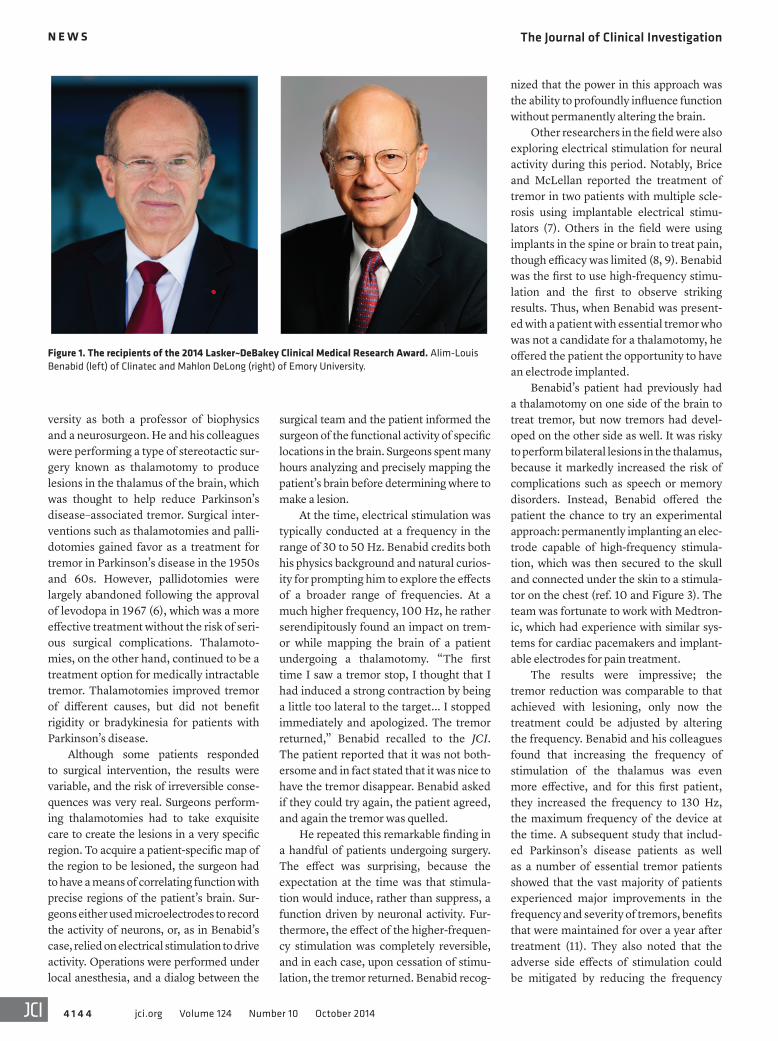
The basal ganglia consist of a cluster of nuclei that includes the caudate nucleus, putamen, globus pallidus, substantia nig-ra, and subthalamic nucleus (Figure 2A). While at the NIH, DeLong found that there were striking differences in the spontane-ous discharge of neurons in the nuclei of the basal ganglia and that neurons in the globus pallidus externus, globus pallidus internus, and subthalamic nucleus were involved in movement (2). DeLong and his colleagues identified specific cell activ-ity that correlated with body movements in distinct regions, such as the face, arms,
and legs (3). They found that cortical cen-tral motor areas of the brain transmit sig-nals through the basal ganglia in an orderly fashion to the thalamus and that the sig-nals then return to the cortex (Figure 2B). Subsequent work showed that neuronal activity in the motor circuit of the basal ganglia was correlated with the amplitude and direction of movement.
After his time at the NIH, DeLong completed his residency and continued working on mapping the circuitry of the basal ganglia at Johns Hopkins Univer-sity. He and his colleagues provided finer detail about the functional organization of the basal ganglia and came to recognize that the basal ganglia were also involved in non-motor activities such as emotional and cognitive functions along similar, but independent, lines. Along with Gar-rett Alexander and Peter Strick, DeLong published a seminal paper in the Annual Review of Neuroscience that proposed the existence of parallel, independent circuits in the basal ganglia that play distinct and separate roles in movement, emotion, and cognition (4). This was a watershed moment for both neurology and psychia-try, because it shifted the field away from the view that the basal ganglia served to funnel commands to the motor regions of the cerebral cortex toward an appreciation of their diverse roles in a wide spectrum of neurologic and psychiatric disorders (5).
A novel strategy to manipulate brain activityWhile DeLong was exploring the basal gan-glia at the NIH, Alim-Louis Benabid was completing his training at Joseph Fourier University. The son of a physician, Benabid had a strong interest in medicine but also sought training in research. Benabid simul-taneously pursued his residency in neuro-surgery while obtaining a PhD in physics. His doctoral studies focused on intracrani-al pressure and provided him with a math-ematical and biophysical framework for thinking about brain physiology.
After completing his training, Benabid continued working at Joseph Fourier Uni-Reference information: J Clin Invest. 2014;124(10):4143–4147. doi:10.1172/JCI78491.
The Journal of Clinical Investigation N e w s
4 1 4 4 jci.org Volume 124 Number 10 October 2014
nized that the power in this approach was the ability to profoundly influence function without permanently altering the brain.
Other researchers in the field were also exploring electrical stimulation for neural activity during this period. Notably, Brice and McLellan reported the treatment of tremor in two patients with multiple scle-rosis using implantable electrical stimu-lators (7). Others in the field were using implants in the spine or brain to treat pain, though efficacy was limited (8, 9). Benabid was the first to use high-frequency stimu-lation and the first to observe striking results. Thus, when Benabid was present-ed with a patient with essential tremor who was not a candidate for a thalamotomy, he offered the patient the opportunity to have an electrode implanted.
Benabid’s patient had previously had a thalamotomy on one side of the brain to treat tremor, but now tremors had devel-oped on the other side as well. It was risky to perform bilateral lesions in the thalamus, because it markedly increased the risk of complications such as speech or memory disorders. Instead, Benabid offered the patient the chance to try an experimental approach: permanently implanting an elec-trode capable of high-frequency stimula-tion, which was then secured to the skull and connected under the skin to a stimula-tor on the chest (ref. 10 and Figure 3). The team was fortunate to work with Medtron-ic, which had experience with similar sys-tems for cardiac pacemakers and implant-able electrodes for pain treatment.
The results were impressive; the tremor reduction was comparable to that achieved with lesioning, only now the treatment could be adjusted by altering the frequency. Benabid and his colleagues found that increasing the frequency of stimulation of the thalamus was even more effective, and for this first patient, they increased the frequency to 130 Hz, the maximum frequency of the device at the time. A subsequent study that includ-ed Parkinson’s disease patients as well as a number of essential tremor patients showed that the vast majority of patients experienced major improvements in the frequency and severity of tremors, benefits that were maintained for over a year after treatment (11). They also noted that the adverse side effects of stimulation could be mitigated by reducing the frequency
surgical team and the patient informed the surgeon of the functional activity of specific locations in the brain. Surgeons spent many hours analyzing and precisely mapping the patient’s brain before determining where to make a lesion.
At the time, electrical stimulation was typically conducted at a frequency in the range of 30 to 50 Hz. Benabid credits both his physics background and natural curios-ity for prompting him to explore the effects of a broader range of frequencies. At a much higher frequency, 100 Hz, he rather serendipitously found an impact on trem-or while mapping the brain of a patient undergoing a thalamotomy. “The first time I saw a tremor stop, I thought that I had induced a strong contraction by being a little too lateral to the target... I stopped immediately and apologized. The tremor returned,” Benabid recalled to the JCI. The patient reported that it was not both-ersome and in fact stated that it was nice to have the tremor disappear. Benabid asked if they could try again, the patient agreed, and again the tremor was quelled.
He repeated this remarkable finding in a handful of patients undergoing surgery. The effect was surprising, because the expectation at the time was that stimula-tion would induce, rather than suppress, a function driven by neuronal activity. Fur-thermore, the effect of the higher-frequen-cy stimulation was completely reversible, and in each case, upon cessation of stimu-lation, the tremor returned. Benabid recog-
versity as both a professor of biophysics and a neurosurgeon. He and his colleagues were performing a type of stereotactic sur-gery known as thalamotomy to produce lesions in the thalamus of the brain, which was thought to help reduce Parkinson’s disease–associated tremor. Surgical inter-ventions such as thalamotomies and palli-dotomies gained favor as a treatment for tremor in Parkinson’s disease in the 1950s and 60s. However, pallidotomies were largely abandoned following the approval of levodopa in 1967 (6), which was a more effective treatment without the risk of seri-ous surgical complications. Thalamoto-mies, on the other hand, continued to be a treatment option for medically intractable tremor. Thalamotomies improved tremor of different causes, but did not benefit rigidity or bradykinesia for patients with Parkinson’s disease.
Although some patients responded to surgical intervention, the results were variable, and the risk of irreversible conse-quences was very real. Surgeons perform-ing thalamotomies had to take exquisite care to create the lesions in a very specific region. To acquire a patient-specific map of the region to be lesioned, the surgeon had to have a means of correlating function with precise regions of the patient’s brain. Sur-geons either used microelectrodes to record the activity of neurons, or, as in Benabid’s case, relied on electrical stimulation to drive activity. Operations were performed under local anesthesia, and a dialog between the
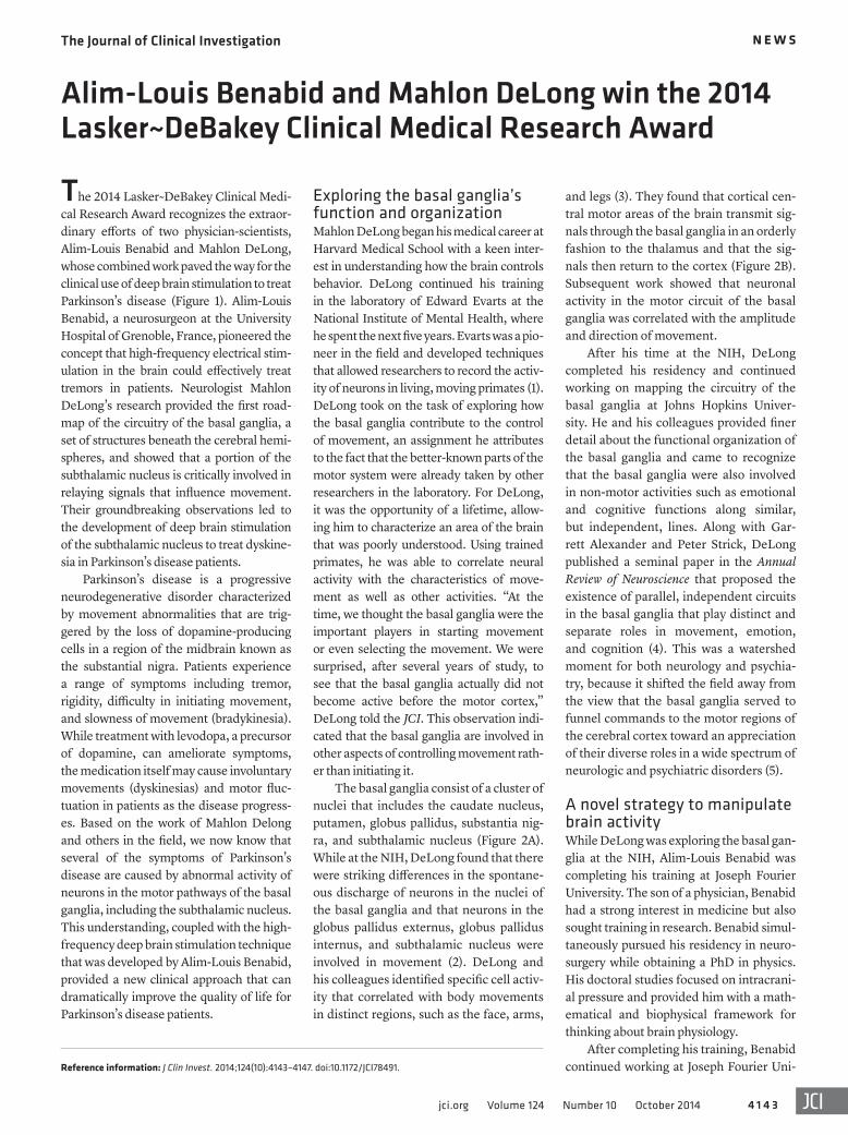
Figure 1. The recipients of the 2014 Lasker~DeBakey Clinical Medical Research Award. Alim-Louis Benabid (left) of Clinatec and Mahlon DeLong (right) of Emory University.
The Journal of Clinical Investigation N e w s
4 1 4 5jci.org Volume 124 Number 10 October 2014
of Parkinson’s disease and provided a pow-erful rationale for surgical intervention.
The notion of lesioning the subtha-lamic nucleus in patients was extremely controversial when DeLong’s Science arti-cle was published in 1990, because lesions targeting the pallidum had been widely used prior to the availability of levodopa, sometimes with disastrous results. Indeed, older physicians recalled cases from the 1950s and 1960s involving patients who were incapacitated; thus, these physicians were hesitant to return to performing palli-dotomies. In addition, Benabid told the JCI, “If you talked at that time about the subthalamic nucleus to a neurosurgeon, this was considered a forbidden place. The subthalamic nucleus is an area, as it happens, that can be destroyed by hem-orrhages in high blood pressure patients. When there is bleeding in this place, it makes a small hematoma, and then you would see the appearance of huge ballis-tic movements of the opposite limbs. This was extremely difficult and exhausting for those patients... And, of course, nobody wanted to be responsible for that.”
In parallel with DeLong’s work, Finn-ish neurosurgeon Lauri Laitinen and col-
a new way to create an accurate animal model of Parkinson’s disease (13). This development allowed DeLong to go beyond his studies of normal function to investigate the changes in neuronal activ-ity in the motor circuit responsible for Parkinson’s disease and, subsequently, to determine whether manipulation of the system could be palliative.
Using the MPTP primate model, Wil-liam Miller and DeLong found increased activity and abnormal output in neurons in the globus pallidus internus and sub-thalamic nucleus (14). With this result in mind, Hagai Bergman, Thomas Wich-mann, and DeLong again used the MPTP primate model to test whether inactivation of the subthalamic nucleus could amelio-rate symptoms (15). In this landmark study, they found that precisely placed lesions in MPTP-treated monkeys effectively reduced motor disturbances, including bradykinesia, tremor, and rigidity. Their findings were soon independently cor-roborated by the Crossman laboratory (16). Cumulatively, these studies confirmed the model that increased activity in the subtha-lamic nucleus and globus pallidus internus was responsible for many of the symptoms
or eliminating stimulation altogether, a unique benefit of this tunable approach. Deep brain stimulation targeting the thala-mus for the treatment of tremor associated with Parkinson’s disease or essential trem-or was approved in Europe in 1993 and subsequently in the United States in 1997.
Hitting the right targetWhile high-frequency deep brain stimu-lation to treat Parkinson’s disease was beginning to emerge, a variety of other approaches continued to be actively investigated. Mahlon DeLong and other researchers in the field uncovered new evidence indicating that the subthalamic nucleus was an important target in Par-kinson’s disease. William Langston and colleagues identified patients who devel-oped Parkinson-like symptoms after tak-ing an illegal synthetic heroin compound, meperidine (12). A contaminant known as MPTP (1-methyl-4-phenyl-1,2,5,6-tetrahydropyridine) was identified as the culprit underlying the striking effects in these patients caused by selective damage to the substantia nigra. This observation, though made under tragic circumstances, subsequently provided researchers with
Figure 2. The basal ganglia play a fundamental role in regulating movement. (A) A schematic representation of key parts of the brain involved in the motor circuit, including the cortex, basal ganglia (caudate nucleus, putamen, globus pallidus, subthalamic nucleus, and substantia nigra), and thalamus. (B) A simplified schematic of the normal motor circuit. The neurotransmitter GABA provides an inhibitory signal, while glutamate stimulates activity. Dopamine stimulates via the D1 receptor but provides an inhibitory signal via the D2 receptor. Movement signals are initiated in the cortex, transmitted to the striatum, and then travel in two different loops. In the direct pathway, dopamine provides an excitatory signal that triggers a net effect of stimulating the motor cortex. In contrast, in the indirect pathway, dopamine provides an inhibitory signal and has a net effect of inhibiting the cortex. In Parkinson’s disease, the loss of dopamine-producing cells in the substantia nigra primarily affects the indirect pathway and causes an increase in the inhibitory signal from the striatum to the globus pallidus externus, which reduces its normal constraint on the subthalamic nucleus. Thus, in Parkinson’s disease patients, increased outputs from the subthalamic nucleus and subsequently the globus pallidus internus are responsible for many of the movement symptoms caused by the loss of dopamine-producing cells.

The Journal of Clinical Investigation N e w s
4 1 4 6 jci.org Volume 124 Number 10 October 2014
Adoption of deep brain stimulation for clinical careBy 1998, deep brain stimulation targeting the subthalamic nucleus or the globus pal-lidus internus was approved in Europe to treat patients with advanced Parkinson’s disease. The benefits of deep brain stimu-lation targeting the subthalamic nucleus or globus pallidus were further demonstrated in large, randomized clinical trials (22), which led to FDA approval of the therapy in the United States in 2002. Since that time, over 100,000 patients have been treated for Parkinson’s symptoms with deep brain stimulation. Deep brain stimulation has proven more effective than standard medi-cal therapy in reducing dyskinesias and improving quality of life (23). The surgery for the implant carries a small risk of hem-orrhage and infection, so it is generally reserved only for patients whose symptoms can no longer be effectively controlled by medication. Surgery seems to work best on patients who respond to levodopa, and, as noted by Benabid’s group and others, can help patients decrease the amount of medication taken, which also helps reduce side effects. Patients must undergo care-ful screening to confirm the diagnosis and rule out contraindications. Once the sur-gery has been completed, patients must return for programming of the device in order to optimize the level of stimulation and adjust their medications. Ultimately, most patients realize substantial benefits such as elimination of drug-induced side effects and increased quality of life. Deep brain stimulation is not a cure for Parkin-son’s disease, but for many patients, the treatment sets the clock back to an earlier point in the disease’s progression, with a better overall level of functioning and few-er complications from medication.
Stimulating new treatment optionsDeep brain stimulation is also approved to treat other neurological movement disorders, including essential tremor and dystonia (under a Humanitarian Device Exemption [HDE]). For cases of primary dystonia, such as those due to genetic causes, deep brain stimulation of the globus pallidus internus can cut involun-tary muscle contractions by half or more. Unlike Parkinson’s disease, dystonia is not a degenerative disorder; therefore,
ganglia that had been developed in animal models to patients. They began perform-ing very carefully guided surgeries using microelectrode mapping, with highly accurate lesion placement in the motor region of the internal palladium, and saw remarkable benefit. DeLong recalled, “Our first patient was such a phenomenal success, I can’t even put it into words. It took us 12 or 13 hours to do the procedure, because we mapped the region very care-fully with microelectrodes and identified his targets.” This success led to NIH-fund-ed clinical trials that went on to confirm the benefits of pallidotomies (18).
The success of surgical lesions in the subthalamic nucleus prompted Benabid and his colleagues to test a new target for high-frequency deep brain stimulation. Because it was readily reversible, Bena-bid and his team decided to implant an electrode for deep brain stimulation into the subthalamic nucleus to treat symp-toms. “We had at hand this method that could alter activity without the complica-tions of a lesion... In the beginning of ’93, I operated on a patient who had severe, advanced Parkinson’s and for the first time put an electrode into the subthalamic nucleus. After four hours of investigation, we found the right place. And when we turned on the stimulation, we had a nice surprise. It worked, and the improvement was extremely impressive.” (19).
Shortly after Benabid and his col-leagues tested chronic stimulation of the subthalamic nucleus in their first patient, Siegfried and Lippitz published a report demonstrating that electrodes implanted into the globus pallidus internus, a down-stream target of the subthalamic nucleus (Figure 2B), improved tremor and rigid-ity in Parkinson’s patients (20). This was a time of incredible excitement for the field, with several lines of evidence now show-ing that two different regions within the basal ganglia could be targeted to alleviate the symptoms of parkinsonism in patients and that deep brain stimulation provided a potent, but reversible, approach. These findings were confirmed in larger studies, and Benabid’s team noted that deep brain stimulation of the subthalamic nucleus reduced the severity of symptoms for patients who were off their medication and allowed patients to decrease their dosage of levodopa (21).
leagues caused a major splash in the field when they published a 1992 article report-ing the benefits of pallidotomy for severely affected Parkinson’s disease patients. They revived techniques previously investigated during the 1950s by their mentor, Lars Lek-sell, that were largely out of practice in most parts of the world. The results were dramat-ic: approximately 80% of patients in their study had complete or nearly complete relief of tremor symptoms (17). Their work lent further support to the notion that it was pos-sible to achieve beneficial effects in patients by targeting the motor circuit portions of the basal ganglia, but subsequent efforts could incorporate the more precise circuitry that DeLong and his colleagues had uncovered to minimize surgical complications that had occurred in decades past.
DeLong, by this time chair of neu-rology at Emory University, along with neurologist Jerrold Vitek and stereotactic neurosurgeon Roy Bakay, began applying the improved understanding of the basal
Figure 3. Schematic of the deep brain stimula-tion implant used to treat Parkinson’s disease symptoms. An electrode is permanently implanted to stimulate the subthalamic nucleus or, alternatively, the globus pallidus internus. Through a mechanism that is still not well understood, stimulation reduces the abnormal discharge that underlies many of the symptoms of Parkinson’s disease. The implant is secured to the skull and connected under the skin to a stimulator and battery pack placed in the chest. An external programming device is used to adjust the frequency following surgery to opti-mize symptom relief and minimize side effects. Figure reproduced from ref. 25.
The Journal of Clinical Investigation N e w s
4 1 4 7jci.org Volume 124 Number 10 October 2014
11. Benabid AL, et al. Long-term suppression of tremor by chronic stimulation of the ven-tral intermediate thalamic nucleus. Lancet. 1991;337(8738):403–406.
12. Langston JW, Ballard P, Tetrud JW, Irwin I. Chronic Parkinsonism in humans due to a prod-uct of meperidine-analog synthesis. Science. 1983;219(4587):979–980.
13. Langston JW, Langston EB, Irwin I. MPTP-induced parkinsonism in human and non-human primates — clinical and experimental aspects. Acta Neurol Scand Suppl. 1984;100:49–54.
14. Miller WC, DeLong MR. Parkinsonian symp-tomatology. An anatomical and physiological analysis. Ann N Y Acad Sci. 1988;515:287–302.
15. Bergman H, Wichmann T, DeLong MR. Reversal of experimental parkinsonism by lesions of the subthalamic nucleus. Science. 1990;249(4975):1436–1438.
16. Aziz TZ, Peggs D, Sambrook MA, Crossman AR. Lesion of the subthalamic nucleus for the alleviation of 1-methyl-4-phenyl-1,2,3,6-tetrahy-dropyridine (MPTP)-induced parkinsonism in the primate. Mov Disord. 1991;6(4):288–292.
17. Laitinen LV, Bergenheim AT, Hariz MI. Leksell’s posteroventral pallidotomy in the treatment of Parkinson’s disease. J Neurosurg. 1992;76(1):53–61.
18. Vitek JL, et al. Randomized trial of pallidotomy versus medical therapy for Parkinson’s disease. Ann Neurol. 2003;53(5):558–569.
19. Limousin P, et al. Effect of parkinsonian signs and symptoms of bilateral subthalamic nucleus stimulation. Lancet. 1995;345(8942):91–95.
20. Siegfried J, Lippitz B. Bilateral chronic electro-stimulation of ventroposterolateral pallidum: a new therapeutic approach for alleviating all parkinsonian symptoms. Neurosurgery. 1994;35(6):1126–1130.
21. Limousin P, et al. Electrical stimulation of the subthalamic nucleus in advanced Parkinson’s disease. N Engl J Med. 1998;339(16):1105–1111.
22. Deep-Brain Stimulation for Parkinson’s Disease Study Group. Deep-brain stimulation of the subthalamic nucleus or the pars interna of the globus pallidus in Parkinson’s disease. N Engl J Med. 2001;345(13):956–963.
23. Weaver FM, et al. Bilateral deep brain stimula-tion vs best medical therapy for patients with advanced Parkinson disease: a randomized con-trolled trial. JAMA. 2009;301(1):63–73.
24. Loher TJ, et al. Deep brain stimulation for dys-tonia: outcome at long-term follow-up. J Neurol. 2008;255(6):881–884.
25. Williams NR, Okun MS. Deep brain stimulation (DBS) at the interface of neurology and psychia-try. J Clin Invest. 2013;123(11):4546–4556.
26. Mallet L, et al. Subthalamic nucleus stimulation in severe obsessive-compulsive disorder. N Engl J Med. 2008;359(20):2121–2134.
27. Holtzheimer PE, et al. Subcallosal cingulate deep brain stimulation for treatment-resistant unipo-lar and bipolar depression. Arch Gen Psychiatry. 2012;69(2):150–158.
28. Fisher R, et al. Electrical stimulation of the ante-rior nucleus of thalamus for treatment of refrac-tory epilepsy. Epilepsia. 2010;51(5):899–908.
of neurologists, neurosurgeons, engineers, mathematicians, and preclinical research-ers. Both Benabid and DeLong hope this sort of incubator atmosphere can speed the discovery of innovative approaches to treat a host of neurological conditions, and both remain deeply engaged in exploring new patient therapies.
This year’s Lasker~DeBakey Clini-cal Medical Research prize pays tribute to the legacy of two men who independently contributed in fundamental ways to the development of deep brain stimulation treatment for Parkinson’s disease. DeLong noted that the award is an incredible honor and described the experience as “unbeliev-able.” For his part, Benabid says that it has been amazing to be recognized by his peers, but he equally treasures hearing from his many patients who, throughout the years, have thanked him for the vast improve-ments in their daily lives. Indeed, he remarked, “What could be a better dream?”
Sarah Jackson
1. Evarts EV. Methods for recording activity of indi-vidual neurons in moving animals. In: Rushmer RF, ed. Methods in Medical Research. Chicago, Illinois, USA: Year Book Medical, 1966:241–250.
2. Georgopoulos AP, DeLong MR, Crutcher MD. Relations between parameters of step-tracking movements and single cell discharge in the globus pallidus and subthalamic nucleus of the behaving monkey. J Neurosci. 1983;3(8):1586–1598.
3. DeLong MR. The neurophysiologic basis of abnormal movements in basal ganglia disorders. Neurobehav Toxicol Teratol. 1983;5(6):611–616.
4. Alexander GE, DeLong MR, Strick PL. Parallel organization of functionally segregated circuits linking basal ganglia and cortex. Annu Rev Neu-rosci. 1986;9:357–381.
5. DeLong MR, Wichmann T. Circuits and circuit disorders of the basal ganglia. Arch Neurol. 2007;64(1):20–24.
6. Gregory R. Surgery for movement disorders. J Neu-rol Neurosurg Psychiatry. 2002;72(suppl 1):I32–I36.
7. Brice J, McLellan L. Suppression of intention tremor by contingent deep-brain stimulation. Lancet. 1980;1(8180):1221–1222.
8. Mullett K. Electrical brain stimulation for the control of chronic pain. Med Instrum. 1978;12(2):88–91.
9. Long DM, Erickson D, Campbell J, North R. Elec-trical stimulation of the spinal cord and peripher-al nerves for pain control. A 10-year experience. Appl Neurophysiol. 1981;44(4):207–217.
10. Benabid AL, Pollak P, Louveau A, Henry S, de Rougemont J. Combined (thalamotomy and stimulation) stereotactic surgery of the VIM thalamic nucleus for bilateral Parkinson disease. Appl Neurophysiol. 1987;50(1–6):344–346.
the benefits for patients who respond can often be sustained (24). The targets used for the treatment of movement disorders such as Parkinson’s and dystonia are the same, i.e., the nodes of the basal ganglia motor circuit in the subthalamic nucleus or globus pallidus internus.
Exciting new applications for deep brain stimulation are beginning to emerge for divergent neurological conditions that have completely different symptoms and different underlying neural circuitry. Alter-ations of the limbic circuit are thought to contribute to the development of Tourette syndrome, obsessive-compulsive disorder, and depression (25). A clinical trial testing stimulation of the subthalamic nucleus in patients with severe obsessive-compul-sive disorder demonstrated a decrease in symptoms and an increase in functionality (26). For patients with treatment-resistant depression, early clinical trial results of deep brain stimulation of the subcallosal cingulate indicated a significant decrease in depression and an increase in function following chronic stimulation (27). Posi-tive results have also been obtained with deep brain stimulation in epilepsy patients, and bilateral stimulation of the thalamus significantly reduced the frequency of sei-zures in patients with medically refractory seizures (28). Further studies are needed to confirm these findings, but these stud-ies illustrate that deep brain stimulation has important applications beyond Parkin-son’s disease and may someday benefit a wide range of patients.
A lifetime of discoveriesBenabid and DeLong both credit their dis-coveries to being in the right place at the right time and being open to new possibili-ties. For DeLong, the opportunity to pur-sue both clinical practice and research has been enormously rewarding. He continues to see patients who come to Emory for deep brain stimulation to treat their movement disorders and is a founding co-director of a new center focused on neuromodulation called ENTICe, which was formed by fac-ulty in neurology, psychiatry, and neuro-surgery at Emory in affiliation with faculty in engineering at the Georgia Institute of Technology. Benabid now heads Clinatec, a multidisciplinary institute in the small city of Grenoble, France, that is home to a team


![Capablanca - Lasker Match 1921 [Capablanca, 1921]](https://img.pdfslide.net/doc/110x75/577cda471a28ab9e78a54085/capablanca-lasker-match-1921-capablanca-1921.jpg)