Embed Size (px)
Citation preview

LETTERS ANDCORRESPONDENCE
Letters and correspondence submitted for possible publication mustbe identified as such. Text length must not exceed 500 words andfive bibliographic references. A single concise figure or table may beincluded if it is essential to support the communication. Letters nottyped double-spaced will not be considered for publication. Letters notmeeting these specifications will not be returned to authors. Letters tothe Editor are utilized to communicate a single novel observation orfinding. Correspondence is to be used to supplement or constructivelycomment on the contents of a publication in the journal and cannotexceed the restrictions for Letters to the Editor. The Editor reservesthe right to shorten text, delete objectional comments, and makeother changes to comply with the style of the journal. Permission forpublication must be appended as a postscript. Submissions must besent to Paul Chervenick, M.D., Editor of Brief Reports/Letters toEditors, American Journal of Hematology, H. Lee Moffitt CancerCenter, University of South Florida, 12902 Magnolia Drive, Tampa,FL 33612 to permit rapid consideration for publication.
All- trans Retinoic Acid-InducedMultiple Mononeuropathies
To the Editor:All-trans retinoic acid (ATRA) is widely used as a differ-entiation therapy to induce a complete remission in patients with acutepromyelocytic leukemia (APL) [1]. The major adverse effect during ATRAtherapy is an increase of leukocytes, which is often accompanied by reti-noic acid syndrome [2]. This reaction is characterized by fever, respiratorydistress, radiographic pulmonary infiltrates, pleural effusions, and weightgain [3]. It has also been reported that ATRA-associated neurologicalsymptoms such as headache and pseudotumor cerebri [4] are mainlycaused by intracranial hypertension. We describe a patient who developedmultiple mononeuropathies during ATRA therapy for APL.
A 23-year-old Japanese female was admitted with fever and purpura inNovember 1997. APL with disseminated intravascular coagulation wasdiagnosed. She received ATRA (45 mg/m2/day) with chemotherapy in-cluding daunorubicin (40 mg/m2/day, on days 9, 10, 21, and 22) andbehenoyl cytarabine (200 mg/m2/day, on days 9, 10, 11, 21, 22, and 23),and supportive therapy for disseminated intravascular coagulation. She hada slight headache after administration of ATRA, but there were no findingson the neurological examination and computed tomography scan of thebrain. On the seventeenth day after admission, a neurosurgical operationwas performed because of acute subdural hematoma. Although ATRA-associated headache persists, no symptoms remained, which were causedby intracranial hemorrhage and operation. Furthermore, she complained ofdiplopia, and burning pain and contact dysaesthesiae at the dorsum of theleft hand and the right foot on day 21 of the ATRA treatment. She also hadweakness in the same extremities. Tendon reflexes were normal. Electro-physiologic study revealed a decrease of right peroneal nerve conductionvelocity and amplitude. Although right abducens nerve palsy and visualdisturbance were present, there were no papilloedema and abnormalities onmagnetic resonance imaging of the brain. Lumbar puncture showed normalcerebrospinal fluid with normal pressure. She had no manifestation ofautonomic disturbances. ATRA was discontinued because a complete re-mission was achieved on day 51 after administration of ATRA. Totaldosage of ATRA was 3,390 mg. She received three times the intensive
postremission chemotherapy (daunorubicin, cytarabine, mitoxantrone,mercaptopurine, and prednisolone). Symptoms of peripheral neuropathyand findings on electrophysiologic study gradually improved as well asheadache and dry skin after discontinuation of ATRA, in spite of additionalchemotherapy. The right abducens nerve palsy also partially improved.
To the best of our knowledge, this is the first report of ATRA-inducedmultiple mononeuropathies. Neurological symptoms might be consideredas atypical pseudotumor cerebri with focal neurological sign [5]. However,there was no evidence of intracranial hypertension. Furthermore, we care-fully excluded a possibility that other medication including anticancerdrugs induced peripheral neuropathy. Clinicians should be aware of thispotential side effect in patients receiving ATRA.
SATOSHI YAMAJI
HEIWA KANAMORI
AKI MISHIMA
SHIN FUJISAWA
SHIGEKI MOTOMURA
HIROSHI MOHRI
The First Department of Internal Medicine, Yokohama City UniversitySchool of Medicine, Yokohama, Japan
REFERENCES
1. Huang ME, Ye YC, Chen SR, Chai JR, Lu JX, Zhoa L, Gu LJ, Wang ZY: Use ofall-trans retinoic acid in the treatment of acute promyelocytic leukemia. Blood72:567–572, 1988.
2. Frankel SR, Eardley A, Lauwers G, Weiss M, Warrell RP Jr: The ‘‘retinoic acidsyndrome’’ in acute promyelocytic leukemia. Ann Intern Med 117:292–296, 1992.
3. Warrell RP, Jr., de The H, Wang ZY, Degos L: Acute promyelocytic leukemia. NEngl J Med 329:177–189, 1993.
4. Digre KB, Corbet JJ: Pseudotumor cerebri in men. Arch Neurol 45:866–872, 1988.5. Round R, Keane JR: The minor symptoms of increased intracranial pressure: 101
patients with benign intracranial hypertension. Neurology 38:1461–1464, 1988.
Soft Drink Abuse, Malnutrition, and Folic Acid Deficiency
To the Editor:Herein, we report a 28-year-old woman who presented withsevere macrocytic anemia in November 1996. Her past medical history wasunremarkable except for asthma and one episode of acute synovitis a fewyears earlier. Her blood count was normal two years earlier.
She is a housekeeper who drank alcohol very rarely and used nonste-roidal anti-inflammatory drugs occasionally. She presented with a historyof weakness, a 7.5 kg weight loss in the past two months and of nausea,vomiting, and loss of appetite in the last three weeks. Her food intake forthis period consisted almost exclusively of the soft drink Coca-Cola. Therewas no malabsorption symptoms. Physical examination revealed a severeobesity (126 kg, 1.58 m, body mass index over 50). She was slightly ictericand her tongue was reddened and smooth with papillary atrophy. Theneurological examination was normal. The first blood count showed asevere anemia with a hemoglobin (Hg) of 51 g/l, a hematocrit of 0.147 L/L(0.37–0.47), a mean corpuscular volume (MCV) of 99.5 fl (normal 82–94),and reticulocytes at 1.8%. Leukocytes were reduced to 3.3 × 10⁹ g/l (nor-mal 150–450).
Blood smear revealed a marked anisocytosis, no ovalocytosis, quite afew dacryocytes, polychromatophilia, and erythroblasts. A discrete my-elemia with a left shift was noted; there were no hypersegmented neutro-
American Journal of Hematology 60:311–314 (1999)
© 1999 Wiley-Liss, Inc.
phils. The biochemical analysis was remarkable only for an elevated lactatedehydrogenase level (3,198 U/l) and bilirubin (52mmol/l). Serum levels ofproteins and albumin were normal as was the iron status. However, serumlevel of folic acid showed a severe deficiency with values below 1.3 nmol/l(normal above 3.5, deficiency below 2.5). Serum level of vitamin B12 was100 pmol/l (normal above 150, deficiency below 100). Intrinsic factorantibody was negative.
A malabsorption evaluation was performed, including duodenal biop-sies, and was noncontributory. A bone marrow aspiration was performedand was compatible with megaloblastic anemia; there was a marked ery-throid dysplasia, a few giant metamyelocytes, and the myeloid on erythroidratio fell to 1.5:1. Alkaline phosphatase of leukocytes score was elevated(212, normal 20–80). Haptoglobin was undetectable and direct Coombs’test was negative. Hepatic function tests, including viral serologies werenormal, as were the thyroid function tests. The sucrose hemolysis test andHam’s acid hemolysis test were negative.
A careful review of the patient’s dietary habits showed poor nutrition,consisting mostly of bread, rice, potatoes, liver, and a great amount of dairyproducts. She regularly ate snacks consisting of candies, ice cream, andpotato chips. However, what was most remarkable was the consumption ofapproximately three liters of Coca-Cola per day. This soft drink does notcontain any folic acid and has no known folic acid inhibitor. Dietaryevaluation showed an energetic intake of 1,500 kcal per day, 40 mg ofproteins per day, and 70 mg per day of folic acid (normal is 185 mg perday). The patient received one transfusion of packed red cells, 1,000 mg ofB12 vitamins and folic acid, 5 mg IV for one dose, then 5 mg po die forfour days, 10 mg po die for two days, 15 mg po die for two monthsfollowed by 5 mg po die for two months. Then it was stopped. She alsoreceived nutritional counseling.
The patient improved rapidly with disappearance of her symptoms ofweakness and vomiting. Ten days after beginning her treatment, her bloodcount showed a Hb increase from 51 to 79 g/l, and an MCV of 97 fl;reticulocytes were increased at 18%. Platelets were still at 141 × 109 g/l butleukocytes were normal. Seventeen days later, Hb was 105 g/l and plateletswere normal.
After one year of follow-up, her blood count remains normal; serumlevels of folic acid and vitamin B12 are normal. She is taking neither folicacid nor B12 vitamin supplements. Her diet has improved but remainsdeficient.
This case is one of severe macrocytic anemia due to a severe deficit infolic acid. This is the first case to our knowledge in which excessiveconsumption of soft drinks combined with malnutrition is reported as themain cause of folic acid deficiency. This deficiency has been reported sofar mainly in a population of alcoholics and substance abusers. The un-derlying mechanism has not been elucidated.
It is interesting to note that this patient was severely obese, suggestingthat malnutrition must not be overlooked, even in this population; intake ofvitamins and elements is expected to be sufficient but quality of intake maycause a significant deficiency in one or more of these.
Finally, it is a reminder that nutrition status must be well assessed andthat counseling is crucial.
GUYLAINE GAUDET
Fellow in Hematology, Hopital du Sacre-Cœur de Montreal, AffiliatedHospital, University of Montreal, Canada
JACQUES LAPLANTE
Hemato-Oncologist, Hopital du Sacre-Cœur de Montreal, AffiliatedHospital, University of Montreal, Canada
Sex Difference in Myeloperoxidase Activity of Neutrophils
To the Editor:Activated neutrophils produce several antimicrobial oxygenmetabolites, including superoxide anion, hydrogen peroxide, hydroxyl
radicals, hypochlorous acid, and single oxygen [1]. These reactive metabo-lites are generated by a reduced nicotinamide adenine dinucleotide phos-phate (NADPH)-dependent oxidase, located on the plasma membrane, anda myeloperoxidase (MPO), located in the lysosome [2]. The former threemetabolites are produced even in MPO deficiency, so it may be difficult toconsider that MPO deficiency is one of the causes for clinically significantfailure of neutrophil function. However, in some patients with MPO defi-ciency, the cytotoxic activities against bacteria and fungi are decreased inneutrophils [3,4]. Therefore, measurement of MPO activity in neutrophilsmay be useful for evaluating a part of the defense mechanism. Recently, anautomated hematology system, which differentiates white blood cells, hascome into widespread use. With this system, MPO activity, which repre-sents the MPO staining intensity, is measured as the mean myeloperoxidaseindex (MPXI) of neutrophils. In this study, therefore, we examined MPOactivities of neutrophils in normal subjects.
The subjects studied were 41 normal men (mean age, 36 years; range,19–57 years), and 46 normal women (mean age, 32 years; range, 21–47years). The MPXI of peripheral blood was measured using the automatedhematological analyzer THMS-H2 (Bayer Technicon, Tarrytown, NY). Wefound that the MPXI in normal women (n4 46, 0.3 ± 2.7, mean ± SD,P< 0.01) was significantly higher than that in normal men (n4 41, −1.8 ±2.4) (Fig. 1). This sex difference suggests that some microbicidal activitymay be stronger in women than in men. In support of this finding, it wasreported in mass surveys that the infection rate of women was lower thanthat in men in the tinea of dermatophytic infection [5]. Furthermore, thissuggests that sex steroid hormones may affect MPO activity, that is theMPXI. Indeed, we found that the change of the MPXI in a woman syn-chronized with that of basal body temperature and that of serum concen-tration of estradiol with some phase shift, during three menstrual cycles.However, the MPXI in a man did not change during the 12 weeks ofmeasurement. Also, the results of nitro blue tetrazolium (NBT) reductionand neutrophil number did not change during this period in both subjects.To study the direct effect of sex hormone on the MPO activity, we exam-
Fig. 1. The mean MPXI of neutrophils in normal men andwomen.
312 Letters and Correspondence
ined the changes of MPXI 2 and 3 hr after the addition of estrone (5,000pg/mL), B-estradiol (5,000 pg/mL), estriol (5,000 pg/mL), or testosterone(100 ng/mL) to peripheral blood in vitro, but we found no effect.
Taken together, these data indicate that: 1. the MPXI of neutrophils,which is measured easily by an automated hematological analyzer andrepresents MPO activity, is higher in women than in men: 2. the menstrualcycle affects the MPXI; and 3. the MPXI may be useful as a partial indexfor microbicidal activity of neutrophils.
OSAMU KABUTOMORI
TAKEHIKO YANAGIHARA
The Central Laboratory for Clinical Investigation, Osaka UniversityHospital, Osaka, Japan
YOSHINORI IWATANI
Department of Clinical Laboratory Science, Faculty of Medicine, OsakaUniversity, Osaka, Japan
ATSUSHI KAWARAZAKI
MIWAKO KABUTOMORI
Kawarazaki Hospital, Osaka, Japan
REFERENCES
1. Borregaard N. Bactericidal mechanisms of the human neutrophil. Scand J Hae-matol 1984;32:225–230.
2. Curnutte JT, Babior BM. Composition of neutrophils. In: Williams WJ, Beutler E,Erslev AJ, Lichman MA, editors. Hematology. 4th ed. New York: McGraw-HillBook Co.; 1990. p 770–774.
3. Cech P, Papathanassiou A, Boreux G, Roth P, Miescher PA. Hereditary myeloper-oxidase deficiency. Blood 1979;53:403–411.
4. Lehrer RI, Cline MJ. Leukocyte myeloperoxidase deficiency and disseminate can-didiasis. J Clin Invest 1969;48:1478–1488.
5. Nowicki R. Dermatophytoses in the Gdansk area. Poland. A 12-year survey.Mycoses 1996;39:399–402.
Case of Hepatosplenic gd T-Cell Lymphoma PresentingWith Severe Hypersplenism
To the Editor:Pancytopenia with severe neutropenia at presentation is arare finding in hepatosplenicgd T-cell lymphoma (TCL) [1–4].
In March 1997, an 18-year-old male was hospitalized with fever, ab-dominal discomfort, and fatigue. Despite the absence of either superficialor profound lymphadenopathies as confirmed by a computerized tomo-graphic scan of the chest, abdomen and pelvis, hepatomegaly with massivesplenomegaly, 3 and 23 cm below the costal margins, respectively, werenoted. The peripheral blood count showed pancytopenia and severe neu-tropenia (Hb 6.3 g/dl, Hct 20%, platelets 20 × 109/l,leukocytes 2 × 10⁹/l,neutrophils 0.68 × 10⁹/l). No abnormal cells were detected in the peripheralblood and in the bone marrow (aspirate and biopsy). Biochemistry resultswere within normal limits except moderate elevations in alkaline phospha-tase, lactic dehydrogenase, andb2-microglobulin levels. In May 1997,splenectomy was performed. Histologic examination of the spleen showedatrophy of the germinal centers, with partial destructions of the white pulp.The red pulp seemed expanded with its sinusoids homogenously infiltratedwith lymphoid cells, which were positive for LCA and CD3. A similarinfiltration pattern was observed in the hepatic sinusoids.
After splenectomy, the liver enlarged to the umbilicus. The pancytopeniaimproved (Hb 11.7 g/dl, Hct 34.7%, platelets 256 × 109/l, neutrophils 4 ×10⁹/l) but the peripheral blood smear showed atypical lymphoid cells vary-ing between 35% and 45%. A second bone marrow biopsy three monthsafter splenectomy showed an infiltration with the same neoplastic cells ina diffuse interstitial pattern. In the immunophenotyping of peripheral bloodafter splenectomy with flow cytometry, the ratio of thegd T-cell receptor
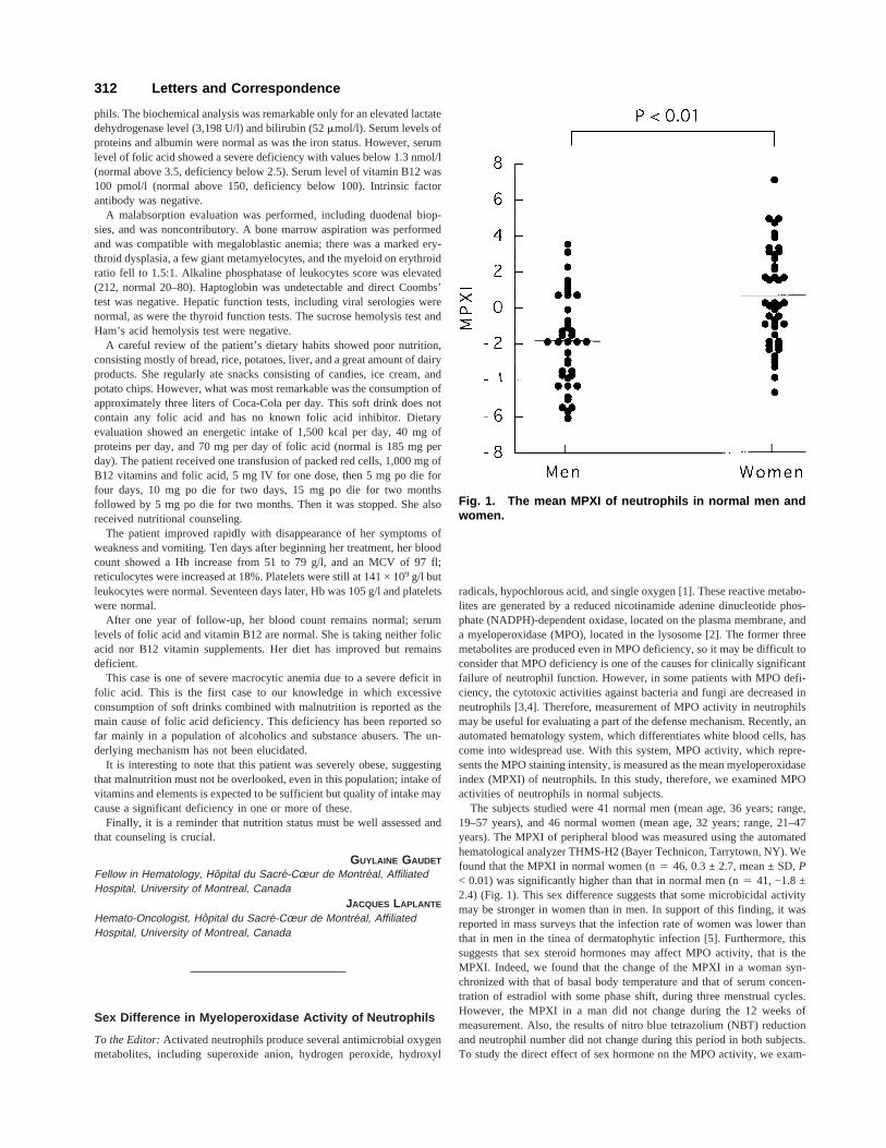
(TCR) bearing lymphoid cells was 59%. CD2, CD3, CD5, CD7, and CD56positivity ranged between 70 and 99%. The heteroduplex analysis of pe-ripheral blood showed that this pattern carried the monoclonal TCR Vg-1gene (Fig. 1).
The patient did not respond to chemotherapy. Neither regimens withconventional doses (CHOP: cyclophosphamide, doxorubicin, vincristine,and prednisone) nor that with high doses of methotrexate and/or cytosinearabinoside (NHL-BFM-90) proved to be effective. In May 1998 his con-dition deteriorated progressively and he died 13 months after diagnosis.
The presence of pancytopenia with severe neutropenia at presentation,seems to be an uncommon manifestation as it has been observed in onlythree of the 23 patients with hepatosplenicgd TCL reported in the medicalliterature until the time of this writing [3,4]. Thus, the patient presentedseems to be the fourth case displaying this feature. Another importantfinding in this case was the presence of hypersplenism due to the infiltra-tion of the red pulp of the spleen in a manner resembling that seen in hairycell leukemia. Splenectomy has, similarly, corrected this hypersplenismonly by increasing the number of peripheral blood cells [5] without anyeffect on the disease process, progression being evident by the appearanceof abnormal lymphoid cells in the peripheral blood. Thus, despite high-dose chemotherapy, the course of the disease was concluded as progressivein this case due to the persistence of B symptoms, increased hepatomegaly,and peripheral blood involvement. It can also be concluded that hyper-splenism may also account for the cytopenias besides bone marrow infil-tration and that splenectomy, like chemotherapy, seems ineffective in al-tering the progressive course of the disease.
ACKNOWLEDGMENTS
The Authors are indebted to Prof. Dr. Andrea Biondi (Milan, Italy) forhis help and interest and who kindly performed the heteroduplex analysisof the peripheral blood.
GÜNCAG DI.NCOL
MELIHA NALCACI
A. SELIM YAVUZ
HÜSEYIN KESKIN
MELIH AKTAN
Division of Hematology, Istanbul Medical School, Istanbul, Turkey
Fig. 1. Polyacrylamide gel electrophoresis with TCR V g1and Vg2 primers after PCR amplification. (PCR with TCRVg1 and Vg2 primers were for lanes 1 to 4 and 6 to 9, re-spectively. Lanes 1 and 6: case A; lanes 2 and 7: our patient;lanes 3 and 8: positive control; lanes 4 and 9: typical poly-clonal smear sample from normal lymphocytes; lane 5:Marker-BM no. VIII)
Letters and Correspondence 313
ONER DOGAN
MEHMET AGAN
Division of Pathology, Istanbul Medical School, Istanbul, TurkeyUGUR OZBEK
Institute for Experimental Medicine, University of Istanbul, Istanbul,Turkey
KORAY DINCOL
Institute of Oncology, University of Istanbul, Capa, Istanbul, Turkey
REFERENCES
1. Farcet JP, Gaulard P, Marolleau JP, Le Couedic JP, Henni T, Gourdin MF, DivineM, Haioun C, Zafrani S, Goossens M, Hercend T, Reyes F. Hepatosplenic T-cell
lymphoma: sinusal/sinusoidal localisation of malignant cells expressing the T-cellreceptorgd. Blood 1990;75:2213.
2. Cooke CB, Krenacs L, Stetler-Stevenson, Greiner TC, Raffeld M, Kingma DW,Abruzzo L, Frantz C, Kaviani M, Jaffe ES. Hepatosplenic T-cell lymphoma: adistinct clinicopathologic entity of cytotoxicgd T-cell origin. Blood 1996;88:4265.
3. Dommann-Scherrer CC, Kurer SB, Zimmerman DR, Odermatt BF, Durs-Zimmermann MT, Briner J, Heitz PU. Occult hepatosplenic T-gamma delta lym-phoma: value of genotypic analysis in the differential diagnosis. Virchows Arch1995;426–629.
4. Salhany KE, Feldman M, Kahn MJ, Peritt D, Schretzenmair RD, Darren M,Wilson DM, Dipaola RS, Glick AD, Kant JA, Nowell PC, Kamoun M. Hepato-splenic gd T-cell lymphoma: ultrastructural, immunophenotypic and functionalevidence for cytotoxic T lymphocyte differentiation. Hum Pathol 1997;28:674.
5. Jansen J, Hermans J. Splenectomy in hairy cell leukemia: a retrospective multi-center analysis. Cancer 1981;47:2066.
314 Letters and Correspondence