Embed Size (px)
Citation preview

Allostasis:
The Fundamental Biology and Implications for Social Standing and Longevity
by
Barry Nguyen
2
Table of Contents
ABSTRACT ................................................................................................................... 3
INTRODUCTION ........................................................................................................... 4
ALLOSTASIS ................................................................................................................. 5
PRIMARY MEDIATORS .......................................................................................................... 5 Glucocorticoids .......................................................................................................... 5 Dehydroepiandrosterone ........................................................................................... 5 Cytokines ................................................................................................................... 5 Catecholamines ......................................................................................................... 6
ALLOSTATIC LOAD ............................................................................................................... 6
CONSEQUENCES .......................................................................................................... 7
NEURODEGENERATION ......................................................................................................... 7 Interleukin-1α & Interleukin 1 β ................................................................................. 7 Glucocorticoid Cascade Hypothesis ........................................................................... 8
SOCIETAL IMPLICATIONS ........................................................................................... 10
SOCIAL STANDING ............................................................................................................. 10 LONGEVITY ...................................................................................................................... 12
CONCLUSION ............................................................................................................. 12
3
ABSTRACT
In the face of external adversities, the human body utilizes a vast array of physiological systems to combat the perceived threat in order to keep the internal conditions in balance. This intricate and well-coordinated process is known as allostasis. Among the various hormones that are secreted by the allostatic networks, four play a prominent role in the stress response: glucocorticoids, dehydroepiandrosterone, cytokines, and catecholamines. To counteract the effects of the stress stimulus, the hormones work to keep systems in balance, shifting internal conditions as needed. However, as the body continuously deals with stress through the activation of the allostatic process, the physiological processes associated with the response slowly degrades, and the body enters a state known as allostatic overload. In addition to negatively affecting the body's capacity to deal with stress, allostatic overload entails an even greater risk: neurological degeneration. Increased stress levels associated with increased hormonal secretion have been observed to be closely linked to certain degenerative changes in the brain. Stress, a determinant of allostatic load in particular, is derived from external forces or experiences. Such experiences differ greatly across social classes and therefore, confer differential effects. Furthermore, inferences can be made on differential manifestations of allostatic load across differing social standings. In doing so, the effects of allostasis can be quantified and implications on a larger scale can be made.

4
INTRODUCTION
Allostasis is an important biological process and it describes the process in which the body regains homeostasis during a stress response. It aids greatly to survival in the short run, but as the human body ages, the mechanistic dysfunctionalities that arise give way to internal pathophysiology, posing a threat to overall longevity.
In the 1900s, the general adaptation syndrome, coined by scientist Selye, was largely used to describe the body’s stress response pathway (Jansen & Roberta 2016). The process is broken up into 3 stages: alarm, resistance, and exhaustion (Higuera 2018). The alarm stage is the fight or flight response where the body experiences a surge of energy in the face of a stress stimulus. If the stressor persists, the body enters the resistance stage. In this stage, the body adapts to a higher stress level through the continual secretion of hormones. Prolonged stress leads the body to the last stage, exhaustion. At this final stage, the body is stripped of its energy and is no longer able to deal with stress, leaving the individual vulnerable to stress-related diseases.
Fig. 1. A model of the stages of the general adaptation syndrome (Redrawn from Myers, 2008).
Adapting some of the core concepts of the general adaptation syndrome, scientists Sterling and Eyer introduced allostasis in 1988, which they defined as achieving stability through change (Ramsay & Woods 2014). It is the process in which the body utilizes a vast array of physiological processes to maintain stability in the internal environment. It is analogous to homeostasis, the process of mitigating deviations from acceptable ranges using positive and negative feedback loops. Unlike homeostasis, allostasis is operated by four primary hormones and they are key players in the maintenance of the body's internal parameters during a stress response. While homeostasis keeps conditions within narrow ranges, allostasis adjusts the internal parameters to match the perceived circumstances. In the initial stage of a stress response pathway, the allostatic mediators are released to vary the body's internal parameter. Consequently, chronic stress can lead to the dysregulation of the allostatic mediating systems, propelling the body toward a state of systematic wear and tear. The resulting ramifications can have detrimental consequences on the brain.
5
ALLOSTASIS
In the initial stages of the stress responses pathway, four primary mediators are secreted from the body: glucocorticoids, dehydroepiandrosterone, cytokines, and catecholamines. This interconnected network of hormones plays an essential part in helping the body respond to a stress stimulus. The interplay of such hormones have subsequent effects on one another, attributing to the activity of the other and ultimately, contributing to the strength of the overall allostatic response.
Primary Mediators
Glucocorticoids
Glucocorticoids (GC) are released by the hypothalamic-pituitary-adrenal (HPA) axis during a stress response and are one of the most versatile hormones (McEwen 2003). Among the many domains they play a role in, GCs play a key part in the regulation of cardiovascular functions, inflammation, and fluid volume. During cardiovascular distress, they help enhance cardiovascular function by increasing blood pressure and cardiac output. They also suppress sites of inflammation and mobilize immune cells to sites of infection. In other words, they shape and direct the nature of an immune response (Frank et al. 2013). During a hemorrhage stressor, vasoconstrictive stress hormones are released, and GCs have been observed to repress these secretions.
Dehydroepiandrosterone
Dehydroepiandrosterone (DHEA) is secreted by the adrenal gland; it is another prominent entity during the stress response (McEwen 2003). Although there are no receptors known currently in tissues for DHEA, it still has a prominent role due to its regulation of GCs. DHEA can be seen as a functional antagonism of the actions of GCs. The antagonistic dynamic of the two mediate opposing physiological functions. For example, DHEA regulates the immune system passively by suppressing GC-induced immune responses (Kamin & Kertes 2017). Additionally, DHEA has been shown to negate the neurotoxic effects of corticosterone, a GC and suppress cortisol induced neurodegeneration. In the short run, DHEA can be viewed to offset the negative effects caused by an over-active, overproduced GC and keep its activity under control.
Cytokines
Many cytokines are referred to as interleukins due to being secreted by leukocytes, and they are generally classified into two categories: proinflammatory and anti-inflammatory (McEwen 2003). One major difference between the two types of cytokines is that the latter inhibits the former's production. This class of mediators is produced primarily in immune cells but have been found to be produced in the brain and liver. When the body elicits an inflammatory response, cytokine production increases (Zhang & An 2009). Among the many allostatic synergies, there exists an additional regulatory property between cytokines and glucocorticoids: the latter has been found to induce the former's synthesis.
6
Catecholamines
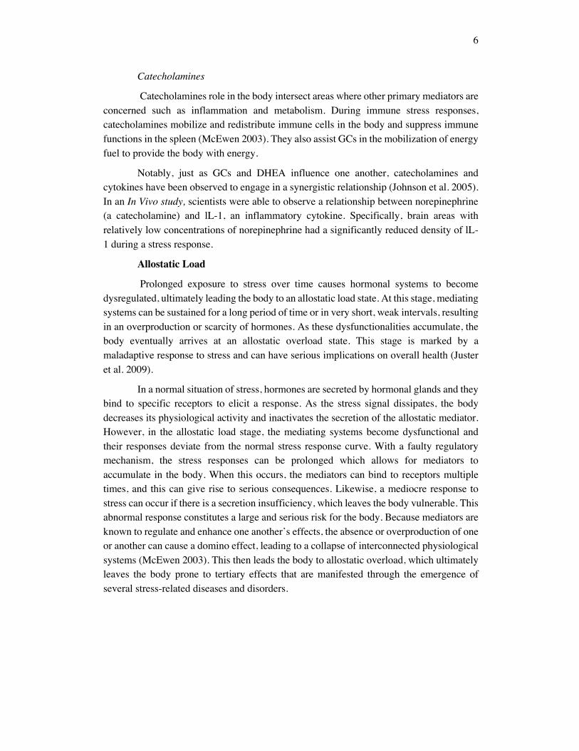
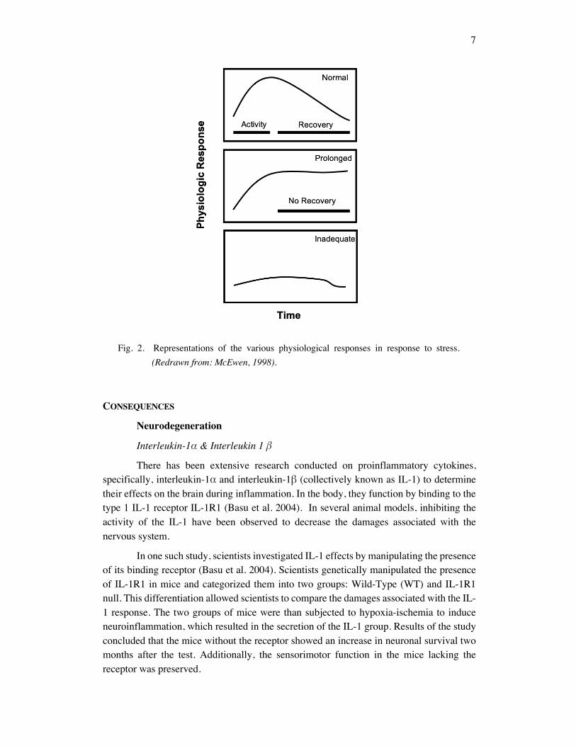
Catecholamines role in the body intersect areas where other primary mediators are concerned such as inflammation and metabolism. During immune stress responses, catecholamines mobilize and redistribute immune cells in the body and suppress immune functions in the spleen (McEwen 2003). They also assist GCs in the mobilization of energy fuel to provide the body with energy.
Notably, just as GCs and DHEA influence one another, catecholamines and cytokines have been observed to engage in a synergistic relationship (Johnson et al. 2005). In an In Vivo study, scientists were able to observe a relationship between norepinephrine (a catecholamine) and lL-1, an inflammatory cytokine. Specifically, brain areas with relatively low concentrations of norepinephrine had a significantly reduced density of lL-1 during a stress response.
Allostatic Load
Prolonged exposure to stress over time causes hormonal systems to become dysregulated, ultimately leading the body to an allostatic load state. At this stage, mediating systems can be sustained for a long period of time or in very short, weak intervals, resulting in an overproduction or scarcity of hormones. As these dysfunctionalities accumulate, the body eventually arrives at an allostatic overload state. This stage is marked by a maladaptive response to stress and can have serious implications on overall health (Juster et al. 2009).
In a normal situation of stress, hormones are secreted by hormonal glands and they bind to specific receptors to elicit a response. As the stress signal dissipates, the body decreases its physiological activity and inactivates the secretion of the allostatic mediator. However, in the allostatic load stage, the mediating systems become dysfunctional and their responses deviate from the normal stress response curve. With a faulty regulatory mechanism, the stress responses can be prolonged which allows for mediators to accumulate in the body. When this occurs, the mediators can bind to receptors multiple times, and this can give rise to serious consequences. Likewise, a mediocre response to stress can occur if there is a secretion insufficiency, which leaves the body vulnerable. This abnormal response constitutes a large and serious risk for the body. Because mediators are known to regulate and enhance one another’s effects, the absence or overproduction of one or another can cause a domino effect, leading to a collapse of interconnected physiological systems (McEwen 2003). This then leads the body to allostatic overload, which ultimately leaves the body prone to tertiary effects that are manifested through the emergence of several stress-related diseases and disorders.
7
Fig. 2. Representations of the various physiological responses in response to stress. (Redrawn from: McEwen, 1998).
CONSEQUENCES
Neurodegeneration
Interleukin-1α & Interleukin 1 β
There has been extensive research conducted on proinflammatory cytokines, specifically, interleukin-1α and interleukin-1β (collectively known as IL-1) to determine their effects on the brain during inflammation. In the body, they function by binding to the type 1 IL-1 receptor IL-1R1 (Basu et al. 2004). In several animal models, inhibiting the activity of the IL-1 have been observed to decrease the damages associated with the nervous system.
In one such study, scientists investigated IL-1 effects by manipulating the presence of its binding receptor (Basu et al. 2004). Scientists genetically manipulated the presence of IL-1R1 in mice and categorized them into two groups: Wild-Type (WT) and IL-1R1 null. This differentiation allowed scientists to compare the damages associated with the IL-1 response. The two groups of mice were than subjected to hypoxia-ischemia to induce neuroinflammation, which resulted in the secretion of the IL-1 group. Results of the study concluded that the mice without the receptor showed an increase in neuronal survival two months after the test. Additionally, the sensorimotor function in the mice lacking the receptor was preserved.
8
Cytokines can impact the brains functionality due to inhibition of neurogenesis (Borsini et al. 2015). Neurogenesis is the process in which new neurons are generated from neural stem cells (NSC). In adult brains, this primarily occurs in the subventricular zone and sub granular zone (SGZ). The newly formed neurons are then differentiated and are integrated in different parts of the nervous system where they serve their function. Neurons with the capabilities to renew themselves are classified as neural progenitor cells. Considering that cytokines have multiple pathways for communicating and interacting with the nervous system, they can easily threaten the nervous system's stability by interfering with the NSC (Fan & Pang 2017).
Chronic secretion of the inflammatory mediators has been experimentally proven to impose a serious threat to the brain's neural plasticity. It is important to note that neural plasticity is a process in which the brain generates new neurons. This allows for the brain to continue to adapt and to learn. IL-1 and IL-6 have shown to reduce the proliferation and differentiation of newborn cells and inhibit SGZ neuroprogenitors, hence inhibiting neurogenesis (Sierra 2014). In mice models, the same cytokines have shown to be pivotal in the suppression of hippocampal neurogenesis (Das 2008). By interrupting crucial stages like cell proliferation and differentiation, the inflammatory mediators adversely affect neurogenesis, resulting in the net decrease of neurons. It is important to note that the harmful effects of inflammation are widely determined by the levels of inflammatory mediators produced. Furthermore, in cases of physiological dysfunction, uncontrolled cytokine responses can promote neurodegeneration.
Glucocorticoid Cascade Hypothesis
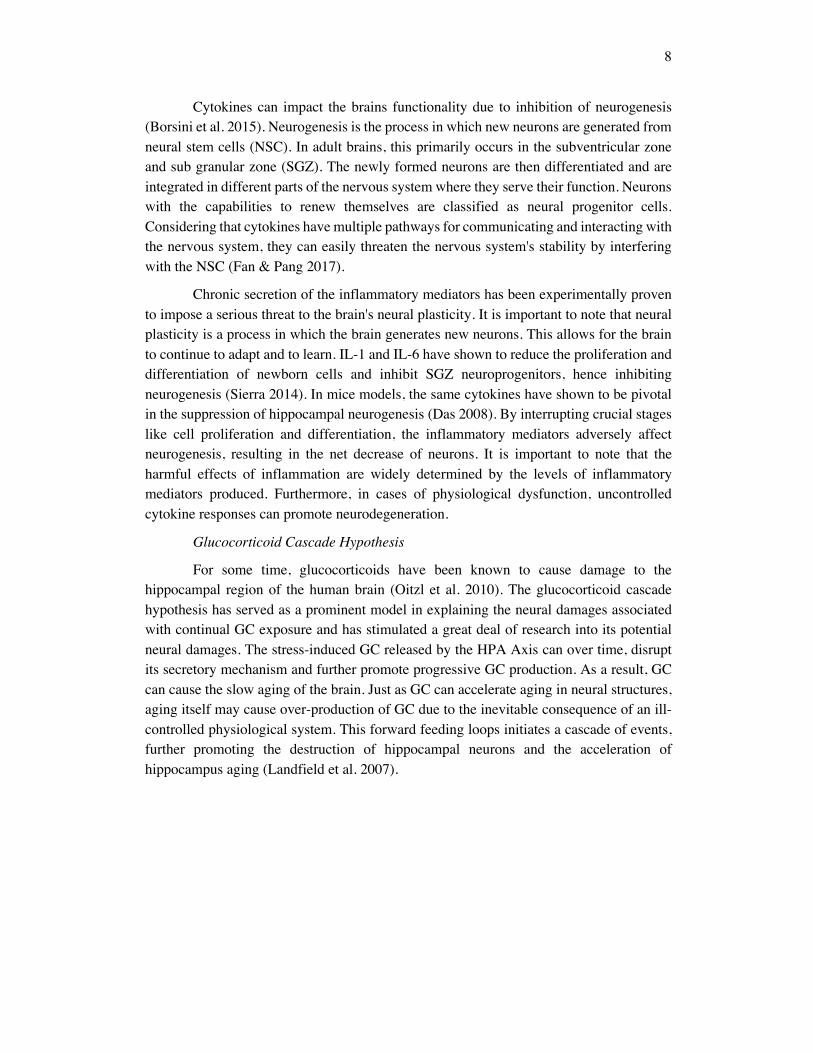
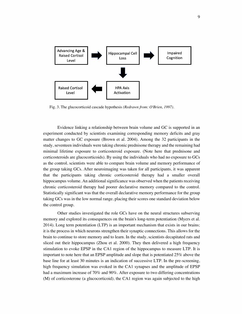
For some time, glucocorticoids have been known to cause damage to the hippocampal region of the human brain (Oitzl et al. 2010). The glucocorticoid cascade hypothesis has served as a prominent model in explaining the neural damages associated with continual GC exposure and has stimulated a great deal of research into its potential neural damages. The stress-induced GC released by the HPA Axis can over time, disrupt its secretory mechanism and further promote progressive GC production. As a result, GC can cause the slow aging of the brain. Just as GC can accelerate aging in neural structures, aging itself may cause over-production of GC due to the inevitable consequence of an ill-controlled physiological system. This forward feeding loops initiates a cascade of events, further promoting the destruction of hippocampal neurons and the acceleration of hippocampus aging (Landfield et al. 2007).
9
Fig. 3. The glucocorticoid cascade hypothesis (Redrawn from: O'Brien, 1997).
Evidence linking a relationship between brain volume and GC is supported in an experiment conducted by scientists examining corresponding memory deficits and gray matter changes to GC exposure (Brown et al. 2004). Among the 32 participants in the study, seventeen individuals were taking chronic prednisone therapy and the remaining had minimal lifetime exposure to corticosteroid exposure. (Note here that prednisone and corticosteroids are glucocorticoids). By using the individuals who had no exposure to GCs as the control, scientists were able to compare brain volume and memory performance of the group taking GCs. After neuroimaging was taken for all participants, it was apparent that the participants taking chronic corticosteroid therapy had a smaller overall hippocampus volume. An additional significance was observed when the patients receiving chronic corticosteroid therapy had poorer declarative memory compared to the control. Statistically significant was that the overall declarative memory performance for the group taking GCs was in the low normal range, placing their scores one standard deviation below the control group.
Other studies investigated the role GCs have on the neural structures subserving memory and explored its consequences on the brain's long-term potentiation (Myers et al. 2014). Long term potentiation (LTP) is an important mechanism that exists in our brains; it is the process in which neurons strengthen their synaptic connections. This allows for the brain to continue to store memory and to learn. In the study, scientists decapitated rats and sliced out their hippocampus (Zhou et al. 2000). They then delivered a high frequency stimulation to evoke EPSP in the CA1 region of the hippocampus to measure LTP. It is important to note here that an EPSP amplitude and slope that is potentiated 25% above the base line for at least 30 minutes is an indication of successive LTP. In the pre-screening, high frequency simulation was evoked in the CA1 synapses and the amplitude of EPSP had a maximum increase of 70% and 90%. After exposure to two differing concentrations (M) of corticosterone (a glucocorticoid), the CA1 region was again subjected to the high
10
frequency stimulation and LTP was unable to persist within 60 minutes. Scientists regarded this reduced interval of potentiation as short term potentiation (STP). In total, only 30% of the hippocampal slices after corticosteroid treatment retained its LTP and the remaining 70% displayed STP. From these results, it can be concluded that the brain's synaptic plasticity can be great impaired when in the presence of GCs.
GCs play an important role in the stress response due to its ability to regulate a multitude of internal parameters. However, its ability to inhibit neurogenesis hence interfering with neural plasticity, can compromise the brain's ability to function. The brain's functionality and structure can be seriously jeopardized when it is excessively exposed to GCs. Like many mediators, GCs subdue the problem in the short run. But the consequences of repeatedly activating the mediating system overtime causes mechanistic dysfunctionalities. In addition to leaving our bodies prone to stress-related diseases, these dysfunctionalities allow for the emergence of unintended health consequences.
SOCIETAL IMPLICATIONS
Social Standing
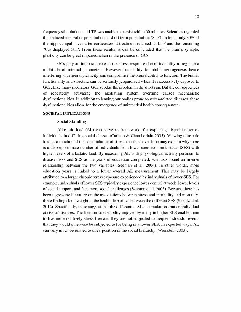
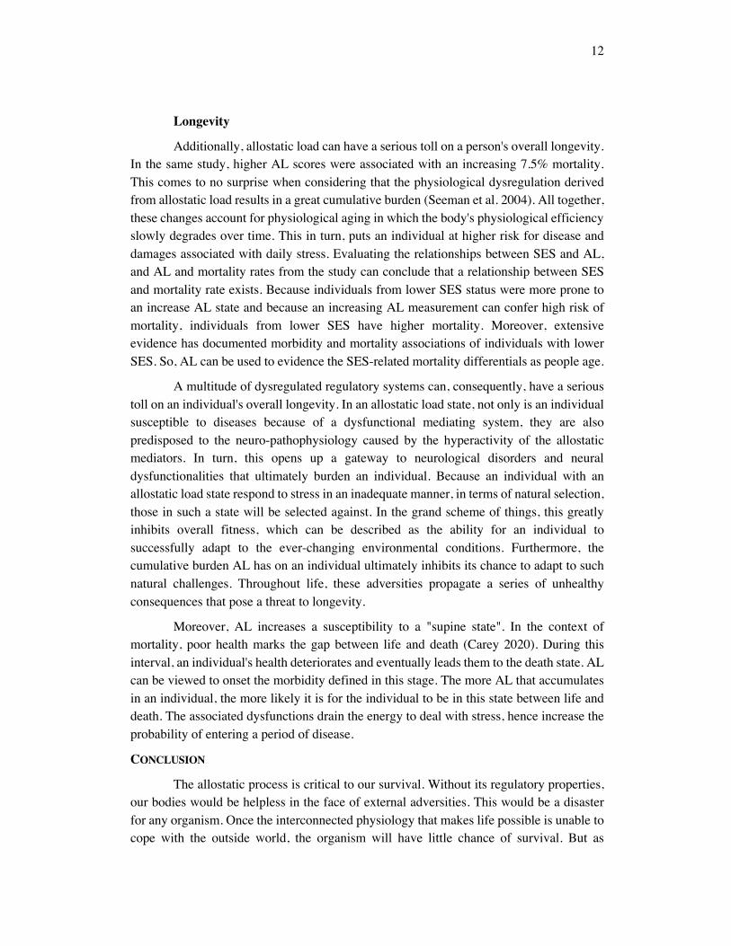
Allostatic load (AL) can serve as frameworks for exploring disparities across individuals in differing social classes (Carlson & Chamberlain 2005). Viewing allostatic load as a function of the accumulation of stress-variables over time may explain why there is a disproportionate number of individuals from lower socioeconomic status (SES) with higher levels of allostatic load. By measuring AL with physiological activity pertinent to disease risks and SES as the years of education completed, scientists found an inverse relationship between the two variables (Seeman et al. 2004). In other words, more education years is linked to a lower overall AL measurement. This may be largely attributed to a larger chronic stress exposure experienced by individuals of lower SES. For example, individuals of lower SES typically experience lower control at work, lower levels of social support, and face more social challenges (Szanton et al. 2005). Because there has been a growing literature on the associations between stress and morbidity and mortality, these findings lend weight to the health disparities between the different SES (Schulz et al. 2012). Specifically, these suggest that the differential AL accumulations put an individual at risk of diseases. The freedom and stability enjoyed by many in higher SES enable them to live more relatively stress-free and they are not subjected to frequent stressful events that they would otherwise be subjected to for being in a lower SES. In expected ways, AL can very much be related to one's position in the social hierarchy (Weinstein 2003).
11
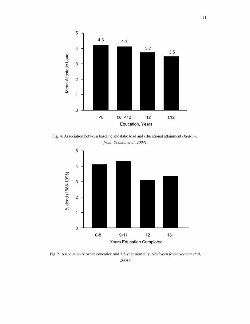
Fig. 4. Association between baseline allostatic load and educational attainment (Redrawn from: Seeman et al, 2004).
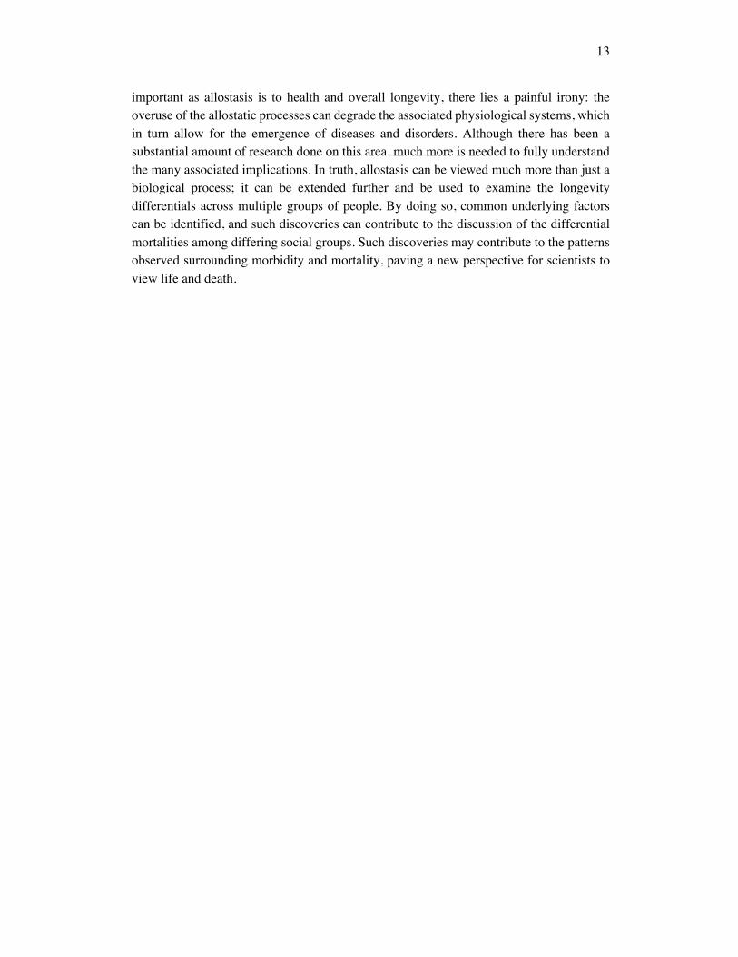
Fig. 5. Association between education and 7.5-year mortality. (Redrawn from: Seeman et al, 2004).
0
1
2
3
4
54.3 4.1
3.73.5
<8 12 ≥12≥8, <12
Mea
n Al
lost
atic
Loa
d
Education, Years
0
1
2
3
4
5
0-8 12 13+9-11
% d
ead
(198
8-19
95)
Years Education Completed
12
Longevity
Additionally, allostatic load can have a serious toll on a person's overall longevity. In the same study, higher AL scores were associated with an increasing 7.5% mortality. This comes to no surprise when considering that the physiological dysregulation derived from allostatic load results in a great cumulative burden (Seeman et al. 2004). All together, these changes account for physiological aging in which the body's physiological efficiency slowly degrades over time. This in turn, puts an individual at higher risk for disease and damages associated with daily stress. Evaluating the relationships between SES and AL, and AL and mortality rates from the study can conclude that a relationship between SES and mortality rate exists. Because individuals from lower SES status were more prone to an increase AL state and because an increasing AL measurement can confer high risk of mortality, individuals from lower SES have higher mortality. Moreover, extensive evidence has documented morbidity and mortality associations of individuals with lower SES. So, AL can be used to evidence the SES-related mortality differentials as people age.
A multitude of dysregulated regulatory systems can, consequently, have a serious toll on an individual's overall longevity. In an allostatic load state, not only is an individual susceptible to diseases because of a dysfunctional mediating system, they are also predisposed to the neuro-pathophysiology caused by the hyperactivity of the allostatic mediators. In turn, this opens up a gateway to neurological disorders and neural dysfunctionalities that ultimately burden an individual. Because an individual with an allostatic load state respond to stress in an inadequate manner, in terms of natural selection, those in such a state will be selected against. In the grand scheme of things, this greatly inhibits overall fitness, which can be described as the ability for an individual to successfully adapt to the ever-changing environmental conditions. Furthermore, the cumulative burden AL has on an individual ultimately inhibits its chance to adapt to such natural challenges. Throughout life, these adversities propagate a series of unhealthy consequences that pose a threat to longevity.
Moreover, AL increases a susceptibility to a "supine state". In the context of mortality, poor health marks the gap between life and death (Carey 2020). During this interval, an individual's health deteriorates and eventually leads them to the death state. AL can be viewed to onset the morbidity defined in this stage. The more AL that accumulates in an individual, the more likely it is for the individual to be in this state between life and death. The associated dysfunctions drain the energy to deal with stress, hence increase the probability of entering a period of disease.
CONCLUSION
The allostatic process is critical to our survival. Without its regulatory properties, our bodies would be helpless in the face of external adversities. This would be a disaster for any organism. Once the interconnected physiology that makes life possible is unable to cope with the outside world, the organism will have little chance of survival. But as
13
important as allostasis is to health and overall longevity, there lies a painful irony: the overuse of the allostatic processes can degrade the associated physiological systems, which in turn allow for the emergence of diseases and disorders. Although there has been a substantial amount of research done on this area, much more is needed to fully understand the many associated implications. In truth, allostasis can be viewed much more than just a biological process; it can be extended further and be used to examine the longevity differentials across multiple groups of people. By doing so, common underlying factors can be identified, and such discoveries can contribute to the discussion of the differential mortalities among differing social groups. Such discoveries may contribute to the patterns observed surrounding morbidity and mortality, paving a new perspective for scientists to view life and death.
REFERENCES
Basu. A, Lazovic. J., Krady, J., Mauger, D., Rothstein, R., Smith, M., and S. Levison. 2005. Interleukin-1 and the interleukin-1 type 1 receptor are essential for the progressive neurodegeneration that ensues subsequent to a mild hypoxic/ischemic injury. Journal of Cerebral Blood Flow and Metabolism 25:17-29.
Borsini, A., Zunszain, P., Thuret, S., and C. Pariante. 2015. The role of inflammatory cytokines as key modulators of neurogenesis. Trends in Neurosciences 38:145-157.
Brown, E.S., Woolston, D., Frol, A., Bobadilla, L., Khan, D., Hanczyc, M., Rush, A.J., Fleckenstein, J., Babcock, E., and C.M. Cullum. 2004. Hippocampal volume, spectroscopy, cognition, and mood in patients receiving corticosteroid therapy. Biological Psychiatry 55:538-545.
Carey, J.R. 2020, June 13. Limits of morbidity compression. Longevity (HDE/ENT 117) lecture notes, UC Davis.
Carlson, E.D. and R.M. Chamberlain. 2005. Allostatic load and health disparities: a theoretical orientation. Research in Nursing & Health 28:306-315.
Das, S. and A. Basu. 2008. Inflammation: A New Candidate in Modulating Adult Neurogenesis. Journal of Neuroscience Research 586:1199-1208.
Fan, L.W. and Y. Pang. 2017. Dysregulation of neurogenesis by neuroinflammation: key differences in neurodevelopmental and neurological disorders. Neural Regeneration Research 12:366-371.
Frank, M., Watkins, L., and S. Maier. 2013. Stress-induced glucocorticoids as a neuroendocrine alarm signal of danger. Brain, Behavior, and Immunity 33:1-6.
Higuera, V. 2012, March 19. General Adaptation Syndrome: Your Body's Response to Stress. Retrieved July 05, 2020 from https://www.healthline.com/health/general-adaptation-syndrome
Johnson, J.D., Campisi, J., Sharkey, C.M., Kennedy, S.I., Nickerson, M., Greenwood, B.N., and M. Fleshner. 2005. Catecholamines mediate stress-induced increases in peripheral and central inflammatory cytokines. Neuroscience 135:1295-1307.
Juster, R.P., McEwen, B., and S. Lupien. 2010. Allostatic Load Biomarkers of Chronic Stress and Impact on Health and Cognition. Neuroscience and Biobehavioral Reviews 35:2-16.
Kamin, H. and D. Kertes. 2017. Cortisol and DHEA in development and psychopathology. Hormones and Behavior 89:69-85.
Landfield, P., Blalock, E., Chen, K.C., and N. Porter. 2007. A New Glucocorticoid Hypothesis of Brain Aging: Implications for Alzheimer’s Disease. Current Alzheimer Research 4:205-212.
McEwen, B. 2003. Interacting mediators of allostasis and allostatic load: towards an understanding of resilience in aging. Metabolism 52:10-16.
McEwen, B. and J. Wingfield. 2003. The concept of allostasis in biology and biomedicine. Hormones and Behavior 43:2-15.
Miller, D.B. and J.P. O’ Callaghan. 2005. Aging, stress, and the hippocampus. Ageing Research Reviews 4:123-140.
Myers, B., Mcklveen, J., and J. Herman. 2014. Glucocorticoid actions on synapses, circuits, and behavior: Implications for the energetics of stress. Frontiers in Neuroendocrinology 35:180-196.
O'Brien, J. 1997. The 'glucocorticoid cascade' hypothesis in man. British Journal of Psychiatry 170:199-201.
Oitzl, M., Champagne, D., Veen, R., and E. Kloet. 2010. Brain development under stress: Hypotheses of glucocorticoid actions revisited. Neuroscience & Biobehavioral Reviews 34:853-866.
Ramsay, D. and S. Woods. 2014. Clarifying the Roles of Homeostasis and Allostasis in Physiological Regulation. Psychological Review 121:225-247.
Schulz A, Mentz G, Lachance, L., Johnson, J., Gaines, C., and I. Barbara. 2012. Associations between Socioeconomic Status and Allostatic Load: Effects of Neighborhood Poverty and Tests of Mediating Pathways. American Journal of Public Health 102:1706-1714.
Seeman, T., Crimmins, E., Huang, M.H., Singer, B., Bucur, A., Gruenwald, T., Berkman, L., and D. Reuben. 2004. Cumulative biological risk and socio-economic differences in mortality: MacArthur Studies of Successful Aging. Social Science & Medicine 58:1985-1997.
Seeman, T., Epel, E., Gruenewald, T., Karlamangla, A., and B. McEwen. 2010. Socio-economic differentials in peripheral biology: Cumulative allostatic load. Annals of the New York Academy of Sciences 1186:223-239.
Sierra, A., Beccari, S., Diaz-Aparicio, I., Encinas, J., Comeasu, S., and M.E. Tremblay. 2014. Surveillance, Phagocytosis, and Inflammation: How Never-Resting Microglia Influence Adult Hippocampal Neurogenesis. Neural Plasticity 2014:1-15.
Szanton, S., Gill, J., and J. Allen. 2005. Allostatic Load: A Mechanism of Socioeconomic Health Disparities? Biological Research for Nursing 7:7-15.
Themes, U. 2016, September 03. Homeostasis and Adaptive Responses to Stressors. Retrieved July 05, 2020 from https://basicmedicalkey.com/homeostasis-and-adaptive-responses-to-stressors/
Weinstein, M., Goldman, N., Hedley, A., Yu-Hsuan, and T. Seeman. 2003. Social Linkages to Biological markers of Health Among the Elderly. Journal of Biosocial Science 35:433-453.
Zhang, J. and J. An. 2007. Cytokines, Inflammation and Pain. International Anesthesiology Clinics 45:27-37.
Zhou, J., Zhang, F., and Y. Zhang. 2000. Corticosterone inhibits generation of long-term potentiation in rat hippocampal slice: involvement of brain-derived neurotrophic factor. Brain Research 885:182-191.