Embed Size (px)
Citation preview

Journal of Cell Science 102, 821-832 (1992)Printed in Great Britain © The Company of Biologists Limited 1992
821
Alteration in glycosaminoglycan metabolism and surface charge on human
umbilical vein endothelial cells induced by cytokines, endotoxin and
neutrophils
NIGEL J. KLEIN*, GRAHAM I. SHENNAN, ROBERT S. HEYDERMAN and MICHAEL LEVIN
Department of Paediatrics, Queen Elizabeth the Queen Mother Wing, St Mary's Hospital Medical School, South Wharf Road, London W21NY, England
*Author for correspondence
Summary
There is increasing evidence that the glycosaminoglycan(GAG) component of the vascular endothelium isimportant in regulating vascular permeability, throm-boresistance and cellular interactions. We have investi-gated the GAG metabolism of cultured human umbilicalvein endothelial cells (HUVEC) in response to a range ofinflammatory stimuli. Using both chemical measure-ment of cellular and supernatant GAGS and 35Slabelling to identify newly synthesised GAGS, inter-leukin 1 (IL1), tumour necrosis factor (TNF) andinterferon gamma (IFNy) were shown to influencesulphated GAG metabolism significantly. IL1 and TNFcaused a marked increase in culture supernatant GAGSand a concomitant reduction in cell-associated GAGS.
This was shown histochemically to be associated with amarked reduction and redistribution of endothelialsurface anionic sites. The addition of neutrophils toHUVEC pretreated with Escherichia coli endotoxin, IL1or TNF resulted in a further reduction in both cellularGAGS and surface anionic sites. These results suggestthat changes in endothelial cell GAG metabolism duringinflammation may contribute to the disturbance ofvascular endothelial homeostasis associated with infec-tious and inflammatory states.
Key words: glycosaminoglycans, endothelium,inflammation, neutrophils, endotoxin, cytokines.
Introduction
The pathophysiology of serious sepsis is extremelycomplex. Three of the most important clinical featuresare capillary leakage, intravascular thrombosis andderangement of vascular tone, and this triad suggeststhat an alteration in the normal homeostatic propertiesof the vascular endothelium may be central to thepathogenesis of this condition (Tracey et al. 1986;Mercier et al. 1988; Levin, 1990).
Histochemical examination of vascular endothelialcells with cationic probes such as ruthenium red, Alcianblue and cationic ferritin, have shown the luminalsurface to be highly negatively charged (Brenner et al.1978; Brody et al. 1984; Kanwar, 1984; Rounds andVaccaro, 1987). This property is largely attributed tothe presence of the sulphated glycosaminoglycans(GAGS), heparan and dermatan sulphate, with asmaller contribution from sialoglycoproteins (Kanwaret al. 1981; Wight et al. 1986).
There is now evidence that these sulphated GAGSare important in maintaining vascular homeostasis by:(a) modulating antithrombotic and coagulant activity
on the endothelial cell surface through specific interac-tions with antithrombin III and heparin cofactor II(Marcum and Rosenberg, 1984; Platt et al. 1990); (2)providing an electrostatic barrier to similarly chargedcell surfaces, including those of platelets and red bloodcells (Danon and Shutelsky, 1976; Springer, 1990); (3)binding important macromolecules such as lipoproteinlipase, fibroblast growth factor and superoxide dismu-tase to the cell surface (Gallagher et al. 1986; Karlssonand Marklund, 1988); and (4) regulating the vascularpermeability of the endothelium to circulating mol-ecules and particularly plasma proteins (Lindahl andHook, 1978).
It is now well established that vascular permeabilityto macromolecules is dependent not only on molecularsize, but also on electrostatic charge. Neutral proteinsand dextrans are cleared more rapidly from thecirculation than similarly sized anionic polymers, andthe clearance of cationic proteins is greatly enhanced(Bennet et al. 1976; Bohrer et al. 1978). The importanceof endothelial charge and GAGS in the regulation ofvascular permeability has been demonstrated by enzy-matic digestion of GAGS and their neutralisation by

822 N. J. Klein and others
polycations, both of which increase the clearance ofnormally restricted molecules from the renal andsystemic vasculature (Kelley and Cavallo, 1978; Kan-war et al. 1980; Vehaskari et al. 1984). Endothelialsurface charge is particularly important in restrictingalbumin, the major protein responsible for plasmaoncotic pressure, within the intravascular space. Theclearance of albumin from the kidney is significantlyless than that of similarly sized neutral molecules(Brenner et al. 1978; Kanwar, 1984) and indeed thefractional clearance of the cationic albumin exceedsthat of native albumin by a factor of 300 (Purtell et al.1979). Furthermore, experimental neutralisation ofendothelial anionic sites, either in the glomerulus or inthe systemic circulation, increases the clearance ofnative albumin to within the range of similarly sizedneutral molecules (Hunsicker et al. 1981; Vehaskari etal. 1984). Patients with septic shock invariably have aprofound capillary leak (Mercier et al. 1988), and maylose several times their circulating volume into theinterstitial compartment. Albumin is the predominantprotein lost and is therefore central to the process ofcapillary leakage of plasma.
We have postulated that alteration in endothelial cellsurface charge and GAG composition might underlythe increased vascular permeability in septic shock, andsuch an alteration would also be expected to influencethe thromboresistant properties of the endothelium. Inview of the emerging evidence that cytokines andneutrophils are important mediators of the tissue andorgan damage seen in overwhelming sepsis, we havestudied the effect of cytokines, endotoxin and neutro-phils on endothelial cell GAG metabolism and surfacecharge.
Materials and methods
ReagentsHrlLl and EL6 (interleukin) were donated by Dr S. Gillis,Immunex Corporation, Seattle, WA. HrlFNy (interferon)was obtained from Biogen, Geneva, Switzerland, and HrTNF(tumour necrosis-factor) supplied by Dr. A. Gearing, NIBSC,South Mimms, GB. Lipopolysaccharide B (LPS) Escherichiacoli 0128:B12, heparan sulphate (HS) from bovine kidney,dermatan sulphate (DS) from porcine skin, chondroitin 4- and6-sulphate (CS) from bovine trachea and shark cartilage,respectively, protease E, chondroitinase ABC, heparinase IIand neuraminidase type X were purchased from Sigma,London, UK. Alcian blue 8GX was from Imperial ChemicalIndustries Ltd., Blackley, Manchester, UK.
Endothelial cultureEndothelial cells were obtained from human unbilical veinsby digestion with 0.1% collagenase as previously described(Jaffe et al. 1973). Cells were cultured in Dulbecco's minimalessential medium (Gibco, Paisley, Scotland) with L-glutamine(1.6 mM), penicillin (80i.u./ml), streptomycin (80mg/ml) and20% fetal calf serum (Gibco). Cells were grown to confluencein 25 cm2 flasks (Nunc, Denmark) and passaged by exposureto EDTA10 mM (BDH, Essex, England) either to gelatinised13 mm glass coverslips or to 6-well culture plates (Nunc).Human umbilical vein endothelial cells (HUVEC) used in all
the experiments were always from the second passage. Cellswere verified as endothelial by morphology, the presence ofVon Willibrand factor and prostacyclin production.
Preparation of neutrophilsVenous blood from adult donors was collected into 3.8%trisodium citrate, divided into aliquots and placed in test tubescontaining mono-poly resolving medium (Flow Laboratories,Herts, England). After centrifugation at 300 g for 30 minutes,a discrete band of neutrophils could be easily located andpipetted free from contaminating blood constituents. Theneutrophils were washed twice in Hanks' buffered saltsolution, counted and reconstituted in culture medium.Morphological assessment of neutrophil purity and viabilitywere estimated to be >93% and >95%, respectively.
Glycosaminoglycan isolationGAGS were isolated from both culture supematants andendothelial cell monolayers. At specific times during theexperiments (see below for details of sampling times) culturesupematants were removed and the monolayers washed twicewith warm (37°C) Dulbecco's phosphate buffered saline(PBS). The washings were added to the supematants and thenspun at 1200 g to remove any cellular debris. GAGS werereleased free from protein by incubation with Pronase (finalconcentration of 50 mg/ml) at 37°C for 20 hours. Aftercentrifugation to remove any precipitate the supematantswere stored at —70°C so that an entire experiment could besimultaneously analysed. The cell layer was digested withPronase (50 mg/ml) in PBS. After 5 minutes the cells haddetached from the well and were counted in a haemocyt-ometer. The cells were then digested for a further 20 hours at37°C, spun at 1200 g and the supernatant was stored forfurther processing. To assess any degradation of GAGSduring this processing, known quantities of GAG standardscontaining heparan, dermatan and chondroitin 4- and 6-sulphate were incubated with each new batch of Pronase andalso with samples of culture supematants and cells. Thesewere treated identically to the test samples.
GAGS were then isolated using a modification of themethod of Whiteman (1973). All samples were incubated for 6hours with 10 volumes of alcian blue 8GX solution (alcianblue, 0.05%, 50 mM sodium acetate buffer, pH 5.8 and 50mM MgCl2). The GAG/dye complex was isolated bycentrifugation at 1000 g. After removal of the supernatant, theAlcian blue was dissociated from the GAGS with NaCl andmethanol (final concentration 2.67 M and 33%, v/v) respect-ively, and the Alcian blue denatured with Na2 CO3 (finalconcentration 12.5 mM). The mixture was sonicated in awater bath sonicator for 2 minutes before removing the alcianblue by centrifugation at 10 000 g and precipitating the GAGSfrom the clear supernatant with 3 volumes of ethanol. Aftercentrifugation to 10 000 g, the supernatant was removed, andthe GAGS left to dry in air overnight. The sample was thendissolved in distilled water, spun at 10 000 g to remove anynon-soluble material, before electrophoresis.
GAG characterisationOne-dimensional electrophoresis was performed by applying0.7 jA of the GAG solution as a 3 mm band to a celluloseacetate sheet (Sartorious, Gottigen, W. Germany). Afterelectrophoresis for 4 hours in 0.1 M barium acetate, pH 6.0, at7.5 V/cm, the sheets were then developed in alcian bluesolution for 30 minutes and destained in 5% acetic acid. Theidentity of the bands was established by comparison with theposition of standards (HS, DS, CS), which were included ineach run. To verify the identity of the bands, two-dimensional

Endothelial GAG modulation in inflammation 823
electrophoresis was undertaken after application of 1 [A ofsample as a spot to a cellulose acetate sheet. Electrophoresiswas performed first in pyridine/acetate buffer, pH 6.0, at 7.5V/cm for 75 minutes and the cellulose acetate sheet was driedin air before electrophoresis at 90° to the first run in bariumacetate buffer as described above. GAG standards wereapplied prior to the second run. Further confirmation of bandidentities was by electrophoresis of enzymatic digests of thesample with 0.5 unit/ml of chondroitinase ABC or withnitrous acid (made with equal volumes of 5% NaNO2 and33% acetic acid and then mixed 1:1 with the sample).Degraded GAGS did not appear on developed sheets.
Enzyme specificityThe specificities of the GAG-degrading enzymes wereassessed by their activity on purified GAGS using celluloseacetate electrophoresis and by assaying their ability to inhibitthe heparan sulphate stimulation of antithrombin III activityusing an amidolytic assay system (Larson et al. 1978). Usingcellulose acetate electrophoresis we found that the chondroi-tin and dermatan sulphate bands were both removed bychondroitinase ABC and that heparan sulphate was degradedby nitrous acid but not by heparinase EL This enzyme did,however, completely inhibit the heparan sulphate stimulationof antithrombin III activity assessed by using the amidolyticasssay (data not shown).
GAG quantitationBands identified by reference to GAG standards werequantified using a laser densitometer (Pharmacia). Standardcurves were constructed from the optical densities of bandpeaks, above background, from a range of known concen-trations of GAG standards. Sample bands were thencalculated as micrograms of heparan, dermatan or chondroi-tin sulphate.
Electrophoretic bands containing more than 0.5 microgramof GAG were too dense for densitometry. They weretherefore cut out and dissolved in dimethylsulphoxidecontaining 0.5 g anhydrous sodium acetate, 1.27 g MgCL; and1.56 ml acetic acid/250 ml as previously described (Vermylenet al. 1989) and their absorbance was read at 678 nm in aspectrophotometer (LKB). Standard curves were constructedfrom GAG standards of known concentrations, which wereelectrophoresed and treated as above. Exact quantities ofindividual GAGS could then be calculated from the samplebands.
In vitro labelling of GAGS with 35STo ascertain the newly synthesised component of both cellularand supernatant sulphated GAGS, 20 /iCi and 35S as H2SO4(ICN, U.K., sp. activity 43 Ci/l g SO4) was added at statedtimes (see below) and durations of cell cultures. GAGS wereisolated and characterised as described above.
QuantificationThe radiolabelled GAGS were quantified by autoradiographyof the cellulose acetate sheets for 7 days at —70°C (KodakXAR-5 film) and measurement of the absorbance of theautoradiographic bands using the laser densitometer. Whenthe autoradiograph was faint, bands were cut from celluloseacetate sheets and 35S incorporation was measured bystandard scintillation counting techniques (Scintillationcounter from LKB Instruments, Bromma, Sweden).
GAG release and synthesis in unstimulated culturesEight wells were seeded with 5 x 10⁵ cells/well from the sameprimary source. The medium was changed every 24 hours so
that GAG metabolism could be measured for each 24 hourperiod as cells grew to confluence and beyond. The cells andsupernatants from sequential duplicate wells were harvestedevery 24 hours for a total of four days and GAGS measured asalready described. In order to measure newly synthesisedGAGS, ^S was added to the duplicate wells at the beginningof each 24 hour period. GAGS were undetectable in the tissueculture medium alone.
Endothelial incubation with cytokinesAt confluence, wells were washed and 2.0 ml of fresh mediumadded and incubated for up to 48 hours with either LPS 1-10ng/ml, IL1 20 units/ml, IL6 50 units/ml, IFN gamma 500units/ml or TNF 75 units/ml. These concentrations wereselected from previous studies (Pober et al. 1986; Broudy etal. 1986) and from other experiments performed in ourdepartment. 35S was included at the start of the incubation.Every incubation was performed in duplicate wells with aninternal control of unstimulated cells and on at least threeseparate occasions. At 12, 24 and 48 hours the culturesupernatants and cells were pooled from two wells and boththe total GAG and radiolabelled GAG content calculated asdescribed above.
Endothelial incubation with cytokines and neutrophilsEL 1, TNF and LPS have been shown to increase endothelialcell adherence for neutrophils (Pober et al. 1986; Smedley etal. 1986). This effect is maximal 4 to 6 hours followingtreatment with these inflammatory mediators. Endothelialcells were therefore preincubated with either IL1, TNF orLPS for 4 hours before adding neutrophils at a finalconcentration of 0.5 x 10⁶ per ml. Cytokines were presentthroughout the experiment and 35S was added with theneutrophils. GAGS were determined from both cells andsupernatants at 4 hours (early) and 48 hours (late) andprocessed as for cytokine stimulation alone. All experimentswere performed on at least 3 occasions. Controls includedGAG measurements of neutrophils with and without cytok-ines added to empty wells and unstimulated endothelial cellswith and without neutrophils.
GAG recoveryTo assess the efficiency of individual GAG extraction, GAGstandards were added to culture medium and then extracted,electrophoresed and quantified. Recoveries were 58% ± 6%of CS, 56% ± 4% of DS and 24% ± 5% of HS (mean ±s.e.m. from 15 samples). Pronase digestion had no effect onthese results, indicating that there was no detectable enzymicdegradation of the GAGS during processing.
AnalysisData presented from unstimulated cultures were expressedeither as micrograms GAG/106 cells or as cts min"1/!©6 cellsafter correction for extraction efficiencies. To allow compari-son of data derived from the various methods of quantifi-cation utilised in this study, results from experimentsinvolving cytokines, LPS and neutrophils were corrected forcell numbers and then expressed as a percentage of the valuesobtained from the internal control.
Experimental results were analysed using a Student's two-tailed Mest.
Visualisation of endothelial anionic sitesTo ascertain the correlation between cellular GAGS andsurface charge, we developed a morphological light-micro-scopic technique based upon the charged interaction of acationic probe with endothelial anionic sites, which has

824 N. J. Klein and others
previously been used for ultrastructural studies (Skutelskyand Roth, 1986). Endothelial cells were grown to confluenceon gelatinised coverslips and treated with cytokines, endo-toxin and neutrophils as described above. At similar timepoints to those used for the biochemical GAG estimations,coverslips were washed in warm (37°C) PBS and then fixed incold (—20°C) methanol. Anionic sites were visualised with apoly-L-lysine probe conjugated to 5 nm gold particles (BiocellResearch Laboratories, Cardiff, UK). The probe was appliedto the cells for 60 minutes, washed off with deionised waterand developed with a silver enhancer (Biocell) for 15 minutesat room temperature (Volker et al. 1991). Binding was pHdependent, but was found to be most specific for glycosamino-glycans at pH 1.2. Addition of 25 mmol MgCl2 increased theGAG specificity. The cells were counterstained in Meyer'shaematoxylin for 1 minute and mounting in an Aquamount(BDH, Essex, England).
The charge-dependent nature of the binding was demon-strated by inhibition of binding by other cationic moleculesincluding poly-L-lysine and DEAE-dextran. Staining was notinhibited by neutral dextran. In order to establish the identityof endothelial anionic sites, live cultures were incubated with10 units/ml of heparinase II, 0.5 unit/ml of chondroitinaseABC and 0.5 unit/ml of neuraminidase for 4 hours and fixedcultures were treated with nitrous acid for 2 hours. Thesewere then stained as described above.
Results
GAG metabolism in unstimulated culturesDuring growth of freshly passaged cells to confluence,there was a progressive increase in GAGS, bothreleased into the culture supernatant and associatedwith endothelium. Fig. 1 shows a typical electrophoreticprofile of supernatant GAGS at 48 hours. Fig. 2 depictsa representive experiment in which the GAG content ofsupernatants and cells for each 24 hour epoch over a 96hour period. HS, DS and CS were detected in culturesupernatants at 24 hours, with maximal levels occurringat confluence (24-48 hours) and then declining steadilyover the following 48 hours (Fig. 2A). Cell layer-associated GAGS constituted less than 25% of releasedGAGS, (Fig. 2B); however, the time course ofdetectable HS and DS was similar to that seen in culturesupernatants. CS was rarely recovered from theendothelial cell monolayer.
As shown in Fig. 2C and D, there was little 35Sincorporation into GAGS isolated from both super-natants and cell layer during the first 24 hours ofculture. There was then a marked increase in 35Sincorporation into HS and DS at 48 hours, followed bya progressive decrease over the next 2 days. Radio-labelled CS was only detected in culture supernatants.Synthesis was rapid over the initial 48 hours, and thendeclined over the subsequent 48 hours. Comparison ofFig. 2A,B and C,D indicates that GAGS detected in thefirst 24 hours were predominantly synthesised prior tosubculture, whereas those detected after 24 hours weremostly newly synthesised.
GAG release, content and synthesis were extremelysensitive to a number of variables including the type oftissue culture medium, culture flask, batch of serum,umbilical cords and as shown in Fig. 2 by proliferation
Fig. 1. Cellulose acetate electrophoresis of HUVECsupernatant GAGS. GAGS were precipitated from the 24-48 hour HUVEC culture supernatants using Alcian blue.After isolation of GAGS free of cationic dye,electrophoresis was performed on cellulose acetate in 0.1M barium acetate. Lanes 1 and 3 are GAG samples fromtwo separate endothelial culture supernatants and lane 2contains the GAG standards of 0.5 mg/ml of heparansulphate (HS), dermatan sulphate (DS), or chondroitinsulphate (CS). In some experiments two HS bands couldbe seen (shown here).
and age of the culture. Subsequent experiments weretherefore always performed on confluent monolayersand were internally controlled using identical cells andconditions.
Effect of cytokines on GAG metabolismAddition of TNF and IL1 to the culture mediuminduced an increase in total GAGS detected insupernatants at 12, 24 and 48 hours. Quantification ofindividual GAG components established that CS wasmost affected, with smaller increases in DS and HS(Table 1). In contrast, there was a small, but consistent,decrease in cell-associated GAGS. Measurement ofnewly synthesised GAGS, by 35S incorporation (Table2) indicated that CS production was increased through-out the experiment, whereas there was a biphasicalteration in HS and DS synthesis. 35S-labelled HS andDS were reduced relative to the control at 12 hours, butincreased incorporation was observed at 24 and 48hours. Cell-layer-associated 35S-labelled GAG wasdepressed at all three time points.
IFN gamma did not influence GAG metabolism inthe first 24 hours, but after 48 hours all three classes ofGAGS had increased on both cells and in thesupernatant. Table 2 indicates that this was the result ofstimulated GAG synthesis.

Endothelial GAG modulation in inflammation 825
96 96
8-
q5 6 '
4H
2-
BD HS• DS 8-
6-
O
HSDS
24 48 72 96
Time (h)
48 72
Time (h)
96
Fig. 2. Time course of GAG production by cultured endothelial cells. A typical time course of GAGS isolated, asdescribed above, from supernatants (A) and endothelial cell monolayers (B) showing the pattern of HS, DS, and CS in 24hour epochs for 96 hours. Newly synthesised GAGS, as assessed by S incorporation into isolated GAGS, are shown forthe supernatant (C) and cell layer (D). GAG synthesis was not very marked until 24 hours of culture, whereas GAGSdetected after 48 hours, were predominantly newly synthesised.
LPS and EL6 had no demonstrable effect on endo-thelial GAG metabolism (data not shown).
The effect of neutrophils together with cytokines onGAG metabolismIn the absence of cytokines or endotoxin, co-cultivationof neutrophils and EC resulted in a modest increase insupernatant GAGS at 4 hours with a 23% reduction incell layer-associated GAGS (Table 3). After 48 hours asignificant elevation in supernatant GAGS was ob-served, together with a marked loss of cell layer-associated GAGS. Pretreatment of HUVEC with IL1and TNF reduced cellular GAGS by over 60% at 4hours with minimal change in the supernatant. Thecombination of LPS and neutrophils reduced bothsupernatant and cellular GAGS by 4 hours. After 48hours of IL1, TNF and LPS incubation, GAGS wereundetectable from the cells, but were increased in
concentration in culture supernatants (Table 3). Thisincrease in release of GAGS was predominantly due toincreased biosynthesis, as shown by the results of 35Sincorporation (Table 4). Release and synthesis ofGAGS were not detected from the neutrophils that hadbeen added to empty wells.
Histochemical detection of anionic sitesUsing the poly-L-lysine gold probe to detect endothelialanionic sites, unstimulated cells were found to have anextensive nbrillar network of poly-L-lysine binding sitesextending across and between the cultured cells, with afine punctate array of sites located on the cellularmembrane overlying the endothelial cytoplasm (Fig.3a). Acellular regions of the culture, where cells haddetached, were also densely stained with cationic gold,whereas there was minimal staining of gelatinisedcoverslips that had not been exposed to HUVEC (not


Tab
le 3
. T
he in
fluen
ce o
f cy
toki
nes,
end
otox
in a
nd n
eutr
ophi
ls o
n en
doth
elia
l G
AG
met
abol
hm
Med
iato
r
Sup
erna
tant
C
ell
Tot
al G
AG
HS
DS
CS
Tot
al G
AG
H
S D
S
- - -
4 ho
urs
ILl+
neut
roph
ils
109
(95
- 1%
) la
3
(91
- 11
4)
91
(87
- 95)
1%
(10
5 - 1
35)
55 (
46 -
62)
* 58
(48
- 64
)*
51 (
43 -
59)*
,t T
NF
+ne
utro
phil
s %
(9
1 - 1
02)
91
(86 -99)
SS
(83
- W)
112
(104
- 12
1)
59 (
53 -
70)*
65
(59
- 72
)*
52 (
48 -
67)*
L
PS
+ne
utro
phil
s 63
(5
1 -
69).
63
(53
- 71
)*
60
(48
-71
)'
88
(71
- 10
6)
52 (
40 -
63)
* 57
(44
- 66)*
46 (
36 -
59)
* N
eutr
ophi
ls a
lone
10
9 (1
06 -
113)
93
(8
7 -
98)
101
(88
- 112
) 12
0 (1
12 -
131)
77
(68
- 8
7)
85 (
74 -
91)
74 (
61 -
89)
48 h
ours
IL
l+ n
eutr
ophi
ls
173
(159
- IN
)*
191
(162
- 21
5)*
162
(U9
- 18
7)*
148
(131
- 16
3)*
<w
TN
F+
neut
roph
ils
178
(171
- 19
3)t
212
(184
- 24
3)'
142
(122
- 15
8)
1% (
159
- 23
7)
<m
L
PS
+ne
utro
phil
s 19
1 (1
81 -
198)
t 21
8 (1
95 -
240
)*
153
(141
- 16
3)*
229
(198
- %
I)*
<m$
Neu
trop
hils
alo
ne
137
(130
- 14
9)*
142
(127
- 1
54)'
1% (
118
- I%
)*
1M (
129
- 17
3)
39 (
34 -
43)t
37
(29
- 47
)t
39 (
31 -
49)t
Neu
trop
hils
wer
e ad
ded
to H
UV
EC
pre
stim
ulat
ed w
ith E
l,
TN
F o
r L
PS.
GA
GS
wer
e is
olat
ed f
rom
the
cells
and
cul
ture
sup
erna
tant
s an
d qu
anti
fied
. A
fter
4 h
ours
, ne
utro
phil
s ca
used
a s
igm
lican
t red
ucti
on in
cel
l-as
soci
ated
GA
GS
in t
he p
rese
nce
of all o
f th
e m
edia
tors
use
d. B
y 48
hou
rs G
AG
S w
ere
unde
tect
able
(le
ss th
an 2
0% t
hat
of u
nsti
mul
ated
cul
ture
s)
Thi
s was
acco
mpa
nied
by
a m
arke
d el
evat
ion
in s
uper
nata
nt G
AG
S.
The
res
ults
sho
w t
he G
AG
con
tent
, ex
pres
sed as a
per
cent
age
of u
nsti
mul
ated
cul
ture
s. T
he m
ean
and
rang
e of
th
ree
expe
rim
ents
at
4 an
d 48
hou
rs a
re s
how
n. *
P<
0.05
, tP
<O
.Ol,
tP<
0.00
1.
Tab
le 4
. T
he e
ffec
t of
cyt
okin
es a
nd n
eutr
ophi
ls o
n en
doth
elia
l GA
G s
ynth
esh
? h E
Sup
erna
tant
C
ell
Med
iato
r T
otal
GA
G
HS
DS
CS
Tot
al G
AG
H
S D
S
a (3
48 h
ours
3
207
(186
- 2
39)'
241 (207 -
271)
. 16
3 (1
47 -
183)
' 21
2 (1
7 -
214)
* <
m
0
ILl +
neut
roph
ib
TN
F+
neut
roph
ils
351
(269
- 405)'
388
(330
- 44
1)*
309
(237
- %
I)*
37
7 (2
61 -
473)
* <
m
LP
S+ n
eutr
ophi
ls
321
(277
- 34
8)t
1% (
152
- 25
3)
413
(297
- 50
3)'
528
(329
- 56
0)
<a$
Neu
trop
hils
alo
ne
147
(142
- 15
3)t
151
(129
- 1
81)
134
(118
- 15
7)
163
(154
- 19
8)
49 (
42 -
56)t
45
(38
- 5
3)t
53 (
46 -
59)
t
z 3 3
35
~
inco
rpor
atio
n in
to s
uper
nata
nt a
nd c
ell-
asso
ciat
ed G
AG
S w
as m
easu
red
follo
win
th
e in
cuba
tion
of
cyto
kine
- or
endo
toxi
n-pr
etre
ated
end
othe
lial
cul
ture
s w
ith n
eutr
ophi
ls f
or 4
8 3"'
hour
s. S
uper
nata
nt G
AG
S i
sola
ted
afte
r th
is t
ime
had
inco
rpor
ated
si@
cant
ly
mor
e 'S
than
uns
tim
ulat
ed c
ultu
res,
ind
icat
ing
that
the
rel
ease
d G
AG
S w
ere
the
resu
lt o
f de
nov
o sy
nthe
sis.
The
res
ults
are
exp
ress
ed a
s a
perc
enta
ge o
f 3
5~
in
corp
orat
ion
com
pare
d w
ith t
hat
of u
nsti
mul
ated
cul
ture
s. M
ean
and
rang
e of
thr
ee e
xper
imen
ts a
re s
how
n. I
Pc0
.05
, tP
<O
.Ol,
$P<
0.00
1.
3 3

828 N. J. Klein and others
Fig. 3. Cationic gold staining of: (a) unstimulated cultures. An extensive fibrillar network can be seen overlying the monolayerwith fine punctate staining of the cell surface, (b) HUVEC treated with heparinase n . Live cultures treated with 10 units/ml for2 hours have almost completely lost their fibrillar network. Occassional fibrils are still present and moderate surface punctatestaining remains, (c) HUVEC following incubation with TNF. Cultures exposed to TNF for 12 hours have lost much of theirsurface charge, with the remaining staining located at the intercellular junctions, (d) Cationic staining after IFNy stimulation. 48hours of IFNy treatment induces cellular elongation and a redistribution of anionic sites to the intercellular and pericellularmatrix, (e) Surface charge following the addition of neutrophils to LPS-stimulated endothelium. Neutrophils, added to HUVECpreincubated for 4 hours with LPS, cause a dramatic reduction in cationic gold.

Endothelial GAG modulation in inflammation 829
shown). In order to elucidate the nature of the cationicbinding, HUVEC were exposed to neuraminidase,chondroitinase ABC, heparinase II and nitrous acid.Although neuraminidase had a minimal effect on thedistribution of charged sites, heparinase II and nitrousacid caused significant disruption of the fibrillar patternand also reduced the intensity of punctate staining.These results indicate that the cationic gold bindingobserved in this study was predominantly directedagainst cell layer-associated heparan sulphate (Fig. 3b).Chondroitinase ABC had little effect on the fibrils, butinhibited cationic gold binding to the acellular zones ofthe culture and also reduced the membrane-associatedpunctate staining (data not shown).
Addition of IL1 (not shown) or TNF to the culturemedium altered both the intensity and distribution ofanionic sites. Fig. 3c shows that TNF induced ageneralised reduction in the binding of cationic gold,and that in contrast to control cultures staining wasmainly limited to the intercelluar junctions.
The pattern of staining after the HUVEC werestimulated with IFN gamma differed significantly fromthat seen in the control and TNF-treated cells (Fig. 3d).Gold was distributed in clumps running between thelongitudinal axes of the cells with minimal stainingdetected on the cell surface.
Neutrophils added to IL1-, TNF- (not shown) andLPS-treated cultures, caused a considerable reductionin cell surface staining by 4 hours, and by 12 hours therewas only minimal cationic gold deposition (Fig. 3e).Trypan blue exclusion demonstrated that the neutro-phil/cytokine combinations used in these experimentshad not significantly altered HUVEC viability.
Cationic gold staining was very sensitive to fixation.The fibrillar structures seen with cold methanol werenot observed if FIUVEC were fixed in either warmmethanol or an aldehyde fixative. Paraformaldehydeand glutaraldehyde fixation produced a more homo-geneous distribution of staining, predominantly locatedon the exposed surface of the endothelial cell, whichwas removed following treatment with heparinase II.Cells fixed with these aldehyde fixatives did howeverdisplay significant alterations in response to the stimuliused in this study. In particular, neutrophils added tostimulated endothelium almost completely eradicatedthe cationic staining.
Discussion
Despite increasing recognition that GAGS are involvedin many biological processes, elucidation of theirfunctional significance has proved difficult to defineaccurately. The methods developed in this study toexamine the metabolism of endothelial cell GAGS invitro enabled both cell layer-associated and supernatantGAGS to be determined simultaneously. In addition,by labelling GAGS with 35S the contribution of newlysynthesised molecules relative to the total extractableGAG could be assessed. Furthermore, poly-L-lysine/gold histochemistry has also enabled us to visualise the
surface distribution of endothelial anionic sites inresponse to cytokines, endotoxin and neutrophils.
Unstimulated FIUVEC in culture release heparan,dermatan and chondroitin sulphate into the culturesupernatant with maximal release and synthesis occur-ring during growth to confluence. Only heparan anddermatan sulphate were detectable as cell layer-associated GAGS, and whilst these constituted less than25% of supernatant levels, their rate of synthesisfollowed a similar time course. These observations areconsistent with the reports of other investigators(Oohira et al. 1983; Gordon et al. 1985; Wight et al.1986). Using a combination of cationic gold histo-chemistry, nitrous acid and enzymic treatment ofendothelial cells, we have determined the surfacelocation of these endothelial GAGS. Heparan sulphatewas predominantly seen as an extensive network offibrils extending above and between the cultured cells,wheras chondroitinase ABC-sensitive GAGS werelocated on the surface of the cell, and also constituted asignificant component of subendothelial matrix.
Following the addition of TNF and IL1 to culturedendothelial cells, increased quantities of GAGS werecontinuously released into the HUVEC supernatants.However, using 35S incorporation as an indicator of denovo synthesis, it was apparent that the GAGS releasedin the first 12 hours were predominantly preformed andwere not the result of increased biosynthesis. Indeed,GAG synthesis was actually depressed during the first12 hours of cytokine incubation. Throughout theexperiment, biochemical detection of cell layer-associ-ated GAGS was diminished. Although this loss couldbe either from the intracellular compartment or fromthe cell surface, the dramatic changes seen in both thecontent and distribution of negatively charged sitessuggests that at least a proportion of the releasedGAGS were derived from the endothelial cell surface.By 48 hours there was a significant increase in thesynthesis of supernatant GAGS; however, this did notserve to replete the endothelial cell of its GAGS. Thisstimulation of GAG synthetic activity may be the resultof ELI- and TNF-directed stimulation or, alternatively,it may represent recovery from earlier cytokine supres-sion.
In contrast to IL1 and TNF, IFN gamma had nomeasurable effect on GAG metabolism until 48 hoursof incubation, but by this time there was an increase inall three species of sulphated GAG in both cell layer-associated and supernatant fractions. For all threespecies, this was as a result of increased GAGbiosynthesis. The microscopic pattern of cationic golddeposition was quite distinct from that seen with theother cytokines. Very little of the fibrillar networkremained and the greatest proportion of negative siteswere located at the periphery of the cells and in theextracellular matrix. The timing of these changes maybe significant as many of the known effects of IFNgamma on HUVECs, including ICAM1 expression andneutrophil adhesion, are often observed later thanthose induced by ELI and TNF (Dustin et al. 1986;Pober et al. 1986).
830 N. J. Klein and others
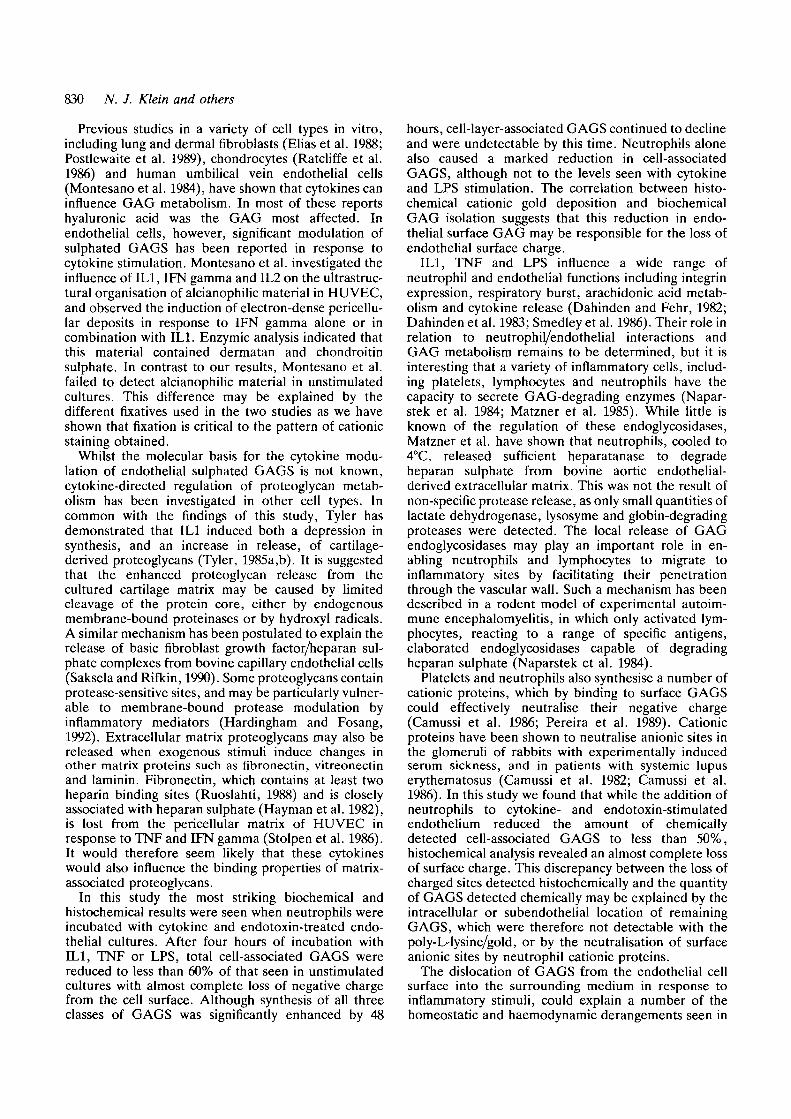
Previous studies in a variety of cell types in vitro,including lung and dermal fibroblasts (Elias et al. 1988;Postlewaite et al. 1989), chondrocytes (Ratcliffe et al.1986) and human umbilical vein endothelial cells(Montesano et al. 1984), have shown that cytokines caninfluence GAG metabolism. In most of these reportshyaluronic acid was the GAG most affected. Inendothelial cells, however, significant modulation ofsulphated GAGS has been reported in response tocytokine stimulation. Montesano et al. investigated theinfluence of IL1, IFN gamma and IL2 on the ultrastruc-tural organisation of alcianophilic material in HUVEC,and observed the induction of electron-dense pericellu-lar deposits in response to IFN gamma alone or incombination with ILL Enzymic analysis indicated thatthis material contained dermatan and chondroitinsulphate. In contrast to our results, Montesano et al.failed to detect alcianophilic material in unstimulatedcultures. This difference may be explained by thedifferent fixatives used in the two studies as we haveshown that fixation is critical to the pattern of cationicstaining obtained.
Whilst the molecular basis for the cytokine modu-lation of endothelial sulphated GAGS is not known,cytokine-directed regulation of proteoglycan metab-olism has been investigated in other cell types. Incommon with the findings of this study, Tyler hasdemonstrated that IL1 induced both a depression insynthesis, and an increase in release, of cartilage-derived proteoglycans (Tyler, 1985a,b). It is suggestedthat the enhanced proteoglycan release from thecultured cartilage matrix may be caused by limitedcleavage of the protein core, either by endogenousmembrane-bound proteinases or by hydroxyl radicals.A similar mechanism has been postulated to explain therelease of basic fibroblast growth factor/heparan sul-phate complexes from bovine capillary endothelial cells(Saksela and Rifkin, 1990). Some proteoglycans containprotease-sensitive sites, and may be particularly vulner-able to membrane-bound protease modulation byinflammatory mediators (Hardingham and Fosang,1992). Extracellular matrix proteoglycans may also bereleased when exogenous stimuli induce changes inother matrix proteins such as fibronectin, vitreonectinand laminin. Fibronectin, which contains at least twoheparin binding sites (Ruoslahti, 1988) and is closelyassociated with heparan sulphate (Hayman et al. 1982),is lost from the pericellular matrix of HUVEC inresponse to TNF and IFN gamma (Stolpen et al. 1986).It would therefore seem likely that these cytokineswould also influence the binding properties of matrix-associated proteoglycans.
In this study the most striking biochemical andhistochemical results were seen when neutrophils wereincubated with cytokine and endotoxin-treated endo-thelial cultures. After four hours of incubation withIL1, TNF or LPS, total cell-associated GAGS werereduced to less than 60% of that seen in unstimulatedcultures with almost complete loss of negative chargefrom the cell surface. Although synthesis of all threeclasses of GAGS was significantly enhanced by 48
hours, cell-layer-associated GAGS continued to declineand were undetectable by this time. Neutrophils alonealso caused a marked reduction in cell-associatedGAGS, although not to the levels seen with cytokineand LPS stimulation. The correlation between histo-chemical cationic gold deposition and biochemicalGAG isolation suggests that this reduction in endo-thelial surface GAG may be responsible for the loss ofendothelial surface charge.
ELI, TNF and LPS influence a wide range ofneutrophil and endothelial functions including integrinexpression, respiratory burst, arachidonic acid metab-olism and cytokine release (Dahinden and Fehr, 1982;Dahinden et al. 1983; Smedley et al. 1986). Their role inrelation to neutrophil/endothelial interactions andGAG metabolism remains to be determined, but it isinteresting that a variety of inflammatory cells, includ-ing platelets, lymphocytes and neutrophils have thecapacity to secrete GAG-degrading enzymes (Napar-stek et al. 1984; Matzner et al. 1985). While little isknown of the regulation of these endoglycosidases,Matzner et al. have shown that neutrophils, cooled to4°C, released sufficient heparatanase to degradeheparan sulphate from bovine aortic endothelial-derived extracellular matrix. This was not the result ofnon-specific protease release, as only small quantities oflactate dehydrogenase, lysosyme and globin-degradingproteases were detected. The local release of GAGendoglycosidases may play an important role in en-abling neutrophils and lymphocytes to migrate toinflammatory sites by facilitating their penetrationthrough the vascular wall. Such a mechanism has beendescribed in a rodent model of experimental autoim-mune encephalomyelitis, in which only activated lym-phocytes, reacting to a range of specific antigens,elaborated endoglycosidases capable of degradingheparan sulphate (Naparstek et al. 1984).
Platelets and neutrophils also synthesise a number ofcationic proteins, which by binding to surface GAGScould effectively neutralise their negative charge(Camussi et al. 1986; Pereira et al. 1989). Cationicproteins have been shown to neutralise anionic sites inthe glomeruli of rabbits with experimentally inducedserum sickness, and in patients with systemic lupuserythematosus (Camussi et al. 1982; Camussi et al.1986). In this study we found that while the addition ofneutrophils to cytokine- and endotoxin-stimulatedendothelium reduced the amount of chemicallydetected cell-associated GAGS to less than 50%,histochemical analysis revealed an almost complete lossof surface charge. This discrepancy between the loss ofcharged sites detected histochemically and the quantityof GAGS detected chemically may be explained by theintracellular or subendothelial location of remainingGAGS, which were therefore not detectable with thepoly-L-lysine/gold, or by the neutralisation of surfaceanionic sites by neutrophil cationic proteins.
The dislocation of GAGS from the endothelial cellsurface into the surrounding medium in response toinflammatory stimuli, could explain a number of thehomeostatic and haemodynamic derangements seen in

Endothelial GAG modulation in inflammation 831
inflammatory conditions. Vascular endothelium isknown to exert a regulating influence on both procoa-gulant and anticoagulant mechanisms. IL1 and TNFhave been shown to enhance the thrombogenicity ofvascular tissue by increasing the surface expression oftissue factor, decreasing endothelial surface thrombo-modulin and increasing the secretion of tissue plasmin-ogen activator inhibitor (Bevilacqua et al. 1986;Nachman et al. 1986; Nawroth et al. 1986; Nawroth etal. 1988). The roles of heparin, heparan sulphate anddermatan sulphate in preventing thrombosis throughthe stimulation of antithrombin III and heparin cofactorII activity have now also been well documented(Lindahl and Hook, 1978; Wright, 1980). The reductionin HUVEC surface GAG that we have observed inresponse to inflammatory stimuli would therefore beexpected to result in a local reduction of endothelial cellsurface anticoagulant activity. Paradoxically, GAGSreleased from the vascular wall could also behave ascirculating anticoagulants as described in some meta-static and inflammatory diseases (Palmer et al. 1984;Tefferi et al. 1990).
In this study we have shown that cytokines, endo-toxin and neutrophils can modulate endothelial sul-phated GAG metabolism and thereby influence boththe content and distribution of endothelial anionic sites.A number of recent studies have implicated hostinflammatory mediators as important components inthe pathophysiology of Gram-negative sepsis (Waage etal. 1987; Brandtzaeg et al. 1989). Our own results,considered in the context of these findings, maycontribute to the further understanding of the vascularchanges associated with endotoxic shock.
We thank Suzanne Peddie for her help in typing thismanuscript and Roland Levinsky, Kevin Forsyth and RobinCallard for their support and technical advice.
This work was supported through grants obtained from theMeningitis Research Appeal and the Child Health ResearchAppeal Trust.
References
Bennet, C. M., Glassock, R. J., Chang, R. L. S., Deen, W. M.,Robertson, C. R. and Brenner, B. M. (1976). Permselectivity of theglomerular capillary wall: studies of experimentalglomerulonephitis in the rat using dextran sulphate. /. Clin. Invest.57, 1287-1294.
Bevilacqua, M. P., Schleef, R. R., Gimbrone, M. A., Jr andLoskutofT, D. J. (1986). Regulation of the fibrinolytic system ofcultured human vascular endothelium by interleukin 1. J. Clin.Invest. 78, 587-591.
Bohrer, M. P., Bayliss, C , Humes, H. D., Glassock, R. J., Robertson,C. R. and Brenner, B. M. (1978). Permselectivity of the glomerularcapillary wall: facilitated filtration of polycations. J. Clin. Invest.61, 72-78.
Brandtzaeg, P., Klerulf, P., Gaustad, P., Skulberg, A., Bruun, J. N.,Halvorsen, S. and Sorensen, E. (1989). Plasma endotoxin as apredictor of multiple organ failure and death in systemicmeningococcal disease. J. Infect. Dis. 159, 195-204.
Brenner, B. M., Hosteller, M. D. and Humes, H. D. (1978). Molecularbasis of proteinuria of glomerular origin. N. Engl. J. Med. 298, 826-833.
Brody, J. S., Vaccaro, C. A., Hill, N. S. and Rounds, S. (1984).Binding of charged ferritin to alveolar wall components and charge
selectivity of macromolecular transport in permeability pulmonaryedema in rats. Circ. Res. 55, 155-167.
Broudy, V. C , Kanshansky, K., Segal, G. M. and Harian, J. M.(1986). Tumour necrosis factor type a stimulates humanendothelial cells to produce granulocyte/macrophage colony-stimulating factor. Proc. Nat. Acad. Sci. USA 83, 7467-7471.
Camussi, G., Tetta, C , Coda, R., Segoloni, G. P. and VerceUone, A.(1982). Localisation of neutrophil cationic proteins and loss ofanionic charges in glomeruli of patients with systemic lupuserythematosus glomerulonephritis. Clin. Immun. Immunopathol.24, 299-314.
Camussi, G., Tetta, T., Meronl, M., Toni-Tarelli, L., Roffineilo, C ,Alberton, A., Deregibus, C. and Sessa, A. (1986). Localisation ofcationic proteins derived from platelets and polymorphonuclearneutrophils and local loss of anionic sites in glomeruli of rabbitswith experimentally-induced acute serum sickness. Lab. Invest. 55,56-62.
Dahinden, C. and Fehr, J. (1982). Granulocyte activation byendotoxin. /. Immunol. 130, 863-868.
Dahinden, C , Galanos, C. and Fehr, J. (1983). Granulocyteactivation by endotoxin. / . Immunol. 130, 857-862.
Danon, D. and Shutelsky, E. (1976). Endothelial surface charge andits possible relationship to thrombogenesis. Ann. N.Y. Acad. Sci.275, 47-63.
Dustin, M. I ., Rothlein, R., Bhan, A. K., Dinarello, C. A. andSpringer, T. A. (1986). Induction of I LI and interferon gamma,tissue distribution, biochemistry and function of a naturaladherence molecule (ICAM1). J. Immunol. 137, 245-254.
Ellas, J. A., Krol, R. C , Freundlich, B. and Sampson, P. M. (1988).Regulation of human fibroblast glycosaminoglycan production byrecombinant interferons, tumor necrosis factor, and lymphotoxin./. Clin. Invest. 81, 325-333.
Gallagher, J. T., Lyon, M. and Steward, W. P. (1986). Structure andfunction of heparan sulphate proteoglycans. Biochem. J. 236, 313-325.
Gordon, P. B., Conn, G. and Hatcher, V. B. (1985).Glycosaminoglycan production in cultures of early and late passagehuman endothelial cells: the influence of an anionic endothelial cellgrowth factor and the extracellular matrix. /. Cell. Physiol. 125,596-607.
Hardingham, T. E. and Fosang, A. J. (1992). Proteoglycans: manyforms and many functions. FASEB J. 6, 861-870.
Hayman, E. G., OWberg, A., Martin, G. R. and Ruoslahti, E. (1982).Codistribution of heparan sulphate proteoglycan, laminin, andfibronectin in the extracellular matrix of normal rat kidney cells andtheir coordinate absence in transformed cells. J. Cell Biol. 94, 28-35.
Hunsicker, L. G., Shearer, T. P. and Shaffer, S. J. (1981). Acutereversible proteinuria induced by infusion of the polycationhexadimethrine. Kidney Int. 20, 7-13.
Jaffe, E. A., Nachman, N. L., Becker, C. G. and Minick, C. R. (1973).Culture of human endothelial cells derived from umbilical veins. J.Clin. Invest. 52, 2745-2764.
Kanwar, Y. S. (1984). Biology of disease: Biophysiology ofglomerular filtration and proteinuria. Lab Invest. 51, 7-20.
Kanwar, Y. S., Hascall, V. C. and Farquar, G. (1981). Partialcharacterisation of newly synthesised proteoglycans isolated fromthe glomerular basement membrane. J. Cell Biol. 90, 527-532.
Kanwar, Y. S., Linker, A. and Farquar, M. G. (1980). Increasedpermeability of the glomerular basement membrane to ferritinafter removal of glycosaminoglycans (heparan sulphate) byenzymatic digestion. /. Cell Biol. 86, 688-694.
Karlsson, K. and Marklund, S. L. (1988). Plasma clearance ofextracellular-superoxide dismutase c in rabbits. J. Clin. Invest. 82,762-766.
Kelley, V. E. and Cavallo, T. (1978). Glomerular permeability:Transfer of native ferritin in glomeruli with decreased anionic sites.Lab. Invest. 390, 547-553.
Larson, M. L., Abildgaard, U., Teien, A. N. and Gjesdal, K. (1978).Assay of plasma heparin using thrombin and the chromogenicsubstrate H-D-Phe-Pip-Arg-pNA (S-2238). Thromb. Res. 13, 285-288.
Levin, M. (1990). The inflammatory response to infections. In

832 N. J. Klein and others
Infection in the Newborn (ed. J. de Louvois and D. Harvey), pp. 1-12. John Wiley and Sons Ltd, London.
Lindahl, U. and Hook, M. (1978). Glycosaminoglycans and theirbinding to biological macromolecules. Annu. Rev. Biochem. 47,387-417.
Marcum, J. A. and Rosenberg, R. D. (1984). Anticoagulantly activeheparin-like molecules from vascular tissue. Biochemistry Z3,1730-1737.
Matzner, Y., Bar-Ner, M., Yahalom, J., Ishai-Michadi, R., Fuks, Z.and Vlodavsky, I. (1985). Degradation of heparan sulfate in thesubendothelial extracellular matrix by a readily releasedheparanase from human neutrophils. J. Clin. Invest. 76,1306-1313.
Mercier, J.-C., Beaufils, F., Hartman, J.-F. and Aze"ma, D. (1988).Hemodynamic patterns of meningococcal shock in children. Crit.Care Med. 16, 27-33.
Montesano, R., Mossaz, A., Ryser, J. E., Oric, L. and Vassalli, P.(1984). Leukocyte interleukins induced cultured endothelial cells toproduce a highly organised, glycosaminoglyan-rich pericellularmatrix. J. Cell Biol. 99, 1706-1715.
Nachman, R. L., Hajjar, K. A., Silverstein, R. I. and D1narelk>, C. A.(1986). Interleukin 1 induces endothelial cell synthesis ofplasminogen activator inhibitor. J. Exp. Med. 163, 1595-1600.
Naparstek, Y., Cohen, I. R., Fuks, Z. and Vlodavsky, I. (1984).Activated T lymphocytes produce a matrix-degrading heparansulphate endoglycosidase. Nature 310, 241-244.
Nawroth, P. P., Handley, D. A., Esmon, C. T. and Stern, D. M.(1986). Interleukin 1 induces endothelial cell procoagulant whilesuppressing cell-surface anticoagulant activity. Proc Nat. Acad. Sci.USA 83, 3460-3464.
Nawroth, P., Handley, D., Matsueda, G., De Waal, R., Gerlacb, H.,Blohm, D. and Stern, D. (1988). Tumour necrosis factor/cachectin-induced intravascular fibrin formation in meth A fibrosarcomas. J.Exp. Med. 168, 637-647.
Oohira, A., Wight, T. N. and Bornstcin, P. (1983). Sulfatedproteoglycans synthesized by vascular endothelial cells in culture.J. Biol. Chem. 258, 2014-2021.
Palmer, R. N., Rkk, M. E., Rick, P. D., ZeUer, J. A. and Gralnkk,H. R. (1984). Circulating heparan sulfate anticoagulant in a patientwith a fatal bleeding disorder. N. Engl. J. Med. 310, 1696-1699.
Pereira, H. A., Martin, L. E. and Spitmagel, J. K. (1989).Quantitation of a cationic antimicrobial granule protein of humanpolymorphonuclear leukocytes by ELISA. J. Immunol. Meth. 117,115-120.
Platt, J. L., VerceJlotti, G. M., Lindman, B. J., Oegema, T. R., Bach,F. H. and Dalmasso, A. P. (1990). Release of heparan sulphatefrom endothelial cells. J. Exp. Med. 1363-1368.
Pober, J. S., Gunbrone, M. A., Lapierre, L. A., Mendrick, D. L.,Fiers, W., Rothlein, R. and Springer, T. A. (1986). Overlappingpatterns of activation of human endothelial cells by interleukin 1,tumor necrosis factor, and immune interferon. J. Immunol. 137,1893-18%.
Postlewaite, A. E., Smith, G. N., Lachman, L. B., Endres, R. O.,Poppleton, H. M., Hasty, K. A., Seyer, J. M. and Kang, A. H.(1989). Stimulation of glycosaminoglycan synthesis in culturedhuman dermal fibroblasts by interleukin 1. J. Clin. Invest. 83, 629-636.
PurteU, J. N., Pesce, A. J., Clyne, D. H., Millar, W. C. and Pollak, V.E. (1979). Isoelectric point of albumin:affect on renal handling ofalbumin. Kidney Int. 16, 366-376.
RatcUfTe, A., Tyler, J. A. and Hardingham, T. E. (1986). Articularcartilage cultured with interleukin 1. Biochem. J. 238, 571-580.
Rounds, S. and Vaccaro, C. A. (1987). The binding of cationic probesto apical and basal surfaces of rat lung capillary endothelium and of
endothelial cells in tissue culture. Amer. Rev. Resp. Dis. 135, 725-730.
Ruoslahti, E. (1988). Fibronectin and its receptors. Annu. Rev.Biochem. 57, 375^13.
Saksela, O. and Rlfldn, D. B. (1990). Release of basic fibroblastgrowth factor-heparan sulfate complexes from endothelial cells byplasminogen activator-mediated proteolytic activity. J. Cell Biol.110, 767-775.
Skutelsky, E. and Roth, J. (1986). Cationic colloidal gold - a newprobe for the detection of anionic cell surface sites by electronmicroscopy. /. Histochem. Cytochem. 34, 693-696.
Smedley, L. A., Tonnesen, M. G., Sandhaus, R. A., Haslett, C ,Guthrie, L. A., Johnston, R. B., Henson, P. M. and S., W. G.(1986). Neutrophil-mediated injury to endothelial cells. J. Clin.Invest. 77, 1233-1243.
Springer, T. A. (1990). Adhesion receptors of the immune system.Nature 346, 425-434.
Stolpen, A. H., Guinan, E. C , Fiers, W. and Pober, J. S. (1986).Recombinant tumour necrosis factor and immune interferon actsingly and in combination to reorganize human vascular endothelialcell monolayers. Amer. J. Pathol. 123, 16-24.
TefTeri, A., Nichols, W. L. and WalterBowie, E. J. (1990). Circulatingheparin-like anticoagulants: report of five consecutive cases and areview. Amer. J. Med. 88, 184-188.
Tracey, K. J., Beutler, B., Lowry, S. F., Merryweather, J., Wolpe,S., Milsark, I. W., Hariri, R. J., Fahey III, T. J., ZenteUa, A.,Albert, J. D., Shires, G. T. and Cerami, A. (1986). Shock and tissueinjury induced by recombinant human cachectin. Science 234, 470-474.
Tyler, J. A. (1985a). Articular cartilage cultured with catabolin (piginterleukin 1) synthesisezes a decreased number of normalproteoglycan molecules. Biochem. J. 227, 869-878.
Tyler, J. A. (1985b). Chondrocyte-mediated depletion of articularcartilage proteoglycans in vitro. Biochem. J. 225, 493-507.
Vehaskari, V. M., Chang, C. T.-C., Stevens, J. K. and Robson, A. M.(1984). The effects of polycations on vascular permeability in therat. J. Clin. Invest. 84, 1053-1059.
Vermylen, C , Levin, M., Mosman, J. and Barrett, T. M. (1989).Decreased heparan sulphate content of the glomcrular basementmembrane, and increased urinary excretion of heparan sulphate inthe congenital nephrotic syndrome. Paediatr. Nephrol. 3, 122-129.
Volker, W., Schon, P. and Vischer, P. (1991). Binding andendocytosis of thrombospondin and thrombospondin fragments inendothelial cell cultures analysed by cuprolinic blue staining,colloidal gold labelling and silver enhancement techniques. J.Histochem. Cytochem. 39, 1385-1394.
Waage, A., Halstensen, A. and Epsevik, T. (1987). Associationbetween tumour necrosis factor in serum and fatal outcome inpatients with meningococcal disease. Lancet I, 355-357.
Whiteman, P. (1973). The quantitative determination ofglycosaminoglycans in urine with alcian blue 8GX. Biochem. J.131, 351-357.
Wight, T. N., Kinsella, M. G., Lark, M. W. and Potter-perigo, S.(1986). Vascular cell proteoglycans: evidence for metabolicmodulation. In Functions of Proteoglycans, Ciba FoundationSymposium, pp 241-259. John Wiley and Sons Ltd, Chichester.
Wright, T. N. (1980). Vessel proteoglycan and thrombogenesis. InProgress in Haemostasis and Thrombosis, vol. 5 (Ed T. H. Spaet),pp. 1-39. Grune and Stratton, New York.
(Received 28 February 1992 - Accepted 28 April 1992)