Embed Size (px)
Citation preview
Alterations of GABAB binding caused by acute and chronic lead administration
Jolanta WaSkiewicz
Department of Neurochemistry, Medical Research Centre, Polish Academy of Sciences, 3 Dworkowa St.,00-784 Warsaw, Poland
Abstract. The effect of lead on GABAB binding was studied on membranes obtained from acute and chronically lead-treated rats. Acute lead treatment reduced both GABAB affinity (KD) by about 30% and density of receptor (Bmax) by about 15%. On the contrary chronic lead treatment increased receptor capacity by about 20% in spite of decreased receptor affinity by 25%. Both acute and chronic lead treatment shiffted displacement curves toward higher concentration of non-labelled compound(decreased affinity). The results show that lead can affect the GABAB binding in two ways: by reducing affinity of binding and by altering capacity of binding.
Key words: GABA , gamma-aminobutyric acid, KD, affinity of receptor, Bm, , density of receptor
Lead ions are known to be toxic to the nervous system of man and animal (Davis et al. 1993). Both acute and chronic exposures to lead result in beha- vioural and neurological symptomes. In lead neurotoxicology an important issue relates to how lead interferes with chemical neurotrans- mission. Numerous investigations have been under- taken in order to explain the possible molecular mechanism of lead toxicity (Silbergeld 1982, Tschudy and Lamon 1990, Zareba and Chrnielnicka 1992, Guilarte 1993, Jabonska et al. 1994). It has been postulated that PbICa interactions may play an important role in the process (Silbergeld et al. 1974, Kolton and Yassi 1982).
Gamma-aminobutyric acid (GABA) is known as one of inhibitory neurotransmitters in the mamma- lian brain that induces the hyperpolarization of membrane potential by activating GABA receptor coupled with chloride ion channel (Olsen 198 1). The observation that some effects of GABA in the CNS are not antagonized by the GABA receptor an- tagonist bicuculline (Hill and Bowery 1981), has led to the discovery of the GABAB class of GABA receptor, which is insensitive to bicuculline but sen- sitive to (-)-baclofen(Hil1 and Bowery 1981). It has been reported that the activation of GABAB recep- tor causes reduction of evoked transmitter release (Bowery et al. 1984).
Activation of GABAB receptors by baclofen or GABA increases membrane K+ conductance post- -synaptically and decreases ca2+ conductance presynaptically to depress transmitter release; these receptors are directly coupled to the Kt and ca2+ channels through GiIGo proteins and are modu- lated by guaninenucleotides (Bowery 1993).
The aim of this work was to assess the sensitivity of Ca-dependent GABA binding to lead toxicity,es- pecially its influence on affinity and capacity of GABAB binding. As shown previously, pb2+ level in synaptosomal fraction (obtained from P2 fraction) was more than twice higher than in control fraction (Struzynska and Rafalowska 1994). Membranes for GABAB binding were obtained from crude P2 fraction.
The study was performed on male Wistar rats. Two models of poisoning were used:
1. In the acute model 15 mg Pb(CH3C00)2/kg b.w. was injected intraperitoneally for 7 days into rats weighting 150 g. The control animals were in- jected with distilled water. The animals were de- capitated and brains were removed.
2. In the chronic model of toxicity , lead acetate (200 mgll H20) was given to 3-week old rats in drinking water for 3 months. Control animals re- ceived distilled water without Pb(CH3C00)2 . The mechanism of the gamma-aminobutyric acid recep- tor (GABAB) binding was examined using crude membrane fraction (P2) obtained from the rat brain according to Zukin et al. (1974). For the assay pel- lets (P2 fraction) which had been frozen for at least 16 h at - 15' C were allowed to thow for 20 min at room temperature before resuspension in Tris-HC1 buffer (50 mM pH 7,4) plus CaClz (2.5 mM).
The pellet obtained from the equivalent of one rat brain was resuspended in 10 ml Tris-HC1. The suspension was incubated for 45 min at 2 0 ' ~ before centrifugation at 7,000 g for 10 min. This working procedure was repeated three more times allowing 15 min incubation with addition of Tris-HC1 plus CaC12 each time. The final pellet was resuspended in Tris-HC1 buffer+CaC12 (- 1 mg protein per 0.8 ml buffer plus 40 yM isoguvacine for the assay. Pro- tein concentrations were determined by the method of Lowry et al. (1951 ). To each 0.8 ml aliquot of membrane suspension 0.1 ml Tris-HC1 plus CaClz containing various concentrations of unlabelled GABA and 0.1 ml Tris-HC1 containing a fixed con- centration(1-3 nM) of [ 3 ~ ] - ~ ~ ~ ~ was added. The range of final concentrations of unlabelled GABA was 1 nM-100 pM. Nonspecific binding was deter- mined by adding 100 pM baclofen. The mixture was incubated for 15 min at 2 0 ' ~ . Each incubation was terminated by filtration under vacuum through Whatman GFIB glass filter and then the filter was rinsed twice with 5 ml of ice-cold distilled water ac- cording to Ohmori et al. (1990). The radioactivity trapped on each filter was measured by a liquid scin- tillation spectrometer at a counting efficiency of 40%.
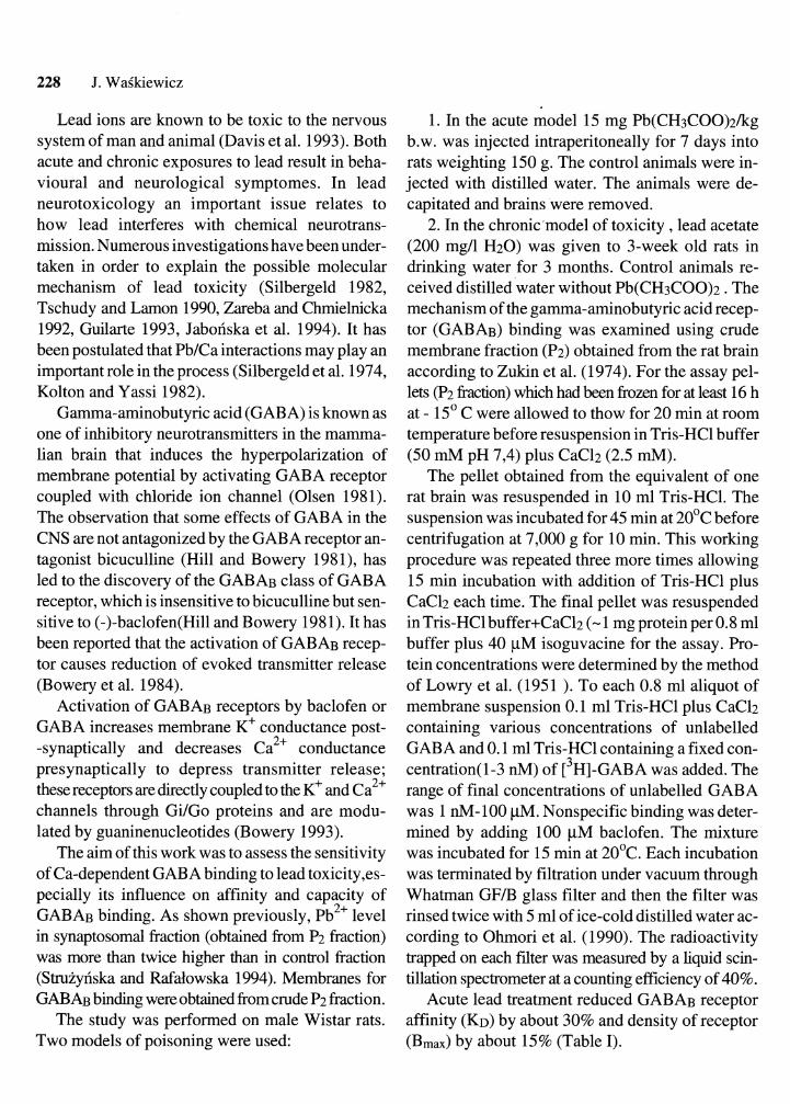
Acute lead treatment reduced GABAB receptor affinity (KD) by about 30% and density of receptor (Bmax) by about 15% (Table I).
Lead and GABAB receptor 229
TABLE I TABLE I1
Kinetic parameters for [ 3 ~ ] GABA binding to GABAB re- ceptors after acute lead treatment
Group K D ( ~ M ) Bmax(fm011mg protein)
control 105f 1 1 952f93 lead treated 136f 14 707f 79
Kinetic parameters f ~ r [ ~ H ] GABA binding to the GABAB receptors after chronic lead treatment
Group K D ( ~ M ) Bmm(fmolImg protein)
control 99f11 984f 9 1 lead treated 126k12 1210k111
The values represent mean f SEM from 4 experiments; P<0.05 (Student's t test).
The results represent mean + SEM from 4 experiments; P<0,05 (Student's t test).
Chronic lead treatment decreased receptor af- finity (KD) by about 25% but increases receptor ca- pacity (Bmax) by about 20% (Table 11).
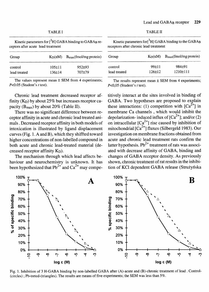
There was no significant difference between re- ceptor affinity in acute and chronic lead treated ani- mals . Decreased receptor affinity in both models of intoxication is illustrated by ligand displacement curves (Fig. 1. A and B), which they shiffted toward higher concentrations of non-labelled compound in both acute and chronic lead-treated material (de- creased receptor affinity KD).
The mechanism through which lead affects be- haviour and neurochemistry is unknown. It has been hypothesized that pb2+ and ca2+ may compe-
titively interact at the sites involved in binding of GABA. Two hypotheses are proposed to ex lain $ these interactions: (1) competition with [Ca ] in membrane Ca channels , which would inhibit the depolarization- induced influx of [ca2+]; and/or (2) on intracellular [ca2+] rise caused by inhibition of mitochondria1 [ca2+] fluxes (Silbergeld 1983). Our investigation on membrane fractions obtained from acute and chronic lead treatment rats confirm the latter hypothesis. pb2+ treatment of rats was associ- ated with decrease affinity of GABA, binding and changes of GABA receptor density. As previously shown, chronic treatment of rat results in the inhibi- tion of KC1 dependent GABA release (Struiyriska
Fig. 1. Inhibition of 3 H-GABA binding by non-labelled GABA after (A)-acute and (B)-chronic treatment of lead . Control- (circles) ; Pb-treted-(triangles). The results are means of five experiments; the SEM was less than 5%.