Embed Size (px)
Citation preview

Altered mental status is a sign of seriousneurologic or systemic disease. Thephysician must rapidly assess the depthof coma and risk of intracranialhypertension, then determine theetiology and prescribe appropriatemanagement. This is no small task,considering the variety and multitude ofillnesses that may present with alteredmental status. The etiologies can bebroken down into structural and medicalcauses. The structural causes morefrequently affect the brainstem centersadjacent to the ascending reticularactivating system that are responsible forpupillary response and oculocephalicreflexes. Medical causes generally sparethese structures. Management ofstructural lesions requires promptdiagnosis and neurosurgical input.Medical etiologies are protean andtreatment is often supportive.Clin Ped Emerg Med 4:171-178.© 2003 Elsevier Inc. All rights reserved.
Altered Mental Status
By Diana King, MD, and Jeffrey R. Avner, MDBRONX, NEW YORK
CONSCIOUSNESS IS GENERALLY THOUGHT OF as aware-ness of one’s self and environment while coma is unrespon-siveness, even to a painful stimulus. However, between theconditions of consciousness and coma, there are several
states with definitions that are often misused, including confu-sion, delirium, obtundation, and stupor. A confused person hasslowed or impaired cognitive abilities manifested by disorienta-tion, memory deficits, or difficulty following commands. Stimuliare misinterpreted and the person is often drowsy. Delirium is achain of unconnected ideas such that the patient appears disori-ented, fearful, agitated, and irritable. Misperception of sensorystimuli can lead to hallucinations. This state is usually associatedwith a toxic/metabolic etiology. Obtundation is decreased alert-ness and limited interest in the environment. More time is spentsleeping and when awakened, the patient is still drowsy. Instupor, the patient is responsive only to vigorous, repeated stim-uli and returns to an unresponsive state when left alone.1 Al-though each of the above terms connote a specific altered state,the clinician should appreciate that they are quite often misusedand can therefore lead to poor communication between healthcare providers. For this reason the Glasgow Coma Scale (GSC) isoften a much more reliable means of conveying the level ofconsciousness in a patient.
Clinical Assessment
Normal consciousness requires both awareness and arousal.Awareness is the combination of cognition and affect that can beinferred based on the patient’s interaction with the environment.Thus, alterations of consciousness may be the result of deficits inawareness, arousal, or both. Awareness is determined by thecerebral hemispheres whereas arousal is controlled by the as-cending reticular activating system (ARAS) commonly called thesleep center. A helpful analogy is a bulb-switch model where thecerebral hemispheres function as a light bulb and the ARAS
From the Division of PediatricEmergency Medicine, Department ofPediatrics, Children’s Hospital atMontefiore, Albert Einstein College ofMedicine, Bronx, NY.
Address reprint requests to Diana King,MD, Pediatric Emergency Medicine,Children’s Hospital at Montefiore, 111East 210th Street, Bronx, NY 10467.E-mail: [email protected]
© 2003 Elsevier Inc. All rightsreserved.
1522-8401/03/0403-0000$30.00/0doi:10.1016/S1522-8401(03)00058-2
AL TERED MENTAL S T A TU S / K ING AND AVNER 171

functions as a light switch. Normal consciousnessrequires the light bulb to be lit; to do so requiresboth the bulb and the switch to function properly. Ifthe bulb is “out,” there can either be a problemwith the bulb itself, the switch, or both. Similarly,altered mental status can result from depression ofboth cerebral hemispheres, localized abnormalityof the sleep center, or global central nervous systemdysfunction.1 Components necessary for the bulbto function are relative normothermia and bloodflow, delivery of energy substrates (oxygen and glu-cose), and absence of toxins (metabolic waste prod-ucts, poisons, and infectious material).2
The ARAS is a core brain structure that coursesfrom the medulla to the thalamus. Serendipitously,the ARAS’ location overlaps several brain stem re-flex pathways, in particular those responsible forthe pupillary light reflex and the reflex eye move-ments that allow conjugate gaze. Thus, preservationof these reflexes often means that ARAS function isnormal and therefore implies that the alteration inmental status is the result of deficits in both cere-bral hemispheres. Conversely, pupillary asymmetryor dysconjugate gaze imply deficits in the area ofthe ARAS.1
It is often helpful to divide the etiologies of al-tered mental status into 2 groups: structural andmedical (toxic-infectious-metabolic) (Table 1).Structural etiologies usually cause compression ordysfunction in the area of the ARAS whereas mostmedical etiologies lead to general dysfunction ofboth cerebral hemispheres. Common etiologies ac-cording to age are presented in Table 2. Since struc-tural etiologies may require operative interventionas opposed to the supportive care required for mostmedical etiologies, it is important for the emer-gency physician to make a rapid assessment ofthe likelihood of each of these conditions. This
can be done through the recognition of an asym-metric neurologic examination and the system-atic assessment of 3 physical exam findings:pupillary response, extraocular movements, andmotor response to pain.
Pupillary Response
The areas of the brainstem that control con-sciousness and pupillary response are anatomicallyadjacent. The sympathetic and parasympatheticnervous systems control pupillary dilatation andconstriction, respectively. The sympathetic path-way originates in the hypothalamus, fibers descendto the spinal cord, preganglionic fibers synapse inthe superior cervical ganglion, and postganglionicfibers travel with the internal carotid artery into theskull. The parasympathetic pathway originates inthe midbrain and the postganglionic fibers accom-pany the oculomotor nerve. Knowledge of thesepathways helps localize where a lesion may bebased upon pupillary signs. If damage occurs in themidbrain region, the parasympathetic pathway isinterrupted and pupils will be slightly enlarged andnot responsive to light. Pontine lesions interferewith the descending sympathetic fibers and resultin small pupils. The light reflex may be present, butdifficult to visualize without magnification. Lesionsthat compress the third nerve will result in a dilatedand unresponsive pupil on the same side as theinsult.
Pupillary response is usually preserved when al-tered mental status is secondary to medical etiologies(especially toxic and metabolic causes). The pupilsmay be small, but they are generally symmetric andreactive. Therefore, the presence of the pupillary lightreflex may be the most important sign that differen-tiates structural from medical coma.1
TABLE 1. Common Etiologies of AlteredMental Status
Structural Medical
Trauma Infection
Intracranial bleed Toxin
Cerebral edema Seizure
Shaken baby syndrome Metabolic
Tumor Intussusception
Stroke Hemolytic-uremic syndrome
Hydrocephalus Psychogenic
TABLE 2. Common Diagnoses of Altered MentalStatus by Age
Infant Child Adolescent
Infection Toxin Toxin
Metabolic Infection Trauma
Inborn Error Seizure Psychiatric
Seizure Intussusception Seizure
Abuse Abuse/Trauma
172 AL TERED MENTAL S T A TU S / K ING AND AVNER

Extraocular Movements
Areas of the brainstem adjacent to those respon-sible for consciousness also mediate oculomotorreflexes. The ability to maintain conjugate gazerequires preservation of the internuclear connec-tions of cranial nerves 3, 6, and 8 via the mediallongitudinal fasciculus (MLF). For example, propri-oceptive inputs from cervical muscles (eg, when thehead is turned to a side) ascend through the MLF inthe brainstem to reach the ipsilateral abducens nu-cleus. The stimulus then crosses and continues toascend through the MLF to reach the contralateraloculomotor nucleus. These pathways, therefore, al-low for the coordination of the ipsilateral abducensand contralateral oculomotor nerves. The abducensnerve (through the lateral rectus muscle) moves theipsilateral eye laterally and the oculomotor nerve(through the medial rectus) moves the contralateraleye medially; hence, there is conjugate gaze. Aswith pupillary responses, structural lesions that im-pinge on these pathways will cause dysfunctionranging from disconjugate gaze to opthalmoparesis.Therefore, deficits in extraocular movements usu-ally accompany a structural etiology.
There are several reflexes that can test extraoc-ular function in an unconscious patient. The ocu-locephalic (Doll’s eyes) reflex is elicited by holdingthe eyelids open and turning the head briskly toeach side. Normal response is for the eyes to shiftleft when the head is turned right and vice versa. Ifa low brainstem lesion is present, the eyes willmove along with the head mimicking oculoparesis.This reflex should not be performed in a patientwith suspected spinal cord injury. The oculoves-tibular reflex (cold caloric) is elicited by elevatingthe head 30 degrees and inserting a small catheterinto the external auditory canal, near the tympanicmembrane. The eyes are held open while 120cc ofice water is flushed into the ear. The normal re-sponse in an unconscious patient is nystagmus withthe slow component toward the ear being irrigatedand the fast component away from the irrigatedside (the reverse is true in conscious patients).Patients with unilateral MLF lesions will deviate theeye only on the unaffected side while those with lowbrainstem lesions will not move either eye in re-sponse to this maneuver.1
Motor Response to Pain
Abnormal motor movements may also help pin-point the location of a lesion. Decorticate posturing(abnormal flexion) can be seen with damage to the
diencephalon (uppermost brainstem). Decerebrateposturing (abnormal extension) can be seen withdamage to the midbrain and pons. Flaccid posturingis an ominous sign and indicates compression of themedulla, a terminal event.1
In summary, structural causes will often lead tocompression or dysfunction of the brain stem andtherefore present with pupillary abnormality (un-equal, unreactive, or sluggishly reactive) and focalneurologic findings. Medical causes usually resultfrom involvement of both cerebral hemispheres;the pupils are generally equal and reactive (al-though the reaction may be sluggish) and the neu-rologic examination is non-focal. There are,however, some notable exceptions. Structural dis-orders without focality include acute bilateral cere-brovascular disease and early acute hydrocephalus.Medical encephalopathies with focality include hy-perglycemia, hypoglycemia, hypercalcemia, he-patic encephalopathy, uremia, and some toxicingestions.
Etiologies
A complete list of conditions that may presentwith altered level of consciousness is not practical.We will therefore discuss only the major categorieswith the most common illnesses.
Structural Neurologic Derangement
Trauma
The typical mechanism for accidental or in-flicted trauma is rapid deceleration that causesshearing of axons connecting cell bodies (diffuseaxonal injury). The shearing of axons that connectthe ARAS to higher brain centers results in loss ofconsciousness. Contusions are most common in thefrontal and temporal regions. The shearing forcescan also rupture blood vessels and result in epi-dural, subdural, or intraparenchymal hemorrhage.Subdural and epidural hematomas may requireemergent evacuation. The role of the emergencyphysician is centered on preventing secondary in-juries that occur due to hypoxia, ischemia, hypo-tension, seizures, and cerebral edema.3 TheGlasgow Coma Scale is a helpful measure of sever-ity of head injury, and intubation and controlledhyperventilation is the standard of care for a scoreof 8 or less.4
AL TERED MENTAL S T A TU S / K ING AND AVNER 173

Tumors
Primary brain tumors that affect either cerebralhemispheres or the ARAS of the brainstem mayaffect the level of consciousness by direct effect onthe neural pathways. Generalized effects of tumorsthat affect level of consciousness include seizures,intracranial hypertension due to enlarging mass, orcerebral edema surrounding the mass. Brainstemand cerebellar tumors are more likely to cause ob-structive hydrocephalus by blocking the third andfourth ventricles. Symptoms may be present forweeks to months before presentation. Commonsymptoms include headache, vomiting, alteredmental status, and focal neurologic deficit.5
Vascular
Ischemic, thrombotic, or hemorrhagic strokesmay cause altered mental status by interfering withcerebral blood flow and by causing intracranial hy-pertension secondary to cerebral edema around theinfarction. Hemorrhagic and ischemic strokes oc-cur with the same frequency in children. The mostcommon cause of hemorrhagic stroke in children isarteriovenous malformation (AVM).6 Thromboticand ischemic strokes are most commonly seen inchildren with sickle cell disease and congenitalheart disease. Other etiologies include hypercoag-ulable states, metabolic disorders (MELAS andhomocystinuria), vasculitis (systemic lupus erythe-matosis, Henoch Schonlein purpura, and poly-arteritis nodosa), and other vascular abnormalities(Moyamoya, arterial dissection and sinus thrombo-sis).7 Ischemic strokes present with focal deficitsand hemorrhagic strokes present with altered men-tal status and headache.6
Hydrocephalus
Hydrocephalus occurs when there is an imbal-ance between the production and absorption ofcerebrospinal fluid (CSF). This causes dilatation ofthe ventricles and displacement of the cerebral cor-tex. Communicating hydrocephalus occurs whenthe arachnoid villi are unable to absorb CSF nor-mally due to infection or hemorrhage. Noncommu-nicating hydrocephalus occurs when there is ablockage of the normal circulation of CSF, usuallydue to congenital malformations or acquired tu-mors. Infants present with head enlargement, thinscalp with distended veins, and full or bulging fon-tanel. They may have irritability, vomiting, andpoor feeding. “Setting sun” sign may be seen, whichis created by weakness of cranial nerve VI, resulting
in decreased upward gaze. Once the fontanels haveclosed the typical symptoms are headache, nausea,and vomiting. As intracranial pressure rises, pa-tients become lethargic and may have increasedmuscle tone and posturing.8 The treatment of hy-drocephalus requires diverting CSF via a shunt sys-tem. The ventriculoperitoneal shunt is mostcommon. Tissue debris, choroid plexus, infection,or migration of the catheter can obstruct the shuntproximally. Distal obstruction results from kinking,omentum, infection and migration. The signs andsymptoms of shunt obstruction are the same asthose for hydrocephalus.9
Medical Causes of Neurologic Derangement
Any process that decreases or interferes with thedelivery or utilization of substrate to the brain canalter brain activity. Hyper- or hypothermia, hyper-or hypotension, hyper- or hypoglycemia, hypoxia,and hypercarbia can all result in altered conscious-ness. Abnormal electrolyte concentrations (partic-ularly sodium and calcium) can lead to seizures andcoma. Build-up of waste products due to renal orhepatic failure can also alter the level of conscious-ness.
Infections
Central nervous system infections can rapidlyprogress to coma and death. Fever, irritability,vomiting, and lethargy often accompany meningi-tis. Headache and neck stiffness are manifested inchildren older than two years of age.10 Subduralempyema can occur secondary to meningitis or,more commonly, from direct extension of paranasalsinus infection or otitis media. Infection can extendto both hemispheres. The presentation is similar tothat of meningitis and seizures occur in two-thirdsof these patients.11 Mortality is higher in patientswho present in coma.12 Older children and adoles-cents can develop an epidural abscess as a result ofcontiguous spread of infection from the sinuses ormiddle ear. Patients do not come to attention untilfocal neurologic deficits or seizures ensue.11 Sepsiscan also cause depressed mental status secondaryto circulating proinflammatory mediators (cyto-kines, endotoxins, etc) and shock.
Toxins
Accidental and intentional ingestions of drugsand toxins are a common pediatric occurrence.Many ingestions present with altered mental status
174 AL TERED MENTAL S T A TU S / K ING AND AVNER
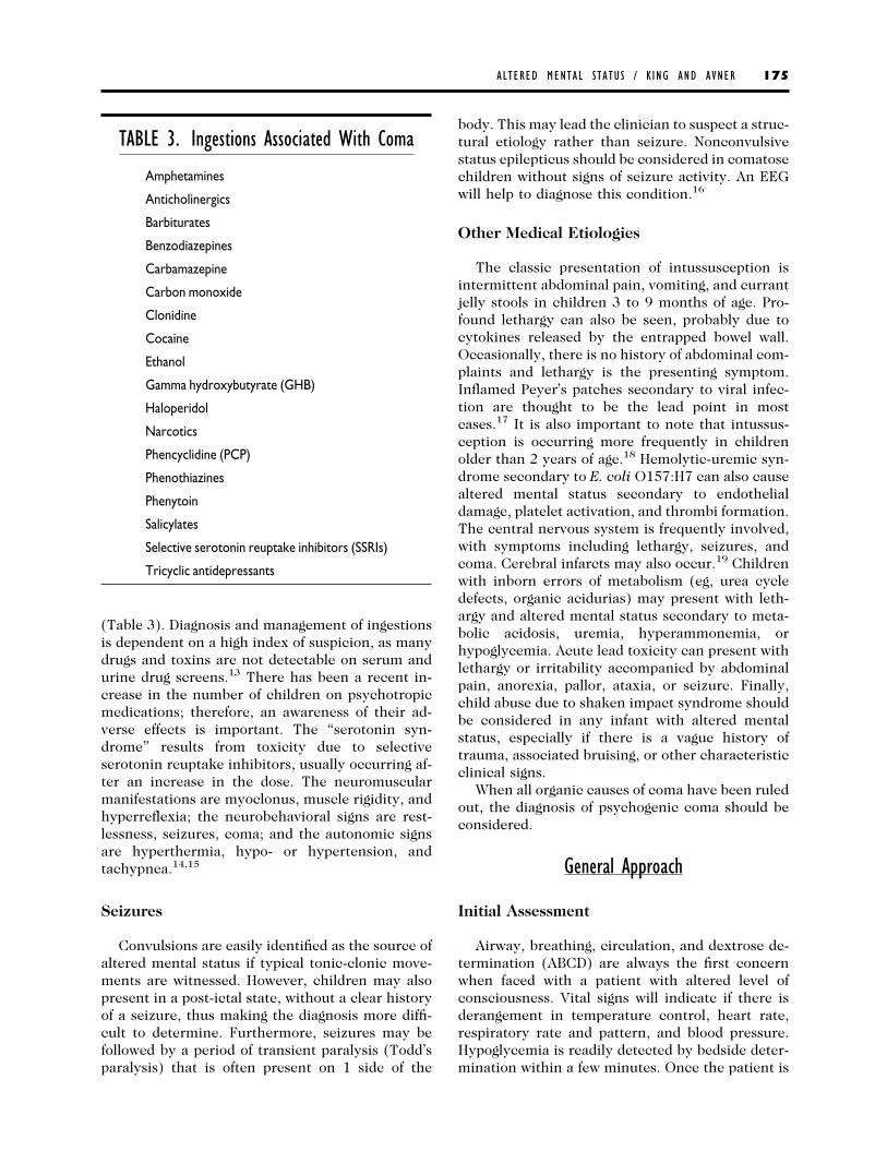
(Table 3). Diagnosis and management of ingestionsis dependent on a high index of suspicion, as manydrugs and toxins are not detectable on serum andurine drug screens.13 There has been a recent in-crease in the number of children on psychotropicmedications; therefore, an awareness of their ad-verse effects is important. The “serotonin syn-drome” results from toxicity due to selectiveserotonin reuptake inhibitors, usually occurring af-ter an increase in the dose. The neuromuscularmanifestations are myoclonus, muscle rigidity, andhyperreflexia; the neurobehavioral signs are rest-lessness, seizures, coma; and the autonomic signsare hyperthermia, hypo- or hypertension, andtachypnea.14,15
Seizures
Convulsions are easily identified as the source ofaltered mental status if typical tonic-clonic move-ments are witnessed. However, children may alsopresent in a post-ictal state, without a clear historyof a seizure, thus making the diagnosis more diffi-cult to determine. Furthermore, seizures may befollowed by a period of transient paralysis (Todd’sparalysis) that is often present on 1 side of the
body. This may lead the clinician to suspect a struc-tural etiology rather than seizure. Nonconvulsivestatus epilepticus should be considered in comatosechildren without signs of seizure activity. An EEGwill help to diagnose this condition.16
Other Medical Etiologies
The classic presentation of intussusception isintermittent abdominal pain, vomiting, and currantjelly stools in children 3 to 9 months of age. Pro-found lethargy can also be seen, probably due tocytokines released by the entrapped bowel wall.Occasionally, there is no history of abdominal com-plaints and lethargy is the presenting symptom.Inflamed Peyer’s patches secondary to viral infec-tion are thought to be the lead point in mostcases.17 It is also important to note that intussus-ception is occurring more frequently in childrenolder than 2 years of age.18 Hemolytic-uremic syn-drome secondary to E. coli O157:H7 can also causealtered mental status secondary to endothelialdamage, platelet activation, and thrombi formation.The central nervous system is frequently involved,with symptoms including lethargy, seizures, andcoma. Cerebral infarcts may also occur.19 Childrenwith inborn errors of metabolism (eg, urea cycledefects, organic acidurias) may present with leth-argy and altered mental status secondary to meta-bolic acidosis, uremia, hyperammonemia, orhypoglycemia. Acute lead toxicity can present withlethargy or irritability accompanied by abdominalpain, anorexia, pallor, ataxia, or seizure. Finally,child abuse due to shaken impact syndrome shouldbe considered in any infant with altered mentalstatus, especially if there is a vague history oftrauma, associated bruising, or other characteristicclinical signs.
When all organic causes of coma have been ruledout, the diagnosis of psychogenic coma should beconsidered.
General Approach
Initial Assessment
Airway, breathing, circulation, and dextrose de-termination (ABCD) are always the first concernwhen faced with a patient with altered level ofconsciousness. Vital signs will indicate if there isderangement in temperature control, heart rate,respiratory rate and pattern, and blood pressure.Hypoglycemia is readily detected by bedside deter-mination within a few minutes. Once the patient is
TABLE 3. Ingestions Associated With Coma
Amphetamines
Anticholinergics
Barbiturates
Benzodiazepines
Carbamazepine
Carbon monoxide
Clonidine
Cocaine
Ethanol
Gamma hydroxybutyrate (GHB)
Haloperidol
Narcotics
Phencyclidine (PCP)
Phenothiazines
Phenytoin
Salicylates
Selective serotonin reuptake inhibitors (SSRIs)
Tricyclic antidepressants
AL TERED MENTAL S T A TU S / K ING AND AVNER 175

stabilized, a focused history should be obtained.Important features of the history include the cir-cumstances of the onset of the neurologic symp-toms (eg, gradual or abrupt onset), precedingneurologic symptoms (weakness, headache, sei-zure, dizziness, diplopia, vomiting), trauma (wit-nessed or suspected), drug use or access to drugs,bloody stools (hemolytic uremic syndrome or in-tussusception), and whether the history is incon-sistent with the injury noted (child abuse).Contributing past medical history may includebrain tumor, VP shunt, seizure disorder, sickle celldisease, metabolic disorder, diabetes, renal failure,and liver disease. Physical examination will helpdifferentiate structural neurologic injury from asystemic abnormality. Rapid recognition of intra-cranial hypertension is crucial.
Intracranial Hypertension
There are 3 components within the cranium:brain, cerebrospinal fluid, and blood. When there isan increase in the volume of any of these compo-nents, the intracranial pressure (ICP) will increase.Elevated intracranial pressure can cause hernia-tion, which may result in irreversible brain damageor death. A history of severe headaches (especiallythose that improve with elevation), vomiting, visualchanges, and altered behavior or level of conscious-ness may indicate elevated ICP. Physical signs thatmay point to increased ICP include papilledema,cranial nerve palsies, abnormal mental status, andposturing. The ominous Cushing’s triad (bradycar-dia, hypertension, and irregular respirations) is asign of impending herniation.8
Physical Examination
A thorough physical and neurologic evaluationshould be performed. Assessment of the patient’slevel of conscious as well as frequent reassessmentof the mental status is crucial in following thecourse of illness. As reviewed earlier, pupillary size(normal or asymmetric) and reflex (fixed or reac-tive), extraocular movements (normal, asymmetricor absent), and motor response to pain (normal,decorticate, decerebrate, or flaccid) are importantclues to determining whether the etiology of theillness is structural or medical. Assymetry to theexamination also points towards a structural lesion.The GSC score can also be helpful in assessing thedepth of coma in patients with head trauma. Ascore of 8 or less indicates severe injury.20
Identifying abnormal respiratory patterns canalso assist in differentiating a structural neurologic
condition from a medical condition. Cheyne-Stokesrespiration is characterized by hyperpnea in a cre-scendo and decrescendo pattern followed by anapneic phase. It is seen in patients with bilateralhemispheric disease, hypertensive encephalopa-thy, conditions which cause cerebral hypoxia, andmetabolic conditions. Central neurogenic hyper-ventilation may occur with lesions of the midbrainand pons. It is a sustained, rapid, and deep respira-tory pattern that results in a respiratory alkalosis.Apneustic breathing consists of end-inspiratorypauses alternating with end-expiratory pauses. Thispattern is consistent with damage to the pons. Re-spiratory centers in the medulla are responsible forthe normal rhythm of breathing. Damage at thislevel produces ataxic breathing, which is a com-pletely irregular pattern that may progress to ap-nea.1
The physical exam should include a search forsigns of trauma: bruises, hematomas, hemotympa-num, Battle’s sign, raccoon eyes, and retinal hem-orrhages. The breath may indicate alcohol use ordiabetic ketoacidosis. Patients who are feigning un-responsiveness will have an increase in heart ratein response to painful stimuli, may resist eye open-ing, and usually avoid hitting themselves whentheir hand is allowed to drop to their face.
Management
A management algorithm is shown in Figure 1.The patient should be placed on a cardiorespiratorymonitor and a pulse oximeter. Oxygen should beroutinely administered. If the patient has an unsta-ble airway, abnormal breathing pattern, or a GCS of8 or less, rapid sequence intubation should be per-formed with a attention to cerebral protection. In-travenous (IV) access should be obtained and blooddrawn for laboratory studies and a bedside bloodglucose level. Fluid boluses should be given if hy-potension or poor perfusion are noted, and dextrosegiven for hypoglycemia. Hypertensive crisis shouldbe treated with antihypertensives, but hyperten-sion in a patient with increased ICP may be anappropriate physiologic response for maintainingcerebral perfusion pressure.
Patients with traumatic injuries or structural ab-normalities and signs of increased ICP should havean emergent head computed tomography (CT) andneurosurgical consult. They should be mechani-cally ventilated to a pCO2 of 35, and given IVmannitol (1g/kg). The cerebral perfusion pressure(CPP) is the mean arterial pressure (MAP)–ICP, anda minimum of 70 mmHg is the goal of therapy.Neurosurgery may place a ventriculostomy, which
176 AL TERED MENTAL S T A TU S / K ING AND AVNER

serves to measure ICP and drain CSF (which re-duces ICP).20 Obstructive hydrocephalus from tu-mor or obstructed shunt may need to be relievedemergently via VP shunt tap or ventriculostomy.9
Patients with new-onset seizures may warrant ahead CT, especially if the seizures are focal. Pa-tients with known seizure disorder should have an-ticonvulsant levels checked.
Infants under the age of 1 year who present witha change in mental status, seizures, apnea, or pos-turing may have suffered non-accidental trauma. Ahead CT should be performed and fundoscopy mayreveal retinal hemorrhages. Coagulation profile andliver function tests should be sent as coagulopathymay be associated with brain injury and elevatedtransaminases may indicate occult intraabdominal
trauma. The goal of management is to maintainCPP.21
Consider giving activated charcoal if ingestion issuspected; a cuffed endotracheal tube may be re-quired in patients with obtundation or decreasedgag reflex to prevent aspiration. Adolescents fre-quently ingest multiple substances and an electro-cardiogram should be performed to detect anychanges in conduction intervals. Naloxone can begiven if narcotic overdose is suspected.13
Patients with fever should be given intravenousantibiotics. A lumbar puncture should be per-formed, but only if the child is hemodynamicallystable, has no sign of increased intracranial pres-sure, and has a maintainable airway. Antibiotic ad-ministration should be prompt regardless if the
Figure 1. Management algorithm for a child with altered mental status.
AL TERED MENTAL S T A TU S / K ING AND AVNER 177

lumbar puncture is delayed or deferred. Any focal-ity to the exam warrants a CT scan; finding empy-ema or abscess with mass effect contraindicateslumbar puncture and emergent neurosurgical con-sultation is indicated.
Blood sent for blood gas, chemistries, and liverfunction might reveal acid-base and electrolyte dis-turbances as well as renal and hepatic dysfunction.Ammonia and coagulation profile assist in diagnos-ing hepatic encephalopathy.
Guaiac-positive stools may indicate intussuscep-tion and abdominal sonogram is an excellentscreening tool. If intussusception is suspected, aircontrast enema is the first treatment modality uti-lized. If the enema is unsuccessful in reducing theintussusception, laparotomy with manual reduc-tion is required.17
Summary
When presented with a patient with altered levelof consciousness, the goal of the emergency physi-cian is to stabilize vital functions, perform diagnos-tic tests that will clarify the etiology, and dictatemanagement. The rapid recognition of intracranialhypertension is essential and access to neurosurgi-cal consultation may be life-saving. Patients shouldbe transferred to a critical care setting until theyreturn to a normal level of consciousness.
References
1. Plum F, Posner J: The Diagnosis of Stupor andComa. Philadelphia, PA, FA Davis, 1980.
2. Adams RD, Victor M, Ropper AH (eds): Principlesof Neurology (ed 6). New York, NY, McGraw-Hill, 1997.
3. Johnston MV, Gerring JP: Head trauma and itssequelae. Pediatr Ann 21:362-368, 1992.
4. Meyer PG, Ducrocq S, Carli P: Pediatric neuro-logic emergencies. Curr Opin Crit Care 7:81-87, 2001.
5. Snyder H, Robinson K, Shah D, et al: Signs andsymptoms of patients with brain tumors presenting to theemergency department. J Emerg Med 11:253-258, 1993.
6. Earley CJ, Kittner SJ, Feeser BR, et al: Stroke inchildren and sickle cell disease: Baltimore Washington co-operative young stroke study. Neurology 51:169-176, 1998.
7. Carlin TM, Chanmugam A: Stroke in children.Emerg Med Clin North Am 20:671-685, 2002.
8. Pattisapu JV: Etiology and clinical course of hy-drocephalus. Neurosurg Clin North Am 36:651-659,2001.
9. Key CB, Roghrock SG, Falk JL: Cerebrospinalfluid shunt complications: An emergency medicine per-spective. Pediatr Emerg Care 11:265-273, 1995.
10. Fleisher GR: Infectious disease emergencies, In:Fleisher GR, Ludwig S (eds): Textbook of Pediatric Emer-gency Medicine (ed 4). New York, Lippincott Williamsand Wilkins, 2000, pp. 725-793.
11. Bockova J, Rigamoniti D: Intracranial empyema.Pediatr Infect Dis J 19:735-737, 2000.
12. Krauss WE, McCormick PC: Infections of the duralspaces. Neurosurg Clin North Am 3:421-433, 1992.
13. Powers KS: Diagnosis and management of com-mon toxic ingestions and inhalations. Pediatr Ann 29:330-347, 2000.
14. Spirko BA, Wiley JF, III: Serotonin syndrome: Anew pediatric intoxication. Pediatr Emerg Care 15:440-443, 1999.
15. Jacobs ES, Dickstein DP, Liebelt EL: Novel psych-otropic medications in children: New toxicities to master.Pediatr Emerg Care 17:226-231, 2001.
16. Towne AR, Waterhouse EF, Boggs JG, et al: Prev-alence of nonconvulsive status epilepticus in comatosepatients. Neurology 54:1421-1423, 2000.
17. Birkhahn R, Fiorini M, Gaeta TJ: Painless intus-susception and altered mental status. Am J Emerg Med17:345-347, 1999.
18. Luks FI, Yazbeck S, Perreault G, et al: Changes inthe presentation of intussusception. Am J Emerg Med10:574-576, 1992.
19. Grimm PC, Ogborn MR: Hemolytic uremic syn-drome: The most common cause of acute renal failure inchildhood. Peditr Ann 23:505-511, 1994.
20. Ghajar J, Hariri RJ: Management of pediatric headinjury. Pediatr Clin North Am 39:1093-1125, 1992.
21. Conway EE: Nonaccidental head injury in infants:The shaken baby syndrome revisited. Pediatr Ann 27:677-690, 1998.
178 AL TERED MENTAL S T A TU S / K ING AND AVNER


![Altered Mental Status · 6/1/2018 2 Overview •Altered mental status: It Could Be [almost] Anything! requires a thorough work-up •What is the differential for altered mental status?](https://img.pdfslide.net/doc/110x75/5e771bc68f2c7b2c9440a58e/altered-mental-status-612018-2-overview-aaltered-mental-status-it-could-be.jpg)