Embed Size (px)
Citation preview
Altered Metabolic and Adhesive Properties and Increased Tumorigenesis Associated with Increased Expression of Transforming Growth Factor Bradley A. Arrick,*'l Alfredo R. Lopez,*** Fred Elfman,w Reinhard Ebner,** Carol ine H. Damsky,w and Rik Derynck**w * Department of Developmental Biology, Genentech Inc., South San Francisco, California 94080; Departments of*Growth and Development, ~ Anatomy, and II Stomatology, Programs in Cell Biology and Developmental Biology, University of California San Francisco, San Francisco, California 94143; I Department of Medicine, Dartmouth Medical School, Hanover, New Hampshire 03755; * * Department of Medicine and Cancer Research Institute, University of California San Francisco, Hematology/Oncology Section 11 l-H, V. A. Medical Center, San Francisco, California 94121
Abstract. Transforming growth factor-/3 (TGF-~) is a potent mediator of cell proliferation and extracellular matrix formation, depending on the cell type and the physiological conditions. TGF-~ is usually secreted in a "latent" complex that needs activation before it can exert its effects. Several observations correlate increased expression of TGF-/31 with tumorigenesis. To evaluate the physiological relevance of increased TGF-/31 syn- thesis in tumor cells we established cell clones over- expressing TGF-/~I and observed the resulting physio- logical changes in TGF-~ overproducing cells in vitro and in vivo. As a model system we used the human E1A-transformed 293 tumor cells, which are insensi- tive to the direct growth modulatory effects of TGF-/L The selection of this cell line allows an assessment of physiological alterations independent of TGF-/~ induced proliferative changes.
The use of two TGF-/~ 1 expression vectors contain- ing either the natural or a modified TGF-/~I precursor cDNA permitted the establishment of separate 293 cell lines overexpressing latent or active TGF-/L Com- parison of the resulting changes in glycolytic rate, ad- hesiveness and integrin and plasminogen activator ex- pression established that, in vitro, both types of clones behaved similarly, indicating that expression of latent
TGF-/$ induces autocrine changes in the tumor cells and thus suggesting that some level of cell-associated activation occurs. TGF-/3 overexpression resulted in an increased metabolic rate due to enhanced glycolysis, a property long associated with tumor cells. This in- creased glycolysis was not associated with altered pro- liferation. Cells overexpressing TGF-~ also displayed enhanced fibronectin mRNA and plasminogen activa- tor synthesis and increased adhesiveness in vitro. They showed enhanced survival when plated sparsely on plastic in the absence of serum, and attached more readily to laminin. In addition, synthesis of several ~/1 integrins, in particular the oil//31, ot2/~l, and a3/~ll, all of which recognize laminin, were enhanced. Finally, cells overexpressing active TGF-/~, but not latent TGF- ~, also showed increased tumorigenicity in nude mice. Thus, an increase in endogenous TGF-/3 synthesis con- fers several proliferation-independent phenotypic changes which may be of significance for the survival of the tumor cell inoculum or its subsequent growth, and for tumor formation and development. In the case of cells expressing active TGF-/3, the release of active TGF-/~ into the vicinity of the tumor cells may also result in a more hospitable environment for tumor growth.
T HE transforming growth factor-~ (TGF-~y family of structurally related growth and differentiation factors has been reported to display diverse biological activ-
ities in a wide range of experimental settings. The effects of "I'GF-~ differ considerably, depending on cell type and physi- ological conditions. In vitro experiments have revealed that members of the "I'GF-/~ family are able to function as potent
1. Abbreviation used in this paper: TGF-/~, transforming growth factor-/L
regulators of cellular proliferation and differentiation, as well as immunosuppression. It is therefore assumed that TGF-~ plays a crucial role in these different processes in vivo. In addition, TGF-# is known to modulate the expres- sion of various proteins that contribute to extracellular ma- trix formation and to cell-matrix interaction. Such altera- tions are expected to exert some effects on phenotypic parameters of the ceils, such as increased extracellular ma- trix deposition and cellular adhesiveness. Many cell types
�9 The Rockefeller University Press, 00269525/92/08/715/12 $2.00 The Journal of Cell Biology, Volume 118, Number 3, August 1992 715-726 715
on April 4, 2019jcb.rupress.org Downloaded from http://doi.org/10.1083/jcb.118.3.715Published Online: 1 August, 1992 | Supp Info:
and tissues express mRNAs for one or more species of TGF- /3, each encoded by genes with a distinctive complement of transcriptional and translational regulatory elements. Since most cells have functional TGF-/3 receptors, it is generally assumed that the biological activities of TGF-/3 are exerted in an autocrine or paracrine fashion (Roberts and Sporn, 1990).
TGF-/3 is a disulfide-linked homodimeric protein which is secreted as part of a complex consisting of two units of the large precursor segment of the TGF-/3 propolypeptide linked in a non-covalent association with the mature TGF-/3 dimer. This complex is "latent" in the sense that it does not bind to TGF-/3 receptors and therefore cannot exert any biological activities associated with TGF-/3. The release under physio- logical conditions of active TGF-/3 from the latent complex may be a finely regulated event in which specific proteases are involved (Sato et al., 1990). Thus, localized proteolytic activation of the complex may be a critical determinant of the local concentration of the active form of TGF-~.
Several observations correlate increased expression of TGF-/3 with tumorigenesis. Viral transformation of normal rat kidney cells or NIH-3T3 fibroblasts was associated with a greater than 40-fold increase in TGF-/3 secretion (Anzano et al., 1985). In addition, transformation of 10T1/2 fibro- blasts by radiation resulted in increased rates of TGF-/3 secre- tion, and rat tracheal epithelial cells transformed by carcino- gens or irradiation contained elevated levels of TGF-~I mRNA (Ferriola et al., 1989; Schwarz et al., 1990). More- over, analysis of TGF-ffl mRNA levels in specimens of hu- man tumors of various histological types demonstrated in- creased levels of expression in comparison with adjacent normal tissues (Derynck et al., 1987). Similarly, breast, hepatocellular, and renal cell carcinomas, as well as T-cell leukemia cells, displayed elevated levels of TGF-/31 mRNA compared with their non-malignant counterparts (Gomella et al., 1989; Ito et al., 1991; Niitsu et al., 1988; Travers et al., 1988). Furthermore, TGF-/3 has recently been impli- cated as playing a critical role in the development of tumors in Rous sarcoma virus-infected chickens following wound- ing (Sieweke et al., 1990). Finally, metastatic fibrosarcomas derived by transfection of fibroblasts with any of four on- cogenes were reported to release TGF-/~ in its active form at significantly higher levels than the corresponding non- transfected cells (Schwarz et al., 1990). Activated TGF-/3 has been detected in conditioned medium from several other tu- mor cell lines as well (Knabbe et al., 1987; Liu et al., 1988; Schwarz et al., 1990; Jennings et al., 1991; Constam et al., 1992).
These observations suggest that TGF-~ synthesized by the tumor cells themselves may facilitate tumorigenesis and regulate tumor cell behavior. However, by their very nature, data obtained from such descriptive approaches are limited and correlative. Studies comparing TGF-/3 expression by surgically obtained tissue specimens are unavoidably com- plicated by cellular heterogeneity. Similarly, the multitude of cellular and genetic changes that follow viral transformation or oncogene transfection make assessment of the particular importance of TGF-/3 expression impossible.
We addressed these issues in designing this study to evalu- ate the importance of endogenous 1GF-/3 synthesis for tumor cells by generating stable transfected tumor cell lines that produce elevated levels of TGF-/3. We specifically wanted to
evaluate its importance for tumor cell behavior in the ab- sence of a direct contribution by TGF-/~ to cellular prolifera- tion, which might otherwise interfere with an evaluation of other TGF-/3-mediated phenotypic changes, e.g., altered matrix production. In addition, many tumor cell lines have lost their sensitivity to the antiproliferative effects of TGF-/3 and it has been proposed that a loss of the autocrine an- tiproliferative effects of TGF-/3 may be a necessary step for progression to malignancy (Wakefield and Sporn, 1990), a possibility recently reinforced by the observation that carci- nomas, but not papillomas have lost their responsiveness to the direct effects of TGF-/3 on proliferation (Haddow et al., 1991). We have therefore chosen the human embryonic kid- ney cell line 293, which has incorporated a functional E1A gene derived from adenovirus 5 (Graham et al., 1977). The formation of E1A protein complexes with the endogenous retinoblastoma gene product and/or several other nuclear proteins renders these ceils insensitive to the TGF-/3-in- duced modulation of proliferation, thus suggesting an in- volvement of the retinoblastoma gene product in TGF-/3- induced growth inhibition (Pietenpol et al., 1990). From the parental 293 cells, we generated recombinant cell lines trans- fected with a TGF-/31 expression vector, that secreted high levels of either latent TGF-~ complex or biologically active TGF-#I.
Using these cell lines, which had an unaltered prolifera- tion rate, we asked several questions. First, we determined whether overexpression of biologically inactive, latent TGF- /3 resulted in any autocrine effects at all and in this context compared the physiological consequences of increased ex- pression of latent versus active TGF-/3. Second, we evaluated the behavior of these cell lines overexpressing latent or active TGF-/~, in vitro, focusing on the most striking changes in glycolysis and adhesiveness. Finally, we examined the effect of overexpression of TGF-/3 on tumor formation in vivo.
Materials and Methods
Expression Plasmids for Latent and Active TGF-~I
The parent plasmid used for expression of the TGF-~I cDNA was pRK5 (Graycar et al., 1989) (see Fig. 1). This plasmid incorporates a human cyto- megalovirus promoter which controls the transcription of the cDNA and has a high efficiency of transcription in human 293 cells. The promoter is fol- lowed by a short intron containing an sp6 promoter which in turn is followed by a polylinker in which the cDNA will be incorporated. The polylinker site is followed by a sequence from the SV-40 late region containing the sig- nais for polyadenylation and transcriptional termination.
Plasmid pRKS-/~E is a derivative of pRK5 in which the human TGF-/31 cDNA encoding the unmodified wild type TGF-/31 precursor (Derynck et ai., 1985) is inserted (see Fig. 1). The TGF-~I cDNA was engineered to obtain a high level of translation. The modifications of the cDNA, intro- duced by site-directed mutagenesis of the single-stranded cDNA in recom- binant MI3 derivatives (Zoller and Smith, 1984) were as follows. One muta- tion centered around the initiator ATG and converted the original sequence (Derynck et ai., 1985) into TCTAGAAACAATG-CK2GCCC. This mutation resulted in the introduction of a unique restriction site for XbaI closely preceding the start codon. The Xbal site was subsequently used for incor- porating the cDNA into pRK5, so that virtually nothing of the originai long 5' untranslated sequence was retained in the expression vector. This muta- genesis also generated a sequence context for the initiator ATG which con- formed well to the consensus sequence for eukaryotic mRNAs (Kozak, 1989). The sequence surrounding the ATG, incorporating an A-residue at the - 3 position and a G at +4, is predicted to result in a high efficiency of translation. The second mutation removed the 86 bp CG-rich sequence immediately following the stop codon. Finally, a HindllI recognition site was introduced to immediately follow the Pst I site downstream from the
The Journal of Cell Biology, Volume 118, 1992 716
3' untranslated region (position 2740; Derynck et al., 1985). After these mutations were introduced, the eDNA was excised and introduced as an Xbal-HindIII fragment into the Xbal-HindRI-opened pRK5 plasmid.
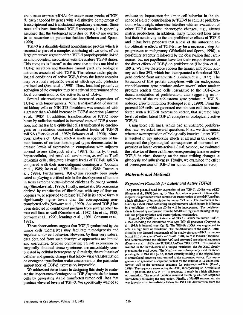
Plasmid pRK5-151EC2S2 was derived from pRK5-/~IE (Fig. 1). Again using site-directed mutagenesis, codons for the two cysteine residues at po- sitions 223 and 225 in the TGF-/31 precursor were replaced by TCT and AGC codons for serine. As shown by Brunner et al. (1989), these mutations in the TGF-/51 precursor eDNA result in expression of TGF-/51 that is largely biologically active and thus apparently no longer in a latent complex.
Generation of Stable Tmnsfected Clones Subconfluent 293 cells were transfected with 30 ~g of pRK5, pRKS-/31E, and pRKS-/31EC2S2 plasmid DNA mixed with 1.5/~g pRSV-Neo plasmid DNA by the calcium phosphate co-precipitation method (Gorman el al., 1983; Ausubel et al., 1991). Cells were maintained in RPMI with 5-10% FBS, and during exposure to precipitated DNA, cultures were incubated in F12/DME medium with 5 % FBS. Media were supplemented with I00 U/m1 penicillin and 100/zg/rnl streptomycin. Cells were incubated with the DNA precipitate for 5 h and then exposed to 15% glycerol in PBS at room temper- ature for 90 s. Two days later, cells were split 1:20 and grown in the presence of G418 (600-800/~g/ml) until colonies formed. Colonies were expanded and RNA was prepared (Chomczynski and Sacchi, 1987) for subsequent Northern analysis to detect'rGF-/31 mRNA. The transfected clonal cell lines with the highest plasmid-derived TGF-/31 mRNA levels were selected for characterization. All selected colonies were tested for mycoplasma contam- ination and found to be negative.
mRNA Analysis Northern analysis for the presence of specific mRNAs proceeded as follows: Total RNA was electrophoresed in 1% agarose, 5 % formaldehyde gels, fol- lowed by etectrotransfer to nylon membranes (C~nescreen) and UV cross- linking. Membranes were probed with eDNA fragments radiolaboled by the random priming method (Bochringer Mannheim Biochemicals, Indi- anapolis, IN) according to the manufacturer's directions. The TGF-/51 probe was the 1.9-kbp EcoRI-HindllI eDNA insert of plasmid pRK5-/31E. The fibronectin probe was the 1.0-kbp ClaI-BamHI fragment from plasmid pFH23 (obtained from Dr. Z. Werb, University of California, San Fran- cisco, CA). All hybridizations were performed under high-stringency con- ditions. Unhybridized probe was removed by washing membranes with 0.1 x SSC, 0.2% SDS at 600C.
RNase protection analyses to detect and quantitate levels of TGF-/31, -/32, and -/33 mRNA were performed as previously described (Arrick el al., 1990).
Quantitation of TG F-I~ Secretion To prepare conditioned medium, cells were incubated for 24 h in serum-free FI2/DME medium supplemented with transferrin (13.5 gg/ml), BSA (300 gg/ml), and Hepes (10 raM, pH 7.2). The conditioned medium was col- lected and assayed for TGF-~-induced inhibition of proliferation of CCL64 mink lung cells, with and without acid activation, as previously described (Arrick et al., 1990; Meager, 1991). The monolayers were then trypsinized and counted in a Coulter counter to allow normalization of the levels of secreted TGF-B against cell number.
Assay for Lactate Production Cells harvested from routine cultures with detaching solution (125 mM NaC1, 5 mM KC1, 5 mM glucose, 1 mM EDTA, 50 mM Hepes, pH Z4), were washed with serum-free medium, and replated in triplicate in serum- free medium at a density of 4 x 10⁵tmi on laminin-coated 24-well plates at 1 ml per well and incubated overnight at 37"C. Laminin-coated plates were used in order to obtain an adherent monolayer. Medium was then replaced with sodium phosphate-containing buffer supplemented with 20 mM glucose. After a 1-h incubation, lactate levels were measured as de- scribed (Resnick el al., 1986). In this assay, lactate-dependent reduction of nicotine adenine dinucleotide in the presence of lactate dehydrogenase was quantitated by measurement of OD at 339 nm. Cell counts obtained after trypsinization of the monolayers were used to normalize lactate determina- tions.
Cell Adherence on Defined Substrates We evaluated the adherence and morphology of control and transfected cells
plated on plastic in the presence and absence of serum. Cell suspensions in serum-free DME medium were obtained as described above, and 5 x lif t cells (or 5 x 10³ cells in some experiments) in 1 mt were plated per well in 24-well plates in the presence and absence of serum.
Experiments were also carded out on surfaces coated with iaminin (from Engelbreth-Holms-Swarm tumor, Collaborative Research) or plasma fibronectin (Collaborative Research Inc., Lexington, MA) in the presence of serum-free medium containing 2 % of Nutridoma (Boehringer Mannheim Biochemicals), a medium supplement that lacks the attachment factors present in serum. The wells were coated for 16 h at 40C with laminin or fibronectin at 10 and 5 ~g/ml, respectively. Unoccupied sites on the sub- strates were blocked with 0.2% BSA in Ca 2+- and Mg2+-free PBS (CMf- PBS) for 1 h before plating cells (Hall el al., 1990). Cells were plated in 24-well plates at a density of 4 x 10⁴ cells/ml. Morphology was evaluated at 4 h and at 2 and 4 d.
Detection of lntegrins by lraraunoprecipitations Antibodies used to detect integrins were obtained as follows: rat mAbs against the human integrin subunits ~1 (AIIB2), or5 (BIIG2: Werb et al., 1989; Hall et al., 1990), and a6 (J1B5; Damsky et al., 1992) were produced as described. Other antibodies were obtained as follows: mouse monoclonal anti-u2 (P1H5) and c~3 (P1B5) were gifts of Drs. E. Wayner (Fred Hutchin- son Cancer Center, Seattle, WA) and W. Carter (Fred Hutchinson Cancer Center) (Wayner and Carter 1987); mouse monoclonal anti-cd (TS217; Hemler et al., 1984) was purchased as ACT-T-SET from T-Cell Sciences, (Cambridge, MA); mouse anti-oN (LM142) was a gift of Dr. David Cheresh (Scripps Institute, La Jolla, CA) (Cheresh and Harper, 1987). Uncon- jugated rabbit anti-mouse IgG was pumhased from Jackson Immunore- search Lab Inc. (West Grove, PA). Protein A-conjugated egarose beads were purchased from Sigma Chemical Co. (St. Louis, MO).
Cells were either metabolically labeled or surface labeled. For metabolic labeling, cells were harvested using trypsin-EDTA, plated at a density of 2 x 10 ~ cells per T75 flask in DME medium with 10% FBS and radiola- beled for 24 h with 50 ~Ci/ml 3H-glucosamine. The cells were then har- vested with 2 mM EDTA in CMf-PBS, washed in PBS containing Ca 2+ and Mg 2+, and lysed in 25 mM "Iris buffer, pH Z2, containing 150 mM NaCI, 0.5% NP-40, and 1 mM PMSE Alternatively, cells were harvested with EDTA as described and surface-labeled with t2sI-iodine using the lac- toperoxidase/glucose oxidase method. After being washed in PBS contain- ing 50 mM Na-iodide and again in Ca 2+- and Mg2+-containing PBS, these cells were lysed as described for metabolically labeled cells. Lysntes were centrifuged at 12,000 g for 10 min. All procedures were carried out at 4~ The supernatant was precleared with Sepharose 4B beads, and aliquots con- taining equal protein amounts (determined using the Lowry method; Aus- ubel el al., 1991) were incubated for 2 h or overnight with one of the follow- ing subunit-specific monoclonal anti-integrin antibodies: AIIB2 (anti-t31), TS2/7 (anti-al), P1H5 (anti-ct2), P1B5 (anti-c~3), BIIG2 (anti-cr J1B5 (anti-c~6), or LM142 (anti-oN). The antibodies were present in excess such that addition of another aliquot of primary antibody, following the first round of precipitation, did not bring down any additional material. The samples were next incubated with a 1:100 dilution of rabbit anti-rat or anti-mouse IgG for 1 h, and then with 100 IA packed protein A-agarose beads for 2 h. The beads with bound immune complexes were washed se- quentially with 10 mM Tris-acelate, pH 8.0, 0.5 mM Ca 2+, and 0.5% NP-40 (TNC), TNC with 1 M NaC1, TNC with 0.1% SDS, and again with TNC. The proteins bound to the washed beads were analyzed by SDS- polyacrylamide electrophoresis in 7% polyacrylamide gels under non- reducing conditions, followed by autoradiography.
Detection of Proteases by Substrate Gel Electrophoresis To detect expression of extracellular matrix-degrading metaUoproteinases and plasminogen activators, cell extracts and conditioned media were ana- lyzed on 10 % polyacrylamide-SDS gels copolymerized with either 1 mg/ml gelatin (for the detection of metalloproteinases) or 13/~g/ml plasminogen and 1 mg/ml casein (for the detection of plasminogen activators) as de- scribed previously (Unemori and Werb, 1986; Fisher and Werb, 1991). Be- fore this analysis, the conditioned media were concentrated by dialysis using Centriprep concentrator tubes with a 10-kD exclusion filter (Amicon, Beverly, MA). To determine whether the substratum influenced protein- ase expression, cells were plated on fibronectin- or laminin-cuated plates in serum-free, Nutridoma-containing medium, or on plastic in serum- containing medium as described above.
Ardck et al. Proliferation-independent Function of TGF-13 in Tumor Cells 717
Assessment of lnvasiveness by a Matrigel Invasion Assay
Cells were harvested and 6 x 10⁴ cells in 200 /41 of serum-free, Nutridoma-containin 8 medium were added to the upper section of Costar Transwetl filter culture chambers partitioned by a Nucleopore filter (growth surface area: 0.28 cm2; Costar, Cambridge, MA) that had been coated with a thin layer of Matrigel (Collaborative Research Inc., Lexington, MA), the reconstituted basement membrane-like extract of the EHS tumor (Librach et al., 1991). After 24 or 48 h, the cultures were fixed with 2% glutaraldehyde and processed for scanning electron microscopy by standard methods (Librach et al., 1991). Filters were mounted such that either the top or the underside of the filter could be viewed. The presence of ceils on the underside of the filter constituted evidence of invasive activity.
Tumor Formation in Nude Mice
Subcordiuent cultures were detached with detaching solution (125 mM NaCl, 5 mM KCI, 5 mM glucose, 1 mM EDTA, 50 mM Hepes pH 7.4). Cells were resuspended in FI2:DME medium at a density of 3 x 10~/ml and kept on ice until injection. Female nude mice between 4- and 6-wk old (Charles River) were injected subcutaneously with 0.2 ml of the cell suspen- sion. Some mice were injected with cell suspensions at two different sites. After 4 wk, mice were killed by cervical dislocation and the sizes of the subcutaneous tumor masses were measured in three dimensions (x, y, and z). From animals with a palpable tumor mass, we removed the lung, liver, kidneys, spleen, femur, and tumor, fixed the tissues in 10% formalin, and evaluated the organs and tissues macroscopically and histologically for the appearance of metastases. The microscopical examination of histological sections of the tissues was done by a veterinary pathologist. The volumes of the tumors were calculated as 0.Sx the product of the measurements in three dimensions (Tomayko and Reynolds, 1989).
Resul ts
Characterization of the Parent 293 Cells
In this study we evaluated the effects of TGF-/~ overexpres- sion in 293 cells, a transformed and tumorigenic human kid- ney fibroblast cell line with an incorporated, functional E1A gene from adenovirus 5 (Graham et al., 1977). Analysis of the TGF-B expression pattern in the untransfected cells re- vealed that they synthesized only TGF-/Yl mRNA of the ex- pected size of 2.5 kb. No traces of TGF-/~2 or -/33 mRNA were detected by RNAse protection assay (data not shown). Measurements of the TGF-/~ levels using a TGF-~ bioassay indicated that 0.3-0.4 rig/106 cells was secreted per 24 h. The requirement for acid activation to detect the TGF-B indi- cated that most, if not all, of the TGF-B was secreted in a latent form.
The presence of TGF-/~ receptors in the nontransfected 293 cells was assayed using specific cross-linking of cell sur- face proteins in the presence of ~2q-TGF-B1 essentially as described by Frolik et al. (1984). Analysis of the cross- linking pattern revealed the presence of all three known spe- cies of TGF-B cell surface receptors or binding proteins, i.e., the type 165-kD receptor, the type I185-kD receptor and the large type HI proteoglycan (data not shown). Thus, the pa- rental 293 cells synthesize and secrete low levels of TGF-~ and have TGF-B receptors, suggesting that changes in endog- enous TGF-/3 synthesis could affect their cellular physiology in an autocrine fashion.
Generation of Stable Cell Lines Overexpressing Latent or Active TGF-fl We constructed two mammalian expression plasmids for TGF-fll from the parental expression vector pRK5 as de-
pBR322 pUC118 ori
CMV �9 , ~ p r o m o t e r / l ~4~J early enhancer i ~ J S V 4 0
Intron '4 ~ V 4 0 / ~ ~J L. ~ ~ PolyA
c oo
I~&S I [(///////////~J ) [}IE rGF-,~I Matore
Precursor TGF-,61
c 1
--ISSl ...... I] P'II///I/III/5a /}IEC2S2 / \ s s
Figure 1. Schematic diagram of the TGF-~I expression plasmids pRK5-/31E and pRK5-~IEC2S2. The cytomegalovirus promoter segment with the direction of transcription is shown as a heavy black arrow, and the segments derived from SV-40 are shown as shaded boxes. The two TGF-B1 eDNA transcription units, corre- sponding to either plasmid, are shown with their flanking restric- tion sites. SS, signal sequence.
scribed in Materials and Methods and diagrammed in Fig. 1. Plasmid pRK5-BIE incorporates a human "I'GF-fll cDNA (Derynck et al., 1985), the expression of which should yield an unmodified human TGF-01 precursor protein, and thus TGF-/3 in its latent or biologically inactive complex. The sec- ond plasmid, pRKS-fllEC2S2, is identical to pRK5-BIE ex- cept for the replacement of the codons for two cysteines at positions 223 and 225 in the TGF-ffl precursor with serine codons, a mutation that results in secretion of TGF-B largely in its biologically active form ~runner et ai., 1989).
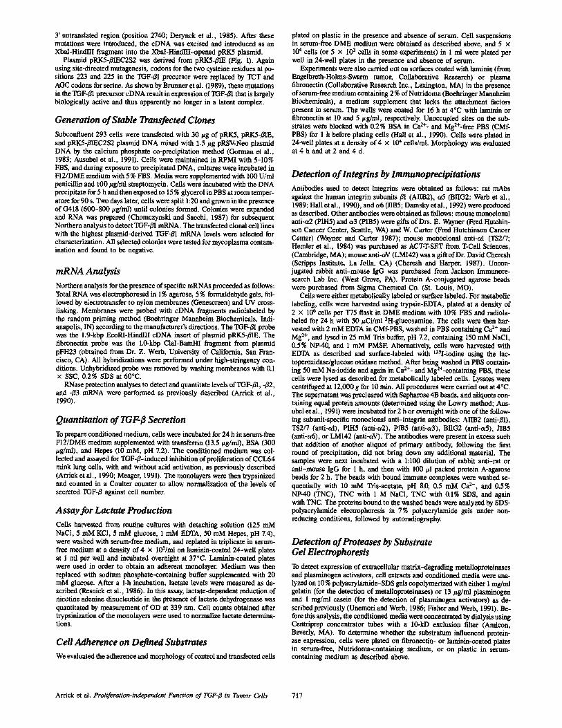
Transfection of the individual TGF-/31 expression plasmids along with the pRSV-Neo plasmid containing the neomycin- resistance transcription unit, followed by selection for G418- resistant clones, resulted in the generation of stable trans- fected 293 cell clones. Northern hybridization indicated that several of the G418-resistant cell lines expressed elevated levels of TGF-/31 mRNA (Fig, 2), the size of which agreed with the size predicted from the design of the expression plasmids. Two clonal cell lines transfeeted with pRKS-~E (B6 and B9) and two transfected with plasmid pRKS- /31EC2S2 (C15 and C19) were selected for further analysis. As controls, two G418-resistant clones cotransfected with the parent pRK5 lacking any cDNA sequence (R1 and R6) were characterized in parallel with the TGF-13-overexpress- ing cell lines. RNase protection assays indicated that the neomycin-resistant control clones expressed 2nGF-t31 mRNA at levels equivalent to untransfected 293 cells and that the TGF-/3-overexpressing B6 and C19 clones had about 100- fold higher levels of TGF-/31 mRNA (not shown).
We measured secretion of TGF-B by the selected 293 clones by assaying the serum-free conditioned medium for TGF-B bioactivity in a cell growth inhibition assay (Table I).
The Journal of Cell Biology, Volume I18, 1992 718
PTgure 2. Expression of mRNA encoding TGF-B1 by trans- fected cell clones, determined by Northern hybridization. Cells were transfected with pa- rental plasmid pRK5 lacking TGF-B eDNA (R), with pRKS- B1E (B), or with pRK5-BIE
C2S2 (C). Each lane contained 18 ~g total RNA prepared from transfected cell clones as indicated. Equivalent RNA loading and transfer was confirmed by subsequent probing with a glyceralde- hyde-6-phosphate dehydrogenase eDNA. The TGF-B1 mRNA ex- pressed from the transfected expression plasmid is about 2-kb long on the basis of migration in the gel. The low endogenous TGF-B1 mRNA levels are not detectable using the short duration of auto- radiography of this experiment and are more easily assessed by RNAse protection assay.
The assay was done with and without acid activation, which quantitatively converts latent TGF-/3 to its biologically active form. The control transfected clones, like the parental 293 cells, secreted low levels of TGF-B in the latent form. In con- trast, the transfected clones transfected with pRK5-B1E or pRK5-~EC2S2 secreted TGF-B levels 100 to 300 times higher than the control clones. Their relative TGF-B levels were in agreement with the relative TGF-BI mRNA levels. As expected, ,,,99% or more of the TGF-B secreted by the clones transfected with pRK5-B1E was in the latent form. We do not know whether the very low levels of active TGF-/~ in the conditioned medium of these clones are a result of techni- cal manipulations, temperature- or protease-induced activa- tion, or activation in the bioassay system. In contrast, 50% of the TGF-B released by the stable clones transfected with the pRK5-B1EC2S2 plasmid, with its mutated precursor eDNA, was in a biologically active form. Remarkably, the total TGF-B expression from this plasmid was generally much higher than with the same expression vector contain- ing the unmodified TGF-/~ precursor eDNA (Arrick, B., and R. Derynck, unpublished data).
Analysis of the TGF-B receptors by cross-linking, using ~25I-TGF-B as ligand, indicated that all clones had the three TGF-B receptor types at their cell surface. Some differences in receptor levels between the different clones were appar- ent, suggesting clonal heterogeneity (data not shown).
Table L Secretion of TGF-[3 by Transfected 293 Cell Clones
TGF-$ bioactivity (ng/106 cells)
Percent active Cell done Without acid With acid without acidification
R1 ND 0.42 - R6 ND 0.41 -
B6 0.49 95 0.5 B9 0.60 55 1.1
C 15 58 110 52 C19 69 125 56
Serum-free conditioned medium was collected af~er a 24-h incubation and as- sayed for TGF-~ bioactivity by inhibition of [3H]TdR uptake by mink lung cells CCL64 with and without prior acid activation. TGF-/~ bioactivity in the conditioned medium of the two control clones was not detectable (ND) without acid activation. Values reported are means from triplicate determinations. Average coefficient of variance was 7.6%.
Table II. Lactate Production by Transfected 293 Cell Clones
Cell clone Lactate
(mmol/lO 5 cells~h) R1 37.3 + 0.73 R6 39.0 • 1.3
B6 59.3 • 4.1 B9 53.6 • 0.74
C15 54.1 • 1.4 C19 50.1 • 0.79
Cells were incubated overnight in laminin-coated plates under serum-free con- ditions, followed by a 1-h incubation in a phosphate- and glucose-containing buffer. Lactate production was determined by quantitative conversion to pyru- rate by lactate dehydrogenase, with stoichiometric formation of NADH (Res- nick et al., 1986). Data are normalized to cell number and presented as means 5: SD for triplicate determinations.
Increased Glycolysis in TGF-fl Overexpressing Cells
Increased aerobic glycolysis by tumors has been noted for over half a century. More recently it was shown that TGF-/5 was able to stimulate glycolysis in normal rat fibroblasts mitogenically stimulated by TGF-~ (Boerner et al., 1985). During the course of our experiments, we noted a more pro- nounced acidification of the medium by several of the clones overexpressing TGF-B, even though, as expected, there were no differences in proliferation rate between the overexpress- ing clones and the control clones. We therefore assessed the glycolytic activity of our transfected clones by measuring lactate production. We found that the pRK5-~E- and pRK5- B1EC2S2-transfected clones demonstrated a greater rate of lactate production than the control clones (Table II). This in- crease in lactate production was about equal to that observed upon exposure of rat kidney fibroblasts to exogenous TGF-B (Boerner et al., 1985).
It is important to observe that there was no major differ- ence in lactate production between the pRK5-/~IE clones B6 and B9, and the pRK5-/SqEC2S2 clones C15 and C19. Thus, at least in this respect, the clones producing latent TGF-B be- haved similarly to those secreting active TGF-~5. This sug- gests that the clones producing latent TGF-B have the ability to activate it and are responsive to it.
Adhesive and Invasive Properties
During cell culture propagation of the transfected cells, we noted that clones overexpressing TGF-~ required longer ex- posure to trypsin-EDTA than the control clones. To evaluate this further, we studied the adhesive properties of these cells to plastic, laminin, and fibronectin.
In one set of experiments we plated and incubated the cells (5 x 10⁴/ml) in the presence or absence of serum in plastic culture wells. When cultured on plastic in serum-free condi- tions, the control clones proliferated as non-adherent clumps of cells. In contrast, the TGF-B-overproducing clones 136, B9, C15, and C19 adhered to the plastic, spread out, and proliferated into a monolayer. Again, there were no apparent differences between clones overproducing latent or active TGF-/~ (Fig. 3). However, when plated in serum-free medium at very low density (5 x l(P/ml), only the C15 and C19 clones, which expressed active TOF-/$1 attached, sur- vived, and proliferated (as assessed by phase-contrast mi-
Arrick et al. Proliferation-independent Function of lXTF-B in Tumor Cells 719
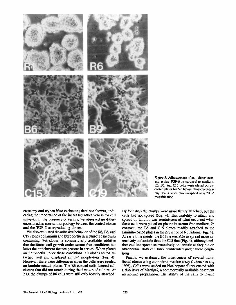
Figure 3. Adhesiveness of cell clones over- expressing TGF-/3 in serum-free medium. R6, B6, and C15 cells were plated on un- coated plates for 5 d before photomicrogra- phy. Ceils were photographed at a 200)< magnification.
croscopy and trypan blue exclusion; data not shown), indi- cating the importance of the increased adhesiveness for cell survival. In the presence of serum, we observed no differ- ences in adherence or morphology between the control clones and the TGF-B-overproducing clones.
We also evaluated the adhesive behavior of the R6, B6, and C15 clones on laminin and fibronectin in serum-free medium containing Nutridoma, a commercially available additive that facilitates cell growth under serum-free conditions but lacks the attachment factors present in serum. When plated on fibronectin under these conditions, all clones tested at- tached well and displayed similar morphology (Fig. 4). However, there were differences when the cells were seeded on laminin-coated plates. The R6 control cells formed cell clumps that did not attach during the first 4 h of culture. At 2 D, the clumps of R6 cells were still only loosely attached.
By four days the clumps were more firmly attached, but the cells had not spread (Fig, 4). This inability to attach and spread on laminin was reminiscent of what occurred when these cells were plated on plastic in serum-free medium. In contrast, the B6 and C15 clones readily attached to the laminin-coated plates in the presence of Nutridoma (Fig. 4). At early time points, the B6 line was able to spread more ex- tensively on laminin than the C15 line (Fig. 4), although nei- ther cell line spread as extensively on laminin as they did on fibronectin. Both cell lines proliferated under these condi- tions.
Finally, we evaluated the invasiveness of several trans- fected clones using an in vitro invasion assay (Librach et al., 1991). Cells were seeded on Nucleopore filters coated with a thin layer of Matrigel, a commercially available basement membrane preparation. The ability of the cells to invade
The Journal of Cell Biology, Volume 118, 1992 720
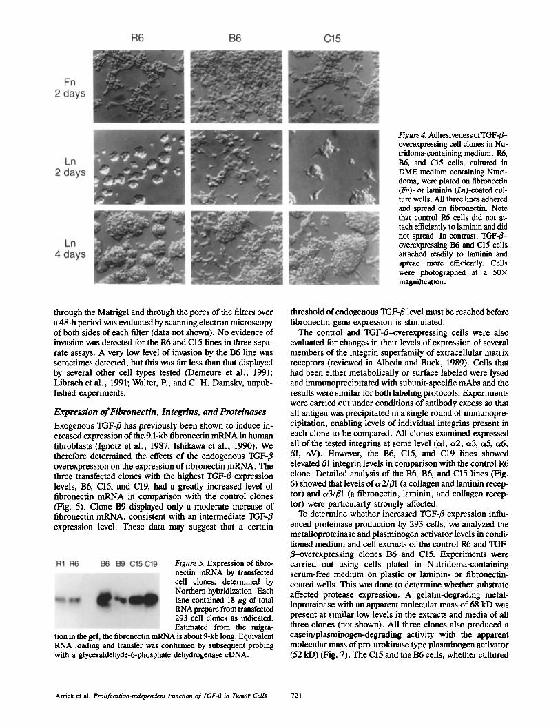
Figure 4. Adhesiveness of TGF-#- overexpressing cell clones in Nu- tridoma-containing medium. R6, B6, and C15 cells, cultured in DME medium containing Nutri- doma, were plated on fibronectin (Fn)- or laminin (Ln)-coated cul- ture wells. All three lines adhered and spread on fibronectin. Note that control R6 cells did not at- tach efficiently to laminin and did not spread. In contrast, TGF-~- overexpressing B6 and C15 cells attached readily to laminin and spread more efficiently. Cells were photographed at a 50x magnification.
through the Matrigel and through the pores of the filters over a 48-h period was evaluated by scanning electron microscopy of both sides of each filter (data not shown). No evidence of invasion was detected for the R6 and C15 lines in three sepa- rate assays. A very low level of invasion by the B6 line was sometimes detected, but this was far less than that displayed by several other cell types tested (Demeure et al., 1991; Librach et al., 1991; Walter, P., and C. H. Damsky, unpub- lished experiments.
Expression of ~'bronectin, lntegrins, and Proteinases
Exogenous TGF-/3 has previously been shown to induce in- creased expression of the 9.1-kb fibronectin mRNA in human fibroblasts (Ignotz et al., 1987; Ishikawa et al., 1990). We therefore determined the effects of the endogenous TGF-/~ overexpression on the expression of fibronectin mRNA. The three transfected clones with the highest TGF-~ expression levels, B6, C15, and C19, had a greatly increased level of fibronectin mRNA in comparison with the control clones (Fig. 5). Clone B9 displayed only a moderate increase of fibronectin mRNA, consistent with an intermediate TGF-/3 expression level. These data may suggest that a certain
Figure 5. Expression of fibro- nectin mRNA by transfected cell clones, determined by Northern hybridization. Each lane contained 18 #g of total RNA prepare from transfected 293 cell clones as indicated. Estimated from the migra-
tion in the gel, the fibronectin mRNA is about 9-kb long. Equivalent RNA loading and transfer was confirmed by subsequent probing with a glyceraldehyde-6-phosphate dehydrogenase cDNA.
threshold of endogenous TGF-~ level must be reached before fibronectin gene expression is stimulated.
The control and TGF-~-overexpressing cells were also evaluated for changes in their levels of expression of several members of the integrin superfamily of extracellular matrix receptors (reviewed in Albeda and Buck, 1989). Cells that had been either metabolically or surface labeled were lysed and immunoprecipitated with subunit-specific mAbs and the results were similar for both labeling protocols. Experiments were carried out under conditions of antibody excess so that all antigen was precipitated in a single round of immunopre- cipitation, enabling levels of individual integrins present in each clone to be compared. All clones examined expressed all of the tested integrins at some level (or, or2, o~3, or5, ol6, /~1, oN). However, the B6, C15, and C19 lines showed elevated/~1 integrin levels in comparison with the control R6 clone. Detailed analysis of the R6, B6, and C15 lines (Fig. 6) showed that levels of c~2/~1 (a collagen and laminin recep- tor) and o~3//31 (a fibronectin, laminin, and collagen recep- tor) were particularly strongly affected.
To determine whether increased TGF-~ expression influ- enced proteinase production by 293 cells, we analyzed the metalloproteinase and plasminogen activator levels in condi- tioned medium and cell extracts of the control R6 and TGF- /3-overexpressing clones B6 and C15. Experiments were carried out using cells plated in Nutridoma-containing serum-free medium on plastic or laminin- or fibronectin- coated wells. This was done to determine whether substrate affected protease expression. A gelatin-degrading metal- loproteinase with an apparent molecular mass of 68 kD was present at similar low levels in the extracts and media of all three clones (not shown). All three clones also produced a casein/plasminogen-degrading activity with the apparent molecular mass ofpro-urokinase type plasminogen activator (52 kD) (Fig. 7). The C15 and the B6 cells, whether cultured
Arrick et al. Proliferation-independent Function of TGF-{3 in Tumor Cells 721
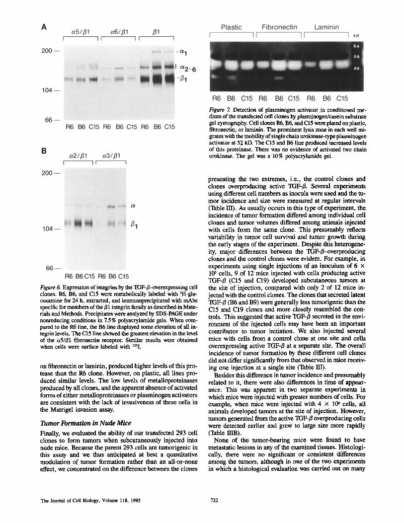
Figure Z Detection of plasminogen activator in conditioned me- dium of the transfected cell clones by plasminogen/casein substrate gel zymography. Cell clones R6, B6, and C15 were plated on plastic, fibronectin, or laminin. The prominent lysis zone in each well mi- grates with the mobility of single chain urokinase-type plasminogen activator at 52 kD. The C15 and B6 line produced increased levels of this proteinase. There was no evidence of activated two chain urokinase. The gel was a 10% polyacrylamide gel.
Figure 6. Expression of integrins by the TGF-/%overexpressing cell clones. R6, B6, and C15 were metabolically labeled with 3H-glu- cosarnine for 24 h, extracted, and immunoprecipitated with mAbs specific for members of the/31 integrin family as described in Mate- rials and Methods. Precipitates were analyzed by SDS-PAGE under nonreducing conditions in 7.5% polyaerylamide gels. When com- pared to the R6 line, the B6 line displayed some elevation of all in- tegrin levels. The C15 line showed the greatest elevation in the level of the ~5/~1 fibronectin receptor. Similar results were obtained when cells were surface labeled with ~2~I.
on fibronectin or laminin, produced higher levels of this pro- tease than the R6 clone. However, on plastic, all lines pro- duced similar levels. The low levels of metalloproteinases produced by all clones, and the apparent absence of activated forms of either metalloproteinases or plasminogen activators are consistent with the lack of invasiveness of these cells in the Matrigel invasion assay.
Tumor Formation in Nude Mice
Finally, we evaluated the ability of our transfected 293 cell clones to form tumors when subcutaneously injected into nude mice. Because the parent 293 cells are tumorigenic in this assay and we thus anticipated at best a quantitative modulation of tumor formation rather than an all-or-none effect, we concentrated on the difference between the clones
presenting the two extremes, i.e., the control clones and clones overproducing active TGF-/3. Several experiments using different cell numbers as inocula were used and the tu- mor incidence and size were measured at regular intervals (Table HI). As usually occurs in this type of experiment, the incidence of tumor formation differed among individual cell clones and tumor volumes differed among animals injected with cells from the same clone. This presumably reflects variability in tumor cell survival and tumor growth during the early stages of the experiment. Despite this heterogene- ity, major differences between the TGF-/3-overproducing clones and the control clones were evident. For example, in experiments using single injections of an inoculum of 6 x 1@ cells, 9 of 12 mice injected with cells producing active TGF-/3 ( 0 5 and C19) developed subcutaneous tumors at the site of injection, compared with only 2 of 12 mice in- jected with the control clones. The clones that secreted latent TGF-/3 (B6 and B9) were generally less tumorigenic than the C15 and C19 clones and more closely resembled the con- trols. This suggested that active TGF-/3 secreted in the envi- ronment of the injected cells may have been an important contributor to tumor initiation. We also injected several mice with cells from a control clone at one site and cells overexpressing active TGF-/3 at a separate site. The overall incidence of tumor formation by these different cell clones did not differ significantly from that observed in mice receiv- ing one injection at a single site (Table III).
Besides this difference in tumor incidence and presumably related to it, there were also differences in time of appear- ance. This was apparent in two separate experiments in which mice were injected with greater numbers of cells. For example, when mice were injected with 4 x 10⁶ cells, all animals developed tumors at the site of injection. However, tumors generated from the active TGF-/3 overproducing cells were detected earlier and grew to large size more rapidly (Table IIIB).
None of the tumor-beating mice were found to have metastatic lesions in any of the examined tissues. Histologi- cally, there were no significant or consistent differences among the tumors, although in one of the two experiments in which a histological evaluation was carried out on many
The Journal of Cell Biology, Volume 118, 1992 722
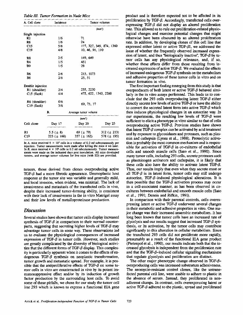
Table IlL Tumor Formation in Nude Mice
A. Cell clone Incidence Tumor volumes
(ram 3)
Single injection R1 1/6 71 R6 1/6 24 C15 5/6 177, 327, 349, 874, 1360 C19 4/6 10, 46, 91, 110
R8 2/5 149, 649 R4 1/5 451 B9 1/5 29
R6 2/6 213, 1073 B6 2/6 25, 51
Double injection R1 (shoulder) 2/4 255, 2230 C15 (flank) 4/4 475, 622, 1340, 2260
R6 (shoulder) 0/6 C19 (flank) 3/6 69, 73, 77
B. Average tumor volume
Cram ~;
Cell clone Day 17 Day 20 Day 25
R1 5.5 ( + 8) 69 ( + 79) 312 ( • 213) C19 223 ( • 160) 357 ( + 162) 578 ( • 195)
In A, mice received 6 x l0 s cells in a volume of 0.2 ml subcutaneously per injection. Tumor measurements were made after killing the mice 4 wk later. In B, mice received 4 x 10⁶ cells in 0.2 ml subcutaneously. Tumor measure- merits were made on the indicated days post inoculation. All mice developed tumors, and average tumor volumes for five mice (with SD) are provided.
tumors, those derived from clones overproducing active TGF-/~ had a more fibrotic appearance. Desmoplastic host response at the tumor site was variable and generally mild, and local invasion, when evident, was minimal. The lack of invasiveness and metastasis of the transfected cells in vivo, despite their increased tumor-forming ability, is consistent with their lack of invasiveness in the in vitro Matrigel assay and their low levels of metalloproteinase production.
Discussion
Several studies have shown that tumor cells display increased synthesis of TGF-/3 in comparison to their normal counter- parts, suggesting that secreting higher levels of TGF-/3 may advantage tumor cells in some way. These observations led us to evaluate the physiological consequences of increased expression of TGF-~ in tumor cells. However, such studies are greatly complicated by the diversity of biological activi- ties that the different forms of TGF-/3 display. This complex- ity is particularly apparent when it comes to the effects of en- dogenous TGF-/3 synthesis on neoplastic transformation, tumor growth and metastatic spread. For example, it is pos- sible that the antiproliferative effects of TGF-~ on some tu- mor cells in vitro are counteracted in vivo by its potent im- munosuppressive effect and/or by its induction of growth factor production by the surrounding host cells. To avoid some of these pitfalls, we chose for our study the tumor cell line 293 which is known to express a functional E1A gene
product and is therefore expected not to be affected in its proliferation by TGF4/. Accordingly, transfected cells over- expressing TGF-/3 did not display an altered proliferation rate. This allowed us to rule out proliferation-related physio- logical changes and examine potential changes that might otherwise have been obscured by an altered proliferation rate. In addition, by developing clones of this cell line that expressed either latent or active TGF-~I, we addressed the issue of whether the frequently observed increased expres- sion of latent, and thus "biologically inactive", TGF-/3 by tu- mor cells has any physiological relevance, and, if so, whether these effects differ from those resulting from in- creased expression of active TGF-~. We evaluated the effects of increased endogenous TGF-~ synthesis on the metabolism and adhesive properties of these tumor cells in vitro and on tumor formation in vivo.
The first important finding emerging from this study is that overproducers of both latent or active TGF-/3 behaved simi- larly in the in vitro assays performed. This leads us to con- clude that the 293 cells overproducing latent TGF-/3 either directly secrete low levels of active TGF-~/or have the ability to convert the secreted latent form into active TGF-B which then induces physiological changes in an autocrine way. In our experiments, the resulting low levels of TGF-/~ were sufficient to elicit a phenotype in vitro similar to that of cells overproducing active TGF-~/. Previous studies have shown that latent TGF-~/complex can be activated by acid treatment and by exposure to glycosidases and proteases, such as plas- min and cathepsin (Lyons et al., 1988). Proteolytic activa- tion is probably the most common mechanism and is respon- sible for activation of TGF-# in co-cultures of endothelial cells and smooth muscle cells (Sato et al., 1990). Since many tumor cells, including 293 cells, secrete proteases such as plasminogen activators and cathepsins, it is likely that these cells also have the ability to activate latent TGF-~/. Thus, our results imply that even when they secrete most or all TGF-j3 in its latent form, tumor cells may still undergo autocrine, TGF-/3-induced physiological alterations. It is then possible that the TGF-/3 activation process may occur in a cell-associated manner, as has been observed in co- cultures between endothelial and smooth muscle cells (Sato et al., 1991; Dennis and Rifkin, 1991).
In comparison with their parental controls, cells overex- pressing latent or active TGF-/3 underwent several changes in their metabolic and adhesive properties in vitro. One ma- jor change was their increased anaerobic metabolism. It has long been known that tumor cells have an increased rate of glycolysis and our results suggest that increased TGF-/3 syn- thesis, or its activation, by the tumor cells may contribute significantly to this alteration in cellular metabolism. Since the transfected 293 cells did not proliferate more rapidly, presumably as a result of the functional E1A gene product (Pietenpol et al., 1990), our results indicate both that the in- creased glycolysis is independent from the proliferation rate and that the TGF-/3-induced cellular signalling mechanisms that regulate glycolysis and proliferation are distinct.
The other major phenotypic change observed in TGF-/3- overproducing cells was increased substratum adhesiveness. The neomycin-resistant control clones, like the untrans- fected parental cell line, were unable to adhere to plastic in the absence of serum. Instead, they proliferated in non- adherent clumps. In contrast, cells overexpressing latent or active TGF-/3 adhered to the plastic, spread and proliferated
Arrick et al. Proliferation-independent Function of TGF4t in Tumor Cells 723

in a monolayer. Of even greater potential significance in view of our in vivo results, was the observation that only the C15 and C19 clones overproducing active TGF-/3, and not those overproducing latent TGF-/3, adhered and survived at low cell inoculum. In parallel experiments using different culture conditions, the control cell line R6 was unable to attach rapidly or to spread on laminin when cultured in serum-free medium in the presence of Nutridoma, whereas the TGF-~ overproducing clones 136 and C15 attached readily and spread on laminin. Thus, at least under these conditions, expression of TGF-j3 strongly influenced the ability of 293 cells to ad- here to the substratum. Such differences in adhesive proper- ties could be of importance in vivo in regulating the ability of small numbers of cells to initiate tumor formation.
Consistent with the changes in adhesive behavior were the enhanced levels of expression of fibronectin mRNA and of several E1 integrin complexes. This observation is consistent with a large body of information documenting the ability of TGF-B to enhance extracellular matrix production (Mas- sagu6 et al., 1990; Bamard et al., 1990). Production of a broad range of integrins was increased in the B6 as well as the C15 clones overproducing TGF-/3. This may favor oppor- tunistic adhesive behavior which could enhance survival in vivo. The observed increased expression of o~ 3/~, a promis- cuous receptor that recognizes laminin, collagen, and fibro- nectin, may be of particular importance, as it has been re- ported that enhanced expression of a 3/~ or elevated ratios of c~ 3//51 to ~x5//31 accompany oncogenic transformation in several cell lines (Plantefaber and Hynes, 1989). Taken to- gether these data support the idea that acquisition of an op- timal adhesion phenotype is permissive for a more rapid and successful colonization by tumor cells of their immediate en- vironment.
Having stably transfected clones that produced increased amounts of TGF-/5 gave us the unique opportunity to deter- mine the consequences of TGF-t3 overexpression in vivo, be- cause local concentration gradients of tumor-derived factors and their effect on nearby host cells could be reproduced. Two published studies have used stable clone transfected with the TGF-~I eDNA and have demonstrated significant in vivo suppression of cytolytic T cell and natural killer cell ac- tivities (Torre-Amione et al., 1990; Wallick et al., 1990). In the present study, we found that at a low tumor cell inocu- lure, 293 cells that overexpressed active TGF-/3 produced subcutaneous tumors more readily than control 293 cells. This may reflect some degree of immunosuppression or a growth advantage for these cells when few in number. This increase in tumorigenicity may also be related to our obser- vation that only the clones overexpressing active TGF-/3 sur- vived when plated in serum-free medium at very low density. In addition, the increased ability of cells overexpressing TGF-/3 to produce and interact with extraceilular matrix could create a more permissive environment for tumor cell proliferation and tumor formation. This explanation would be consistent with recent results, documenting an increase in tumor formation when tumor cells were injected together with the basement membrane analog Matrigel (Fridman et al., I99I). The ~eater incidence of tumor formation with the C15 and C19 clones was apparently not the result of a sys- temic effect from the secreted TGF-/3 since tumor formation from control clones injected into the same animals was not increased. Although enhanced production of TGF-/5 by 293
cells may generate a particular adhesion phenotype that is favorable for their anchorage and permissive for their sur- vival in vivo, such an adhesion phenotype is clearly not sufficient for successful tumor initiation. Only the clones producing active TGF-/3 had enhanced rates of tumor forma- tion when injected into nude mice at low inoculum levels, suggesting that the active TGF43 may have profound effects on the imme~ate environment of the tumor cells, e.g., by inducing enhanced matrix formation or growth factor production by the neighboring ceils, or increased prolifera- tion by host fibroblasts. In this context it is important to note that histological evaluation of some, but not all, tumors de- rived from cells overproducing active TGF-/3 showed in- creased matrix formation and interstitial fibroblast prolifera- tion in comparison with tumors derived from the control clones. Neither the parental cell line, nor any of the TGF-/~- overexpressing clones were metastatic in this animal model. Consistent with these observations, the transfected clones did not produce higher levels of metalloproteinases and did not display activated forms of metaUoproteinases or plas- minogen activators.
The observed increased tumorigenicity by 293 cells over- expressing active TGF-/3 is not necessarily inconsistent with some recent results indicating that a colon carcinoma cell line transfected with an antisense mRNA expression vector for TGF-131 caused a higher tumor incidence in nude mice (Wu et al., 1992) or that a rat prostate cell line transfected with a latent TGF-B expression vector demonstrated en- hanced tumor growth (Steiner and Barrack, 1992). These re- cent studies were performed in cell lines that were sensitive to the growth-regulatory effects of TGF-t3, whereas we inten- tionally focused on a tumor cell line which is insensitive to the direct growth modulatory activities of TGF-/3 and have evaluated the tumorigenicity of clones with similar rates of proliferation, thus allowing a more direct evaluation of, e.g., the physiological consequences of alterations in cell-matrix interactions. It is thus possible that the inhibitory effects of TGF-13 on cell proliferation could counteract altered adhe- sive interactions of the cells with the environment in deter- mining tumor cell proliferation in vivo. Thus, the advantage of an increased endogenous TGF-# synthesis for tumorige- nicity may depend on the balance of these two parameters, and on the degree of local TGF-15-induced immunosuppres- sion, and thus may vary depending on the tumor type or cell line. On the other hand, it is also important to realize that many tumor cells have lost their sensitivity to the growth in- hibitory activities of TGF-#, as is the case with the 293 cells, possibly as a consequence of functional inactivation of the Rb gene product (Pietenpol et al., 1990) and possibly also as a step in the progression of tumor cells of full malignancy (Wakefield and Sporn, 1990).
In conclusion, we have evaluated the role of endogenous TGF43 overexpression on several physiological parameters of tumor cells and tumor formation, using an E1A trans- formed tumor cell line. We found that cells overexpressing latent or active TGF-tS, compared with control transfected cells, displayed similar physiological changes in vitro with- out a change in proliferation rate. The major effects of the increased TGF-~ synthesis, evaluated in this study, were in- creased glycolysis and enhanced cell-matrix adhesiveness. In addition, increased release of active TGF43 resulted in higher incidence of tumor formation. These findings may
The Journal of Cell Biology, Volume 118, 1992 724
have relevance to pathological tumor formation since, at least in several systems, it has been documented that tumor cells release higher levels of TGF-/3 compared to their non- malignant controls. In addition, several tumor cell lines have been shown to release significant levels of already activated TGF-fl (Knabbe et al., 1987; Schwarz et al., 1990; Constam et al., 1992). The cell line used in our study was minimally invasive and non-metastatic. As such, it does not provide an adequate assessment of the potential role of TGF-fl in tumor cell metastatic behavior. Similar experiments with meta- static tumor cell lines are in progress to address this issue.
We thank Dr. Z. Werb for valuable discussions. The work in the laboratory of C. Damsky was supported by National
Institutes of Health grant CA42032.
Received for publication I l March 1992 and in revised form 14 May 1992.
R@f@renc@$
Albeda, S. M., and C. A. Buck. 1990. Integrins and other cell adhesion mole- cules. FASEB (Fed. Am. Soc. Exp. BioL) J. 4:2866-2880.
Anzano, M. A., A. B. Roberts, J. E. DeLarco, L. M. Wakefield, R. K. As- soian, N. S. Roche, J. M. Smith, J. E. Lazarus, and M. B. Sporn. 1985. Increased secretion of type beta transforming growth factor accompanies vi- ral transformation of cells. Mol. Cell Biol. 5:242-247.
Arrick, B. A., M. Korc, and R. Derynck. 1990. Differential regulation of ex- pression of three transforming growth factor E species in human breast can- cer cell lines by estradiol. Cancer Res. 50:299-303.
Ausubel, F. M., R. Blent, R. E. Kingston, et al. 1991. Current Protocols in Molecular Biology. John Wiley & Sons, New York.
Barnard, J. A., R. M. Lyons, andH. L. Moses. 1990. The cell biology of trans- forming growth factor E. Biochem. Biophys. Acts. Rev. Cancer. 1032: 79-87.
Bocrner, P., R. J. Resnick, and E. Racker. 1985. Stimulation of glycolysis and amino acid uptake in NRK-49F cells by transforming growth factor E and epidermal growth factor. Proc. Natl. Acad. Sci. USA. 82:1350-1353.
Brunner, A. M., H. Marquardt, A. R. Malacko, M. N. Lioubin, and A. F. Pur- chin. 1989. Site-directed mutagenesis of cysteine residues in the pro region of the transforming growth factor E 1 precursor. Expression and characteriza- tion of mutant proteins. J. Biol. Chem. 264:13660-13664.
Cheresh, D. A., and J. R. Harper. 1987. Arg-Gly-Asp recognition by a cell adhesion receptor requires its 130-kDa r subunit. J. Biol. Chem. 262: 1434-1437.
Cbomczynski, P., and N. Saochi. 1987. Single-step method of RNA isolation by acid guanidinium thiocyanate-phenol-cldoroform extraction. Anal. Bin- chem. 162:156-159.
Constam, D. B., J. Phillip, U. V. Malipiero, P. ten Dijke, M. Schachner, and A. Fontana. 1992. Differential expression of transforming growth factor-E 1, -E2 and -E3 by gliobtastoma cells, astrocytes, and microglia. J. Immunol. 148:1404-1410.
Damsky, C. H., M. L. Fitzgerald, and S. J, Fisher. 1992. Distribution patterns of extracellular matrix components and adhesion receptors are intricately modulated during first trimester cytotrophoblast differentiation along the in- vasive pathway in vivo. J. Clin. Invest. 92:210-222.
Demeure, M., C. M. Damsky, F. Elfman, P. Goretzki, M. G. Wong, and O. Clark. 1992. Invasion by cultured human follicular thyroid cancer cells correlates with increased fll integrins and production of proteases. World J. Surgery. In press.
Dennis, P. A., and D. B. Rifkin. 1991, Cellular activation of latent transform- ing growth factor E requires binding to the cation-independent manoose 6-phosphate/insalin-like growth factor type H receptor, Proc. Natl. Acad. Sci. USA. 88:580-584.
Derynek, R., D. V. Goeddel, A. Ullrich, J. U. Gutterman, R. D. Williams, T. S. Bringman, and W. H. Berger. 1987. Synthesis of messenger RNAs for transforming growth factors ~ and E and the epidermal growth factor receptor by human minors. Cancer Res. 47:707-712.
Derynck, R., J. A. Jarrett, E. Y. Chen, D. H. Eaton, J. R. Bell, R. K. Assoian, A. B. Roberts, M. B. Spore, and D. V. Goeddel. 1985. Human transforming growth factor-E eDNA sequence and expression in tumor cell lines. Nature (Lond.). 316:701-705.
Fisher, S. J., and Z. Werb. 1992. In Extracellular Matrix Molecules, a Practical Approach. H. A. Harealson, and J. R. Hassell, editors. IRL Press/Oxford University Press, Oxford, UK. In press.
Ferriola, P. C., C. Walker, A. T. Robertson, H. S. Earp, D. W. Rusnak, and R. Nettesheim. 1989. Altered growth factor dependence and transforming factor gene expression in transformed rat tracheal epithelial cells. Idol. Car. cinog. 2:336-344.
Fridman, R., M. C. Kibbey, L. S. Royce, M. Zain, M. Sweeney, D. L. Jicha, J. R. Yannelli, G. R. Martin, and H. K. Kieinman. 1991. Enhanced tumor
growth of both primary and established human and murine tumor cells in athymic mice after coinjection with Matrigel. J. Natl. Cancer Inst. 83: 769-774.
Frolik, C. A., L. M. Wakefield, D. M. Smith, and M, B. Spore. 1984. Charac- terization of a membrane receptor for transforming growth factor-beta in normal kidney fibroblasts. J. Biol. Chem. 259:10995-11000.
Giltay, J. C., H.-J. Brinkman, M. P. W. Modderman, A. E. G. K. yon dem Borne, and J. A. van Mourik. 1989. Human vascular endothelial cells ex- press a membrane protein complex immunochemically indistinguishable from the platelet VLA-2 (glycoprotein Is-Ha) complex. Blood. 73:1235- 1241.
GomeUa, L. O., E. R. Sargent, T. P. Wade, P. Anglard, W. M. Linehan, and A. Kasid. 1989. Expression of transforming growth factor-a in normal hu- man adult kidney and enhanced expression of transforming growth factors c~ and E1 in renal cell carcinoma. Cancer Res. 49:6972-6975.
Gorman, C., R. Padmanablmn, and B. H. Howard. 1983. High efficiency DNA- mediated transformation of primate cells. Science (Wash. DC). 221:551- 553.
Graham, F. L., J. Smiley, W. C. Russell, and R. Nalm, 197% Characteristics of a human cell line transformed by DNA from human adenovirus type 5. J. Gen. Virol. 36:59-72.
Graycar, J. L , D. A. Miller, B. A. Arrick, R. M. Lyons, H. L. Moses, and R. Derynck. 1989. Human transforming growth factor-~3: Recombinant ex- pression, purification, and biological activities in comparison with trans- forming growth factors-E1 and-#~2, biol. Endocrinol. 3:1977-1986.
Haddow, S., D. J. Fowlis, K. Parkinson, R. J. Akhurst, and A. Belmain. 1991. Oneogene. 6:1465-1470.
Hall, D. L., L. F. Reichardt, E. Crowley, B. Holley, H. Moezzi, A. Sorman- berg, and C. H. Darnsky. 1990. The c~1/#31 and c~6/~1 integrin hetarodimers mediate cell attachment to distinct sites on laminin. J. Cell Biol. 110:2175- 2184.
Heine, U. I., J. K. Burmester, K. C. Flanders, D. Danielpour, E. F. Munoz, A. B. Roberts, and M. B. Sporn. 1991. Localization of transforming growth factor-E1 in mitochondria of murine heart and liver. Cell Regut. 2:467-477.
Hemler, M. F,, F. Sanchez-Madrid, T. J. Fiotta, A. M. Krensky, S. J. Burakoff, A. K. Balm, T. A. Springer, and J. L. Strominger. 1984. Glyco- proteins of 210,000 and 130,000 M. W. on activated T cells: cell distribution and antigenic relation to components on resting cells and T cell lines. J. lm- munol. 132:3011-3018.
Ignotz, R. A., T. Endo, and J. Massagu& 1987. Regulation of fibronectin and type I collagen mRNA levels by transforming growth factor-E. J. Biol. Chem. 262:6443-6446.
Ishikawa, O., A. Yamakage, E. C. LeRoy, and M. Trojanowska. 1990. Persis- tent effect of TGF-E1 on extracellular matrix gene expression in human der- mal fibroblasts. Biochem. Biophys. Res. Commun. 169:232-238.
Ito, N., S. Kawata, S. Tamura, K. Takaishi, Y. Shirai, S. Kiso, I. Yabuuchi, Y. Matsuda, M. Nishioka, and S. Tarni. 1991. Elevated levels of transform- ing growth factor #~ messenger RNA and its polypeptide in human hepatocel- lular carcinoma. Cancer Res. 51:4080-4083.
Jennings, M. T., R. L Maciunas, R. Carver, C. C. Bascom, P. Juneau, K. Mis- ulis, and H. L. Moses. 1991. TGF-E1 and TGF-~2 are potent growth regula- tors for low-grade and malignant glinmas in vitro: evidence in support of an autocrine hypothesis. Int. J. Cancer. 49:129-139.
Knabbe, C., M. E. Lippman, L. M. Wakefield, K. C. Flanders, A. Kasid, R. Derynck, and R. B. Dickson. 1987. Evidence that transforming growth factor-E is a bormonally regulated negative growth factor in human breast cancer cells. Cell. 48:417-428.
Kozak, M. 1989. The scanning model for translation: an update. J. Cell Biol. 108:229-241.
Librach, C. L., Z. Werb, M. L. Fitzgerald, K. Chiu, N. M. Corwin, R. A. Esteves, D. Groboiny, R. Galardy, C. H. Damsky, and S. J. Fisher. 1991. 92 kD type IV collagenase mediates invasion of human cytotrophoblasts. J. Cell Biol. 113:437-449.
Liu, C., M. S. Tsao, and J. W. Grisham. 1988. Transforming growth factors produced by normal and neoplastically transformed rat liver epithelial cells in culture. Cancer Res. 48:850-855.
Lyons, R. M., J. Keski-Oja, and H. L. Moses. 1988. Proteolytic activation of latent transforming growth factor-E from fibroblast-conditionad medium. J. Cell Biol. 106:1659-1665.
Massagud, J. 1990. The transforming growth factor-~ family. Ann. Rew Cell Biol. 6:597-641.
Meager, A. 1991. Assays for transforming growth factor-E. J. Immunol. Meth. 141:1-14.
Morhenn, V. B., A. B. Schreiber, O. Soriero, W. McMillan, and A. C. Alli- son. 1985. A monoclonal antibody against basal ceils of human epidermis. J. Clin. Invest. 76:1978-1983.
Niitsu, Y., Y. Urushizaki, Y. Koshida, K. Teroi, K. Mahara, Y. Kohgo, and I. UrushizakL 1988. Expression of TGF-beta gene in adult T cell leukemia. Blood. 71:263-266.
Pietenpol, J. A., R. W. Stein, E. Moran, P. Yaciuk, R. Schlegel, R. M. Lyons, M. R. Pittalkow, K. Mtinger, P. M. Howley, and H. L. Moses. 1990. TGF-
1 inhibition of c-myc transcription and growth in keratinocytes is abrogated by viral transforming proteins with pRB binding domains. Cell. 61:777-785.
Plantefaber, L. C., and R. O. Hynes. 1989. Changes in integrin receptors on oncogenically ~ansformed cells. Cell. 56:281-290.
Resnick, R. J., R. Feldman, J. Willard, and E. Racker. 1986. Effect of growth
Arrick et al. Proliferation-independent Function of TGF-E in Tumor Cells 725
factors and methionine on glycolysis and methionine transport in rat fibro- blasts and fibroblasts transfected with myc and ras genes. Cancer Res. 46:1800-1804.
Roberts, A. B., and M. B. Sporn. 1990. The transforming growth factor-/3s. In Peptide Growth l~actors and Their Receptors. Handbook of Experimental Pharmacology. M. B. Sporn and A. B. Roberts, editors. Springer Verlag, Heidelberg. 419-472.
Sato, Y., R. Tsnboi, R. Lyons, H. Moses, and D. B. Rifkin. 1990. Character- ization of the activation of latent TGF-19 by co-cultures of endothelial cells and pericytas or smooth muscle cells: a self-regulating system. J. Cell Biol. 111:757-763.
Schwarz, L. C., J. A. Wright, M.-C. Gingras, P. Kondalah, D. Danielpour, M. Pimentel, M. B. Sporn, and A. H. Greenberg. 1990. Aberrant TGF-/3 production and regulation in metastatic malignancy, Growth Factors. 3:115-127.
Sieweke, M. H., N. L. Thompson, M. B. Spore, and M. J. Bisscll. 1990. Medi- ation of wound-related Rous sarcoma virus tumorigenesis by TGF-~. Science (Wash. DC). 248:1656-1660.
Steiner, M. S., and E. R. Barrack. 1992, Transforming growth factor-~l over- production in prostate cancer: effects on growth in vitro and in vivo, Mol. Endocrinol. 6:15-25.
Tonmyko, M. M., and C. P. Reynolds. 1989. Determination of subcutaneous tumor size in athymic (nude) mice. Cancer Chemother. Pharmacol. 24: 148-154.
Torre-Amione, G., R. D. Beauchamp, H. Koeppan, B. H. Park, H. Schreiber, H. L. Moses, and D. A. Rowley, 1990. A highly immunogenic tumor trans-
fected with a murine transforming growth factor type/31 cDNA escapes im- mune surveillance. Proc. Natl. Acad. Sci. USA. 87:1486-1490.
Travers, M. T., P. J. Barrett-Lee, U. Berger, Y. A. Luqmani, J.-C. Gazet, T. J. Powers, and R. C. Coombes. 1988. Growth factor expression in nor- mal, benign, and malignant breast tissue. Br. Med. J. 296:1621-1624.
Unemori, E., and Z. Werb. 1986. Reorganization of polymerized actin: a possi- ble trigger for the induction of procollagenase in fibroblasts cultured in and on collagen gels. J. Cell Biol. 103:1021-1031.
Wakefield, L. M., and M. B. Sporn. 1990. In Tumor Suppressor Genes. G. Klein, editor,
Walllck, S. C., I. S. Figari, R. E. Morris, A. D. Levinson, and M. A. Pal- ladino. 1990. Immunoregulatory role of transforming growth factor ~ (TGF- ~) in development of killer cells: comparison of active and latent TGF-~. J. Exp. Med. 172:1777-1784.
Wayner, E, A., and W. G. Carter. 1987. Identification of multiple receptors for flbronectin and type VI collagen. J. Cell Biol. 105:1873-1884.
Werb, Z., P. M. Tremble, O. Behrendtsen, E. Crowley, and C. H. Damsky. 1989. Signal transduction through the fibronectin receptor induces col- lagenase stromelysin gene expression. J. Cell Biol. 109-877-889.
Wu, S,, D. Theodorescu, R. S. Kerbei, J. K. V. Willson, K. M. Mulder, L. E. Humphrey, and M. G. Brattain. 1992. TGF-~I is an antocrine-negutive growth regulator of human colon carcinoma FET cells in vivo as revealed by transfection of an antisense expression vector. J. Cell Biol. 116:187-196.
Zoller, M. J., and M. Smith. 1984. Oligonucleotide-directed mutagunesis: a simple method using two oligunucleotide primers and a single-stranded DNA template. DNA. 3:479-488.
The Journal of Cell Biology, Volume 118, 1992 726