Embed Size (px)
Citation preview

© 1991 Nature Publishing Group
LETTERS TO NATURE
but not or very weakly with the other four mutants (Fig. 3a, lanes 8-14). These results indicated that the regions of Rb involved in RbAP46 binding coincide precisely with the two regions necessary for binding to T.
If the 46K protein indeed binds to the same domain of Rb as does T, then T antigen should compete effectively with RbAP46 for binding to Rb. In addition, T antigen should be replaceable in competition studies by T peptide (a synthetic 18-residue polypeptide containing amino-acid residues 101-118 ofT antigen which are necessary and sufficient for Rb-binding) but not by mutant K peptide which contains the same sequence but with a Lys ~ Glu substitution at position 107 (ref. 14). This experiment was performed by mixing HeLa cell lysates with p56-Rb, alone or together with T antigen or peptides. Effective competition was observed with 40 nM T antigen (Fig. 4a, lane 4) or 50 1-1-M T peptide (Fig. 4b, lane 10), although identical amounts of unrelated proteins (data not shown) or mutant K peptide (Fig. 4b, lane 8) were less potent. These results further supported the conclusion that RbAP46 binds to Rb at its T antigen binding domain.
In short, RbAP46 shares the properties and features of Rb binding with T antigen and E1A, oncoproteins of two DNA tumour viruses. Beyond this, the function of the 46K protein and of its association with Rb protein are obscure. It has been proposed that oncoproteins of DNA tumour viruses may transform cells by binding to and inactivating Rb protein. Analogously, one can imagine that the 46K protein may serve as a regulator of Rb to neutralize its biological function under certain cellular conditions. Alternatively, the 46K protein may be a downstream mediator of Rb's normal physiological function. In either case, the lack of RbAP46-Rb complex formation might result in loss of growth control. Consistent with this notion, the regions of Rb involved in T-or RbAP46-binding are common sites for naturally occurring mutations in human tumour cells10
• For example, we have found that a single aminoacid substitution in exon 21,.as in small-cell lung tumour cell line H209 15
, abolished binding to both T and RbAP46 (data not shown). Although the biological function of RbAP46 has yet to be defined, the inverse correlation of RbAP46 binding to, and biologically significant mutations of, the Rb gene product strongly suggests that the interaction with RbAP46 plays a pivotal role in normal Rb function.
It is not known whether RbAP46 contacts Rb directly, or if intermediate proteins are involved. RbAP46 seems to be a major protein bound to Rb detected by our method, although other
(/ h T peptide -
T anugcn :un\1 ~tl n\t K peptide - - '~M ~I~M -
p56Rb p56Rb + + + +
p46- p46-
2 4 5 6 7 9 10
FIG. 4 T antigen and T peptide compete with RbAP46 for binding to Rb. Radiolabelled Hela cell lysates were mixed with different samples as indicated on top of each lane and incubated for 90 min on ice. lmmunoprecipitation was done by anti-Rb 0.495 and subsequently analysed as described in Fig. 2. The amino-acid sequence (one-letter code) of T peptide is ENLFCSEEMPSSDDEAAT, and the K peptide is ENLFCSKEMPSSDDEAAT. These two peptides differ only in one amino acid at position 7. About 20 nM of p56-Rb was used where indicated.
162
protein species have been inconsistently seen. Perhaps different members of Rb protein can interact with different sets of cellular proteins, depending on cellular growth status. Recently, transcription of the c-fos gene has been shown to be downregulated when cells were transfected with Rb16. It is plausible that Rbassociated protein complexes could bind directly to the c-fos promoter, and RbAP46 is a candidate for membership in such complexes. Further characterization of RbAP46 will certainly replace these provocative speculations with real insight into the molecular mechanisms of tumour suppression. 0
Received 20 September: accepted 21 December 1990.
1. Klein. G. Science 238, 1539-1545 (1987). 2. Huang. H.·J. S. et at. Science 242, 1563-1566 (1988). 3. 8ookstein. R.. Shew, J .• Y .. Chen. P.-L., Scully, P. & Lee. W.-H. Science 247, 712-715 (1990). 4. Sumegi. J .. Uzvolgyi, E. & Klein. G. Cell Growth Differ.1, 247-250 (1990). 5. Lee. W.-H., Sookstein. R. & Lee. E. Y.-H. P. in Tumor Suppressor Genes (ed. Klein. G.) 169-200
(Marcel Dekker. New York. 1990). 6. Whyte, P. et at. Nature 334, 124-129 (1988). 7. DeCaprio, J. A. et at. Cell 54, 275-283 (1988). 8. Dyson. N .. Howley. P. M .. Munger. K. & Harlow. E. Science 243, 934-937 (1989). 9. Hu. Q .. Dyson. M. N. & Harlow, E. EMBO 19, 1147-1155 (1990).
10. Huang, S .. Wang, N.-P., Tseng, B. Y., Lee, W.-H. & Lee. Y.-H. P. EMBO 1 9, 1815-1822 (1990). 11. Xu. H.-J .. Hu. S.-X .. Hashimoto, T .. Takashi. R. & Benedict, W. F. Oncogene 4, 807-812 (1989). 12. Rosenberg. A. H. et at. Gene 56, 125-135 (1987). 13. Wang, N.-P .. Qian. Y.-W .. Chung. A. E .. Lee. W.-H. & Lee, E. Y.-H. P. Cell Growth Differ. 1, 429-437
(1990). 14. DeCaprio, J. A. et at. Cell 58, 1085-1095 (1989). 15. Bignon. Y.-J. et at. Cell Growth Differ. 1, 647-651 (1990). 16. Robbins. P. D .. Horowitz. J. M. & Mulligan. R. C. Nature 346, 668-671 (1990). 17. Wang. N.-P. et at. Cell Growth Differ. 1, 233-239 (1990). 18. Shew. J .• Y .. Ling. N., Yang. X .. Fodstad. 0. & Lee. W.-H. Oncogene Res. 1, 205-214 (1989).
ACKNOWLEDGEMENTS. We thank F. W. Studier for plasmids and strains of the T7 expression system. and F. Hong, C. Lin, M. Kapiloff and R. Chen for discussions concerning the use of the system; N.~P. Wang and Y.-W. Qian for p110-Rb from insect cells. and for anti-Rb antibodies 0.495 and 0.47: B. Tseng for SV40 T antigen; N. Lin for peptide synthesis and purification; and R. Bookstein for reading the manuscript. This work was supported by the NIH and Council for Tobacco Research to E. Y.-H.P.L. and w .. H.L. S.H. was supported in part by CRCC, University of California. and by a Fight For Sight-Prevent Blindness Postdoctoral Fellowship.
Alternative temporal control systems for hypodermal cell differentiation in Caenorhabditis elegans Zhongchi Liu & Victor Ambros
Department of Cellular and Developmental Biology, Harvard University, 16 Divinity Avenue, Cambridge, Massachusetts 02138, USA
IN certain multicellular organisms, genetic regulatory systems that specify the timing of cell division, differentiation and morphogenesis1-3 must accommodate environmental and physiological contingencies that perturb or arrest development. For example, Caenorhabditis elegans can either develop continuously through four larval stages (Ll-IA) or arrest indefinitely as a 'dauer larva' at the second larval (L2) moult, and later resume L3 and IA development4-7
• At the larva-to-adult (IA) moult of both continuous and 'post-dauer' development, hypodermal cells switch (the 'L/ A switch') from a proliferating state to the terminally differentiated state. Four temporal regulators, lin-4, lin-14, lin-18 and lin-19, have been identified in C. elegans by mutations that cause precocious or retarded expression of stage-specific post-embryonic development events, including the L/ A switch (refs 3, 8, 9; Fig. la). These genes have been organized into a genetic pathway that controls the timing of the L/ A switch during continuous development10: lin-19 activates the switch and the other heterochronic genes regulate it indirectly by regulating lin-19. We have now examined how the proper timing of this event is specified in alternative developmental pathways. In continuously developing lin-4, lin-14 and lin-18 mutants the L/ A switch occurs at abnormally early or late moults3·8 , but during post-dauer development of the same mutants the L/ A switch occurs normally. Thus
NATURE · VOL 350 · 14 MARCH 1991

© 1991 Nature Publishing Group
LETTERS TO NATURE
TABLE 1 Expression of the L/ A switch in heterochronic mutants during continuous and post-dauer development
Genotype
Wild type lin-14 n355 n679ts 15 °C
n5361+ n355 n679ts 25 oc n727 n360 ma135 n179ts 20 °C
lin-4(e912), lin-14(n179ts) 20 oc /in-28 n1119
n719 lin-29 n333
n1440
Precocious Continuous
100% (40/40) 100% (55/55) 100% (n > 100) 100% (n > 100)
O%(n>100)
100% (33/33) 100% (n > 100)
Heterochronic expression of the U A switch Retarded
Post-dauer Continuous Post-dauer
100% (21/21) 19%* (4/21) 100% (n > 100) 3%* (2/73)
0%(0/36) 00!6 (0/51) 0%(0/28) 0%(0/39) 00/o (n > 100)
100%(9/9) 23%(8/35) 3%t (1/31) 8%t (3/37)
100% (n > 100) 1000!6 (55/55) 100% (n > 100) 1000!6 (n > 100)
During continuous development, loss-of-function (/f) mutations of lin-14 and /in-28 cause the UA switch to occur precociously (refs 3, 10; Fig. 1a). 'Precocious' UA switch refers to the formation of adult lateral alae and absence of seam cell division at the L3 moult. In contrast, lin-4(e912), /in-29(/f) and the semi-dominant gain-of-function (gf) mutations of lin-14(n536) delay or completely inhibit the UA switch (refs 3, 8, 9; Fig. 1a); their seam cells synthesize a larval cuticle and continue cell divisions at the L4 moult and they develop through supernumerary larval moults. 'Retarded' U A switch refers to absence of lateral alae and occurrence of seam cell division at the L4 moult. The percentage of animals that express a precocious or retarded UA switch is given. Where known precisely, number of animals examined is indicated in parentheses; 'n > 100' is an estimation of the number of animals examined. - Corresponding mutant phenotype was never observed. Seam cell divisions were observed using Nomarski optics18
. Adult lateral alae were visualized by side illumination under X50 magnification in the dissecting microscope3
. The lin-14(n355n679ts) (strain MT1388) was derived by reverting the semi-dominant gain-of-function lin-14(n355) mutation, and thus contains both the original semi-dominant mutation n355 and the new temperaturesensitive intragenic suppressor n679ts. lin-14(n355n679ts) at 15 oc is semi-dominant and results in retarded defects. At 25 °C, /in-14(n355n679ts) exhibits precocious defects9
. As severely retarded mutants such as lin-14(n536) and lin-4(e912) do not form dauer larvae, we used less retarded strains, lin-14(n5361 +) and lin-4(e912); /in-14(n179ts). In the lin-4(e912); lin-14(n179ts) double mutant at 20 °C, the retarded L/ A switch is likely to be solely caused by the lin-4(e912) mutation, because lin-14(n179ts) animals at 20 oc are weakly precocious.
* Animals expressed the precocious or the retarded U A switch in only local area(s) of the body after post-dauer development (see text). t Occasionally, lin-28 mutants displayed very small patches (corresponding to 1 or 2 seam cells) of precocious adult cuticle even after post-dauer
development. This may result from the fact that some lin-28 animals express patches of adult cuticle at the L2-to-dauer moult (data not shown), and thus· these cells may have terminally differentiated before dauer larva formation.
hypodermal cell differentiation is regulated by separate temporal control systems, depending on the developmental history.
Dauer larvae were isolated from starved cultures by SDS selection4
, placed on food, and post-dauer development was examined. The morphological and behavioural defects that result from abnormal timing of the L/ A switch in both retarded and precocious mutants of lin-14, lin-4 and lin-28 were efficiently
FIG. 1 Lateral hypodermal cell lineages during continuous and post-dauer development of wild type and heterochronic mutants. The vertical axis indicates the four post-embryonic larval stages, with horizontal marks indicating the times of hatching and each larval moult. U A switch is indicated by triple bars. Cell lineages were followed in living nematodes at 20 oc using Nomarski optics18
. a, Representative lateral hypodermal cell lineages, beginning at the L2 moult of continuous development (previous cell divisions of this lineage are not shown) of the wild type, two precocious mutants (fin-14(ma135) and lin-28(n719)) and three retarded mutants (/in-4(e912); lin-14(n179ts), lin-14(n536/ +) and lin-29(n333))3
. b, Representative lateral hypodermal cell lineages during post-dauer development in precocious and retarded mutants beginning at the time of feeding developmentally arrested dauer larvae. PD1 and PD2 refer to the two post-dauer larval stages. Dauer larva arrest occurred at the second (L2) moult in all these animals. The diagrams are composites of lineages observed in several animals of each genotype. Some post-dauer lineages were followed intermittently1°; the results of intermittent observation of multiple animals were confirmed by continuous observation of one or more individuals.
NATURE · VOL 350 · 14 MARCH 1991
suppressed in animals that developed from dauer larvae (Table 1). Cell-lineage analysis revealed that the precocious or retarded L/ A switch defects were suppressed by the expression of a wild-type post-dauer developmental programme consisting of two larval stages (PD1 and PD2, analogous to L3 and L4) with the L/ A switch at the PD2-to-adult moult (Fig. 1 b). Suppression absolutely required dauer larva formation: mutant animals that
a Continuous development
fin-14(ma 135) fin-4(e912); /in-14(n 179ts) Wild type fin-28(n719) lin-14(n536/ +)
fin-29(n333) L1 L1
L2 L2
L3
L4 ~ 1 L3
=UAswitch
b Post-dauer development
L1
L2
PD1
PD2
Wild type
L1
L2
~ PD1
1 PD2
-UAswttch
L1 L1
L2 L2
L3
~ L3
L4 L4
L5 L5
-
fin-4(e912); fin-14(n 179ts) fin-14(n536/ +) fin-14(ma135)
\ fin-28(n719) lin-29(n333)
L1
L2
PD1
PD2
PD3
163
© 1991 Nature Publishing Group
LETTERS TO NATURE
did not become dauer larvae in starved and crowded cultures were not suppressed, but dauer larvae from the same cultures were all suppressed. Suppression was not a function of time spent in dauer larva arrest, and dauer larvae induced by growth in the presence of the dauer-promoting pheromone6, by starvation, or by the ts dauer-constitutive mutations11 were equally suppressed. Finally, the mechanism of suppression does not result from heritable suppressor mutations or a restoration of lin-4, lin-28 or lin-14 gene activity. Presumptive null alleles, such as lin-14(ma135) and lin-28(n719) (ref. 10), show essentially full suppression. Thus the post-dauer developmental programme does not require lin-4, lin-14 or lin-28.
The lin-4 and lin-14 mutants show an essentially wild-type post-dauer developmental programme even when they begin post-dauer development at abnormal stages. Although wild-type animals form dauer larvae only at the L2 moult, certain heterochronic mutants sometimes arrest as dauer larvae at abnormal stages-at the L1 moult in precocious lin-14 mutants, or at the L3 moult in retarded lin-14 and lin-4 mutants12
• The L1 or L3 dauer larvae execute an essentially normal post-dauer pathway, consisting of two larval stages and two rounds of lateral hypo-
(./ h c d
" ~ . . ...
~
l •. ,..
j g
.~ I
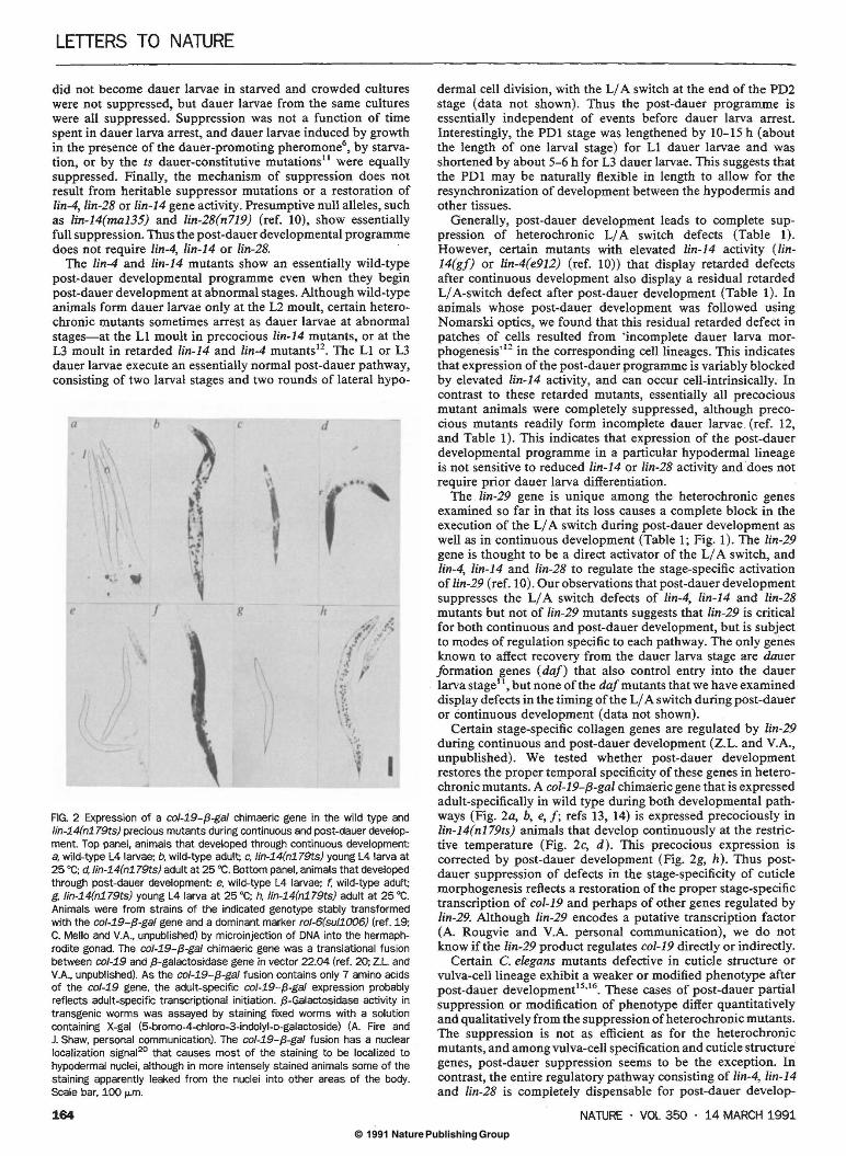
FIG. 2 Expression of a co/-19-{3-ga/ chimaeric gene in the wild type and lin-14(n179ts) precious mutants during continuous and post-dauer development. Top panel, animals that developed through continuous development: a, wild-type L4 larvae; b, wild-type adult; c, lin-14(n179ts) young L4 larva at 25 •c; d, lin-14(n179ts) adult at 25 •c. Bottom panel, animals that developed through post-dauer development: e, wild-type L4 larvae; f. wild-type adult; g, lin-14(n179ts) young L4 larva at 25 •c; h, lin-14(n179ts) adult at 25 •c. Animals were from strains of the indicated genotype stably transformed with the col-19-{3-ga/ gene and a dominant marker rol-6(sul1006) (ref. 19; C. Mello and V.A., unpublished) by microinjection of DNA into the hermaphrodite gonad. The co/-19-{3-ga/ chimaeric gene was a translational fusion between col-19 and {3-galactosidase gene in vector 22.04 (ref. 20; Z.l. and V.A., unpublished). As the co/-19-{3-ga/ fusion contains only 7 amino acids of the co/-19 gene, the adult-specific co/-19-{3-ga/ expression probably reflects adult-specific transcriptional initiation. {3-Galactosidase activity in transgenic worms was assayed by staining fixed worms with a solution containing X-gal (5-bromo-4-chloro-3-indolyl-o-galactoside) (A. Fire and J. Shaw, personal communication). The col-19-{3-gal fusion has a nuclear localization signal20 that causes most of the staining to be localized to hypodermal nuclei, although in more intensely stained animals some of the staining apparently leaked from the nuclei into other areas of the body. Scale bar, 100 ,...m.
164
dermal cell division, with the L/ A switch at the end of the PD2 stage (data not shown). Thus the post-dauer programme is essentially independent of events before dauer larva arrest. Interestingly, the PD1 stage was lengthened by 10-15 h (about the length of one larval stage) for L1 dauer larvae and was shortened by about 5-6 h for L3 dauer larvae. This suggests that the PD1 may be naturally flexible in length to allow for the resynchronization of development between the hypodermis and other tissues.
Generally, post-dauer development leads to complete suppression of heterochronic L/ A switch defects (Table 1). However, certain mutants with elevated lin-14 activity (lin-14(gf) or lin-4(e912) (ref. 10)) that display retarded defects after continuous development also display a residual retarded L/ A-switch defect after post-dauer development (Table 1). In animals whose post-dauer development was followed using Nomarski optics, we found that this residual retarded defect in patches of cells resulted from 'incomplete dauer larva morphogenesis'12 in the corresponding cell lineages. This indicates that expression of the post-dauer programme is variably blocked by elevated lin-14 activity, and can occur cell-intrinsically. In contrast to these retarded mutants, essentially all precocious mutant animals were completely suppressed, although precocious mutants readily form incomplete dauer larvae (ref. 12, and Table 1). This indicates that expression of the post-dauer developmental programme in a particular hypodermal lineage is not sensitive to reduced lin-14 or lin-28 activity and does not require prior dauer larva differentiation .
The lin-29 gene is unique among the heterochronic genes examined so far in that its loss causes a complete block in the execution of the L/ A switch during post-dauer development as well as in continuous development (Table 1; Fig. 1). The lin-29 gene is thought to be a direct activator of the L/ A switch, and lin-4, lin-14 and lin-28 to regulate the stage-specific activation of lin-29 (ref. 10). Our observations that post-dauer development suppresses the L/ A switch defects of lin-4, lin-14 and lin-28 mutants but not of lin-29 mutants suggests that lin-29 is critical for both continuous and post-dauer development, but is subject to modes of regulation specific to each pathway. The only genes known to affect recovery from the dauer larva stage are dauer formation genes ( daf) that also control entry into the dauer larva stage11 , but none of the daf mutants that we have examined display defects in the timing of the L/ A switch during post-dauer or continuous development (data not shown).
Certain stage-specific collagen genes are regulated by lin-29 during continuous and post-dauer development (Z.L. and V.A., unpublished). We tested whether post-dauer development restores the proper temporal specificity of these genes in heterochronic mutants. A col-19-{3-gal chimaeric gene that is expressed adult-specifically in wild type during both developmental pathways (Fig. 2a, b, e, f; refs 13, 14) is expressed precociously in lin-14(n179ts) animals that develop continuously at the restrictive temperature (Fig. 2c, d). This precocious expression is corrected by post-dauer development (Fig. 2g, h). Thus postdauer suppression of defects in the stage-specificity of cuticle morphogenesis reflects a restoration of the proper stage-specific transcription of col-19 and perhaps of other genes regulated by lin-29. Although lin-29 encodes a putative transcription factor (A. Rougvie and V.A. personal communication), we do not know if the lin-29 product regulates col-19 directly or indirectly.
Certain C. elegans mutants defective in cuticle structure or vulva-cell lineage exhibit a weaker or modified phenotype after post-dauer development15·16. These cases of post-dauer partial suppression or modification of phenotype differ quantitatively and qualitatively from the suppression ofheterochronic mutants. The suppression is not as efficient as for the heterochronic mutants, and among vulva-cell specification and cuticle structure genes, post-dauer suppression seems to be the exception. In contrast, the entire regulatory pathway consisting of lin-4, lin-14 and lin-28 is completely dispensable for post-dauer develop-
NATURE · VOL 350 · 14 MARCH 1991

© 1991 Nature Publishing Group
ment. These differences may reflect particularly acute constraints imposed on temporal control systems (as opposed to spatial patterning or cuticle morphogenesis) by the suspension of development at dauer larva arrest.
What mechanistic or developmental constraints account for the existence of separate temporal signals for continuous and post-dauer development? Heterochronic genes may encode components of a temporally dynamic system of interacting gene products. The lin-14 activity must decrease between L1 and L2 to specify the proper timing and sequence of diverse events, including the L/ A switch9
•1u,I
7; lin-4 and lin-28, which interact
with lin-14, might also experience precise temporal changes. The indefinite period of developmental arrest in the dauer larva stage would inevitably interrupt the dynamics of this system, and thus render lin-4, lin-14 and lin-28 useless for conveying temporal information to cells during post-dauer development. Other organisms with optional developmentally arrested stages, such as insects that undergo diapause, may use a similar strategy to cope with the temporal control problems necessarily associated with alternative developmental histories. 0
Received 5 December; accepted 27 December 1990.
1. Alberch. P., Gould, S. J., Oster, G. F. & Wal<e, D. B. Paleobiology 5, 296-317 (1979). 2. Gould, S. J. Ontogeny and Phylogeny (Harvard University Press, Cambridge, Massachusetts, 1977). 3. Ambros, V. & Horvitz, H. R. Science 226, 409-416 (1984). 4. Cassada, R. C. & Russell, R. L. Devl Bioi. 46, 326-342 (1975). 5. Evans, A. A. F. & Perry, R. M. in The Organization of Nematodes (ed. Croll, N. A.) (Academic, New
Yol1<, 1976). 6. Golden, J. M. & Riddle, D. L. Science 218, 578-580 (1987). 7. Riddle, D. L. in The Nematode Gaenorhabditis elegans (eds Wood, W. B. and the community of
C. elegans researchers) (Cold Spring Harbor Laboratory, New Yol1<, 1988). 8. Chalfie, M., Horvitz, R. H. & Sulston, J. E. Cell 24, 59-69 (1981). 9. Ambros, V. & Horvitz, H. R. Genes Dev. 1, 398-414 (1987).
10. Ambros, V. Cell 57, 49-57 (1989). 11. Riddle, D. L., Swanson, M. M. & Alberts, P. S. Nature 290, 668-671 (1981). 12. Liu, Z. & Ambros, V. Genes Dev. 3, 2039-2049 (1989). 13. Cox, G. N., Fields, C., Kramer, J. M., Rosenzweig, B. & Hirsh, D. Gene 76, 331-344 (1989). 14. Cox, G. N. & Hirsh, D. Melee. cell. Bioi. 5, 363-372 (1985). 15. Ferguson, E. & Horvitz, H. R. Genetics UO, 17-72 (1985). 16. Cox, G. N., Laufer, J. S. Kusch, M. & Edgar, R. Genetics 95, 317-339 (1980). 17. Ruvkun, G. & Guisto, J. Nature 338, 313-319 (1989). 18. Sulston, J. E. & Horvitz, R. H. Devl Bioi. 56, 110-156 (1977). 19. Kramer, J. M .. French, R. P., Pall<, E. & Johnson, J. J. Melee. cell. Bioi. 10 (1990). 20. Fire, A .. Harrison, S. & Dixon, D. Gene 93, 189-198 (1990).
ACKNOWLEDGEMENTS. We thank S. Euling, R. Feinbaum, G. Struhl, D. Levitan, C. Mello, A. Rougvie and other members of the laboratory for critical reading of the manuscript, and P. Cherbas for comments.
Function of DnaJ and DnaK as chaperones in origin-specific DNA binding by RepA Sue Wickner*, Joel Hoskinst & Keith McKenneyt
* Laboratory of Molecular Biology, National Cancer Institute, National Institutes of Health, Bethesda, Maryland 20892, USA t Center for Advanced Research in Biotechnology, National Institute of Standards and Technology, Rockville, Maryland 20850, USA
HEAT-shock proteins are normal constituents of cells whose synthesis is increased on exposure to various forms of stress. They are interesting because of their ubiquity and high conservation during evolution. Two families of heat-shock proteins, hsp60s and hsp70s, have been implicated in accelerating protein folding and oligomerization and also in maintaining proteins in an unfolded state, thus facilitating membrane transpore-s. The Escherichia coli hsp70 analogue, DnaK, and two other heat-shock proteins, DnaJ and GrpE, are required for cell viability at high temperatures and are involved in DNA replication of phage A and plasmids Pl and F'-10
• These three proteins are involved in replication in vitro of Pl DNA along with many host replication proteins and the Pl RepA initiator protein11
•12
• RepA exists in a stable protein complex with DnaJ containing a dimer each of RepA and DnaJ11
• We
NATURE · VOL 350 · 14 MARCH 1991
LETTERS TO NATURE
TABLE 1 Temperature requirement for activation of RepA
Incubation at 24 oc Incubation at 0 oc oriP1 DNA bound (15min) (15min) (%)
Complete No additions 26 No additions Complete <2 Minus ATP +ATP <2 Minus oriP1 DNA +oriP1 DNA 32 Minus DnaJ +DnaJ 4 Minus DnaK +DnaK 4 Minus RepA +Rep A 3
DNA binding was measured by retention on nitrocellulose filters. Reaction mixtures for the first incubation at 24 oc contained the components described in Fig. 1 with the omissions indicated and with 20 ng RepA, 30 ng DnaJ and 100 ng DnaK. After 15 min, the reactions were chilled on ice, the omitted components were added, and the reactions were continued for 15 min at 0 °C.
report here that DnaK and DnaJ mediate an alteration in the Pl initiator protein, rendering it much more active for oriPl DNA binding.
RepA binds specifically to five 19-base-pair (bp) direct repeats in the Pl origin13
• We observed that the DnaJ-RepA protein complex also binds to the Pl origin (Fig. la). Surprisingly, about a 100-fold molar excess of RepA dimers to binding sites was required for binding by RepA alone or in a complex with DnaJ (Fig. la). These results suggested that the preparations of RepA and DnaJ-RepA complex were largely inactive or the affinity of RepA for DNA was very low.
Because it seemed likely that the other heat-shock proteins may function with DnaJ, we added DnaJ, DnaK and GrpE to DNA-binding reactions with RepA. About 100-fold less RepA was required for oriPl DNA binding when DnaJ, DnaK and ATP were added to the reaction mixtures (Fig. la-c). GrpE was not required for RepA activation (Fig. lc). DnaJ-RepA protein complexes could be activated by DnaK and ATP, without additional DnaJ. ATP hydrolysis is probably required because the non-hydrolysable ATP analogue ATP-y-S was inactive and was a competitive inhibitor of the reaction.
The stimulation by DnaJ, DnaK and ATP of oriPl DNA binding by RepA depended both on the time of incubation and the temperature. DNA binding increased linearly at 24 oc for 20 min, but was barely detectable when the reactions were carried out at 0 °C (Fig. 2). Because protein-DNA binding is expected to occur at 0 °C, we attempted to divide the DnaJ- and DnaK-stimulated binding reaction. In a first reaction at 24 °C, we incubated all five components (DnaJ, DnaK, RepA, ATP and oriPl DNA) or four of them. Then in a second incubation at 0 oc, we added the omitted component and measured oriPl DNA bound (Table 1). DnaK, DnaJ, RepA and ATP were each required for DNA binding in the first incubation, but oriPl DNA was not. The first reaction in the absence of DNA proceeded linearly for 20 min at 24 oc, whereas the DNA-binding reaction was complete by 30 sat 0 °C (Fig. 2). The same requirements were necessary when the first reaction was carried out at 30 °C or 42 oc. When DnaJ and RepA were replaced by the DnaJ-RepA complex, the activation reaction still required a 20-min incubation at 24 oc with DnaK and ATP. This is consistent with our previous observation that the DnaJ-RepA complex forms rapidly at 0 oc on mixing the two proteins11
• These experiments show that DnaJ and DnaK., in an ATP and temperature-dependent reaction, activate RepA in the absence of DNA so that it can then rapidly bind to oriPl DNA.
Of the three proteins required for the reaction, only RepA ends up bound to the Pl DNA. We prepared a large reaction mixture, isolated the protein-DNA complex by gel filtration, subjected the material to SDS-PAGE, and stained for protein. Densitometer analysis of the gel showed that RepA was the only protein detectable (Fig. 3). Protein-DNA complexes similarly
165