Embed Size (px)
Citation preview
i
Alveolar Bone and Root Length Changes Following En-masse Retraction with Corticotomy assisted Orthodontic Treatment
Satinee Narupakorn
A Thesis Submitted in Partial Fulfillment of the Requirements for the Degree of Master of Science in Oral Health Sciences
Prince of Songkla University 2017
Copyright of Prince of Songkla University
ii
Thesis Title Alveolar Bone and Root Length Changes following En-masse Retraction with Corticotomy assisted Orthodontic Treatment
Author Miss Satinee Narupakorn Major Program Oral Health Sciences
Major Advisor ……………….…………………………. (Asst.Prof.Dr.Bancha Samruajbenjakun)
Examining Committee : ………………….………….…….…Chairperson (Prof.Smorntree Viteporn) ……………….…………………………. (Asst.Prof.Dr.Bancha Samruajbenjakun) …………….……………………………. (Assoc.Prof.Dr. Srisurang Suttapreyasri)
The Graduate School, Prince of Songkla University, has approved this thesis as partial fulfillment of the requirements for the Master of Science Degree in Oral Health Sciences
………………………………………… (Assoc.Prof.Dr.Teerapol Srichana)
Dean of Graduate School
iii
This is to certify that the work here submitted is the result of the candidate’s own investigations. Due acknowledgement has been made of any assistance received. …………………………………………Signature (Asst. Prof. Dr. Bancha Samruajbenjakun) Major Advisor …………………………………………Signature (Miss Satinee Narupakorn) Candidate

iv
I hereby certify that this work has not been accepted in substance for any degree, and is not being currently submitted in candidature for any degree. …………………………………………Signature (Miss Satinee Narupakorn) Candidate
v
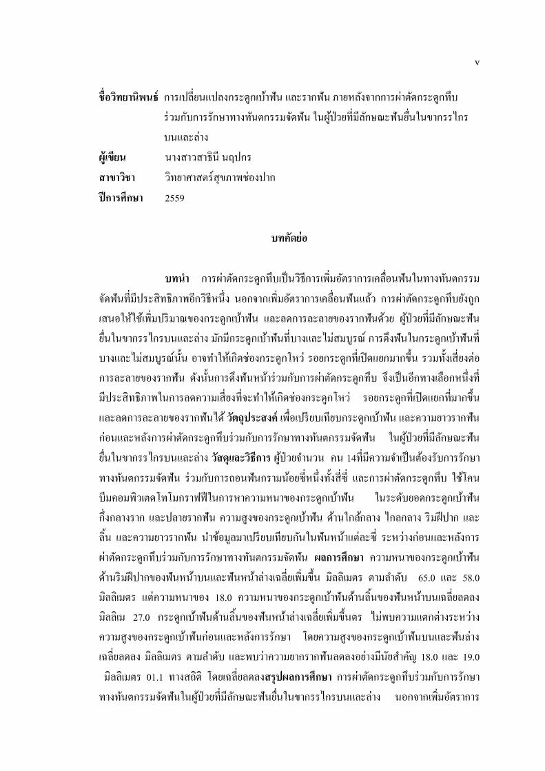
ช่ือวทิยานิพนธ์ การเปล่ียนแปลงกระดูกเบา้ฟัน และรากฟัน ภายหลงัจากการผา่ตดักระดูกทึบร่วมกบัการรักษาทางทนัตกรรมจดัฟัน ในผูป่้วยท่ีมีลกัษณะฟันยืน่ในขากรรไกรบนและล่าง
ผู้เขียน นางสาวสาธินี นฤปกร สาขาวชิา วิทยาศาสตร์สุขภาพช่องปาก ปีการศึกษา 2559
บทคดัย่อ
บทน า การผา่ตดักระดูกทึบเป็นวิธีการเพ่ิมอตัราการเคล่ือนฟันในทางทนัตกรรมจดัฟันท่ีมีประสิทธิภาพอีกวิธีหน่ึง นอกจากเพ่ิมอตัราการเคล่ือนฟันแลว้ การผา่ตดักระดูกทึบยงัถูกเสนอใหใ้ชเ้พ่ิมปริมาณของกระดูกเบา้ฟัน และลดการละลายของรากฟันดว้ย ผูป่้วยท่ีมีลกัษณะฟันยืน่ในขากรรไกรบนและล่าง มกัมีกระดูกเบา้ฟันท่ีบางและไม่สมบูรณ์ การดึงฟันในกระดูกเบา้ฟันท่ีบางและไม่สมบูรณ์นั้น อาจท าใหเ้กิดช่องกระดูกโหว่ รอยกระดูกท่ีเปิดแยกมากข้ึน รวมทั้งเส่ียงต่อการละลายของรากฟัน ดงันั้นการดึงฟันหนา้ร่วมกบัการผา่ตดักระดูกทึบ จึงเป็นอีกทางเลือกหน่ึงท่ีมีประสิทธิภาพในการลดความเส่ียงท่ีจะท าใหเ้กิดช่องกระดูกโหว ่ รอยกระดูกท่ีเปิดแยกท่ีมากข้ึน และลดการละลายของรากฟันได ้วตัถุประสงค์ เพื่อเปรียบเทียบกระดูกเบา้ฟัน และความยาวรากฟัน ก่อนและหลงัการผา่ตดักระดูกทึบร่วมกบัการรักษาทางทนัตกรรมจดัฟัน ในผูป่้วยท่ีมีลกัษณะฟันยืน่ในขากรรไกรบนและล่าง วสัดุและวธิีการ ผูป่้วยจ านวน 14 คน ท่ีมีความจ าเป็นตอ้งรับการรักษาทางทนัตกรรมจดัฟัน ร่วมกบัการถอนฟันกรามนอ้ยซ่ีหน่ึงทั้งส่ีซ่ี และการผา่ตดักระดูกทึบ ใชโ้คนบีมคอมพิวเตดโทโมกราฟฟีในการหาความหนาของกระดูกเบา้ฟัน ในระดบัยอดกระดูกเบา้ฟัน ก่ึงกลางราก และปลายรากฟัน ความสูงของกระดูกเบา้ฟัน ดา้นใกลก้ลาง ไกลกลาง ริมฝีปาก และล้ิน และความยาวรากฟัน น าขอ้มูลมาเปรียบเทียบกนัในฟันหนา้แต่ละซ่ี ระหว่างก่อนและหลงัการผา่ตดักระดูกทึบร่วมกบัการรักษาทางทนัตกรรมจดัฟัน ผลการศึกษา ความหนาของกระดูกเบา้ฟันดา้นริมฝีปากของฟันหนา้บนและฟันหนา้ล่างเฉล่ียเพ่ิมข้ึน 0.58 และ 0.65 มิลลิเมตร ตามล าดบั
ความหนาของกระดูกเบา้ฟันดา้นล้ินของฟันหนา้บนเฉล่ียลดลง 0.18 มิลลิเมตร แต่ความหนาของกระดูกเบา้ฟันดา้นล้ินของฟันหนา้ล่างเฉล่ียเพ่ิมข้ึน 0.27 มิลลิเม ตร ไม่พบความแตกต่างระหว่าง
ความสูงของกระดูกเบา้ฟันก่อนและหลงัการรักษา โดยความสูงของกระดูกเบา้ฟันบนและฟันล่างเฉล่ียลดลง 0.19 และ 0.18 มิลลิเมตร ตามล าดบั และพบว่าความยากรากฟันลดลงอยา่งมีนยัส าคญั
ทางสถิติ โดยเฉล่ียลดลง 1.01 มิลลิเมตร สรุปผลการศึกษา การผา่ตดักระดูกทึบร่วมกบัการรักษาทางทนัตกรรมจดัฟันในผูป่้วยท่ีมีลกัษณะฟันยืน่ในขากรรไกรบนและล่าง นอกจากเพ่ิมอตัราการ

vi
เคล่ือนฟันในทางทนัตกรรมจดัฟันแลว้ ในการศึกษาน้ีพบว่า สามารถเพ่ิมความหนาของกระดูกเบา้ฟันได ้ ยกเวน้ ความหนาของกระดูกเบา้ฟันดา้นล้ินของฟันหนา้บน โดยไม่พบความแตกต่างระหว่างความสูงของกระดูกเบา้ฟัน แต่พบว่าความยากรากฟันลดลงอยา่งมีนยัส าคญัทางสถิติ
vii
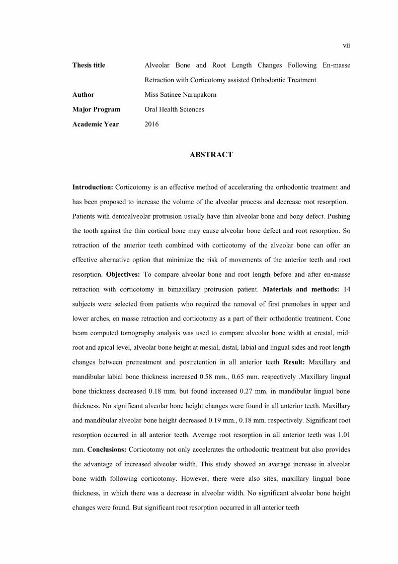
Thesis title Alveolar Bone and Root Length Changes Following En-masse Retraction with Corticotomy assisted Orthodontic Treatment
Author Miss Satinee Narupakorn Major Program Oral Health Sciences Academic Year 2016
ABSTRACT Introduction: Corticotomy is an effective method of accelerating the orthodontic treatment and has been proposed to increase the volume of the alveolar process and decrease root resorption. Patients with dentoalveolar protrusion usually have thin alveolar bone and bony defect. Pushing the tooth against the thin cortical bone may cause alveolar bone defect and root resorption. So retraction of the anterior teeth combined with corticotomy of the alveolar bone can offer an effective alternative option that minimize the risk of movements of the anterior teeth and root resorption. Objectives: To compare alveolar bone and root length before and after en-masse retraction with corticotomy in bimaxillary protrusion patient. Materials and methods: 14 subjects were selected from patients who required the removal of first premolars in upper and lower arches, en masse retraction and corticotomy as a part of their orthodontic treatment. Cone beam computed tomography analysis was used to compare alveolar bone width at crestal, mid-root and apical level, alveolar bone height at mesial, distal, labial and lingual sides and root length changes between pretreatment and postretention in all anterior teeth Result: Maxillary and mandibular labial bone thickness increased 0.58 mm., 0.65 mm. respectively .Maxillary lingual bone thickness decreased 0.18 mm. but found increased 0.27 mm. in mandibular lingual bone thickness. No significant alveolar bone height changes were found in all anterior teeth. Maxillary and mandibular alveolar bone height decreased 0.19 mm., 0.18 mm. respectively. Significant root resorption occurred in all anterior teeth. Average root resorption in all anterior teeth was 1.01 mm. Conclusions: Corticotomy not only accelerates the orthodontic treatment but also provides the advantage of increased alveolar width. This study showed an average increase in alveolar bone width following corticotomy. However, there were also sites, maxillary lingual bone thickness, in which there was a decrease in alveolar width. No significant alveolar bone height changes were found. But significant root resorption occurred in all anterior teeth
viii
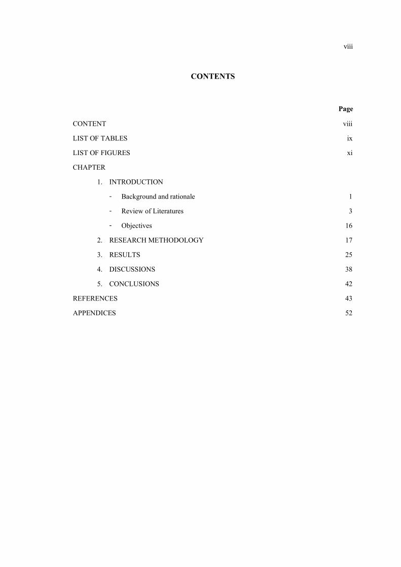
CONTENTS
Page CONTENT viii LIST OF TABLES ix LIST OF FIGURES xi CHAPTER
1. INTRODUCTION - Background and rationale 1 - Review of Literatures 3 - Objectives 16
2. RESEARCH METHODOLOGY 17 3. RESULTS 25 4. DISCUSSIONS 38 5. CONCLUSIONS 42
REFERENCES 43 APPENDICES 52
ix
LISTS OF TABLES
Table Page 1. Age, the total treatment time and post retention phase in subjects 25
who were treated with decortication-facilitated en-masse retraction. 2. Comparison of mean values of maxillary labial alveolar bone width 29
measured from CT scans before and after en-masse retraction with corticotomy assisted orthodontic treatment
3. Comparison of mean values of mandibular labial alveolar bone width 30 measured from CT scans before and after en-masse retraction with corticotomy assisted orthodontic treatment
4. Comparison of mean values of maxillary lingual alveolar bone width 31 measured from CT scans before and after en-masse retraction with corticotomy assisted orthodontic treatment
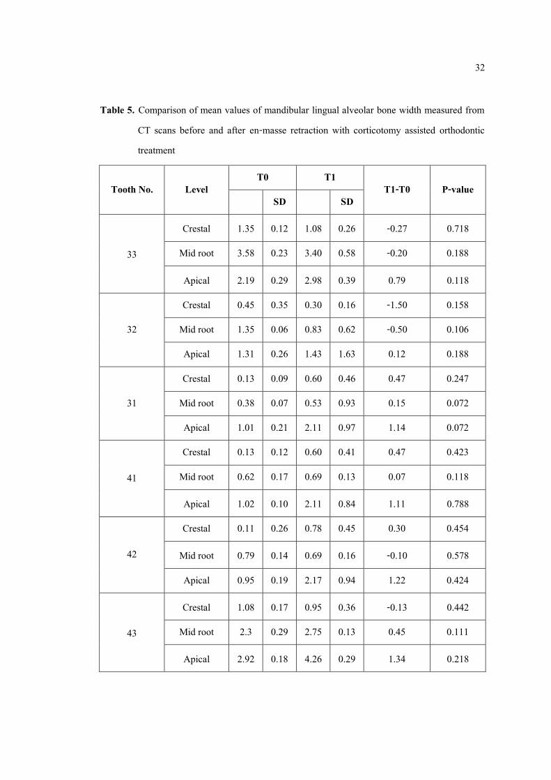
5. Comparison of mean values of mandibular lingual alveolar bone width 32 measured from CT scans before and after en-masse retraction with corticotomy assisted orthodontic treatment
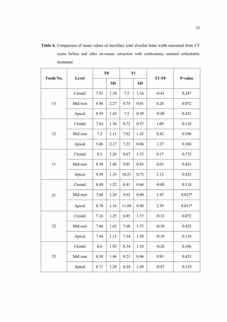
6. Comparison of mean values of maxillary total alveolar bone width 33 measured from CT scans before and after en-masse retraction with corticotomy assisted orthodontic treatment
7. Comparison of mean values of mandibular total alveolar bone width 34 measured from CT scans before and after en-masse retraction with corticotomy assisted orthodontic treatment
8. Comparison of mean values of maxillary alveolar bone height 35 measured from CT scans before and after en-masse retraction with corticotomy assisted orthodontic treatment
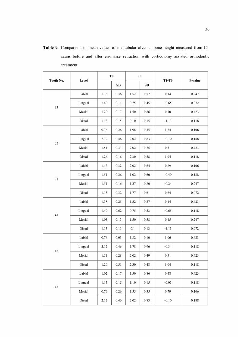
9. Comparison of mean values of mandibular alveolar bone height 36 measured from CT scans before and after en-masse retraction with corticotomy assisted orthodontic treatment
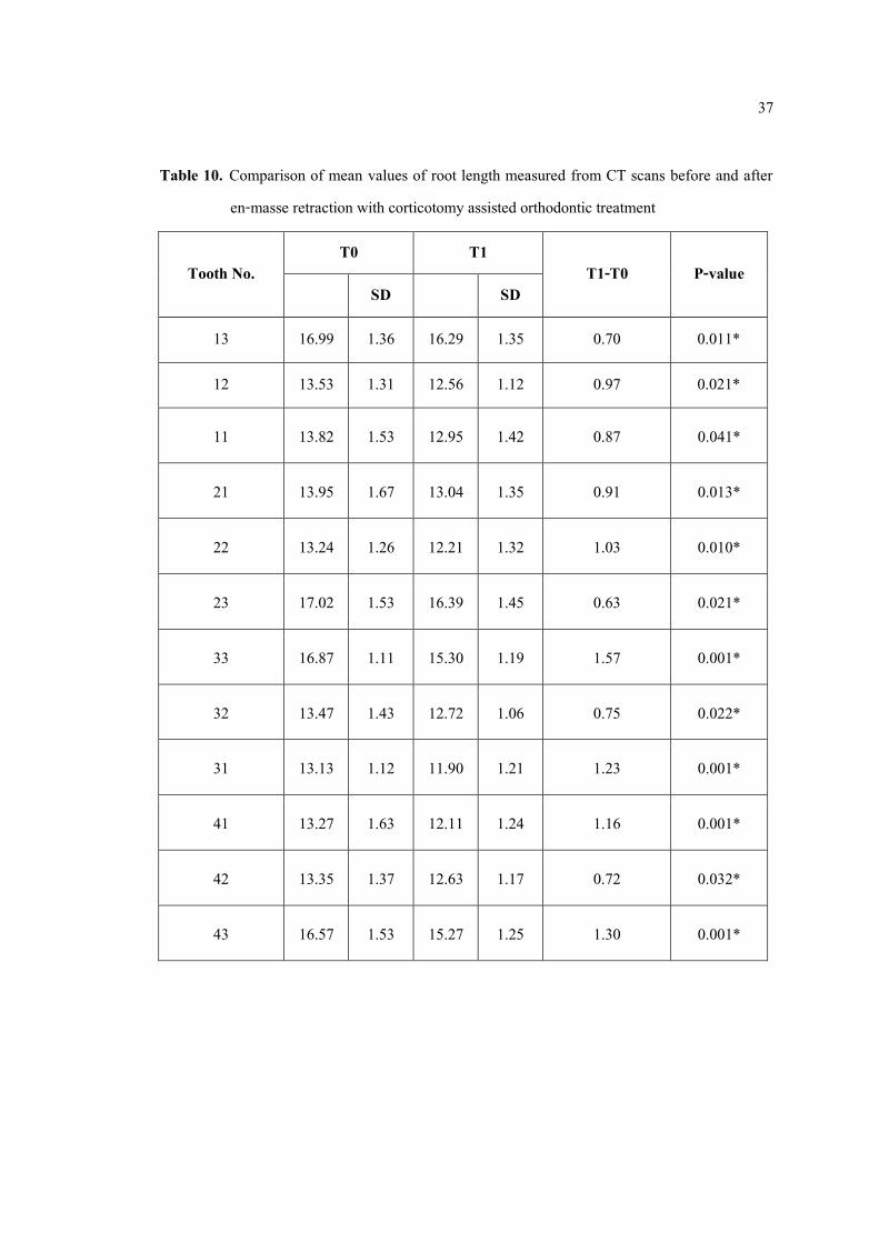
10. Comparison of mean values of root length measured from CT scans 37 before and after en-masse retraction with corticotomy assisted orthodontic treatment

x
LISTS OF TABLES (CONTINUED)
Table Page 11. Comparing the alveolar bone height changes 38
xi
LISTS OF FIGURES
Figure Page 1 The decortications 19 2 Bone grafting on the alveolar decortication area 19 3 Flap repositioning and suturing 19 4 T-loop was used to en masse retraction 20
5 Location of bone thickness measurement pretreatment and postretention 22 6 Measurement of bone thickness 22 7 Alveolar bone height measurement 23 8 Root length measurement 23 9 Controlled tipping orthodontic tooth movement 39
1
CHAPTER 1
INTRODUCTION
Background and rationale
Nowadays, not only children and adolescent seek for orthodontic treatment. Late adolescent and adult patients are increasing and it’s quite challenging because adult patients always expect short treatment time.1, 2 Orthodontists are looking forward to any alternative options for accelerating orthodontic tooth movement. Among the five interventions which are corticotomy procedure, pulsed electromagnetic fields, low-level laser technique, electrical current process and periodontal distraction, corticotomy is effective and safe to accelerate orthodontic tooth movement.3, 4
If discrepancies are borderline or beyond envelope of tooth movement and no longer possible for growth modification, orthognathic surgery might be necessary to obtain treatment objectives.5 By an osteotomy procedure, the trabecular and cortical bone is cut and the surgeon move the segments as needed, then the nerves and blood supply damage may be occur and leading to many complications. For mild dentoskeletal discrepancies patients, appropriate option may not be an orthognathic surgery. Corticotomy has been proposed, the cuts are made only on shallow surface of the cortical alveolar bone, that it is different from an osteotomy. Orthodontic force is applied shortly after surgery. It has been claimed that tooth move faster, and that the treatment results are more stable following a corticotomy with minimal risk of complications.6
Corticotomy with bone augmentation has been proposed to increase the volume of the alveolar process, to accelerate tooth movement, to prevent dehiscences and fenestrations and to increase the metabolic response during orthodontic treatment.7, 8 A retrospective study by Rothe9 show that decreased bone thickness, bone mass, bone density and size on panoramic and lateral cephalograms are shown to be a risk factor for orthodontic relapse. It is further interesting questioned that increased bone thickness might improve stability because it is related with increased bone volume. So alveolar bone size might relate to stability following orthodontic
2
treatment. Many studies claimed that orthodontic relapse after corticotomy-facilitated treatment is minimal because of increased root support after healing and loss of tissue memory by the high turnover and remodeling processes,2, 6-8, 10 and also found better score of American Board of Orthodontics objective grading system and a more stable treatment result in the corticotomy group.11, 12 Recently, clinical trials address about corticotomy enhancing 10 years long-term stability in non-extraction cases.13
Corticotomy is quite safe for periodontium and shows little risk of root resorption.4, 14 The explanation of decreased root resorption in the corticotomy is that localized selective decortication combined with orthodontic tooth movement casues a rapid alveolar bone remodeling or “Regional Acceleratory Phenomenon” in bone marrow cavities, decreases bone density, decreased hyalinization of the periodontal ligament and leads to absence of the lag phase during later stages of orthodontic tooth movement.15
Bimaxillary protrusion is common in Asian and African American populations.16 There are two treatment options for bimaxillary protrusion. The first is conventional orthodontic treatment and the second is anterior segmental osteotomy. Conventional orthodontic treatment is the most common treatment of bimaxillary protrusion. There are many side effects in conventional orthodontic treatment such as gingival recession, bone dehiscence, bone fenestration and root resorption. To overcome the limitations of conventional orthodontic treatment, sometimes anterior segmental osteotomy is recommended. Anyway, there are many disadvantages from postoperative complications of anterior segmental osteotomy such as delayed wound healing, ischemic necrosis, wound dehiscence and non-vital teeth beside the osteotomy site.17 So the most recent treatment method for bimaxillary protrusion is corticotomy-assisted orthodontic treatment, which is introduced as a compromise option between anterior segmental osteotomy and conventional orthodontic treatment.18 By corticotomy, the reducing cortical bone allows the anterior alveolar segment to bending when retraction force is applied.
Fuhrmann19 stated that retraction of the upper anterior tooth cause dehiscence and fenestration in alveolar bone around the roots. And alveolar bone modeling and remodeling during retraction and intrusion of upper incisors are limited20. So it is necessary to evaluate dentoalveolar changes during incisor retraction and intrusion. Three-dimensional evaluation is necessary for this purpose, which could provide three dimensional displacements.21 Risk of alveolar bone loss should
3
be cautioned during incisor retraction and intrusion, several treatment techniques are suggested.22 For example, distance of tooth movement and bone quantity limitation should be considered, light force should be used, direction should be controlled, periodontal injury should be routine evaluated and corticotomy with bone graft is another alternative way of decreasing the anatomical limitation.23, 24 Corticotomy can decrease the alveolar bone dehiscence and maintain the health, function, and esthetics of the periodontium in the orthodontic treatment.
The etiology of root resorption still remains unclear, complex and multifactorial,25 both genetic and environmental factors are involved.26, 27 And root resorption is quite common during orthodontic tooth movement. The explanation of the decreased root resorption in the corticotomy area is that decortication results in a rapid alveolar bone remodeling in bone marrow cavities, leads to absence of the lag phase during later stages of orthodontic tooth movement and decreased hyalinization of the periodontal ligament.15 However, there is only limited evidence in the orthodontic literature that demonstrates a root resorption in corticotomy procedures.28
This study is therefore undertaken to evaluate alveolar bone and root length changes after corticotomy in bimaxillary protrusion group.
Review of literatures
Corticotomy-facilitated tooth movement
Corticotomy-facilitated tooth movement was first stated by L.C. Bryan in 1893 as a surgical approach to facilitate orthodontic treatment with incisions to the cortical alveolar bone to splint teeth into new positions. However it was first introduced in 1959 by Kole6, corticotomy was reintroduced as a surgical procedure to facilitate orthodontic treatment by penetrating the buccal and palatal cortical layers while leaving the spongiosa intact. Kole explained that this method was used to move teeth faster than usual, leading to a shorter orthodontic treatment period because the teeth are moved together with the bone block. However, Kole’s technique involves the full thickness flaps reflection to expose both labial and palatal alveolar bone, followed by
4
interdental cuts through the cortical bone and penetrated the medullary bone. Connecting the interdental by the subapical horizontal cuts were osteotomy style, penetrating the full thickness of the alveolar bone. It was never widely accepted due to invasive technique.
Wilcko et al.7, 8 have noted that orthodontic tooth movement is accelerated by decrease of bone density and increase of bone turnover by increased osteoclasts and osteoblasts, which called a regional acceleratory phenomenon (RAP) that first described by Frost HM in 198329, Frost found a direct relation between the intensity of the healing process and the severity of bone injury, leading to accelerated bone turnover at the surgical area. The duration of RAP depends on the tissue type and usually stay around four months in human bone. This RAP phenomenon causes faster bone healing than normal bone turnover 10–50 times.
Wilcko introduced a recent surgical orthodontic technique8, 10 which combined corticotomy technique with alveolar grafting, which called Accelerated Osteogenic Orthodontics (AOO) and more recently with periodontally accelerated osteogenic orthodontics (PAOO). Several studys indicated that PAOO is safe, effective, extremely predictable, less root resorption occured and short treatment time. This technique also could reduce the need for orthognathic surgery in some situations.8, 30-32
In addition to faster treatment time, corticotomy treatment provide a healthier periodontium than would be achieved with traditional orthodontics. In their original article,8 the Wilcko brothers claimed no periodontal pocketing and no significant apical root resorption. They believe that a greater extent of movement can be achieved with corticotomy, thus reducing the need for extractions while providing increased support to the periodontium.33 Shoreibah34 showed similar results, twenty adult patients who presented with 3-5 mm of lower anterior crowding were equally divided into two groups. Group 1 treatment included corticotomy-facilitated orthodontic therapy alone, whereas group 2 treatment involved the addition of bone grafting to the corticotomy site. Pre-surgical, post-treatment, and 6 months post-treatment measurements of bone density and clinical probing depths were obtained for comparison. Their results demonstrated no significant difference in treatment time between the two groups as well as a significant reduction in probing depths in both groups from pretreatment to 6 months post-retention. With the augmentation of bone following corticotomy, a obviously increase in labiolingual alveolar cortical width has been
5
observed radiographically and remains stable at two-years post-treatment.35 And also eliminate of pre-treatment fenestrations and dehiscences.
Another often claimed advantage of corticotomy-facilitated orthodontic treatment is a decrease in apical root resorption. It has been hypothesized that the osteopenia induced by corticotomies allows for diminished osseous resistance to tooth movement. Therefore, less strain on the cemental surface of the root may result in a decreased incidence of root resorption. Interestingly, a diminished extent of hyalinization of the PDL occurs during tooth movement after corticotomies36 and hyalinization has been known to be a precursor to root resorption.37 To assess root resorption, Shoreibah34 compared root length between pre-treatment and post-treatment from the cementoenamel junction to the tooth apex on periapical films. Their results showed no significant root resorption in patients treated with corticotomy. These results are similar with report by Wang38 who using CBCT, found no significant difference of root length between pre-treatment and post-treatment in their corticotomy group, but an average of 1.55 mm resorption in patients treated with orthodontic therapy alone.
Orthodontic stability is dependent on the ability of the periodontium to regenerate, reorganize, and adapt to the final position of the teeth. Due to the increased tissue turnover that occurs at the corticotomy site, increased alveolar thickness from grafting and tissue memory after dramatic tissue turnover, researchers evaluated post-treatment stability after corticotomy.13
As all mentioned above, there are many clinical applications for corticotomy with bone graft have been addressed.8, 30, 39, 40 This technique use to accelerate orthodontic tooth movement and to overcome conventional orthodontic treatment limitations such as the long treatment time and envelope of tooth movement. The corticotomy procedure can be use for shorten treatment time, crowding correction, increased alveolar bone support, accelerate canine retraction, reinforced anchorage, open bite correction, decreased risk of root resorption and improve stability.41
Contraindications include patients with active periodontal disease and gingival recession. Moreover, corticotomy should not be for severe posterior cross-bite surgically and should not be used in cases which bimaxillary protrusion is combined with a gummy smile.30
6
Bone grafting and corticotomy
There are three types of bone graft material. Autografts, the first one, are get from their own hosts. The second type, allografts, are derived from the same species as the host. The third type is xenografts which are taken from another species. And the last one is alloplasts which are synthetic materials. The gold standard is autogenous bone for the defects of the facial area.42 Because of three potential biological processes in autogenous bone. The first process is osteogenesis which new bone are formed from osteocompetent cells. is Osteoinduction, the second process is the formation of new bone from osteoprogenetor cells. The third one is osteoconduction, the formation of new bone at the recipient site along a scaffold of osteocompetent cells.43 Autogenous bone can be harvested from intraoral sites for example mandibular ramus and maxillary tuberosity. And from extraoral sites such as the iliac crest, tibia, rib and calvarium.
In corticotomy, bone grafting material is needed quite small volume. So if autogenous bone is chosen, intraoral sites are usually harvested to decrease morbidity. Nowadays, allografts are most frequently used during corticotomy because of elimination of donor site morbidity, ease of use, and accessibility. Although there are only potential osteoinductive and osteoconductive properties. In the oral surgery, many studies reveal that there are advantage outcomes from allografts. Allografts are usually harvested from cadaveric bone in human, but may also get from live human donors and can be prepared as many processes such us fresh, frozen, freeze-dried, mineralized and demineralized. Grafts are processed by different ways such as ultrasonic, physical debridement, antibiotic washing and freeze-drying to make sure that no remaining diseases to the recipient. When the graft is processed, it relates to osteoinductive properties. For example, demineralization of the freeze-dried bone result in more bone morphogenic proteins created, which are important for bone formation. So freeze-dried bone is mostly osteoconductive, but demineralized freeze-dried bone provides osteoinduction in addition to an osteoconductive scaffold.44, 45 From reasons mentioned above, the most commonly allograft for the corticotomy augmentation procedure is demineralized freeze-dried bone allograft.
Xenografts are taken from the inorganic portion of bone in different species. Bovine bone is the most common used in this type. Xenografts will be complete deproteined by high temperature procedure to eliminate risk of immune response.43 The crystalline structure of
7
xenografts is similar to human cancellous bone. Total resorption of the xenografts had taken place with new bone formation by 14 weeks.46 Alloplasts are synthetic materials used as bone substitutes and include various combinations such as hydroxyapatite and calcium carbonate. For new bone growth, only osteoconductive properties are available in alloplasts.42 The major disadvantage of alloplasts is the lack of osteoinductive properties.
Rate of resorption and new bone formation depend on the physical and chemical properties of graft materials.47 Incorporation of bone grafts are expected, the first process involves the bony union along the edges of the graft and native bone segments followed by the gradual resorption of the graft material itself and replacement by new bone.48, 49 Duration and amount of incorporation into host bone depends on multiple factors such as graft type, processing method particle size, porosity, crystallinity and the pH of surrounding tissues. All factors play a role in the resorption rate. For example, the larger the particle size, the longer the material will remain at the site. A cortical graft may never be fully resorbed due to incomplete vascular penetrance into the tightly packed lamellar bone. Because of the great variability in grafting materials, it is difficult to definitively define an exact time to full graft resorption and bone replacement. One study showed that within 8 weeks, most of the graft has been remodeled.50 However, another study reported the presence of grafting granules even after 44 months.51 Despite the range in numbers, most of orthopedic and dental literature agrees that the majority of cancellous bone graft replacement occurs in 3-4 months.
The process of incorporation of new bone within the implanted grafting material is biologically similar to normal bone healing with a reactive, reparative, and remodeling phase. Like long bone healing, adequate blood supply and stability are imortant for graft survival, and periosteal preservation has been documented to enhance incorporation of the graft.52 Urist first described the five stages of graft incorporation in 1976.53 Stage one, the inflammatory stage, immediately follows bone grafting. A blood clot forms to stop the bleeding. Necrosis of the graft occurs and a subsequent inflammatory response is established. During the second stage, the osteoblast stage, osteoblasts, lymphocytes and plasma cells are attracted by platelet derived growth factor. Granulation tissue forms together with capillary buds bringing macrophages and mesenchymal cells. A fibrovascular stroma develops with an influx of osteogenic precursors and blood vessels. Osteoblasts and osteoclasts are stimulated and the osteoclasts initiate graft

8
resorption. Stage three is the osteoinductive stage, whereby mesenchymal cells differentiate into osteoblasts and new bone is laid down by endochondral ossification. Next is the osteoconduction stage, during which the graft serves as a passive template for ingrowth of vascular and cellular activity. Lastly, stage five is the remodeling stage where final incorporation and remineralization occurs. It is important to recognize that these final three stages are closely entwined and occur simultaneously during graft healing.
In the grafting procedure known as guided bone regeneration, a resorbable or non-resorbable membrane is used as a mechanical barrier to stabilize particulate graft material while simultaneously maintaining space around the bony defect to discourage in-growth of soft tissue at the osseous healing site. A graft has the potential to lose up to 25% of its volume after 4 months when a membrane is not used.54 In general, both resorbable and non-resorbable membrane materials are equally effective in attaining adequate bony defect fill, but the use of a resorbable membrane does not require a second removal surgery.55 In a systematic review of bone replacement grafts used in the treatment of intrabony periodontal defects, increases in bone level and in clinical attachment levels, as well as decreases in probing depths were found, when compared to simply open flap.56 Additionally, when compared to grafting alone, grafting with a barrier membrane increases clinical attachment level and decreases probing depth.56
Traditionally, particulate bone grafts are used in dentistry to fill bony defects or gain additional bone height and width. Jensen and Terheyden57 reported a mean increase in horizontal and vertical dimensions of 2.6 mm and 3.6 mm, respectively, following the guided bone regeneration technique. There is no consensus in the literature, however, on the biological events that take place after grafting with DFDBA. Smukler58 demonstrated the presence of residual DFDBA particles within a network of newly bone formation. They concluded that it may take many months or years for complete particulate resorption and total replacement by new bone.
In 2001, Wilcko introduced the use of the afore-mentioned particulate bone grafts following a corticotomy procedure.8 The case report described a patient with severe maxillary transverse constriction whose premolars were expanded over 3 mm. The authors claimed an increase in the buccolingual thickness of the cortical bone. A bone biopsy taken two years post-retention qualitatively revealed the presence of lamellar bone. Later, in 2008, Nowzari31 published the first case report using autogenous particulate bone as the grafting material after a corticotomy
9
procedure. One year after treatment was complete, the surgical sites were reentered and a visual report of both resorption and remodeling of the particulate material grafted from the patient’s mandibular ramus and lingual exostoses was described. They claimed no loss in alveolar height. Since the Wilckos’ first augmentation procedure with corticotomy, many others are also using bone grafting to supposedly build bone. Recently, Shoreibah34 has conducted the only study comparing corticotomy with and without bone augmentation. One aim of their study was to evaluate the effect of bone grafting on the periodontium. Using bioactive glass as their grafting material of choice, they sought to assess bone density in patients receiving either corticotomy or corticotomy with bone grafting. Measurements of bone density were obtained through calculation of mean gray value in an area of interest by periapical radiographs. As expected, they found a mean decrease in bone density following the corticotomy surgery. By 6 months post-treatment, a significantly greater percent increase in bone density was discovered in the group receiving bone augmentation. When comparing the groups from the start of orthodontic treatment and six months into retention, however, no significant differences in bone density changes could be noted. However, using periapical radiographs to measure bone density is not accurately quantified and it does not provide clear information on the condition of the grafted bone.
Dehiscences and fenestrations
The bony defects of the alveolar bone that found on the labial or lingual side of a
tooth and may extend over 2 mm. below the cementoenamel junction to the apex of the root with normal interproximal bone levels, it is called dehiscences.59
There are 2 types of gingiva. The first type is thin and scalloped, the second type is thick and flat.60 The first type, scalloped and thin gingiva. This thin gingiva takes much more risk for gingival recession. Because the thin gingiva loss easily when the base of the dehiscence is reached. The etiology of dehiscences and fenestrations is multifactorial. Severe anterior crowding and ectopically positioned of the teeth which make the tooth outside the alveolar bone often leading to dehiscences.19,61 Inappropriate eruption that the roots erupt more labially compared to the crown results in a dehiscence, especially found in lower anterior teeth.61 Frenum attachments also cause dehiscenses,61 they can create pressure on the bone and finally result in bone recession.
10
The upper and lower mid-labial frenum often result in the recession on the central incisors.61 The traumatic occlusion strongly relates with periodontal defects and can cause bone destruction.62 There is a significant reduction in periodontal destruction when traumatic occlusion is removed.62,
63 Patient habits are another cause of dehiscences and fenestrations such as the use of smokeless tobacco products. Iatrogenic treatment also cause the defects such as the orthodontic treatment move teeth outside of the alveolar bone or the dental restoration invade through the biologic width.64
The normal attachment of the gingiva combines with one millimeter of sulcular depth, one millimeter of epithelial attachment and one millimeter of connective tissue attachment above the crestal alveolar bone. If this distance is invaded, the bone and epithelial attachment will move apically to maintain the biologic width. Inappropriate tooth brushing and aging also cause recession of bone and also gingiva.61 The etiology of periodontal breakdown can be multifactorial factors or just one factor.61 The dehiscences and fenestration correlated with thin alveolar bone.65 The dehiscences and fenestrations are found around twenty percent in the population.59 Dehiscences were found quite often at mid-labial side of the lower canines. Before orthodontic treatment, CBCT scans is needed for the detection and measurement of dehiscences.66 Because of a long junctional epithelial attachment of the gingiva to the cementum, although the probing depth level and gingival margin is examined as normal, dehiscences and fenestrations often presented. The study showed that the depth of the gingival recession does not always relate with the depth of the dehiscence.67 So the orthodontist must aware about dehiscences and fenestrations in orthodontic treatment plan.
Periodontium and orthodontics
The orthodontic treatment plan depends on each individual patient, one of the
most important factor is periodontium. The buccal and lingual alveolar bone need to be evaluated, the orthodontists must know the limitation of the alveolar bone.68
Unwanted results may happen during orthodontic treatment, although the periodontium has effective regenerative properties. From a systematic review of the literature, there is no reliable study presenting positive results of periodontal health during orthodontic
11
treatment. The report about periodontium during orthodontic treatment compared with no treatment was related with gingival recession around 0.03 mm., 0.23 mm. of increased pocket depth and 0.13 mm. of alveolar bone loss.69
The most important about periodontal tissue respones is the type of the gingiva. Thin gingiva takes much higher risk for gingival recession and periodontal problems.60 If gingival recession is presented and attached gingiva is less than 2 mm., grafting should be done before orthodontic treatment.70, 71 If the grafting is proposed for esthetic aspectes, so the grafting should be done after orthodontic treatment.70 Lower incisors inclination may cause gingival recession and also bone loss. But most of the studies do not agree and found that proclination does not significantly relate the gingival level.72, 73 Anyway, many studies compare models between pre-treatment and posttreatment. Measurments were made at the heights of gingiva but the underlying bone may changed significantly.
Handleman68 determined that there was orthodontic wall, these walls were the thickness of the alveolar bone. The alveolar bone widths should be considered before orthodontic treatment. And it is useful for orthodontic treatment plan about space available to tooth movement. Long face type usually presents less alveolar bone width than short face type.74 Thin alveolar bone were often found in lower incisors area in high angle patients and in upper central incisors area in high angle class II patients. Considerably iatrogenic effects may be occured by tooth movement that beyond the limits of the alveolar bone housing.68
Several patients developed alveolar bone loss that may not visible from visual inspection or 2D film. Sarikaya75 used CBCT scans to evaluate the 3D alveolar bone thickness after retraction of maxillary and mandibular incisors in orthodontic treatment with first premolar extractions. This study showed that bone thickness following retraction on the lingual side in both arches reduced and also found at the alveolar crest on the buccal of the lower incisors. When the alveolar bone housing for tooth movement is limited, the root movement may move against the cortical plate and may cause adverse effects.19, 75 There are studies show that the effects of orthodontic treatment on the underlying bone may not always relate the overlying gingiva.76, 77 During excessive tooth movement, the dehiscences and fenestrations may repair if the teeth are moved back to the alveolar bone housing.
12
Periodontium and CBCT
Vandenberghe78 found that conventional radiography presented more bone details.
And CBCT provided a better morphologic data of both alveolar bone and periodontal defects.78 So intraoral radiography got better image for bone quality, contrast and lamina dura consideration. But periodontal defects were detected better from CBCT.78
Intraoral radiography, conventional CT and CBCT were compared in measurements of 14 dehiscences, 14 fenestrations and 14 intrabony defects on human skulls and dry pig.79 The mean difference of the CT for all defects was 0.16 mm and 0.19 mm for CBCT. For 12mm high and 3mm wide dehiscences measured by CBCT, the deviations were 0.28 for height, 0.21 for width and 0.88 for depth. Another similar results from study using dental implants in dry pig mandibles found that the dehiscence construction was not thinned as natural dehiscences. Mengel concluded that CBCT provided better image quality than CT.80 At least one alveolar defect found on 78% of subjects.
Dehiscences are the most common periodontal defect. And dehiscences were found most often on the mid-labial of the lower canines. So, the detection and measurement of dehiscences before orthodontic treatment is necessary.66 CBCT scans provide an important data for the orthodontic treatment plan based on alveolar bone support. When the alveolar bone was limited, the orthodontic wall, the treatment plan must be concern about tooth movement amount and direction. The orthodontic mechanics would be cautioned to reduce the movement of teeth outside of the alveolar housing. The 3D scans provided adequate data to orthodontist, so these data could help orthodontist to reduce iatrogenic effects.
Willerhausen and Kasaj conclude that CBCT was the low dosage and superior image quality when using for periodontal problems detection, especially for intra-bony defect,cyst recognition dehiscence and fenestration.81
Recently, CBCT has the opportunity to gradually replace conventional radiography. And provide diagnostic decisions based on the bony envelope found on the 3D data.82
13
Marginal alveolar bone and orthodontics
There are many adverse side effects occurred during orthodontic tooth movement such as gingival recession, dental caries progression, tooth mobility, tooth vitality, root resorption and alveolar bone loss. Most research mainly focused on root resorption that resulted from inflammatory process during orthodontic treatment.83 Anyway, factors that can cause root resorption may also make the alveolar bone loss in all directions.19, 84
Bitewing and periapical radiography have mostly used to evaluate alveolar bone changes in orthodontic patients for long time. 2D conventional radiograph has its limitation including the proximal bone surfaces assessment due to structure superimposition.85
In orthodontically treated patients, marginal bone loss has been found more than in untreated patients. But the amount of bone loss between studies are quite different. A marginal bone loss was more in orthodontic treated patients, the study found that marginal bone loss more than 2 mm. was occurred in 16.2% of 14 years old orthodontically treated patients and in 4.3% of untreated patients.86 One reason why marginal bone loss was found more in orthodontically treated is that marginal widening of the periodontal ligament space was included as bone loss. Another study showed that both control and treatment groups did not suffer from marginal bone loss of more than 2 mm over 5 years during orthodontic treatment.87
CBCT scans provide an important data for the orthodontic treatment plan based on alveolar bone support. CBCT was the low dosage and superior image quality when using for periodontal problems detection. Fuhrmann19 concluded that 2D conventional radiography is not clearly visualized for marginal bone assessments because of superimposed structures. When examining thin structures, a smaller voxel size and a smaller field of view cone beam computed tomography (CBCT) that commonly used for orthodontic purposes should be used.88 Alveolar bone height measurements can be made with good to excellent intra-rater and inter-rater repeatability by CBCT. When the bone thickness or bone height was smaller or similar to the voxel size of CBCT, the measurement could be overestimated. The measurement accuracy was improved when decrease in voxel size from 0.4 to 0.25 mm.89
14
Root resorption and corticotomy
The etiology of root resorption still remains unclear, complex and multifactorial.25 Both genetic and environmental factors are involved.26, 27 Root resorption is quite common during orthodontic tooth movement. This problem of root resorption as a consequence of orthodontic treatment was first discussed in 1927.90 The risk for root resorption associated with the length of orthodontic treatment, history of trauma91, orthodontic treatment plan with premolar extraction, the use of intermaxillary elastics, thin, tapered and dilacerated root92 and root resorption from previous orthodontic treatment may result in further root shortening.93
Corticotomy has been proposed for accelerating the rate of tooth movement for over 100 years.94 (Wilcko, 2003). Corticotomy increases rate of orthodontic tooth movement by the activation of a regional acceleratory phenomenon. The RAP occurs within a few days of injury, peaks at 1 to 2 months and lasts for 2 to 4 months with up to 6 to 24 months.94, 95 The corticotomy accelerates tooth movement by producing temporary osteopenia in the bone surrounding the roots. The activated area gets more rapid and extensive alveolar bone and periodontal ligament turnover.96 Corticotomy can increase the rate of orthodontic tooth movement 2 to 3 times.97, 98 Moreover, corticotomy is safe for periodontal structure and causes little risk of root resorption.4, 14 The explanation of the decreased root resorption in the corticotomy area is that decortication results in a rapid alveolar bone remodeling in bone marrow cavities, leads to absence of the lag phase during later stages of orthodontic tooth movement and decreased hyalinization of the periodontal ligament.15 However, there is only limited evidence in the orthodontic literature that demonstrates a root resorption in corticotomy procedures.28, 34
15
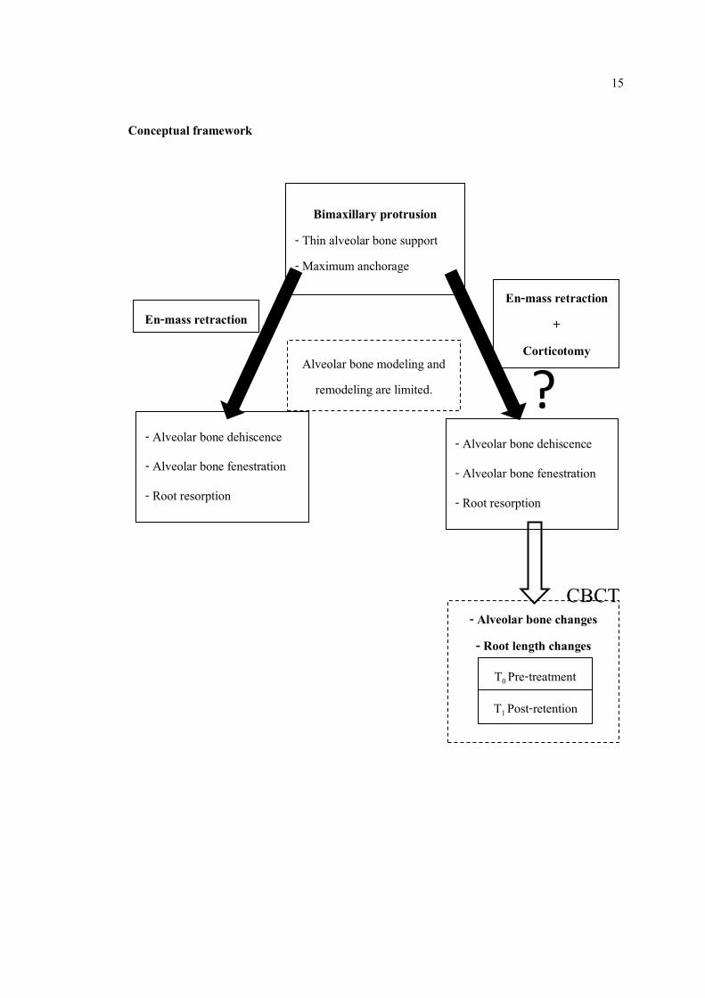
Conceptual framework
Alveolar bone modeling and remodeling are limited.
- Alveolar bone changes - Root length changes
Bimaxillary protrusion - Thin alveolar bone support - Maximum anchorage
T0 Pre-treatment
T1 Post-retention
- Alveolar bone dehiscence
- Alveolar bone fenestration
- Root resorption
En-mass retraction
- Alveolar bone dehiscence
- Alveolar bone fenestration
- Root resorption
En-mass retraction +
Corticotomy ?
CBCT
16
Objectives
1. To compare alveolar bone width and height before and after en-masse retraction with corticotomy in bimaxillary protrusion patient.
2. To compare root length before and after en-masse retraction with corticotomy in bimaxillary protrusion patient.
Hypothesis
En-masse retraction with corticotomy increase alveolar bone width and height and reduce root resorption in bimaxillary protrusion patient.
Significances of the study
1. To basically understand alveolar bone and root length changes following en-masse retraction with corticotomy in bimaxillary protrusion patient.
2. To provide the information for clinical application in bimaxillary protrusion patient.
17
CHAPTER 2
RESEARCH METHODOLOGY
Samples
This study was approved by Ethics committee on human experimental of Faculty of
Dentistry, Prince of Songkla University. The subjects selected from patients who received orthodontic treatment at orthodontic clinic, faculty of dentistry, Prince of Songkla University.
The inclusion criteria
1. Age at beginning of treatment between 18-35 years. 2. Healthy patients
- No allergy or medical problems especially uncontrolled osteoporosis or other bone disease - No long term use of medications such as anti-inflammatory, immunosuppressive, or steroids. - No long-term use of bisphosphonates.
3. No active periodontal disease. 4. No sign and symptoms of temporomandibular disorders. 5. Bimaxillary protrusion (The patients required the removal of first premolars in upper
and lower arches as a part of their orthodontic treatment.) - Interincisal angle < 117º - Class I malocclusion 6. Treatment plan required maximum anchorage. 7. Well-aligned maxillary and mandibular incisors with minimal crowding (≤ 3 mm.) 8. Treatment plan required anterior retraction more than lingual bone thickness of the
lower anterior teeth at the level of the incisor apices.
18
9. The orthodontic treatment completed at least 1 year. 10. CBCT was available at pretreatment and postretention.
The exclusion criteria
1. Medical problems especially uncontrolled osteoporosis or other bone diseases. 2. Long-term use of medications that are anti-inflammatory, immunosuppressive, steroid
or bisphosphonates. 3. CBCT was unavailable at pretreatment and postretention.
Materials and Methods
1. Initial record included (T0)
- Extraoral and intraoral photos. - Lateral cephalometric radiographs. - Cone beam computed tomography scan. 2. Pre-adjusted edgewise appliances (Roth prescription) with 0.018”-slot in anterior teeth and 0.022”-slot in posterior teeth were used for full arch. The teeth were aligned and leveled until complete on 0.016”x0.022” stainless steel archwire. 3. The patients referred to oral surgery clinic for alveolar decortications and bone graft with the same surgeon. The surgical procedures were performed following these step: - The surgical procedures were performed under local anesthesia and conscious oral sedation (Diazepam 5 mg.) - The mucoperiosteal incision were made along the buccal and lingual mucosa, the bone was exposed. - First premolars were extracted - After the mucoperiosteum had been undermined, vertical decortications were made across both first premolar sites with the proper size of round carbide burs in
19
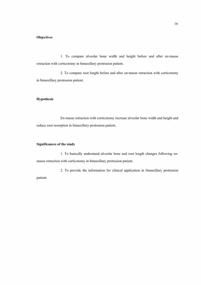
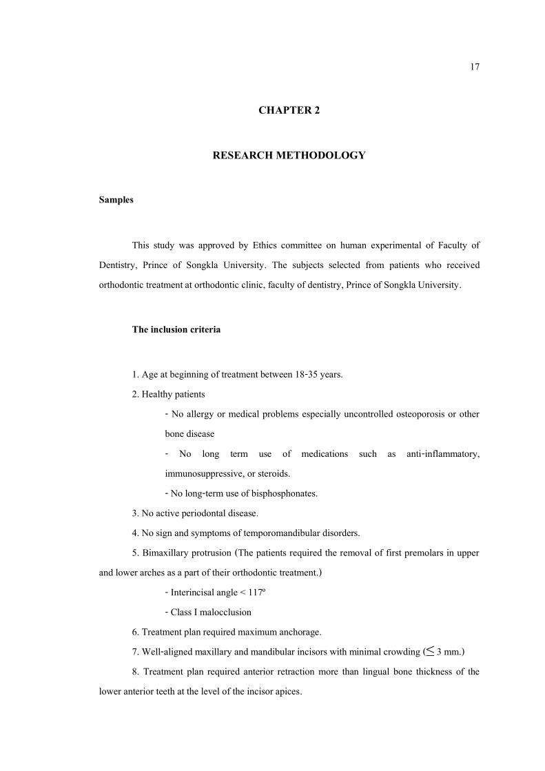
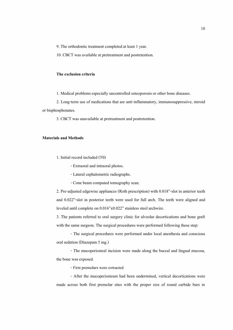
difference areas. The horizontal decortications were made 2 cm. below the apices of anterior teeth. (Figure 1.) Figure 1. The decortications - Bone grafting with allograft (Freeze dried bone from Faculty of Medicine, Siriraj Hospital) and autogenous bone (from decortications procedure) were adjusted to the corticotomy site. (Figure 2.) Figure 2. Bone grafting on the alveolar decortication area - Flap repositioning and suturing were made using a vertical double mattress technique. (Figure3.) Figure 3. Flap repositioning and suturing 4. Two weeks after alveolar decortications (Two weeks was optimal interval for sufficient healing and less patient anxiety), the maxillary and mandible anterior teeth and
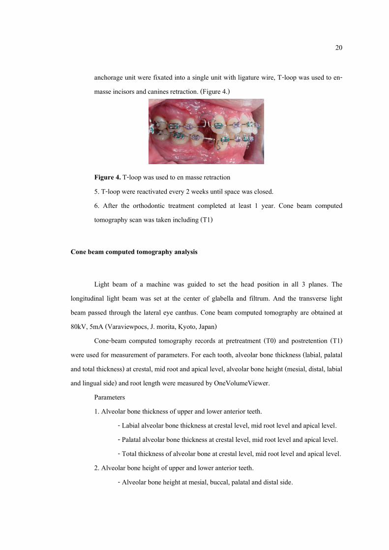
20
anchorage unit were fixated into a single unit with ligature wire, T-loop was used to en-masse incisors and canines retraction. (Figure 4.) Figure 4. T-loop was used to en masse retraction 5. T-loop were reactivated every 2 weeks until space was closed. 6. After the orthodontic treatment completed at least 1 year. Cone beam computed tomography scan was taken including (T1)
Cone beam computed tomography analysis
Light beam of a machine was guided to set the head position in all 3 planes. The
longitudinal light beam was set at the center of glabella and filtrum. And the transverse light beam passed through the lateral eye canthus. Cone beam computed tomography are obtained at 80kV, 5mA (Varaviewpocs, J. morita, Kyoto, Japan)
Cone-beam computed tomography records at pretreatment (T0) and postretention (T1) were used for measurement of parameters. For each tooth, alveolar bone thickness (labial, palatal and total thickness) at crestal, mid root and apical level, alveolar bone height (mesial, distal, labial and lingual side) and root length were measured by OneVolumeViewer.
Parameters 1. Alveolar bone thickness of upper and lower anterior teeth.
- Labial alveolar bone thickness at crestal level, mid root level and apical level. - Palatal alveolar bone thickness at crestal level, mid root level and apical level. - Total thickness of alveolar bone at crestal level, mid root level and apical level.
2. Alveolar bone height of upper and lower anterior teeth. - Alveolar bone height at mesial, buccal, palatal and distal side.
21
3. Root length of upper and lower anterior teeth.
Alveolar bone thickness
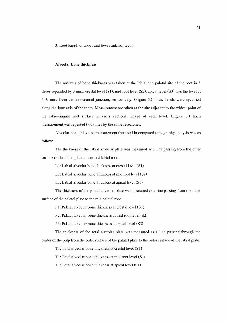
The analysis of bone thickness was taken at the labial and palatal site of the root in 3
slices separated by 3 mm., crestal level (S1), mid root level (S2), apical level (S3) was the level 3, 6, 9 mm. from cementoenamel junction, respectively. (Figure 5.) These levels were specified along the long axis of the tooth. Measurement are taken at the site adjacent to the widest point of the labio-lingual root surface in cross sectional image of each level. (Figure 6.) Each measurement was repeated two times by the same researcher.
Alveolar bone thickness measurement that used in computed tomography analysis was as follow:
The thickness of the labial alveolar plate was measured as a line passing from the outer surface of the labial plate to the mid labial root.
L1: Labial alveolar bone thickness at crestal level (S1) L2: Labial alveolar bone thickness at mid root level (S2) L3: Labial alveolar bone thickness at apical level (S3) The thickness of the palatal alveolar plate was measured as a line passing from the outer
surface of the palatal plate to the mid palatal root. P1: Palatal alveolar bone thickness at crestal level (S1) P2: Palatal alveolar bone thickness at mid root level (S2) P3: Palatal alveolar bone thickness at apical level (S3) The thickness of the total alveolar plate was measured as a line passing through the
center of the pulp from the outer surface of the palatal plate to the outer surface of the labial plate. T1: Total alveolar bone thickness at crestal level (S1) T1: Total alveolar bone thickness at mid root level (S1) T1: Total alveolar bone thickness at apical level (S1)
22
Figure 5. Location of bone thickness measurement pretreatment and postretention. Figure 6. Measurement of bone thickness
Alveolar bone height
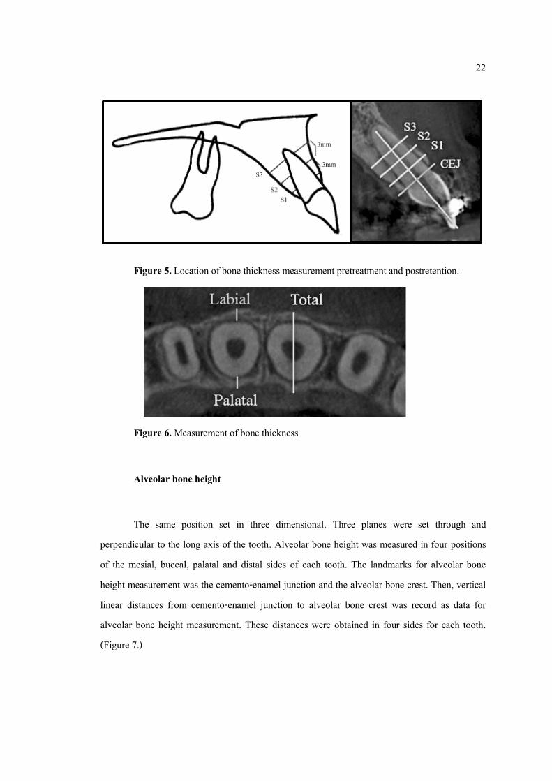
The same position set in three dimensional. Three planes were set through and
perpendicular to the long axis of the tooth. Alveolar bone height was measured in four positions of the mesial, buccal, palatal and distal sides of each tooth. The landmarks for alveolar bone height measurement was the cemento-enamel junction and the alveolar bone crest. Then, vertical linear distances from cemento-enamel junction to alveolar bone crest was record as data for alveolar bone height measurement. These distances were obtained in four sides for each tooth. (Figure 7.)
23
Figure 7. Alveolar bone height measurment
Root length
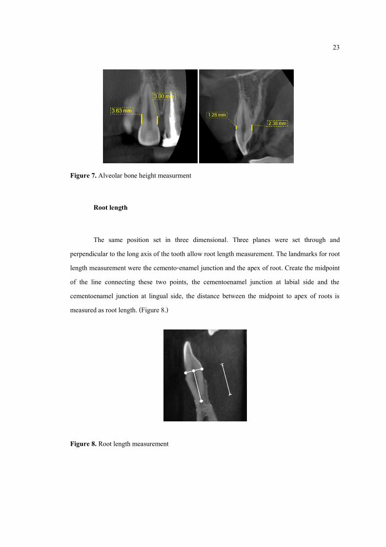
The same position set in three dimensional. Three planes were set through and
perpendicular to the long axis of the tooth allow root length measurement. The landmarks for root length measurement were the cemento-enamel junction and the apex of root. Create the midpoint of the line connecting these two points, the cementoenamel junction at labial side and the cementoenamel junction at lingual side, the distance between the midpoint to apex of roots is measured as root length. (Figure 8.)
Figure 8. Root length measurement
24
Statistical Analysis
Measurement errors
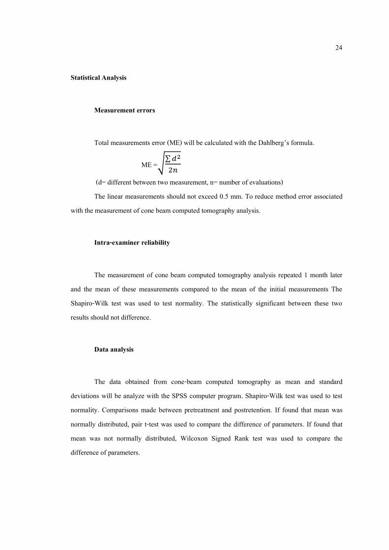
Total measurements error (ME) will be calculated with the Dahlberg’s formula.
ME =√∑𝑑2
2𝑛
(d= different between two measurement, n= number of evaluations) The linear measurements should not exceed 0.5 mm. To reduce method error associated
with the measurement of cone beam computed tomography analysis.
Intra-examiner reliability
The measurement of cone beam computed tomography analysis repeated 1 month later
and the mean of these measurements compared to the mean of the initial measurements The Shapiro-Wilk test was used to test normality. The statistically significant between these two results should not difference.
Data analysis
The data obtained from cone-beam computed tomography as mean and standard
deviations will be analyze with the SPSS computer program. Shapiro-Wilk test was used to test normality. Comparisons made between pretreatment and postretention. If found that mean was normally distributed, pair t-test was used to compare the difference of parameters. If found that mean was not normally distributed, Wilcoxon Signed Rank test was used to compare the difference of parameters.
25
CHAPTER 3
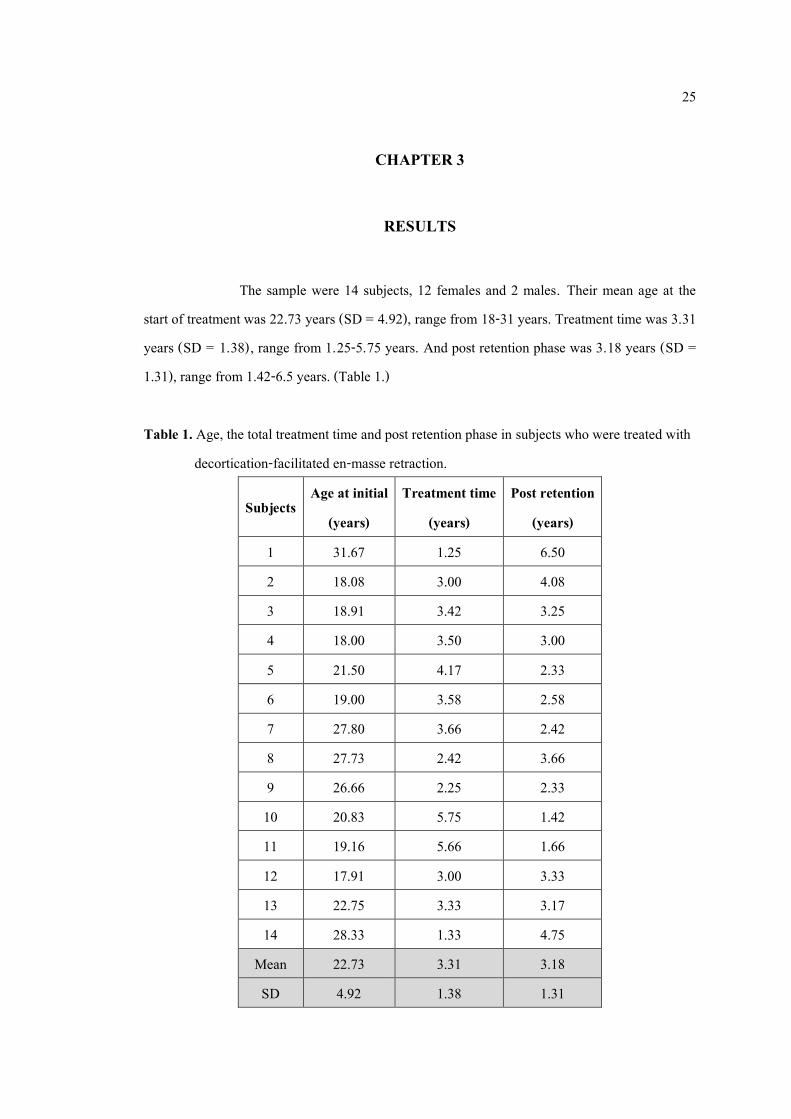
RESULTS The sample were 14 subjects, 12 females and 2 males. Their mean age at the start of treatment was 22.73 years (SD = 4.92), range from 18-31 years. Treatment time was 3.31 years (SD = 1.38), range from 1.25-5.75 years. And post retention phase was 3.18 years (SD = 1.31), range from 1.42-6.5 years. (Table 1.) Table 1. Age, the total treatment time and post retention phase in subjects who were treated with
decortication-facilitated en-masse retraction.
Subjects Age at initial
(years) Treatment time
(years) Post retention
(years) 1 31.67 1.25 6.50 2 18.08 3.00 4.08 3 18.91 3.42 3.25 4 18.00 3.50 3.00 5 21.50 4.17 2.33 6 19.00 3.58 2.58 7 27.80 3.66 2.42 8 27.73 2.42 3.66 9 26.66 2.25 2.33
10 20.83 5.75 1.42 11 19.16 5.66 1.66 12 17.91 3.00 3.33 13 22.75 3.33 3.17 14 28.33 1.33 4.75
Mean 22.73 3.31 3.18 SD 4.92 1.38 1.31
26
Cone beam computed tomography analysis Measurement error analysis
All measurements were repeated 1month apart and calculated to determine the intraobserver reliability. Cone beam computed tomography analysis were measured by same investigator. Systematic error was not significant. The random measurement error ( ME) was calculated according to Dalberg’ s formula. The linear measurement error was found to be less than 0.2 mm. No statistically significant differences were found between the 2 measurements at 2 different times for purpose of error testing.
Alveolar bone thickness
The results for changes in alveolar bone thickness as measured on the cone beam computed tomography from T0 to T1 are listed in Table 2-7.
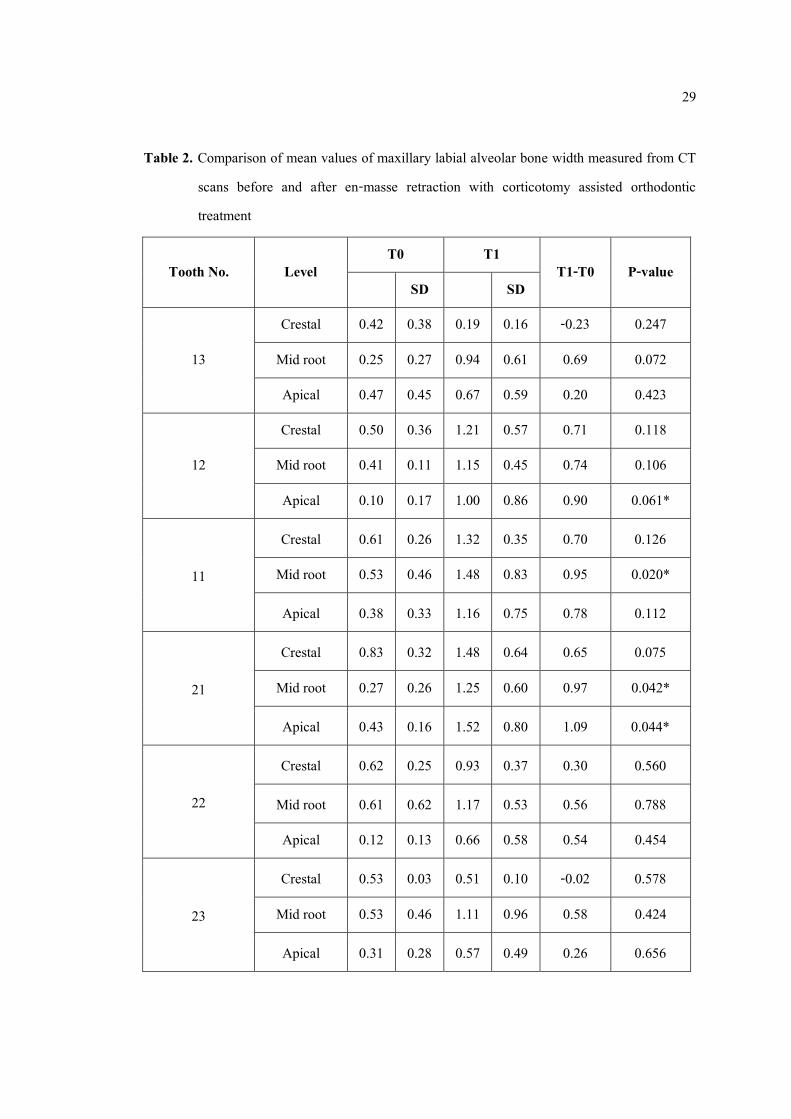
Maxillary labial alveolar bone width
The average increase in maxillary labial bone thickness was 0.58 mm. All levels found that the maxillary labial bone thickness was increased except crestal level of maxillary canines. The changes were not significant except apical level of maxillary right lateral incisor, mid root level of maxillary central incisors and apical level of maxillary left central incisor. (Table 2.)
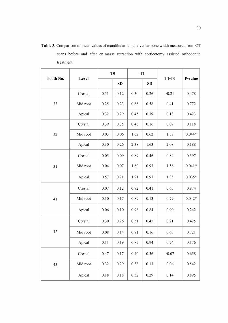
Mandibular labial alveolar bone width
The average increase in mandibular labial bone thickness was 0. 65 mm. All levels found that the mandibular labial bone thickness was increased except crestal level of mandibular canines. The changes were not significant except mid root level of mandibular left lateral incisor, mid root level of mandibular central incisors and apical level of mandibular left central incisor. (Table 3.)
27
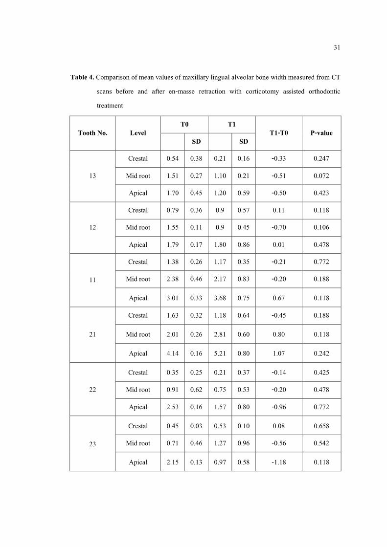
Maxillary lingual alveolar bone width
The average decrease in maxillary lingual bone thickness was 0. 18 mm. All levels found that the maxillary labial bone thickness was decreased except crestal and apical level of maxillary right lateral incisor, apical level of maxillary right central incisor, mid root and apical level of maxillary left central incisor and crestal level of maxillary left canine. The changes were not significant. (Table 4.)
Mandibular lingual alveolar bone width
The average increase in mandibular lingual bone thickness was 0.27 mm. All levels found that the mandibular lingual bone thickness was increased except crestal and mid root level of mandibular left canine and lateral incisor, mid root level of mandibular right lateral incisors and crestal level of mandibular right canine. The changes were not significant. (Table 5.)
Maxillary total alveolar bone width
The average increase in maxillary total bone thickness was 0.43 mm. All levels found that the maxillary labial bone thickness was increased except crestal and apical level of maxillary canine, crestal level of maxillary left central incisor and all levels of maxillary left lateral incisor. The changes were not significant except mid root and apical level of maxillary left central incisor. (Table 6.)
Mandibular total alveolar bone width
The average increase in mandibular total bone thickness was 0.91 mm. All levels found that the mandibular lingual bone thickness was increased except mid root level of mandibular left canine. The changes were not significant except all level of mandibular left central incisor, crestal and apical level of mandibular right central incisor, apical level of mandibular right lateral incisor and crestal level of mandibular right canine. (Table 7.)
28
Alveolar bone height
The results for changes in alveolar bone height as measured on the cone beam computed tomography from T0 to T1 are listed in Table 8-9.
Maxillary alveolar bone height
The average decrease in maxillary alveolar bone height was 0. 19 mm. The changes of all levels in maxillary anterior teeth were not significant. (Table 8.)
Mandibular alveolar bone height
The average decrease in maxillary alveolar bone height was 0. 18 mm. The changes of all levels in mandibular anterior teeth were not significant. (Table 9.)
Root length
The results for changes in root length as measured on the cone beam computed tomography from T0 to T1 are listed in Table 10. The root resorption was observed in range 1.06-1.42 mm. Average root resorption in all anterior teeth was 1.01 mm. The lowest root resorption was shown in upper canines and the highest was observed in lower canines. Significant root resorption occurred in all anterior teeth. During the assessment of each anterior teeth with CBCT. No considerable root damage from corticotomy was shown.
29
Table 2. Comparison of mean values of maxillary labial alveolar bone width measured from CT scans before and after en-masse retraction with corticotomy assisted orthodontic treatment
Tooth No. Level T0 T1
T1-T0 P-value
SD
SD
13
Crestal 0.42 0.38 0.19 0.16 -0.23 0.247
Mid root 0.25 0.27 0.94 0.61 0.69 0.072
Apical 0.47 0.45 0.67 0.59 0.20 0.423
12
Crestal 0.50 0.36 1.21 0.57 0.71 0.118
Mid root 0.41 0.11 1.15 0.45 0.74 0.106
Apical 0.10 0.17 1.00 0.86 0.90 0.061*
11
Crestal 0.61 0.26 1.32 0.35 0.70 0.126
Mid root 0.53 0.46 1.48 0.83 0.95 0.020*
Apical 0.38 0.33 1.16 0.75 0.78 0.112
21
Crestal 0.83 0.32 1.48 0.64 0.65 0.075
Mid root 0.27 0.26 1.25 0.60 0.97 0.042*
Apical 0.43 0.16 1.52 0.80 1.09 0.044*
22
Crestal 0.62 0.25 0.93 0.37 0.30 0.560
Mid root 0.61 0.62 1.17 0.53 0.56 0.788
Apical 0.12 0.13 0.66 0.58 0.54 0.454
23
Crestal 0.53 0.03 0.51 0.10 -0.02 0.578
Mid root 0.53 0.46 1.11 0.96 0.58 0.424
Apical 0.31 0.28 0.57 0.49 0.26 0.656
30
Table 3. Comparison of mean values of mandibular labial alveolar bone width measured from CT scans before and after en-masse retraction with corticotomy assisted orthodontic treatment
Tooth No. Level T0 T1
T1-T0 P-value
SD
SD
33
Crestal 0.51 0.12 0.30 0.26 -0.21 0.478
Mid root 0.25 0.23 0.66 0.58 0.41 0.772
Apical 0.32 0.29 0.45 0.39 0.13 0.423
32
Crestal 0.39 0.35 0.46 0.16 0.07 0.118
Mid root 0.03 0.06 1.62 0.62 1.58 0.044*
Apical 0.30 0.26 2.38 1.63 2.08 0.188
31
Crestal 0.05 0.09 0.89 0.46 0.84 0.597
Mid root 0.04 0.07 1.60 0.93 1.56 0.041*
Apical 0.57 0.21 1.91 0.97 1.35 0.035*
41
Crestal 0.07 0.12 0.72 0.41 0.65 0.874
Mid root 0.10 0.17 0.89 0.13 0.79 0.042*
Apical 0.06 0.10 0.96 0.84 0.90 0.242
42
Crestal 0.30 0.26 0.51 0.45 0.21 0.425
Mid root 0.08 0.14 0.71 0.16 0.63 0.721
Apical 0.11 0.19 0.85 0.94 0.74 0.176
43
Crestal 0.47 0.17 0.40 0.36 -0.07 0.658
Mid root 0.32 0.29 0.38 0.13 0.06 0.542
Apical 0.18 0.18 0.32 0.29 0.14 0.895
31
Table 4. Comparison of mean values of maxillary lingual alveolar bone width measured from CT scans before and after en-masse retraction with corticotomy assisted orthodontic treatment
Tooth No. Level T0 T1
T1-T0 P-value
SD
SD
13
Crestal 0.54 0.38 0.21 0.16 -0.33 0.247
Mid root 1.51 0.27 1.10 0.21 -0.51 0.072
Apical 1.70 0.45 1.20 0.59 -0.50 0.423
12
Crestal 0.79 0.36 0.9 0.57 0.11 0.118
Mid root 1.55 0.11 0.9 0.45 -0.70 0.106
Apical 1.79 0.17 1.80 0.86 0.01 0.478
11
Crestal 1.38 0.26 1.17 0.35 -0.21 0.772
Mid root 2.38 0.46 2.17 0.83 -0.20 0.188
Apical 3.01 0.33 3.68 0.75 0.67 0.118
21
Crestal 1.63 0.32 1.18 0.64 -0.45 0.188
Mid root 2.01 0.26 2.81 0.60 0.80 0.118
Apical 4.14 0.16 5.21 0.80 1.07 0.242
22
Crestal 0.35 0.25 0.21 0.37 -0.14 0.425
Mid root 0.91 0.62 0.75 0.53 -0.20 0.478
Apical 2.53 0.16 1.57 0.80 -0.96 0.772
23
Crestal 0.45 0.03 0.53 0.10 0.08 0.658
Mid root 0.71 0.46 1.27 0.96 -0.56 0.542
Apical 2.15 0.13 0.97 0.58 -1.18 0.118
32
Table 5. Comparison of mean values of mandibular lingual alveolar bone width measured from CT scans before and after en-masse retraction with corticotomy assisted orthodontic treatment
Tooth No. Level T0 T1
T1-T0 P-value
SD
SD
33
Crestal 1.35 0.12 1.08 0.26 -0.27 0.718
Mid root 3.58 0.23 3.40 0.58 -0.20 0.188
Apical 2.19 0.29 2.98 0.39 0.79 0.118
32
Crestal 0.45 0.35 0.30 0.16 -1.50 0.158
Mid root 1.35 0.06 0.83 0.62 -0.50 0.106
Apical 1.31 0.26 1.43 1.63 0.12 0.188
31
Crestal 0.13 0.09 0.60 0.46 0.47 0.247
Mid root 0.38 0.07 0.53 0.93 0.15 0.072
Apical 1.01 0.21 2.11 0.97 1.14 0.072
41
Crestal 0.13 0.12 0.60 0.41 0.47 0.423
Mid root 0.62 0.17 0.69 0.13 0.07 0.118
Apical 1.02 0.10 2.11 0.84 1.11 0.788
42
Crestal 0.11 0.26 0.78 0.45 0.30 0.454
Mid root 0.79 0.14 0.69 0.16 -0.10 0.578
Apical 0.95 0.19 2.17 0.94 1.22 0.424
43
Crestal 1.08 0.17 0.95 0.36 -0.13 0.442
Mid root 2.3 0.29 2.75 0.13 0.45 0.111
Apical 2.92 0.18 4.26 0.29 1.34 0.218
33
Table 6. Comparison of mean values of maxillary total alveolar bone width measured from CT scans before and after en-masse retraction with corticotomy assisted orthodontic treatment
Tooth No. Level T0 T1
T1-T0 P-value
SD
SD
13
Crestal 7.91 1.38 7.5 1.16 -0.41 0.247
Mid root 8.46 2.27 8.74 0.61 0.28 0.072
Apical 8.39 1.45 7.5 0.59 -0.89 0.423
12
Crestal 7.64 1.36 8.73 0.57 1.09 0.118
Mid root 7.2 1.11 7.62 1.45 0.42 0.106
Apical 5.86 2.17 7.23 0.86 1.37 0.188
11
Crestal 8.5 1.26 8.67 1.35 0.17 0.772
Mid root 8.38 1.46 9.01 0.83 0.63 0.423
Apical 9.38 1.33 10.51 0.75 1.13 0.423
21
Crestal 8.49 1.32 8.41 0.64 -0.08 0.118
Mid root 7.68 1.26 9.63 0.60 1.95 0.022*
Apical 8.70 1.16 11.09 0.80 2.39 0.011*
22
Crestal 7.16 1.25 6.85 1.37 -0.31 0.072
Mid root 7.66 1.62 7.60 1.53 -0.10 0.423
Apical 7.44 1.13 7.34 1.58 -0.10 0.118
23
Crestal 8.6 1.93 8.34 1.10 -0.26 0.106
Mid root 8.38 1.46 9.21 0.96 0.83 0.423
Apical 8.71 1.28 8.24 1.49 -0.47 0.118

34
Table 7. Comparison of mean values of mandibular total alveolar bone width measured from CT scans before and after en-masse retraction with corticotomy assisted orthodontic treatment
Tooth No. Level T0 T1
T1-T0 P-value
SD
SD
33
Crestal 9.97 1.12 10.14 0.16 0.17 0.247
Mid root 11.49 1.23 11.09 1.61 -0.4 0.072
Apical 9.12 1.29 9.66 2.59 0.54 0.423
32
Crestal 7.2 1.35 7.49 0.57 0.29 0.118
Mid root 7.27 1.06 8.24 1.45 0.97 0.106
Apical 6.8 1.26 8.31 1.86 1.51 0.188
31
Crestal 5.84 0.79 7.28 0.65 1.44 0.022*
Mid root 5.41 1.07 7.12 1.83 1.71 0.041*
Apical 5.82 1.21 7.21 1.75 1.39 0.001*
41
Crestal 5.65 0.82 6.92 0.64 1.27 0.011*
Mid root 5.63 1.17 6.60 1.60 0.97 0.442
Apical 5.25 1.10 7.31 1.80 1.06 0.010*
42
Crestal 5.32 0.56 6.97 0.37 0.65 0.423
Mid root 6.29 1.14 6.64 1.53 0.35 0.118
Apical 5.94 1.19 8.51 2.08 1.57 0.010*
43
Crestal 8.85 0.17 11.05 0.10 1.10 0.022*
Mid root 9.91 0.59 10.77 1.96 0.86 0.247
Apical 9.21 1.18 10.18 1.49 0.97 0.072

35
Table 8. Comparison of mean values of maxillary alveolar bone height measured from CT scans before and after en-masse retraction with corticotomy assisted orthodontic treatment
Tooth No. Level T0 T1
T1-T0 P-value
SD
SD
13
Labial 1.13 0.38 2.02 0.16 0.89 0.423
Lingual 1.51 0.27 1.01 0.61 -0.50 0.118
Mesial 1.51 0.45 1.27 0.59 -0.24 0.106
Distal 1.13 0.16 1.77 0.15 0.64 0.188
12
Labial 1.38 0.36 1.52 0.57 0.14 0.247
Lingual 1.40 0.11 0.75 0.45 -0.65 0.072
Mesial 1.01 0.17 1.50 0.86 0.49 0.423
Distal 1.13 0.15 0.10 0.15 -1.13 0.118
11
Labial 0.76 0.26 2.02 0.35 1.26 0.106
Lingual 2.12 0.46 2.05 0.83 -0.12 0.188
Mesial 1.51 0.33 2.02 0.75 0.51 0.423
Distal 1.26 0.16 2.30 0.58 1.04 0.118
21
Labial 1.13 0.32 2.02 0.64 0.89 0.106
Lingual 1.51 0.26 1.02 0.60 -0.51 0.188
Mesial 1.51 0.16 1.27 0.80 -0.24 0.247
Distal 1.13 0.32 1.77 0.61 0.64 0.072
22
Labial 1.38 0.25 1.52 0.37 0.14 0.423
Lingual 1.40 0.62 0.75 0.53 -0.65 0.118
Mesial 1.03 0.13 1.5 0.58 0.47 0.247
Distal 1.13 0.11 0.10 0.13 -1.13 0.072
23
Labial 0.76 0.03 1.98 0.10 1.22 0.423
Lingual 2.12 0.46 2.05 0.96 -0.07 0.118
Mesial 1.51 0.28 2.02 0.49 0.51 0.423
Distal 1.26 0.51 2.30 0.48 1.04 0.118
36
Table 9. Comparison of mean values of mandibular alveolar bone height measured from CT scans before and after en-masse retraction with corticotomy assisted orthodontic treatment
Tooth No. Level T0 T1
T1-T0 P-value
SD
SD
33
Labial 1.38 0.36 1.52 0.57 0.14 0.247
Lingual 1.40 0.11 0.75 0.45 -0.65 0.072
Mesial 1.20 0.17 1.50 0.86 0.30 0.423
Distal 1.13 0.15 0.10 0.15 -1.13 0.118
32
Labial 0.76 0.26 1.98 0.35 1.24 0.106
Lingual 2.12 0.46 2.02 0.83 -0.10 0.188
Mesial 1.51 0.33 2.02 0.75 0.51 0.423
Distal 1.26 0.16 2.30 0.58 1.04 0.118
31
Labial 1.13 0.32 2.02 0.64 0.89 0.106
Lingual 1.51 0.26 1.02 0.60 -0.49 0.188
Mesial 1.51 0.16 1.27 0.80 -0.24 0.247
Distal 1.13 0.32 1.77 0.61 0.64 0.072
41
Labial 1.38 0.25 1.52 0.37 0.14 0.423
Lingual 1.40 0.62 0.75 0.53 -0.65 0.118
Mesial 1.05 0.13 1.50 0.58 0.45 0.247
Distal 1.13 0.11 0.1 0.13 -1.13 0.072
42
Labial 0.76 0.03 1.82 0.10 1.06 0.423
Lingual 2.12 0.46 1.78 0.96 -0.34 0.118
Mesial 1.51 0.28 2.02 0.49 0.51 0.423
Distal 1.26 0.51 2.30 0.48 1.04 0.118
43
Labial 1.02 0.17 1.50 0.86 0.48 0.423
Lingual 1.13 0.15 1.10 0.15 -0.03 0.118
Mesial 0.76 0.26 1.55 0.35 0.79 0.106
Distal 2.12 0.46 2.02 0.83 -0.10 0.188
37
Table 10. Comparison of mean values of root length measured from CT scans before and after en-masse retraction with corticotomy assisted orthodontic treatment
Tooth No. T0 T1
T1-T0 P-value
SD
SD
13 16.99 1.36 16.29 1.35 0.70 0.011*
12 13.53 1.31 12.56 1.12 0.97 0.021*
11 13.82 1.53 12.95 1.42 0.87 0.041*
21 13.95 1.67 13.04 1.35 0.91 0.013*
22 13.24 1.26 12.21 1.32 1.03 0.010*
23 17.02 1.53 16.39 1.45 0.63 0.021*
33 16.87 1.11 15.30 1.19 1.57 0.001*
32 13.47 1.43 12.72 1.06 0.75 0.022*
31 13.13 1.12 11.90 1.21 1.23 0.001*
41 13.27 1.63 12.11 1.24 1.16 0.001*
42 13.35 1.37 12.63 1.17 0.72 0.032*
43 16.57 1.53 15.27 1.25 1.30 0.001*
38
CHAPTER 4
DISCUSSION
Orthodontic walls is the anatomical limits that set by the cortical bone.70 Orthodontic tooth movement are limited due to a dense cortical bone plate on the both labial and lingual sides around the roots of the anterior teeth. Moreover, patients with dentoalveolar protrusion usually have thin and bony defect before treatment,77,95 pushing the tooth against the thin cortical plate may cause alveolar bone defect and root resorption. So, retraction of the anterior teeth combined with corticotomy of the alveolar bone can offer an effective alternative with which to minimize the risk of movements of the anterior teeth.30
Lateral cephalograms presented only in midsagittal plane, the cortical plates and the symphysis are shown only in 2 dimensions and the orthodontic tooth movement effect cannot clearly be seen. So the limitation of the alveolar bone housing and the symphysis at the midline may be narrower than lateral cephalograms presented. For this reason, 3 dimensional evaluation is required to provide 3 dimensional displacement of alveolar bone changes. Cone beam computed tomography (CBCT) is now used to evaluated alveolar bone and CBCT scan have proven that statistically similar to histologic finding.96 In many study, the reduced alveolar bone thickness was found in the direction of tooth movement.97,98 Recently in CBCT study found that the maxillary and mandibular alveolar bone thickness decreased during orthodontic tooth movement in bimaxillary protrusion with 4 bicuspids extraction.22,77,99,100 Alveolar bone loss was shown more at the crestal region especially on the lingual side. Ten Hoeve and Mulie101 said that the cortical bone would be repaired within 6 months, no matter how much the tooth has been moved. In contrast to many studies77,102 when perforation was developed, newly thin cortical plate do not form in that patients. And complete repair may be seen if relapse occurs.97
There are many methods to measure alveolar bone before and after orthodontic treatment. It is difficult to accurately measure, since the teeth have been moved orthodontically. It is possible that they have been moved into a region with thicker alveolar housing or grafting area. The method of alveolar bone changes measurement perpendicular to the tooth axis cannot detect
39
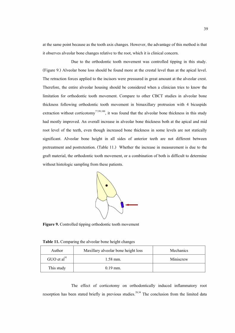
at the same point because as the tooth axis changes. However, the advantage of this method is that it observes alveolar bone changes relative to the root, which it is clinical concern. Due to the orthodontic tooth movement was controlled tipping in this study. (Figure 9.) Alveolar bone loss should be found more at the crestal level than at the apical level. The retraction forces applied to the incisors were pressured in great amount at the alveolar crest. Therefore, the entire alveolar housing should be considered when a clinician tries to know the limitation for orthodontic tooth movement. Compare to other CBCT studies in alveolar bone thickness following orthodontic tooth movement in bimaxillary protrusion with 4 bicuspids extraction without corticotomy77,99,100, it was found that the alveolar bone thickness in this study had mostly improved. An overall increase in alveolar bone thickness both at the apical and mid root level of the teeth, even though increased bone thickness in some levels are not statically significant. Alveolar bone height in all sides of anterior teeth are not different between pretreatment and postretention. (Table 11.) Whether the increase in measurement is due to the graft material, the orthodontic tooth movement, or a combination of both is difficult to determine without histologic sampling from these patients. Figure 9. Controlled tipping orthodontic tooth movement Table 11. Comparing the alveolar bone height changes
Author Maxillary alveolar bone height loss Mechanics GUO et al91 1.58 mm. Miniscrew This study 0.19 mm.
The effect of corticotomy on orthodontically induced inflammatory root
resorption has been stated briefly in previous studies.28,34 The conclusion from the limited data
40
indicated that corticotomy did not result in more root resorption than conventional orthodontic treatment. And there was less root resorption in the other studies.103 However, the accuracy of these studies could be questioned, they examined from periapical radiographs. Traditional 2D imaging has several limitations when root resorption was assessed. For example, magnification, superimposition of structures and lack of reproducibility and sensitivity of technique. These limitations of 2D imaging result in detecting only advanced root. CBCT allow better detection of root resorption than do 2D imaging. CBCT study found that corticotomy facilitated orthodontics resulted in an average of 0.6 mm of apical root loss, less than this study, whereas conventional orthodontics resulted in 1.5 mm.
Significant root resorption occurred in all anterior teeth in this study. However, an average of apical root loss is 1.01 mm still less than 1.5 mm. found in conventional orthodontic treatment.104 There were individual variations in root resorption values among subjects and this was expected because of the impact of individual susceptibility on the root resorption process. The biologic mechanisms behind the acceleration of tooth movement in corticotomy and their effects on root resorption are complex and unclear. RAP increases the turnover rate of alveolar bone and the periodontal ligament by increasing the activity of associated cells. An increased bone turnover rate leads to a reduction in the resistance of teeth moving through alveolar bone. By reducing the resistance and the hyalinization, corticotomy could reduce the amount of root resorption, but the relationship between alveolar bone density and the root resorption process is not clear.105 Some studies found that increased bone turnover and reduced bone density favor remodeling of bone over root surface.106 Another study shown that root resorption increased due to increased bone turnover.107 The treatment duration is positively correlated with root resorption, although some recent studies do not agree.108,109 Corticotomy accelerated tooth movement, so this should reduce in root resorption. Future research still required to provide information how the various biologic mechanisms of corticotomy facilitated tooth movement interact and influence the overall root resorption process.
The drawback of this study is that it was a retrospective study to look at only those patients who had corticotomy combined with orthodontic treatment. There were no matched controls who had similar alveolar bone and orthodontic treatment done without the use of the corticotomy and bone grafting procedure.

41
Overall, corticotomy seems to be a safe procedure that can shorten treatment time and potentially provide for better post-operative stability by slightly increasing the width of the alveolar housing. So in patients with thin buccal or lingual alveolar bone, corticotomy may be alternative procedure to reduce treatment time and possible periodontal complications such as dehiscence, fenestration and recession. However, the disadvantages such as cost and complications of an additional surgical procedure must be considered and discussed with the patient.
42
CHAPTER 5
CONCLUSION
Corticotomy not only accelerates the orthodontic treatment but also provides the advantage of increased alveolar width. This study showed an average increase in alveolar bone width following corticotomy. However, there were also sites, maxillary lingual bone thickness, in which there was a decrease in alveolar width. No significant alveolar bone height changes were found. But significant root resorption occurred in all anterior teeth.
43
1. The average increase in maxillary and mandibular labial bone thickness was 0.58 mm. 0.65 mm. respectively. The average decrease in maxillary lingual bone thickness was 0.18 mm. but found average increase 0.27 mm. in mandibular lingual bone thickness.
2. No significant alveolar bone height changes were found in all anterior teeth. The average decrease in maxillary and mandibular alveolar bone height was 0.19 mm., 0.18 mm. respectively.
3. Significant root resorption occurred in all anterior teeth. Average root resorption in all anterior teeth was 1.01 mm.
44
Thesis title Alveolar Bone and Root Length Changes Following En-masse Retraction with Corticotomy assisted Orthodontic Treatment
Author Miss Satinee Narupakorn Major Program Oral Health Sciences Academic Year 2017
ABSTRACT Introduction: Corticotomy is an effective method of accelerating the orthodontic treatment.
Corticotomy also has been proposed to increase the volume of the alveolar process and decrease
root resorption. Objectives: To compare alveolar bone and root length before and after en-masse
retraction with corticotomy in bimaxillary protrusion patient. Materials and methods: Fourteen
subjects were selected from patients who required the removal of first premolars in upper and
lower arches, en masse retraction and corticotomy as a part of their orthodontic treatment. Cone
beam computed tomography analysis was used to compare alveolar bone width at crestal, mid-
root and apical level, alveolar bone height at mesial, distal, labial and lingual sides and root length
changes between pretreatment and postretention in all anterior teeth Results: The average
increase in maxillary and mandibular labial bone thickness was 0.58 mm. 0.65 mm. respectively.
The average decrease in maxillary lingual bone thickness was 0.18 mm. but found average
increase 0.27 mm. in mandibular lingual bone thickness. No significant alveolar bone height
changes were found in all anterior teeth. The average decrease in maxillary and mandibular
alveolar bone height was 0.19 mm., 0.18 mm. respectively. Significant root resorption occurred in
all anterior teeth. Average root resorption in all anterior teeth was 1.01 mm. Conclusions:
Corticotomy not only accelerates the orthodontic treatment but also provides the advantage of
increased alveolar width. This study showed an average increase in alveolar bone width following
corticotomy. However, there were also sites, maxillary lingual bone thickness, in which there was
a decrease in alveolar width. No significant alveolar bone height changes were found. But
significant root resorption occurred in all anterior teeth.
43
REFERENCES
1. Dibart, S., J.D. Sebaoun, and J. Surmenian, Piezocision: a minimally invasive,
periodontally accelerated orthodontic tooth movement procedure. Compend Contin Educ Dent 2009. 30(6): p. 342-4, 346, 348-50.
2. Hernandez-Alfaro, F. and R. Guijarro-Martinez, Endoscopically assisted tunnel approach for minimally invasive corticotomies: a preliminary report. J Periodontol 2012. 83(5): p. 574-80.
3. Abdallah, M.N. and C. Flores-Mir, Are interventions for accelerating orthodontic tooth movement effective? Evid Based Dent 2014. 15(4): p. 116-7.
4. Long, H., et al., Interventions for accelerating orthodontic tooth movement: a systematic review. Angle Orthod 2013. 83(1): p. 164-71.
5. Roblee, R.D., S.L. Bolding, and J.M. Landers, Surgically facilitated orthodontic therapy: a new tool for optimal interdisciplinary results. Compend Contin Educ Dent 2009. 30(5): p. 264-75.
6. Kole, H., Surgical operations on the alveolar ridge to correct occlusal abnormalities. Oral
Surg Oral Med Oral Pathol 1959. 12(5): p. 515-29. 7. Wilcko, M.T., W.M. Wilcko, and N.F. Bissada, An Evidence-Based Analysis of
Periodontally Accelerated Orthodontic and Osteogenic Techniques: A Synthesis of Scientific Perspectives. Semin Orthod 2008. 14(4): p. 305-316.
8. Wilcko, W.M., et al., Rapid orthodontics with alveolar reshaping: two case reports of decrowding. Int J Periodontics Restorative Dent 2001. 21(1): p. 9-19.
9. Rothe, L.E., et al., Trabecular and cortical bone as risk factors for orthodontic relapse. Am J Orthod Dentofacial Orthop 2006. 130(4): p. 476-84.
10. Wilcko, M.T., et al., Accelerated osteogenic orthodontics technique: a 1-stage surgically facilitated rapid orthodontic technique with alveolar augmentation. J Oral Maxillofac Surg 2009. 67(10): p. 2149-59.
11. Nazarov AD, F.D., Wilcko WM, Wilcko MT, Improved retention following corticotomy using ABO objective grading system. J Dent Res 2004. 83.
44
12. Skountrianos HS, F.D., Wilcko WM, Wilcko MT, Maxillary arch decrowding and stability with and without corticotomy-facilitated orthodontics. J Dent Res 2004. 83.
13. Makki, L., et al., Mandibular irregularity index stability following alveolar corticotomy and grafting: A 10-year preliminary study: Mandibular Irregularity Index Stability. Angle
Orthod 2014. 14. Hassan, A.H., et al., Corticotomy-assisted orthodontic treatment. A systematic review of
the biological basis and clinical effectiveness. Saudi Med J 2015. 36(7): p. 794-801. 15. Baloul, S.S., et al., Mechanism of action and morphologic changes in the alveolar bone in
response to selective alveolar decortication-facilitated tooth movement. Am J Orthod Dentofacial Orthop 2011. 139(4): p. S83-101.
16. Farrow, A.L., K. Zarrinnia, and K. Azizi, Bimaxillary protrusion in black Americans--an esthetic evaluation and the treatment considerations. Am J Orthod Dentofacial Orthop 1993. 104(3): p. 240-50.
17. Scheideman, G.B., et al., Wound healing after anterior and posterior subapical osteotomy. J Oral Maxillofac Surg 1985. 43(6): p. 408-16.
18. Chung, K.R., M.Y. Oh, and S.J. Ko, Corticotomy-assisted orthodontics. J Clin Orthod 2001. 35(5): p. 331-9.
19. Fuhrmann, R., Three-dimensional interpretation of periodontal lesions and remodeling during orthodontic treatment. Part III. J Orofac Orthop 1996. 57(4): p. 224-37.
20. Hwang, C.J., and Moon, The limitation of alveolar bone remodeling during retraction of the upper anterior teeth. Korean J Orthod 2001. 31(1): p. 97-105.
21. Vannier, M.W., Craniofacial computed tomography scanning: technology, applications and future trends. Orthod Craniofac Res 2003. 6 Suppl 1: p. 23-30; discussion 179-82.
22. Guo, Q.Y., et al., Three-dimensional evaluation of upper anterior alveolar bone dehiscence after incisor retraction and intrusion in adult patients with bimaxillary protrusion malocclusion. J Zhejiang Univ Sci B 2011. 12(12): p. 990-7.
23. Kim, S.J., Y.G. Park, and S.G. Kang, Effects of Corticision on paradental remodeling in orthodontic tooth movement. Angle Orthod 2009. 79(2): p. 284-91.
45
24. Alghamdi, A.S., Corticotomy facilitated orthodontics: Review of a technique. Saudi Dent J 2010. 22(1): p. 1-5.
25. Weltman, B., et al., Root resorption associated with orthodontic tooth movement: a systematic review. Am J Orthod Dentofacial Orthop 2010. 137(4): p. 462-76; discussion 12A.
26. Abass, S., Hartsfield, J., Orthodontics and external apical root resorption. Semin Orthod 2007. 13: p. 246-256.
27. Al-Qawasmi, R.A., et al., Genetic predisposition to external apical root resorption in orthodontic patients: linkage of chromosome-18 marker. J Dent Res 2003. 82(5): p. 356-60.
28. Shoreibah EA, S.A., Attia MS, Abu-Seida SM., Corticotomy facilitated orthodontics in adults using a further modified technique. J Int Acad Periodontol 2012(14): p. 97-104.
29. Frost, H.M., The regional acceleratory phenomenon: a review. Henry Ford Hosp Med J 1983. 31(1): p. 3-9.
30. Lee, J.K., K.R. Chung, and S.H. Baek, Treatment outcomes of orthodontic treatment, corticotomy-assisted orthodontic treatment, and anterior segmental osteotomy for bimaxillary dentoalveolar protrusion. Plast Reconstr Surg 2007. 120(4): p. 1027-36.
31. Nowzari, H., F.K. Yorita, and H.C. Chang, Periodontally accelerated osteogenic orthodontics combined with autogenous bone grafting. Compend Contin Educ Dent 2008. 29(4): p. 200-6.
32. Sebaoun, J.D., et al., [Alveolar osteotomy and rapid orthodontic treatments]. Orthod Fr 2007. 78(3): p. 217-25.
33. Wilcko, W. and M.T. Wilcko, Accelerating tooth movement: the case for corticotomy-induced orthodontics. Am J Orthod Dentofacial Orthop 2013. 144(1): p. 4-12.
34. Shoreibah, E.A., et al., Clinical and radiographic evaluation of bone grafting in corticotomy-facilitated orthodontics in adults. J Int Acad Periodontol 2012. 14(4): p. 105-13.
35. B, T., et al., Dentoalveolar bone density changes following corticotomy facilitated orthodontics. J Dent Res 2002. 20: p. 301.
46
36. Iino, S., et al., Acceleration of orthodontic tooth movement by alveolar corticotomy in the dog. Am J Orthod Dentofacial Orthop 2007. 131(4): p. 448.
37. Brudvik, P. and P. Rygh, The initial phase of orthodontic root resorption incident to local compression of the periodontal ligament. Eur J Orthod 1993. 15(4): p. 249-63.
38. Wang, B., et al., Augmented corticotomy-assisted presurgical orthodontics of class III malocclusions: a cephalometric and cone-beam computed tomography study. J Craniofac Surg 2013. 24(6): p. 1886-90.
39. Kim, S.H., et al., Corticotomy-assisted decompensation for augmentation of the mandibular anterior ridge. Am J Orthod Dentofacial Orthop 2011. 140(5): p. 720-31.
40. Hassan, A.H., A.A. Al-Fraidi, and S.H. Al-Saeed, Corticotomy-assisted orthodontic treatment: review. Open Dent J 2010. 4: p. 159-64.
41. Amit, G., et al., Periodontally accelerated osteogenic orthodontics (PAOO) - a review. J Clin Exp Dent 2012. 4(5): p. 292-6.
42. Costantino, P.D., et al., Bone healing and bone substitutes. Facial Plast Surg 2002. 18(1): p. 13-26.
43. S, B., B. RB, and K. H, Current Therapy in Oral and Maxillofacial Surgery. 2012, St. Louis, MO: Elsevier.
44. Wikesjo, U.M., et al., RhBMP-2/alphaBSM induces significant vertical alveolar ridge augmentation and dental implant osseointegration. Clin Implant Dent Relat Res 2002. 4(4): p. 174-82.
45. Kubler, N., et al., Osteoinductive, morphologic, and biomechanical properties of autolyzed, antigen-extracted, allogeneic human bone. J Oral Maxillofac Surg 1993. 51(12): p. 1346-57.
46. Klinge, B., et al., Osseous response to implanted natural bone mineral and synthetic hydroxylapatite ceramic in the repair of experimental skull bone defects. J Oral Maxillofac Surg 1992. 50(3): p. 241-9.
47. Misch, C.E. and F. Dietsh, Bone-grafting materials in implant dentistry. Implant Dent 1993. 2(3): p. 158-67.
47
48. Elsalanty, M.E. and D.G. Genecov, Bone grafts in craniofacial surgery. Craniomaxillofac Trauma Reconstr 2009. 2(3): p. 125-34.
49. Bauer, T.W. and G.F. Muschler, Bone graft materials. An overview of the basic science. Clin Orthop Relat Res 2000(371): p. 10-27.
50. Virolainen, P., et al., Expression of matrix genes during incorporation of cancellous bone allografts and autografts. Clin Orthop Relat Res 1995(317): p. 263-72.
51. Skoglund, A., P. Hising, and C. Young, A clinical and histologic examination in humans of the osseous response to implanted natural bone mineral. Int J Oral Maxillofac Implants 1997. 12(2): p. 194-9.
52. Thompson, N. and J.A. Casson, Experimental onlay bone grafts to the jaws. A preliminary study in dogs. Plast Reconstr Surg 1970. 46(4): p. 341-9.
53. G., F., Current Concept Reviews: Bone Grafts. J Bone Joint Surg Am 1987. 69(A): p. 786-90.
54. Zitzmann, N.U., R. Naef, and P. Scharer, Resorbable versus nonresorbable membranes in combination with Bio-Oss for guided bone regeneration. Int J Oral Maxillofac Implants 1997. 12(6): p. 844-52.
55. Simion, M., et al., Effect of different microstructures of e-PTFE membranes on bone regeneration and soft tissue response: a histologic study in canine mandible. Clin Oral Implants Res 1999. 10(2): p. 73-84.
56. Reynolds, M.A., et al., The efficacy of bone replacement grafts in the treatment of periodontal osseous defects. A systematic review. Ann Periodontol 2003. 8(1): p. 227-65.
57. Jensen, S.S. and H. Terheyden, Bone augmentation procedures in localized defects in the alveolar ridge: clinical results with different bone grafts and bone-substitute materials. Int
J Oral Maxillofac Implants 2009. 24 Suppl: p. 218-36. 58. Smukler, H., L. Landi, and R. Setayesh, Histomorphometric evaluation of extraction
sockets and deficient alveolar ridges treated with allograft and barrier membrane: a pilot study. Int J Oral Maxillofac Implants 1999. 14(3): p. 407-16.
59. Rose L, M.B., Genco R., Periodontics: Medicine, Surgery and Implants. 1st ed. 2004, St. Louis: Mosby, Inc.
48
60. Yared, K.F., E.G. Zenobio, and W. Pacheco, Periodontal status of mandibular central incisors after orthodontic proclination in adults. Am J Orthod Dentofacial Orthop 2006. 130(1): p. 6 e1-8.
61. Kassab, M.M. and R.E. Cohen, The etiology and prevalence of gingival recession. J Am
Dent Assoc 2003. 134(2): p. 220-5. 62. Nunn, M.E. and S.K. Harrel, The effect of occlusal discrepancies on periodontitis. I.
Relationship of initial occlusal discrepancies to initial clinical parameters. J Periodontol 2001. 72(4): p. 485-94.
63. Harrel, S.K. and M.E. Nunn, The effect of occlusal discrepancies on periodontitis. II. Relationship of occlusal treatment to the progression of periodontal disease. J Periodontol 2001. 72(4): p. 495-505.
64. Sanavi, F., A.S. Weisgold, and L.F. Rose, Biologic width and its relation to periodontal biotypes. J Esthet Dent 1998. 10(3): p. 157-63.
65. Rupprecht, R.D., et al., Prevalence of dehiscences and fenestrations in modern American skulls. J Periodontol 2001. 72(6): p. 722-9.
66. Mostafa, Y.A., F.A. El Sharaby, and A.R. El Beialy, Do alveolar bone defects merit orthodontists' respect? World J Orthod 2009. 10(1): p. 16-20.
67. Lost, C., Depth of alveolar bone dehiscences in relation to gingival recessions. J Clin Periodontol 1984. 11(9): p. 583-9.
68. Handelman, C.S., The anterior alveolus: its importance in limiting orthodontic treatment and its influence on the occurrence of iatrogenic sequelae. Angle Orthod 1996. 66(2): p. 95-109; discussion 109-10.
69. Bollen, A.M., et al., The effects of orthodontic therapy on periodontal health: a systematic review of controlled evidence. J Am Dent Assoc 2008. 139(4): p. 413-22.
70. Mathews, D.P. and V.G. Kokich, Managing treatment for the orthodontic patient with periodontal problems. Semin Orthod 1997. 3(1): p. 21-38.
71. Newman, G.V., M.J. Goldman, and R.A. Newman, Mucogingival orthodontic and periodontal problems. Am J Orthod Dentofacial Orthop 1994. 105(4): p. 321-7.
49
72. Melsen, B. and D. Allais, Factors of importance for the development of dehiscences during labial movement of mandibular incisors: a retrospective study of adult orthodontic patients. Am J Orthod Dentofacial Orthop 2005. 127(5): p. 552-61; quiz 625.
73. Artun, J. and D. Grobety, Periodontal status of mandibular incisors after pronounced orthodontic advancement during adolescence: a follow-up evaluation. Am J Orthod
Dentofacial Orthop 2001. 119(1): p. 2-10. 74. Gracco, A., et al., Upper incisor position and bony support in untreated patients as seen on
CBCT. Angle Orthod 2009. 79(4): p. 692-702. 75. Sarikaya, S., et al., Changes in alveolar bone thickness due to retraction of anterior teeth.
Am J Orthod Dentofacial Orthop 2002. 122(1): p. 15-26. 76. Rungcharassaeng, K., et al., Factors affecting buccal bone changes of maxillary posterior
teeth after rapid maxillary expansion. Am J Orthod Dentofacial Orthop 2007. 132(4): p. 428 e1-8.
77. Garib, D.G., et al., Periodontal effects of rapid maxillary expansion with tooth-tissue-borne and tooth-borne expanders: a computed tomography evaluation. Am J Orthod Dentofacial Orthop 2006. 129(6): p. 749-58.
78. Vandenberghe, B., R. Jacobs, and J. Yang, Diagnostic validity (or acuity) of 2D CCD versus 3D CBCT-images for assessing periodontal breakdown. Oral Surg Oral Med Oral Pathol Oral Radiol Endod 2007. 104(3): p. 395-401.
79. Mengel, R., et al., Digital volume tomography in the diagnosis of periodontal defects: an in vitro study on native pig and human mandibles. J Periodontol 2005. 76(5): p. 665-73.
80. Mengel, R., B. Kruse, and L. Flores-de-Jacoby, Digital volume tomography in the diagnosis of peri-implant defects: an in vitro study on native pig mandibles. J Periodontol 2006. 77(7): p. 1234-41.
81. Kasaj, A. and B. Willershausen, Digital volume tomography for diagnostics in periodontology. Int J Comput Dent 2007. 10(2): p. 155-68.
82. Tyndall, D.A. and S. Rathore, Cone-beam CT diagnostic applications: caries, periodontal bone assessment, and endodontic applications. Dent Clin North Am 2008. 52(4): p. 825-41, vii.
50
83. Brezniak, N. and A. Wasserstein, Root resorption after orthodontic treatment: Part 1. Literature review. Am J Orthod Dentofacial Orthop 1993. 103(1): p. 62-6.
84. Wehrbein, H., R.A. Fuhrmann, and P.R. Diedrich, Human histologic tissue response after long-term orthodontic tooth movement. Am J Orthod Dentofacial Orthop 1995. 107(4): p. 360-71.
85. Janson, G., et al., Comparative radiographic evaluation of the alveolar bone crest after orthodontic treatment. Am J Orthod Dentofacial Orthop 2003. 124(2): p. 157-64.
86. Aass, A.M. and P. Gjermo, Changes in radiographic bone level in orthodontically treated teenagers over a 4-year period. Community Dent Oral Epidemiol 1992. 20(2): p. 90-3.
87. Bondemark, L., Interdental bone changes after orthodontic treatment: a 5-year longitudinal study. Am J Orthod Dentofacial Orthop 1998. 114(1): p. 25-31.
88. Molen, A.D., Considerations in the use of cone-beam computed tomography for buccal bone measurements. Am J Orthod Dentofacial Orthop 2010. 137(4 Suppl): p. S130-5.
89. Sun, Z., et al., Effect of bone thickness on alveolar bone-height measurements from cone-beam computed tomography images. Am J Orthod Dentofacial Orthop 2011. 139(2): p. e117-27.
90. Ketcham, A preliminary report of an investigation of apical root resorption of permanent teeth. Int. J. Orthod. Oral Surg 1927. 13: p. 97-115.
91. Malmgren, O., et al., Root resorption after orthodontic treatment of traumatized teeth. Am J Orthod 1982. 82(6): p. 487-91.
92. Sameshima, G.T. and P.M. Sinclair, Predicting and preventing root resorption: Part II. Treatment factors. Am J Orthod Dentofacial Orthop 2001. 119(5): p. 511-5.
93. Brezniak, N. and A. Wasserstein, Orthodontically induced inflammatory root resorption. Part II: The clinical aspects. Angle Orthod 2002. 72(2): p. 180-4.
94. Wilcko W, F.D., Bouquot J, Wilcko M., Rapid orthodontic decrowding with alveolar augmentation: case report. World J Orthod 2003. 4: p. 197-205.
95. Buschang PH, C.P., Ruso S., Accelerating tooth movement with corticotomies: is it possible and desirable? Semin Orthod 2012. 18: p. 286-94.
51
96. Mostafa YA, F.M., Mehanni S, ElBokle NN, Heider AM., Comparison of corticotomy-facilitated vs standard tooth-movement techniques in dogs with miniscrews as anchor units. Am J Orthod Dentofacial Orthop 2009(136): p. 570-7.
97. Aboul-Ela SM, E.-B.A., El-Sayed KM, Selim EM, El-Mangoury NH, Mostafa YA., Miniscrew implant-supported maxillary canine retraction with and without corticotomy facilitated orthodontics. Am J Orthod Dentofacial Orthop 2011(139): p. 252-9.
98. Al-Naoum F, H.M., Al-Jundi A., Does alveolar corticotomy accelerate orthodontic tooth movement when retracting upper canines? A split-mouth design randomized controlled trial. J Oral Maxillofac Surg 2014(72): p. 1880-9.
99. Nahm, K.Y., et al., Alveolar bone loss around incisors in Class I bidentoalveolar protrusion patients: a retrospective three-dimensional cone beam CT study. Dentomaxillofac Radiol 2012. 41(6): p. 481-8.
100. Fuhrmann, R.A., et al., Assessment of the dentate alveolar process with high resolution computed tomography. Dentomaxillofac Radiol 1995. 24(1): p. 50-4.
101. Wainwright, W.M., Faciolingual tooth movement: its influence on the root and cortical plate. Am J Orthod 1973. 64(3): p. 278-302.
102. Vardimon, A.D., E. Oren, and Y. Ben-Bassat, Cortical bone remodeling/tooth movement ratio during maxillary incisor retraction with tip versus torque movements. Am J Orthod Dentofacial Orthop 1998. 114(5): p. 520-9.
103. Ahn, H.W., S.C. Moon, and S.H. Baek, Morphometric evaluation of changes in the alveolar bone and roots of the maxillary anterior teeth before and after en masse retraction using cone-beam computed tomography. Angle Orthod 2013. 83(2): p. 212-21.
104. Nayak Krishna, U.S., et al., Changes in alveolar bone thickness due to retraction of anterior teeth during orthodontic treatment: a cephalometric and computed tomography comparative study. Indian J Dent Res 2013. 24(6): p. 736-41.
105. Ten Hoeve, A. and R.M. Mulie, The effect of antero-postero incisor repositioning on the palatal cortex as studied with laminagraphy. J Clin Orthod 1976. 10(11): p. 804-22.
106. Karring, T., et al., Bone regeneration in orthodontically produced alveolar bone dehiscences. J Periodontal Res 1982. 17(3): p. 309-15.
52
107. Akay, M.C., et al., Enhanced effect of combined treatment with corticotomy and skeletal anchorage in open bite correction. J Oral Maxillofac Surg 2009. 67(3): p. 563-9.
108. Dudic, A., et al., Detection of apical root resorption after orthodontic treatment by using panoramic radiography and cone-beam computed tomography of super-high resolution. Am J Orthod Dentofacial Orthop 2009. 135(4): p. 434-7.
109. Brezniak, N. and A. Wasserstein, Orthodontically induced inflammatory root resorption. Part II: The clinical aspects. Angle Orthod 2002. 72(2): p. 180-4.
110. Goldie, R.S. and G.J. King, Root resorption and tooth movement in orthodontically treated, calcium-deficient, and lactating rats. Am J Orthod 1984. 85(5): p. 424-30.
111. Engstrom, C., G. Granstrom, and B. Thilander, Effect of orthodontic force on periodontal tissue metabolism. A histologic and biochemical study in normal and hypocalcemic young rats. Am J Orthod Dentofacial Orthop 1988. 93(6): p. 486-95.

53
APPENDICES
54
55
56
ใบเชิญชวน
เร่ือง ขอเชิญเขา้ร่วมโครงการวิจยัเร่ือง การเปล่ียนแปลงกระดูกเบา้ฟันและรากฟัน ภายหลงัจาก
การผา่ตดักระดูกทึบร่วมกบัการรักษาทางทนัตกรรมจดัฟันในผูป่้วยท่ีมีลกัษณะฟันยืน่ในขากรรไกรบนและล่าง
เรียน ผูส้นใจเขา้ร่วมโครงการวิจยัฯทุกท่าน
ขา้พเจา้ ทพญ.สาธินี นฤปกร นักศึกษาระดบัปริญญาโท สาขาทนัตกรรมจดัฟัน ภาควิชาทันตกรรมป้องกัน คณะทันตแพทยศาสตร์ มหาวิทยาลยัสงขลานครินทร์ ขอแจ้งรายละเอียดเก่ียวกบัโครงการวิจยัและขอเชิญชวนท่านผูส้นใจเขา้ร่วม โครงการฯ ดงัน้ี
โครงการวิจยัน้ีจดัท าข้ึนเพ่ือศึกษา การเปล่ียนแปลงกระดูกเบา้ฟันและรากฟัน ภายหลงัจากการผา่ตดักระดูกทึบร่วมกบัการรักษาทางทนัตกรรมจดัฟันในผูป่้วยท่ีมีลกัษณะฟันยืน่ในขากรรไกรบนและล่าง และมีกระดูกทางดา้นใกลเ้พดานและใกลล้ิ้นบาง ซ่ึงเป็นลกัษณะทางกายวิภาคท่ีจ ากดัการเคล่ือนฟัน ซ่ึงเดิมหากรักษาทางทันตกรรมจัดฟันทั่วไปเพียงอย่างเดียว จะก่อให้เกิดภาวะแทรกซอ้น ไดแ้ก่ การละลายของรากฟัน การละลายของกระดูกเบา้ฟันและเหงือกร่นได ้อีกทั้งยงัใชเ้วลาในการรักษานาน ซ่ึงจากการศึกษาท่ีผา่นมาพบว่า วิธีการรักษาท่ีใชใ้นการศึกษาน้ีสามารถลดภาวะแทรกซ้อนดังกล่าวได ้และใช้เวลาในการรักษาน้อยกว่าการรักษาทางทนัตกรรมจดัฟันทัว่ไปเพียงอยา่งเดียว
ผูเ้ขา้ร่วมโครงการวิจยัฯ จะเขา้รับบริการท่ีคณะทนัตแพทยศาสตร์ มหาวิทยาลยัสงขลานครินทร์ โดยเป็นผูป่้วยท่ีไดรั้บการรักษาทางทนัตกรรมจดัฟันร่วมกบัการถอนฟันกรามนอ้ยซ่ีท่ี 1 ในขากรรไกรบนและล่าง มีการดึงฟันหนา้บนเขา้ในปริมาณท่ีมากกว่าความหนาของกระดูกดา้นล้ินท่ีระดบัปลายรากฟัน โดยการรักษาทางทนัตกรรมจดัฟันเสร็จส้ินแลว้เป็นระยะเวลาอยา่งนอ้ย 6 เดือน ผูป่้วยจะไดรั้บการถ่ายภาพรังสีคอมพิวเตอร์ จ านวน 1 คร้ัง เพ่ือประเมินการเปล่ียนแปลงกระดูกเบา้ฟันและรากฟัน ภายหลงัจากการผา่ตดักระดูกทึบร่วมกบัการรักษาทางทนัตกรรมจดัฟัน ซ่ึงผูเ้ขา้ร่วมวิจยัไม่ตอ้งรับผดิชอบค่าใชจ่้ายในส่วนน้ี
ทั้งน้ี ในขั้นตอนของการเก็บขอ้มูล ผูเ้ขา้ร่วมวิจยัจ าเป็นตอ้งไดรั้บการถ่ายภาพรังสีคอมพิวเตอร์เพ่ิมเติมนอกเหนือไปจากการเก็บขอ้มูลพ้ืนฐานตามปกติ ซ่ึงการถ่ายภาพรังสีแต่ละคร้ังนั้นมีความเส่ียงท่ีจะเกิดอนัตรายต่อผูเ้ขา้ร่วมวจิยันอ้ยมาก เน่ืองจากปริมาณรังสีท่ีไดรั้บในแต่ละคร้ังของการถ่ายนั้นนอ้ยมาก โดยใหส้วมเส้ือตะกัว่เพ่ือป้องกนัรังสีใหแ้ก่ผูเ้ขา้ร่วมวิจยัทุกรายและทุก
57
คร้ังท่ีตอ้งถ่ายภาพรังสี และพยายามใหมี้ความผดิพลาดในการถ่ายภาพรังสีนอ้ยท่ีสุด เพ่ือลดปริมาณรังสีท่ีผูเ้ขา้ร่วมวิจยัจะไดรั้บโดยไมจ่ าเป็น
ถา้ท่านตดัสินใจเขา้ร่วมโครงการวิจยัฯน้ี จะมีขั้นตอนของการวจิยัท่ีจ าเป็นตอ้งขอความร่วมมือของท่าน คือ การถ่ายภาพรังสีคอมพิวเตอร์ ภายหลงัจากการผา่ตดักระดูกทึบร่วมกบัการรักษาทางทนัตกรรมจดัฟัน โดยผูเ้ขา้ร่วมโครงการฯตอ้งมารับการถ่ายภาพรังสีคอมพิวเตอร์ ณ คณะทนัตแพทยศาสตร์ มหาวิทยาลยัสงขลานครินทร์ โดยไม่เสียค่าใชจ่้าย
หากท่านมีขอ้สงสยัประการใดหรือเกิดผลขา้งเคียงจากการวิจยัจะสามารถติดต่อกบั ทพญ.สาธินี นฤปกร ไดท่ี้ภาควิชาทนัตกรรมป้องกนั คณะทนัตแพทยศาสตร์ มหาวิทยาลยัสงขลานครินทร์ หมายเลขโทรศพัท ์ 081-7022077 หรือ เม่ือมีปัญหาใดๆ เกิดข้ึนเน่ืองจากการท าวิจยัในเร่ืองน้ี ขา้พเจา้สามารถร้องเรียนไดท่ี้คณบดี คณะทนัตแพทยศาสตร์ มหาวิทยาลยัสงขลานครินทร์ อ.หาดใหญ่ จ.สงขลา 90112 หมายเลขโทรศพัท ์074-287500
ไม่ว่าท่านจะเขา้ร่วมในโครงการวจิยัน้ีหรือไม่ ท่านจะยงัคงไดรั้บการรักษาท่ีดีเช่นเดียวกบัผูป่้วยคนอ่ืนๆ และถา้ท่านตอ้งการท่ีจะถอนตวัออกจากการศึกษาน้ีเม่ือใด ท่านก็สามารถกระท าได้อยา่งอิสระ ถา้ท่านมีค าถามใดๆ ก่อนท่ีจะตดัสินใจก่อนเขา้ร่วมโครงการน้ี โปรดซกัถามคณะผูว้ิจยัไดอ้ยา่งเต็มท่ี
ขอแสดงความนบัถือ
ทพญ.สาธินี นฤปกร
58
แบบยินยอมเข้าร่วมการศึกษา
โครงการวิจยัเร่ือง การเปล่ียนแปลงกระดูกเบา้ฟันและรากฟัน ภายหลงัจากการผา่ตดั
กระดูกทึบร่วมกบัการรักษาทางทนัตกรรมจดัฟัน ในผูป่้วยท่ีมีลกัษณะฟันยืน่ในขากรรไกรบน และล่าง
วนัท่ี_______เดือน___________________พ.ศ._______
ขา้พเจา้_____________________________________________________อาย_ุ_______ปี
อาศยัอยูบ่า้นเลขท่ี________________หมู_่__________________ถนน______________________
ต าบล_______________________อ าเภอ______________________จงัหวดั__________________
ไดรั้บการอธิบายถึงวตัถุประสงคข์องการวิจยั วิธีการวิจยั อนัตรายท่ีอาจเกิดข้ึนจากการวิจยั
รวมทั้งประโยชน์ท่ีจะเกิดข้ึนจากการวิจยัอยา่งละเอียดและมีความเขา้ใจดีแลว้
หากขา้พเจา้มีขอ้สงสยัประการใด หรือเกิดผลขา้งเคียงจากการวจิยัจะสามารถติดต่อกบั
ทพญ.สาธินี นฤปกร ไดท่ี้ ภาควิชาทนัตกรรมป้องกนั คณะทนัตแพทยศาสตร์
มหาวิทยาลยัสงขลานครินทร์ หมายเลขโทรศพัท ์ 074-429876 หรือ เม่ือมีปัญหาใดๆ เกิดข้ึน
เน่ืองจากการท าวิจยัในเร่ืองน้ี ขา้พเจา้สามารถร้องเรียนไดท่ี้คณบดี คณะทนัตแพทยศาสตร์
มหาวิทยาลยั สงขลานครินทร์ อ.หาดใหญ่ จ.สงขลา 90112 หมายเลขโทรศพัท ์074-287500
หากผูว้ิจยัมีขอ้มูลเพ่ิมเติมทั้งทางดา้นประโยชน์และโทษท่ีเก่ียวข้องกบัการวิจัยน้ี ผูว้ิจ ัย
จะแจง้ใหข้า้พเจา้ทราบอยา่งรวดเร็วโดยไม่มีปิดบงั
ขา้พเจา้มีสิทธ์ิท่ีจะของดการเขา้ร่วมโครงการวิจยั โดยจะแจง้ใหท้ราบล่วงหนา้โดยการงด
การเขา้ร่วมการวิจยัน้ีจะไม่มีผลต่อการไดรั้บบริการหรือการรักษาท่ีขา้พเจา้จะไดรั้บแต่อยา่งใด
ผูว้ิจยัรับรองว่าจะเก็บขอ้มูลเฉพาะท่ีเก่ียวกบัตวัขา้พเจา้เป็นความลบั จะไม่เปิดเผยขอ้มูล
หรือผลการวิจยัของขา้พเจา้เป็นรายบุคคลต่อสาธารณชน จะเปิดเผยไดใ้นรูปท่ีเป็นสรุปผลการวิจยั
หรือการเปิดเผยขอ้มูลต่อผูม้ีหนา้ท่ีท่ีเก่ียวขอ้งกบัการสนบัสนุนและก ากบัดูแลการวิจยั
59
ขา้พเจ้าได้อ่านข้อความข้างตน้แลว้ และมีความเขา้ใจดีทุกประการ จึงได้ลงนามในใบ
ยนิยอมน้ีดว้ยความเต็มใจ โดยผูว้ิจยัไดใ้ห้ส าเนาแบบยนิยอมท่ีลงนามแลว้กบัขา้พเจา้เพื่อเก็บไวเ้ป็น
หลกัฐานจ านวน 1 ชุด ลงช่ือ..………………………………………ผูย้นิยอม ( ) ลงช่ือ……………………………………….หวัหนา้โครงการ ( ทพญ. สาธินี นฤปกร ) ลงช่ือ..………………………………………พยาน ( ) ลงช่ือ..………………………………………พยาน ( )
60
VITAE
Name Miss. Satinee Narupakorn Student ID 5810820028 Educational Attainment
Degree Name of Institution Year of Graduation Doctor of Dental Surgery Chulalongkorn University 2008 Work-Position and Address
Dental Department, Nuea Khlong hospital, Krabi, Thailand
List of Publication and Proceeding Narupakorn S., Samruajbenjakun B. Root Length Changes following En masse-Retraction with
Corticotomy-assisted Orthodontic Treatment using Cone-beam Computed Tomography Analysis. Proceedings of The 7th Conference on the topic CREATIVE SCHOOLS; 2017 February 24; Nakhon Si Thammarat, Thailand. Southern College of Technology; 2017.