Embed Size (px)
Citation preview

Supplement to July/August 2015
AMD, RVO, & DME Update:
Case Studies in Patient & Practice
Management
Jonathan L. Prenner, MD, ModeratorK. Bailey Freund, MDRichard S. Kaiser, MD
Elias Reichel, MD
CME ACTIVITY
A CME activity jointly provided by The Dulaney Foundation, Retina Today, and New Retina MD.
Supported through an unrestricted educational grant by Regeneron Pharmaceuticals.
RetinaMDNew

CONTENT SOURCEThis continuing medical education (CME) activity is based on
content from a roundtable held in May 2015 in Denver, CO.
STATEMENT OF NEEDThe impact of vision loss due to macular degeneration and
ocular manifestations of diabetes is a major public health bur-den facing our society, given the large aging population at risk for sight-threatening ocular conditions. Significant challenges lie ahead in addressing the needs of patients at risk for vision loss, as well as the impact on society that comes with an increasing population with impaired vision. Macular degeneration, retinal vein occlusion (RVO), and diabetic macular edema (DME) pres-ent related physiologic problems for retinal specialists and oph-thalmologists in the management of these conditions. Given the coincident systemic disease associated with diabetic retinopathy, the present and predicted burden and health care impact is substantial.
According to the 2012 Vision Problems in the US Report from the Prevent Blindness America Foundation,1 diabetic retinopathy affects more than 7.6 million people aged 40 years and older. This contributes significantly to the more than $50 billion in direct economic costs due to vision disorders in people aged 40 years and older.
As new therapies enter the market, therapeutic options and dosing strategies can be affected by the cost of treatment, which continues to be a major factor in treatment planning.2 Clinicians need to consider multiple options for therapy in order to prop-erly gauge the right treatment plan for any given patient’s needs.
More broadly, the American Diabetes Association con-firms that more than 150 million people across the world are affected by diabetes. By 2025, that number will likely increase to 324 million, including 35% who are expected to develop diabetic retinopathy (DR).3 For nearly 20 years, DR has been documented as the leading cause of blindness and decreased vision-related quality of life in working-age Americans.4-6
DME frequently follows the onset of nonproliferative diabetic retinopathy, resulting from abnormal capillary permeability and associated leakage of fluid leakage into the tissue of the retina. In recent years, new understanding of the pathophysiology of DME has focused researchers on the involvement of intracel-lular hyperglycemia, which induces free radicals (oxidative stress), protein kinase C (PKC) activation, and the formation of advanced glycation end products (AGE).7 This process results in hypoxia, ischemia, inflammation, and alteration of vitreomacular interface. Inflammation produces an increase in VEGF production, endothe-lial dysfunction, leukocyte adhesion, and PKC production. In fact, DR is now considered to be a state of low-grade inflammation.8
When not treated properly, which is often the case, DME progresses to proliferative DR (PDR) and retinal neovasculariza-tion, hemorrhaging, and permanent loss of vision. Approximately 50% of untreated patients with PDR will become blind within 5 years of the initial diagnosis.9 Such outcomes can frequently be avoided, however. Both decreased vision and decreased vision-related quality of life may be modified by treatment, including new modalities that provide practitioners with the flexibility of customizing management based on each patient’s needs.
Focal macular laser photocoagulation (FML) has been the primary treatment for DME for more than 2 decades. The Early Treatment Diabetic Retinopathy Study (ETDRS) outcomes focused on the preservation of vision, finding a 50% reduction in the likelihood of severe vision loss with grid-style FML.10 In 2010, the Diabetic Retinopathy Clinical Research Network (DRCR.net) reported a 10-letter gain in nearly one-third of patients treated with a laser, but 19% of subjects experienced progressive visual loss.11 Emerging therapies have recently shown promise, both as adjunctive and possibly first-line alternatives to laser therapy. Several pharmaceutical therapies for DME are currently in clinical development, the majority of which are intravitreally injected anti-inflammatory or anti-angiogenic agents. These include VEGF inhibitors such as ranibizumab (Lucentis, Genentech), aflibercept (VEGF Trap-Eye, Regeneron), and pegaptanib sodium (Macugen, OSI Eyetech), as well as intravitreal delivery systems, which release corticosteroids, such as fluocinolone acetonide (Iluvien, Alimera), dexamethasone (Ozurdex, Allergan), and triamcinolone aceton- ide (I-vation SurModics).
A full knowledge of the dynamics of retinal therapeutic treat- ment options will be beneficial for arming both specialists and general ophthalmologists who use these drugs with a more complete understanding when counseling patients and initiating treatment. It is expected that providing this education would remove a potential barrier to greater acceptance of this area of disease management. Finally, in the interest of providing more complete care to patients, arming clinicians with current insight into the management strategies for retinal therapeutics may assist in the reduction of treatment complications and prevent further loss of vision.
1. Prevent Blindness America, 2012 Vision Problems in the U.S. Available at: http://www.preventblindness.org/ sites/default/files/national/documents/state-fact-sheets/VPUS%2BCOV_FS_US.pdf.2. Akpek EK, Smith RA. Overview of age-related ocular conditions managed care Iimplications of age-related ocular conditions. Available at: http://www.ajmc.com/publications/supplement/2013/ACE011_13may_AgingEye/ ACE011_13May_AgingEye_Akpek.3. Hogan P, Dall T, Nikolov P; American Diabetes Association. Economic costs of diabetes in the US in 2002. Diabetes Care. 2003;26(3):917-932.4. National Institutes of Health. Diabetes in America, 2nd ed. National Institutes of Health, National Institute of Diabetes and Digestive and Kidney Diseases: Bethesda, MD, 1995.5. Klein R, Knudtson MD, Lee KE, et al. The Wisconsin epidemiologic study of diabetic retinopathy: XVIII. The 14-year incidence and progression of diabetic retinopathy and associated risk factors in type 1 diabetes. Ophthalmology. 1998;105:1801-1815.6. Hariprasad SM, Mieler WF, Grassi M, et al. Vision-related quality of life in patients with diabetic macular oedema. Br J Ophthalmol. 2008;92:89-92.7. Bhagat N, Grigorian RA, Tutela A, Zarbin MA. Diabetic macular edema: pathogenesis and treatment. Surv Ophthalmol. 2009;54(1):1-32.8. Singh A, Stewart JM. Pathophysiology of diabetic macular edema. Int Ophthalmol Clin. 2009;49(2):1-11.9. Hamilton AMP, Ulbig MW, Polkinghorne P. Management of diabetic retinopathy. BMJ Publishing Group: London, 1996. 10. Photocoagulation for diabetic macular edema. Early treatment diabetic retinopathy study report number 1. Early treatment diabetic retinopathy study research group. Arch Ophthalmol. 1985;103:1796-1806.11. Diabetic Retinopathy Clinical Research Network. Factors associated with improvement and worsening of visual acuity 2 years after focal/grid photocoagulation for diabetic macular edema. Ophthalmology. 2010;117:946-953.
TARGET AUDIENCEThis certified CME activity is designed for retina specialists
and general ophthalmologists involved in the management of patients with retinal disease.
LEARNING OBJECTIVESUpon completion of this activity, participants should be able to:
Jointly provided by The Dulaney Foundation, Retina Today, and New Retina MD Supported through an unrestricted educational grant by Regeneron PharmaceuticalsRelease Date: August 1, 2015Expiration Date: August 1, 2016
2 SUPPLEMENT TO RETINA TODAY/NEW RETINA MD JULY/AUGUST 2015
JULY/AUGUST 2015 SUPPLEMENT TO RETINA TODAY/NEW RETINA MD 3
• Describe the current epidemiology of major retinal diseases, including AMD, RVO, and DME
• Assess clinical studies involving new approaches to treat DME
• Utilize expert case examples to differentiate between clini-cal study dosing protocols and alternative dosing schedules
• Interpret retinal imaging case examples describing the treat-ment of DME
• Explore the management of treatment complications and secondary therapies
• Educate patients on ophthalmic implications of systemic diabetes management
• Demonstrate optimized patient flow, inventory flow, and office efficiency
ACCREDITATION STATEMENTThis activity has been planned and implemented in accor-
dance with the accreditation requirements and policies of the Accreditation Council for Continuing Medical Education (ACCME) through the joint providership of The Dulaney Foundation, Retina Today, and New Retina MD. The Dulaney Foundation is accredited by the ACCME to provide continuing medical education for physicians.
CREDIT DESIGNATION STATEMENTThe Dulaney Foundation designates this enduring material for
a maximum of 1 AMA PRA Category 1 Credit.™ Physicians should claim only the credit commensurate with the extent of their participation in the activity.
FACULTYJonathan L. Prenner, MDRetina Vitreous Center Robert Wood Johnson Medical School of the University of Medicine and DentistryNew Brunswick, New Jersey
K. Bailey Freund, MDVitreous- Retina-Macula Consultants of New YorkNew York University School of Medicine New York Presbyterian HospitalManhattan Eye Ear & Throat InstituteLenox Hill HospitalNew York, New York
Richard S. Kaiser, MDThomas Jefferson UniversityWills Eye HospitalPhiladelphia, Pennsylvania
Elias Reichel, MD New England Eye CenterTufts University School of MedicineBoston, Massachusetts
DISCLOSURE POLICY It is the policy of The Dulaney Foundation that faculty and
other individuals who are in the position to control the content of this activity disclose any real or apparent conflict of interests relating to the topics of this educational activity. The Dulaney
Foundation has full policies in place that will identify and resolve all conflicts of interest prior to this educational activity.
The following faculty/staff members have the following finan-cial relationships with commercial interests:
Jonathan L. Prenner, MD, has had a financial agreement or affiliation during the past year with the following com-mercial interests in the form of Consultant/Advisory Board: Genentech; Neurotech LLC; Ophthotech; PanOptica; Regeneron Pharmaceuticals; and ThromboGenics NV. Stockholder: Ophthotech.
K. Bailey Freund, MD, has had a financial agreement or affili-ation during the past year with the following commercial inter-ests in the form of Consultant/Advisory Board: Alimera Sciences; Genentech; Heidelberg Engineering GmbH; Ohr Pharmaceutical; Optos; Optovue; and ThromboGenics.
Richard S. Kaiser, MD, has had a financial agreement or affiliation during the past year with the following commercial interests in the form of Consultant/Advisory Board: Ophthotech; PanOptica; and Regeneron Pharmaceuticals. Stockholder: Ophthotech.
Elias Reichel, MD, has had a financial agreement or affiliation during the past year with the following commercial interests in the form of Consultant/Advisory Board: Genentech; Regeneron Pharmaceuticals; and ThromboGenics NV.
Cheryl Cavanaugh, MS, director of operations, The Dulaney Foundation; Michelle Dalton, medical writer; and Melanie Lawler, PhD, reviewer, have no financial relationships with com-mercial interests.
OFF-LABEL STATEMENTThis educational activity may contain discussion of published
and/or investigational uses of agents that are not indicated by FDA. The opinions expressed in the educational activity are those of the faculty. Please refer to the official prescribing infor-mation for each product for discussion of approved indications, contraindications, and warnings
DISCLAIMERThe views and opinions expressed in this educational activ-
ity are those of the faculty and do not necessarily represent the views of The Dulaney Foundation, Retina Today, New Retina MD, or Regeneron Pharmaceuticals.
To view the digital version of this print activity, scan the QR code or visit www.eyetube.net/cme-center and choose the appropriate title. Part 1 of this activity, published in the January/February issue of Retina Today, is also available at www.eyetube.net/cme-center.
eyetube.net

4 SUPPLEMENT TO RETINA TODAY/NEW RETINA MD JULY/AUGUST 2015
AMD, RVO, & DME Update: Case Studies in Patient & Practice Management
Supported by an educational grant from Regeneron Pharmaceuticals.
AMD, RVO, & DME Update: Case Studies in Patient & Practice Management
AGE-RELATED MACULAR DEGENERATIONJonathan L. Prenner, MD: What is your primary
approach to the management of patients with neovascular AMD? Do you currently prefer a PRN, monthly, or a treat-and-extend (TAE) hybrid regimen?
Elias Reichel, MD: The study results from the CATT2 and IVAN3 trials have shown that PRN is an appropriate way of managing these patients. These patients need close monitoring at the beginning of their disease treatment process. They require monthly evaluations for the first 6 to 9 months to determine the cyclic nature of their disease. There are some patients who might need one, two, or three injections in the first 6 months and then may not need another injection for a very long period of time. But there are some patients who show improvement 2 months after the injection and then they recur, and that pattern seems to hold. So you have patients who need injections every month, every 2 months, and every 3 months. The only way to determine this pattern is by using the PRN approach, withholding treatment, and not doing TAE.
Dr. Prenner: Do you utilize a PRN approach if the patient has inactive disease demonstrated only on optical coherence tomography (OCT), or do you require multi-modal imaging to determine neovascular quiescence for this purpose?
Dr. Reichel: I use OCT only for this purpose. It is possible that a patient might never need another injection after the
first round, but rare. This happens in 4% of the patients with a single injection, and in 14% or 15% of the patients with one, two, or three injections.2,3
Dr. Prenner: It is also possible that your patient may not need further injections at all, particularly if you were able to initiate therapy early enough in their disease process when their neovascularization has not matured.
Dr. Reichel: Yes. I want to stress that there is a cyclic nature to the disease for some of these patients. It is almost like clockwork. We schedule some patients to come in every 3 months, and they are going to have a wet macula. By following them every month, we have found their dis-ease can remain inactive for some time.
Dr. Prenner: Does everyone else agree with this?
K. Bailey Freund, MD: I have moved away from a gener-ic TAE regimen to a more individualized strategy. I often base my treatment regimen on the initial presentation. I use multimodal imaging to define the anatomic lesion sub-type of the neovascular process. I classify these sub-retinal pigment epithelial (RPE) lesions as type 1, sub-retinal lesions as type 2, predominantly intra-retinal lesions type 3 or retinal angiomatous proliferation (RAP). Some eyes will present with mixed lesions. In my experience, I have seen that if you catch type 3 lesions very early, they may regress and not recur for extended periods. So, in these cases, I choose to go to PRN immediately. Like Dr. Reichel, I am
It is well accepted that the US population is aging rapidly, and about 20% of the population will be older than 65 years by 2030.
The high incidence and prevalence rate of leading retina conditions, including age-related macular degeneration (AMD), dia-betic macular edema (DME), and retinal vein occlusion (RVO), has placed a new set of challenges on physicians taking care of these diseases, and it appears that we will continue to become more efficient to meet the coming demand for our services.
Prospective trial results demonstrate that frequent, fixed dosing schedules return excellent outcomes for patients, but we often modify those treatment regimens with what we hope are similar outcomes. Patients demand variable dosing strategies, but to date there is not necessarily enough evidence to suggest modifying treatment regimens is more beneficial (or at least not deleterious) to patient outcomes. One should keep in mind Dr. Mant’s comments from The Lancet, that the “paradox of the clinical trial is that it is the best way to assess whether an intervention works, but is arguably the worst way to assess who will benefit from it.” 1
In this roundtable discussion, we tackle that dilemma, and proffer advice on how we each determine what treatment strategy will work best for our patients.
—Jonathan L. Prenner, MD

JULY/AUGUST 2015 SUPPLEMENT TO RETINA TODAY/NEW RETINA MD 5
AMD, RVO, & DME Update: Case Studies in Patient & Practice Management
also concerned that chronic vascular endothelial growth factor (VEGF) suppression in type 3 might be associated with a greater rate of geographic atrophy (GA) with long-term frequent anti-VEGF dosing. However, when a patient with a very large type 1 fibrovascular pigment epithelial detachment (PED) comes in with some polypoidal ele-ments, I am a less concerned about long-term GA and more concerned about the possibility of catastrophic hem-orrhage with under-treatment. My experience shows that such a patient is likely to need chronic therapy and also probably has a higher risk of bleeding if we do OCT-guided therapy. Those are cases for which I prefer TAE. I currently use both TAE and PRN, based on my experience dealing with the different types of initial lesions.
Dr. Prenner: When you decide to treat someone in that highly tailored manner, is dose-loading a part of the regimen?
Dr. Freund: I do not feel that a monthly loading sequence of injection is needed in every case. There will be an article out in Retina soon that is a consensus manu-script by experts from many countries on what constitutes the basic TAE regimen. We treat monthly until we reach what we consider is a maximal response, which is either an absence of fluid or no further improvement of fluid or ana-tomic findings on OCT. This could potentially be achieved at the second visit following the first injection. If you were to see a patient one month following an initial injection, and he had an absence of fluid and normal visual acuity, you might treat and extend the treatment interval at that point. In this scenario, the loading would consist of only two monthly injections, and you would start to extend the treatment by a week or two after that.
Dr. Prenner: So it sounds like you attempt to individual-ize treatment frequency based on CNVM sub-type; do you also tailor your drug choice by disease sub-type?
Dr. Freund: Yes. As mentioned previously, I often prefer PRN in patients with type 3 lesions and TAE in many eyes with type 1 lesions. When I do choose a TAE regimen, it is fairly generic, regardless of the lesion sub-type. As far as the drug choice is concerned, there is a variant of neovascular AMD that occurs in patients who have choroidal features similar to what we see in central serous chorioretinopathy (CSC). These eyes have a pachychoroid or thick choroid with large dilated choroidal vessels, which I call patchy ves-sels. These patients may develop the same type of polyps we see in patients with polypoidal choroidal vasculopathy. Eyes with these polyps seem to respond better to afliber-cept (Eylea, Regeneron) than to the other anti-VEGF drugs. But, for most other anatomic lesion sub-types, I do not have a specific choice of drug.
Dr. Reichel: What about patients with type 2 lesions?
Dr. Freund: We looked at several hundred newly diag-nosed cases of neovascular AMD and found that pure type 2 lesions are extremely uncommon in our population of predominantly Caucasian patients. We found that when we looked with fluorescein angiography (FA) and OCT togeth-er, only 9% of the newly diagnosed cases of neovascular AMD had pure type 2 lesions, and these pure type 2 lesions occurred almost exclusively in patients who had reticular pseudodrusen; we hope to publish these data shortly. You can, however, get mixed lesions with a sub-retinal type 2 component. I just do not see enough type 2 lesions to have specific guidelines on how to treat them. I have observed that if you catch these lesions early, they tend to respond very robustly to the anti-VEGF agents, but may often leave behind some component of sub-retinal fibrosis that can lead to recurrence. When these lesions recur, there is pre-sumably a risk of a sub-retinal hemorrhage due to their location in this space. The risk is particularly worrisome if the lesion is close to the fovea. In a monocular patient, particularly one who has lost vision in the fellow eye from neovascular AMD, I would likely choose a TAE regimen to minimize the risk of hemorrhagic recurrence. But, fortu-nately, most eyes recur with fluid, not blood.
Dr. Prenner: But it is a disaster when it happens.
Dr. Prenner: Dr. Kaiser, how do you approach your stan-dard AMD patient?
Richard S. Kaiser, MD: A year or 2 ago, I preferred the TAE regimen with a standard loading dose of 3 to 4 month-ly treatments. I still have a bias toward the TAE approach, but I will move to the extended period quicker now. If I see a great response after one injection, I will immediately start to extend. I will also repeat the angiogram periodically, especially after 4 to 5 months of treatment, to make sure I am extending these treatments far enough apart and that I am not completely relying on the OCT to show me what is happening.
Dr. Prenner: If we look at the Preferences and Trends (PAT) survey, 2 out of 3 physicians managing AMD are
“A year or 2 ago, I preferred the TAE
regimen with a standard loading
dose of 3 to 4 monthly treatments.
I still have a bias toward the TAE
approach, but I will move to the
extended period quicker now.”—Dr. Kaiser
6 SUPPLEMENT TO RETINA TODAY/NEW RETINA MD JULY/AUGUST 2015
AMD, RVO, & DME Update: Case Studies in Patient & Practice Management
Presented by K. Bailey Freund, MD
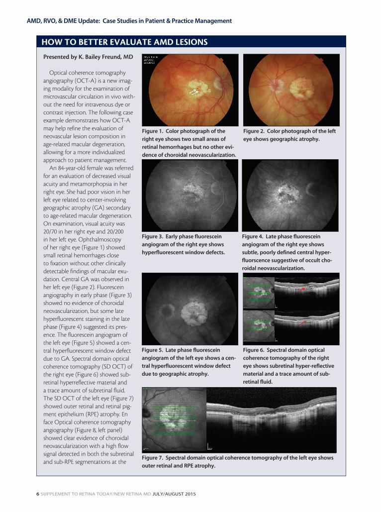
Optical coherence tomography angiography (OCT-A) is a new imag-ing modality for the examination of microvascular circulation in vivo with-out the need for intravenous dye or contrast injection. The following case example demonstrates how OCT-A may help refine the evaluation of neovascular lesion composition in age-related macular degeneration, allowing for a more individualized approach to patient management.
An 84-year-old female was referred for an evaluation of decreased visual acuity and metamorphopsia in her right eye. She had poor vision in her left eye related to center-involving geographic atrophy (GA) secondary to age-related macular degeneration. On examination, visual acuity was 20/70 in her right eye and 20/200 in her left eye. Ophthalmoscopy of her right eye (Figure 1) showed small retinal hemorrhages close to fixation without other clinically detectable findings of macular exu-dation. Central GA was observed in her left eye (Figure 2). Fluorescein angiography in early phase (Figure 3) showed no evidence of choroidal neovascularization, but some late hyperfluorescent staining in the late phase (Figure 4) suggested its pres-ence. The fluorescein angiogram of the left eye (Figure 5) showed a cen-tral hyperfluorescent window defect due to GA. Spectral domain optical coherence tomography (SD OCT) of the right eye (Figure 6) showed sub-retinal hyperreflective material and a trace amount of subretinal fluid. The SD OCT of the left eye (Figure 7) showed outer retinal and retinal pig-ment epithelium (RPE) atrophy. En face Optical coherence tomography angiography (Figure 8, left panel) showed clear evidence of choroidal neovascularization with a high flow signal detected in both the subretinal and sub-RPE segmentations at the
HOW TO BETTER EVALUATE AMD LESIONS
Figure 1. Color photograph of the
right eye shows two small areas of
retinal hemorrhages but no other evi-
dence of choroidal neovascularization.
Figure 2. Color photograph of the left
eye shows geographic atrophy.
Figure 3. Early phase fluorescein
angiogram of the right eye shows
hyperfluorescent window defects.
Figure 4. Late phase fluorescein
angiogram of the right eye shows
subtle, poorly defined central hyper-
fluorscence suggestive of occult cho-
roidal neovascularization.
Figure 5. Late phase fluorescein
angiogram of the left eye shows a cen-
tral hyperfluorescent window defect
due to geographic atrophy.
Figure 6. Spectral domain optical
coherence tomography of the right
eye shows subretinal hyper-reflective
material and a trace amount of sub-
retinal fluid.
Figure 7. Spectral domain optical coherence tomography of the left eye shows
outer retinal and RPE atrophy.
JULY/AUGUST 2015 SUPPLEMENT TO RETINA TODAY/NEW RETINA MD 7
AMD, RVO, & DME Update: Case Studies in Patient & Practice Management
utilizing a classic TAE approach, which is something that was considered very forward thinking 5 years ago.4 I think we now agree that this is an individualized disease that requires very careful attention and therapy tailored to the particular patient and the nuances of their clinical presen-tation. In my opinion, AMD should be treated by a retina specialist who has access to multimodal imaging and can utilize advanced imaging like OCT angiography, indocyanine green (ICG) angiography, and FA, and characterize the sub-type of lesion. The specialist can then tailor the treatment to that particular patient. Overtreatment in certain cases can lead to sub-optimal outcomes. Clinical trials teach us how to treat a population of patients the best possible way, but it is suboptimal in terms of figuring out how to treat any individual patient who is sitting in front of you.1
Dr. Freund: It will be interesting to see this year’s PAT survey to see if TAE peaked last year. I predict it might have.
Dr. Reichel: I agree. But another issue here is that TAE means different things to different people, and the survey does not quantify the definition.
Dr. Freund: Yes, I believe last year’s PAT survey was set up so that a TAE response meant that you were exclusively treating with TAE. I think more retinal specialists are begin-ning to use individualized treatment approaches.
Dr. Kaiser: What are people doing later in the disease?
What do you do when you start to see atrophy develop-ing 2 years after the diagnosis, and yet you still see some activity on OCT? How are you changing your approach in these cases?
Dr. Freund: Atrophy is definitely a concern. Although we do not have strong evidence that anti-VEGF accelerates atrophy, we have three studies showing that monthly treat-ment is associated with a higher risk of atrophy.5-7 So, it is definitely a concern. If I see atrophy progressing on OCT, near infrared reflectance, or the fundus autofluorescence, I consider transitioning the patient to an OCT-guided regimen, particularly if the atrophy is associated with the original site of the neovascularization. If GA surrounds the original site of neovascularization, I believe that is an eye that can be more safely switched to PRN, because recur-rences seem less frequent when GA has already consumed the area surrounding the initial lesion.
Dr. Kaiser: But usually, that atrophy would be quicker. I am referring to the more long-term atrophy. I push for fewer injections if I can and try to correlate what I am see-ing on the OCT with the clinical picture. Sometimes you see cystic spaces, or trace amount of fluid, which have no clinical relevance. If the vision is not changing and the lesion is stable, then I will withhold treatment to see if further changes develop. It goes without saying that close observation is necessary.
Dr. Prenner: Another thing to remember is that if a patient is not responding despite years of treatment, the possibility of a wrong working diagnosis should be consid-ered. One of my patients received 26 aflibercept and ranibi-zumab (Lucentis, Genentech) injections, and responded to the treatment intermittently. After 26 injections, I decided to reconsider the diagnosis of central serous retinopathy,
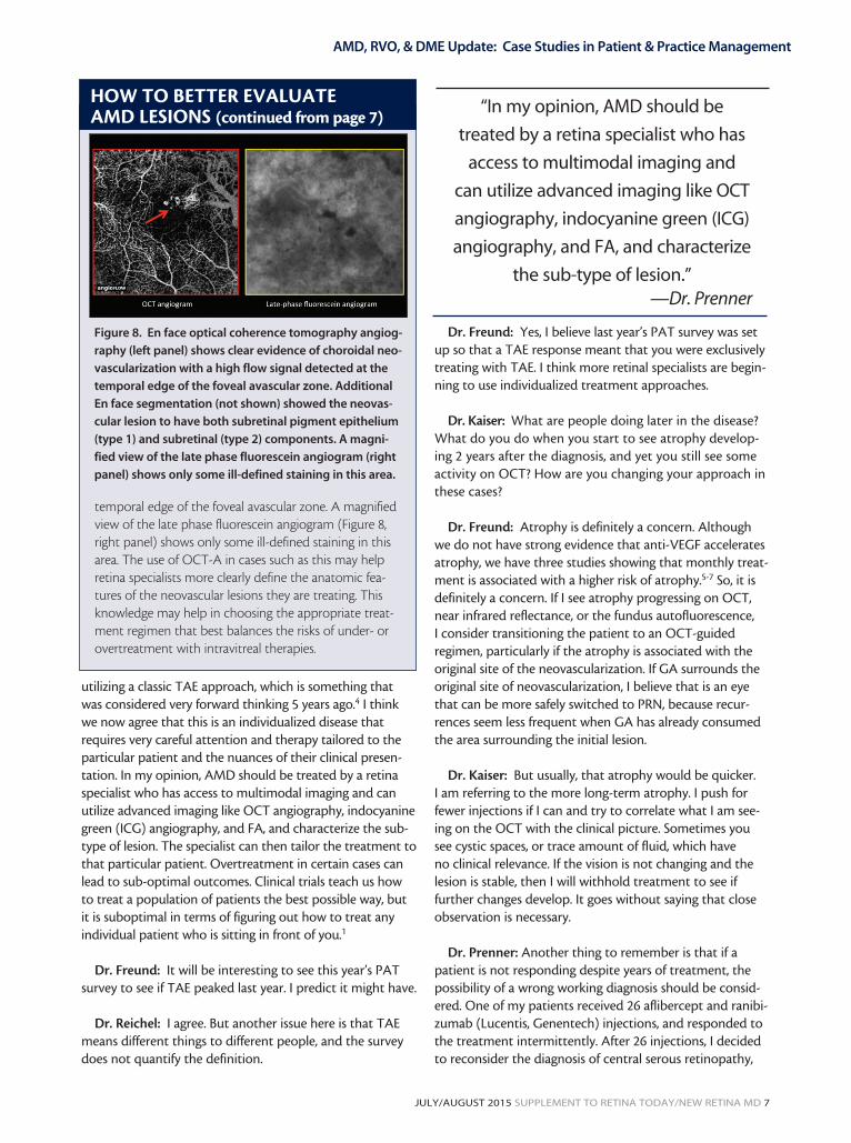
temporal edge of the foveal avascular zone. A magnified view of the late phase fluorescein angiogram (Figure 8, right panel) shows only some ill-defined staining in this area. The use of OCT-A in cases such as this may help retina specialists more clearly define the anatomic fea-tures of the neovascular lesions they are treating. This knowledge may help in choosing the appropriate treat-ment regimen that best balances the risks of under- or overtreatment with intravitreal therapies.
HOW TO BETTER EVALUATE AMD LESIONS (continued from page 7)
Figure 8. En face optical coherence tomography angiog-
raphy (left panel) shows clear evidence of choroidal neo-
vascularization with a high flow signal detected at the
temporal edge of the foveal avascular zone. Additional
En face segmentation (not shown) showed the neovas-
cular lesion to have both subretinal pigment epithelium
(type 1) and subretinal (type 2) components. A magni-
fied view of the late phase fluorescein angiogram (right
panel) shows only some ill-defined staining in this area.
“In my opinion, AMD should be
treated by a retina specialist who has
access to multimodal imaging and
can utilize advanced imaging like OCT
angiography, indocyanine green (ICG)
angiography, and FA, and characterize
the sub-type of lesion.”—Dr. Prenner

8 SUPPLEMENT TO RETINA TODAY/NEW RETINA MD JULY/AUGUST 2015
AMD, RVO, & DME Update: Case Studies in Patient & Practice Management
which it turned out to be. So, if you are only achieving suboptimal responses, you should get the ICG, OCT with enhanced-depth imaging, and auto-fluorescence to confirm the correct diagnosis.
Dr. Freund: The risk for GA also varies quite a bit from patient to patient. In our evaluation of this, we found that
patients presenting with type 1 sub-RPE lesions seem to be more resistant to GA than those presenting with other lesion sub-types.8 We all have patients whom we have treated continuously for close to 10 years and given 100 injections maybe, and they can tolerate continuous VEGF suppression without atrophy. Many of us have eyes that still show no GA despite continuous monthly anti-VEGF treatment for
Presented by Richard Kaiser, MD
A 58-year-old African American male presented with a 14-year history of type 2 diabetes. Optical coherence tomography (OCT) showed subretinal fluid (Figure 1) after receiving intravitreal bevacizumab for 3 months in October 2012. During 2013, we confirmed our OCT findings with both fluorescein angiography and a color photo; areas of active neovascularization were easily identified (Figure 2). We switched the patient to intravitreal ranibizumab, but OCT showed continued anatomic worsening after four injections in January 2014 (Figure 3). In April 2015, after two injections of intravitreal aflibercept, there is clearly anatom-ic improvement on OCT (Figure 4). A color photo (Figure 5) has confirmed findings of regressed neovascularization.
SWITCHING ANTI-VEGF DRUGS IN A CASE OF DME
Figure 1. Optical coherence tomography (OCT) showing
subretinal fluid during initial presentation.
Figure 2. During a follow-up visit, areas of active neovas-
cularization were identified via FA and color photo.
Figure 4. About 15 months later, after two injections of
intravitreal aflibercept, anatomic improvement is noted
and shown on OCT.
Figure 3. OCT shows continued anatomic worsening after
four injections in less than 12 months.
Figure 5. This color photo confirms findings of regressed
neovascularization.

JULY/AUGUST 2015 SUPPLEMENT TO RETINA TODAY/NEW RETINA MD 9
AMD, RVO, & DME Update: Case Studies in Patient & Practice Management
up to 10 years. These cases demonstrate great variability in susceptibility to GA and show that some eyes can tolerate continuous long-term VEGF suppression without develop-ing atrophy. In both our study and in the CATT trial, eyes presenting with type 3 neovascularization (RAP) appeared more likely to develop GA.2,8
Dr. Reichel: One other thing to consider is that there are some patients who are on anti-VEGF and their atrophy is progressing, but they still need more anti-VEGF. I will switch these patients to bevacizumab (Avastin, Genentech), because in the CATT trial, there was a lower trend of GA associated with bevacizumab.2 However, there was not a difference in the IVAN trial.3 This also suggests that there is probably less of an anti-VEGF effect with bevacizumab compared to the other anti-VEGFs.
Dr. Freund: Do you reduce the anti-VEGF dose? I have done that on occasion, to 0.3 mg.
Dr. Reichel: I have not. But that is an excellent idea.
RETINAL VEIN OCCLUSION TREATMENTS IN THE REAL WORLD
Dr. Prenner: What is your algorithm for drug choice in RVO? How do you determine when to use regional depot steroids in RVO? How about laser?
Dr. Freund: For the large majority of eyes with CME secondary to RVO, I treat with intravitreal anti-VEGF monotherapy. I tend to prefer aflibercept in those cases presenting with more severe occlusions with florid CME or those that cannot be extended beyond monthly treatment when attempting a TAE regimen. I will occasionally use grid laser for an eye with a BRVO that has some mild persistent CME following multiple anti-VEGF injections when I think this may achieve resolution of the edema and eliminate the need for further injections. I do not use steroids very often to treat CME secondary to RVO.
Dr. Prenner: At this point in time, I will choose anti-VEGF therapy as my primary intervention for RVO. I gener-ally will treat center-involving macular edema if the patient has either visual symptoms or visual compromise on func-tional testing. In some cases of RVO, particularly BRVO, I will hold on intervention for several months if the acuity is not compromised, as these are likely cases of well perfused RVO that may resolve without intervention. I use steroids rarely for these cases, as most RVO will respond to anti-VEGF therapy, particularly to aflibercept.
DIABETIC MACULAR EDEMA TREATMENTS IN THE REAL WORLD
Dr. Prenner: In DME, we have had a lot of new data over the course of the past year, particularly Protocol T, which
compared ranibizumab, aflibercept, and bevacizumab in a prospective, randomized, controlled clinical trial.9 We also have new indications for both ranibizumab and aflibercept in terms of treating diabetic retinopathy (DR). Novel ste-roid delivery systems have been approved as well, including the fluocinolone and dexamethasone implants.10,11 It will be interesting to hear how everyone has been incorporating the lessons learned from these trials and these medications into their clinical practices.
So, let us take the case of a 50-year-old patient who has recently been diagnosed with diabetes, and has an A1C of 8. He has similar findings in both eyes, where he is phakic, 20/60 visual acuity, with a mild posterior subcapsular cataract. Fluorescein angiography shows minimal nonperfusion and multiple microaneurysms, most of which are not central. The OCT demonstrates an absence of significant traction, and 400 µm of cystic macular edema, with just a hint of subreti-nal fluid. How do you proceed with the management of this patient?
Dr. Freund: I would get a baseline FA and OCT. Perhaps in the future, OCT-angiography will also be helpful in this patient, but it is not commercially available right now. This patient is likely to benefit from anti-VEGF therapy, since he has 20/60 visual acuity and a fairly edematous macula. If you were to follow the results of Protocol T,9 because his visual acuity is 20/60, which might be around 20/50 on an ETDRS chart, this patient just fits into the category that might do better with aflibercept than with other anti-VEGF drugs. However, if you chose to start the patient on bevaci-zumab or ranibizumab, you could always switch to afliber-cept if the response is suboptimal.
Dr. Prenner: Dr. Freund makes a great point about the discrimination between ETDRS and Snellen visual acuity. When you pool the data from multiple clinical trials, there is about a line-and-a-half difference between the ETDRS and Snellen visual acuity. So when we see those results from Protocol T of 20/50 being the point of differentiation, that is really about 20/70 in my clinical practice. Does everyone agree that anti-VEGF therapy should be the first choice in this patient?
Dr. Reichel: There is definite evidence that anti-VEGF
“When you pool the data from
multiple clinical trials, there is
about a line-and-a-half difference
between the ETDRS and Snellen
visual acuity.”—Dr. Prenner

10 SUPPLEMENT TO RETINA TODAY/NEW RETINA MD JULY/AUGUST 2015
AMD, RVO, & DME Update: Case Studies in Patient & Practice Management
therapy should be the primary approach. Before Protocol T, there was Protocol I, which looked at ranibizumab with early and then delayed laser, steroid with early and then delayed laser, and just laser.9,12 Protocol I clearly showed a benefit of anti-VEGF with laser to follow.12 Based on those data, I want to do monthly anti-VEGF injections until the macula begins to respond on the OCT. Then, I repeat the angiogram and see if I have any leaking microaneurysms. I look for exudates that may be moving in on the fovea, and other sources of leakage that I may want to supplement with laser.
Dr. Prenner: I am considering using laser earlier now that I have started using a micropulse laser delivery system with pattern scanning. It is not clear how well this will work, but if I see non-macular microaneurysms, it seems to be well tolerated.
Dr. Reichel: In patients with DME, I think laser still has a definite role in clinically significant macular edema.
Dr. Prenner: If we are going to follow Protocol T, trans-lating that as a guideline for how we practice may be some-what cumbersome. In Protocol T, the patients received essentially monthly therapy; stopping the treatment was difficult, because it required the stability of vision plus or minus 5 letters.9
Dr. Kaiser: In the first year of treatment in Protocol T, patients received 11 injections, except for aflibercept, which was injected 10 times.9 The trial shows us that, despite the elaborate protocol with the ability to hold off on treatment based on a host of clinical and diagnostic criteria, patients were essentially receiving monthly injections for the first
Presented by Elias Reichel, MD
A 63-year-old man with a 12-year history of insulin-depen-dent diabetes presented with a visual acuity of 20/50 OU. On exam, he showed signs of mild nonproliferative diabetic retinopathy and bilateral diabetic macular edema despite multiple anti-vascular endothelial growth factor injections. Figure 1 shows a color photo at presentation of the left eye; the contralateral eye was similar. Optical coherence tomogra-phy (OCT) confirmed the original findings of anatomic disease (Figure 2). However, Figure 3 shows the left eye after undergo-ing OCT-angiography, superficially. From the highly detailed image, areas of nonperfusion can be observed.
It is now our current practice to use OCT-A in cases where the patient’s first presentation is years after initial diabetes diagnosis. Using OCT-A allows us to have a better understanding of the anatomic issues the patient is facing, and helps us dictate a better treatment regimen.
USING ADVANCED IMAGING IN CASES OF DIABETIC MACULAR EDEMA
Figure 1. Color funds photograph shows minimal changes
of BDR.
Figure 2. OCT images show central macular edema. Figure 3. Superficial OCTA reveals areas of nonperfusion in
the macula.

JULY/AUGUST 2015 SUPPLEMENT TO RETINA TODAY/NEW RETINA MD 11
AMD, RVO, & DME Update: Case Studies in Patient & Practice Management
year. This is a key statistic to share with treatment-naive patients and to keep in mind as treatment unfolds.
Dr. Prenner: Should we follow this approach in our clinical practice, or tailor the treatment to each patient and determine future treatments on previous treatment response?
Dr. Reichel: There are other studies as well where one of the groups got five monthly injections of aflibercept, and then they were injected every other month, so they received eight treatments in total, and they did pretty well.13 This is comparable to what was seen in Protocol T.
This situation is very complicated, and it is further complicated by some of the other DRCR.net studies that showed a reduced number of treatments in the first year.12 So, it is difficult to decide which protocol to follow as clinicians. Even though I believe Protocol T is great, the problem is that following the paradigm is very difficult. The researchers used a computer algorithm to decide what the next step of the management in a patient should be. This is not practical on a day-to-day basis when we have a busy office.
Dr. Freund: These protocols are only targeting the DME. We also have another aspect to consider. We do not have any guidelines about the optimal number of injections needed for the regression or improvement of DR. One of the biggest challenges for me is when I see patients who do not yet seem to need anti-VEGF treatment for their DME, I wonder if I am doing them a disservice by denying them the potential benefit of anti-VEGF therapy in reducing overall DR severity. By withholding treatment, am I allowing their retinopathy to progress in an irreversible way?
WHEN TO CONSIDER TREATMENT AS NEEDED?Dr. Prenner: What if there is a patient who responds
well to aflibercept. The clinical exam and OCT are improv-ing, and the vision is starting to improve. The treating phy-sician starts to filter down to nine injections during the first year. The patient is still doing well. That patient is fairly well “tucked in,” from a treatment algorithm standpoint. Do you agree with this assessment?
Dr. Reichel: We may be getting a little ahead of our-selves here. Personally, I would do a PRN approach for this patient. I would treat with an anti-VEGF, and then follow him or her closely on a monthly basis. If he or she came back at month 2, and I did not see a response, I would con-tinue to treat. If at month 3 the condition has improved, I might withhold treatment in the first year. My algorithm is very different from what Protocol T suggests.
Dr. Prenner: It is rare to get a patient with DME who does not need to continue therapy after receiving a single
anti-VEGF injection. We agree that we will use PRN, and if the patients do well, there is no need to change drugs. However, consider a patient who is recalcitrant to three injections of monthly bevacizumab, with no significant change in his or her OCT, or only an intermittent change. If that is your patient, what do you do?
Dr. Kaiser: I will continue with anti-VEGF therapy for a while.
Dr. Prenner: Will you use the same anti-VEGF drug, or will you switch to another drug?
Dr. Kaiser: Based on Protocol T, I would be more apt to switch drugs. Based on your example, I probably would have switched earlier in the treatment regimen. If the patient is on bevacizumab, it is important to consider that tachyphylaxis could be limiting the clinical response. Protocol T certainly supports why some patients have a suboptimal response to bevacizumab.9 The patient who you are talking about is on the borderline, where he or she is probably 20/40 or 20/50 on the ETDRS, so switching may not be an imperative. But if I am not getting a response, I would certainly switch to aflibercept, which in the trial proved to be superior for patients with worse vision than the patient you describe.
Dr. Reichel: We also need to consider the systemic control as well. If you have a patient who is not responding well, even when you are initiating response, you need to contact the primary care physician or endocrinologist to make sure that he or she is better managed.
Dr. Prenner: Yes, each point of HbA1C control lowers your chance of diabetic retinopathy by 20%.14
Dr. Kaiser: I would also add that blood pressure control is essential, as is fluid retention and renal status. I spend extra time with my diabetic patients reviewing key systemic fea-tures that impact their retinas and also their general health.
Dr. Prenner: Back to our hypothetical patient—assume you have given three bevacizumab injections, but do not see appropriate anatomic response. What do you do then?
“These protocols are only target-
ing the DME. We do not have any
guidelines about the optimal num-
ber of injections needed for the
regression or improvement of DR.”—Dr. Freund

12 SUPPLEMENT TO RETINA TODAY/NEW RETINA MD JULY/AUGUST 2015
AMD, RVO, & DME Update: Case Studies in Patient & Practice Management
Dr. Reichel: I switch them to aflibercept.
Dr. Prenner: Data from Protocol T suggest ranibizumab and aflibercept are more impactful in terms of achieving anatomic improvements than bevacizumab.9
Dr. Freund: If I do not see an anatomic response even after three injections, I would switch to aflibercept. Before considering steroids, I may do an FA, and sometimes I can find a few leaky aneurysms that are easily treatable with laser.
Dr. Kaiser: An FA is also valuable to assess the degree of perfusion. A great deal can be gleaned by assessing changes in the FAZ, peripheral ischemia, as well as focal areas that may contribute to recalcitrant DME. Many of these FA find-ings respond to laser. We cannot underestimate the impor-tance of going back to the angiogram. A new technology, OCT angiography, is helpful and may become the standard of care in the future, given its ease of use and ability to pro-vide clinically relevant information.
WHEN TO CONSIDER STEROIDSDr. Prenner: We agree about switching from bevacizum-
ab to aflibercept after three bevacizumab injections that do not achieve an adequate anatomic response. Now, let us assume that after the switch to aflibercept, we still see most of the macular edema present, despite three more injec-tions and the CTS remains at 350 µm. Would you increase the timing or dose of aflibercept? Do you think about an inflammatory component to the DME and consider adding steroids?
Dr. Reichel: I think we are still learning the definition of a “successful” DME treatment. It may be a plateau that you reach after multiple anti-VEGFs. It may be sufficient to say that is the endpoint. If you are at 350 µm and the patient’s vision is good and stable, I would be happy with that. I might consider adding laser treatment, however, because you might get some added benefit with focal laser, and pos-sibly some with grid laser. Sometimes, you can get the RPE responding, especially with micropulsing, and you may see an added benefit, which was also seen in DRCR Protocol I.12 Delayed laser photocoagulation does play a role here, and we are probably up to about 6 months in treatment with this patient, and that is the correct time to be doing it.
Dr. Prenner: How about steroids? When are you going to start thinking about using a regional depot steroid to aid in this patient’s DME?
Dr. Kaiser: In a recalcitrant case, I consider using a low-dose triamcinolone injection. It is best to identify steroid-responsive patients before using an implant. Lens status can also play a role in my decision-making. In most cases, once we use steroids, we are committing the patient to cataract
surgery. Thus, I have exhausted anti-VEGF therapy before using steroids. This patient already has a cataract, thus surgery is likely inevitable with or without steroid usage. Another key to treatment is reviewing the prior treatment history. On occasion, after review, it becomes apparent that the patient did not have regularly scheduled injections or was not consistently compliant with appointments, thus they never received a fair trial of anti-VEGF.
Dr. Prenner: Do you think about triamcinolone (Triesence, Alcon) or dexamethasone implant 0.7 mg (Ozurdex, Allergan) first?
Dr. Freund: There are two options. One is to continue with the anti-VEGF injections and wait for a response, which I rarely choose. There is some evidence that this approach works, but it is very hard to do that in clini-cal practice when your patient is coming in every month and there has been no response. The second option is to use steroids at this point. I would probably go straight to Ozurdex at that point. The IOP issue is less, or at least no worse, with Ozurdex as compared with 2 mg of Triesence.
Dr. Freund: I think the IOP issues with Ozurdex are, by and large, very manageable.
Dr. Prenner: I typically would use Ozurdex rather than Triesence.
Dr. Reichel: I still use 1 mg of triamcinolone. I have been using it for 14 years now, and I am happy with the results. We will not use an off-label steroid, so I use triamcinolone. The black box warning is, in part, because of benzalkonium chloride (BAK). I think the BAK can be a good thing, you know. It actually kills bacteria so it probably actually lowers your risk of developing cystoid macular edema.
Dr. Kaiser: That is interesting because 54% of us use an off-label anti-VEGF therapy, but we are afraid to use an off-label steroid.
Dr. Prenner: Assume then, that for our hypothetical patient we implant Ozurdex, and we see that it lasts 3 to
“Data from Protocol T suggest
ranibizumab and aflibercept are
more impactful in terms of
achieving anatomic improvements
than bevacizumab.”—Dr. Prenner

JULY/AUGUST 2015 SUPPLEMENT TO RETINA TODAY/NEW RETINA MD 13
AMD, RVO, & DME Update: Case Studies in Patient & Practice Management
4 months, and we are needing to repeat Ozurdex multiple times. Are you then thinking about using Iluvien (fluocino-lone, Alimera Sciences) to have a more prolonged effect, even in those patients who are phakic and relatively young? If they have not had a pressure response to one steroid, it does not predict pressure response to another steroid. Does an absence of steroid response with Ozurdex give you confidence that you can use Iluvein safely?
Dr. Freund: I think it is far more likely that you will get an increase in IOP with Iluvien.
Dr. Prenner: Different genetic pathways determine the risk of elevation in IOP with each particular steroid, so one is not necessarily more protective than another. Would you consider fluocinolone for this patient?
Dr. Kaiser: No. The data from the FAME studies demon-strate a benefit for long-standing DME, specifically recalci-trant DME for longer than 2 to 3 years.15 The hypothetical patient described is a freshly diagnosed DME patient, thus the Iluvien implant would not be ideal. Conversely, if there is clinical evidence of chronic DME, the FAME data is actu-ally quite supportive for use of the implant—chronic DME patients gained 3-lines of vision 35% versus 9% with the implant versus standard of care.15
THE ASYMPTOMATIC PATIENTDr. Prenner: Consider another patient, a 60-year-old
bilateral pseudophake, who has 400 µm of central retinal thickness OU, but the visual acuity is 20/32, and she is not particularly symptomatic. Is there a role for observation in this case?
Dr. Freund: I rarely treat patients the first time I see them in a situation where they have good vision.
I do not think that there is any rush to treat in this disease, unless the retina is swollen with large cysts. In a patient who is asymptomatic and has good vision, I would probably check with his or her primary care physician just to be sure that there’s no urgency to treat.
Dr. Kaiser: Discussing this scenario is valuable, because many of our patients are asymptomatic despite fairly advanced macula changes. In this particular case, I recom-mend treatment, because the vision is starting to decline. Other similar scenarios come up with less macular edema, near-perfect vision, and no symptoms where observation is a reasonable approach. Either way, close follow-up is pru-dent.
Dr. Prenner: I would also obtain an OCT for this patient to make sure he or she does not have a component of pseudophakic macular edema that I could treat with topi-cal anti-inflammatory therapy.
Dr. Reichel: I would not treat this patient, either. But, sometimes you see a patient who has mostly extrafoveal disease, and there is a lot of fluid going into the fovea. These patients respond very exquisitely with laser, and I might do a laser in that case. On the other hand, if a patient has a broad-based macular edema centered on the fovea and a 20/32 visual acuity, I would follow him or her without treatment.
Dr. Freund: Another issue arises in patients who have relatively mild DME, but are at high risk of progressing to proliferative retinopathy or those who already have some early disc or retinal neovascularization. Typically, I will start these patients on an anti-VEGF agent and defer panretinal photocoagulation (PRP). Interestingly, I have found many of these patients, some of whom I have followed for up to 5 years, never end up needing the PRP.
Dr. Prenner: A part of the problem with our current protocols is that we assume that patients are reliable. And DME usually occurs in patients who are not particularly reliable in terms of maintaining their general health. Most of these patients have uncontrolled A1Cs. Therefore, it becomes more challenging to avoid definitive laser than it seems on paper, as these patients are often lost to follow-up until advanced disease develops.
Dr. Freund: For the type of patient with severe non-proliferative disease, it is possible that early treatment with monthly anti-VEGF therapy could prevent progression of the disease to PDR. For patients already at the prolif-erative stage, we will soon have data from the DRCR.net Protocol S exploring whether monthly ranibizumab with deferred PRP is superior to prompt PRP at first detection of high-risk PDR.
Dr. Kaiser: There are two additional points to mention. One is that in patients who respond, sometimes a switch goes off, and the proliferative process simply stops. Unlike AMD, which is relentless, these patients, once treated with anti-VEGF, will sometimes quiet down and remain qui-escent for years. Secondly, I would do an angiogram in a patient like this, because if nonperfusion exists, a quadran-tic PRP can be effective.
Dr. Prenner: What about the situation where you have active DME, high-risk PDR, and a small area of traction? Do you then use PRP or anti-VEGF first? And how do you stage it?
Dr. Reichel: In such patients, I like to get the DME under control first, because there is a thought suggesting that PRP may make it worse, which may or may not be true. So, I would combine injections and PRP. The issue with these patients is that their disease is usually bilateral. So, when
14 SUPPLEMENT TO RETINA TODAY/NEW RETINA MD JULY/AUGUST 2015
AMD, RVO, & DME Update: Case Studies in Patient & Practice Management
they come in, I will use an injection in one eye and PRP in the other eye, and then switch them off.
Dr. Prenner: There is a point where the traction becomes a concern in these patients, and you need to worry about anti-VEGF induced contraction, and secondary detachment.
Dr. Reichel: Yes, and the laser can do that, too.
Dr. Kaiser: I pretreat these patients, or start treatment with the anti-VEGF, and then perform PRP laser. Anti-VEGF helps, but ultimately PRP is the definitive treatment for the proliferative process.
Dr. Reichel: Yes. Suppose you have a patient who has preproliferative or early proliferative disease, and you have treated their DME, and they are doing great. How much more anti-VEGF are you going to use, and at what frequen-cy? You have a great result after 6 months of treatment and the retinopathy score is reduced. Do they not need any more anti-VEGF? I believe that they are probably going to need some anti-VEGF over their lifetime.
Dr. Kaiser: I think you continue to treat that patient and monitor the progression of disease. I find the use of photographs helpful to document subtle disease progres-sion over time.
Dr. Prenner: How about surgery for DME? We saw lots of interest in vitrectomy, hyaloid removal and ILM peeling 5 to 7 years ago as a treatment for DME. Is anyone doing that in the absence of obvious traction?
Dr. Reichel: We looked at this 10 years ago. Although the patients improved anatomically, in my experience, visual function did not change.
Dr. Kaiser: With medical treatment and reduction of edema, contraction of the posterior hyaloid can occur. If the patient is symptomatic, a vitrectomy should be consid-ered, but only if there is substantial traction.
Dr. Prenner: In summary, these chronic retinal disorders are complicated from a management perspective. It is not sufficient to broadly state one treatment regimen is appro-priate for all patients—we need to customize and individu-alize our treatment algorithms. Imaging will continue to help define the treatment course for our patients, and OCT angiography is going to be the way we are going to be fol-lowing diabetics. n
1. Mant D. Can randomised trials inform clinical decisions about individual patients? Lancet. 1999;353(9154):743-746.2. CATT Research Group, Martin DF, Maguire MG, et al. Ranibizumab and bevacizumab for neovascular age-related macular degeneration. N Engl J Med. 2011;364(20):1897-1908.3. Chakravarthy U, Harding SP, Rogers CA, et al. Ranibizumab versus bevacizumab to treat neovascular age-related Macular Degeneration: One-Year Findings from the IVAN Randomized Trial. Ophthalmology. 2012;119(7):1399-1411.4. American Society of Retina Specialists, Stone TW. ASRS 2014 Preferences and Trends Membership Survey. Chicago, IL: American Society of Retina Specialists, 2014.5. Chakravarthy U, Harding SP, Rogers CA, et al. Alternative treatments to inhibit VEGF in age-related choroidal neovascularisation: 2-year findings of the IVAN randomised controlled trial. Lancet. 2013;382(9900):1258-1267.6. Grunwald JE, Daniel E, Huang J, et al. Risk of geographic atrophy in the comparison of age-related macular degeneration treatments trials. Ophthalmology. 2014;121(1):150-161.7. Sadda SR. Development of atrophy in neovascular AMD treated with anti-VEGF therapy: Results of the HARBOR Study. Poster presented at: American Academy of Ophthalmology Retina Subspecialty Day. Chicago, IL; Oct. 17, 2014.8. Xu L, Mrejen S, Jung JJ, et al. Geographic atrophy in patients receiving anti-vascular endothelial growth factor for neovascular age-related macular degeneration. Retina. 2015;35(2):176-186.9. Diabetic Retinopathy Clinical Research Network. Aflibercept, bevacizumab, or ranibizumab for diabetic macular edema. N Engl J Med. 2015; 372:1193-1203.10. Ozurdex [package insert]. Irvine, CA: Allergan Inc., 2014.11. luvien [package insert]. Atlanta, GA: Alimera Sciences Inc., 2014.12. Diabetic Retinopathy Clinical Research N, Elman MJ, Qin H, et al. Intravitreal ranibizumab for diabetic macular edema with prompt versus deferred laser treatment: three-year randomized trial results. Ophthalmology. 2012;119(11):2312-2318.13. Korobelnik JF, Do DV, Schmidt-Erfurth U, et al. Intravitreal Aflibercept for Diabetic Macular Edema. Ophthalmology. 2014;21(11):2247-54.14. Diabetes Control and Complications Trial Research Group. Progression of Retinopathy with Intensive versus Conventional Treatment in the Diabetes Control and Complications Trial. Ophthalmology. 1995;102:647-661.15. Campochiaro PA, Brown DM, Pearson A, et al. Sustained delivery fluocinolone acetonide vitreous inserts provide benefit for at least 3 years in patients with diabetic macular edema. Ophthalmology. 2012;119(10):2125-2132.

INSTRUCTIONS FOR CME CREDIT
1. In neovascular AMD, which presenting lesion type is associated with the lowest risk of long-term GA following the initiation of intravitreal anti-VEGF therapy?
a. Type 1 (sub-RPE) neovascularizationb. Type 2 (sub-retinal) neovascularizationc. Type 3 (intraretinal) neovascularizationd. Mixed lesions
2. Aflibercept and ranibizumab were _______ bevacizumab for anatomic endpoints in DME regardless of visual acuity.a. Inferior tob. Identical toc. Superior tod. None of the above
3. If a patient does not have a steroid-responsive IOP rise after receiving one kind of steroid, they are unlikely to develop an IOP rise with a different steroid.
a. Trueb. False
4. Approximately ________ of patients in the CATT trial required between one and three anti-VEGF injections in the first year when treated with a PRN regimen.
a. 5%b. 10%c. 15%d. 20%
5. Consider the following case: a 60-year-old bilateral pseudophake patient, with 400 µm of central retinal thickness OU, and visual acuity is 20/32. Patient is not particularly symptomatic. According to the panel, what is the preferred first step?
a. Observe the patientb. Begin a course of monthly anti-VEGF injectionsc. Treat the patient with panretinal photocoagulationd. Implant a steroid depot
CME credit is available electronically via www.dulaneyfoundation.org.
To receive AMA PRA Category 1 Credit™, you must complete the Post Test and Activity Evaluation and mail or fax to The Dulaney Foundation; PO Box 358; Pine Brook, NJ 07058; Fax: (610) 771-4443. To answer these questions online and receive real-time results, please visit www.dulaneyfoundation.org and click “Online Courses.” If you are experiencing problems with the online test, please email us at [email protected]. Certificates are issued electronically, please provide your email address below.
Please type or print clearly, or we will be unable to issue your certificate.
Name ________________________________________________________________ o MD participant o non-MD participant
Phone (required) ______________________________ o Email (required) ___________________________________________
Address __________________________________________________________________________________________________
City _______________________________________________________________ State _________________________________
AMD, RVO, & DME UPDATE: CASE STUDIES IN PATIENT & PRACTICE MANAGEMENT
1 AMA PRA Category 1 Credit™ Expires August 1, 2016
Supported through an unrestricted educational grant by Regeneron Pharmaceuticals.
Jointly provided by The Dulaney Foundation, Retina Today, and New Retina MD.
Did the program meet the following educational objectives? Agree Neutral Disagree
Describe the current epidemiology of major retinal diseases, including AMD, RVO, and DME ——— ——— ———
Assess clinical studies involving new approaches to treat DME ——— ——— ———
Utilize expert case examples to differentiate between clinical study
dosing protocols and alternative dosing schedules ——— ——— ———
Interpret retinal imaging case examples describing the treatment of DME ——— ——— ———
Explore the management of treatment complications and secondary therapies ——— ——— ———
Educate patients on ophthalmic implications of systemic diabetes management ——— ——— ———
Demonstrate optimized patient flow, inventory flow, and office efficiency ——— ——— ———
Your responses to the questions below will help us evaluate this CME activity. They will provide us with evidence that improve-ments were made in patient care as a result of this activity as required by the Accreditation Council for Continuing Medical Education (ACCME). Please complete the following course evaluation and return it via fax to (610) 771-4443.
Name and email ______________________________________________________________________________________________________
Do you feel the program was educationally sound and commercially balanced? r Yes r No
Comments regarding commercial bias:
_______________________________________________________________________________________________________________
_______________________________________________________________________________________________________________
Rate your knowledge/skill level prior to participating in this course: 5 = High, 1 = Low __________________________________________
Rate your knowledge/skill level after participating in this course: 5 = High, 1 = Low _____________________________________________
Would you recommend this program to a colleague? r Yes r No
Do you feel the information presented will change your patient care? r Yes r No
If yes, please specify. We will contact you by email in 1 to 2 months to see if you have made this change.
_______________________________________________________________________________________________________________
_______________________________________________________________________________________________________________
If no, please identify the barriers to change.
_______________________________________________________________________________________________________________
_______________________________________________________________________________________________________________
Please list any additional topics you would like to have covered in future Dulaney Foundation CME activities or other suggestions or comments.
_______________________________________________________________________________________________________________
_______________________________________________________________________________________________________________
ACTIVITY EVALUATION