Embed Size (px)
Citation preview
Intestinal Protozoa
AmebaeFlagellate protozoa
Coccidia and others
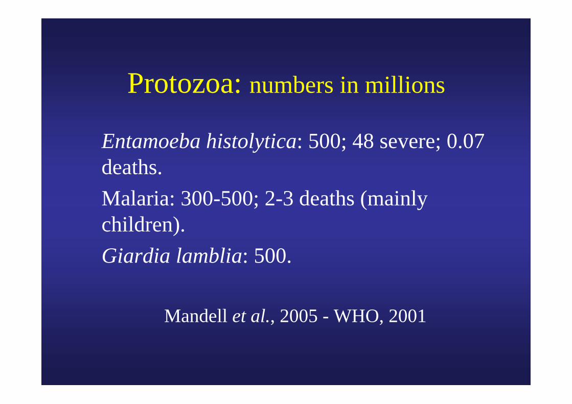
Protozoa: numbers in millions
Entamoeba histolytica: 500; 48 severe; 0.07 deaths.
Malaria: 300-500; 2-3 deaths (mainly children).
Giardia lamblia: 500.
Mandell et al., 2005 - WHO, 2001
Eosinophilia > 10%With helminths,insects (myasis),
not with protozoa excepting Isospora belli and Dientamoeba
fragilis (with pinworms?)
Protozoa in faeces
• Cysts and trophozoites from amebae, flagellates, and coccidia.
• The size is essential for identification (to be measured with a calibrated micrometer).
• The aspect of the nucleus is also important for the identification.
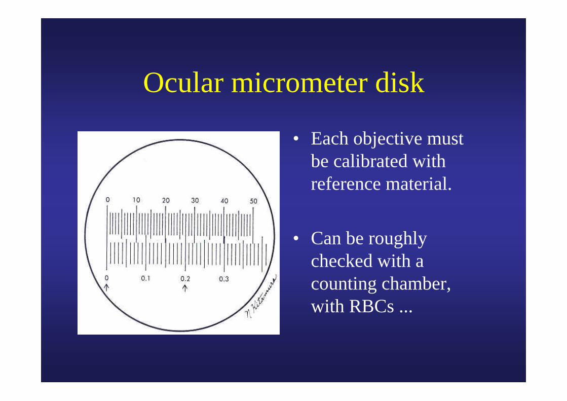
Ocular micrometer disk
• Each objective must be calibrated with reference material.
• Can be roughly checked with a counting chamber, with RBCs ...
Direct examination in saline
• Standard-preparation, containing approximatively 2 mg faeces.
• In fresh faeces it is possible to observe trophozoites (Entamoeba histolytica, ...).
• Cysts of protozoa are difficult to see, because they are colourless.

Ritchie-enrichment and Lugol stain
• Screen the entire preparation with objective 10x.
• Suspect elements (cysts, eggs,…) are checked with higher enlargement (40x, 50x, 100x).
• Amebae and flagellates stain brown-yellow with Lugol.
• The identification of cysts from protozoa is based on the size and the aspect of the nucleus.
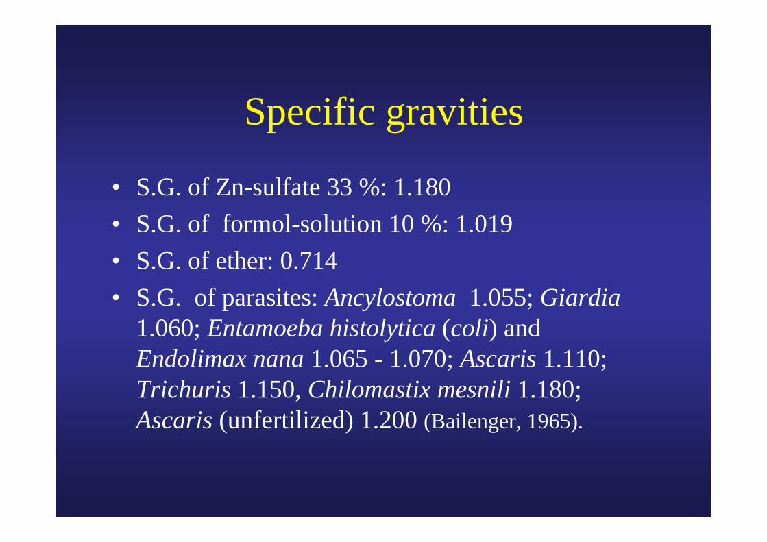
Specific gravities
• S.G. of Zn-sulfate 33 %: 1.180• S.G. of formol-solution 10 %: 1.019• S.G. of ether: 0.714• S.G. of parasites: Ancylostoma 1.055; Giardia
1.060; Entamoeba histolytica (coli) and Endolimax nana 1.065 - 1.070; Ascaris 1.110; Trichuris 1.150, Chilomastix mesnili 1.180; Ascaris (unfertilized) 1.200 (Bailenger, 1965).
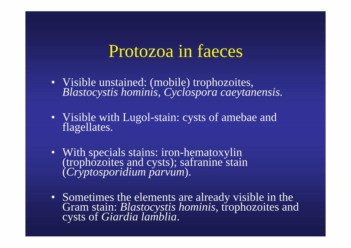
Protozoa in faeces• Visible unstained: (mobile) trophozoites,
Blastocystis hominis, Cyclospora caeytanensis.
• Visible with Lugol-stain: cysts of amebae and flagellates.
• With specials stains: iron-hematoxylin (trophozoites and cysts); safranine stain (Cryptosporidium parvum).
• Sometimes the elements are already visible in the Gram stain: Blastocystis hominis, trophozoites and cysts of Giardia lamblia.
Courtesy ASM
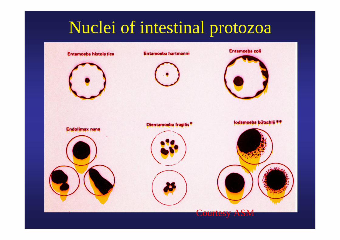
Nuclei of intestinal protozoa
Courtesy ASM
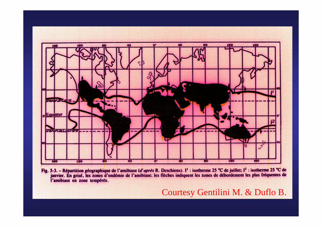
Courtesy Gentilini M. & Duflo B.
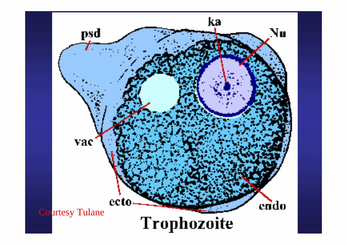
Courtesy Tulane
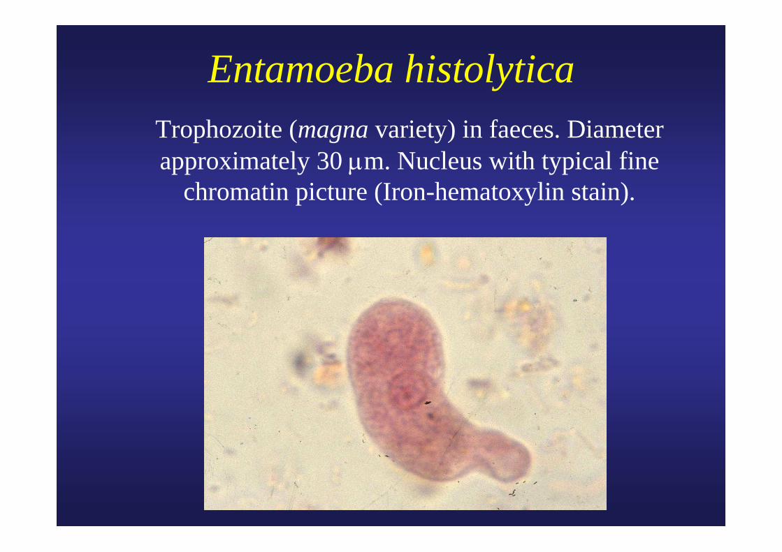
Entamoeba histolytica Trophozoite (magna variety) in faeces. Diameter
approximately 30 μm. Nucleus with typical fine chromatin picture (Iron-hematoxylin stain).
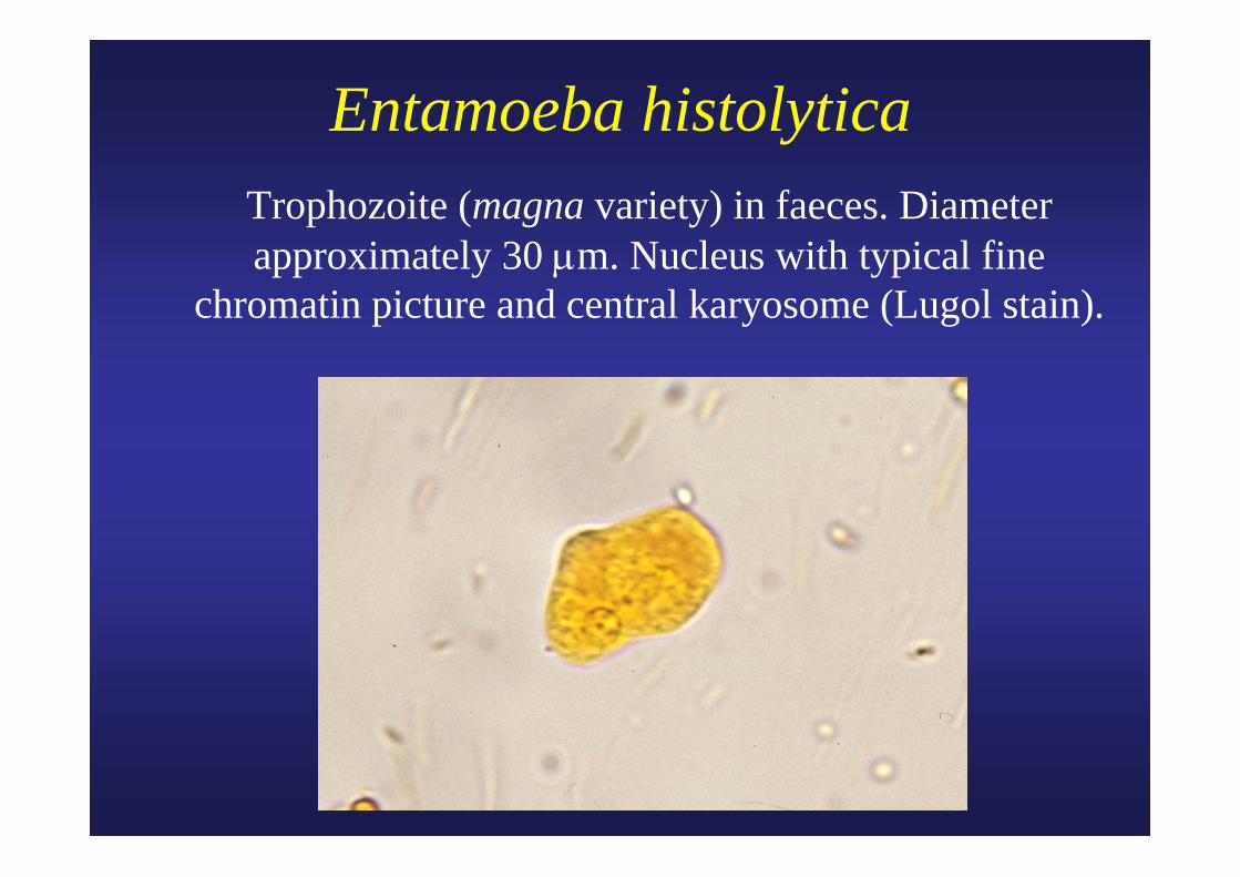
Entamoeba histolytica Trophozoite (magna variety) in faeces. Diameter
approximately 30 μm. Nucleus with typical fine chromatin picture and central karyosome (Lugol stain).
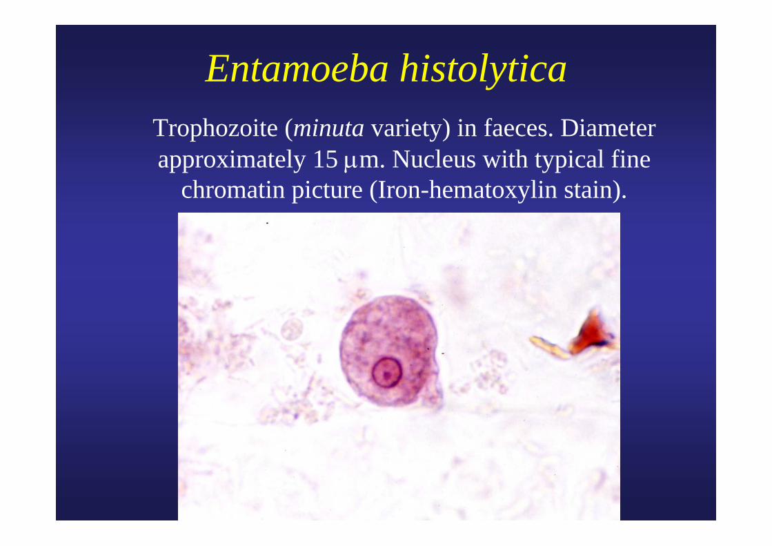
Entamoeba histolytica Trophozoite (minuta variety) in faeces. Diameter
approximately 15 μm. Nucleus with typical fine chromatin picture (Iron-hematoxylin stain).
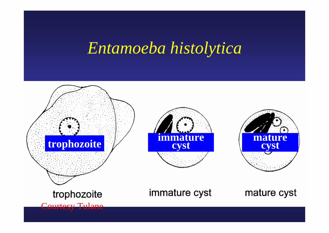
trophozoite immature cyst
mature cyst
Entamoeba histolytica
Courtesy Tulane
Courtesy Tulane
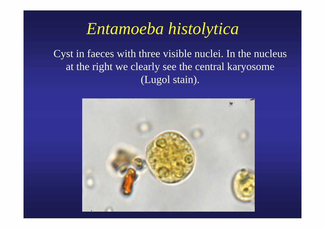
Entamoeba histolytica Cyst in faeces with three visible nuclei. In the nucleus
at the right we clearly see the central karyosome (Lugol stain).
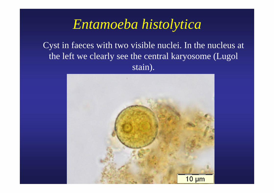
Entamoeba histolytica Cyst in faeces with two visible nuclei. In the nucleus at
the left we clearly see the central karyosome (Lugol stain).
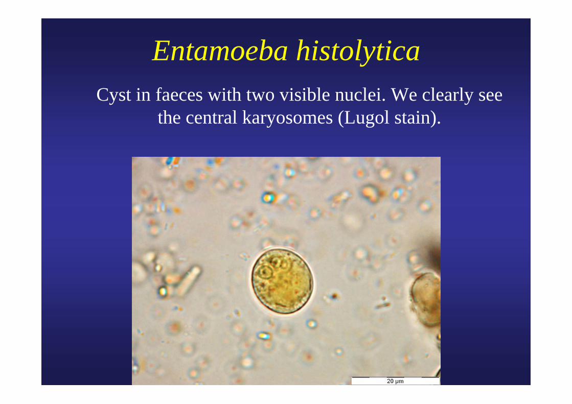
Entamoeba histolytica Cyst in faeces with two visible nuclei. We clearly see
the central karyosomes (Lugol stain).
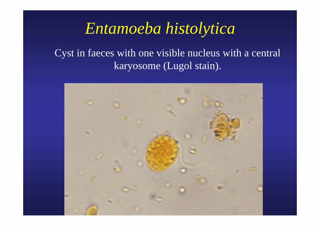
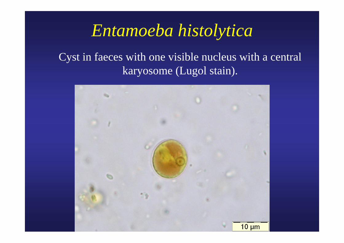
Entamoeba histolytica Cyst in faeces with one visible nucleus with a central
karyosome (Lugol stain).
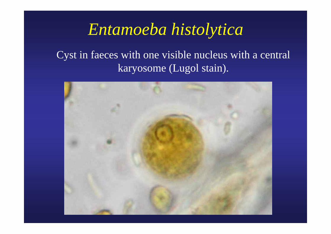
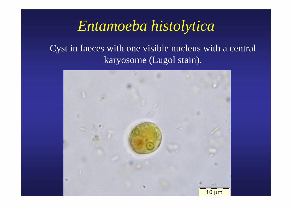
Entamoeba histolytica Cyst in faeces with one visible nucleus with a central
karyosome (Lugol stain).
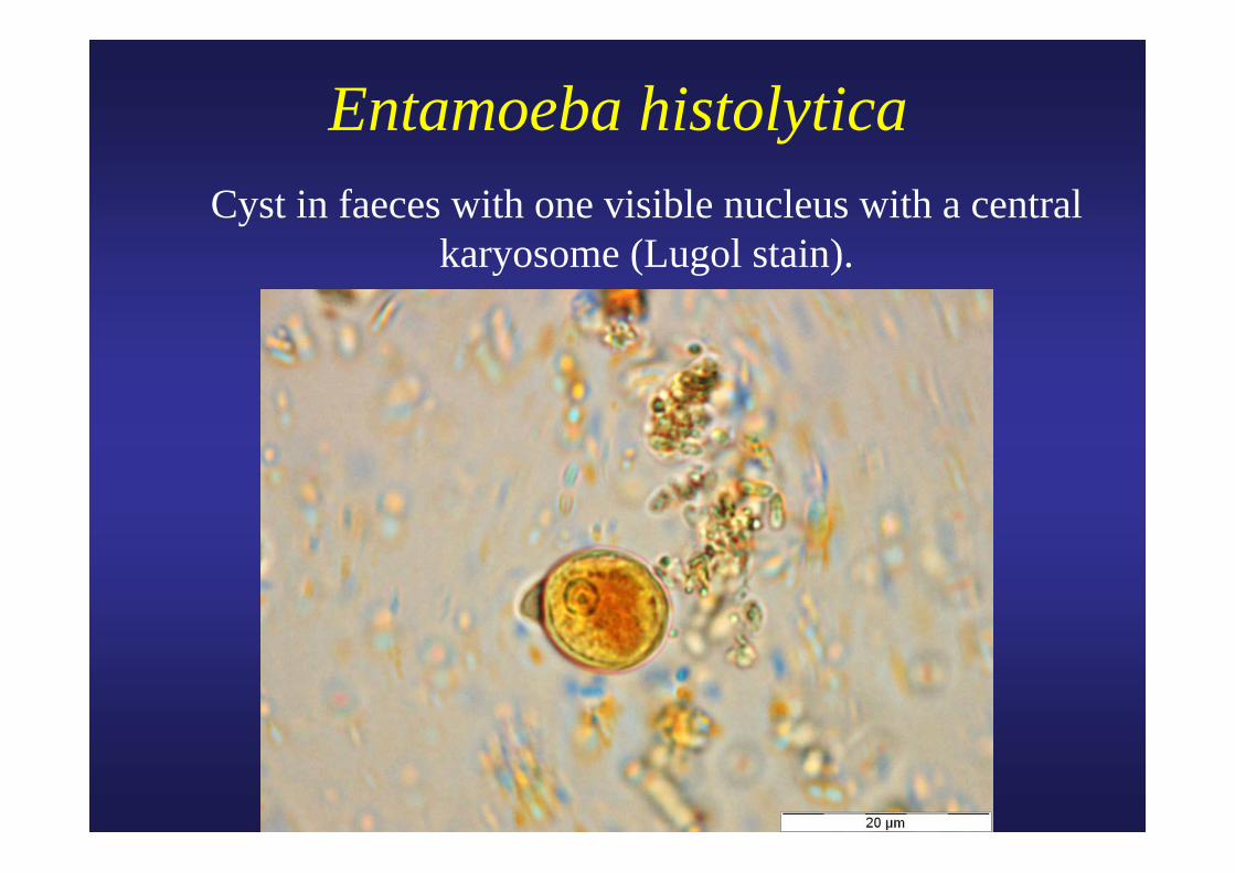
Entamoeba histolytica Cyst in faeces with one visible nucleus with a central
karyosome (Lugol stain).
Entamoeba histolytica Cyst in faeces with one visible nucleus with a central
karyosome (Lugol stain).
Entamoeba histolytica Cyst in faeces with one visible nucleus with a central
karyosome (Lugol stain).
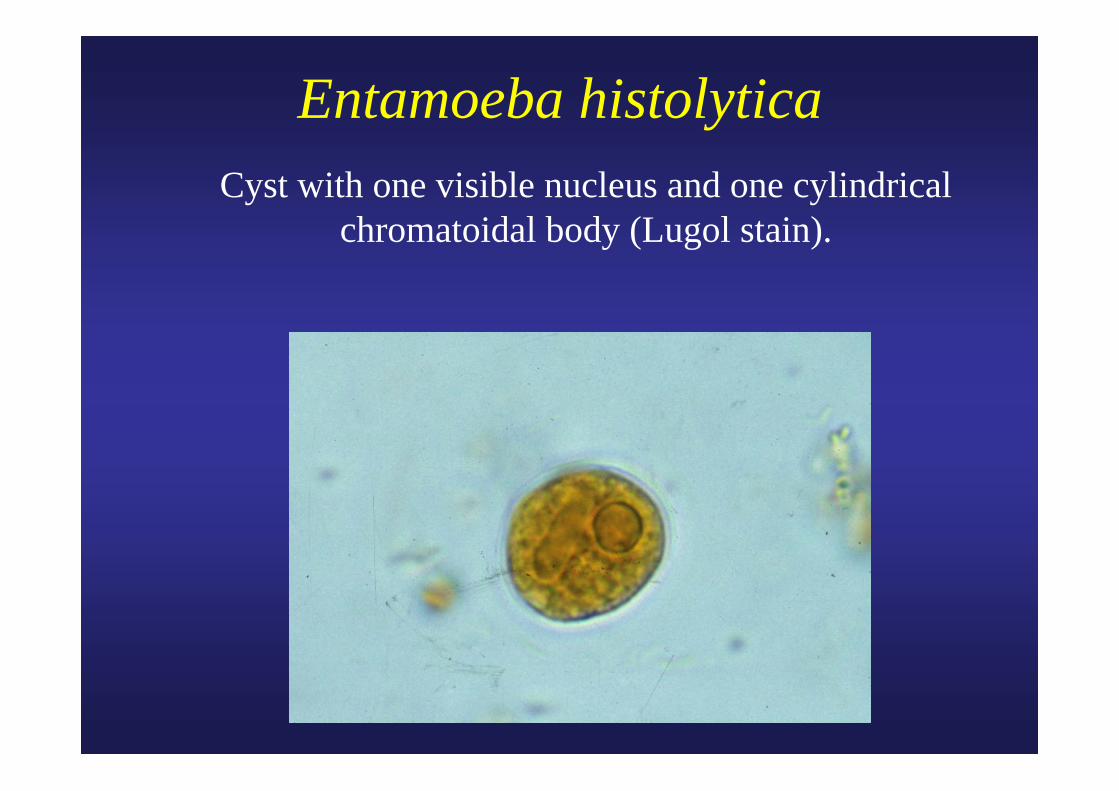
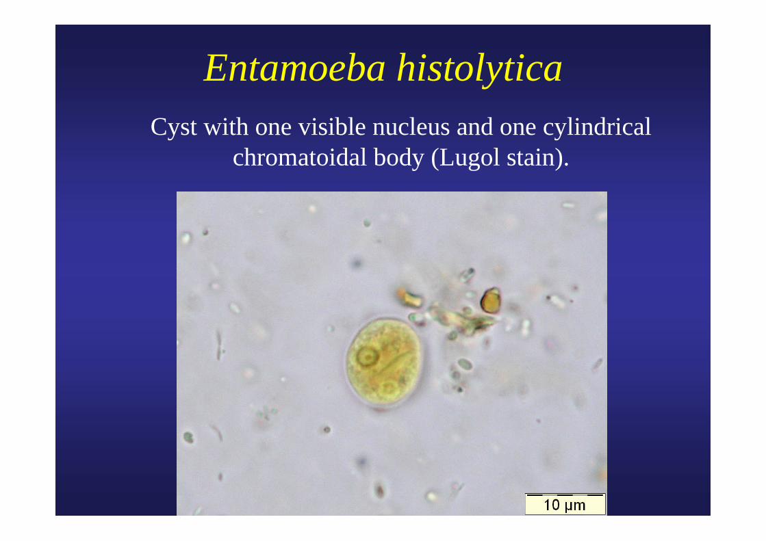
Entamoeba histolytica Cyst with one visible nucleus and one cylindrical
chromatoidal body (Lugol stain).
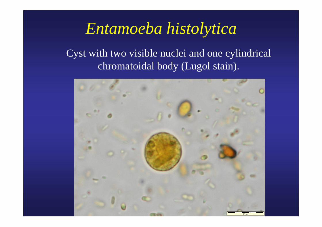
Entamoeba histolytica Cyst with two visible nuclei and one cylindrical
chromatoidal body (Lugol stain).
Entamoeba histolytica Cyst with one visible nucleus and one cylindrical
chromatoidal body (Lugol stain).
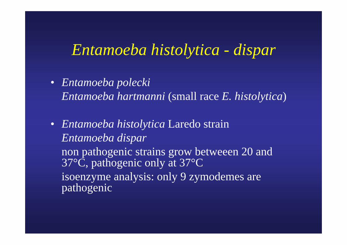
Entamoeba histolytica - dispar
• Entamoeba polecki Entamoeba hartmanni (small race E. histolytica)
• Entamoeba histolytica Laredo strain Entamoeba dispar non pathogenic strains grow betweeen 20 and
37°C, pathogenic only at 37°C isoenzyme analysis: only 9 zymodemes are
pathogenic
Entamoeba histolytica - dispar
• PCR, isoenzyme analysis, and antigen detection (JCM, 1998, 449).
• Monoclonal antibodies (JCM, 2001, 716).
• ITM-Antwerp: PCR on fecal material.
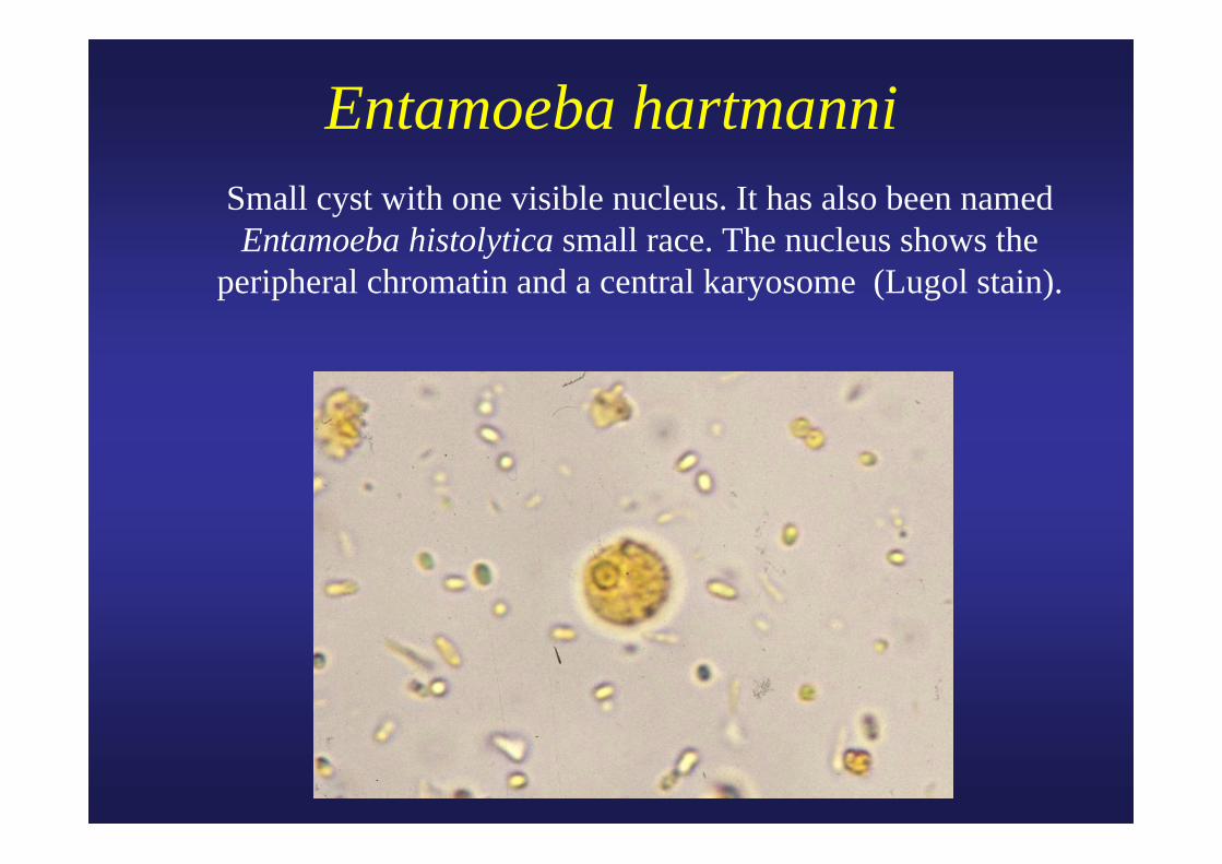
Entamoeba hartmanni Small cyst with one visible nucleus. It has also been named
Entamoeba histolytica small race. The nucleus shows the peripheral chromatin and a central karyosome (Lugol stain).
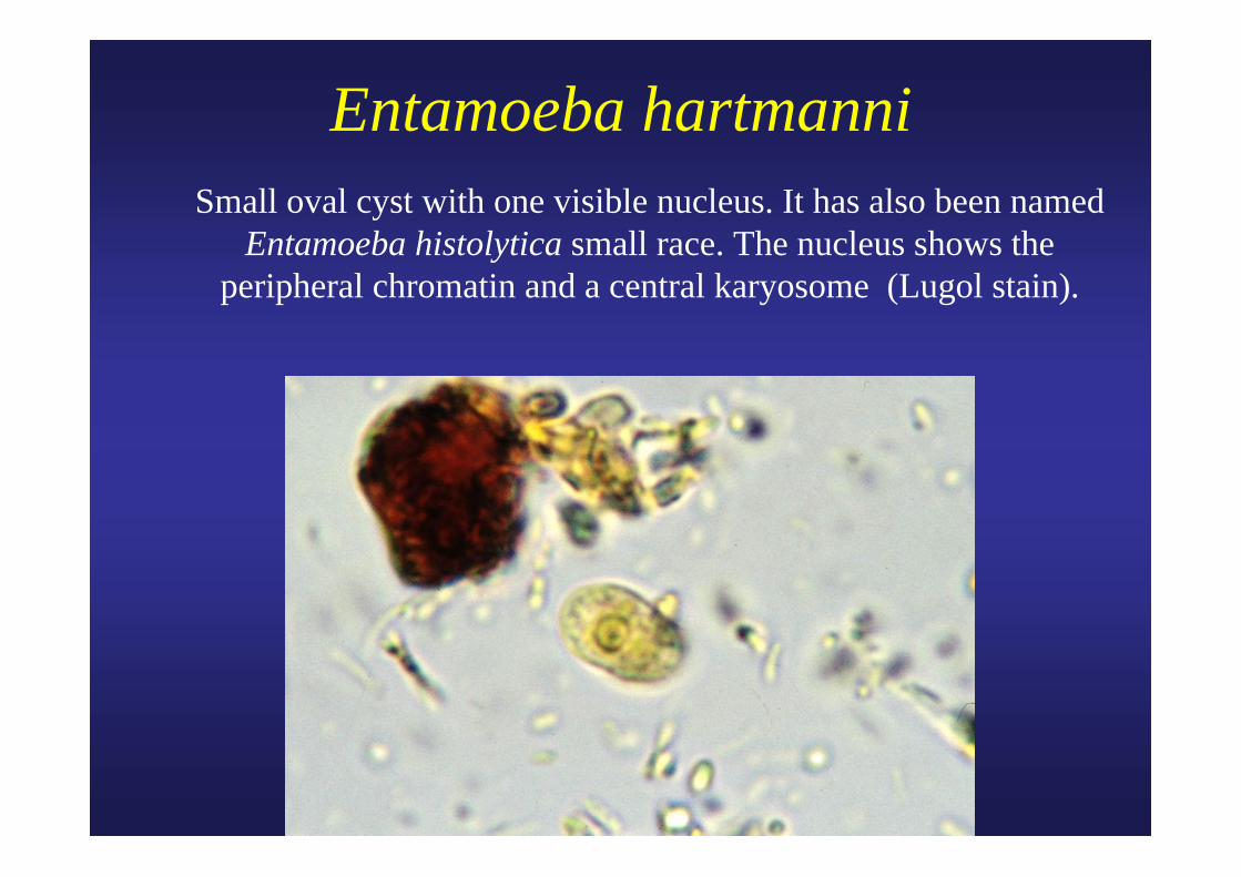
Entamoeba hartmanni Small oval cyst with one visible nucleus. It has also been named
Entamoeba histolytica small race. The nucleus shows the peripheral chromatin and a central karyosome (Lugol stain).

Entamoeba coli Mature cyst with spongy cytoplasm in faeces. Four of the eight
nuclei are visible in this plane. Note the coarse peripheral chromatin and the central karyosome of the nuclei (Lugol stain).
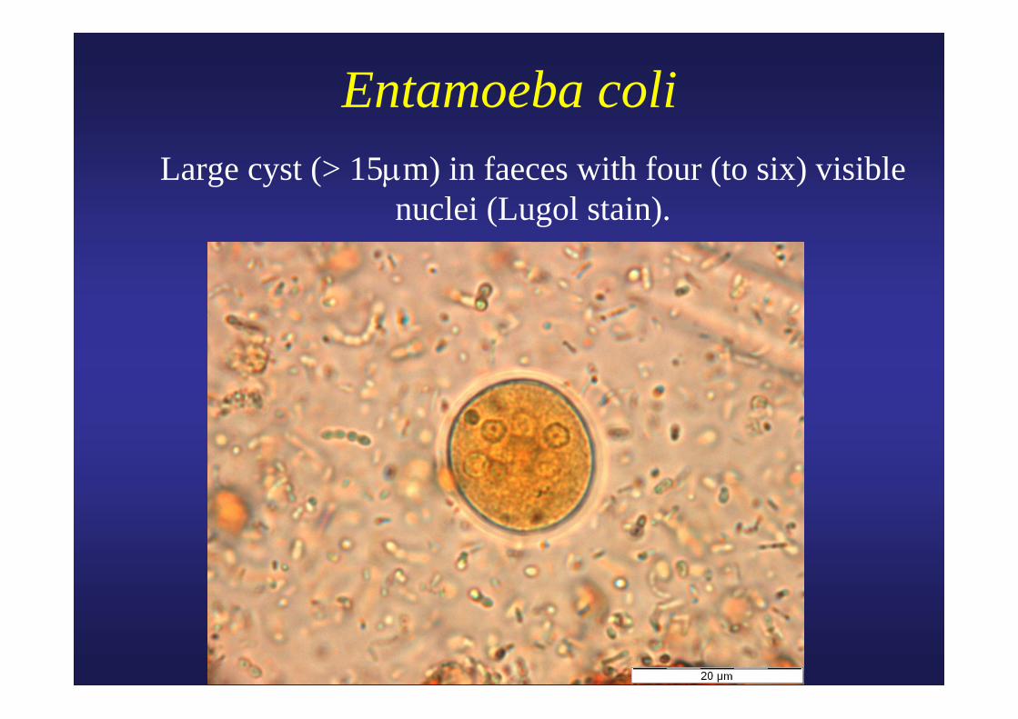
Entamoeba coli Large cyst (> 15μm) in faeces with four (to six) visible
nuclei (Lugol stain).
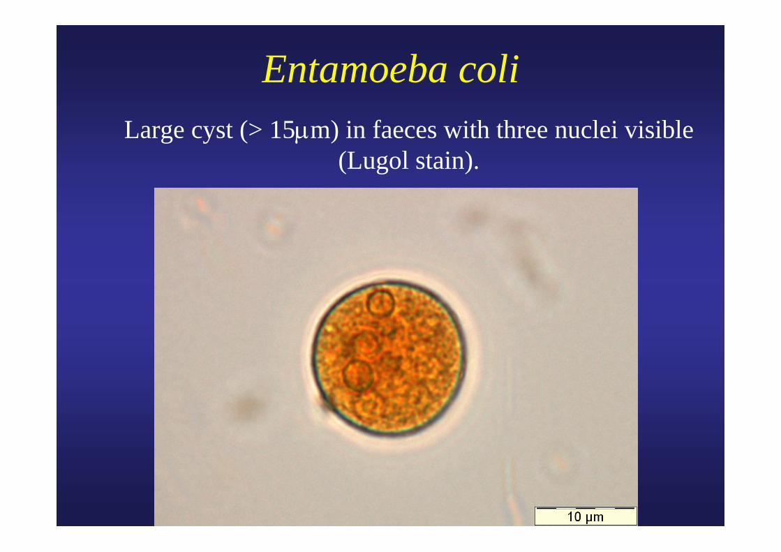
Entamoeba coli Large cyst (> 15μm) in faeces with three nuclei visible
(Lugol stain).
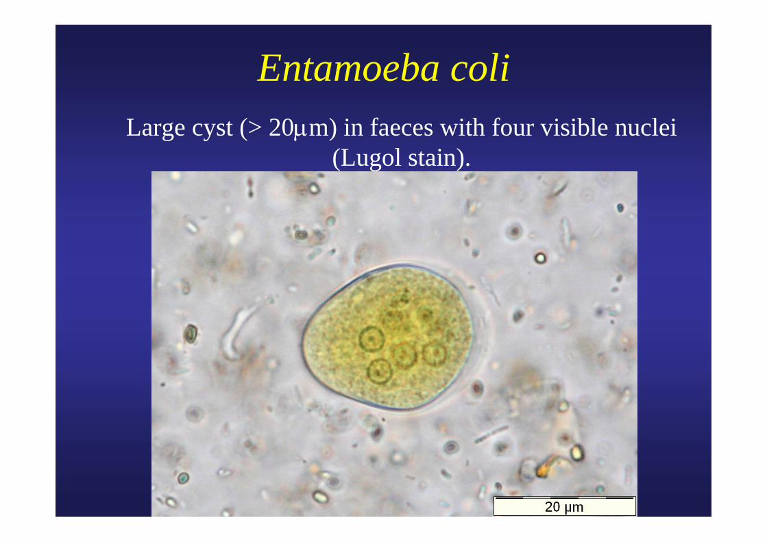
Entamoeba coli Large cyst (> 20μm) in faeces with four visible nuclei
(Lugol stain).
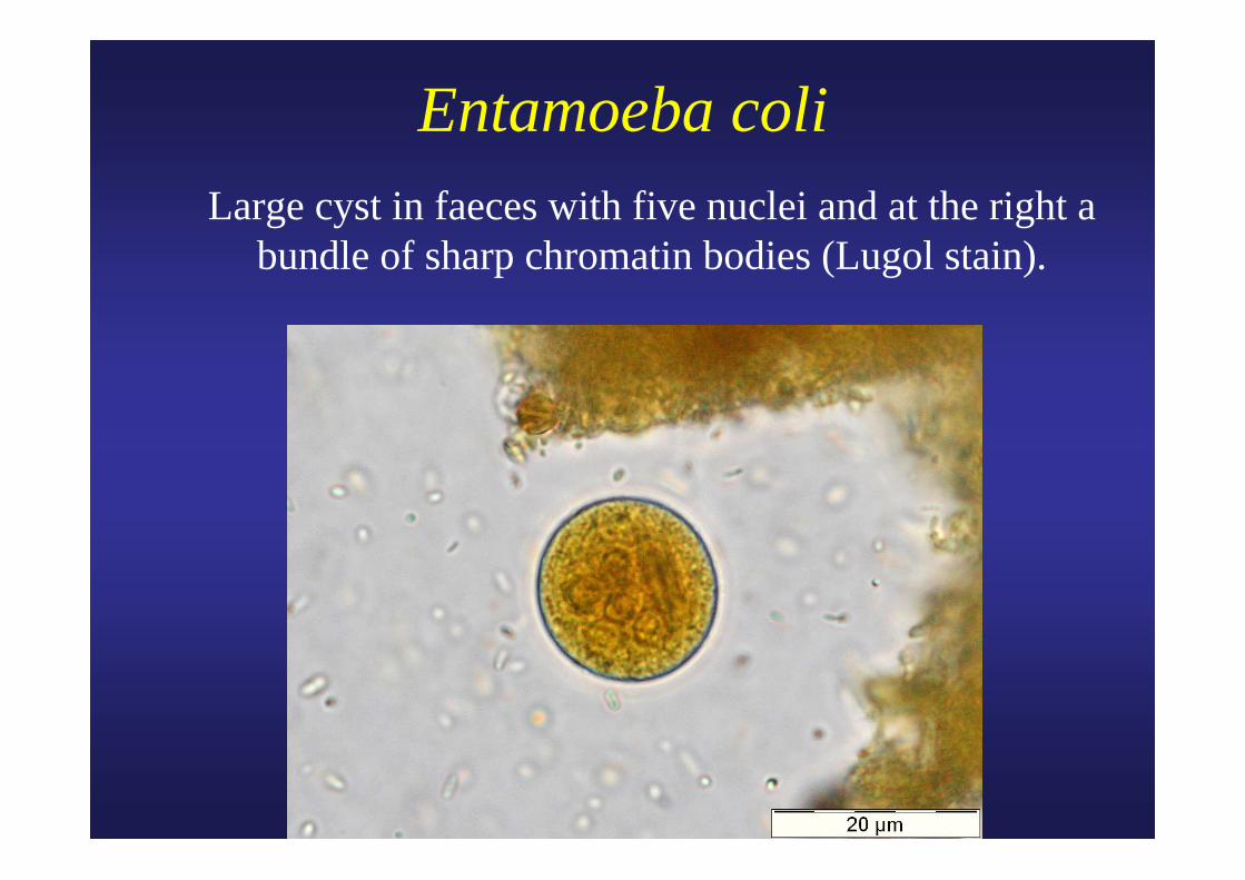
Entamoeba coli Large cyst in faeces with five nuclei and at the right a
bundle of sharp chromatin bodies (Lugol stain).
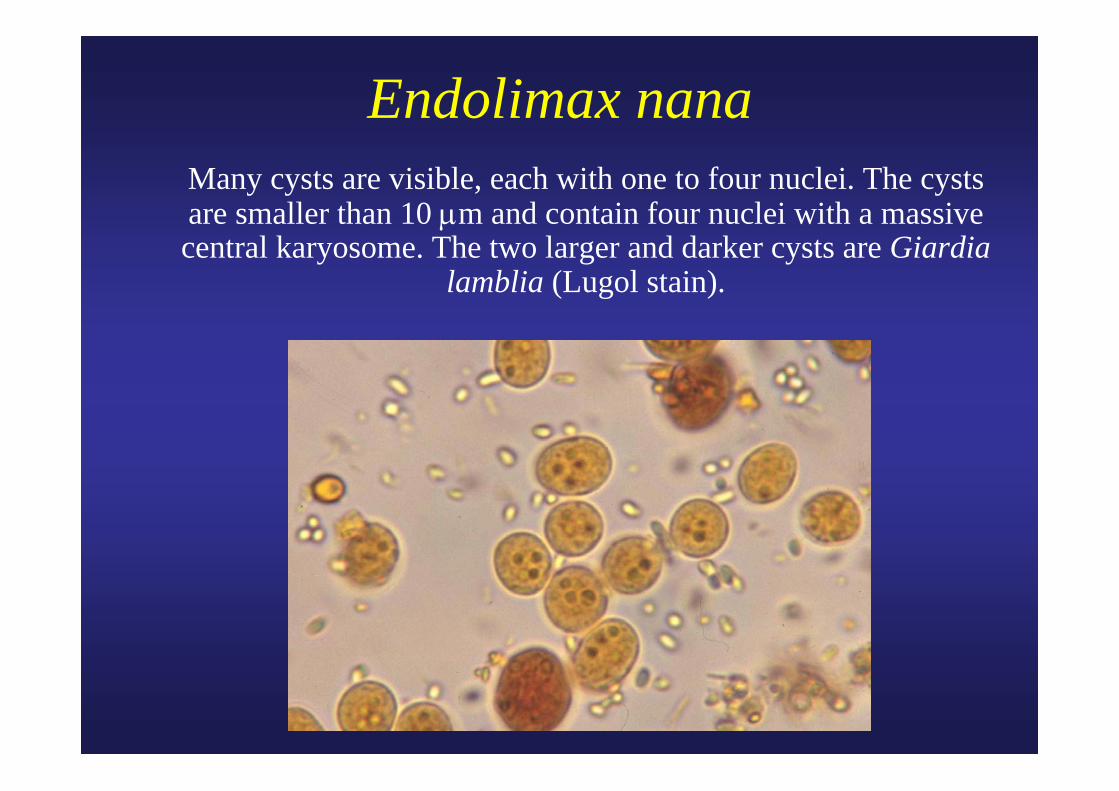
Endolimax nana Many cysts are visible, each with one to four nuclei. The cysts
are smaller than 10 μm and contain four nuclei with a massive central karyosome. The two larger and darker cysts are Giardia
lamblia (Lugol stain).
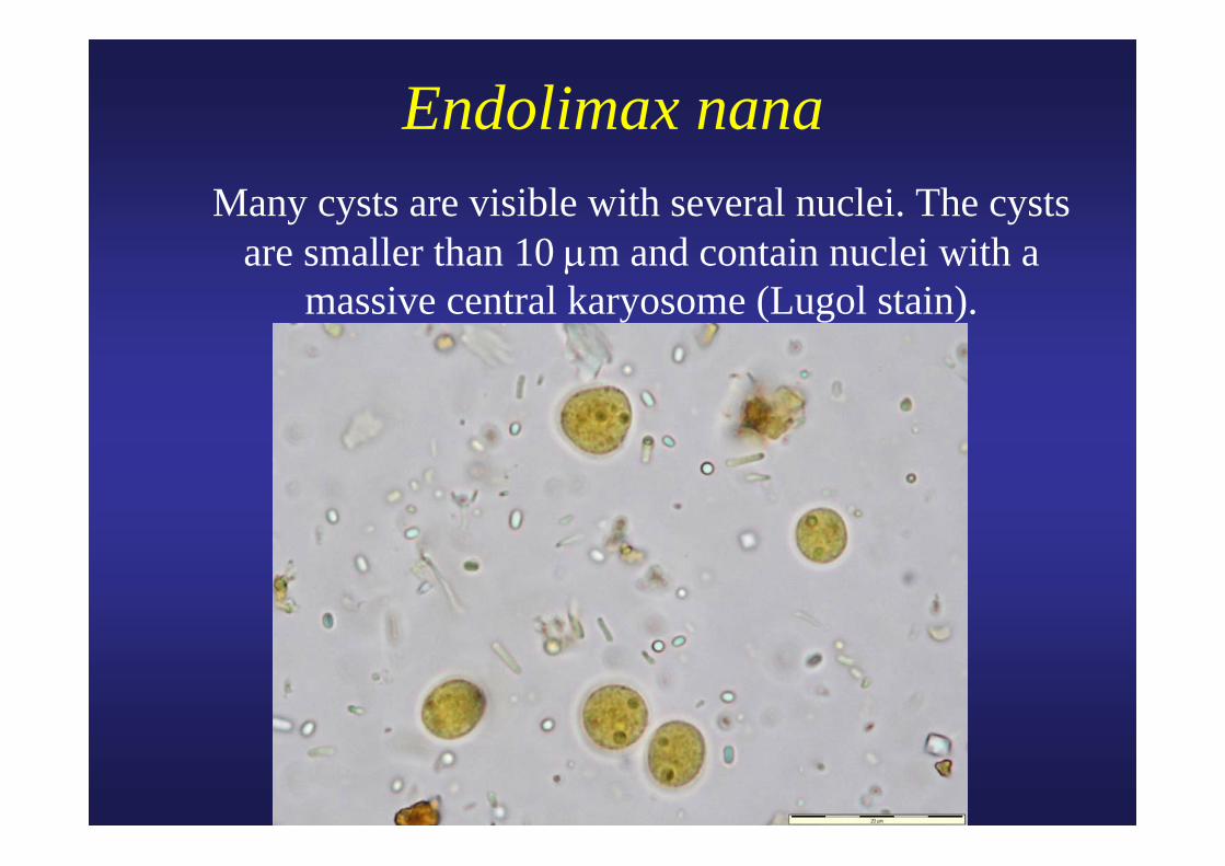
Endolimax nana Many cysts are visible with several nuclei. The cysts
are smaller than 10 μm and contain nuclei with a massive central karyosome (Lugol stain).

Endolimax nana Three cysts are visible with to two to four nuclei. The
cysts are smaller than 10 μm and contain nuclei with a massive central karyosome (Lugol stain).
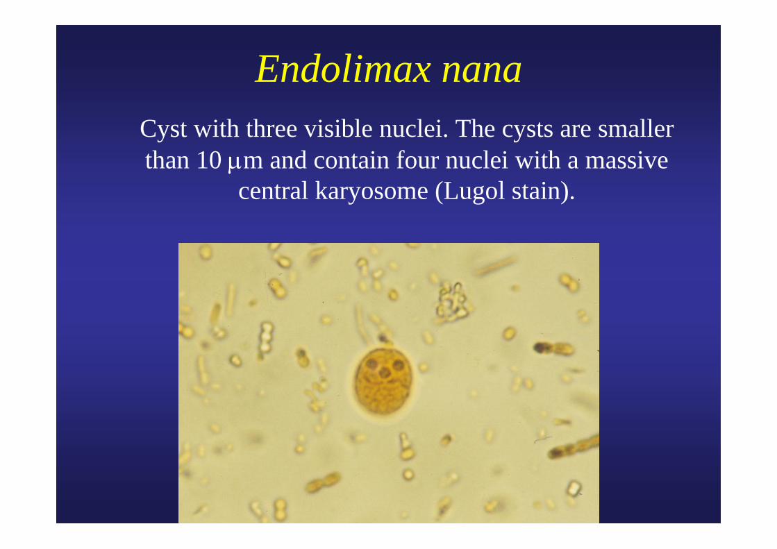
Endolimax nana Cyst with three visible nuclei. The cysts are smaller
than 10 μm and contain four nuclei with a massive central karyosome (Lugol stain).
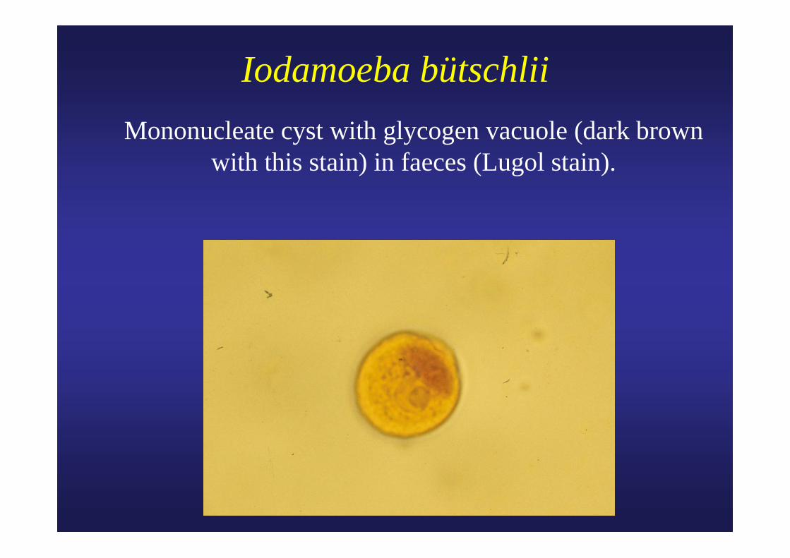
Iodamoeba bütschlii Mononucleate cyst with glycogen vacuole (dark brown
with this stain) in faeces (Lugol stain).
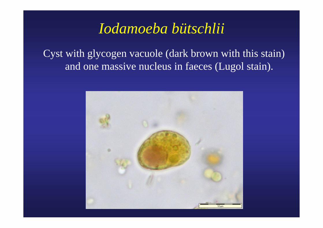
Iodamoeba bütschliiCyst with glycogen vacuole (dark brown with this stain)
and one massive nucleus in faeces (Lugol stain).
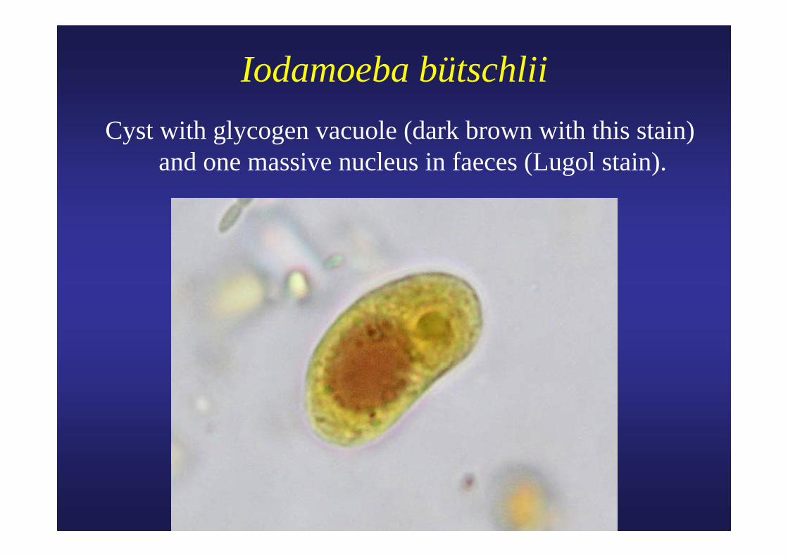
Iodamoeba bütschliiCyst with glycogen vacuole (dark brown with this stain)
and one massive nucleus in faeces (Lugol stain).
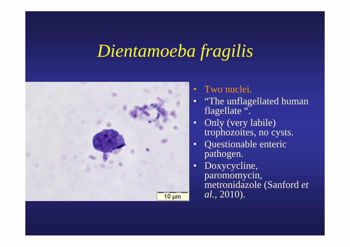
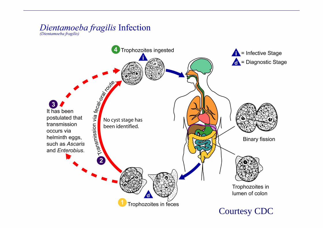
Dientamoeba fragilis
• Two nuclei.• “The unflagellated human
flagellate “.• Only (very labile)
trophozoites, no cysts.• Questionable enteric
pathogen.• Doxycycline,
paromomycin, metronidazole (Sanford et al., 2010).
Courtesy CDC
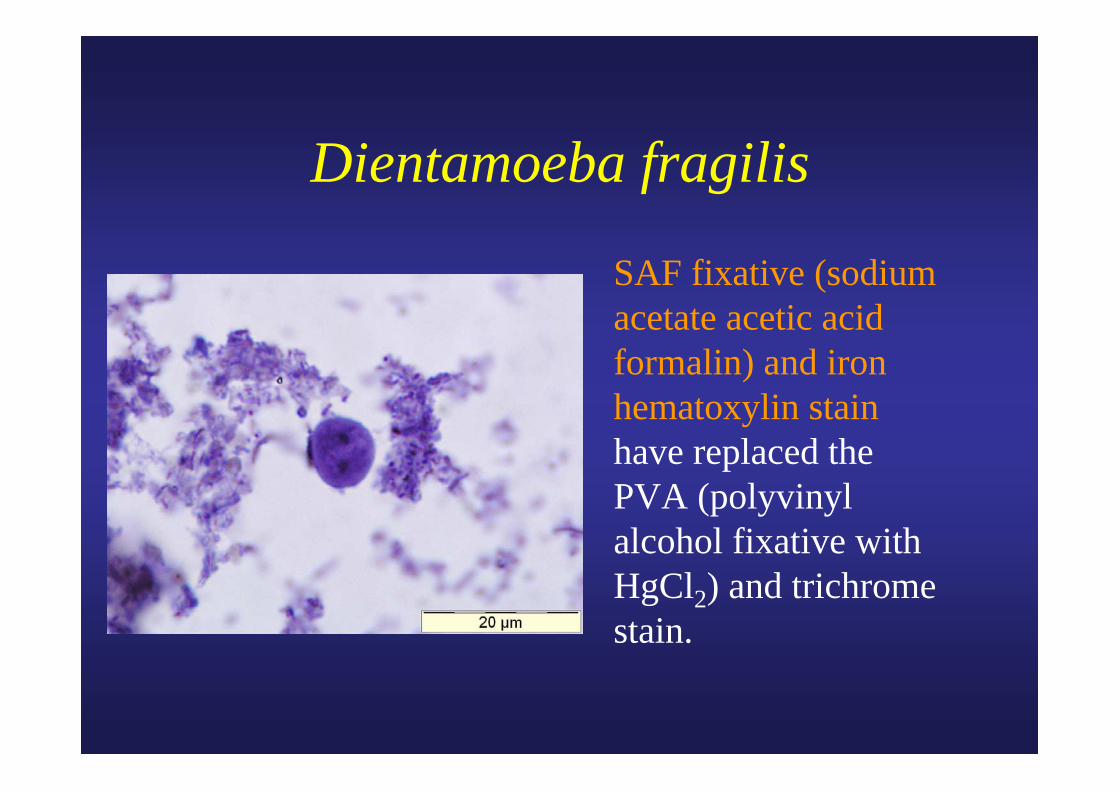
Dientamoeba fragilis
SAF fixative (sodium acetate acetic acid formalin) and iron hematoxylin stain have replaced the PVA (polyvinyl alcohol fixative with HgCl2) and trichrome stain.
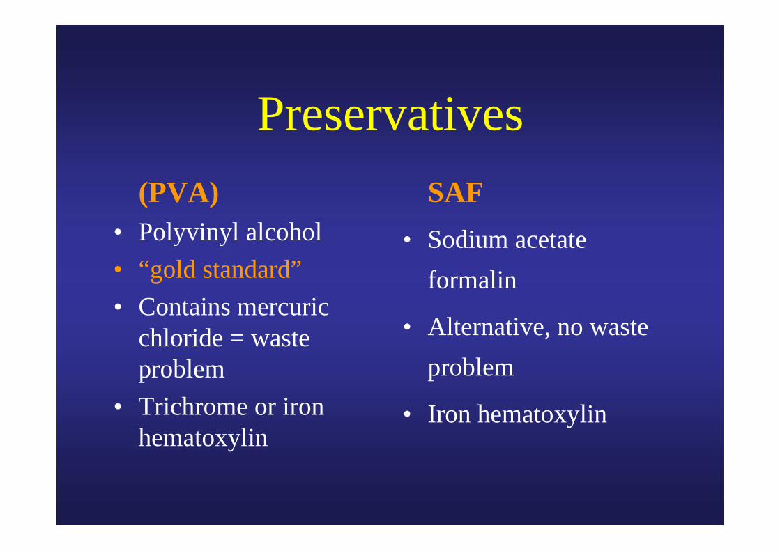
Preservatives (PVA)• Polyvinyl alcohol• “gold standard”• Contains mercuric
chloride = waste problem
• Trichrome or iron hematoxylin
SAF• Sodium acetate
formalin
• Alternative, no waste problem
• Iron hematoxylin
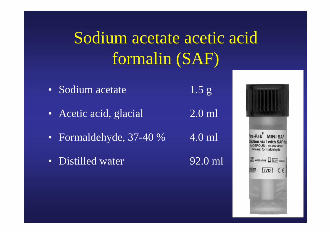
Sodium acetate acetic acid formalin (SAF)
• Sodium acetate 1.5 g
• Acetic acid, glacial 2.0 ml
• Formaldehyde, 37-40 % 4.0 ml
• Distilled water 92.0 ml
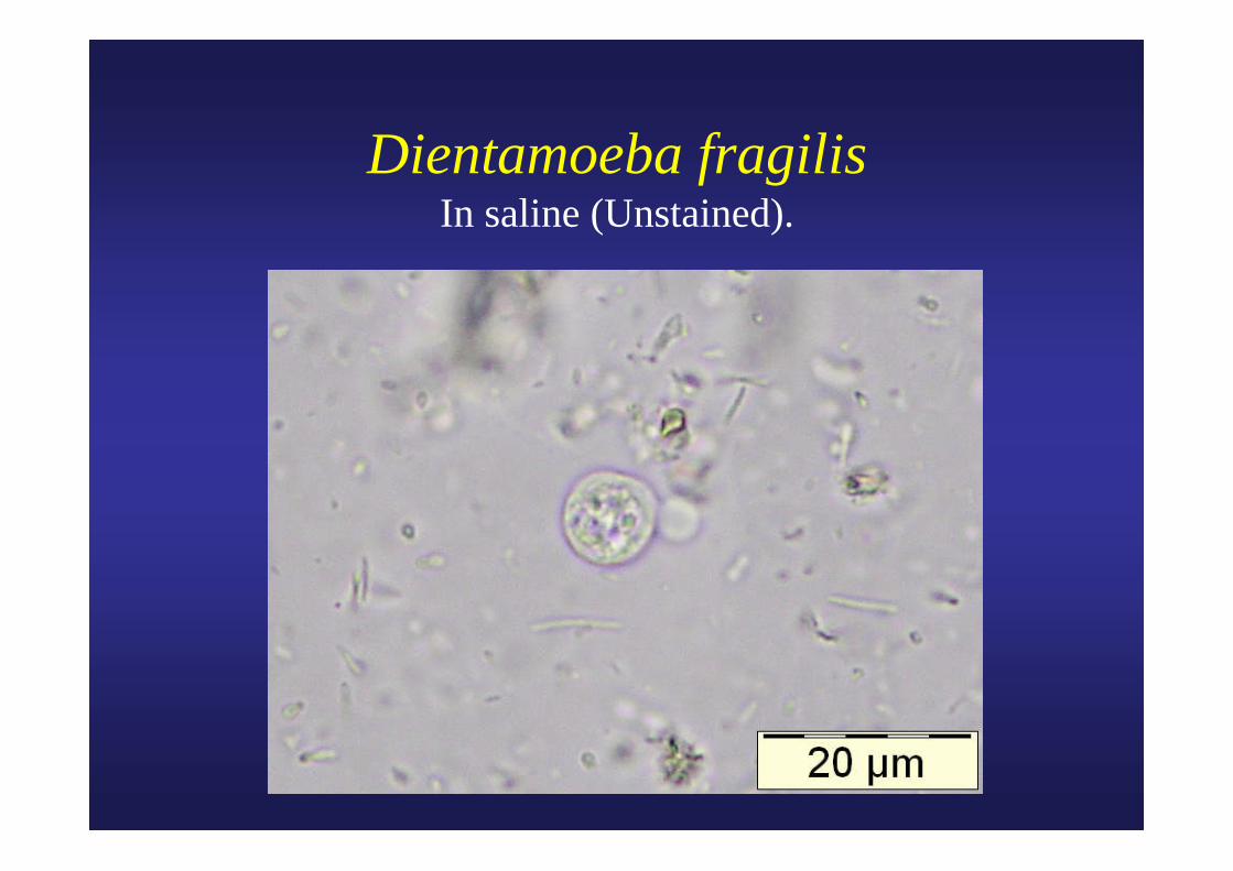
Dientamoeba fragilisIn saline (Unstained).
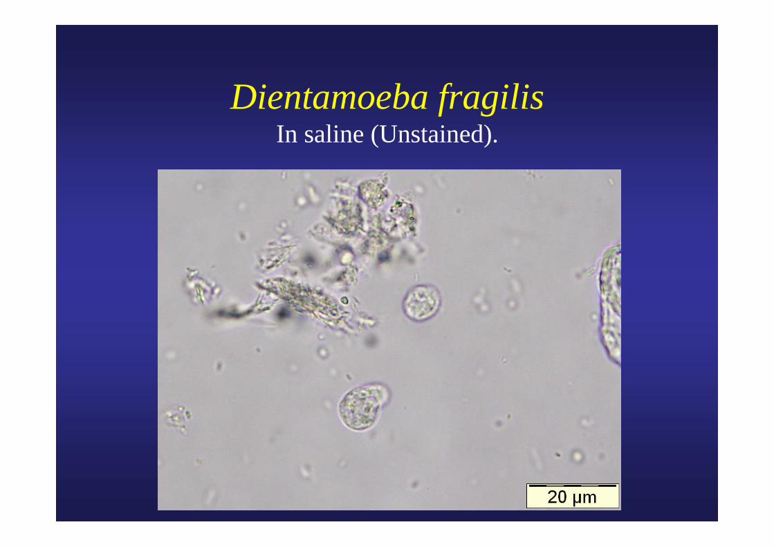
Dientamoeba fragilisIn saline (Unstained).
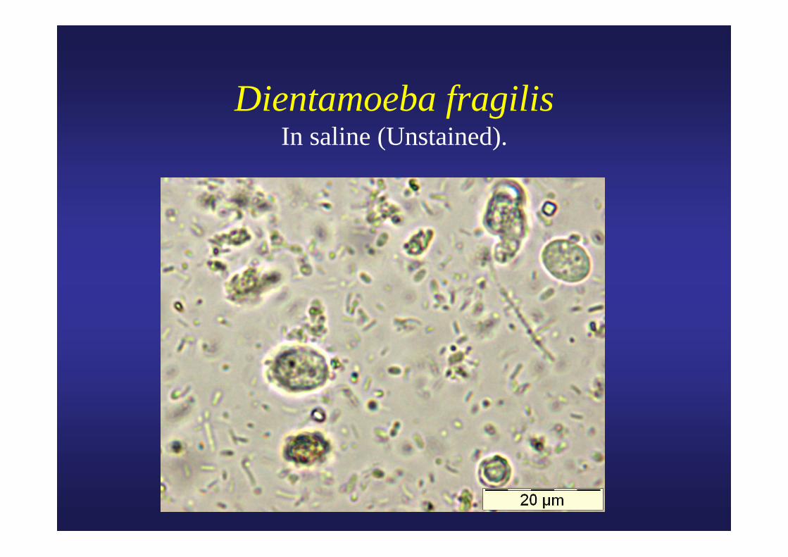
Dientamoeba fragilisIn saline (Unstained).
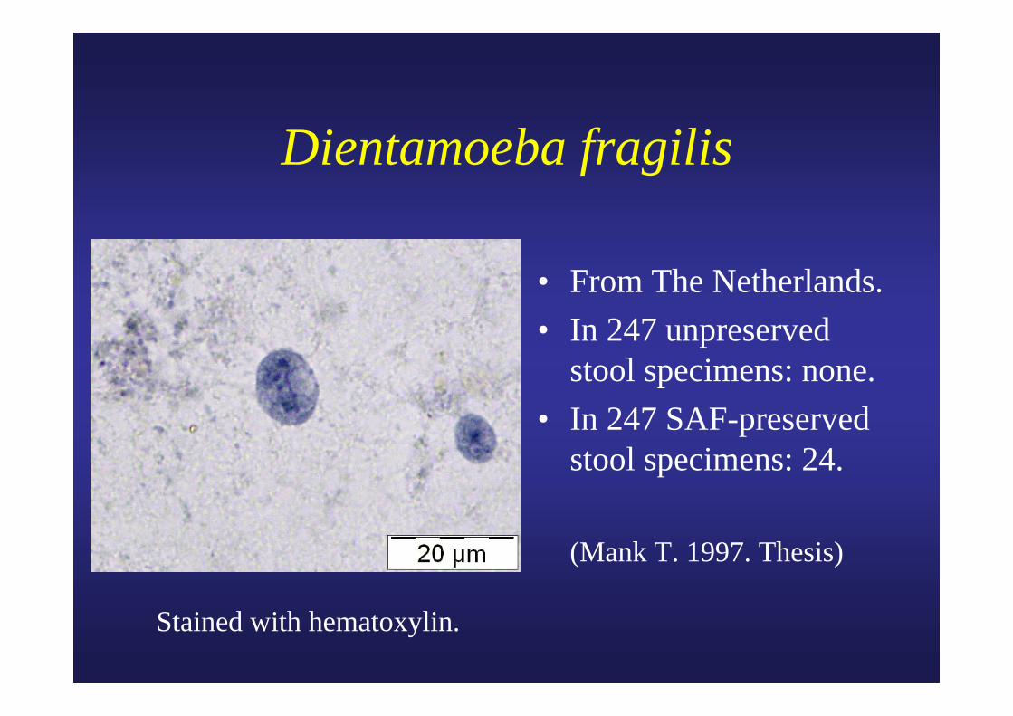
Dientamoeba fragilis
• From The Netherlands.• In 247 unpreserved
stool specimens: none.• In 247 SAF-preserved
stool specimens: 24.
(Mank T. 1997. Thesis)
Stained with hematoxylin.
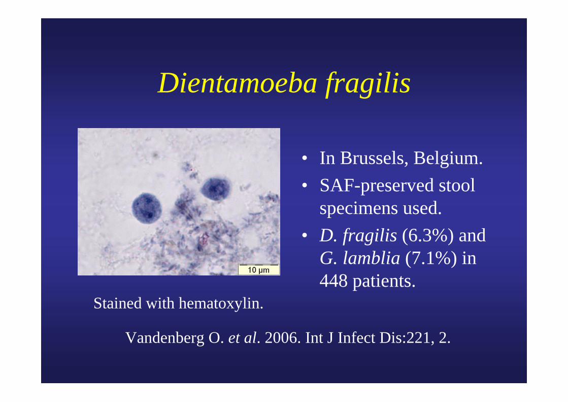
Dientamoeba fragilis
• In Brussels, Belgium.• SAF-preserved stool
specimens used.• D. fragilis (6.3%) and
G. lamblia (7.1%) in 448 patients.
Vandenberg O. et al. 2006. Int J Infect Dis:221, 2.
Stained with hematoxylin.
Courtesy ASM
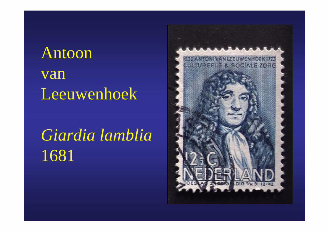
AntoonvanLeeuwenhoek
Giardia lamblia1681
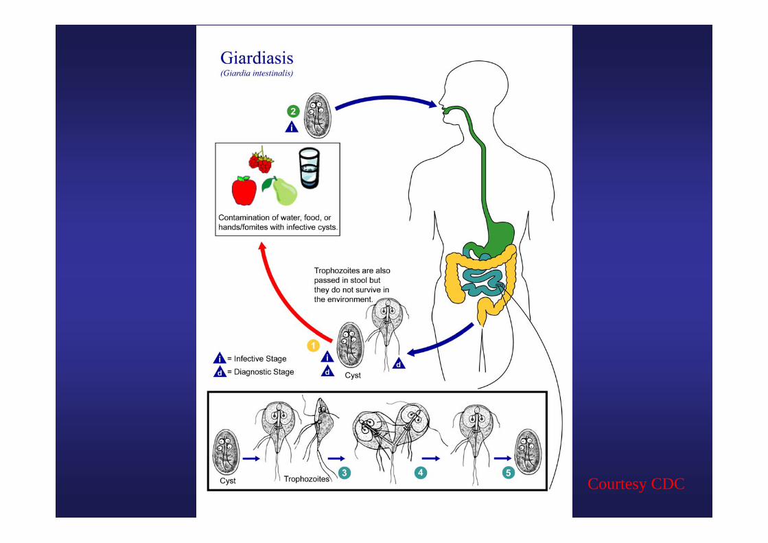
Courtesy CDC
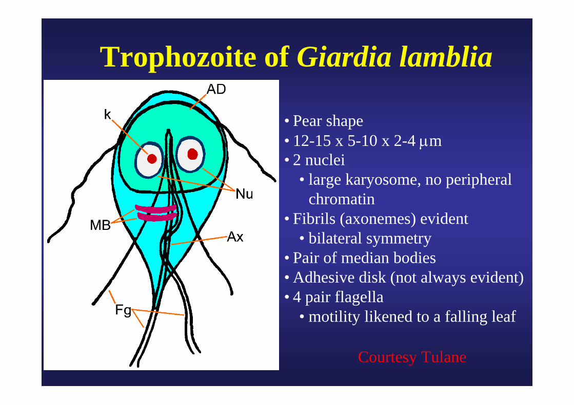
• Pear shape• 12-15 x 5-10 x 2-4 μm• 2 nuclei
• large karyosome, no peripheral chromatin
• Fibrils (axonemes) evident• bilateral symmetry
• Pair of median bodies• Adhesive disk (not always evident)• 4 pair flagella
• motility likened to a falling leaf
Trophozoite of Giardia lamblia
Courtesy Tulane
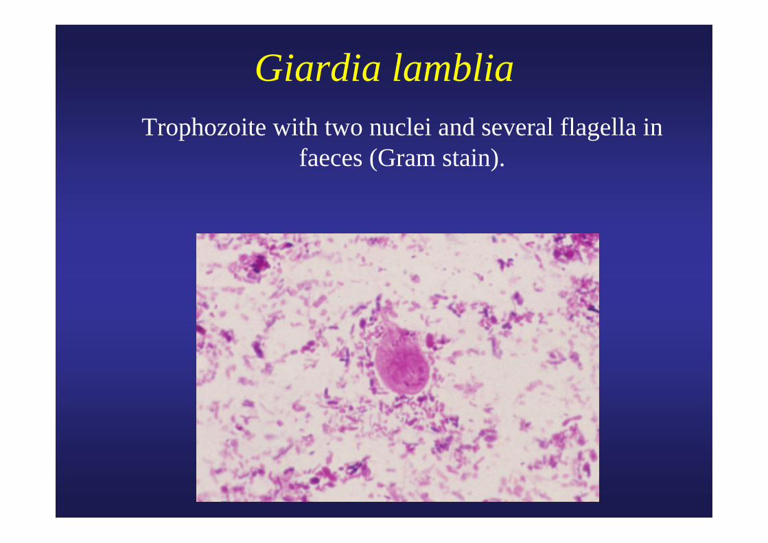
Giardia lamblia Trophozoite with two nuclei and several flagella in
faeces (Gram stain).
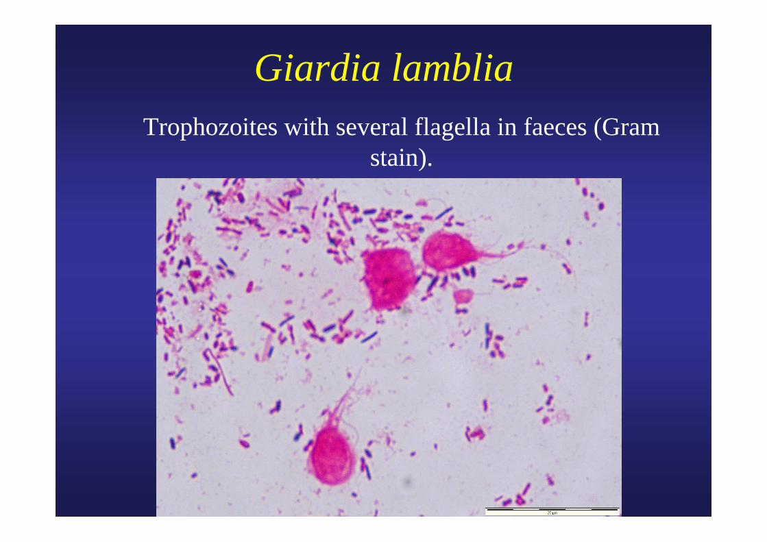
Giardia lamblia Trophozoites with several flagella in faeces (Gram
stain).
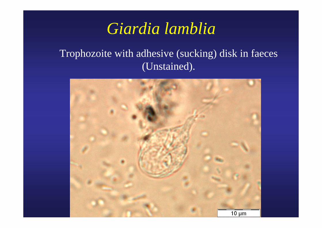
Giardia lamblia Trophozoite with adhesive (sucking) disk in faeces
(Unstained).
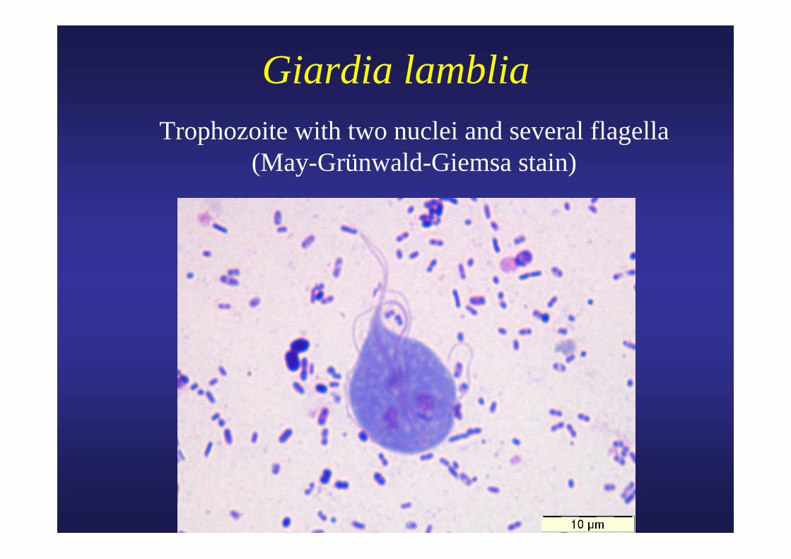
Giardia lamblia Trophozoite with two nuclei and several flagella
(May-Grünwald-Giemsa stain)
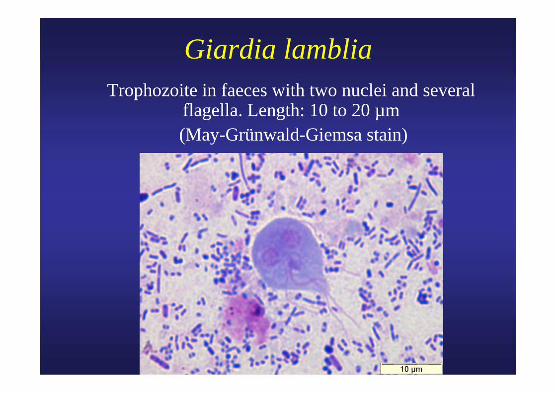
Giardia lamblia Trophozoite in faeces with two nuclei and several
flagella. Length: 10 to 20 µm (May-Grünwald-Giemsa stain)
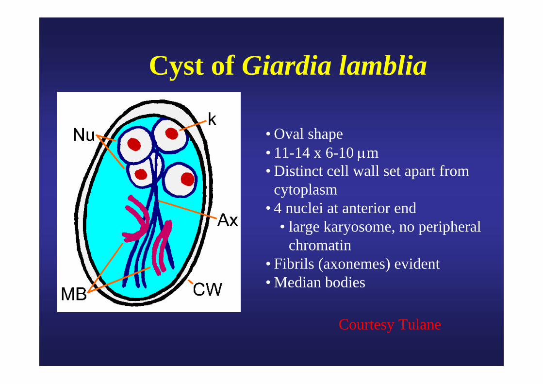
Cyst of Giardia lamblia
• Oval shape• 11-14 x 6-10 μm• Distinct cell wall set apart from
cytoplasm• 4 nuclei at anterior end
• large karyosome, no peripheral chromatin
• Fibrils (axonemes) evident• Median bodies
Courtesy Tulane
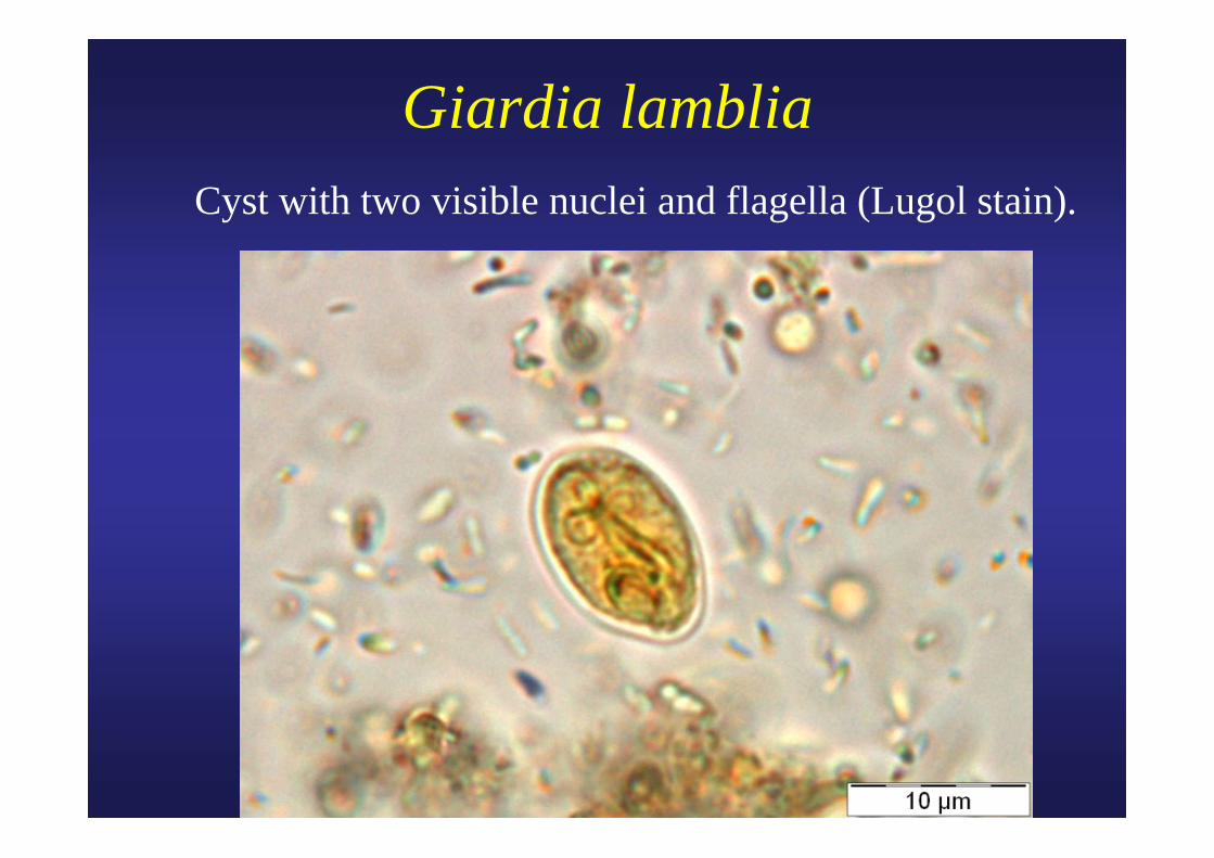
Giardia lamblia Cyst with two visible nuclei and flagella (Lugol stain).
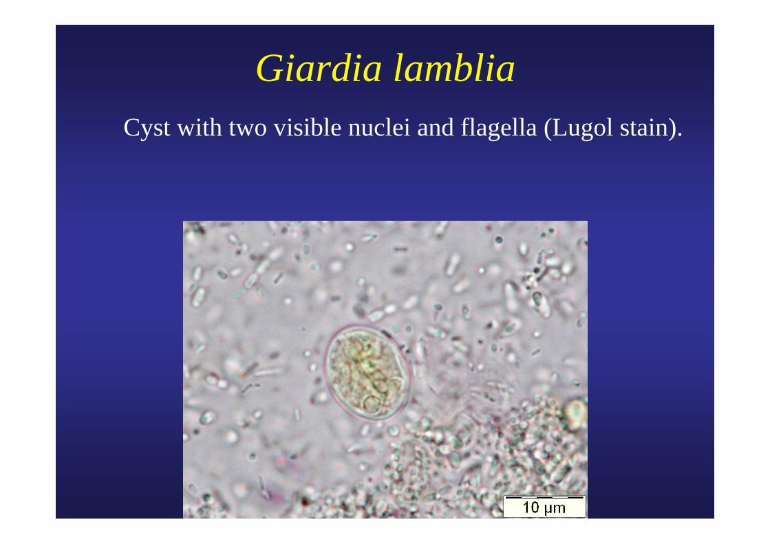
Giardia lamblia Cyst with two visible nuclei and flagella (Lugol stain).
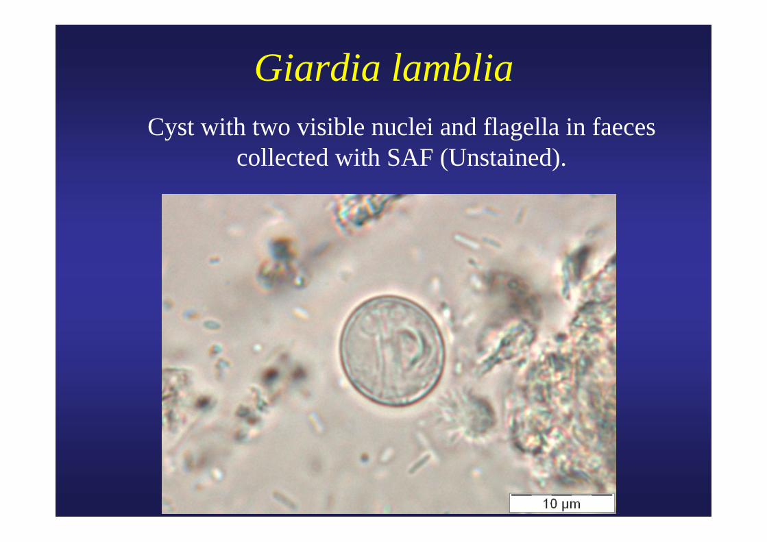
Giardia lamblia Cyst with two visible nuclei and flagella in faeces
collected with SAF (Unstained).
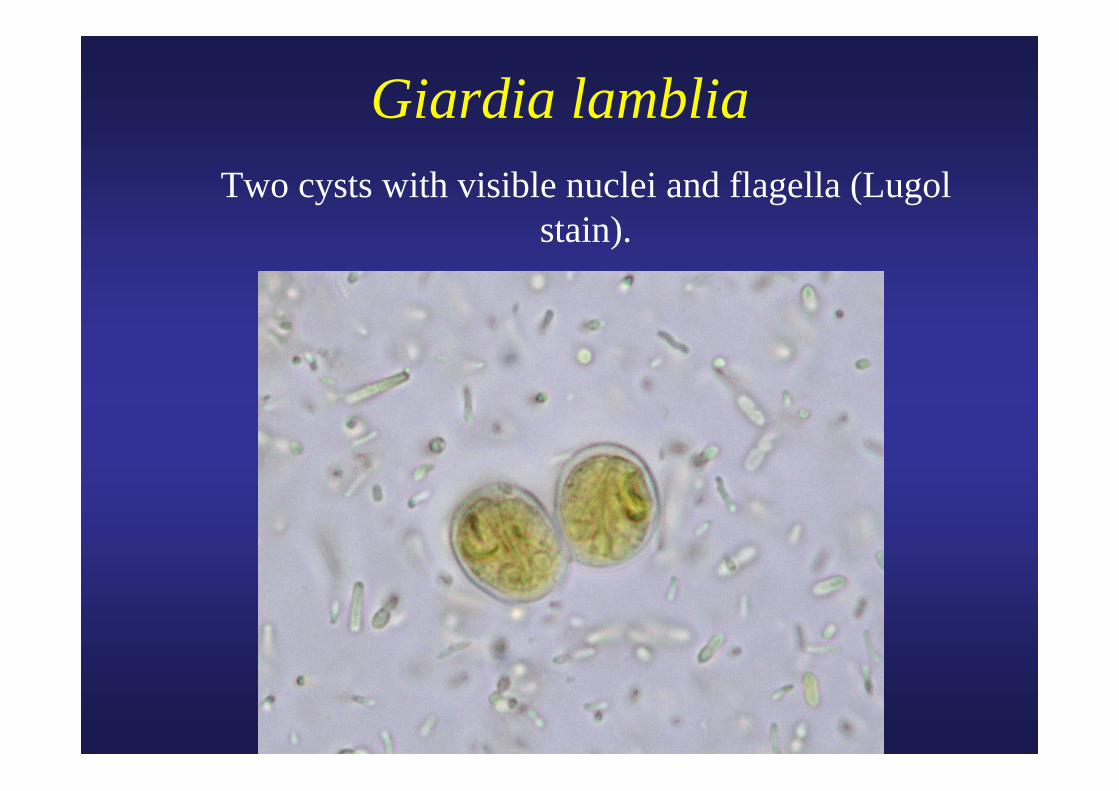
Giardia lamblia Two cysts with visible nuclei and flagella (Lugol
stain).
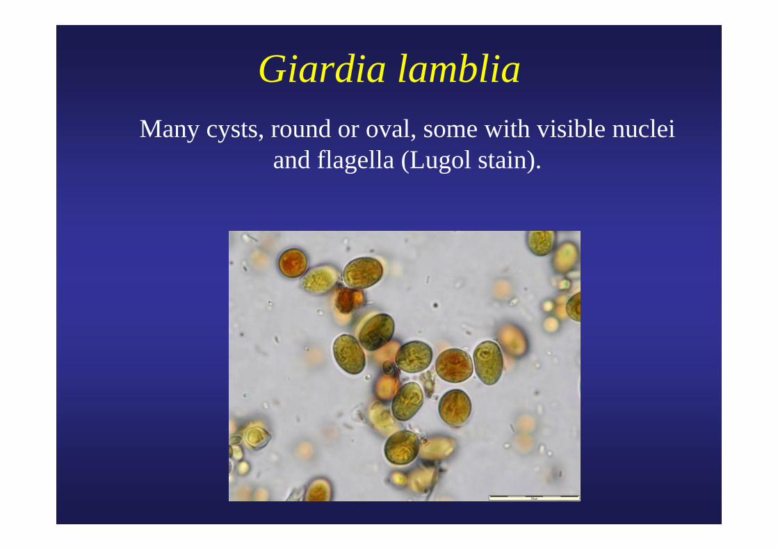
Giardia lamblia Many cysts, round or oval, some with visible nuclei
and flagella (Lugol stain).
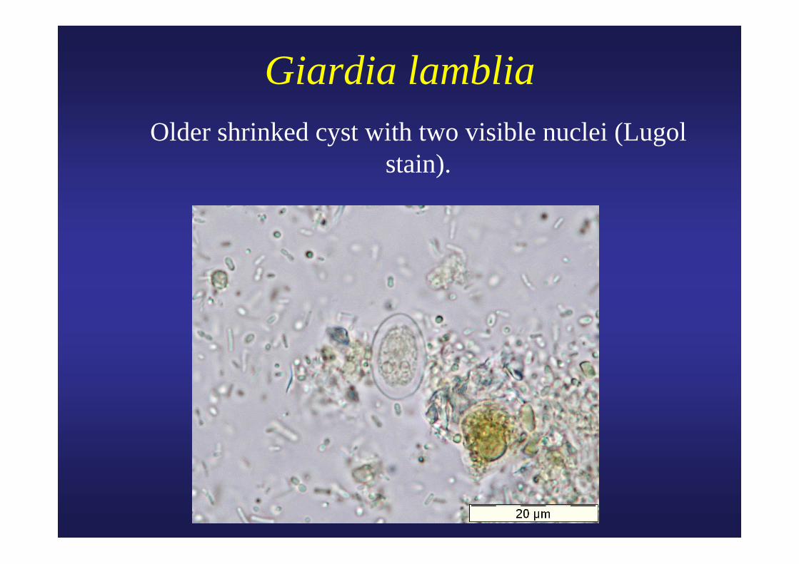
Giardia lamblia Older shrinked cyst with two visible nuclei (Lugol
stain).
Giardia lamblia: antigen detection by IF and ELISA
• Monoclonal antibodies: Merifluor (MERIDIAN) (Cryptosporidium andGiardia).
• 8/9 Giardia ELISAs are OK. (JCM, 1998, 1338).
• Triage parasite panel (BIOSITE) useful. (JCM, 2000, 3337; JCM, 2001, 334).
• One ELISA almost as sensitive as two microscopic examinations. (Mank T. 1997).
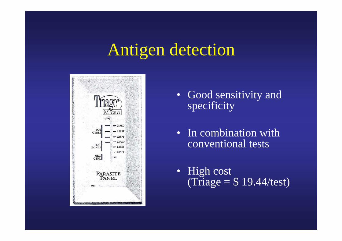
Antigen detection
• Good sensitivity and specificity
• In combination with conventional tests
• High cost (Triage = $ 19.44/test)
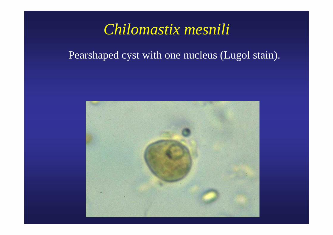
Chilomastix mesnili Pearshaped cyst with one nucleus (Lugol stain).
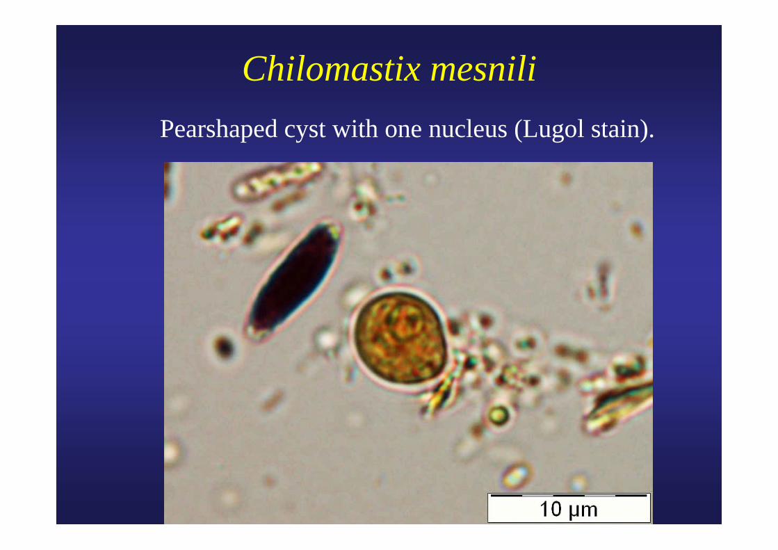
Chilomastix mesnili Pearshaped cyst with one nucleus (Lugol stain).
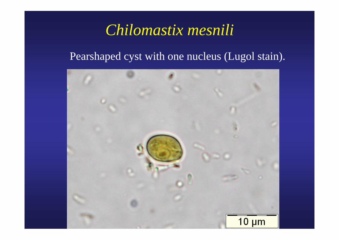
Chilomastix mesnili Pearshaped cyst with one nucleus (Lugol stain).
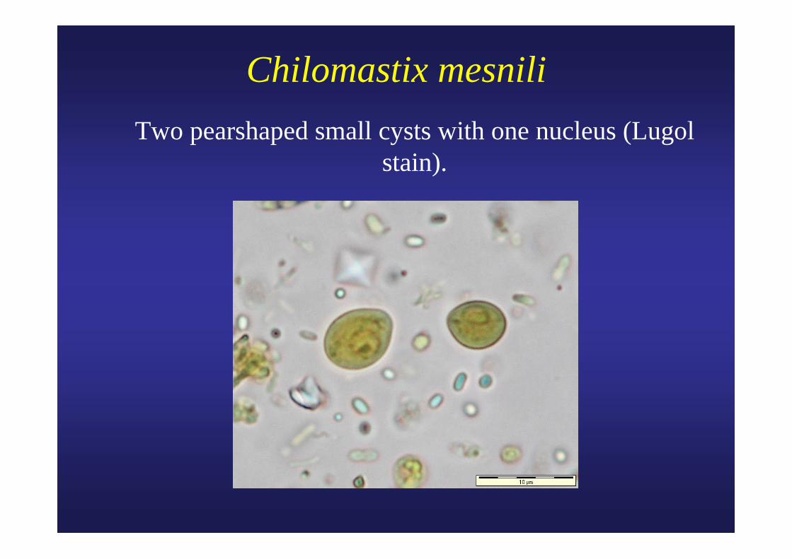
Chilomastix mesnili Two pearshaped small cysts with one nucleus (Lugol
stain).
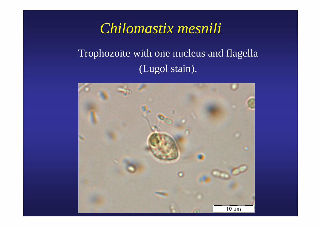
Chilomastix mesnili Trophozoite with one nucleus and flagella
(Lugol stain).
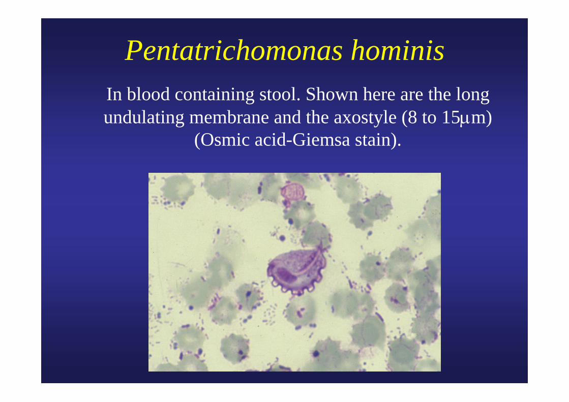
Pentatrichomonas hominis In blood containing stool. Shown here are the long
undulating membrane and the axostyle (8 to 15μm)(Osmic acid-Giemsa stain).
Courtesy ASM
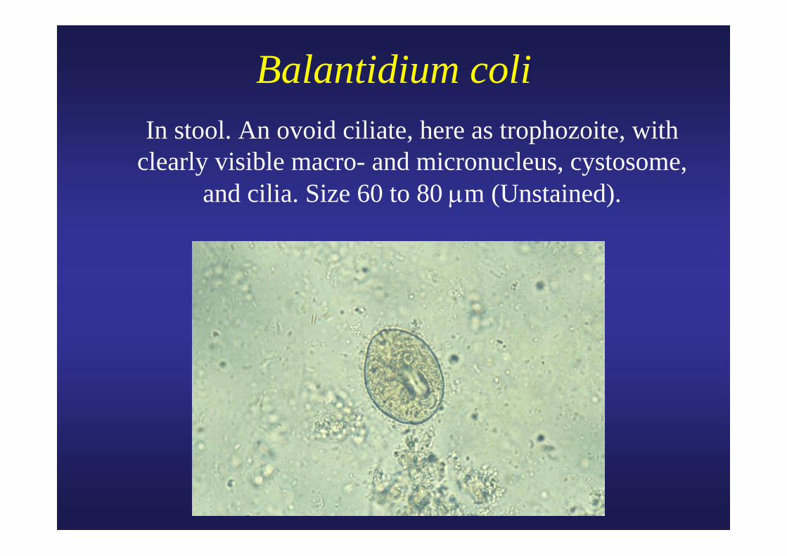
Balantidium coli In stool. An ovoid ciliate, here as trophozoite, with
clearly visible macro- and micronucleus, cystosome, and cilia. Size 60 to 80 μm (Unstained).
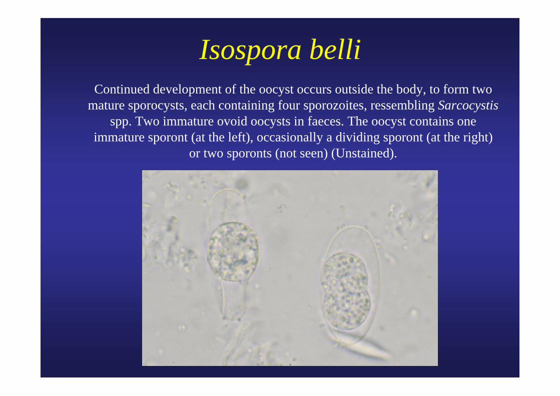
Isospora belli Continued development of the oocyst occurs outside the body, to form two
mature sporocysts, each containing four sporozoites, ressembling Sarcocystisspp. Two immature ovoid oocysts in faeces. The oocyst contains one
immature sporont (at the left), occasionally a dividing sporont (at the right) or two sporonts (not seen) (Unstained).
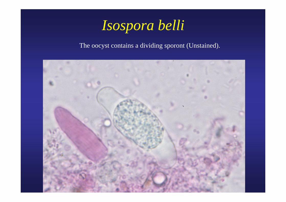
Isospora belli The oocyst contains a dividing sporont (Unstained).

Isospora belli• Human to human transmission.• Eosinophilia may be present.• Worldwide.• Oocyst very pale and
transparent.• Wet-preparation examination
preferred over the stained smear.
• dd. Sarcocystis spp.• Cotrimoxazole (HIV).
Two sporocysts with each four sporozoites.
Fisk T.L et al. 2005.
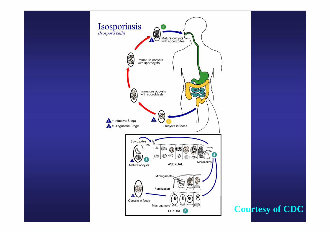
Courtesy of CDC
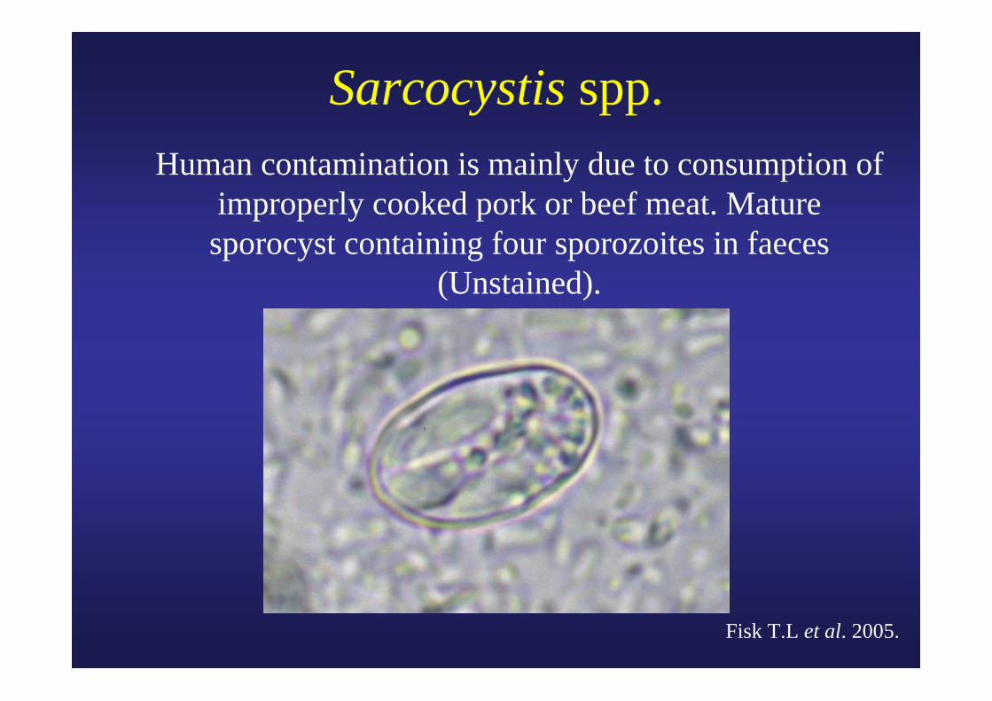
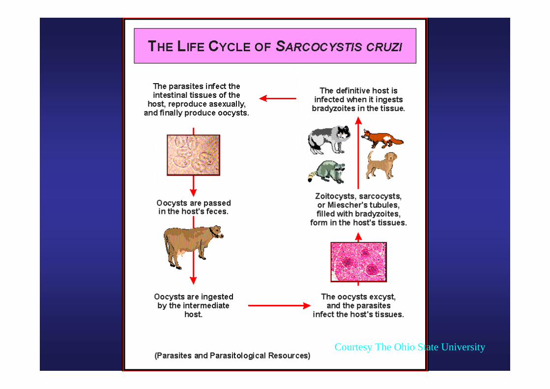
Sarcocystis spp. Human contamination is mainly due to consumption of
improperly cooked pork or beef meat. Mature sporocyst containing four sporozoites in faeces
(Unstained).
Fisk T.L et al. 2005.
Courtesy The Ohio State University
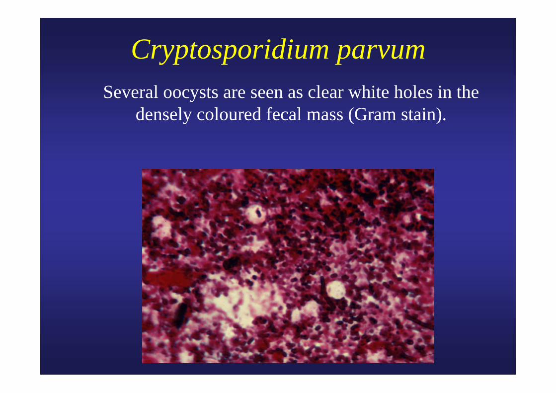
Cryptosporidium parvum Several oocysts are seen as clear white holes in the
densely coloured fecal mass (Gram stain).
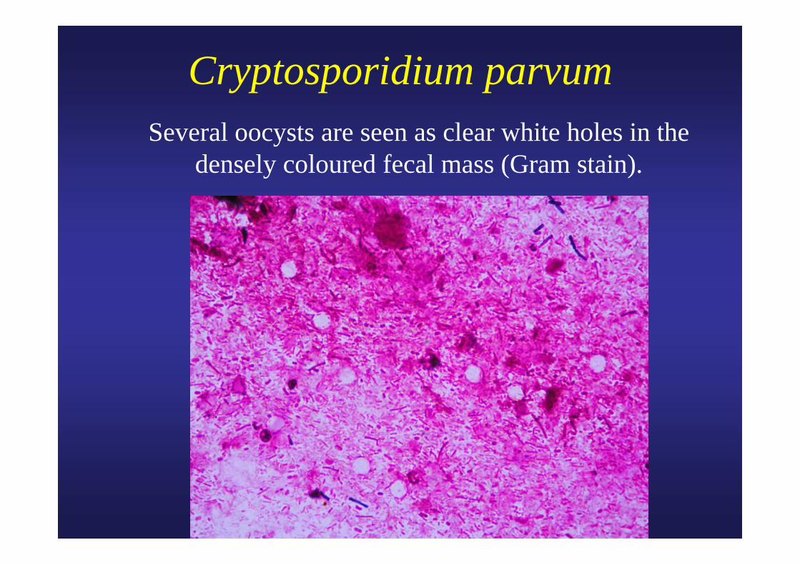
Cryptosporidium parvum Several oocysts are seen as clear white holes in the
densely coloured fecal mass (Gram stain).
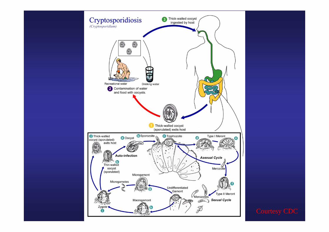
Courtesy CDC
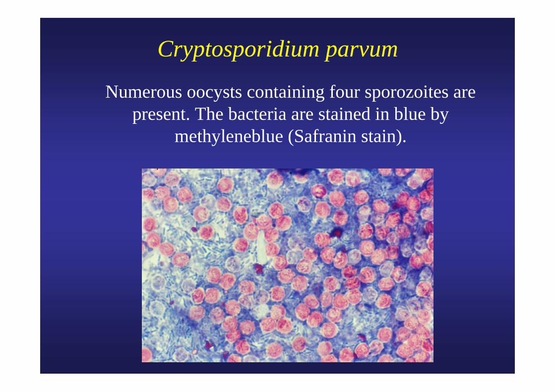
Cryptosporidium parvum
Numerous oocysts containing four sporozoites are present. The bacteria are stained in blue by
methyleneblue (Safranin stain).
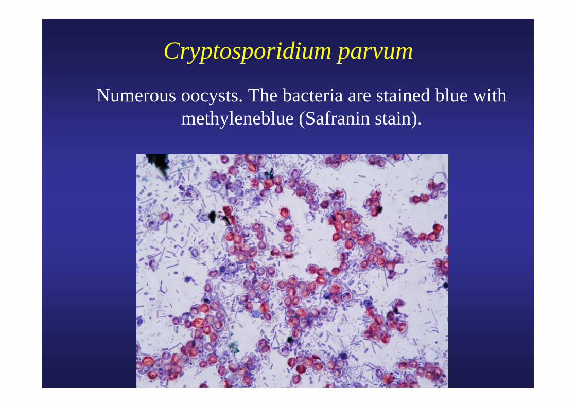
Cryptosporidium parvum
Numerous oocysts. The bacteria are stained blue with methyleneblue (Safranin stain).
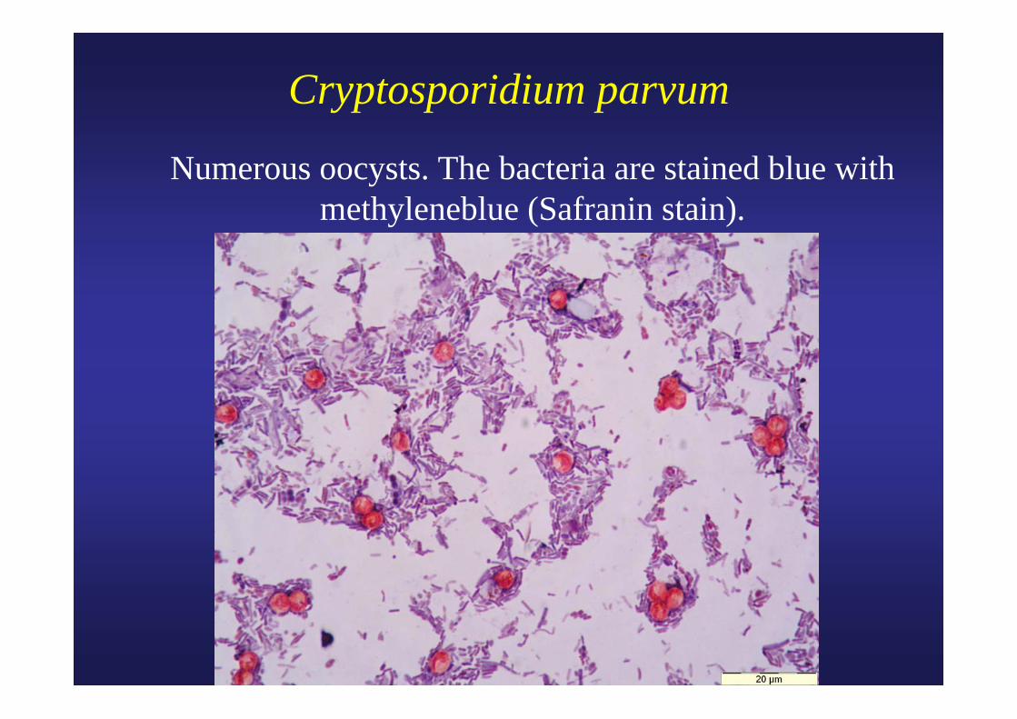
Cryptosporidium parvum
Numerous oocysts. The bacteria are stained blue with methyleneblue (Safranin stain).
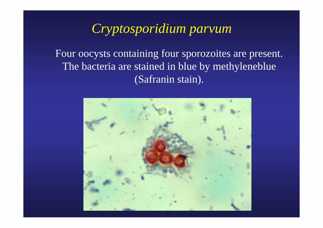
Cryptosporidium parvum
Four oocysts containing four sporozoites are present. The bacteria are stained in blue by methyleneblue
(Safranin stain).
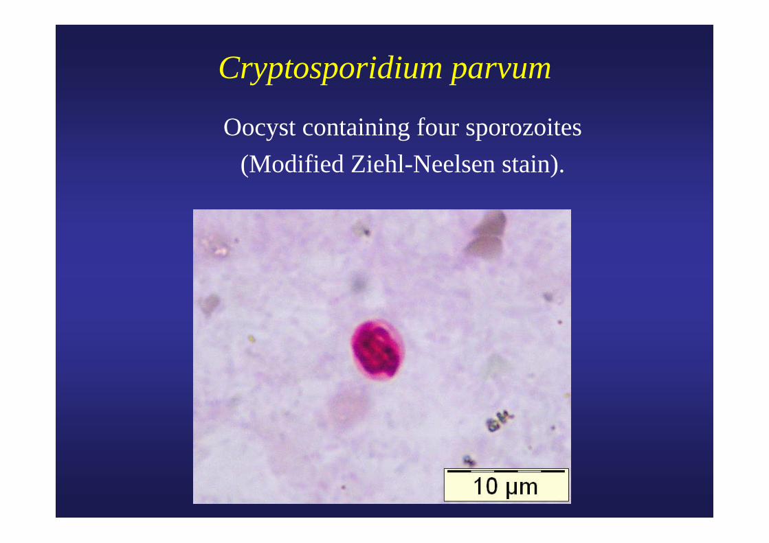
Cryptosporidium parvum
Oocyst containing four sporozoites (Modified Ziehl-Neelsen stain).
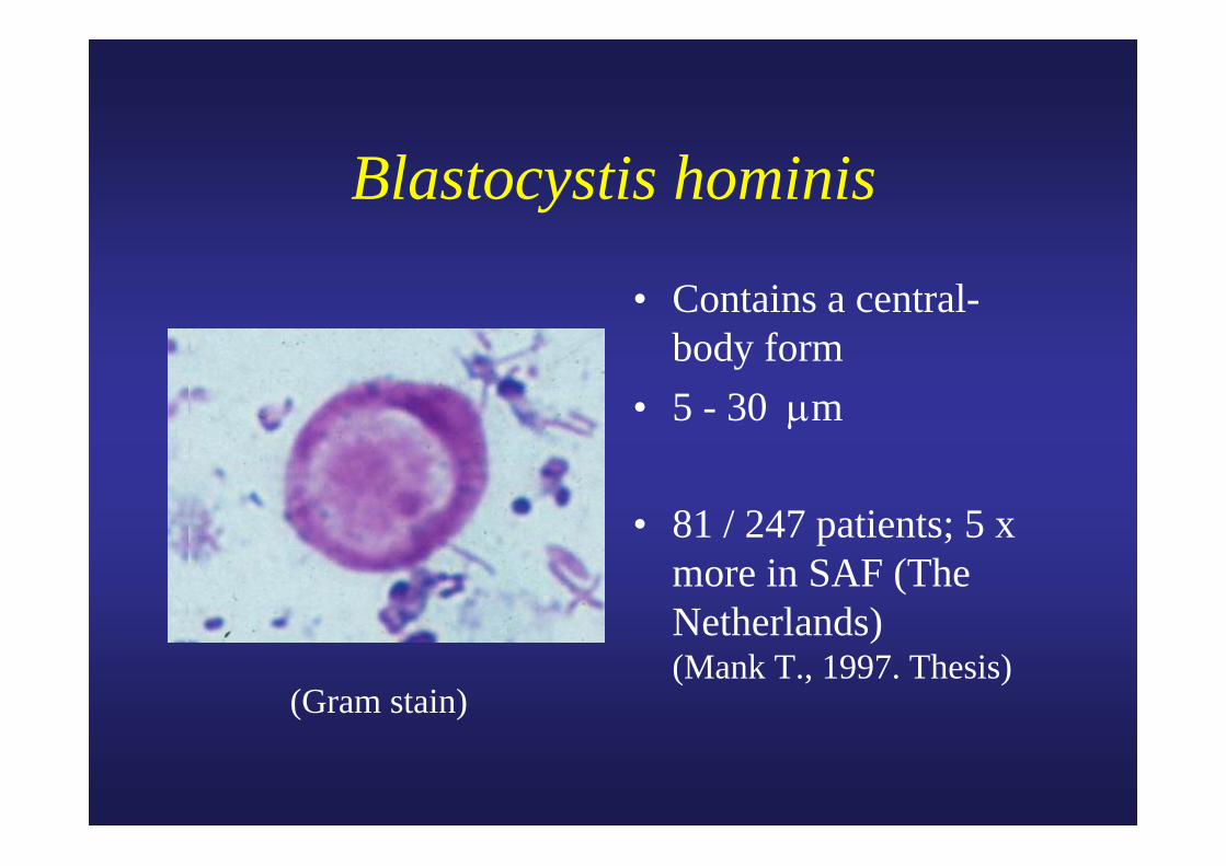
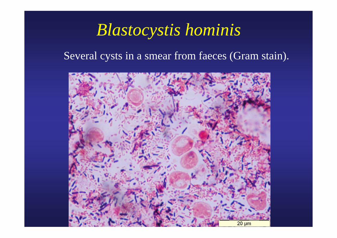
Blastocystis hominis
• Contains a central-body form
• 5 - 30 μm
• 81 / 247 patients; 5 x more in SAF (The Netherlands) (Mank T., 1997. Thesis)
(Gram stain)
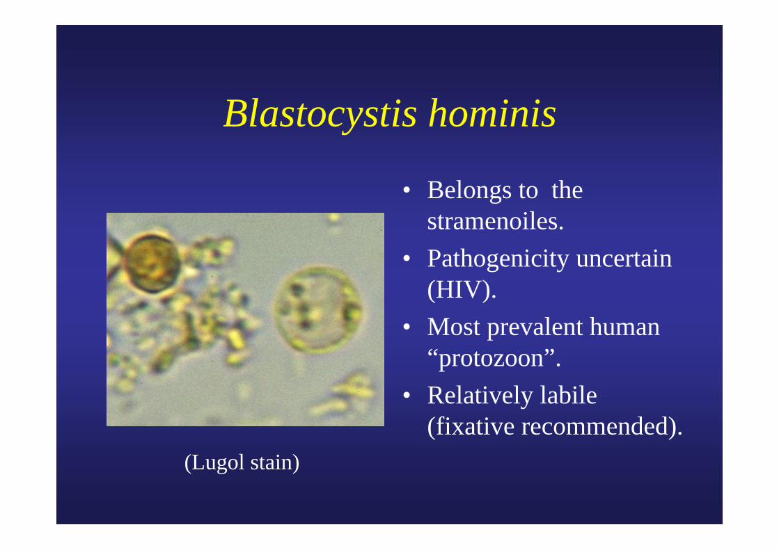
Blastocystis hominis
• Belongs to the stramenoiles.
• Pathogenicity uncertain (HIV).
• Most prevalent human “protozoon”.
• Relatively labile (fixative recommended).
(Lugol stain)
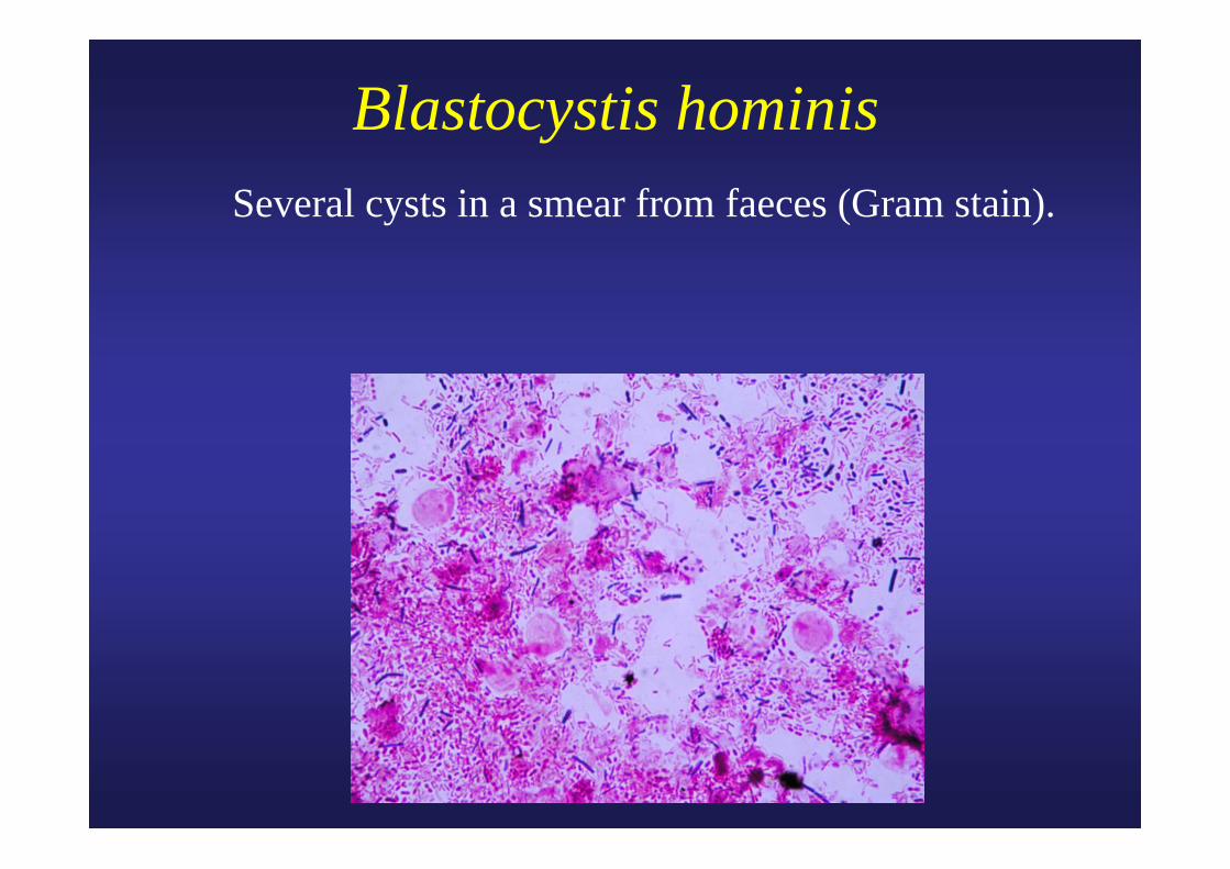
Blastocystis hominis Several cysts in a smear from faeces (Gram stain).
Blastocystis hominis Several cysts in a smear from faeces (Gram stain).
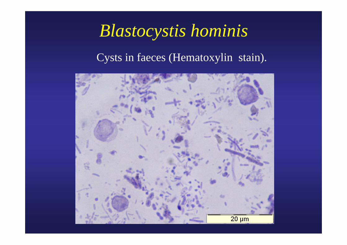
Blastocystis hominis Cysts in faeces (Hematoxylin stain).
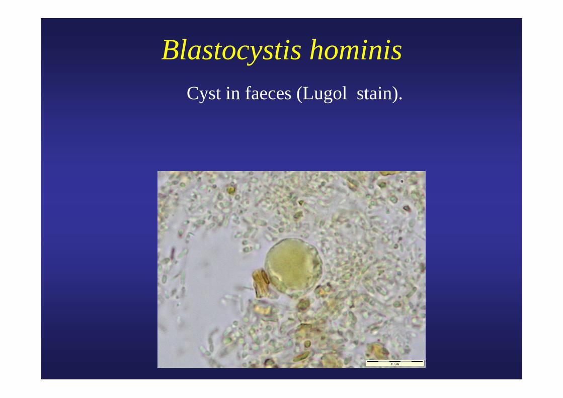
Blastocystis hominis Cyst in faeces (Lugol stain).
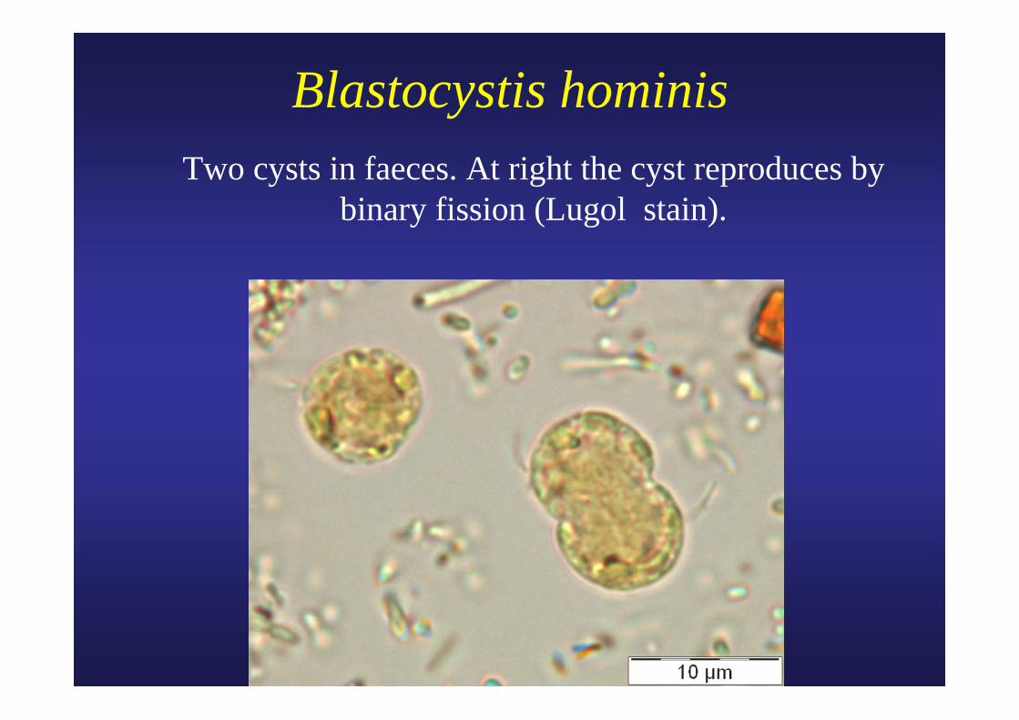
Blastocystis hominis Two cysts in faeces. At right the cyst reproduces by
binary fission (Lugol stain).
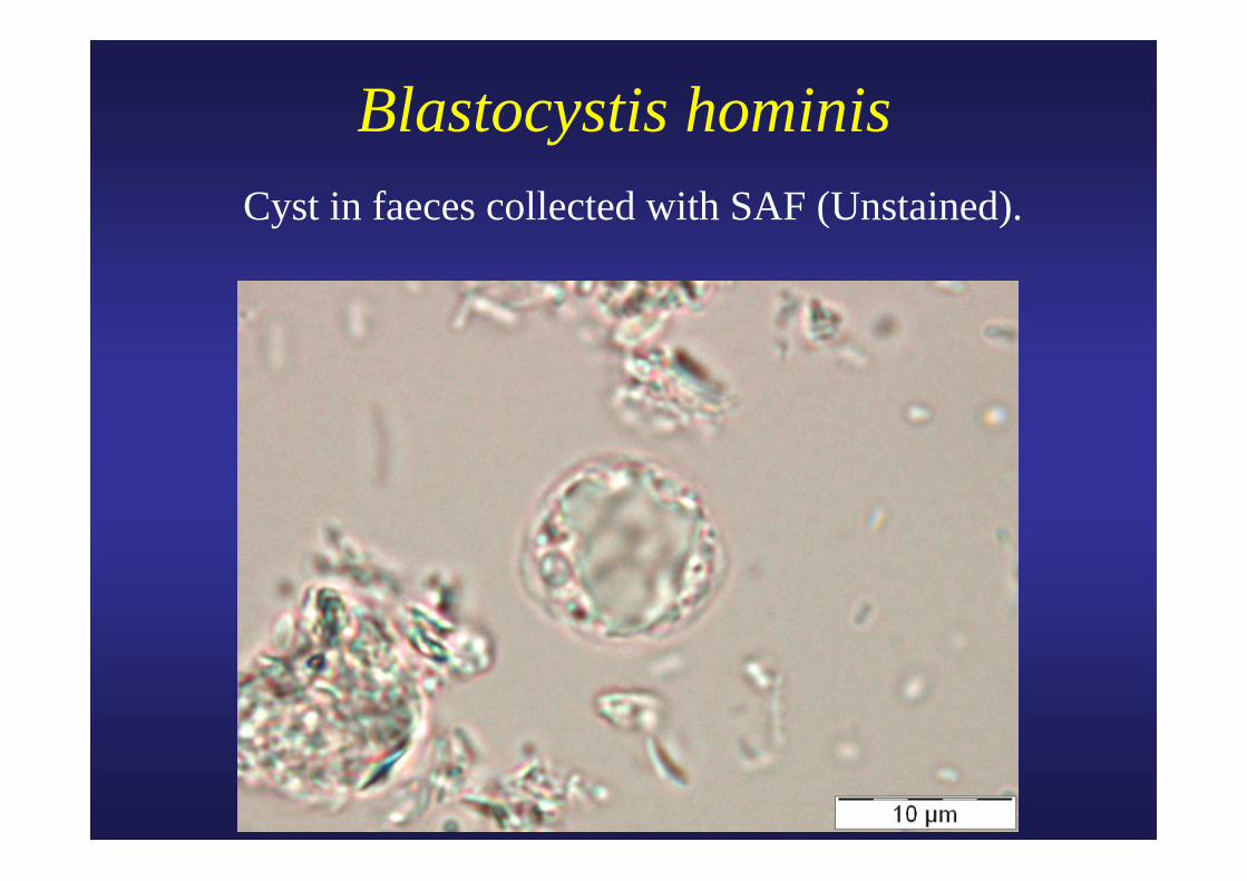
Blastocystis hominisCyst in faeces collected with SAF (Unstained).
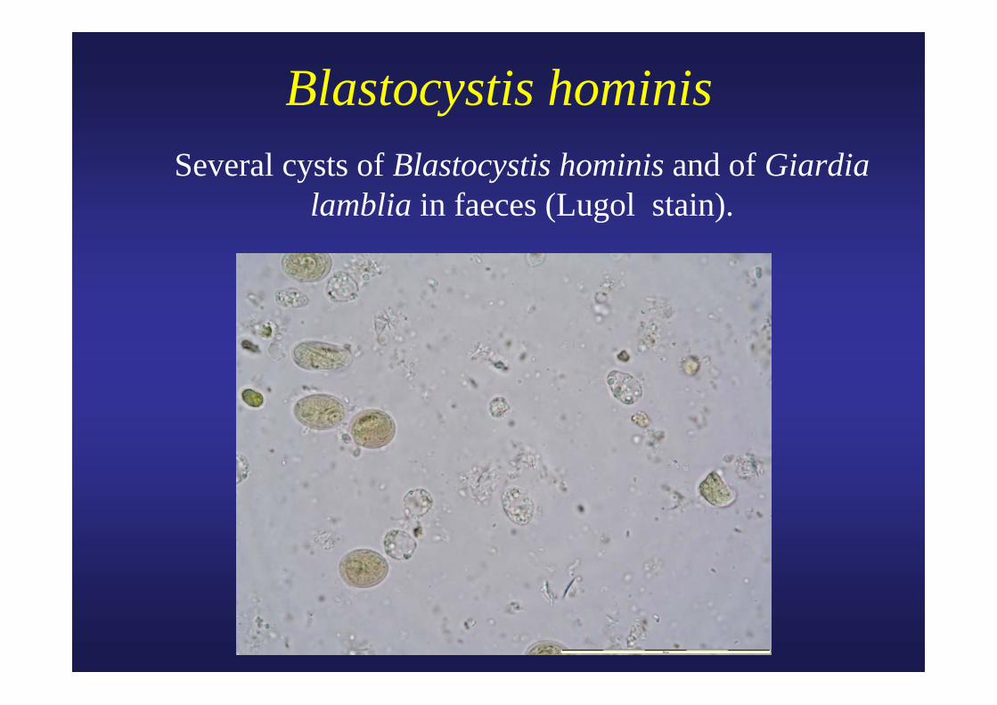
Blastocystis hominis Several cysts of Blastocystis hominis and of Giardia
lamblia in faeces (Lugol stain).
Cyclospora cayetanensis
• Blue-green algae, cyanobacterium-like bodies (CLB).
• Spherical oocyst (8-10 μm) containing small granules with a greenish shade, showing fluorescence under UV illumination.
• Do not stain with Lugol.• Are acid fast with the Ziehl-Neelsen stain.
Lontie M. et al. Acta Clinica Belgica, 1995, 288.
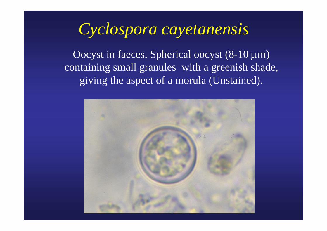
Cyclospora cayetanensis Oocyst in faeces. Spherical oocyst (8-10 μm) containing small granules with a greenish shade,
giving the aspect of a morula (Unstained).
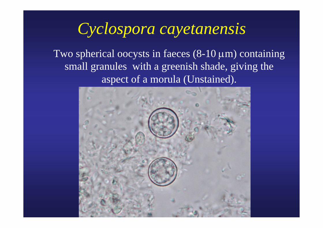
Cyclospora cayetanensis Two spherical oocysts in faeces (8-10 μm) containing
small granules with a greenish shade, giving the aspect of a morula (Unstained).
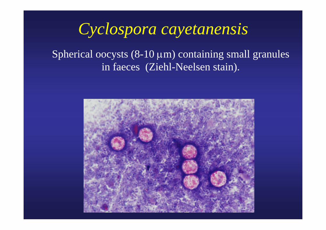
Cyclospora cayetanensis Spherical oocysts (8-10 μm) containing small granules
in faeces (Ziehl-Neelsen stain).
Microsporidia
Immunosuppression (AIDS) Intestine: Enterocytozoon spp., Encephalitozoon
(Septata) spp. Tissues: Nosema spp., Encephalitozoon spp.,
Pleistophora spp., ...
Albendazole (GSK)
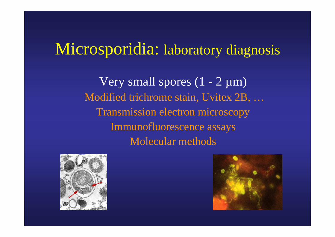
Microsporidia: laboratory diagnosis
Very small spores (1 - 2 µm) Modified trichrome stain, Uvitex 2B, …
Transmission electron microscopy Immunofluorescence assays
Molecular methods
Microsporidia
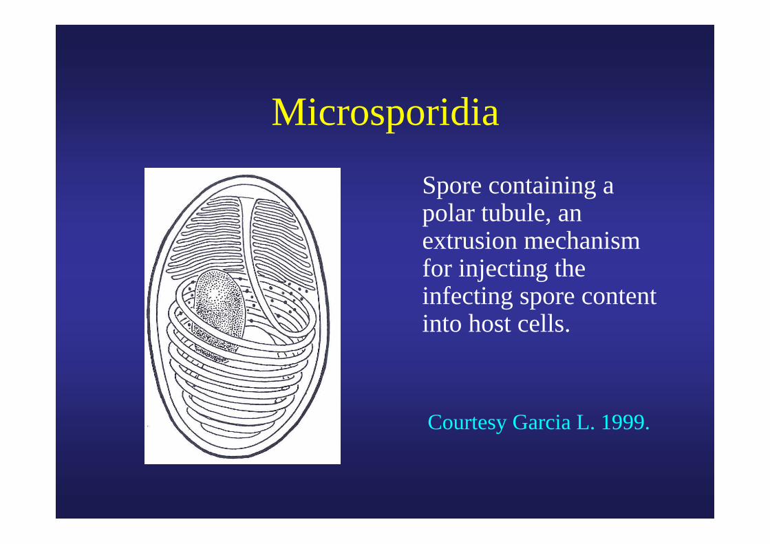
Spore containing a polar tubule, an extrusion mechanism for injecting the infecting spore content into host cells.
Courtesy Garcia L. 1999.

Microsporidia Small round to oval elements in faeces. They are only
slightly larger than the light green bacteria (Trichrome stain).
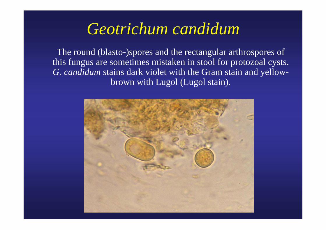
Geotrichum candidum The round (blasto-)spores and the rectangular arthrospores of
this fungus are sometimes mistaken in stool for protozoal cysts.G. candidum stains dark violet with the Gram stain and yellow-
brown with Lugol (Lugol stain).
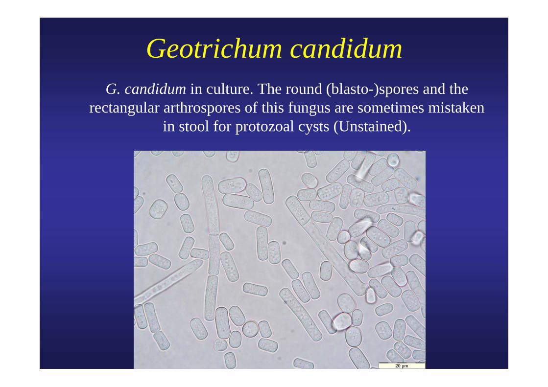
Geotrichum candidum G. candidum in culture. The round (blasto-)spores and the rectangular arthrospores of this fungus are sometimes mistaken
in stool for protozoal cysts (Unstained).