Embed Size (px)
Citation preview
GE HealthcareLife Sciences
Amersham Hybond-PPVDF Membraneoptimized for protein transfer
Product Booklet
Codes: RPN2020F RPN1416F RPN303F 28990983
2
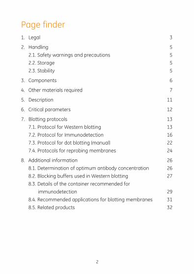
Page finder1. Legal 3
2. Handling 5
2.1. Safety warnings and precautions 5
2.2. Storage 5
2.3. Stability 5
3. Components 6
4. Other materials required 7
5. Description 11
6. Critical parameters 12
7. Blotting protocols 13
7.1. Protocol for Western blotting 13
7.2. Protocol for Immunodetection 16
7.3. Protocol for dot blotting (manual) 22
7.4. Protocols for reprobing membranes 24
8. Additional information 26
8.1. Determination of optimum antibody concentration 26
8.2. Blocking buffers used in Western blotting 27
8.3. Details of the container recommended for
immunodetection 29
8.4. Recommended applications for blotting membranes 31
8.5. Related products 32
1. LegalGE, imagination at work and GE monogram are trademarks of General Electric Company.
AlkPhos Direct, ECF, ECL, ECL Plus, FluorImager, Gene Images, Hybond, Hypercassette, Hyperfilm, Hyperscreen, Molecular Dynamics, Rainbow, Rapid-hyb, Sensitize, and Storm are trademarks of GE Healthcare companies.
ECL Prime Western blotting detection reagent is manufactured and sold under license from Cyanagen Srl and is subject of US patent number 7855287, US patent application number 2008241868 and Italian application number TO2010A000580, together with other equivalent granted patents and patent applications in other countries.
ECL Plus Western Blotting Detection Reagents are manufactured for GE Healthcare by Lumigen, Inc. The PS3 substrate component is covered by US patent numbers 5491072, 5593845, and 5670644, 5686258, 5723295, 5750698 and 6068979 and equivalent patents and patent applications in other countries and is sold under license from Lumigen, Inc.
ECF substrate is manufactured for GE Healthcare by Europa Bioproducts. This component is covered by US patent number 5424440 and EP 424465B1 and is sold under licence from Promega.
All third party trademarks are the property of their respective
owners.
© 2006–2011 General Electric Company – All rights reserved.
First published 2001.
GE Healthcare reserves the right, subject to any regulatory and contractual approval, if required, to make changes in specification
3
4
and features shown herein, or discontinue the product described at any time without notice or obligation.
All goods and services are sold subject to the terms and conditions of sale of the company within GE Healthcare which supplies them. A copy of these terms and conditions is available on request. Contact your local GE Healthcare representative for the most current information.
http//www.gelifesciences.com
GE Healthcare UK Limited. Amersham Place, Little Chalfont, Buckinghamshire, HP7 9NA UK
55
2. Handling
2.1. Safety warnings and precautionsWarning: For research use only. Not recommended or intended for diagnosis of disease in humans or animals. Do not use internally or externally in humans or animals.
We recommend that this product and components are handled only by those persons who have been trained in laboratory techniques and that it is used in accordance with the principles of good laboratory practice. As all chemicals should be considered as potentially hazardous, it is advisable when handling chemical reagents to wear suitable protective clothing such as laboratory overalls, safety glasses and gloves. Care should be taken to avoid contact with skin or eyes. In case of contact with skin or eyes wash immediately with water.
Note that the procedures require the use of: Acrylamide/
NN,–methylene–bis-acrylamide: toxic substance Ammonium persulfate: harmful TEMED: highly flammable, irritant -Mercaptoethanol: poisonous substance
Methanol: toxic substance: highly flammable Sodium dodecyl sulfate: irritant Caution:This product may be used with radioactive materials. Please follow the manu-facturer’s Safety Data Sheet relating to the safe handling and use of these materials.
2.2. Storage Membranes should be stored in a clean, dry atmosphere away from noxious fumes. In order to preserve optimum performance, avoid conditions of extreme humidity.
2.3. Stability Before opening, membranes are stable for one year. Once open keep in the bags in which they were received. Performance is consistent for up to twelve months when stored under the recommended conditions.
6
3. ComponentsRPN2020F 20 × 20 cm, 10 sheets
RPN1416F 14 × 16 cm, 15 sheets
RPN303F 30 cm × 3 m, 1 roll
28990983 8 × 7.5 cm, 10 sheets
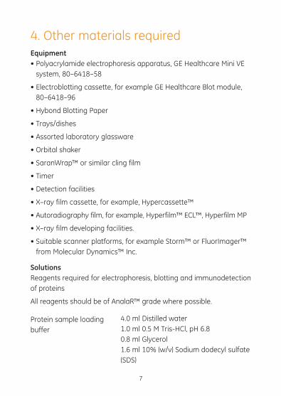
4. Other materials requiredEquipment• Polyacrylamide electrophoresis apparatus, GE Healthcare Mini VE
system, 80–6418–58
• Electroblotting cassette, for example GE Healthcare Blot module, 80–6418–96
• Hybond Blotting Paper
• Trays/dishes
• Assorted laboratory glassware
• Orbital shaker
• SaranWrap™ or similar cling film
• Timer
• Detection facilities
• X–ray film cassette, for example, Hypercassette™
• Autoradiography film, for example, Hyperfilm™ ECL™, Hyperfilm MP
• X–ray film developing facilities.
• Suitable scanner platforms, for example Storm™ or FluorImager™ from Molecular Dynamics™ Inc.
Solutions Reagents required for electrophoresis, blotting and immunodetection of proteins
All reagents should be of AnalaR™ grade where possible.
7
Protein sample loading buffer
4.0 ml Distilled water1.0 ml 0.5 M Tris-HCl, pH 6.8 0.8 ml Glycerol 1.6 ml 10% (w/v) Sodium dodecyl sulfate (SDS)
8
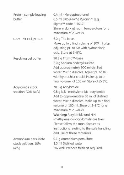
Protein sample loading buffer
0.5M Tris-HCl, pH 6.8
Resolving gel buffer
Acrylamide stock solution, 30% (w/v)
Ammonium persulfate stock solution, 10% (w/v)
0.4 ml –Mercaptoethanol 0.5 ml 0.05% (w/v) Pyronin Y (e.g. Sigma™ code P-7017) Store in dark at room temperature for a maximum of 2 weeks.
6.0 g Tris base Make up to a final volume of 100 ml after adjusting pH to 6.8 with hydrochloric acid. Store at 2–8°C.
90.8 g Trizma™-base 2.0 g Sodium dodecyl sulfate Add approximately 900 ml distilled water. Mix to dissolve. Adjust pH to 8.8 with hydrochloric acid. Make up to a final volume of 100 ml. Store at 2–8°C.
30.0 g Acrylamide 0.8 g N,N -methylene-bis-acrylamide Add to approximately 50 ml of distilled water. Mix to dissolve. Make up to a final volume of 100 ml. Store at 2–8°C for a maximum of 2 weeks. Warning: Acrylamide and N,N -methylene-bis-acrylamide are toxic. Please follow the manufacturer’s instructions relating to the safe handling and use of these materials.
0.1 g Ammonium persulfate 1.0 ml Distilled water Mix well. Prepare fresh as required.
9
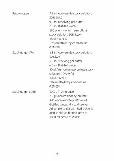
Resolving gel
Stacking gel (4%)
Stacking gel buffer
7.5 ml Acrylamide stock solution, 30% (w/v) 9.0 ml Resolving gel buffer 2.0 ml Distilled water 180 µl Ammonium persulfate stock solution. 10% (w/v) 18 µl N,N,N ,N -Tetramethylethylenediamine (TEMED)
1.8 ml Acrylamide stock solution, 30%(v/v) 5.0 ml Stacking gel buffer 4.0 ml Distilled water 50 µl Ammonium persulfate stock solution, 10% (w/v) 10 µl N,N,N,N-Tetramethylethylenediamine (TEMED)
30.3 g Trizma base 2.0 g Sodium dodecyl sulfate Add approximately 900 ml of distilled water. Mix to dissolve. Adjust pH to 6.8 with hydrochloric acid. Make up final volume to 1000 ml. Store at 2–8°C.
10
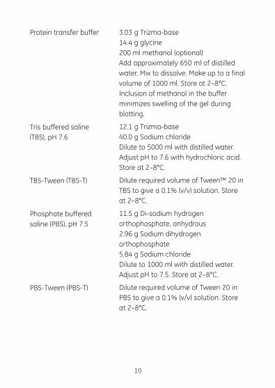
Protein transfer buffer
Tris buffered saline (TBS), pH 7.6
TBS-Tween (TBS-T)
Phosphate buffered saline (PBS), pH 7.5
PBS-Tween (PBS-T)
3.03 g Trizma-base 14.4 g glycine 200 ml methanol (optional) Add approximately 650 ml of distilled water. Mix to dissolve. Make up to a final volume of 1000 ml. Store at 2–8°C. Inclusion of methanol in the buffer minimizes swelling of the gel during blotting.
12.1 g Trizma-base 40.0 g Sodium chloride Dilute to 5000 ml with distilled water. Adjust pH to 7.6 with hydrochloric acid. Store at 2–8°C.
Dilute required volume of Tween™ 20 in TBS to give a 0.1% (v/v) solution. Store at 2–8°C.
11.5 g Di-sodium hydrogen orthophosphate, anhydrous 2.96 g Sodium dihydrogen orthophosphate 5.84 g Sodium chloride Dilute to 1000 ml with distilled water. Adjust pH to 7.5. Store at 2–8°C.
Dilute required volume of Tween 20 in PBS to give a 0.1% (v/v) solution. Store at 2–8°C.
5. DescriptionAll Hybond™ membranes from GE Healthcare are manufactured specifically for Life Science applications. Production runs are carefully controlled and the product exhaustively screened to ensure that only the most consistent product reaches the user.
All GE Healthcare membranes are identical on both sides.
Hybond–P is a hydrophobic polyvinylidene difluoride membrane optimized for protein transfer. It offers the following features:
• high mechanical strength
• high protein binding capacity
• may be used for protein staining and immunoblotting
• chemical stability allowing use of a range of solvents for rapid destaining.
It is particularly recommended for use with:
• ECL Glycoprotein detection system from GE Healthcare
• ECL reagents
• ECL Plus™ reagents
and gives excellent results with: ECF™ substrates and detection system and colorimetric detection reagents such as:
• Colloidal gold
• BCIP/NBT substrate
• DAB substrate
11
12
6. Critical parametersStorage Hybond-P should be stored in a dry and clean environment. Membranes are affected by the environment and so should be kept in the bags and boxes in which they are received.
Handling Membranes should be handled wearing gloves or using blunt ended forceps. All membranes should be cut using clean sharp scissors to avoid damage to the membrane edges.
Pre-wetting Hybond-P should be pre-wet with methanol before use followed by washing in distilled water and equilibration in an appropriate buffer. See page 12 for full wetting instructions. If Hybond-P dries out at any time the wetting procedure must be repeated.
13
Protocol
1. Separate the protein samples using gel electrophoresis or isoelectric focusing.
2. Remove the stacking gel and orientate the resolving gel by removing a corner.
3. Soak the gel in the protein transfer buffer for at least 10–20 minutes.
4. Prepare a sheet of Hybond-P. Cut the membrane to size, a) pre-wet the membrane in 100% methanol (10 seconds),
Notes
3.1. For transfer buffers without methanol it is essential that complete equilibration of the resolving gel is achieved to prevent distortions within the gel which would cause band smearing. Only a brief rinse is required to achieve equilibration if the transfer buffer contains methanol.
4.1. Hybond-P must be kept wet at all times. Should it dry out, re-wet in methanol and water as described in
7. Blotting protocols
7.1. Protocol for Western blottingThere are three basic methods for transferring proteins to membranes, capillary blotting, diffusion blotting, and electroblotting. Capillary and diffusion blotting are relatively slow procedures. As electroblotting is by far the most widely used, this booklet limits itself to this procedure.
14
Protocol
4. Continued b) wash in distilled water 1 × 5 minutes, and then c) equilibrate in the protein
transfer buffer for at least 10 minutes.
5. Assemble the electroblotting cassette and place between the electrodes in the blotting unit, according to the manufacturer’s instructions, using the appropriate transfer buffer.
6. Transfer for 1 hour at 100 V with cooling. This setting is suitable for gels 80 to 320 cm2 in size.
7. Following transfer remove the membrane from the blotting cassette, mark the orientation of the gel on the membrane and rinse briefly in PBS. Trim the membrane/blot.
Notes
4.1. Continued step 4 of this protocol.
6.1. It is recommended that cold transfer buffer is used.
6.2. Extended transfer times of 2 hours at 80 V or 30 V overnight, are also suitable. Cooling is essential for best results.
7.1. Trimming the blot will prevent high background edge effects.
Protocol
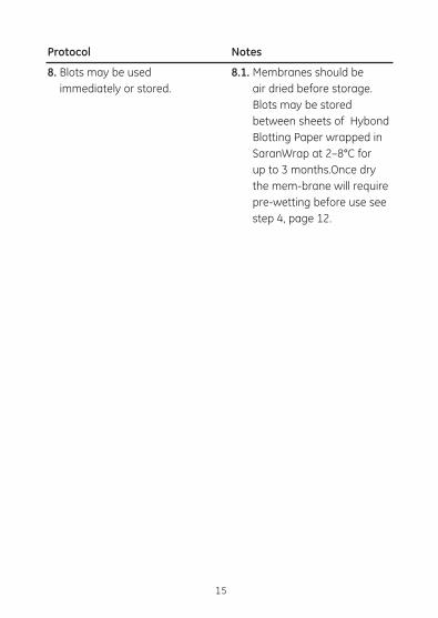
8. Blots may be used immediately or stored.
Notes
8.1. Membranes should be air dried before storage. Blots may be stored between sheets of Hybond Blotting Paper wrapped in SaranWrap at 2–8°C for up to 3 months.Once dry the mem-brane will require pre-wetting before use see step 4, page 12.
15
16
Protocol
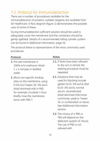
1. Pre-wet membrane in 100% (v/v) methanol. Wash 1 × 5 minutes in distilled water.
2. Block non-specific binding sites on the membrane, using 0.1% (v/v) Tween 20, 5% (w/v) dried skimmed milk in PBS, for example. Incubate 1 hour. Briefly rinse the membrane twice with PBS-T.
Notes
1.1. If blots have been allowed to dry out or stored, the wetting procedure must be repeated.
2.1. Solutions that may be used for blocking include gelatin (0.25–3% (w/v)), BSA (0.25–3% (w/v)), normal serum, reconstituted dried skimmed milk (most commonly used) or Tween 20, in combination or alone. See Additional Information section.
2.2. The choice of a PBS or TBS will depend on the detection system of choice. The use of PBS is not advised with
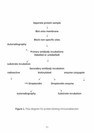
7.2. Protocol for ImmunodetectionThere are a number of procedures available for the immunodetection of proteins, suitable reagents are available from GE Healthcare. A flow diagram (figure 1) demonstrates the possible uses of some of these.
During immunodetection sufficient solution should be used to adequately cover the membrane and the containers should be gently agitated. Details of a recommended rolling cylinder system can be found in Additional Information, page 26.
The protocol below is representative of the most commonly used procedures.
17
Protocol
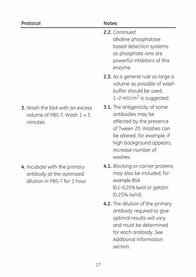
3. Wash the blot with an excess volume of PBS-T. Wash 1 × 5 minutes.
4. Incubate with the primary antibody at the optimized dilution in PBS-T for 1 hour.
Notes
2.2. Continued. alkaline phosphatase
based detection systems as phosphate ions are powerful inhibitors of this enzyme.
2.3. As a general rule as large a volume as possible of wash buffer should be used, 1–2 ml/cm2 is suggested.
3.1. The antigenicity of some antibodies may be affected by the presence of Tween 20. Washes can be altered, for example, if high background appears, increase number of washes.
4.1. Blocking or carrier proteins may also be included, for example BSA (0.1–0.25% (w/v) or gelatin (0.25% (w/v)).
4.2. The dilution of the primary antibody required to give optimal results will vary and must be determined for each antibody. See Additional Information section.
1818
Protocol
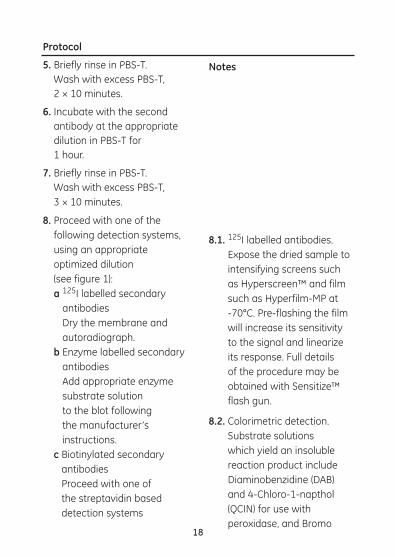
5. Briefly rinse in PBS-T. Wash with excess PBS-T, 2 × 10 minutes.
6. Incubate with the second antibody at the appropriate dilution in PBS-T for 1 hour.
7. Briefly rinse in PBS-T. Wash with excess PBS-T, 3 × 10 minutes.
8. Proceed with one of the following detection systems, using an appropriate optimized dilution (see figure 1): a 125I labelled secondary
antibodies Dry the membrane and autoradiograph.
b Enzyme labelled secondary antibodies Add appropriate enzyme substrate solution to the blot following the manufacturer’s instructions.
c Biotinylated secondary antibodies Proceed with one of the streptavidin based detection systems
Notes
8.1. 125I labelled antibodies. Expose the dried sample to intensifying screens such as Hyperscreen™ and film such as Hyperfilm-MP at -70°C. Pre-flashing the film will increase its sensitivity to the signal and linearize its response. Full details of the procedure may be obtained with Sensitize™ flash gun.
8.2. Colorimetric detection. Substrate solutions which yield an insoluble reaction product include Diaminobenzidine (DAB) and 4-Chloro-1-napthol (QCIN) for use with peroxidase, and Bromo
1919
Protocol
8. Continued. i 125I streptavidin and
autoradiograph ii Streptavidin gold. Incubate
the blot in the reagent at a suitable dilution.
iii Enzyme-labelled streptavidin conjugate, incubate with colour chemiluminescence or chemifluorescent substrates.
Notes
8.2. Continued. chloro indolyl phosphate/
Nitro blue tetrazolium (BCIP/ NBT) for use with alkaline phosphatase. Blots should be incubated at room temperature until the desired band intensity is achieved. In some cases sensitivity can be increased by rendering the membrane transparent, following the enzyme reaction and drying, by immersing in toluene or xylene. Reaction products must be insoluble in such organic solvents.
8.3. Chemifluorescent detection In the presence of the appropriate enzyme (alkaline phosphatase) a chemifluorescent substrate, such as ECF, yields a fluorescent product which may be detected using fluorescence scanning instrumentation for example the Molecular Dynamics FluorImager and Storm.
20
Protocol Notes
8.4. Chemiluminescence detection In the presence of ECL and ECL Plus enzyme chemiluminescence, substrates produce light which can be captured on X-ray film, for example Hyperfilm ECL to give a hard copy result.
21
Separate protein sample
Blot onto membrane
Block non-specific sites
Autoradiography
Primary antibody incubations (labelled or unlabelled)
substrate incubation
Secondary antibody incubation
radioactive biotinylated enzyme conjugate
125I Streptavidin Streptavidin enzyme
autoradiography Substrate incubation
Figure 1. Flow diagram for protein blotting immunodetection
22
Protocol
1. Cut the Hybond-P membrane to size.
2. Using a pencil, mark the membrane lightly with a grid or dots to guide subsequent sample application. There should be a minimum distance of 1 cm between samples applied in a volume of 5 µl or less.
3. Pre-wet the membrane in methanol, wash 1 × 5 minutes in distilled water, followed by TBS or PBS. Place the membrane on a sheet of PBS/TBS soaked Hybond Blotting Paper.
4. Dilute the sample in an appropriate buffer, for example TBS or PBS to the required concentration. A sample size of 1–2 µl is ideal for manual dot blotting.
Notes
4.1. Carrier substances, for example bovine serum albumin, may be included in the diluent buffer to improve retention of very
7.3. Protocol for dot blotting (manual)The following is a general protocol for dot blotting proteins. A number of devices are also commercially available, for example the GE Healthcare slot blotter. These give a more consistent and even application of the sample than the manual procedure described below. This parameter is particularly important in those experiments requiring quantification.
23
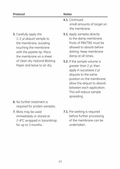
Protocol
5. Carefully apply the 1–2 µl aliquot sample to
the membrane, avoiding touching the membrane with the pipette tip. Place the membrane on a sheet of clean dry Hybond Blotting Paper and leave to air dry.
6. No further treatment is required for protein samples.
7. Blots may be used immediately or stored at 2–8°C wrapped in SaranWrap for up to 3 months.
Notes
4.1. Continued. small amounts of target on
the membrane.
5.1. Apply samples directly to the damp membrane. Pools of PBS/TBS must be allowed to absorb before dotting. Keep membrane damp at all times.
5.2. If the sample volume is greater than 2 µl, then apply in successive 2 µl aliquots to the same position on the membrane, allow the aliquot to absorb between each application. This will reduce sample spreading.
7.1. Pre wetting is required before further processing of the membrane can be undertaken.
24
Protocol
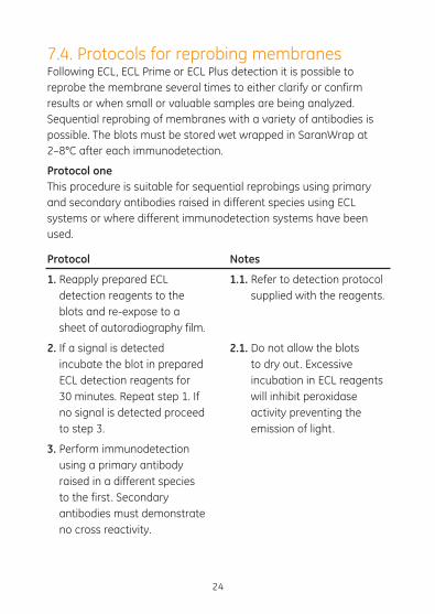
1. Reapply prepared ECL detection reagents to the blots and re-expose to a sheet of autoradiography film.
2. If a signal is detected incubate the blot in prepared ECL detection reagents for 30 minutes. Repeat step 1. If no signal is detected proceed to step 3.
3. Perform immunodetection using a primary antibody raised in a different species to the first. Secondary antibodies must demonstrate no cross reactivity.
Notes
1.1. Refer to detection protocol supplied with the reagents.
2.1. Do not allow the blots to dry out. Excessive incubation in ECL reagents will inhibit peroxidase activity preventing the emission of light.
7.4. Protocols for reprobing membranes Following ECL, ECL Prime or ECL Plus detection it is possible to reprobe the membrane several times to either clarify or confirm results or when small or valuable samples are being analyzed. Sequential reprobing of membranes with a variety of antibodies is possible. The blots must be stored wet wrapped in SaranWrap at 2–8°C after each immunodetection.
Protocol oneThis procedure is suitable for sequential reprobings using primary and secondary antibodies raised in different species using ECL systems or where different immunodetection systems have been used.
25
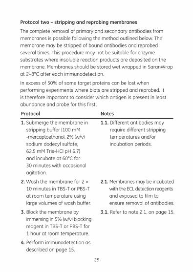
Protocol
1. Submerge the membrane in stripping buffer (100 mM -mercaptoethanol, 2% (w/v) sodium dodecyl sulfate, 62.5 mM Tris-HCl pH 6.7) and incubate at 60°C for 30 minutes with occasional agitation.
2. Wash the membrane for 2 × 10 minutes in TBS-T or PBS-T at room temperature using large volumes of wash buffer.
3. Block the membrane by immersing in 5% (w/v) blocking reagent in TBS-T or PBS-T for 1 hour at room temperature.
4. Perform immunodetection as described on page 15.
Notes
1.1. Different antibodies may require different stripping temperatures and/or incubation periods.
2.1. Membranes may be incubated with the ECL detection reagents and exposed to film to ensure removal of antibodies.
3.1. Refer to note 2.1. on page 15.
Protocol two – stripping and reprobing membranes
The complete removal of primary and secondary antibodies from membranes is possible following the method outlined below. The membrane may be stripped of bound antibodies and reprobed several times. This procedure may not be suitable for enzyme substrates where insoluble reaction products are deposited on the membrane. Membranes should be stored wet wrapped in SaranWrap at 2–8°C after each immunodetection.
In excess of 50% of some target proteins can be lost when performing experiments where blots are stripped and reprobed. It is therefore important to consider which antigen is present in least abundance and probe for this first.
8. Additional information
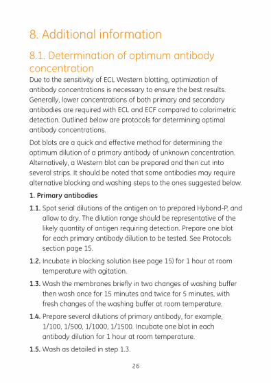
8.1. Determination of optimum antibody concentrationDue to the sensitivity of ECL Western blotting, optimization of antibody concentrations is necessary to ensure the best results. Generally, lower concentrations of both primary and secondary antibodies are required with ECL and ECF compared to colorimetric detection. Outlined below are protocols for determining optimal antibody concentrations.
Dot blots are a quick and effective method for determining the optimum dilution of a primary antibody of unknown concentration. Alternatively, a Western blot can be prepared and then cut into several strips. It should be noted that some antibodies may require alternative blocking and washing steps to the ones suggested below.
1. Primary antibodies
1.1. Spot serial dilutions of the antigen on to prepared Hybond-P, and allow to dry. The dilution range should be representative of the likely quantity of antigen requiring detection. Prepare one blot for each primary antibody dilution to be tested. See Protocols section page 15.
1.2. Incubate in blocking solution (see page 15) for 1 hour at room temperature with agitation.
1.3. Wash the membranes briefly in two changes of washing buffer then wash once for 15 minutes and twice for 5 minutes, with fresh changes of the washing buffer at room temperature.
1.4. Prepare several dilutions of primary antibody, for example, 1/100, 1/500, 1/1000, 1/1500. Incubate one blot in each antibody dilution for 1 hour at room temperature.
1.5. Wash as detailed in step 1.3.
26

1.6. Dilute the secondary antibody and incubate the membranes for 1 hour at room temperature.
1.7. Wash as detailed in step 1.3.
1.8. Detect using the appropriate detection reagents as detailed on page 17.
1.9. The antibody dilution that gives the maximum signal with minimum background should be selected.
2. Secondary antibodies
2.1. Prepare dot blots and block the membranes as described in 1.1 and 1.2 above. Wash as detailed in step 1.3.
2.2. Incubate in an optimized dilution of primary antibody for 1 hour at room temperature.
2.3. Wash as detailed in step 1.3.
2.4. Prepare several dilutions of secondary antibody: for example 1/1500, 1/3000, 1/5000, 1/10 000, 1/50 000. Incubate one blot in each antibody dilution for 1 hour at room temperature.
2.5. Wash as detailed in step 1.3.
2.6. Detect using the appropriate detection reagents as detailed on page 21. The antibody dilution that gives maximum signal with minimum background should be selected.
8.2. Blocking buffers used in Western blottingAfter transfer of proteins to membrane (for example Hybond-P), binding sites must be saturated to prevent non-specific binding of antibodies or other probes. Failure to efficiently block the membrane can lead to high background. Blocking is achieved by incubating the membrane in a relatively concentrated solution of heterologous protein or detergent.
27
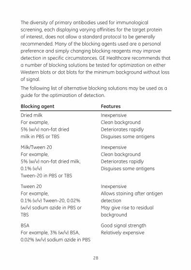
28
Blocking agent
Dried milk For example, 5% (w/v) non-fat dried milk in PBS or TBS
Milk/Tween 20 For example, 5% (w/v) non-fat dried milk, 0.1% (v/v) Tween-20 in PBS or TBS
Tween 20 For example, 0.1% (v/v) Tween-20, 0.02% (w/v) sodium azide in PBS or TBS
BSA For example, 3% (w/v) BSA, 0.02% (w/v) sodium azide in PBS
Features
Inexpensive Clean background Deteriorates rapidly Disguises some antigens
Inexpensive Clean background Deteriorates rapidly Disguises some antigens
Inexpensive Allows staining after antigen detection May give rise to residual background
Good signal strength Relatively expensive
The diversity of primary antibodies used for immunological screening, each displaying varying affinities for the target protein of interest, does not allow a standard protocol to be generally recommended. Many of the blocking agents used are a personal preference and simply changing blocking reagents may improve detection in specific circumstances. GE Healthcare recommends that a number of blocking solutions be tested for optimization on either Western blots or dot blots for the minimum background without loss of signal.
The following list of alternative blocking solutions may be used as a guide for the optimization of detection.
29
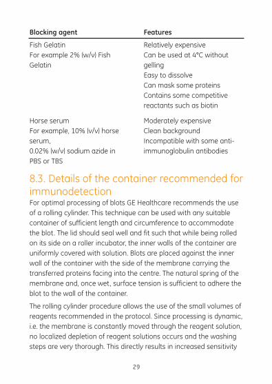
Blocking agent
Fish Gelatin For example 2% (w/v) Fish Gelatin
Horse serum For example, 10% (v/v) horse serum, 0.02% (w/v) sodium azide in PBS or TBS
Features
Relatively expensive Can be used at 4°C without gelling Easy to dissolve Can mask some proteins Contains some competitive reactants such as biotin
Moderately expensive Clean background Incompatible with some anti-immunoglobulin antibodies
8.3. Details of the container recommended for immunodetectionFor optimal processing of blots GE Healthcare recommends the use of a rolling cylinder. This technique can be used with any suitable container of sufficient length and circumference to accommodate the blot. The lid should seal well and fit such that while being rolled on its side on a roller incubator, the inner walls of the container are uniformly covered with solution. Blots are placed against the inner wall of the container with the side of the membrane carrying the transferred proteins facing into the centre. The natural spring of the membrane and, once wet, surface tension is sufficient to adhere the blot to the wall of the container.
The rolling cylinder procedure allows the use of the small volumes of reagents recommended in the protocol. Since processing is dynamic, i.e. the membrane is constantly moved through the reagent solution, no localized depletion of reagent solutions occurs and the washing steps are very thorough. This directly results in increased sensitivity
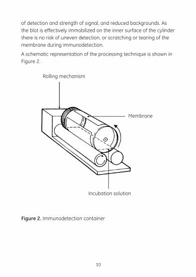
of detection and strength of signal, and reduced backgrounds. As the blot is effectively immobilized on the inner surface of the cylinder there is no risk of uneven detection, or scratching or tearing of the membrane during immunodetection.
A schematic representation of the processing technique is shown in Figure 2.
30
Rolling mechanism
Membrane
Incubation solution
Figure 2. Immunodetection container
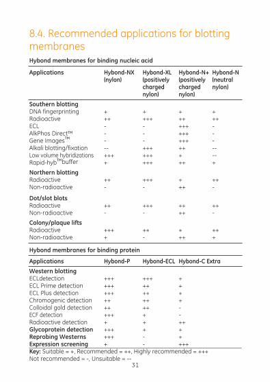
8.4. Recommended applications for blotting membranesHybond membranes for binding nucleic acid
Applications Hybond-NX Hybond-XL Hybond-N+ Hybond-N (nylon) (positively (positively (neutral charged charged nylon) nylon) nylon)Southern blotting DNA fingerprinting + + + +Radioactive ++ +++ ++ ++ECL - - +++ -AlkPhos Direct™ - - +++ -Gene Images™ - - +++ -Alkali blotting/fixation -- +++ ++ --Low volume hybridizations +++ +++ + --Rapid-hyb™buffer + +++ ++ +
Northern blotting Radioactive ++ +++ + ++Non-radioactive - - ++ -
Dot/slot blotsRadioactive ++ +++ ++ ++Non-radioactive - - ++ -
Colony/plaque liftsRadioactive +++ ++ + ++Non-radioactive + - ++ +
Hybond membranes for binding protein
Applications Hybond-P Hybond-ECL Hybond-C Extra Western blotting ECLdetection +++ +++ +ECL Prime detection +++ ++ +ECL Plus detection +++ ++ +Chromogenic detection ++ ++ +Colloidal gold detection ++ ++ -ECF detection +++ + -Radioactive detection + + ++Glycoprotein detection +++ + +Reprobing Westerns +++ - +Expression screening + - +++Key: Suitable = +, Recommended = ++, Highly recommended = +++ Not recommended = -, Unsuitable = --
31
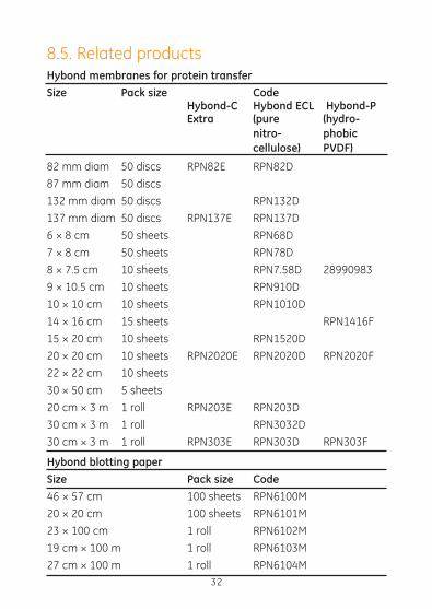
8.5. Related productsHybond membranes for protein transferSize Pack size Code Hybond-C Hybond ECL Hybond-P Extra (pure (hydro- nitro- phobic cellulose) PVDF)
82 mm diam 50 discs RPN82E RPN82D
87 mm diam 50 discs
132 mm diam 50 discs RPN132D
137 mm diam 50 discs RPN137E RPN137D
6 × 8 cm 50 sheets RPN68D
7 × 8 cm 50 sheets RPN78D
8 × 7.5 cm 10 sheets RPN7.58D 28990983
9 × 10.5 cm 10 sheets RPN910D
10 × 10 cm 10 sheets RPN1010D
14 × 16 cm 15 sheets RPN1416F
15 × 20 cm 10 sheets RPN1520D
20 × 20 cm 10 sheets RPN2020E RPN2020D RPN2020F
22 × 22 cm 10 sheets
30 × 50 cm 5 sheets
20 cm × 3 m 1 roll RPN203E RPN203D
30 cm × 3 m 1 roll RPN3032D
30 cm × 3 m 1 roll RPN303E RPN303D RPN303F
Hybond blotting paperSize Pack size Code46 × 57 cm 100 sheets RPN6100M
20 × 20 cm 100 sheets RPN6101M
23 × 100 cm 1 roll RPN6102M
19 cm × 100 m 1 roll RPN6103M
27 cm × 100 m 1 roll RPN6104M32
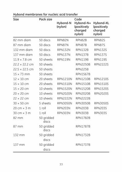
Hybond membranes for nucleic acid transferSize Pack size Code Hybond-N Hybond-N+ Hybond-XL (nylon) (positively (positively charged charged nylon) nylon)
82 mm diam 50 discs RPN82N RPN82B RPN82S
87 mm diam 50 discs RPN87N RPN87B RPN87S
132 mm diam 50 discs RPN132N RPN132B RPN132S
137 mm diam 50 discs RPN137N RPN137B RPN137S
11.9 × 7.8 cm 50 sheets RPN119N RPN119B RPN119S
22.2 × 22.2 cm 50 sheets RPN2250B RPN2222S
22.5 × 22.5 cm 50 sheets RPN225B
15 × 73 mm 50 sheets RPN1567B
12 × 10 cm 20 sheets RPN1210N RPN1210B RPN1210S
15 × 10 cm 20 sheets RPN1510N RPN1510B RPN1510S
15 × 20 cm 10 sheets RPN1520N RPN1520B RPN1520S
20 × 20 cm 10 sheets RPN2020N RPN2020B RPN2020S
22 × 22 cm 10 sheets RPN2222N RPN2222B
30 × 50 cm 5 sheets RPN3050N RPN3050B RPN3050S
20 cm × 3 m 1 roll RPN203N RPN203B RPN203S
30 cm × 3 m 1 roll RPN303N RPN303B RPN303S
82 mm 50 gridded RPN1782B discs
87 mm 50 gridded RPN1787B discs
132 mm 50 gridded RPN1732B
discs
137 mm 50 gridded RPN1737B
discs
33
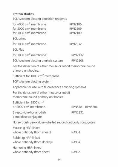
Protein studiesECL Western blotting detection reagents
for 4000 cm2 membrane RPN2106 for 2000 cm2 membrane RPN2209 for 1000 cm2 membrane RPN2109
ECL prime
for 1000 cm2 membrane RPN2232
ECL Plus
for 1000 cm2 membrane RPN2132
ECL Western blotting analysis system RPN2108
For the detection of either mouse or rabbit membrane bound primary antibodies.
Sufficient for 1000 cm2 membrane.
ECF Western blotting system
Applicable for use with fluorescence scanning systems
For the detection of either mouse or rabbit membrane bound primary antibodies.
Sufficient for 2500 cm2 or 5000 cm2 membrane. RPN5781–RPN5784
Streptavidin-horseradish RPN1231 peroxidase conjugate
Horseradish peroxidase-labelled second antibody conjugates
Mouse Ig HRP-linked whole antibody (from sheep) NA931
Rabbit Ig HRP-linked whole antibody (from donkey) NA934
Human Ig HRP-linked whole antibody (from sheet) NA933
34
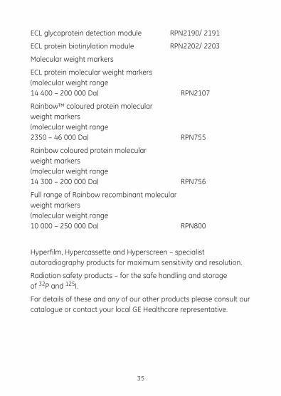
ECL glycoprotein detection module RPN2190/ 2191
ECL protein biotinylation module RPN2202/ 2203
Molecular weight markers
ECL protein molecular weight markers(molecular weight range 14 400 – 200 000 Da) RPN2107
Rainbow™ coloured protein molecular weight markers(molecular weight range 2350 – 46 000 Da) RPN755
Rainbow coloured protein molecular weight markers(molecular weight range 14 300 – 200 000 Da) RPN756
Full range of Rainbow recombinant molecular weight markers(molecular weight range 10 000 – 250 000 Da) RPN800
Hyperfilm, Hypercassette and Hyperscreen – specialist autoradiography products for maximum sensitivity and resolution.
Radiation safety products – for the safe handling and storage of 32P and 125I.
For details of these and any of our other products please consult our catalogue or contact your local GE Healthcare representative.
35

imagination at work
RPN2020FPL Rev AD 02/2011
GE Healthcare offices:
GE Healthcare Bio-Sciences AB
Björkgatan 30 751 84 Uppsala
Sweden
GE Healthcare Europe GmbH
Munzinger Strasse 5 D-79111 Freiburg
Germany
GE Healthcare Bio-Sciences Corp
800 Centennial Avenue
P.O. Box 1327
Piscataway
NJ 08855-1327
USA
GE Healthcare Japan Corporation
Sanken Bldg. 3-25-1
Hyakunincho Shinjuku-ku
Tokyo 169-0073
Japan
For contact information for your local office, please visit: http://www.gelifesciences.com/contact
GE Healthcare UK Limited Amersham Place, Little Chalfont, Buckinghamshire, HP7 9NA UK
2899
3976