Embed Size (px)
Citation preview
Dr. Diala Abu-Hassan, DDS, PhD
All images are taken from Lippincott’s Biochemistry textbook except where noted
Amino Acid Metabolism: Amino Acid Degradation & Synthesis
CATABOLISM OF THE CARBON SKELETONS OF AMINO ACIDS
The pathways by which AAs are catabolized are
organized according to which one (or more) of
the seven intermediates is produced from a
particular amino acid.
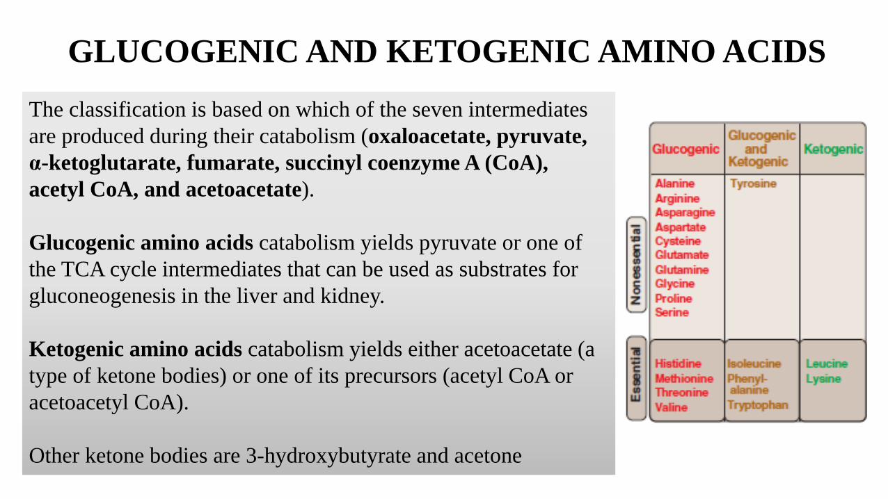
GLUCOGENIC AND KETOGENIC AMINO ACIDS
The classification is based on which of the seven intermediates
are produced during their catabolism (oxaloacetate, pyruvate,
α-ketoglutarate, fumarate, succinyl coenzyme A (CoA),
acetyl CoA, and acetoacetate).
Glucogenic amino acids catabolism yields pyruvate or one of
the TCA cycle intermediates that can be used as substrates for
gluconeogenesis in the liver and kidney.
Ketogenic amino acids catabolism yields either acetoacetate (a
type of ketone bodies) or one of its precursors (acetyl CoA or
acetoacetyl CoA).
Other ketone bodies are 3-hydroxybutyrate and acetone
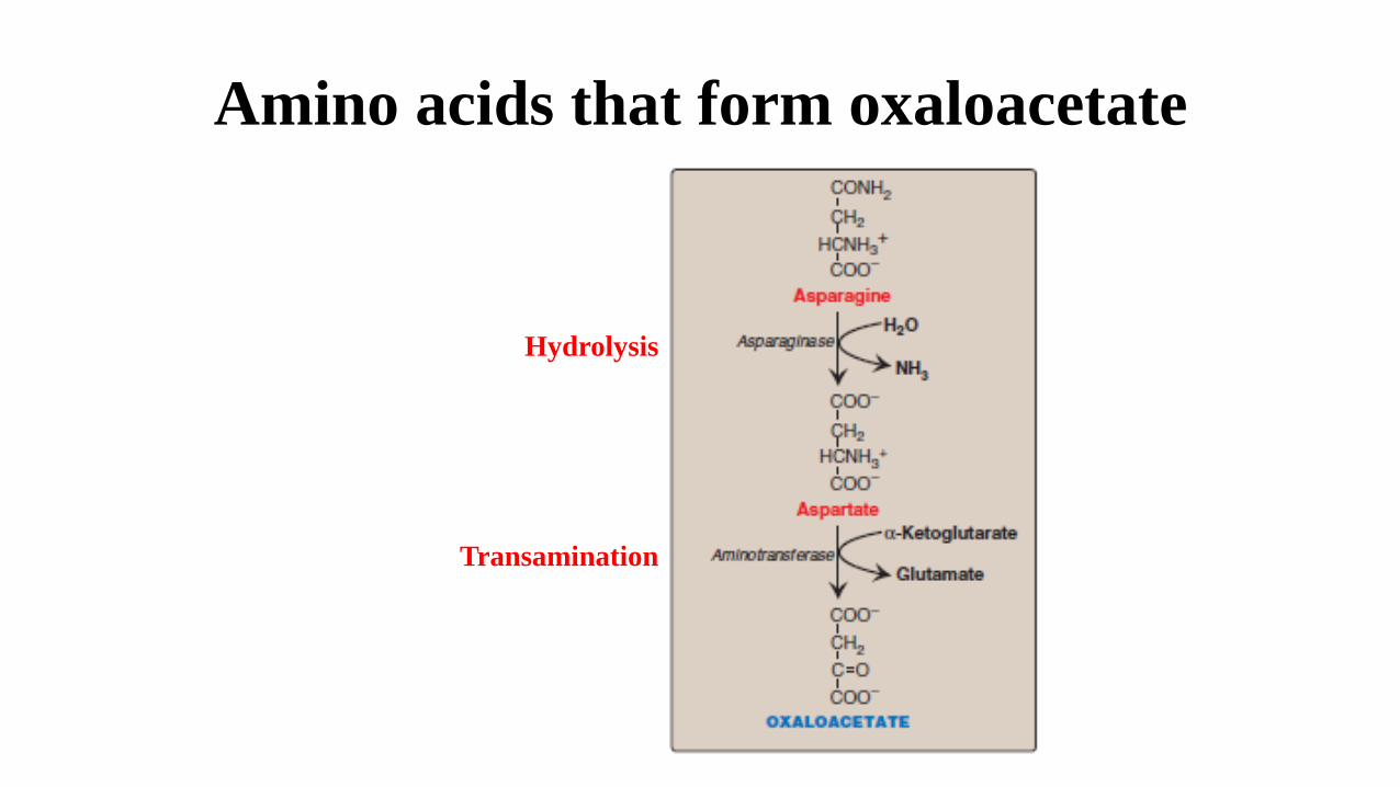
Amino acids that form oxaloacetate
Hydrolysis
Transamination
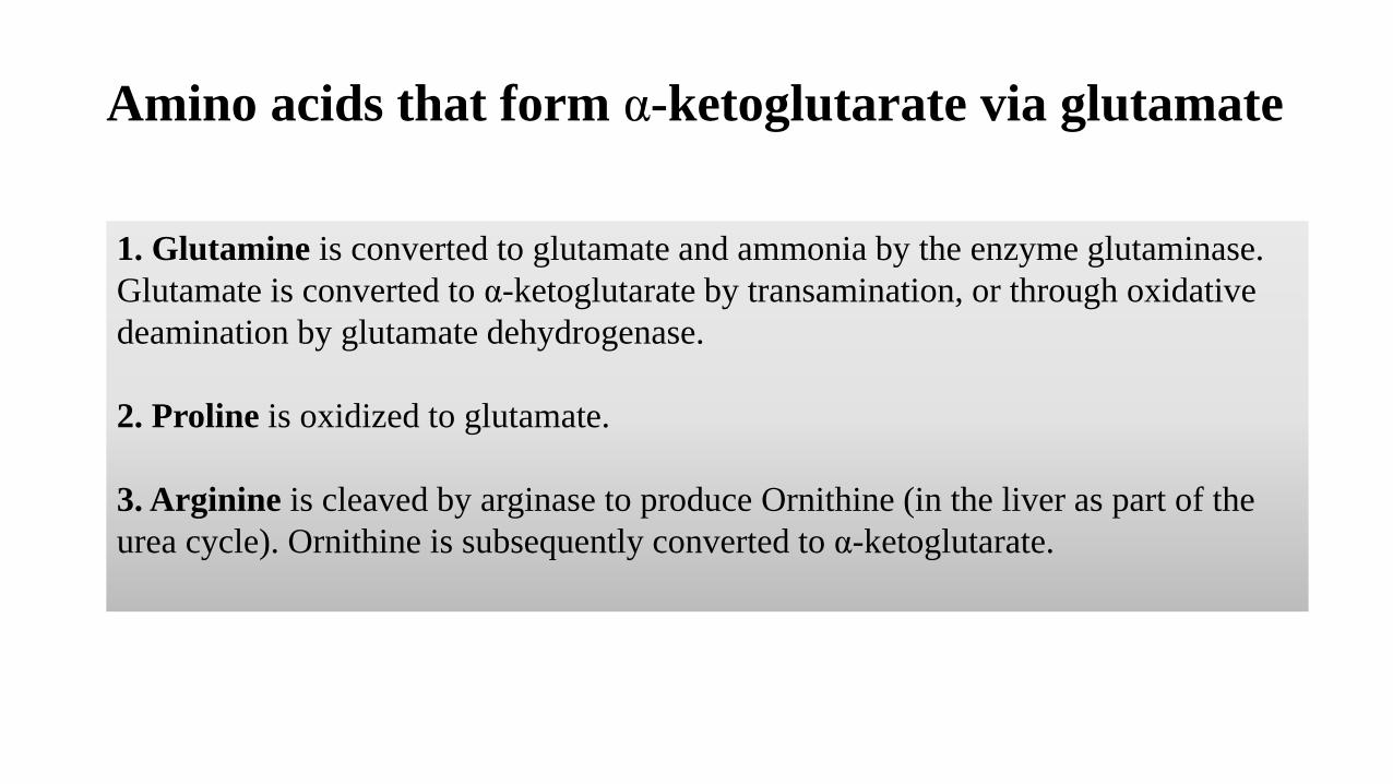
Amino acids that form α-ketoglutarate via glutamate
1. Glutamine is converted to glutamate and ammonia by the enzyme glutaminase.
Glutamate is converted to α-ketoglutarate by transamination, or through oxidative
deamination by glutamate dehydrogenase.
2. Proline is oxidized to glutamate.
3. Arginine is cleaved by arginase to produce Ornithine (in the liver as part of the
urea cycle). Ornithine is subsequently converted to α-ketoglutarate.
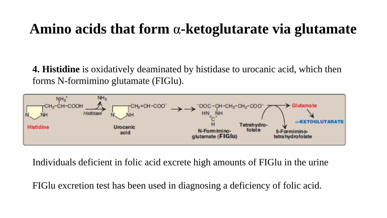
Amino acids that form α-ketoglutarate via glutamate
4. Histidine is oxidatively deaminated by histidase to urocanic acid, which then
forms N-formimino glutamate (FIGlu).
Individuals deficient in folic acid excrete high amounts of FIGlu in the urine
FIGlu excretion test has been used in diagnosing a deficiency of folic acid.
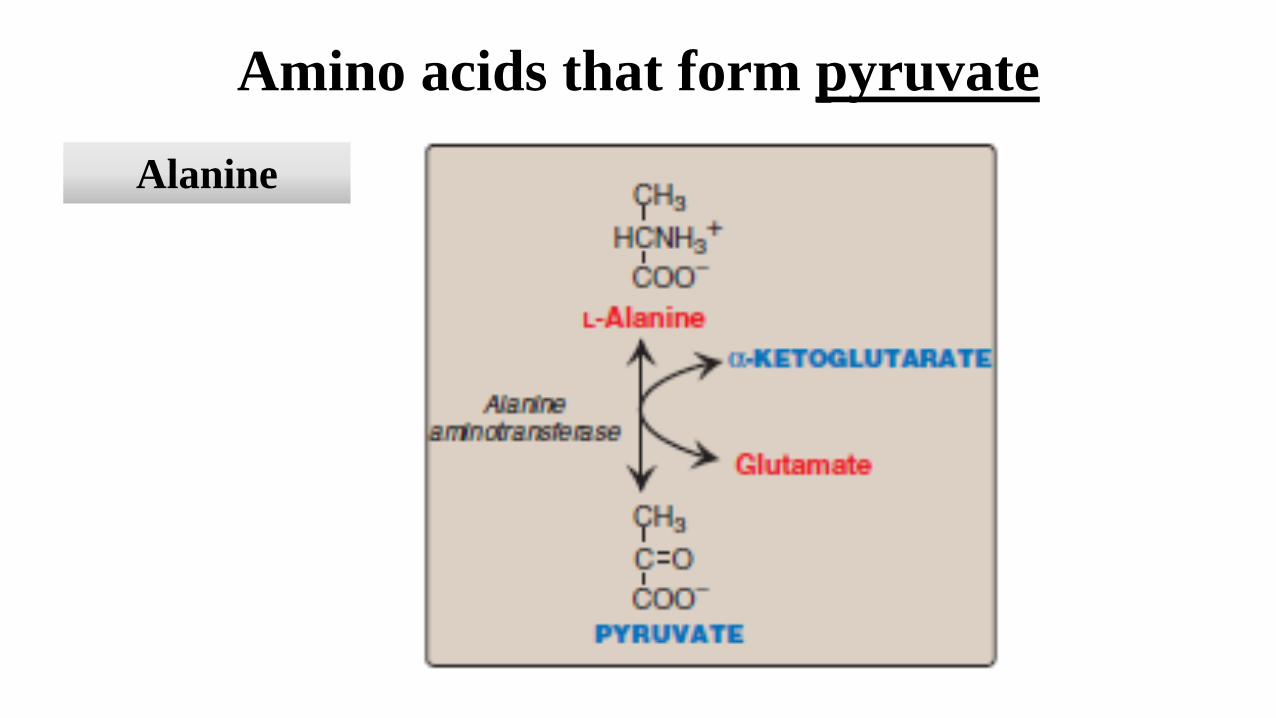
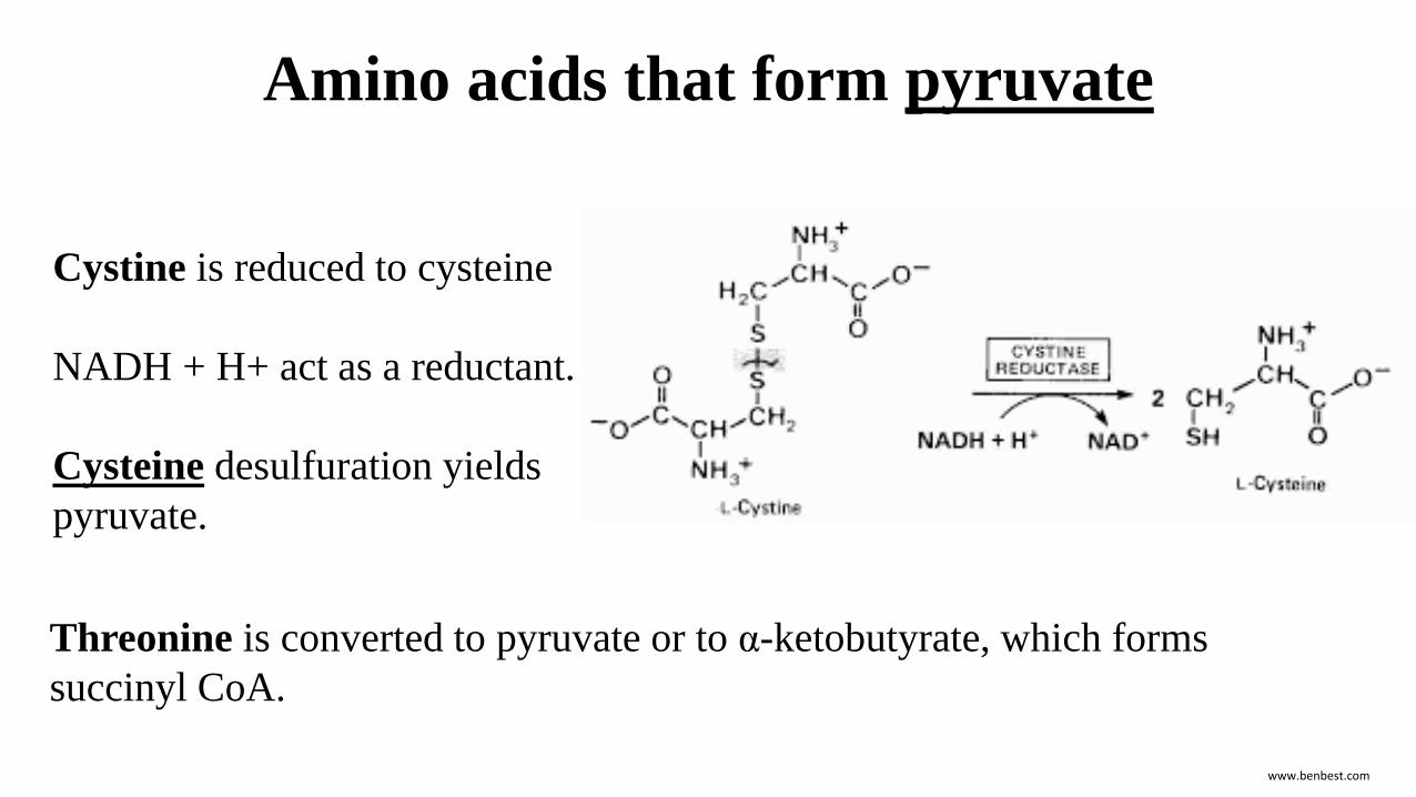
Amino acids that form pyruvate
Alanine
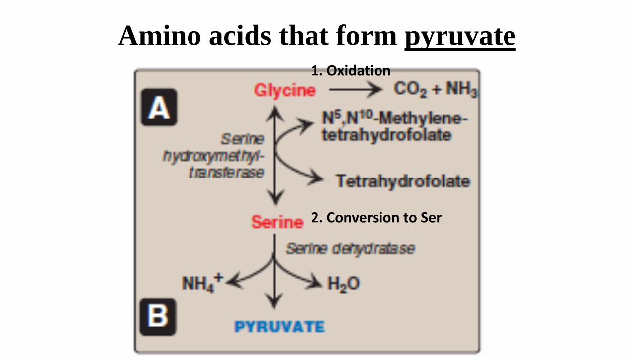
Amino acids that form pyruvate1. Oxidation
2. Conversion to Ser
Amino acids that form pyruvate
www.benbest.com
Cystine is reduced to cysteine
NADH + H+ act as a reductant.
Cysteine desulfuration yields
pyruvate.
Threonine is converted to pyruvate or to α-ketobutyrate, which forms
succinyl CoA.
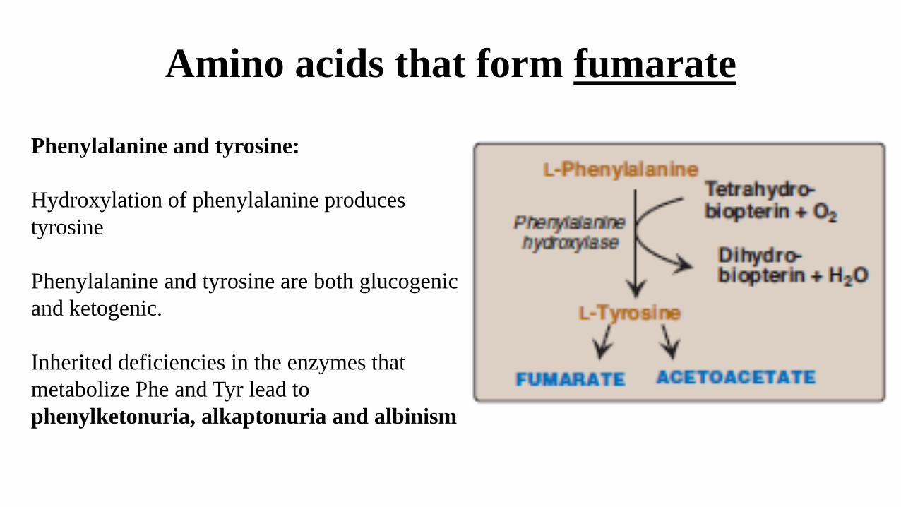
Amino acids that form fumarate
Phenylalanine and tyrosine:
Hydroxylation of phenylalanine produces
tyrosine
Phenylalanine and tyrosine are both glucogenic
and ketogenic.
Inherited deficiencies in the enzymes that
metabolize Phe and Tyr lead to
phenylketonuria, alkaptonuria and albinism
Amino acids that form succinyl CoA(a TCA cycle intermediate and glucogenic compound)
Valine and isoleucine are branched-chain amino acids
They generate propionyl CoA that is converted to succinyl CoA by biotin- and vitamin B12–
requiring reactions
Threonine is dehydrated to α-ketobutyrate, which is converted to propionyl CoA and then to
succinyl CoA.
Thr can also be converted to pyruvate.
Methionine is converted to S-adenosyl methionine (SAM), the major methyl-group donor in
one-carbon metabolism.
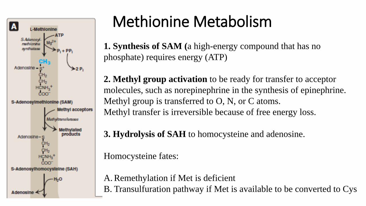
Methionine Metabolism
1. Synthesis of SAM (a high-energy compound that has no
phosphate) requires energy (ATP)
2. Methyl group activation to be ready for transfer to acceptor
molecules, such as norepinephrine in the synthesis of epinephrine.
Methyl group is transferred to O, N, or C atoms.
Methyl transfer is irreversible because of free energy loss.
3. Hydrolysis of SAH to homocysteine and adenosine.
Homocysteine fates:
A. Remethylation if Met is deficient
B. Transulfuration pathway if Met is available to be converted to Cys
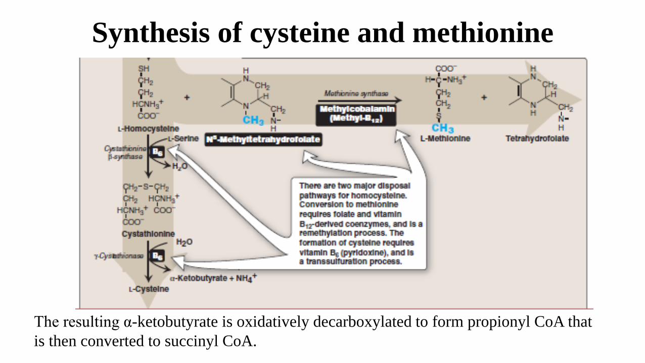
Synthesis of cysteine and methionine
The resulting α-ketobutyrate is oxidatively decarboxylated to form propionyl CoA that
is then converted to succinyl CoA.
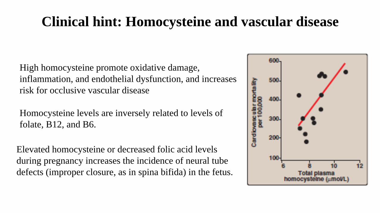
Clinical hint: Homocysteine and vascular disease
High homocysteine promote oxidative damage,
inflammation, and endothelial dysfunction, and increases
risk for occlusive vascular disease
Homocysteine levels are inversely related to levels of
folate, B12, and B6.
Elevated homocysteine or decreased folic acid levels
during pregnancy increases the incidence of neural tube
defects (improper closure, as in spina bifida) in the fetus.
Amino acids that form acetyl CoA or acetoacetyl CoA
Phe and Tyr produce acetoacetate during their catabolism
Leucine is exclusively ketogenic (acetoacetate and acetyl CoA)
Isoleucine is both ketogenic and glucogenic (acetyl CoA, acetoacetyl
CoA and succinyl CoA)
Lysine is an exclusively ketogenic (acetyl CoA and acetoacetyl CoA).
Tryptophan is both glucogenic and ketogenic (acetyl CoA and
acetoacetyl CoA)
Amino acid metabolism and single amino acid groups
Some synthetic pathways require the addition of single carbon groups
Single carbon groups exist in a variety of oxidation states, including
formyl, methenyl, methylene, and methyl.
Single carbon groups can be transferred from carrier compounds such
as THF and SAM to molecules that are being synthesized
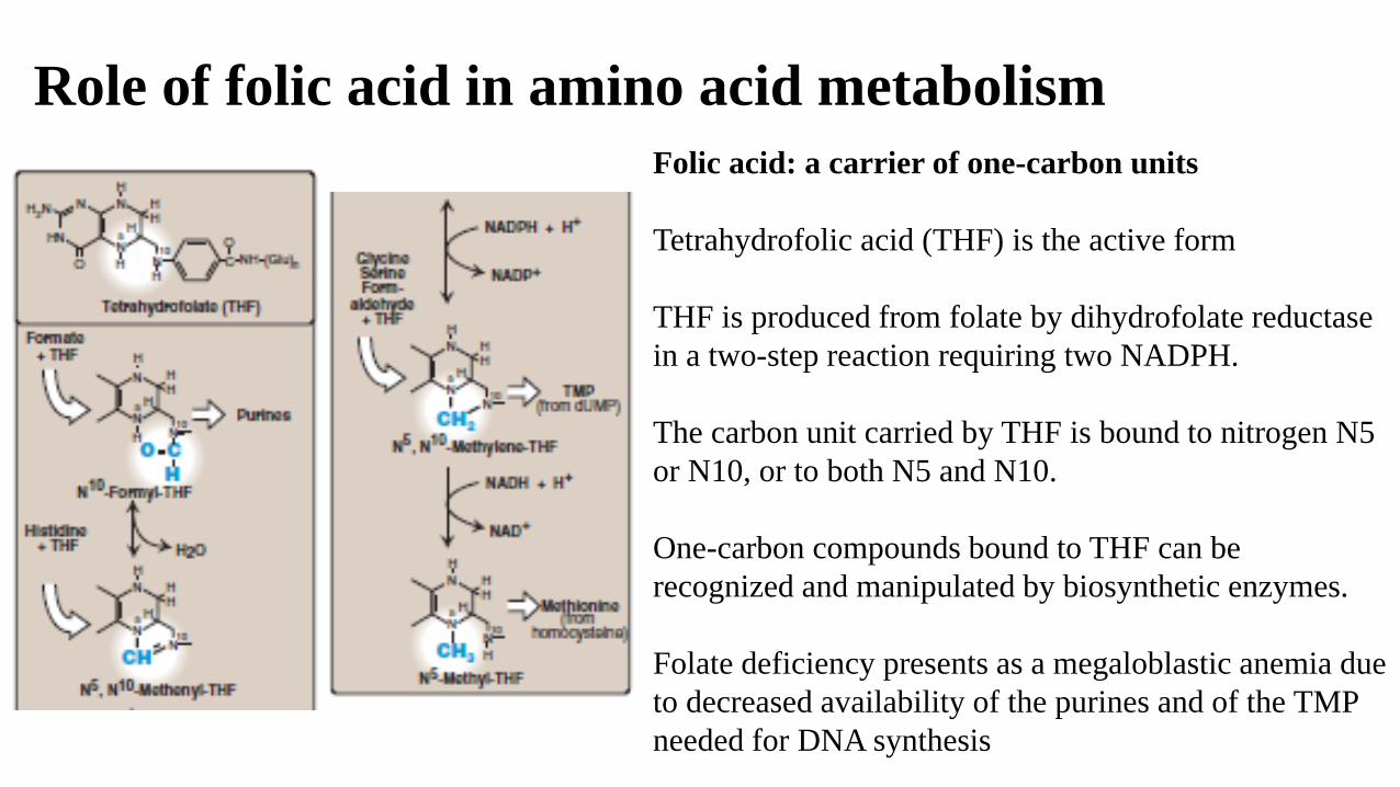
Role of folic acid in amino acid metabolism
Folic acid: a carrier of one-carbon units
Tetrahydrofolic acid (THF) is the active form
THF is produced from folate by dihydrofolate reductase
in a two-step reaction requiring two NADPH.
The carbon unit carried by THF is bound to nitrogen N5
or N10, or to both N5 and N10.
One-carbon compounds bound to THF can be
recognized and manipulated by biosynthetic enzymes.
Folate deficiency presents as a megaloblastic anemia due
to decreased availability of the purines and of the TMP
needed for DNA synthesis
Biosynthesis of Nonessential Amino Acids
Essential: Phe, Val, Thr, Trp, Met, Leu, Ile, Lys & His
Nonessential: Ala, Arg, Asp, Asn, Cys, Glu, Gln, Gly, Pro, Ser & Tyr
Nonessential amino acids are synthesized from:
1. Metabolic intermediates
2. Or from the essential amino acids.
Example: Tyr and Cys are synthesized Phe and Met, respectively.
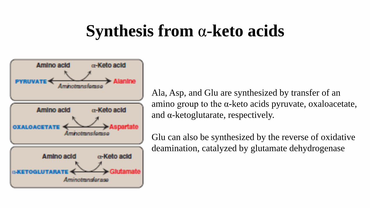
Synthesis from α-keto acids
Ala, Asp, and Glu are synthesized by transfer of an
amino group to the α-keto acids pyruvate, oxaloacetate,
and α-ketoglutarate, respectively.
Glu can also be synthesized by the reverse of oxidative
deamination, catalyzed by glutamate dehydrogenase
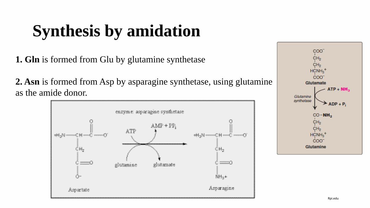
Synthesis by amidation
1. Gln is formed from Glu by glutamine synthetase
2. Asn is formed from Asp by asparagine synthetase, using glutamine
as the amide donor.
Rpi.edu
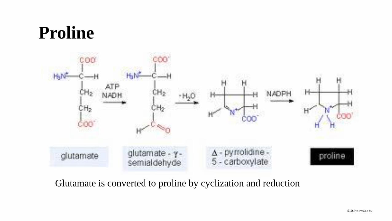
Proline
Glutamate is converted to proline by cyclization and reduction
S10.lite.msu.edu
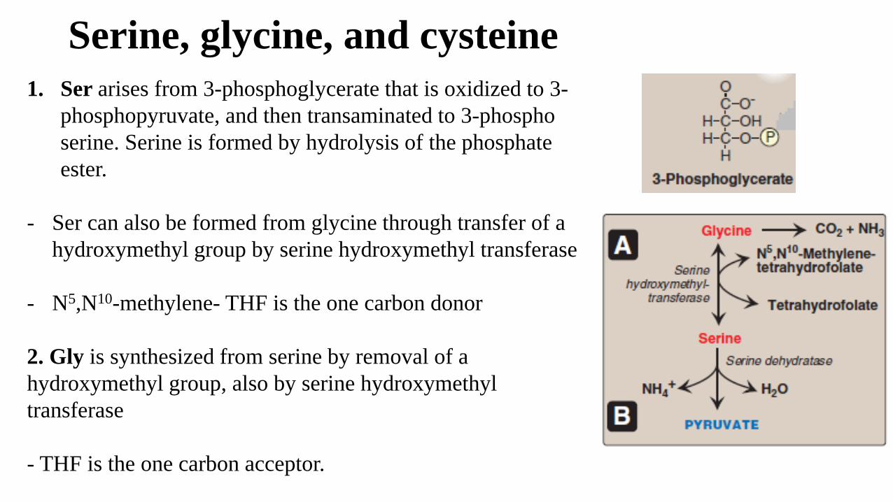
Serine, glycine, and cysteine
1. Ser arises from 3-phosphoglycerate that is oxidized to 3-
phosphopyruvate, and then transaminated to 3-phospho
serine. Serine is formed by hydrolysis of the phosphate
ester.
- Ser can also be formed from glycine through transfer of a
hydroxymethyl group by serine hydroxymethyl transferase
- N5,N10-methylene- THF is the one carbon donor
2. Gly is synthesized from serine by removal of a
hydroxymethyl group, also by serine hydroxymethyl
transferase
- THF is the one carbon acceptor.
Serine, glycine, and cysteine
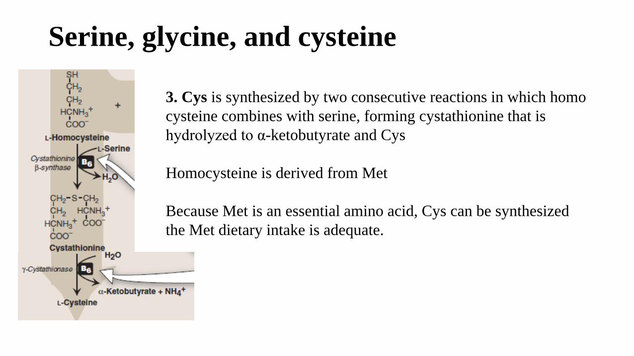
3. Cys is synthesized by two consecutive reactions in which homo
cysteine combines with serine, forming cystathionine that is
hydrolyzed to α-ketobutyrate and Cys
Homocysteine is derived from Met
Because Met is an essential amino acid, Cys can be synthesized
the Met dietary intake is adequate.
Tyrosine
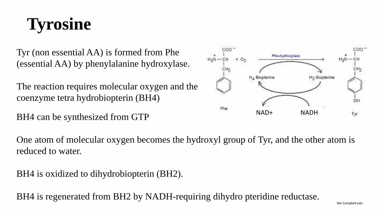
Tyr (non essential AA) is formed from Phe
(essential AA) by phenylalanine hydroxylase.
The reaction requires molecular oxygen and the
coenzyme tetra hydrobiopterin (BH4)
We.Campbell.edu
BH4 can be synthesized from GTP
One atom of molecular oxygen becomes the hydroxyl group of Tyr, and the other atom is
reduced to water.
BH4 is oxidized to dihydrobiopterin (BH2).
BH4 is regenerated from BH2 by NADH-requiring dihydro pteridine reductase.
NAD+ NADH
Metabolic defects in amino acid metabolism
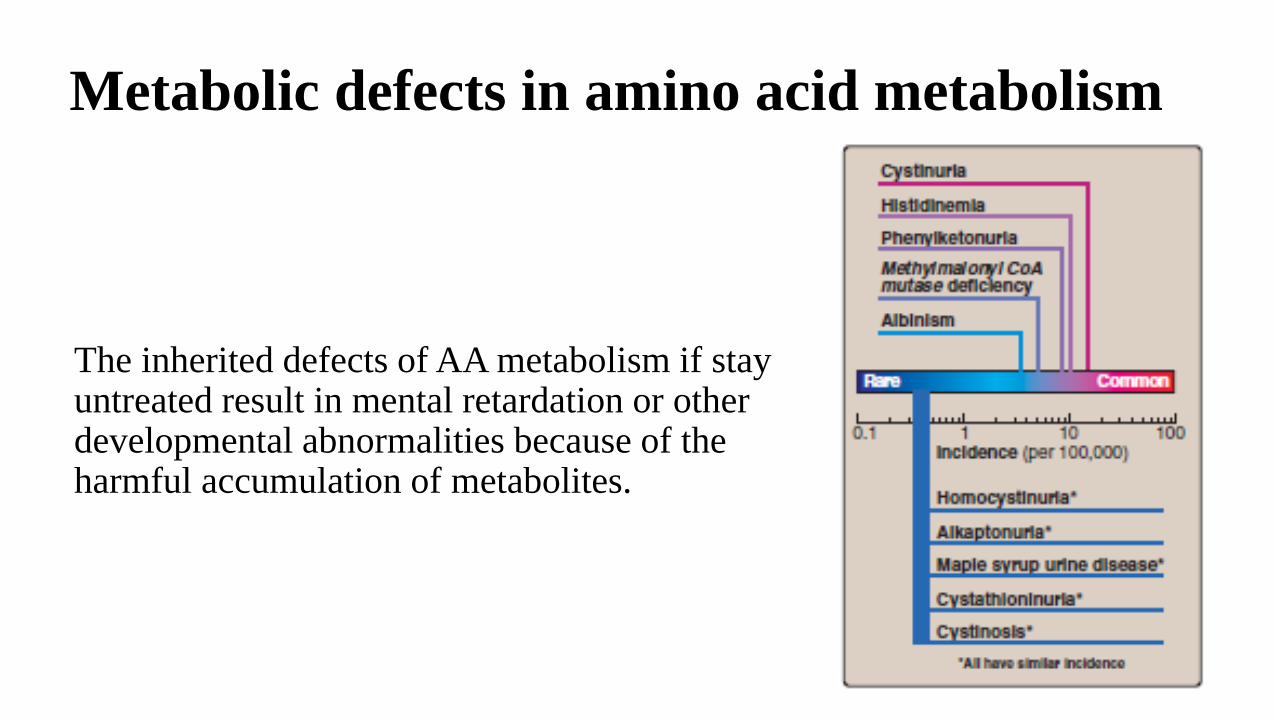
The inherited defects of AA metabolism if stay untreated result in mental retardation or other developmental abnormalities because of the harmful accumulation of metabolites.

Metabolic disorders: Phenylketonuria (PKU)
The most common inborn error of amino acid metabolism
(prevalence 1:15,000).
Due to phenylalanine hydroxylase deficiency
Biochemical changes: accumulation of phenylalanine (and
a deficiency of tyrosine)
Tyr cannot be synthesized from Phe and becomes an
essential amino acid.
Caused by any of 100 or more different mutations in the
gene that codes for phenylalanine hydroxylase (PAH).
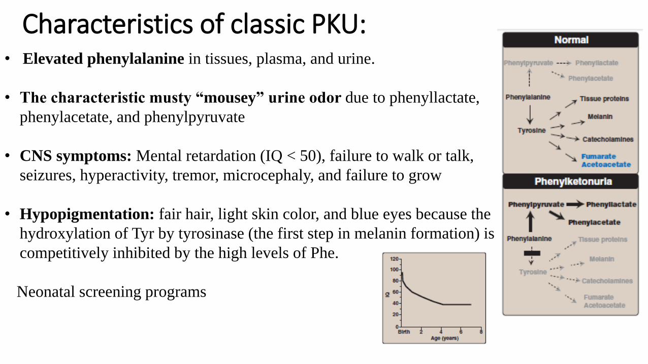
Characteristics of classic PKU:• Elevated phenylalanine in tissues, plasma, and urine.
• The characteristic musty “mousey” urine odor due to phenyllactate,
phenylacetate, and phenylpyruvate
• CNS symptoms: Mental retardation (IQ < 50), failure to walk or talk,
seizures, hyperactivity, tremor, microcephaly, and failure to grow
• Hypopigmentation: fair hair, light skin color, and blue eyes because the
hydroxylation of Tyr by tyrosinase (the first step in melanin formation) is
competitively inhibited by the high levels of Phe.
Neonatal screening programs
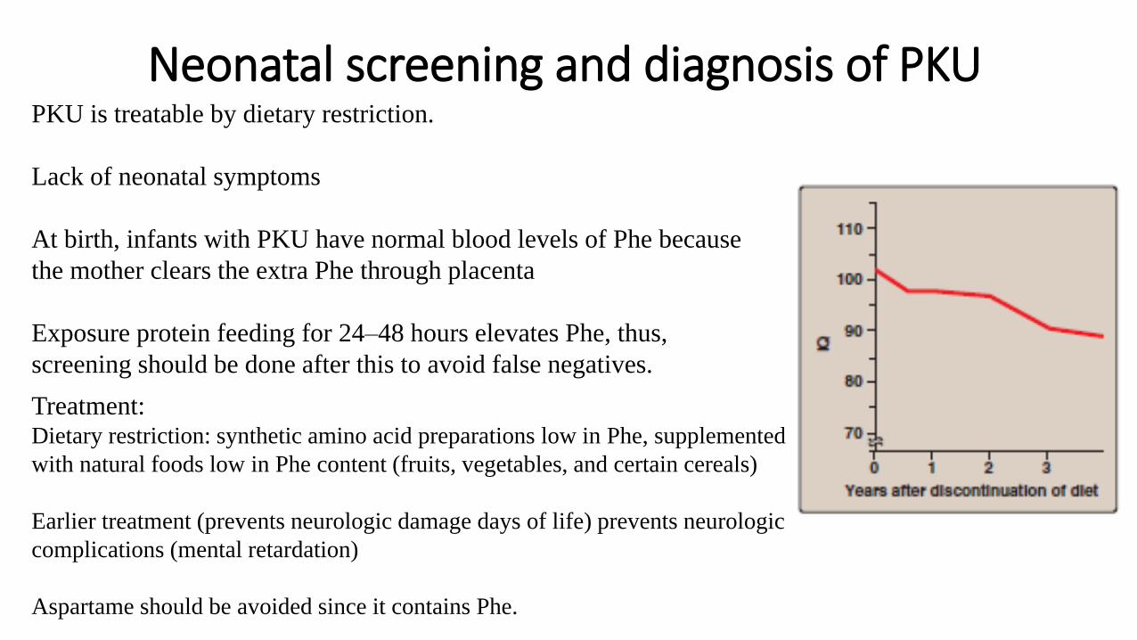
Neonatal screening and diagnosis of PKUPKU is treatable by dietary restriction.
Lack of neonatal symptoms
At birth, infants with PKU have normal blood levels of Phe because
the mother clears the extra Phe through placenta
Exposure protein feeding for 24–48 hours elevates Phe, thus,
screening should be done after this to avoid false negatives.
Treatment: Dietary restriction: synthetic amino acid preparations low in Phe, supplemented
with natural foods low in Phe content (fruits, vegetables, and certain cereals)
Earlier treatment (prevents neurologic damage days of life) prevents neurologic
complications (mental retardation)
Aspartame should be avoided since it contains Phe.
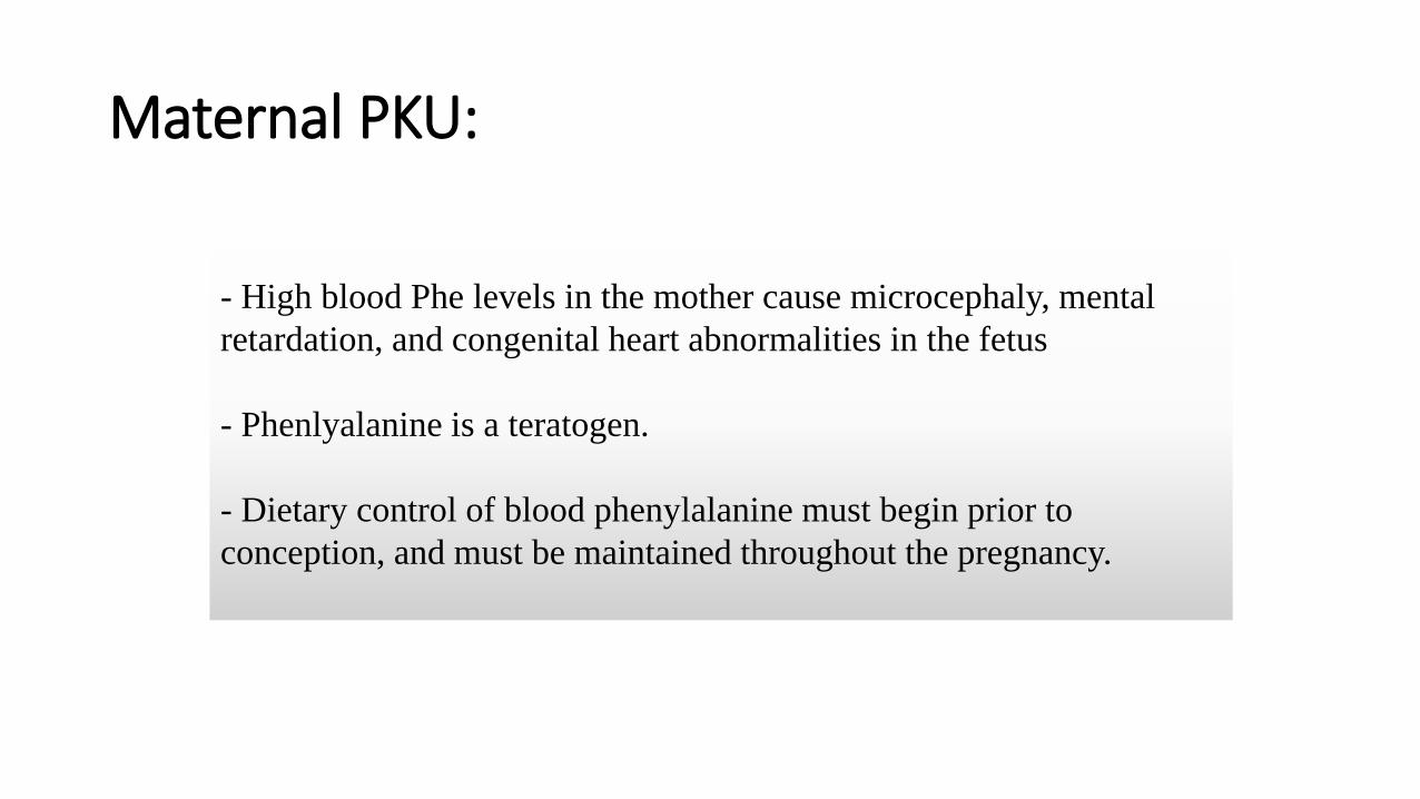
Maternal PKU:
- High blood Phe levels in the mother cause microcephaly, mental
retardation, and congenital heart abnormalities in the fetus
- Phenlyalanine is a teratogen.
- Dietary control of blood phenylalanine must begin prior to
conception, and must be maintained throughout the pregnancy.
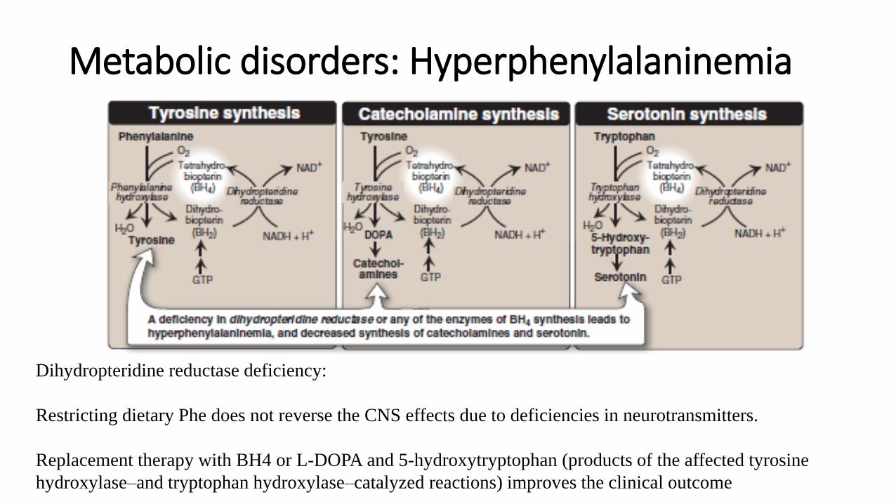
Dihydropteridine reductase deficiency:
Restricting dietary Phe does not reverse the CNS effects due to deficiencies in neurotransmitters.
Replacement therapy with BH4 or L-DOPA and 5-hydroxytryptophan (products of the affected tyrosine
hydroxylase–and tryptophan hydroxylase–catalyzed reactions) improves the clinical outcome
Metabolic disorders: Hyperphenylalaninemia
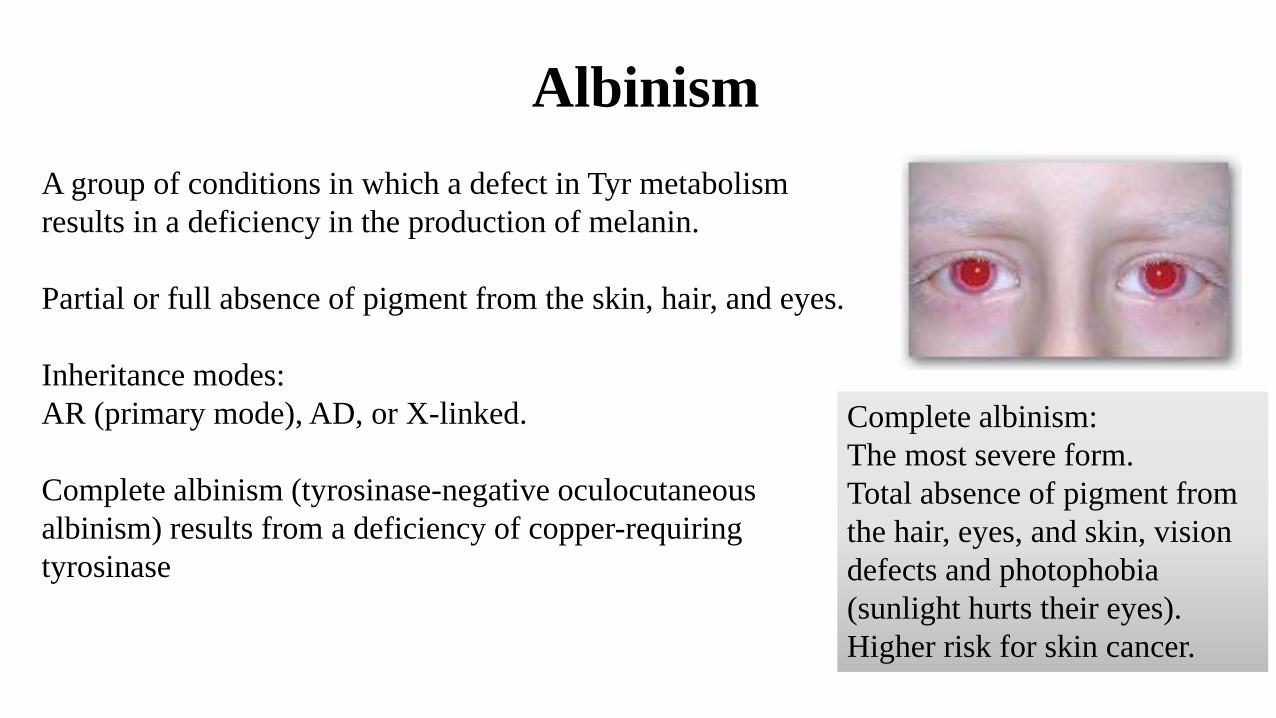
Albinism
A group of conditions in which a defect in Tyr metabolism
results in a deficiency in the production of melanin.
Partial or full absence of pigment from the skin, hair, and eyes.
Inheritance modes:
AR (primary mode), AD, or X-linked.
Complete albinism (tyrosinase-negative oculocutaneous
albinism) results from a deficiency of copper-requiring
tyrosinase
Complete albinism:
The most severe form.
Total absence of pigment from
the hair, eyes, and skin, vision
defects and photophobia
(sunlight hurts their eyes).
Higher risk for skin cancer.
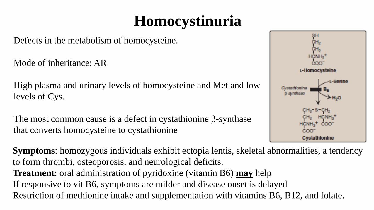
Homocystinuria
Defects in the metabolism of homocysteine.
Mode of inheritance: AR
High plasma and urinary levels of homocysteine and Met and low
levels of Cys.
The most common cause is a defect in cystathionine β-synthase
that converts homocysteine to cystathionine
Symptoms: homozygous individuals exhibit ectopia lentis, skeletal abnormalities, a tendency
to form thrombi, osteoporosis, and neurological deficits.
Treatment: oral administration of pyridoxine (vitamin B6) may help
If responsive to vit B6, symptoms are milder and disease onset is delayed
Restriction of methionine intake and supplementation with vitamins B6, B12, and folate.


![Biochem [Gluconeogenesis]](https://img.pdfslide.net/doc/110x75/577c82b31a28abe054b1e4af/biochem-gluconeogenesis.jpg)